cybertesis.unmsm.edu.pecybertesis.unmsm.edu.pe/bitstream/cybertesis/2787/1/varillas_ce.pdf · El...
63
! ! ! "# $%$ % &$ $! '!&$ $ $ ! "# $% $ ( ) $ & *+ ,--.
Transcript of cybertesis.unmsm.edu.pecybertesis.unmsm.edu.pe/bitstream/cybertesis/2787/1/varillas_ce.pdf · El...


A mis Padres, quienes me enseñaron a escuchar, a comprender, a respetar y a valorar, quienesme levantaron en caídas y tropiezos y me enseñaron a sonreír; a ustedes porque los amo yporque sé que están a mi lado acompañándome siempre en el camino de mi vida. A mihermana Iraida, Por enseñarme que la humildad es un don, por su comprensión, tolerancia ypaciencia; por ser mi ejemplo de una digna mujer. A mi hermana Luciana, Por enseñarme quesiempre llevamos un niño dentro, y le agradezco a Dios que hoy te encuentres a mi lado. A míhermano Ismael, Porque es un incondicional amigo, un buen hermano, y un hombre que me haenseñado valores y virtudes, con toda mi admiración y respeto, gracias. A mis Amigos, Conquienes he compartido momentos de amistad, de confidencias, de lágrimas y alegrías, de metas yobjetivos cumplidos; porque siempre seremos “amigos” aunque la vida nos lleve por diferentescaminos. A mis Maestros, Por darme las herramientas para construir los cimientos de micarrera profesional, por el apoyo y enseñanzas. A la Doctora Doris Salcedo Moncada, por suapoyo y empeño para este trabajo, a la Dra. Ana Maria Díaz y Dra. Antonia Castro, porenseñarme que la perseverancia es la base para cumplir cualquier meta; y un especialagradecimiento a la Dra. Liliana Terán, por su incondicional amistad. Al Coronel de Sanidadde Odontología Pablo Hernández Bautista, a la Dra. Norma Delgado y su asistente Ivonne Paz,quienes me apoyaron para la realización de este trabajo.

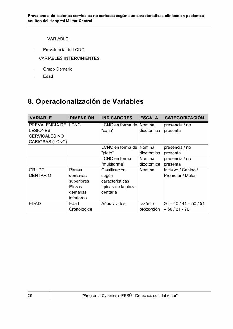
El presente trabajo tiene como propósito evaluar la prevalencia de lesiones cervicales no cariosasen piezas dentarias según sus características clínicas en pacientes adultos en el Servicio deOperatoria del Hospital Militar Central, y su frecuencia y distribución según edad y grupodentario (incisivo. canino, premolar y molar). Esto requiere del conocimiento del aspecto clínicoy la comparación a las diferentes formas de este tipo de desgaste dentario, la Abfracción,Abrasión, Erosión y formas Multiformes.
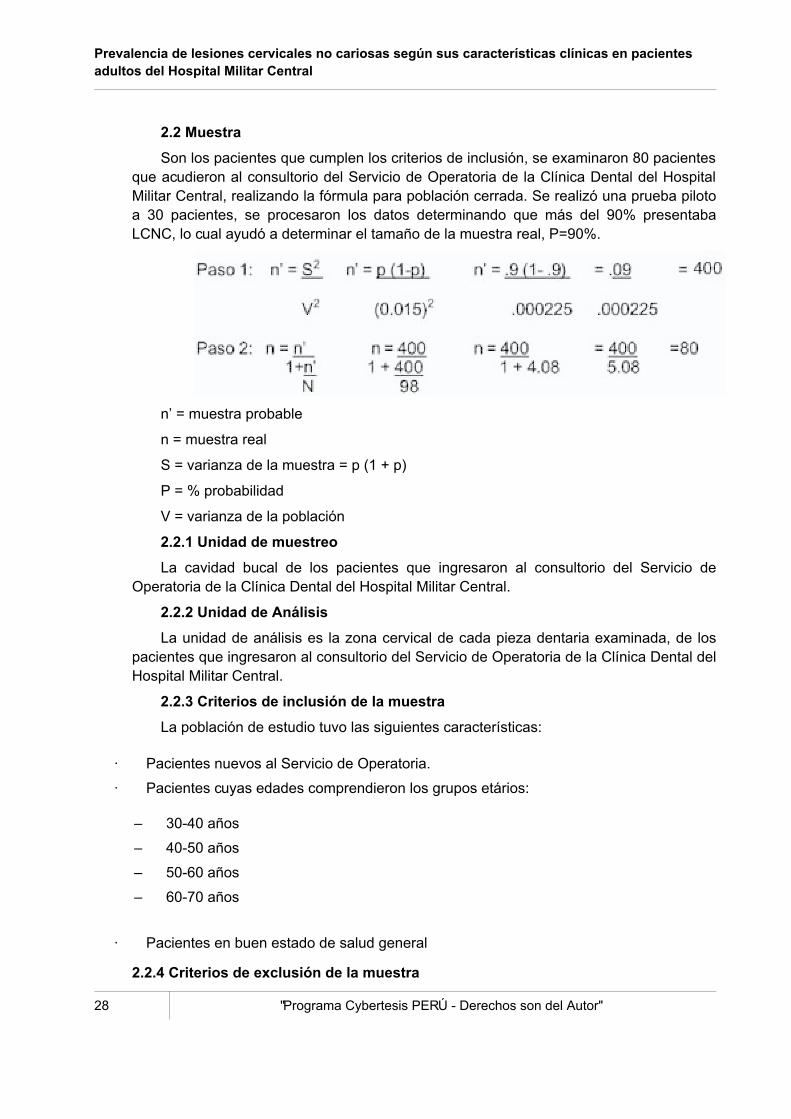
Se evaluó a 80 pacientes nuevos que ingresaron al consultorio del Servicio de Operatoria enel Hospital Militar Central, se encontró una alta prevalencia de estas lesiones cervicales nocariosas, ya que el 97.5% de los pacientes presentaron estas lesiones; se examinaron 1920 piezasdentarias y el 25.9%, 498 piezas, presentaba esta condición dental. El grupo etario de 41-50 añospresentó la mayor cantidad de piezas lesionadas, el grupo dentario con mayor frecuencia fue elgrupo de premolares y de todos los grupos dentarios, las premolares inferiores fueron las másafectadas.
Palabra clave: Prevalencia lesiones cervicales no cariosas


The present work has as intention evaluate the prevalence of non-carious cervical lesions inteeth, according to the clinical characteristics in adult patients in the Service of the MilitaryCentral Hospital, and the frequency and distribution according to age and group toothwort(incisor tooth, canine, premolar and molar). This needs from the knowledge of the clinical aspectand the comparison to the different forms of this type of wear toothwort, the Abfraction, Graze,Erosion and Multiform forms.
There were evaluated 80 new patients who joined to the doctor's office of Operative Servicethe Military Central Hospital, one found a discharge prevalence of these non-carious cervicallesions, 97.5 % of the patients presented these injuries; 1920 pieces were examined and the25.9%, 498 pieces, presented this dental condition. The group of 41-50 years old presented themajor quantity of disabled pieces, the group toothwort with major frequency was the group ofpremolars and the low premolars were the most affected.
Key word: Prevalence non-carious cervical lesions


ADRIAN U.J., YAP and JENNIFER C.L. Neo, “Non-carious Cervical Tooth Loss: Part 1”,Dental Update, 1995, Vol. 22 N° 8. 315-318.
ALEXANDER J.F., SAFFIR A.J., GOLD W., “The measurement of the effect oftoothbrushes on soft tissue”. J. Dent Res 1977; 56:722-7.
AUBRY M., MAFART B., DONAT B., BRAU J.J., “Laboratoire d'Anthropologie”, “Facultede Medecine Secteur Nord, Universite de la Mediterranee”, UMR 6569, 13916Marseille, France; Am J Phys Anthropol 2003 May; 121(1):10-4.
AW T.C., LEPE X., JOHNSON G.H., MANCL L., “Department of Restorative Dentistry,University of Washington”, School of Dentistry, J Am Dent Assoc 2002Jun;133(6):725-33.
BADER J.D., LEVITCH L.C., SHUGARS D.A., HEYMANN H.O., MCCLURE F., “Howdentists classified and treated non-carious cervical lesions”, J Am Dent Assoc 1993.
BARREDA PAREDES Roger, “Abfracciones: lesiones cervicales no cariosas,prevalencia y distribución”. Rev. Visión Dental 2002.
BLACK G. V. Operative Dentistry: Erosion of the teeth. 1914; 39-59.
BOR#I#, JOSIPA, ANI#, IVICA; MUHVI#-UREK, MIRANDA, FERRERI SILVIO, “Theprevalence of non-carious cervical lesions in permanent dentition”, Journal of OralRehabilitation, 2003.
BRADLEY T. PIOTROWSKI; WILLIAM B. GILLETTE; EVERETT B. HANCOCK;

“Examinig the prevalence and characteristics of abfractionlike cervical lesions in apopulation of U.S. veterans”, JADA, Dec 2001.
BRAEM, M. LAMBRECHTS, P. VANHERLE, G., “Stress Induced Cervical Lesions”, J.Prost Dent, 1991.
E.M. SOUZA, VIEIRA, Y F.R. PAGNONCELLI, “Predominio de lesiones cervicalesno-cariosas en estudiantes dentales” escuela dental de PUCPR, el Brasil, de 2 Tuiutide Paraná, el Brasil 2001.
DAVID E., MEYER G., SCHWARTZ P., “the etiology of wedge-shape defects: Amorphological and function-oriented investigation”, The Journal of Gnathology,Volume 10, Number 1, 1991.
GANDARA Beatrice Kay, TRUELOVE Edmond L.; “Diagnosis and Management ofdental erosion”. The Journal of Contemporary Dental Practice, 1999.
GRAEHN G., BERNDT C., STAEGE B. ZUR, “Epidemiologie Keilförmiger Defekte”.Dtsch Stomatol, 1991.
GRIPPO J. O., “A new classification of hard tissue lesions”. J Esthete Dent 1991.
---------- “Noncarious Cervical Lesions: The decision to ignore or to restore”. J.EstheteDent Vol. 4, Suplement, 1992.
---------- “Dental Erosion revisited”. JADA 1995; 126: 619-630.
GROSSKOPF G., “Untersuchungen zur Entstehung der sogenannten keilformigenDefekte am organum dentale” (tesis). Frankfurt/main, 1967.
HEINZ SPRANGER, “Investigación sobre la génesis de lesiones “en cuña” en la regióncervical de los dientes”. Quintessence (Ed. esp.) Vol. 9, núm. 5, 1996: 298-303.
JARVINEN V.K., RYTOMAA I.I., HEINONEN O.P., “Risk factors in dental erosion”. JDent Res 1991; 70:942-947.
JIMÉNEZ LOZANO G., “Restauraciones estéticas de clase 5”, Operatoria Dental, 1999.
JOHANSSON A. ET AL. “Dental erosion, soft-drink intake, and oral health in youngSaudi men, and the development of a system for assessing erosive anterior toothwear”. Acta Odontol Scand 1996; 54: 369-378.
KANTOROWICZ A., “Lehrbuch der klinischen Zarhnheikunde”. Berlin: H Meusser,1924.
KHAN F. ET AL., “Dental cervical lesions associated with occlusal erosion and attrition.Austral Dent J 1999; 44: 176-186.
KÖRBER K.H., “Die elastische Deformierung menschlicher Zähne”. Dtsch Zahnärztl Z1962; 17:691-698.
LEE W.C., EAKLE W.S., “Possible role of tensile stress in the etiology of cervicalerosive lesions of teeth”. J. Prosthet Dent. 1984; 52:374-80.
LEHMAN H., MEYER M., Relationship of dental caries and stress: Concentrations inteeth as revealed by photoelastic test. J Dent Res 1966; 45: 1706-1714.
LEVITCH L ET AL., Epidemiology of non-carious cervical lesions. J Dent Res 1993;IADR abst 715:193.
LUSSI A., SCHAFFNER M., HOTZ P., et al. “Dental erosion in a population of Swissadults”. Community Dent Oral Epidemiol 1991; 19:286-290.

MCCOY G., “On the longevity of teeth”. J. Oral Implant. 1982; II: 249-67.
MELVIN L. ET EL., “root caries and root defects in urban an rural adults: The FloridaDental care study”. JADA 1996; 127: 885-891.
MILLER WD. Experiments and observations on the wasting of tooth tissue variouslydesignated as erosion, abrasion, chemical abrasion, denudation, etc. Dental Cosmos1907; XLIX: 1-23.
NEMCOVSKY C.E, ARTZI Z., “Erosion-Abrasion lesions “Revisited, Compendium 1996,Vol. 17, N° 4.
PEREIRA J.C., “Consideraciones sobre la etiología y el diagnóstico de las lesionescervicales dentarias”. Rev. FOB, Vol. 2, Nº 3, 1994.
RUBIANO M., “Placa neuromiorelajante”, Editorial Médico OdontológicoLatinoamericano, Segunda Edición, 1991.
SOGNNAES R., WOLCOTT R., XHONGA F., “Dental erosion: erosion-like patternsoccurring in association with other dental conditions”. J Am Dent Assoc 1972.
TELLES D., PEGORARO L.F., PEREIRA J.C., “Prevalence of non-carious cervicallesions and their relation to occlusal aspect a clinical study”. J Esthet Dent 2000 vol12., nº1
VILLALOBOS JIMENEZ R., “Tratamiento de lesiones, erosiones, caries ehipersensibilidad radicular”. Universidad de Costa Rica, San José Costa Rica, 1997.
WANNENMACHER E., “Die Veränderungen der Pulpa bei keilförmigen Defekten mitbesonderer Berücksichtigung der Reizdentinbildung” Korrespondenzbl Zahnärzte,1927.
WHITEHEAD S.A., WILSON N.H., WATTS D.C., “Development of non-carious cervicalnotch lesions in vitro”. Coll Antropol 1999.
XHONGA F.A., VALDMANIS S., “Geographical comparisons of the incidence of dentalerosions: a two centre study”. J Oral Rehab., 1983; 10(3): 269-77.
XHONGA F.A., SOGNNAES R. Dental erosion: Clinical measurements of dentalerosion progress. JADA 1972; 84 : 577-582
YETRAM A., WRIGHT K., PICKARD H., “Finite Element Stress Analysis of the crownsof normal and restored teeth”. J Dent Rest Vol. 55
ZSIGMONDY U., “Über die keilförmigen Defekte an den Facialflächen der Zahnhälse”.Österr Ungar Vjhrschr Zahnärzte, 1894.