10businessmodelsofourtimebeta 100204180704-phpapp02-130915004644-phpapp02
Upload
guruindia2012Category
view
128download
0
Chronic Venous Congestion Of
Lung, Liver and Spleen

INTRODUCTION
Congestion is a hemodynamic i.e. circulatory disorder.
Definition: Congestion is a passive process resulting from reduced outflow of blood from a tissue.
It is also called passive hyperemia .
It can be: 1)Systemic or Local And 2)Acute or chronic
Chronic is more common so it is known as Chronic Venous congestion(CVC).

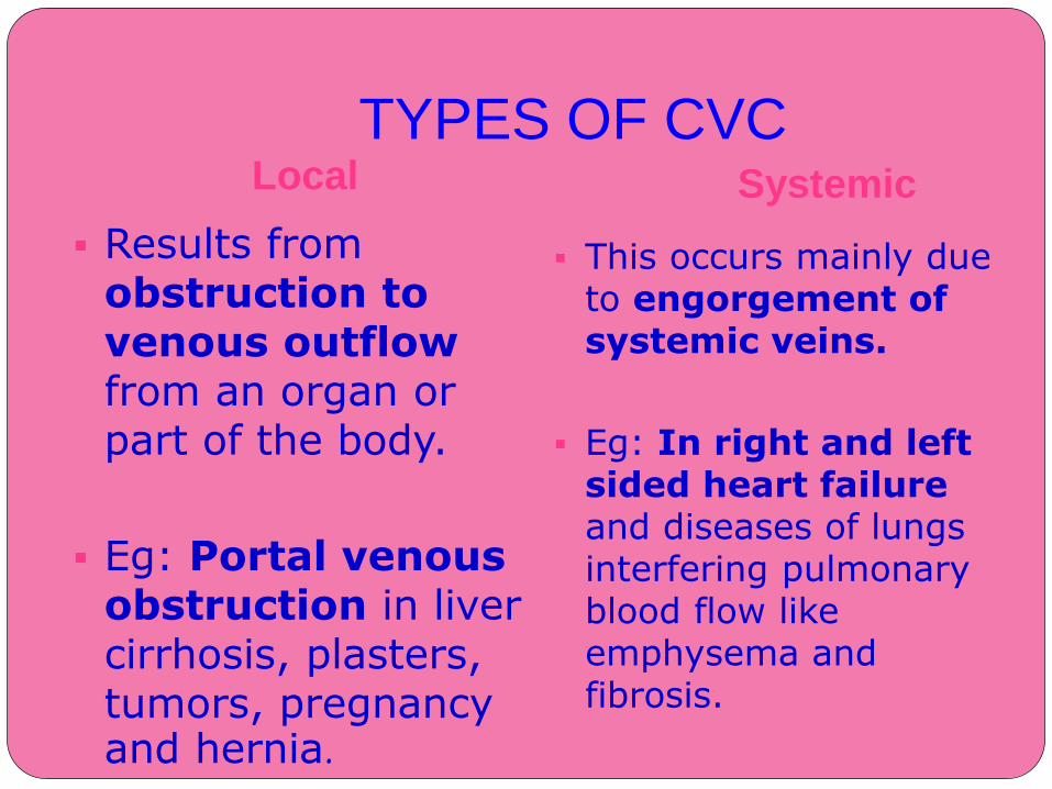
TYPES OF CVCLocal Systemic
Results from obstruction to venous outflowfrom an organ or part of the body.
Eg: Portal venous obstruction in liver cirrhosis, plasters, tumors, pregnancy and hernia.
This occurs mainly due to engorgement of systemic veins.
Eg: In right and left sided heart failure and diseases of lungs interfering pulmonary blood flow like emphysema and fibrosis.
CONSEQUENCES As a result of increased volumes and
pressures, congestion commonly leads to edema.
In passive long standing congestion, lack of blood flow causes chronic hypoxia potentially resulting in ischemic tissue injury and scarring.
Capillary rupture in chronic congestion can also cause small hemorrhagic foci, subsequent catabolism of extravasated red cells can leave residual tell tale clusters of hemosiderin laden macrophages.

GROSS APPEARANCE
Congested tissue is dusky reddish blue in color due to red cell stasis and accumulation of
deoxygenated hemoglobin.
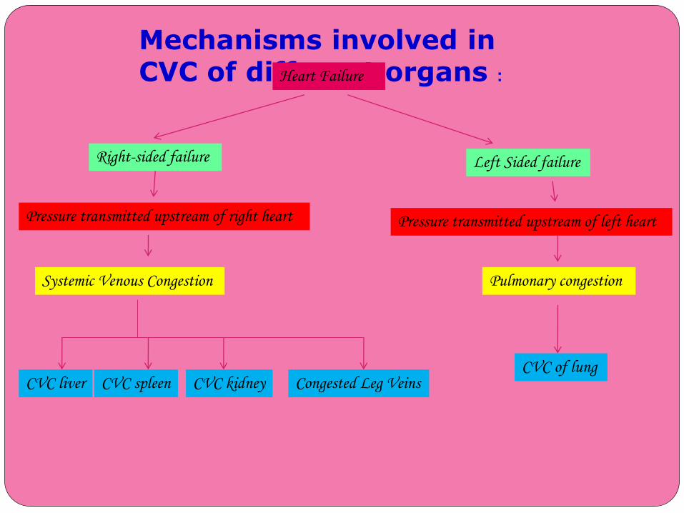
Mechanisms involved in CVC of different organs :Heart Failure
Right-sided failure Left Sided failure
Pressure transmitted upstream of right heart Pressure transmitted upstream of left heart
Systemic Venous Congestion Pulmonary congestion
CVC of lungCVC liver CVC spleen CVC kidney Congested Leg Veins
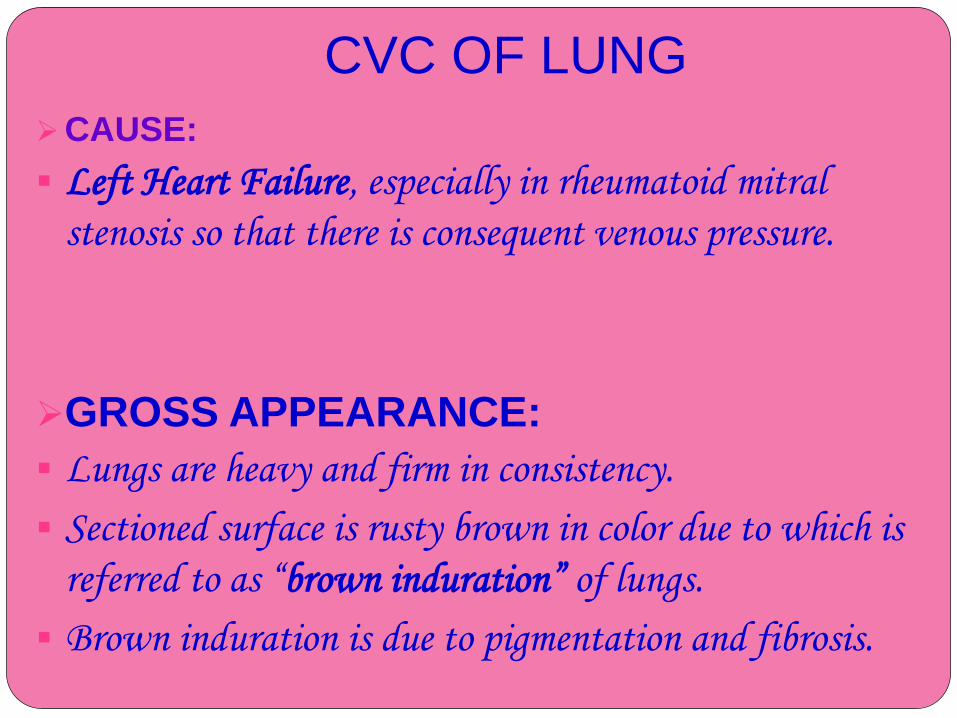
CVC OF LUNG
CAUSE:
Left Heart Failure, especially in rheumatoid mitral
stenosis so that there is consequent venous pressure.
GROSS APPEARANCE:
Lungs are heavy and firm in consistency.
Sectioned surface is rusty brown in color due to which is
referred to as “brown induration” of lungs.
Brown induration is due to pigmentation and fibrosis.
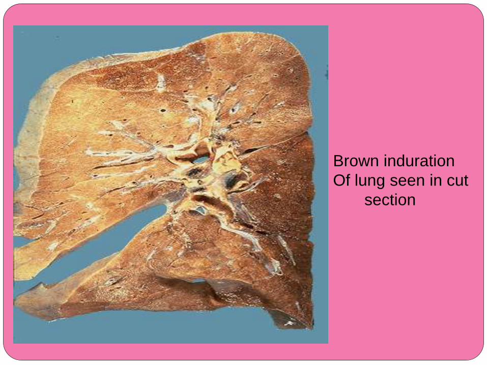
Brown induration
Of lung seen in cut
section
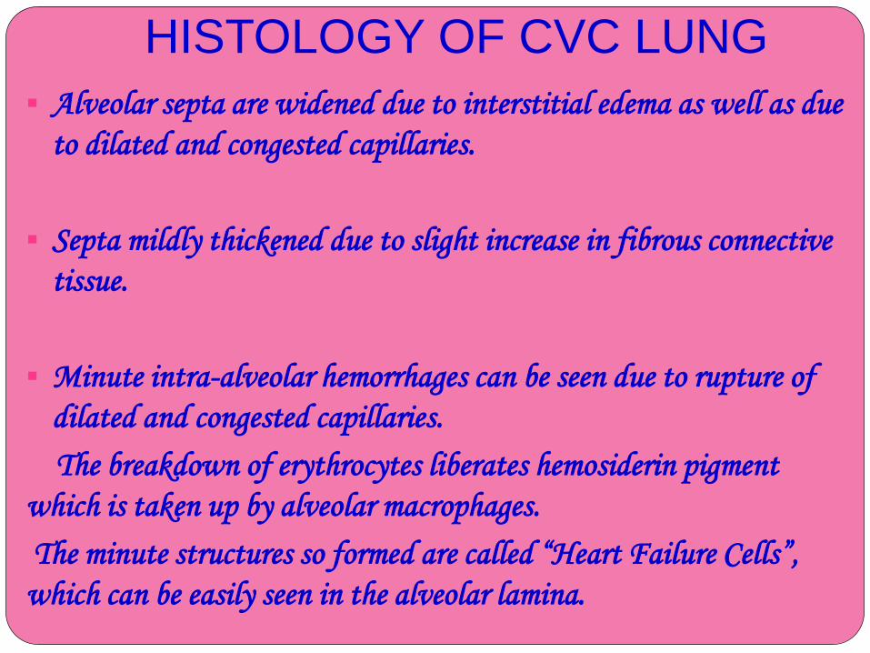
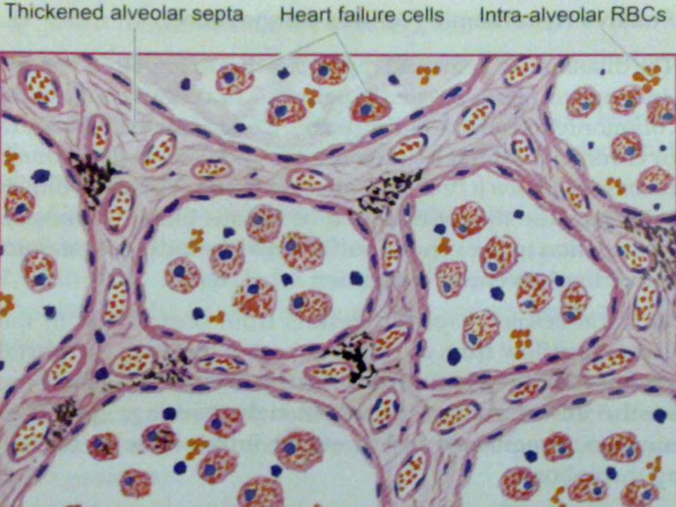
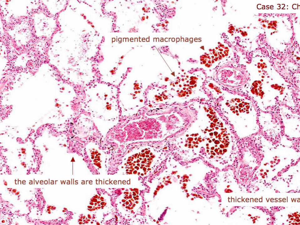
HISTOLOGY OF CVC LUNG
Alveolar septa are widened due to interstitial edema as well as due
to dilated and congested capillaries.
Septa mildly thickened due to slight increase in fibrous connective
tissue.
Minute intra-alveolar hemorrhages can be seen due to rupture of
dilated and congested capillaries.
The breakdown of erythrocytes liberates hemosiderin pigment
which is taken up by alveolar macrophages.
The minute structures so formed are called “Heart Failure Cells”,
which can be easily seen in the alveolar lamina.

CVC OF LIVER
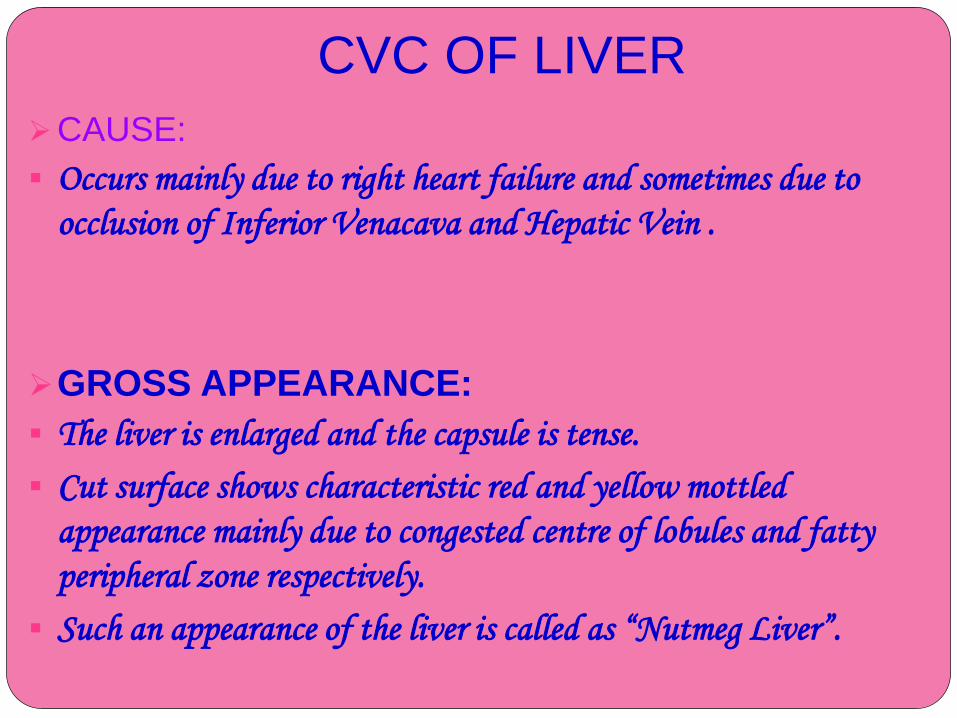
CAUSE:
Occurs mainly due to right heart failure and sometimes due to
occlusion of Inferior Venacava and Hepatic Vein .
GROSS APPEARANCE:
The liver is enlarged and the capsule is tense.
Cut surface shows characteristic red and yellow mottled
appearance mainly due to congested centre of lobules and fatty
peripheral zone respectively.
Such an appearance of the liver is called as “Nutmeg Liver”.
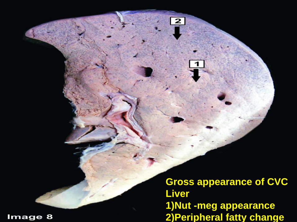
Gross appearance of CVC
Liver
1)Nut -meg appearance
2)Peripheral fatty change
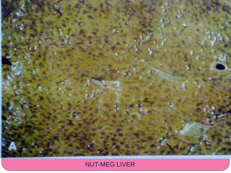
NUT-MEG LIVER


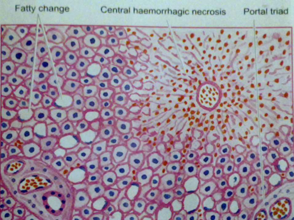
HISTOLOGY OF CVC LIVER Changes are more marked in the
centrilobular zone due to severe hypoxia than in the periphery.
Central vein as well as adjacent sinusoids are distended and filled with blood.
The centrilobular hepatocytes undergo degenerative changes , and eventually “centrilobular hemorrhagic necrosis” can be seen.
Long standing cases may show fine centrilobular fibrosis and regeneration of hepatocytes.
The peripheral zone of the lobule is less severely affected by chronic hypoxia and shows some fatty change in the hepatocytes.
CVC OF SPLEEN
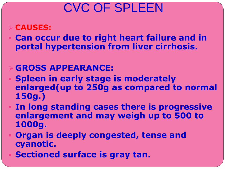
CAUSES:
Can occur due to right heart failure and in portal hypertension from liver cirrhosis.
GROSS APPEARANCE:
Spleen in early stage is moderately enlarged(up to 250g as compared to normal 150g.)
In long standing cases there is progressive enlargement and may weigh up to 500 to 1000g.
Organ is deeply congested, tense and cyanotic.
Sectioned surface is gray tan.
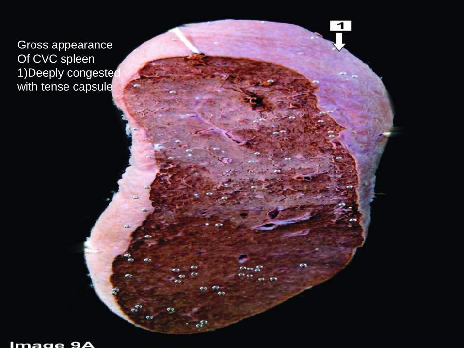
Gross appearance
Of CVC spleen
1)Deeply congested
with tense capsule

2)Cut surface shows gray
tan parenchyma
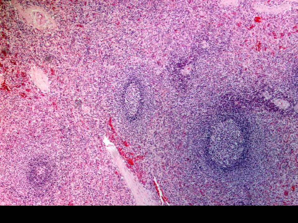
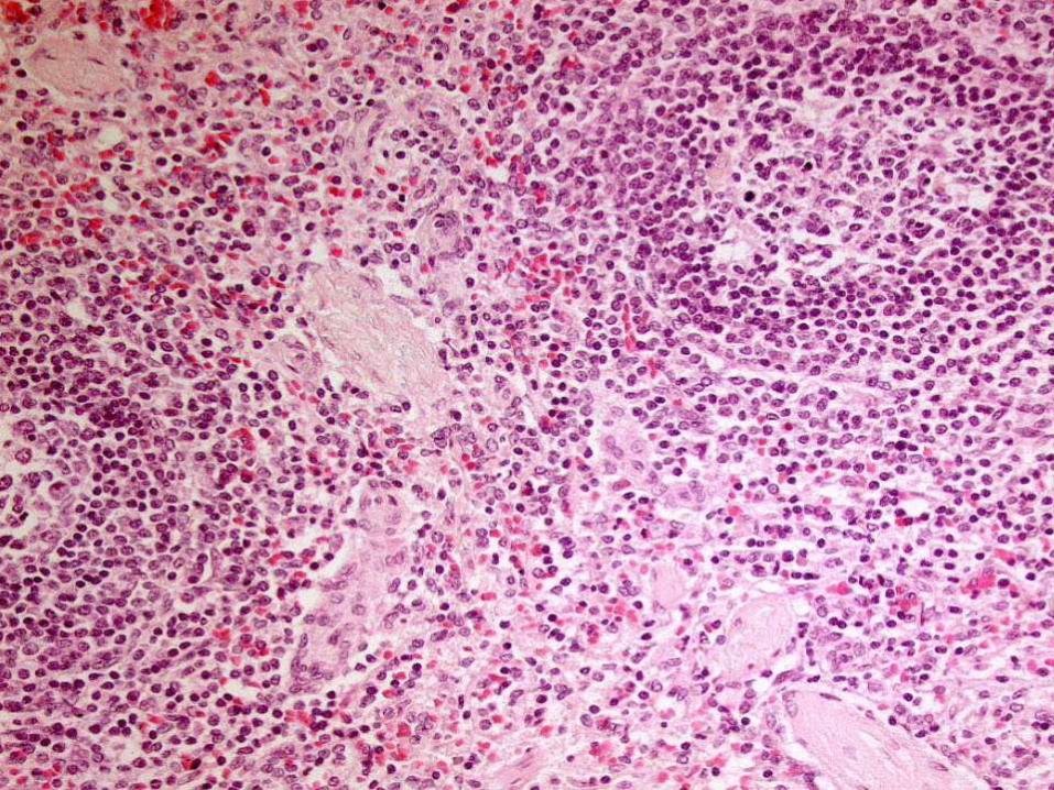
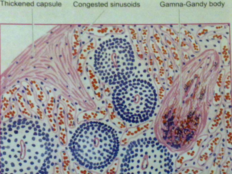
HISTOLOGY OF CVC SPLEEN
Red pulp is enlarged due to congestion and marked sinusoidal dilation and there are many areas of new and old hemorrhages.
Sinusoids may get converted to capillary (capillarisation of sinusoids).
There is hyperplasia of reticuloendothelial cells in the red pulp of the spleen( Splenic macrophages).
There is fibrous thickening of the capsule and the trabeculae.
Some of the hemorrhages overlying fibrous tissue get deposits of the hemosiderin pigment and the calcium salts, these organized structures are called Gamna Gandy bodies or siderofibrotic nodules.
Firmness of spleen in advanced stage is seen more commonly in hepatic cirrhosis(Congestive splenomegaly) and is the most common cause of hypersplenism.