CURRENT UPDATES ON BIOCHEMICAL IDENTIFICATION …
19
www.wjpps.com Vol 9, Issue 5, 2020. 1227 Gajbhiye et al. World Journal of Pharmacy and Pharmaceutical Sciences CURRENT UPDATES ON BIOCHEMICAL IDENTIFICATION METHODS OF GINGIVAL CREVICULAR FLUID MICROFLORA OF SEVERE PERIDONTITIS PATIENT Pranali R. Gajbhiye*, Yogesh N. Gholse, Rahul H. Kasliwal and Dinesh R. Chaple Priyadarshini J. L. College of Pharmacy, Electronic Zone Building, MIDC, Hingna Road, Nagpur-440016, India. ABSTRACT Bacteria are meant to be omnipresent and are mostly involved in lots of human microbial infections. Such bacterial infections can be identified by the different properties of this microorganism. Bacteria are bearing several inherent properties. By using these properties we can differentiate, can check there presence and absence, can check their gram negative and gram positive nature and many more. The present review is therefore focused to combine different biochemical test in one article. The identification of unknown bacteria produces benefits for many aspects of the research of microorganisms and helps physicians correctly treat patients. Multiple biochemical tests were performed to provide the fermentation abilities, presence of certain enzymes, and certain biochemical reactions. Qualitative observations were made on the tests, which were compared to unknown bacteria identification key to aid with the identification process. The identification is required so as to cure the illness or the infection caused due to the bacteria by using appropriate antibiotics. Identificationalso holds significance for epidemiological purposes. KEYWORDS: Bacteria, biochemical test, micro-organisms, bacterial identification. INTRODUCTION Bacteria are present in most habitats on Earth, growing in soil, acidic, radioactive waste, [1] and deep in the Earth's crust, as well as in organic matter and the live bodies of plants and animals, providing outstanding examples of mutualism in the digestive tracts of humans, WORLD JOURNAL OF PHARMACY AND PHARMACEUTICAL SCIENCES SJIF Impact Factor 7.632 Volume 9, Issue 5, 1227-1245 Research Article ISSN 2278 – 4357 *Corresponding Author Pranali R. Gajbhiye Priyadarshini J. L. College of Pharmacy, Electronic Zone Building, MIDC, Hingna Road, Nagpur- 440016, India. Article Received on 04 March. 2020, Revised on 25 March 2020, Accepted on 15 April 2020 DOI: 10.20959/wjpps20205-16056
Transcript of CURRENT UPDATES ON BIOCHEMICAL IDENTIFICATION …

www.wjpps.com Vol 9, Issue 5, 2020.
1227
Gajbhiye et al. World Journal of Pharmacy and Pharmaceutical Sciences
CURRENT UPDATES ON BIOCHEMICAL IDENTIFICATION
METHODS OF GINGIVAL CREVICULAR FLUID MICROFLORA OF
SEVERE PERIDONTITIS PATIENT
Pranali R. Gajbhiye*, Yogesh N. Gholse, Rahul H. Kasliwal and Dinesh R. Chaple
Priyadarshini J. L. College of Pharmacy, Electronic Zone Building, MIDC, Hingna Road,
Nagpur-440016, India.
ABSTRACT
Bacteria are meant to be omnipresent and are mostly involved in lots of
human microbial infections. Such bacterial infections can be identified
by the different properties of this microorganism. Bacteria are bearing
several inherent properties. By using these properties we can
differentiate, can check there presence and absence, can check their
gram negative and gram positive nature and many more. The present
review is therefore focused to combine different biochemical test in
one article. The identification of unknown bacteria produces benefits
for many aspects of the research of microorganisms and helps
physicians correctly treat patients. Multiple biochemical tests were
performed to provide the fermentation abilities, presence of certain
enzymes, and certain biochemical reactions. Qualitative observations were made on the tests,
which were compared to unknown bacteria identification key to aid with the identification
process. The identification is required so as to cure the illness or the infection caused due to
the bacteria by using appropriate antibiotics. Identificationalso holds significance for
epidemiological purposes.
KEYWORDS: Bacteria, biochemical test, micro-organisms, bacterial identification.
INTRODUCTION
Bacteria are present in most habitats on Earth, growing in soil, acidic, radioactive waste,[1]
and deep in the Earth's crust, as well as in organic matter and the live bodies of plants and
animals, providing outstanding examples of mutualism in the digestive tracts of humans,
WORLD JOURNAL OF PHARMACY AND PHARMACEUTICAL SCIENCES
SJIF Impact Factor 7.632
Volume 9, Issue 5, 1227-1245 Research Article ISSN 2278 – 4357
*Corresponding Author
Pranali R. Gajbhiye
Priyadarshini J. L. College
of Pharmacy, Electronic
Zone Building, MIDC,
Hingna Road, Nagpur-
440016, India.
Article Received on
04 March. 2020,
Revised on 25 March 2020,
Accepted on 15 April 2020
DOI: 10.20959/wjpps20205-16056
www.wjpps.com Vol 9, Issue 5, 2020.
1228
Gajbhiye et al. World Journal of Pharmacy and Pharmaceutical Sciences
termites and cockroaches. There are typically 40 million bacterial cells in a gram of soil and a
million bacterial cells in a milliliter of fresh water; in all, there are approximately five
nonillion(5×1030) bacteria on Earth.[2]
If bacteria form a parasitic association with other
organisms, they are classed as pathogens. Pathogenic bacteria are a major cause of human
death and disease and cause infections such as tetanus, typhoid, fever, diphtheria, syphilis,
cholera, foodborn, illness, leprosy and tuberculosis. Conditionally pathogenic bacteria are
only pathogenic under certain conditions, such as a wound that allows for entry into the
blood, or a decrease in immune function. For example, Staphylococcus or Streptococcus are
also part of the normal human flora and usually exist on the skin or in the nose without
causing disease, but can potentially cause skin infections, pneumonia, meningitis and even
overwhelming sepsis, a systemic inflammatory response producing shock, massive
vasodilation and death. Some species of bacteria, such as Pseudomonas aeruginosa,
Burkholderiacenocepacia, and Mycobacterium avium, are opportunistic pathogens and cause
disease mainly in people suffering from immunosuppression or cystic fibrosis.[3]
The
identification is required so as to cure the illness or the infection caused due to the bacteria by
using appropriate antibiotics. Identification also holds significance for epidemiological
purposes.[4]
Bacteria are meant to be omnipresent and are mostly involved in lots of human microbial
infections. Such bacterial infections can be identified by the different properties of this
microorganism. Bacteria are bearing several inherent properties. by using these properties we
can differentiate, can check there presence and absence, can check their gram negative and
gram positive nature and many more. The present review is therefore focused to combine
different biochemical test in one article.[5]

www.wjpps.com Vol 9, Issue 5, 2020.
1229
Gajbhiye et al. World Journal of Pharmacy and Pharmaceutical Sciences
Different biochemicals tests are performed to check and differentiate the bacteria. Different
biochemical test has been mentioned below-
Fig. 1: Different biochemical test performed to check and differentiate the bacteria.
a. Indole Test[5]
Purpose: The indole test screens for the ability of an organism to degrade the amino acid
tryptophan and produce indole. It is used as part of the IMViC procedures, a battery of tests
designed to distinguish among members of the family Enterobacteriaceae.
Principle: Indole test is a biochemical test which differentiates the coliform from other
members of Enterobacteriaceby detecting their ability to produce the enzyme tryptophanase.
This enzyme hydrolyses the amino acid tryptophan into indole, pyruvic acid and ammonia. It
is the intracellular enzyme (endoenzyme).whenindole is combined with Kovac’s Reagent, the

www.wjpps.com Vol 9, Issue 5, 2020.
1230
Gajbhiye et al. World Journal of Pharmacy and Pharmaceutical Sciences
solution turns from yellow to cherry red. Because amyl alcohol is not water soluble, the red
colouration will form in an oily layer of the top of the broth.
Procedure
1. SIM medium broth was prepared and sterilized using autoclave at 151bs for 15 minutes
2. The SIM medium broth was inoculated with test organism and an uninoculated tube
waskept as control
3. The tubes were incubated at 35°C for 48hours; 1ml of Kovac s reagent was added48hrs of
incubation
4. The tubes were shaken gently after intervals of 10 to 15minutes
5. The tubes were allowed to stand for few minutes to permit the reagent to come to the top
6. The tubes were observed for cherry red layers in the top layer
Inference
Positive test: Development of bright fuchsia red colour at the interface of the reagent and
the broth within seconds after adding the reagent.
Example: E.coli and Proteus vulgaris
Negative test: No colour change after addition of reagent
Example: Salmonella spp, Enterobacter aerogens, Klebsiella spp.
Fig. 2: Indole test.

www.wjpps.com Vol 9, Issue 5, 2020.
1231
Gajbhiye et al. World Journal of Pharmacy and Pharmaceutical Sciences
b. Starch Hydrolysis Test[6]
Purpose: This test is performed to test the utilization of starch by Bacteria by producing the
enzyme Amylase.
Principle: In the starch hydrolysis test, the test bacteria are grown on agar plates containing
starch. If the bacteria have the ability to hydrolyze starch, it does so in the medium,
particularly in the areas surrounding their growth while the rest of the area of the plate still
contain non-hydrolysed starch. Since no color change occurs in the medium when organisms
hydrolyze starch, iodine solution is added as an indicator to the plate after incubation. While
the non-hydrolysed starch forms dark blue color with iodine, its hydrolyzed end products do
not acquire such dark blue color with iodine.
Procedure
1. Starch agar media was prepared and sterilized using autoclave at 151bs for 15minutes
2. The media was poured into Petri plate and allowed to solidify
3. The test organism was inoculated on to the plate with a sterile transfer loop
4. The plate was incubated at 35°C for 48hrs
5. After incubation the plate was flooded with Gram’s iodine
6. Plate was observed for clear zone around the test organism
Inference
Positive test: A clear zone around the line of growth after the addition of iodine solution.
Example: Bacillus subtilis
Negative test: Dark blue coloration of the medium.
Example: E.coli
Observation: Examine for the clear zone around the line of bacterial growth.
Fig. 3: Starch hydrolysis test.
www.wjpps.com Vol 9, Issue 5, 2020.
1232
Gajbhiye et al. World Journal of Pharmacy and Pharmaceutical Sciences
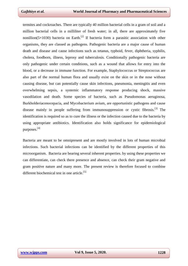
c. Catalase Production Test[7]
Purpose: The test is done to check weather the test organism produce Catalase or not.
Principle:The enzyme catalase mediates the breakdown of hydrogen peroxide into oxygen
and water. The presence of the enzyme in a bacterial isolate is evident when a small inoculum
is introduced into hydrogen peroxide, and the rapid elaboration of oxygen bubbles occurs.
The lack of catalase is evident by a lack of or weak bubble production. The culture should not
be more than 24 hours old. Bacteria thereby protect themselves from the lethal effect of
Hydrogen peroxide which is accumulated as an end product of aerobic carbohydrate
metabolism.
Procedure
1. Nutrient agar medium was prepared
2. The medium was poured into culture tubes and flasks
3. It was sterilized by autoclaving at 151b pressure for 15 minutes
4. The nutrient agar slants were inoculated with test organisms
5. An inoculated nutrient agar slant was kept as control
6. The cultures were incubated at 35°C and 3-4 drops of hydrogen peroxide was added on
the growth of each slant culture
7. The culture was observed for the appearance or absence of gas bubbles.
Inference
Positive: Copious bubbles produced, active bubbling
Examples: Staphylococci, Micrococci, Listeria, Corynebacterium diphtheriae, Burkholderia
cepacia, Nocardia, the family Enterobacteriaceae (Citrobacter, E. coli, Enterobacter,
Klebsiella, Shigella, Yersinia, Proteus, Salmonella, Serratia), Pseudomonas, Mycobacterium
tuberculosis, Aspergillus, Cryptococcus, and Rhodococcus equi.
Negative: No or very few bubbles produced.
Examples: Streptococcus and Enterococcus spp
www.wjpps.com Vol 9, Issue 5, 2020.
1233
Gajbhiye et al. World Journal of Pharmacy and Pharmaceutical Sciences
Fig. 4: Catalase production test.
d. Motility Test[5]
Purpose: The MIO medium is a multitest agar used to test for indole production while
simultaneously determining other characteristics of the bacterium.
Principle: Motility is the ability of an organism to move by itself by means of propeller-like
flagella unique to bacteria or by special fibrils that produce a gliding form of motility.
Motility by bacterium is demonstrated in semi solid agar medium. The medium mainly used
for this purpose is SIM medium (Sulphide Indole Motility medium) which is a combination
differential medium that tests three different parameters, Sulfur Reduction, Indole Production
and Motility. This media has a very soft consistency that allows motile bacteria to migrate
readily through them causing cloudiness. The inoculum is stabbed into the centre of a
semisolid agar deep using a sterile inoculating needle. Bacterial motility is evident by a
diffuse zone of growth extending out from the line of inoculation. Some organisms grow
throughout the entire medium, whereas others show small areas or nodules that grow out
from the line of inoculation. The non-motile bacteria will only grow in the soft agar tube and
only the area where they are inoculated.
Procedure
1. Prepare SIM broth medium and sterilized by autoclaving at 121°c at 15 min.
2. Touch the needle to a colony of young (18-24hrs) culture growing on agar medium.
3. Slab once to a depth of only 1/3 to ½ inch in middle of tube.
4. Be sure to keep the needle in the same line it entered as it is removed from medium.
5. Incubate at 35-37°c & examine daily for upto 7 days.

www.wjpps.com Vol 9, Issue 5, 2020.
1234
Gajbhiye et al. World Journal of Pharmacy and Pharmaceutical Sciences
Inference
Positive: Diffuse, hazy growths that spread throughout the medium rendering it slightly
opaque.
Example: Helicobacter pylori, Salmonella species, Escherichia coli, Pseudomonas
aeruginosa, and Vibrio cholerae.
Negative: Growth that is confined to the stab-line, with sharply defined margins and
leaving the surrounding medium clearly transparent.
Example: Klebsiella pneumoniae, and Yersinia pestis.
Fig. 5: Motility test.
e. Methyl Red Test[8]
Principe: Some bacteria have ability to utilize the glucose and convert it to a stable acid like
lactic acid, acetic acid or formic acid as the end product.These bacteria initially metabolise
glucose to pyruvic acid which is further metabolized through the ‘mixed acid pathway to
produce the stable acid. The type of acid produced differs from species to species and
depends on the specific enzymatic pathways present in the bacteria. The acid so produced
decreases the pH to 4.5 or below, which is indicated by a change in the colour of methyl red
from yellow to red.
Procedure
1. Prepare MR-VP broth mediumand sterilized by autoclaving at 121°c at 15 min.
2. Prior to inoculation, allow medium to equilibrate to room temperature.
3. Using organism taken from 18-24hrs pure culture, lightly inoculate the medium.
4. Incubate aerobically at 37°c for 24hrs. Following 24hrs of incubation, aliquot 1ml of
broth to a clean test tube.

www.wjpps.com Vol 9, Issue 5, 2020.
1235
Gajbhiye et al. World Journal of Pharmacy and Pharmaceutical Sciences
5. Reincubate the remaining broth for an additional 24 hrs.
6. Add 2-3 drops of methyl red indicator to aliquot.
Inference
Positive test: A distinct red color
Examples: E. coli, Yersinia sps, etc.
Negative Reaction: A yellow color
Examples: Enterobacter aerogenes, Klebsiella pneumoniae, etc.
Fig. 6: Methyl red test.
f. Vogus Proskauer Test[5]
Purpose: Voges–Proskauer is a test used to detect acetoin in a bacterial broth culture.
Principle: The Voges-Proskauer (VP) test is used to determine if an organism
produces acetylmethylcarbinol from glucose fermentation. If present, acetylmethylcarbinol is
converted to diacetyl in the presence of∝- naphthol, strong alkali (40%KOH), and
atmospheric oxygen. The αnaphthol was not part of the original procedure but was found to
act as a color intensifier by Barritt and must be added first. The diacetyl andquanidine-
containing compounds found in the peptones of the broth then condense to form a pinkish red
polymer.
Procedure
1. Prepare MR-VP broth mediumand sterilized by autoclaving at 121°c at 15 min.
2. Prior to inoculation, allow medium to equilibrate to room temperature.
3. Using organism taken from 18-24hrs pure culture, lightly inoculate the medium.
4. Incubate aerobically at 37°c for 24 hrs. Following 24 hrsincubation, aliquot 2ml of the
broth to a clean test tube.
5. Reincubate the remaining broth for additional 24 hrs.

www.wjpps.com Vol 9, Issue 5, 2020.
1236
Gajbhiye et al. World Journal of Pharmacy and Pharmaceutical Sciences
6. Add 6 drops of 5% alpha naphthol and mix well to aerate. Add 2 drops of 40% KOH &
mix well to aerate.
7. Observe for a pink-red color at the surface within 30 min. shake the tube vigorously
during the 30 min period.
Inference
Positive test: Development of a red color 15 minutes or more after the addition of the
reagents indicating the presence of diacetyl, the oxidation product of acetoin.
Example: Klebsiella (formerly Enterobacter) aerogenes
Negative test: Produce a creamson or red colour
Example: Escherichia coli
Fig. 7: Vogus proskauer test.
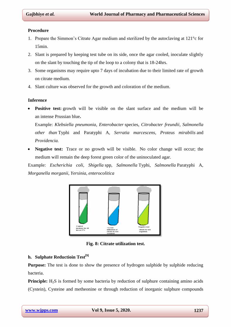
g. Citrate Utilization Test[5]
Purpose :The test is used to check the ability of microorganisms for utilization of citrate.
Principle:Citrate agar is used to test an organism’s ability to utilize citrate as a source of
energy. The medium contains citrate as the sole carbon source and inorganic ammonium salts
(NH4H2PO4) as the sole source of nitrogen.
Bacteria that can grow on this medium produce an enzyme, citrate-permease, capable of
converting citrate to pyruvate. Pyruvate can then enter the organism’s metabolic cycle for
the production of energy. Growth is indicative of utilization of citrate, an intermediate
metabolite in the Krebs cycle.
When the bacteria metabolize citrate, the ammonium salts are broken down to ammonia,
which increases alkalinity. The shift in pH turns the bromthymol blue indicator in the
medium from green to blue above pH 7.6.
www.wjpps.com Vol 9, Issue 5, 2020.
1237
Gajbhiye et al. World Journal of Pharmacy and Pharmaceutical Sciences
Procedure
1. Prepare the Simmon’s Citrate Agar medium and sterilized by the autoclaving at 121°c for
15min.
2. Slant is prepared by keeping test tube on its side, once the agar cooled, inoculate slightly
on the slant by touching the tip of the loop to a colony that is 18-24hrs.
3. Some organisms may require upto 7 days of incubation due to their limited rate of growth
on citrate medium.
4. Slant culture was observed for the growth and coloration of the medium.
Inference
Positive test: growth will be visible on the slant surface and the medium will be
an intense Prussian blue.
Example: Klebsiella pneumonia, Enterobacter species, Citrobacter freundii, Salmonella
other than Typhi and Paratyphi A, Serratia marcescens, Proteus mirabilis and
Providencia.
Negative test: Trace or no growth will be visible. No color change will occur; the
medium will remain the deep forest green color of the uninoculated agar.
Example: Escherichia coli, Shigella spp, Salmonella Typhi, Salmonella Paratyphi A,
Morganella morganii, Yersinia, enterocolitica
Fig. 8: Citrate utilization test.
h. Sulphate Reductioin Test[9]
Purpose: The test is done to show the presence of hydrogen sulphide by sulphide reducing
bacteria.
Principle: H2S is formed by some bacteria by reduction of sulphure containing amino acids
(Cystein), Cysteine and metheonine or through reduction of inorganic sulphure compounds
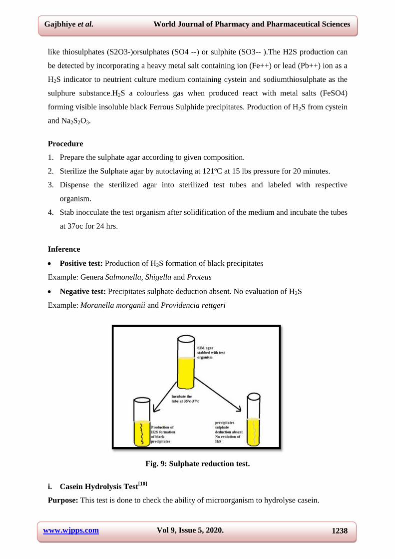
www.wjpps.com Vol 9, Issue 5, 2020.
1238
Gajbhiye et al. World Journal of Pharmacy and Pharmaceutical Sciences
like thiosulphates (S2O3-)orsulphates (SO4 --) or sulphite (SO3-- ).The H2S production can
be detected by incorporating a heavy metal salt containing ion (Fe++) or lead (Pb++) ion as a
H2S indicator to neutrient culture medium containing cystein and sodiumthiosulphate as the
sulphure substance.H2S a colourless gas when produced react with metal salts (FeSO4)
forming visible insoluble black Ferrous Sulphide precipitates. Production of H2S from cystein
and Na2S2O3.
Procedure
1. Prepare the sulphate agar according to given composition.
2. Sterilize the Sulphate agar by autoclaving at 121ºC at 15 lbs pressure for 20 minutes.
3. Dispense the sterilized agar into sterilized test tubes and labeled with respective
organism.
4. Stab inocculate the test organism after solidification of the medium and incubate the tubes
at 37oc for 24 hrs.
Inference
Positive test: Production of H2S formation of black precipitates
Example: Genera Salmonella, Shigella and Proteus
Negative test: Precipitates sulphate deduction absent. No evaluation of H2S
Example: Moranella morganii and Providencia rettgeri
Fig. 9: Sulphate reduction test.
i. Casein Hydrolysis Test[10]
Purpose: This test is done to check the ability of microorganism to hydrolyse casein.

www.wjpps.com Vol 9, Issue 5, 2020.
1239
Gajbhiye et al. World Journal of Pharmacy and Pharmaceutical Sciences
Principle: Casein is a major protein found in milk. It is a macromolecule composed of amino
acid linked together by peptide bond. Some microorganism have the ability to decrease the
protein casein by producing proteolyticexo-enzymes called protease. The process break down
the peptide bond by introducing water into the molecule liberating the soluble amino acid
pool for use in the synthesis of structural and functional cellular protein.
Casein hydrolysis can be demonstrated by supplementing nutrient agar media with milk .The
medium is opaque due to the casein is colloidal suspension formation of a clear zone
surrounded the bacterial growth. after inoculation and incubation of agar plate culture is due
to hydrolysis of casein by protease activity. The medium surrounding the growth of the
organism remains opaque since it defects light rays rather than transmitting which indicates a
negative reaction.
Procedure
1. Prepared the Casein media. Sterilize the media by autoclaving.
2. Then dispense the media in sterilized petriplates.
3. After solidification streak the test organism at the centre of the plate.
4. Incubate the plate at 37oc for 24 hrs.
Inference
Positive test: Clearing is observed around and/or beneath colony growth (hydrolysis).
Example: Bacillus subtilis and Pseudomonas aeroginosa
Negative test: No clearing is observed around and/or beneath the inoculums.
Example: Escherichia coli
Fig. 10: Casein hydrolysis test.
www.wjpps.com Vol 9, Issue 5, 2020.
1240
Gajbhiye et al. World Journal of Pharmacy and Pharmaceutical Sciences
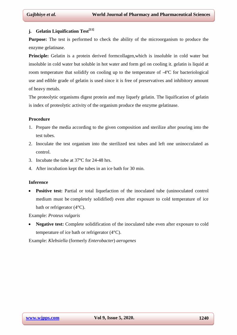
j. Gelatin Liquification Test[11]
Purpose: The test is performed to check the ability of the microorganism to produce the
enzyme gelatinase.
Principle: Gelatin is a protein derived formcollagen,which is insoluble in cold water but
insoluble in cold water but soluble in hot water and form gel on cooling it. gelatin is liquid at
room temperature that solidify on cooling up to the temperature of -4ºC for bacteriological
use and edible grade of gelatin is used since it is free of preservatives and inhibitory amount
of heavy metals.
The proteolytic organisms digest protein and may liquefy gelatin. The liquification of gelatin
is index of proteolytic activity of the organism produce the enzyme gelatinase.
Procedure
1. Prepare the media according to the given composition and sterilize after pouring into the
test tubes.
2. Inoculate the test organism into the sterilized test tubes and left one uninocculated as
control.
3. Incubate the tube at 37ºC for 24-48 hrs.
4. After incubation kept the tubes in an ice bath for 30 min.
Inference
Positive test: Partial or total liquefaction of the inoculated tube (uninoculated control
medium must be completely solidified) even after exposure to cold temperature of ice
bath or refrigerator (4°C).
Example: Proteus vulgaris
Negative test: Complete solidification of the inoculated tube even after exposure to cold
temperature of ice bath or refrigerator (4°C).
Example: Klebsiella (formerly Enterobacter) aerogenes

www.wjpps.com Vol 9, Issue 5, 2020.
1241
Gajbhiye et al. World Journal of Pharmacy and Pharmaceutical Sciences
Fig. 11: Gelatin liquification test.
k. OXIDASE TEST[5]
Purpose: The test is done to detect the presence of cytochrome C and hence the production
of oxidase enzyme by given test organism.
Principle: Cytochrome areheam containing catalytic enzyme which are tightly bound to
(prokaryotic cells) cells of plasma membrane. They are concerned with later stages of
biochemical oxidation. They act as electron or H2 carries of biological oxidation by virtue of
their availability valence by heam ions i.e., they can undergo reduction and oxidation. During
period of great activity they are reduced. Cytochrome C is more abundant and freely soluble
in water. Cytochrome C does not react directly with O2 but reduced form will be oxidized by
cytooxidase with which it is closely associated. Cyto a,a3 is called cytooxidase.
orindophenolase or endophenol oxidase or ferro cytochrome C or oxygen oxidoreductase.
Cyt.a.a3 is the terminal codon in electron transport chain hence called Cytooxidase.This test
tests for lytic and not cytooxidase.
Procedure
1. Place 1-2 drops of 1 % oxidase reagent on 6 cm square piece of whatman filter paper.
2. Transfer a small colony of test organism using a loop onto soaked filter paper
3. Observe for purple colour development.
Inference
Positive test: Development of dark purple color (indophenols) within 10 seconds
Example: Neisseria, Pseudomonas spp, Vibrio cholrae, Helicobacter spp, etc
Negative test: Does not show purple colour
Example: Genera of the Enterobacteriaceae.

www.wjpps.com Vol 9, Issue 5, 2020.
1242
Gajbhiye et al. World Journal of Pharmacy and Pharmaceutical Sciences
Fig. 12: Oxydase test.
l. Urease Test[12]
Purpose: The test is done if the given organism produces enzyme Urease or not.
Pinciple: Urea is a major organic waste production of protein digestion by most vertebrates
and is excreted in urine.Some microorganisms have the ability to produce the enzyme urease.
Urease is a hydrolytic enzyme, which attack the amide linkage liberating Ammonia.
This test distinguishes members of genus Proteus from other lactose non fermenting enteric
microbes.
Procedure
1. Prepare and dispense Urea agar (basal medium) into tubes and sterilized.
2. Glucose and phenol red is added to the basal medium and steamed for 1 Hr.
3. Add filtered sterilized urea solution and mix all contents well and dispense into sterile test
tubes.
4. The test organisms are inoculated and incubated at 37oC for 24-48 Hrs.
5. The slants were observed for colour
Inference
Positive test: Development of an intense magenta to bright pink color in 15 min to 24 h.
Example:Proteus spp, Cryptococcus spp, Corynebacterium spp, Helicobacterpylori, Yersina s
pp, Brucella spp, etc.
Negative test: No color change.
Example: Escherichia, Shigella, Salmonella, etc.

www.wjpps.com Vol 9, Issue 5, 2020.
1243
Gajbhiye et al. World Journal of Pharmacy and Pharmaceutical Sciences
Fig. 13: Urease test.
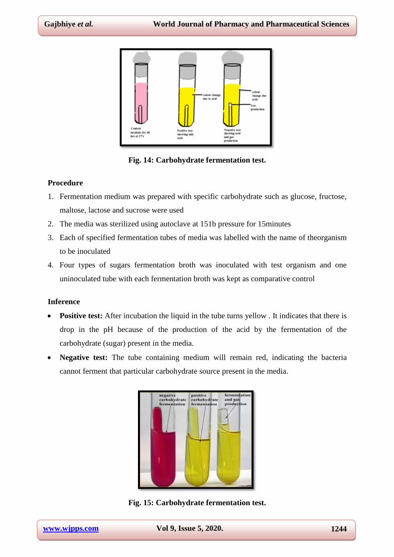
m. Carbohydrate Fermentation Test[5]
Purpose: This test I performed to check the ability of bacteria to check its ability to ferment
sugar.
Principle: Morphological and cultural characteristics are essential in identifying an organism
in the case of bacteria. Physiological information (through physiological reactions) in an
organism contribute informations for specific identification as all the biochemical reactions
are catalysed by enzyme and enzyme specific to species the bio-oxidation reaction could be
oxidative or fermentative oxidizing organic substance s for energy result in the production of
CO2 and water it is oxidative reaction.Aldehydes or alcohols produced with various type of
gases such as CO2,H2,CH4s called fermentive reactions. Fermentation of sugars can be
easily demonstrated and the end products of particular fermentation depends on the nature of
the organism characteristics of substrate and environmental conditions such as temperature
and PH.
Fermentation of sugar is carried out anaerobically with the indicator production incompletely
oxidized acidic or alkaline and products with gas production, to detect fermentation in labs. A
fermentation tube is used with a Durham’s tube for the detection of gas as the end product of
carbohydrate metabolism.
www.wjpps.com Vol 9, Issue 5, 2020.
1244
Gajbhiye et al. World Journal of Pharmacy and Pharmaceutical Sciences
Fig. 14: Carbohydrate fermentation test.
Procedure
1. Fermentation medium was prepared with specific carbohydrate such as glucose, fructose,
maltose, lactose and sucrose were used
2. The media was sterilized using autoclave at 151b pressure for 15minutes
3. Each of specified fermentation tubes of media was labelled with the name of theorganism
to be inoculated
4. Four types of sugars fermentation broth was inoculated with test organism and one
uninoculated tube with each fermentation broth was kept as comparative control
Inference
Positive test: After incubation the liquid in the tube turns yellow . It indicates that there is
drop in the pH because of the production of the acid by the fermentation of the
carbohydrate (sugar) present in the media.
Negative test: The tube containing medium will remain red, indicating the bacteria
cannot ferment that particular carbohydrate source present in the media.
Fig. 15: Carbohydrate fermentation test.

www.wjpps.com Vol 9, Issue 5, 2020.
1245
Gajbhiye et al. World Journal of Pharmacy and Pharmaceutical Sciences
CONCLUSION
The development of rapid, low-cost, sensitive, and reproducible methods for the screening
identification of microorganisms is an important issue in modern science. Replacing
biochemical methods, which are labor intensive and time saving, with modern methods, such
as MALDI-TOFMS or electromigration techniques.Different methodologies have been
developed throughout the years to identify microorganism to improve bioremediation
techniques, determine susceptibility profiles of bacteria and reduce the impact of
microorganisms.
REFERENCES
1. Fredrickson JK, Zachara JM, Balkwill DL. Geomicrobiology of high-level nuclear waste.
App EnvtMicrobiol, 2004; 70(7): 4230–41.
2. Whitman WB, Coleman DC, Wiebe WJ. Prokaryotes: The unseen majority.
ProcNatlAcadSci USA, 1998; 95(12): 657883.
3. Heise E. Diseases associated with immunosuppression. Envt health perspect, 1998; 43:
919.
4. Mishra1 V, Kumar D, Srivastava VK, Gautam A, Shukla S, Verma KR, Verma
AR.Identification of unknown bacteria by using biochemical tests and 16s RNA
sequencing from different soil samples. Int J Adv Res Sci Eng., 2016; 5(4): 2319-8354.
5. Vashist H, Sharma D, Gupta A. A review on commonly used biochemical test for
bacteria. Inn J life sci., 2013; 1(1): 1-7.
6. www.vumicro.com/vumie/help/VUMICRO/Starch_Hydrolysis_Test.html
7. Facklam R, Elliott JA.Identification, classification, and clinical relevance of catalase-
negative, gram-positive cocci, excluding the streptococci and enterococci. ClinMicrobiol
Rev., 1995; 8(4): 479.
8. Crown ST, Gen J. Micromethod for the methyl red test. Microbiol, 1998; 9: 101-109.
9. Barton LL, Fauque G D. Biochemistry, Physiology and Biotechnology of Sulfate
Reducing Bacteri. Adv App Microbiol, 2009; 68: 41–98.
10. Clarke PH, Cowan S T. Biochemical methods for bacteriology. J. Gen. Microbiol, 1952;
6: 187-197.
11. Greene RA, Larks GG. A quick method for the detection ofgelatin liquefying bacteria. J
Bacteriol, 1955; 69: 224.
12. Christensen WB. Urea decomposition as a means of differentiating Proteus and paracolon cultures
from each other and from Salmonella andShigella types. J. Bacteriol, 1946; 52: 461–466.