Current Management of Biliary Atresia - Denver, Colorado · Current Management of Biliary Atresia...
30
Current Management of Biliary Atresia Janeen Jordan, MD PGY5 Surgery Grand Rounds November 19, 2007
-
Upload
truongmien -
Category
Documents
-
view
233 -
download
4
Transcript of Current Management of Biliary Atresia - Denver, Colorado · Current Management of Biliary Atresia...

Current Management of Biliary Atresia
Janeen Jordan, MDPGY5Surgery Grand RoundsNovember 19, 2007
Overview
Etiology and diagnosis Work-up and management History of Kasai Portoenterostomy Studies Advances Recommendations
Transplant Summary

John Thompson in 1891 Holmes (Johns Hopkins) in early 1900s “correctable” biliary atresia
Morio Kasai 1955 pioneered HPE “uncorrectable” biliary atresia
Biliary Atresia (BA)
Type 3Type 2

Biliary Atresia
2 Subtypes Embryonic – associated with other anomalies Perinatal
80-90% Aquired CMV, Reovirus, Rotavirus
Incidence 1/5000-18,000 births (~300 per year) 2/3 female predominance Asian and African descent


Management
Goal to diagnose prior to 60-90days >100days or cirrhosis then transplant Most die within 2yrs from ESLD
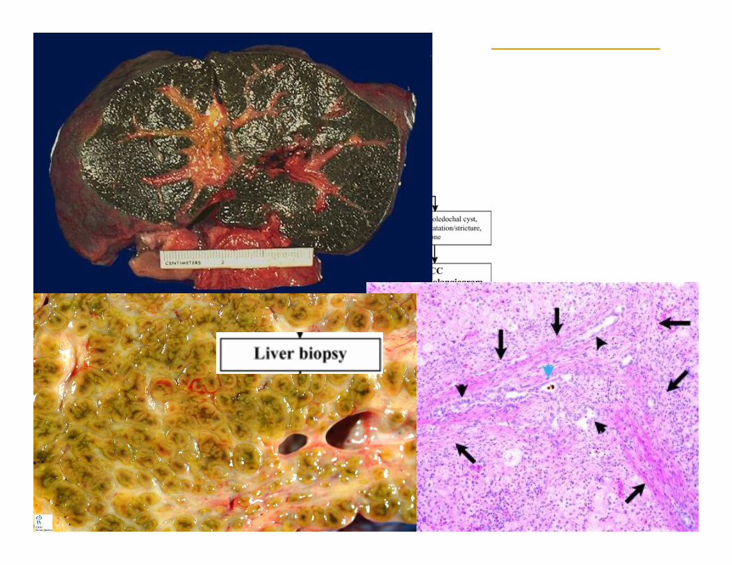
Initial Management is surgical Kasai Portoenterostomy
Excise obliterated biliary tree Roux loop from proximal jejunum anastomosed to porta
hepatis

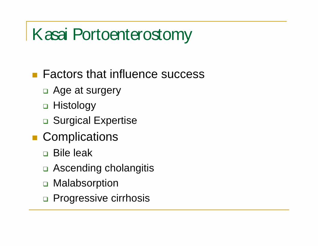
Kasai Portoenterostomy
Factors that influence success Age at surgery Histology Surgical Expertise
Complications Bile leak Ascending cholangitis Malabsorption Progressive cirrhosis
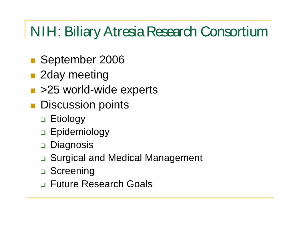
NIH: Biliary Atresia Research Consortium
September 2006 2day meeting >25 world-wide experts Discussion points Etiology Epidemiology Diagnosis Surgical and Medical Management Screening Future Research Goals

Contemporary Outcome of Biliary Atresia Following Kasai Hepatoportoenterostomy (HPE) and Liver Transplantation
Adapted from Chardot C, Serinot M-O. Prognosis of biliary atresia: what can be further improved? J Pediatr 2006;148:432-435.
4 years: 89% (actuarial)
2 years: 89% (actuarial)
4 years: 51% (actuarial)
Median of 54 days
148England and Wales, 1999-2002, 3 centers
4 years: 87.1% (actuarial)
4 years: 88.8% (actuarial)
4 years: 42.7% (actuarial)
Median of 57 days
271France, 1997-2002, 22 centers
2 years: 91.3% (actual)
2 years: 88% (actual)
2 years: 55.8% (actual)
Mean of 61 days
104United States, 1997-2000, 9 centers
5 years: 85% (actuarial)
2.4 years: 89% (actual)
5 years: 30.1% (actuarial)
Median of 54 days
93United Kingdom and Ireland, 1993-1995, 15 centers
5 years: 75.5% (actual)
-5 years: 59.7% (actual)
Median of 61-70 days
1381Japan, 1989-1999, 93 centers
Overall Survival of
Patients
Survival After Liver
Transplantation
Survival with Native
LiverAge at HPE
Number of
PatientsCountry, Year, and Number of Centers

Japan
JBAR 1989 – 1999 93 centers 1381 patients (863girls, 507boys) 84.1% Type III 19% associated anomalies (Polysplenia) HPE 61day Survival with Native Liver 5-yr: 59.7% 10-yr: 52.8%
Nio, et al. J of Ped Surg 2003 Vol 38(7): 997 - 1000
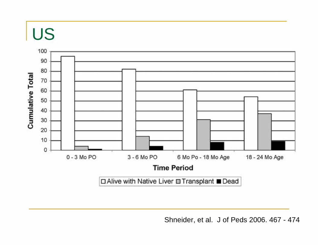
US
9 centers 1997 – 2000 104 patients (60% girls, 63% white) 25% associated anomalies (Polysplenia) Survival with Native Liver 2-yr: 55.8%
40.4% had OLTx 4 patients died from CHD
Shneider, et al. J of Peds 2006. 467 - 474

Cholangitis 47% no cholangitis 31% in the first year (15% at 18-24mos) NOT associated with decreased survival
Ascites 24% of patients in the first year
Esophageal varices 8% only 3 were bleeding
US cont

What’s New
Corticosteroids RCT UK 2007 Hepatology
Type III only given oral prednisolone (2mg/kg/d) Lower at 1month, no diff at 6 or 12months. Reduced need for transplant at 12months.
Laparoscopy Retrospective Brazil 2005 Sem in Ped Surg 41patients
Robotics

So what’s the debate
The disease is progressive Most children will develop portal fibrosis, cirrhosis portal hypertension
WHY NOT JUST TRANSPLANT

Liver Transplantation
70-76% of patients have progressive disease Transplant improves overall survival Indications Failed HPE age >100-120days at diagnosis Progressive cholestasis Development of cirrhosis Portal hypertension unresponsive to medical
management

Liver Transplantation
90% survival at 10yrs Complications Death (25-30%/20yr) HAT(20%) PVT (10%) Biliary complications(12-20%) Rejection (50%) Infectious (25%) Sensorineuronal hearing loss Decreased nutrition, bone density, growth
D’Alessandro, et al. Ped Transp 2007: 661-670

Recommendations
Canada 7% HPE <30d 52% survival with native liver 17yrs 21% survival if 90d or greater
Earlier screening Include a direct bilirubin on serum measurements Visit within 2-4wks Stool color cards

Summary
BA 100% fatal w/i 2yrs without intervention Goal to diagnose and treat w/i 30days Kasai HPE is effective and can delay OLT 5yrs in 60% of patients 10yrs in 50% of patients 20yrs in 25% of patients
Improved steroids Minimally Invasive techniques are available Delay transplant

“The primacy and value of the Kasai operation in biliary atresia should now be unquestioned…”
Mark Davenport, ChM

Current Management of Biliary Atresia
Janeen Jordan, MDPGY5November 19, 2007

Outcomes Following Portoenterostomy
886864%772003van Heurnet al.
4322NA331997Davenport
3330NA271996Valayer et al.
10-Year Overall Survival (%)
10-Year Native Liver Survival (%)
Successful Bile DrainagenYearAuthor/s
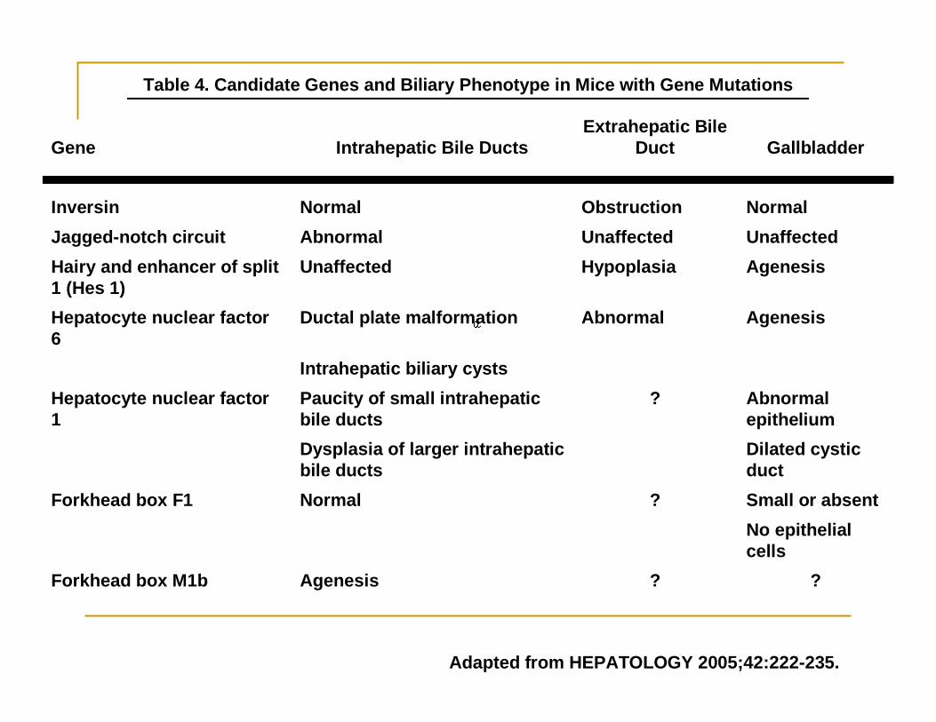
Table 4. Candidate Genes and Biliary Phenotype in Mice with Gene Mutations
Adapted from HEPATOLOGY 2005;42:222-235.
??AgenesisForkhead box M1b
No epithelial cells
Small or absent?NormalForkhead box F1
Dilated cystic duct
Dysplasia of larger intrahepaticbile ducts
Abnormal epithelium
?Paucity of small intrahepaticbile ducts
Hepatocyte nuclear factor 1
Intrahepatic biliary cysts
AgenesisAbnormalDuctal plate malformationHepatocyte nuclear factor 6
AgenesisHypoplasiaUnaffectedHairy and enhancer of split 1 (Hes 1)
UnaffectedUnaffectedAbnormalJagged-notch circuitNormalObstructionNormalInversin
GallbladderExtrahepatic Bile
DuctIntrahepatic Bile DuctsGene
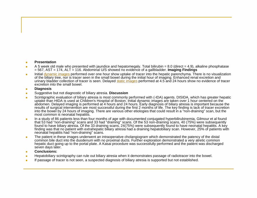
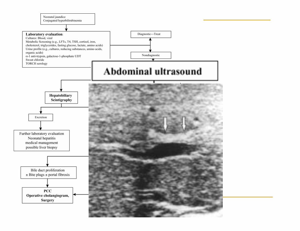
Presentation A 5 week old male who presented with jaundice and hepatomegaly. Total bilirubin = 8.0 (direct = 4.9), alkaline phosphatase
= 567, AST = 174, ALT = 116. Abdominal U/S showed no evidence of a gallbladder. Imaging Findings Initial dynamic images performed over one hour show uptake of tracer into the hepatic parenchyma. There is no visualization
of the biliary tree, nor is tracer seen in the small bowel during the initial hour of imaging. Enhanced renal excretion and urinary bladder collection of tracer is seen. Delayed static images performed at 4.5 and 24 hours show no evidence of tracer excretion into the small bowel.
Diagnosis Suggestive but not diagnostic of biliary atresia. Discussion Scintigraphic evaluation of biliary atresia is most commonly performed with (-IDA) agents. DISIDA, which has greater hepatic
uptake than HIDA is used at Children's Hospital of Boston. Initial dynamic images are taken over 1 hour centered on the abdomen. Delayed imaging is performed at 4 hours and 24 hours. Early diagnosis of biliary atresia is important because the results of surgical intervention are most successful during the first 2 months of life. The key finding is lack of tracer excretion into the bowel by 24 hours of imaging. There are various other etiologies that could result in a "non-draining" scan, but the most common is neonatal hepatitis.
In a study of 86 patients less than four months of age with documented conjugated hyperbilirubinemia, Gilmour et al found that 53 had "non-draining" scans and 33 had "draining" scans. Of the 53 non-draining scans, 40 (75%) were subsequently found to have biliary atresia. Of the 33 draining scans, 24(75%) were subsequently found to have neonatal hepatitis. A key finding was that no patient with extrahepatic biliary atresia had a draining hepatobiliary scan. However, 25% of patients with neonatal hepatitis had “non-draining” scans.
The patient in these images underwent an intraoperative cholangiogram which demonstrated the patency of the distal common bile duct into the duodenum with no proximal ducts. Further exploration demonstrated a very atretic common hepatic duct going up to the portal plate. A Kasai procedure was successfully performed and the patient was discharged seven days later.
Conclusions: Hepatobiliary scintigraphy can rule out biliary atresia when it demonstrates passage of radiotracer into the bowel. If passage of tracer is not seen, a suspected diagnosis of biliary atresia is supported but not established.

Biliary Atresia: Diagnosis
Full term infant Persisting jaundice from day2 Pale stool, dark urine, FTT, hepato/splenomegaly Other anomalies
Polysplenism, interrupted IVC, malrotation, situs inversus Lab abnormalities
Conjugated hyperbilurubinemia >20% of total bili (2mg/dL) Mildly elevated LFTs, Alk Phos (500-100 IU/L)
Late Findings Coagulopathy Malabsorption Fat Sol Vit Deficiency
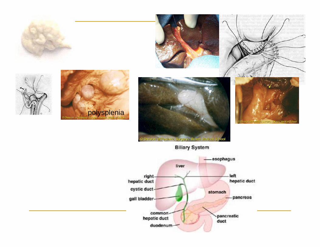
polysplenia


![Ultrasonographic findings of type IIIa biliary atresiabiliary atresia. Further, there have been only a few reports on the US findings of biliary atresia based on its types [33,34].](https://static.fdocuments.us/doc/165x107/60a90a6926e7a533947d7637/ultrasonographic-findings-of-type-iiia-biliary-atresia-biliary-atresia-further.jpg)