Current Applications of MRI-Guided Laser Interstitial Thermal ...
9
REVIEW ARTICLE Current Applications of MRI-Guided Laser Interstitial Thermal Therapy in the Treatment of Brain Neoplasms and Epilepsy: A Radiologic and Neurosurgical Overview R. Medvid, A. Ruiz, R.J. Komotar, J.R. Jagid, M.E. Ivan, R.M. Quencer, and M.B. Desai ABSTRACT SUMMARY: Minimally invasive stereotactic tumor ablation is a viable option for the treatment of benign and malignant intracranial lesions. Although surgical excision constitutes first-line therapy for various brain pathologies, it can cause irreversible neurologic deficits. Additionally, many patients who may benefit from surgery do not qualify as surgical candidates due to multiple comorbidities. Recent advancements in laser interstitial thermal therapy, namely the ability to monitor ablation in real-time under MR imaging, have improved the safety and efficacy of the procedure. MRI-guided laser interstitial thermal therapy is currently used as a minimally invasive treatment for brain metastases, radiation necrosis, glioma, and epilepsy. This article will discuss the principles, suggested indications, complications, and imaging characteristics of MRI-guided laser interstitial thermal therapy as they pertain to the treatment of brain pathology. ABBREVIATIONS: GBM glioblastoma multiforme; LITT laser interstitial thermal therapy; MRgLITT MRI-guided laser interstitial thermal therapy; MRTI MRI thermal imaging; RN delayed radiation necrosis; SRS stereotactic radiosurgery L aser interstitial thermal therapy (LITT) is a stereotactically guided percutaneous minimally invasive procedure, which delivers light energy to target tissue via a fiberoptic catheter, re- sulting in selective thermal ablation of malignant and benign le- sions. LITT was described in 1983 by Bown 1 and was first applied in the treatment of brain lesions in 1990 by Sugiyama et al. 2 Ac- ceptance of the procedure has been slow due to initial technologic shortcomings, which resulted in low efficacy and an unacceptably high risk of thermal damage to the surrounding normal brain parenchyma. Recent improvements have led to the development of percutaneous MRI-guided laser interstitial thermal therapy (MRgLITT), which enables monitoring of tissue ablation in real- time. Multiple academic centers are now using MRgLITT as a minimally invasive treatment for brain metastases, radiation ne- crosis, gliomas, and epilepsy. Successful management of patients undergoing MRgLITT requires interdisciplinary collaboration among neurosurgeons, neuroradiologists, neurologists, anesthe- siologists, radiation oncologists, and neuro-oncologists. This ar- ticle will discuss the principles, suggested indications, complica- tions, and imaging characteristics of MRgLITT as they pertain to the treatment of brain pathology. Background Minimally invasive stereotactic tumor ablation is a viable option for the treatment of benign and malignant intracranial lesions. Surgical excision constitutes first-line therapy for many malig- nant brain tumors followed by chemotherapy and/or radiation therapy. Aggressive complete resection or surgical debulking of newly diagnosed glioblastoma (GBM) improves survival, 3 com- pared with chemotherapy and radiation therapy alone. 4,5 A prob- lem arises when patients do not qualify for surgical resection due to the presence of deep-seated lesions, low functional scores, co- morbidities, or an inability to tolerate general anesthesia. In such cases, survival is limited; this outcome underscores the need for minimally invasive alternatives. In the case of inoperable brain metastases, stereotactic radiosurgery (SRS) presents a viable min- imally invasive option. SRS achieves an 80%–90% local control rate over metastatic lesions 6,7 but plays a limited role in the treat- ment of glioma or medically intractable seizures. Metastatic re- currence after SRS and whole-body radiation therapy is common, with estimates of up to 46.8% at 1-year follow-up. 8 Repeat use of radiation therapy after recurrence is limited due to concerns about cumulative adverse radiation effects, such as radiation necrosis. Delayed radiation necrosis (RN) is a known complication of SRS, with reported rates ranging from 5% to 50%. 9 The true in- cidence of RN in the setting of prior SRS is unknown because of From the Department of Radiology (R.M., A.R., R.M.Q., M.B.D.), Division of Neurora- diology, and Department of Neurological Surgery (J.R.J., R.J.K., M.E.I.), Jackson Me- morial Hospital/University of Miami Hospital, Miami, Florida. Please address correspondence to Rostislav Medvid, MD, Jackson Memorial Hospi- tal, Department of Radiology, 1611 NW 12th Ave, West Wing 279, Miami, FL 33136; e-mail: [email protected] Indicates open access to non-subscribers at www.ajnr.org http://dx.doi.org/10.3174/ajnr.A4362 AJNR Am J Neuroradiol ●:● ● 2015 www.ajnr.org 1 Published June 25, 2015 as 10.3174/ajnr.A4362 Copyright 2015 by American Society of Neuroradiology.
Transcript of Current Applications of MRI-Guided Laser Interstitial Thermal ...
REVIEW ARTICLE
Current Applications of MRI-Guided Laser Interstitial ThermalTherapy in the Treatment of Brain Neoplasms and Epilepsy:
A Radiologic and Neurosurgical OverviewR. Medvid, A. Ruiz, R.J. Komotar, J.R. Jagid, M.E. Ivan, R.M. Quencer, and M.B. Desai
ABSTRACT
SUMMARY: Minimally invasive stereotactic tumor ablation is a viable option for the treatment of benign and malignant intracraniallesions. Although surgical excision constitutes first-line therapy for various brain pathologies, it can cause irreversible neurologic deficits.Additionally, many patients who may benefit from surgery do not qualify as surgical candidates due to multiple comorbidities. Recentadvancements in laser interstitial thermal therapy, namely the ability to monitor ablation in real-time under MR imaging, have improved thesafety and efficacy of the procedure. MRI-guided laser interstitial thermal therapy is currently used as a minimally invasive treatment forbrain metastases, radiation necrosis, glioma, and epilepsy. This article will discuss the principles, suggested indications, complications, andimaging characteristics of MRI-guided laser interstitial thermal therapy as they pertain to the treatment of brain pathology.
ABBREVIATIONS: GBM � glioblastoma multiforme; LITT � laser interstitial thermal therapy; MRgLITT � MRI-guided laser interstitial thermal therapy; MRTI � MRIthermal imaging; RN � delayed radiation necrosis; SRS � stereotactic radiosurgery
Laser interstitial thermal therapy (LITT) is a stereotactically
guided percutaneous minimally invasive procedure, which
delivers light energy to target tissue via a fiberoptic catheter, re-
sulting in selective thermal ablation of malignant and benign le-
sions. LITT was described in 1983 by Bown1 and was first applied
in the treatment of brain lesions in 1990 by Sugiyama et al.2 Ac-
ceptance of the procedure has been slow due to initial technologic
shortcomings, which resulted in low efficacy and an unacceptably
high risk of thermal damage to the surrounding normal brain
parenchyma. Recent improvements have led to the development
of percutaneous MRI-guided laser interstitial thermal therapy
(MRgLITT), which enables monitoring of tissue ablation in real-
time. Multiple academic centers are now using MRgLITT as a
minimally invasive treatment for brain metastases, radiation ne-
crosis, gliomas, and epilepsy. Successful management of patients
undergoing MRgLITT requires interdisciplinary collaboration
among neurosurgeons, neuroradiologists, neurologists, anesthe-
siologists, radiation oncologists, and neuro-oncologists. This ar-
ticle will discuss the principles, suggested indications, complica-
tions, and imaging characteristics of MRgLITT as they pertain to
the treatment of brain pathology.
BackgroundMinimally invasive stereotactic tumor ablation is a viable option
for the treatment of benign and malignant intracranial lesions.
Surgical excision constitutes first-line therapy for many malig-
nant brain tumors followed by chemotherapy and/or radiation
therapy. Aggressive complete resection or surgical debulking of
newly diagnosed glioblastoma (GBM) improves survival,3 com-
pared with chemotherapy and radiation therapy alone.4,5 A prob-
lem arises when patients do not qualify for surgical resection due
to the presence of deep-seated lesions, low functional scores, co-
morbidities, or an inability to tolerate general anesthesia. In such
cases, survival is limited; this outcome underscores the need for
minimally invasive alternatives. In the case of inoperable brain
metastases, stereotactic radiosurgery (SRS) presents a viable min-
imally invasive option. SRS achieves an 80%–90% local control
rate over metastatic lesions6,7 but plays a limited role in the treat-
ment of glioma or medically intractable seizures. Metastatic re-
currence after SRS and whole-body radiation therapy is common,
with estimates of up to 46.8% at 1-year follow-up.8 Repeat use of
radiation therapy after recurrence is limited due to concerns
about cumulative adverse radiation effects, such as radiation
necrosis.
Delayed radiation necrosis (RN) is a known complication of
SRS, with reported rates ranging from 5% to 50%.9 The true in-
cidence of RN in the setting of prior SRS is unknown because of
From the Department of Radiology (R.M., A.R., R.M.Q., M.B.D.), Division of Neurora-diology, and Department of Neurological Surgery (J.R.J., R.J.K., M.E.I.), Jackson Me-morial Hospital/University of Miami Hospital, Miami, Florida.
Please address correspondence to Rostislav Medvid, MD, Jackson Memorial Hospi-tal, Department of Radiology, 1611 NW 12th Ave, West Wing 279, Miami, FL 33136;e-mail: [email protected]
Indicates open access to non-subscribers at www.ajnr.org
http://dx.doi.org/10.3174/ajnr.A4362
AJNR Am J Neuroradiol ●:● ● 2015 www.ajnr.org 1
Published June 25, 2015 as 10.3174/ajnr.A4362
Copyright 2015 by American Society of Neuroradiology.
diagnostic difficulties in distinguishing RN and recurrence. Imag-
ing-wise, radiation necrosis and tumor recurrence can both ap-
pear as enlarging previously treated lesions with a variable in-
crease in internal enhancement or as a new thickening and/or
irregularity of a peripherally enhancing rim with surrounding
edema.10 Although commonly used modalities such as MR per-
fusion imaging, MR imaging spectroscopy, SPECT, and PET may
provide useful clues about the identity of a previously treated
enlarging lesion, their utility is limited. Current MR imaging and
MR spectroscopy concepts under investigation include lesion
quotient,11 T1/T2 mismatch,12 percentage signal recovery,13 rel-
ative cerebral blood volume,14 and multivoxel proton MR spec-
troscopy,15 but specificities and sensitivities are variable. Unfor-
tunately, the specificity of biopsy can also be low due to sampling
error.16 Definitive diagnosis is further limited because up to 33%
of enlarging lesions after treatment with SRS represent a combi-
nation of RN and recurrence rather than a pure form of either
entity.17 Given the common coexistence of RN and recurrence,
Rao et al18 postulated that an ablation technique such as LITT,
which treats both RN and recurrent metastatic lesions, may cir-
cumvent diagnostic difficulties by providing the same standard-
ized treatment for both types of lesions.
Another entity that could benefit from minimally invasive
treatment is epilepsy. One-third of seizures are refractory to
pharmacologic therapy. The success rate of additional phar-
macologic therapy after 2 failed regimens is �3%.19 In the
setting of medically intractable seizures, open resection of
well-defined epileptic foci has been shown to achieve a control
rate of up to 75%– 80%.20 However, many of the lesions are
deep-seated, requiring excessive dissection during resection,
thus increasing the risk of iatrogenic complications and resid-
ual permanent neurologic deficits. Minimally invasive proce-
dures such as SRS and radiofrequency ablation are currently of
limited utility in the treatment of epilepsy due to the inability
to visualize ablation in real-time.
The ideal minimally invasive procedure should reduce intra-
and postoperative morbidity and mortality, shorten hospital
stays, decrease health care costs, and offer effective treatment op-
tions to patients who are otherwise not eligible for open surgery or
other debulking treatments. To date, multiple modalities have
been used for stereotactic lesion ablation, including cryoablation,
radiofrequency, sonography, microwave, ionizing radiation, and
laser. Recent technologic advances in laser ablation have signifi-
cantly improved safety and efficacy by providing the ability to
monitor ablation in real-time. Other advantages include shorter
ablation times and sharper ablation zone boundaries. Both SRS
and LITT can treat difficult-to-access lesions, can be performed
with the patient under minimal sedation, and do not require dis-
continuation of ongoing systemic therapy. Although MRgLITT is
an invasive intracranial procedure, which requires a burr-hole
and possible general anesthesia, it provides a potential benefit
over the noninvasive SRS: MRgLITT delivers nonionizing radia-
tion to target lesions, thus avoiding long-term adverse radiation
effects and theoretically allowing retreatment of the same lesion.
However, the long-term effects of thermal necrosis have not yet
been clearly elucidated.
Physics of MRgLITTDuring LITT, photons emitted from a fiberoptic laser are ab-
sorbed by tumor chromophores, organic molecules that absorb,
transmit, and reflect light. Light absorption leads to molecular
excitation and subsequent release of thermal energy within the
target tissue. Thermally induced irreversible cell damage occurs
between temperatures of 46°C and 60°C.21 Temperatures above
60°C result in instantaneous coagulation necrosis.22 The absorp-
tion coefficient determines the extent of photon absorption
within a target tissue. In general, the absorption coefficient of
pathologic lesions and coagulation necrosis is higher than that in
normal brain parenchyma, leading to preferential ablation of the
target tissue.23 There is a sharp temperature fall-off at the border
of the ablation zone,24 creating a sharp margin between viable and
nonviable tissue, which can be monitored with real-time MRI
thermal imaging (MRTI).
Proton resonance frequency is the most widely used tempera-
ture-sensitive MR imaging parameter for real-time MRTI.25 Pro-
ton resonance frequency– based MRTI is the basis of MRgLITT.
The physical mechanism of proton resonance frequency– based
MRTI depends on the presence of hydrogen bonds within a tis-
sue.26-28 As the temperature increases, the number of hydrogen
bonds decreases. This decrease leads to a more uniform distribu-
tion of electron clouds within a molecule. The more evenly dis-
tributed electron clouds are better able to shield the 1H nuclei
from the full force of the external magnetic field supplied by the
MR imaging machine. Thus, as the temperature increases, the
local magnetic field experienced by a 1H nucleus decreases due to
increased shielding by the surrounding electrons. The decrease in
the local magnetic field decreases the Larmour precession fre-
quency of the 1H nucleus, which, in turn, alters the phase of gra-
dient recalled-echo phase images.25 Measurements are obtained
by subtracting “thermal” fast-spoiled gradient recalled phase im-
ages (obtained after administration of thermal energy) from a
“reference” fast-spoiled gradient recalled phase image (obtained
at body temperature before any energy pulse is delivered).26,27
The phase difference, or phase shift, between the 2 images is
proportional to the overall temperature change. As such, proton
resonance frequency– based MRTI does not measure the absolute
temperature of a sample, but simply measures the temperature
difference between the sample and a designated reference temper-
ature image.25,28 Temperature information and time of ablation
can be incorporated into a mathematic model of thermal tissue
destruction (Arrhenius model) to provide a real-time quantitative
estimate of tissue necrosis,29 displayed in real-time as an orange
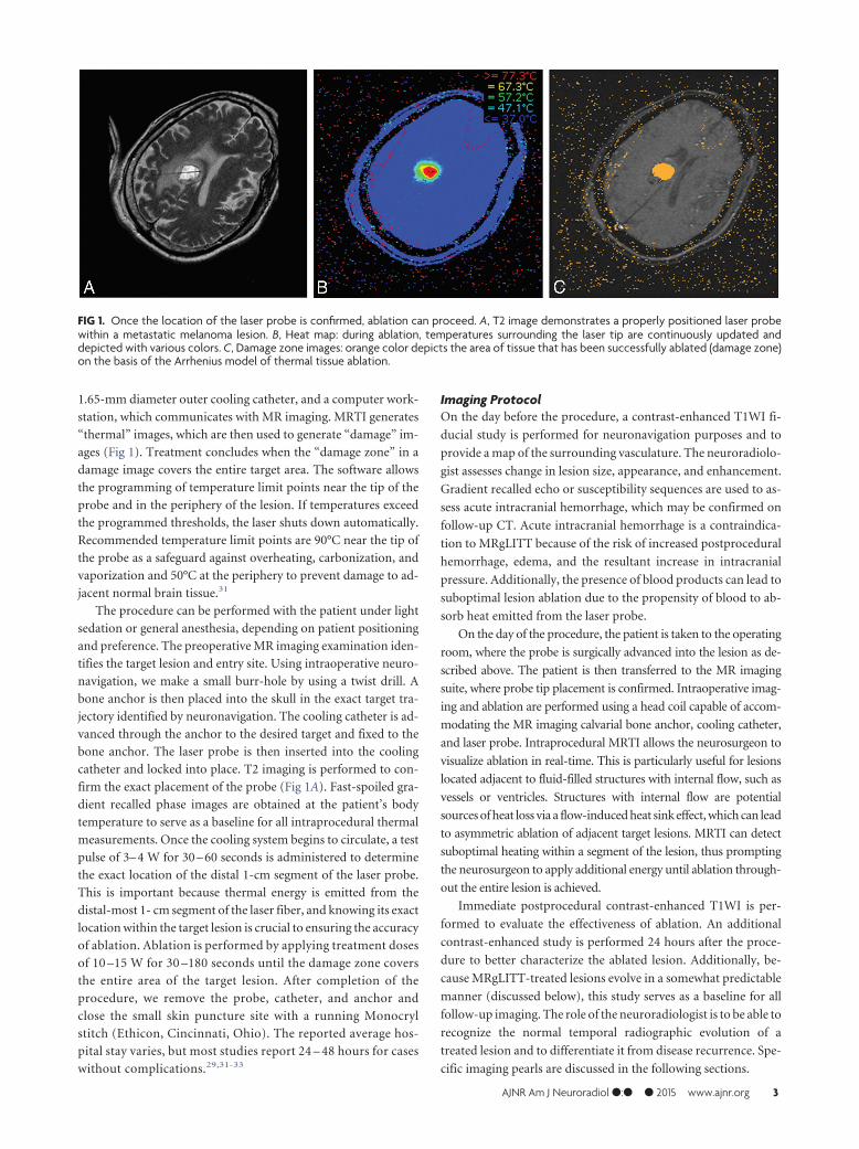
“damage zone” (Fig 1).
LITT Setup and ProcedureTwo major LITT platforms are in use today. NeuroBlate (Mon-
teris Medical Corporation, Minneapolis, Minnesota), which re-
ceived 510(k) FDA clearance in May 2009, uses a 12-W 1064-nm
neodymium-doped yttrium aluminium garnet laser with a CO2
cooled side-firing probe.30 Visualase (Medtronic, Minneapolis,
Minnesota), which received FDA clearance in 2007, is the plat-
form used in our institution. The major components of the Visu-
alase system include a 15-W 980-nm diode laser, a disposable
saline-cooled diffusing laser applicator probe with a 1-cm-long
2 Medvid ● 2015 www.ajnr.org
1.65-mm diameter outer cooling catheter, and a computer work-
station, which communicates with MR imaging. MRTI generates
“thermal” images, which are then used to generate “damage” im-
ages (Fig 1). Treatment concludes when the “damage zone” in a
damage image covers the entire target area. The software allows
the programming of temperature limit points near the tip of the
probe and in the periphery of the lesion. If temperatures exceed
the programmed thresholds, the laser shuts down automatically.
Recommended temperature limit points are 90°C near the tip of
the probe as a safeguard against overheating, carbonization, and
vaporization and 50°C at the periphery to prevent damage to ad-
jacent normal brain tissue.31
The procedure can be performed with the patient under light
sedation or general anesthesia, depending on patient positioning
and preference. The preoperative MR imaging examination iden-
tifies the target lesion and entry site. Using intraoperative neuro-
navigation, we make a small burr-hole by using a twist drill. A
bone anchor is then placed into the skull in the exact target tra-
jectory identified by neuronavigation. The cooling catheter is ad-
vanced through the anchor to the desired target and fixed to the
bone anchor. The laser probe is then inserted into the cooling
catheter and locked into place. T2 imaging is performed to con-
firm the exact placement of the probe (Fig 1A). Fast-spoiled gra-
dient recalled phase images are obtained at the patient’s body
temperature to serve as a baseline for all intraprocedural thermal
measurements. Once the cooling system begins to circulate, a test
pulse of 3– 4 W for 30 – 60 seconds is administered to determine
the exact location of the distal 1-cm segment of the laser probe.
This is important because thermal energy is emitted from the
distal-most 1- cm segment of the laser fiber, and knowing its exact
location within the target lesion is crucial to ensuring the accuracy
of ablation. Ablation is performed by applying treatment doses
of 10 –15 W for 30 –180 seconds until the damage zone covers
the entire area of the target lesion. After completion of the
procedure, we remove the probe, catheter, and anchor and
close the small skin puncture site with a running Monocryl
stitch (Ethicon, Cincinnati, Ohio). The reported average hos-
pital stay varies, but most studies report 24 – 48 hours for cases
without complications.29,31-33
Imaging ProtocolOn the day before the procedure, a contrast-enhanced T1WI fi-
ducial study is performed for neuronavigation purposes and to
provide a map of the surrounding vasculature. The neuroradiolo-
gist assesses change in lesion size, appearance, and enhancement.
Gradient recalled echo or susceptibility sequences are used to as-
sess acute intracranial hemorrhage, which may be confirmed on
follow-up CT. Acute intracranial hemorrhage is a contraindica-
tion to MRgLITT because of the risk of increased postprocedural
hemorrhage, edema, and the resultant increase in intracranial
pressure. Additionally, the presence of blood products can lead to
suboptimal lesion ablation due to the propensity of blood to ab-
sorb heat emitted from the laser probe.
On the day of the procedure, the patient is taken to the operating
room, where the probe is surgically advanced into the lesion as de-
scribed above. The patient is then transferred to the MR imaging
suite, where probe tip placement is confirmed. Intraoperative imag-
ing and ablation are performed using a head coil capable of accom-
modating the MR imaging calvarial bone anchor, cooling catheter,
and laser probe. Intraprocedural MRTI allows the neurosurgeon to
visualize ablation in real-time. This is particularly useful for lesions
located adjacent to fluid-filled structures with internal flow, such as
vessels or ventricles. Structures with internal flow are potential
sources of heat loss via a flow-induced heat sink effect, which can lead
to asymmetric ablation of adjacent target lesions. MRTI can detect
suboptimal heating within a segment of the lesion, thus prompting
the neurosurgeon to apply additional energy until ablation through-
out the entire lesion is achieved.
Immediate postprocedural contrast-enhanced T1WI is per-
formed to evaluate the effectiveness of ablation. An additional
contrast-enhanced study is performed 24 hours after the proce-
dure to better characterize the ablated lesion. Additionally, be-
cause MRgLITT-treated lesions evolve in a somewhat predictable
manner (discussed below), this study serves as a baseline for all
follow-up imaging. The role of the neuroradiologist is to be able to
recognize the normal temporal radiographic evolution of a
treated lesion and to differentiate it from disease recurrence. Spe-
cific imaging pearls are discussed in the following sections.
FIG 1. Once the location of the laser probe is confirmed, ablation can proceed. A, T2 image demonstrates a properly positioned laser probewithin a metastatic melanoma lesion. B, Heat map: during ablation, temperatures surrounding the laser tip are continuously updated anddepicted with various colors. C, Damage zone images: orange color depicts the area of tissue that has been successfully ablated (damage zone)on the basis of the Arrhenius model of thermal tissue ablation.
AJNR Am J Neuroradiol ●:● ● 2015 www.ajnr.org 3

MRTI ArtifactsThe accuracy of MRTI can be hindered by the following: 1) sus-
ceptibility artifacts from calcifications or pre-existing surgical
hardware, 2) the presence of fat, 3) misregistration artifacts due to
motion, and 4) magnetic field inhomogeneities. In general, if a
lesion cannot be adequately visualized, then ablation should not
be attempted. Large amounts of susceptibility artifacts adjacent to
a lesion are a relative contraindication to MRgLITT, though no
publications exist in support of this claim, to our knowledge. The
presence of intralesional calcifications has not been addressed in
the literature, but we have verbal confirmation from Visualase
representatives of successful treatment of partially calcified le-
sions at other institutions (in conversation with David Simon,
PhD, January 22, 2015). The presence of fat is a potential source of
susceptibility in proton resonance frequency– based MRTI be-
cause fat does not contain hydrogen bonds.27 The lack of hydro-
gen bonds in fat makes proton resonance frequency less suscepti-
ble to temperature change, thus leading to inaccurate temperature
measurements and susceptibility effects.26,27 This characteristic
implies that ablation of fat-containing lesions cannot be accu-
rately monitored with proton resonance frequency– based MRTI.
To our knowledge, there are no published reports on the use of
MRgLITT in the treatment of fat-containing lesions. Misregistra-
tion artifacts from motion can be prevented with mechanical im-
mobilization of the cranium, proper sedation, and stabilization of
the laser tip with a bone anchor. Magnetic field inhomogeneities
can be eliminated by subtracting baseline reference images from
thermal images.25
Imaging and Radiologic-Pathologic Correlation of LesionsTreated with LITTAblated lesions demonstrate a thin peripheral rim of enhance-
ment, variable T1 and T2 central signal due to presence of blood
and protein products and surrounding edema on T1WI contrast-
enhanced images obtained 24 hours after treatment. At the mi-
croscopic level, these changes correspond to 5 histologically sep-
arate concentric zones (Fig 2).34 At the core is the probe track,
which may be filled with CSF or blood. The probe track is sur-
rounded by the central zone of coagulation necrosis, which con-
tains damaged cell membranes and stains positive for markers of
apoptosis. On 24-hour follow-up, the central zone may appear
hyperintense on T1 and hypointense on T2 because of the pres-
ence of subacute blood products and protein coagulation. Alter-
natively, it may appear hypointense on T1 and hyperintense on
T2, depending on the age of the blood products and relative con-
centration of protein.
The peripheral zone makes up the next concentric layer, which
contains thrombosed vessels and distended cell bodies. This area
undergoes delayed liquefaction necrosis and tends to enlarge dur-
ing the first 1– 40 days, followed by a continuous reduction in size
thereafter.35 On MR imaging, this layer
appears hypointense on T1 and hyperin-
tense on T2 due to edema. The periph-
eral zone contains a thin peripherally
enhancing rim on T1-weighted con-
trast-enhanced images, secondary to
blood-brain barrier damage. The pe-
ripheral rim gradually changes in cir-
cumference and enhancement in accor-
dance with changes in the entire
peripheral zone. It generally decreases
with time or may remain stable (Fig 3).
Residual enhancement persists on long-
term-follow-up, likely due to reactive
inflammatory/granulation tissue. Ac-
cording to Rao et al,18 most lesions re-
FIG 2. Concentric zones: T1 contrast-enhanced (T1C) and T2WI 24 hours after laser ablation of arecurrent right cerebellar metastatic lesion in a 71-year-old female patient with history of breastcarcinoma: 1) probe track, 2) central zone, 3) peripheral zone, 4) peripherally enhancing rim, 5)marginal zone. Note that the concentric zones appear as inverse images on T1C and T2 images.
FIG 3. T1 contrast-enhanced images demonstrating the normal evolution of a LITT-treated metastatic left cerebellar lesion, which recurredafter SRS in a 70-year-old female patient with history of ovarian adenocarcinoma. Note the expected increase in the size of the treated lesionat 2-month follow-up and a steady decrease in size on subsequent follow-up.
4 Medvid ● 2015 www.ajnr.org

turn to pretreatment size within 16 weeks. The outermost layer of
the LITT lesion is the marginal zone, an area of reversible post-
surgical perifocal edema, which appears hypointense on T1 and
hyperintense on T2. This layer contains viable edematous tissue
and demonstrates axonal swelling without thrombosis. It in-
creases in size, reaching maximum dimensions at 1–3 days and
gradually decreases in size during the course of 15 days to 2
months (Fig 4). In some cases, the marginal zone may demon-
strate high T1 signal without corresponding susceptibility arti-
facts, likely due to the presence of myelin breakdown products.
Patients are instructed to return for follow-up 1 month after ab-
lation. Depending on imaging findings, clinical presentation,
and type of disease, subsequent follow-up may be performed
on a monthly basis or may be extended to longer intervals.
There are no official follow-up recommendations.
RecurrenceSerial follow-up performed �40 days after the procedure should
demonstrate a continuous decrease in the size of the ablated lesion
and stable or decreased enhancement (Fig 3). Overall, the entire
ablated lesion decreases to 50% of its original size within 93 days
of treatment35 and continues to decrease in size for 6 –15 months,
becoming more homogeneous in appearance. Peripheral en-
hancement that persists or gradually decreases in size is a sign of
the normal evolution of the ablated lesion. Any interval increase
in lesion size, heterogeneity, peripheral nodular enhancement,
restricted diffusion, CBF/CBV, and surrounding edema in a le-
sion treated �40 – 60 days prior should raise suspicion for recur-
rence (Fig 5).36,37 Recurrence usually occurs within the peripheral
rim of enhancement and presents as new or enlarging peripheral
enhancing nodularity (Figs 5) or simply as thickening of the rim
of enhancement (Fig 6). Comparison with prior images is vital in
monitoring tumor recurrence because normally evolving lesions
can demonstrate asymmetric enhancement similar to that of re-
curring lesions. Therefore, irregularly enhancing lesions require
close-interval follow-up. Any increase in size, enhancement, or
surrounding edema should be further assessed with adjunctive
techniques such as MR spectroscopy, perfusion imaging, or PET.
Clinical ApplicationsStudies during the past 20 years report the use of LITT to
treat a variety of brain lesions. The most studied lesions in-
clude glioma2,30,31,33,38-48 and metastases.2,18,29,32,40,41,44,49-51
Epilepsy51-55 and radiation necrosis50,56 represent a much smaller
subset of treated lesions reported in the literature. Additionally,
MRgLITT has been used to treat refractory cerebral edema57 and
tumors such as ependymoma, meningioma, primitive neuroecto-
dermal tumor, hemangioblastoma, and chordoma.31,58 The sur-
FIG 4. T2WI demonstrating the normal evolution of an LITT-treated left splenial low-grade astrocytoma in a 30-year-old male patient with ahistory of type 1 neurofibromatosis. Note the expected increase in the size of the lesion and marginal zone at 1-month follow-up and subsequentdecrease at 3 months.
FIG 5. Disease recurrence after treatment with LITT. T1 contrast-enhanced images in a 58-year-old man with GBM status post surgical excision,chemoradiation, and SRS for a recurrent left parietal lobe lesion. The second recurrence was treated with LITT. Note irregular peripheral nodularenhancement at 1-month follow-up (white arrow), which progressively increases in size at 2 and 6 months. Findings are consistent with diseaserecurrence.
AJNR Am J Neuroradiol ●:● ● 2015 www.ajnr.org 5

vival benefits of LITT after treatment of various brain lesions vary
from favorable to statistically insignificant. Evidence is limited
because to date, all studies consist of noncontrolled, nonrandom-
ized retrospective reports, case series, or case reports, thus predis-
posing to selection bias. Many of the studies mix multiple disease
entities to increase the number of enrolled subjects; this mixture
makes the evaluation of survival benefits for a given disease entity
difficult. Another major limitation is the use of variable inclusion
criteria by selecting patients with either recurrent, newly diag-
nosed, previously treated, or untreated tumors or a mixture of any
of the above. However, the above studies offer a plethora of evi-
dence on the safety profile of the procedure. The variable compli-
cations of MRgLITT will be addressed below after a brief discus-
sion of the 4 most common indications for the procedure:
gliomas, metastatic lesions, radiation necrosis, and medically in-
tractable epilepsy.
GliomasGliomas are subdivided into multiple subtypes, of which GBM is
the most common primary brain neoplasm in adults. Median
survival of newly diagnosed patients after treatment with maximal
safe resection, radiation therapy, and adjuvant chemotherapy is
12–15 months.59 Survival decreases with inoperable deep-seated
lesions. GBM poses multiple treatment challenges due to its dif-
fusely infiltrative nature and strong resistance to therapy. As a
result, all GBMs recur, and median survival after recurrence is 3–5
months.60 Surgical resection increases survival but is not feasible
in cases of difficult-to-access tumors, thus the need for minimally
invasive surgical procedures. The use of SRS in newly diag-
nosed GBM demonstrated no survival benefit,61 while data on
MRgLITT are inconclusive. Local therapies, such as MRgLITT,
SRS, or open surgical resection, do not address the infiltrative
component of GBM and can only be used for palliative/salvage
therapy. Examples of MRgLITT-treated gliomas are provided
in Figs 4 and 5.
Metastatic Brain TumorsMetastatic brain tumors are 10 times more prevalent than pri-
mary brain tumors, accounting for approximately 200,000 cases
of the total of 225,000 cases of brain tumors per year.32 The inci-
dence of brain metastases is increasing due to effective oncologic
treatments of primary malignancies, resulting in longer survival.
The first-line treatment for new brain metastatic lesions is radia-
tion therapy (with SRS, whole-brain radiation therapy, or both)
and surgical resection.29 Local recurrence within 1 year of treat-
ment is approximately 10% after resection and radiation therapy,
compared with 46% after surgical resection alone.62 Similar to
GBM, no consensus exists on the treatment of recurrent meta-
static brain lesions, and repeat use of radiation therapy is limited
due to concerns over adverse cumulative radiation effects. To
date, studies have shown that MRgLITT is a safe, minimally inva-
sive alternative. Further evidence is needed to determine whether
there are definitive survival benefits over currently accepted treat-
ments. Examples of treated metastatic lesions are provided in Figs
1–3 and 6.
Radiation NecrosisRadiation necrosis is a common entity in neuro-oncology. Esti-
mated incidence varies between 5% and 10% for all radiation
therapy modalities, but the risk may be as high as 50% in the
setting of prior SRS, particularly at treatment doses between 16
and 22 Gy.9 RN, also known as delayed neurotoxicity, represents a
specific type of radiation injury and occurs at least 3 months after
radiation therapy. The process is irreversible, and approximately
85% of cases occur within 2 years of treatment.10 Histologically,
RN consists of a central zone of necrosis surrounded by a periph-
eral zone of altered astrocytes, which release large quantities of
proinflammatory factors such as hypoxia-inducing factor 1� and
FIG 6. LITT of radiation necrosis with subsequent disease recurrencein a 68-year-old female patient with lung squamous cell carcinomastatus post surgical excision and SRS of a metastatic brain lesion in theleft parietal lobe, which subsequently resulted in radiation necrosis.Medically intractable radiation necrosis was treated with LITT. Pre-LITT imaging demonstrated an enhancing lesion in the left parietallobe on T1 contrast-enhanced (not shown) with significant vasogenicedema on T2 images (dashed white arrow on T2 image labeled “pre”).Dynamic imaging pre-LITT (not shown) did not demonstrate a signif-icant increase in CBF or CBV. The patient was not treated with bev-acizumab, and RN was favored over recurrence. Note a significantdecrease in vasogenic edema 1 month after treatment (dashed whitearrow), coinciding with symptomatic improvement. T2 images ob-tained at 4-month follow-up demonstrate a significant increase inperitumoral vasogenic edema. There is significant thickening of theperipheral zone of enhancement (white arrow) on T1 contrast-en-hanced images at 4 months compared with 1 month. Findings areconcerning for tumor recurrence within a treated RN lesion, whichwas corroborated on PET CT (not shown).
6 Medvid ● 2015 www.ajnr.org

vascular endothelial growth factor, inducing severe inflammation
and edema.63 First-line treatment is steroids to decrease
inflammation. A new effective non-FDA-approved treatment is
bevacizumab, a humanized mouse monoclonal vascular endothe-
lial growth factor antibody acting as vascular endothelial growth
factor inhibitor, though its use is limited due to high cost, in-
creased risk of deep venous thrombosis and pulmonary emboli,
and the need to stop other concurrent systemic therapy.9 Surgical
resection is reserved for medically refractory RN, but its role is
limited in the setting of deep or difficult-to-access lesions and in
patients with multiple comorbidities who are unable to tolerate
general anesthesia. LITT induces resolution of RN,50,56 but long-
term data are limited due to low numbers and lack of sufficient
long-term follow-up. The postulated mechanism of action of
LITT in the setting of RN is ablation of the peripheral zone of
altered astrocytes, thus terminating the proinflammatory signal-
ing cascade induced by vascular endothelial growth factor.9 Fig-
ure 6 demonstrates an example of MRgLITT-treated RN in which
metastatic disease subsequently recurred.
EpilepsyMedically intractable focal epilepsy is generally treated with surgical
resection of epileptic foci. Given the deep location of many seizure
foci, surgical resection can be difficult and can leave patients with
persistent neurologic and cognitive deficits secondary to collateral
tissue damage. Available minimally invasive treatment options such
as SRS64,65 and RF ablation65 offer the potential benefit of decreasing
iatrogenic complications, but the inability to monitor ablation in
real-time diminishes their margin of safety. A pilot study by Curry et
al52 first demonstrated the feasibility of MRgLITT in the successful
treatment of medically intractable epileptic foci. To date, studies re-
port laser ablation of tuberous sclerosis, hypothalamic hamartoma,
mesial temporal sclerosis, cortical dysplasia, and periventricular nod-
ular hyperplasia with follow-up ranging from 2 to 13 months.51-54
Seven of a total of 9 patients remained seizure-free at 6- to 13-month
follow-up, depending on the study. The remaining 2 patients expe-
rienced seizure recurrence at 2 and 3 months after LITT and under-
went subsequent definitive open surgical treatment.52,54 This result
suggests that MRgLITT does not preclude future invasive therapy.
Given these findings, Esquenazi et al54 postulated that laser ablation
could be used as a first-line treatment for deep-seated medically in-
tractable epileptic foci and that surgical resection be reserved for pa-
tients who fail therapy with LITT. More recently, a report by Willie et
al55 showed that seizure-free rates for mesiotemporal epilepsy by us-
ing MRgLITT closely approximate those of open temporal lobecto-
mies while potentially improving postprocedural neurocognitive
outcomes. Figure 7 provides an imaging example of ablation of the
left hippocampo-amygdalar area to treat mesial temporal sclerosis.
ComplicationsAlthough the survival benefits and clinical outcomes of MRgLITT are
difficult to estimate on the basis of the currently available studies,
enough data are available to evaluate the safety profile of the proce-
dure. We performed an analysis of all LITT studies conducted on
human subjects to date with complications as an end point. We tal-
lied the different complications and calculated their rates. The most
common reported complication of LITT is transient neurologic def-
icit, accounting for 13% of all complications. Reported symptoms
include dysphagia, weakness, hemianopsia, or minor seizures. The
symptoms either resolved spontaneously or responded to steroid ad-
ministration within days to weeks.
The next most common complications include new pro-
gressive or permanent neurologic symptoms (3%), intracra-
nial hemorrhage (2.5%), infection (2.5%), and deep venous
thrombosis (2.5%). Life-threatening complications include in-
tracranial hemorrhage, ventriculitis, meningitis, and 1 case of
refractory intracranial hypertension after simultaneous use of
multiple probes to treat a large irregular lesion.31,33,48,51 Two
deaths have been reported, both in patients with GBM, one
from intractable intracranial hemorrhage and the other from
meningitis.48,51 Sloan et al33 suggested that pretreatment MRA
or CTA and fiber-tract imaging with DTI may be useful mo-
dalities in presurgical planning to identify and potentially
avoid critical vascular and white matter structures. Jethwa et
al58 recommended that LITT treatment of lesions of �3 cm
FIG 7. Mesial temporal sclerosis confirmed on preprocedural FDG-PET (black arrow), which showed decreased FDG uptake in the left hip-pocampal/parahippocampal region. The lesion was not noted on prior contrast-enhanced MR imaging and did not demonstrate enhancement(not shown). Intraprocedural T2 ablation map demonstrates the laser probe tip within the left medial temporal lobe (white arrow). T1 and T1contrast-enhanced images 24 hours after LITT demonstrate an oval, rather than round, postablation lesion with signal characteristics similar tothose of contrast-enhancing lesions (see Fig 2). The elongated shape is due to sequential probe retraction during ablation to cover the entire lefthippocampo-amygdalar area.
AJNR Am J Neuroradiol ●:● ● 2015 www.ajnr.org 7

should be staged. Administration of high-dose preprocedural
steroids should be considered.31
CONCLUSIONSLITT appears to be a safe palliative alternative for the treatment of
malignant high-grade gliomas and recurrent metastatic lesions.
While several life-threatening complications have been reported
in the setting of GBM, preliminary data suggest that their overall
incidence is acceptably low. Furthermore, the incidence of such
complications may be decreased with appropriate preoperative
imaging and as neurosurgeons overcome the steep learning curve
associated with the procedure. Because research on the use of
MRgLITT is in its infancy, indications and contraindications are
relative and are still being worked out. The most common post-
procedural complications of MRgLITT are non-life-threatening
and transient. LITT may also provide a safe curative option in
cases of radiation necrosis and in certain types of medically intrac-
table epilepsy.
REFERENCES1. Bown S. Phototherapy in tumors. World J Surg 1983;7:700 – 092. Sugiyama K, Sakai T, Fujishima I, et al. Stereotactic interstitial laser-
hyperthermia using Nd-YAG laser. Stereotact Funct Neurosurg1990;54:501– 05
3. Sanai N, Polley MY, McDermott MW, et al. An extent of resectionthreshold for newly diagnosed glioblastomas. J Neurosurg 2011;115:3– 8
4. Simpson JR, Horton J, Scott C, et al. Influence of location and extentof surgical resection on survival of patients with glioblastomamultiforme: results of three consecutive Radiation Therapy Oncol-ogy Group (RTOG) clinical trials. Int J Radiat Oncol Biol Phys 1993;26:239 – 44
5. Vuorinen V, Hinkka S, Farkkila M, et al. Debulking or biopsy ofmalignant glioma in elderly people—a randomised study. ActaNeurochir (Wien) 2003;145:5–10
6. Loeffler JS, Barker FG, Chapman PH. Role of radiosurgery in themanagement of central nervous system metastases. Cancer Che-mother Pharmacol 1999;43(suppl):S11–14
7. Young RF. Radiosurgery for the treatment of brain metastases. Se-min Surg Oncol 1998;14:70 –78
8. Aoyama H, Shirato H, Tago M, et al. Stereotactic radiosurgery pluswhole-brain radiation therapy vs stereotactic radiosurgery alonefor treatment of brain metastases: a randomized controlled trial.JAMA 2006;295:2483–91
9. Rahmathulla G, Marko NF, Weil RJ. Cerebral radiation necrosis: areview of the pathobiology, diagnosis and management consider-ations. J Clin Neurosci 2013;20:485–502
10. Shah R, Vattoth S, Jacob R, et al. Radiation necrosis in the brain:imaging features and differentiation from tumor recurrence. Ra-diographics 2012;32:1343–59
11. Dequesada IM, Quisling RG, Yachnis A, et al. Can standard magneticresonance imaging reliably distinguish recurrent tumor from radia-tion necrosis after radiosurgery for brain metastases? A radiographic-pathological study. Neurosurgery 2008;63:898–903; discussion 904
12. Kano H, Kondziolka D, Lobato-Polo J, et al. T1/T2 matching to dif-ferentiate tumor growth from radiation effects after stereotacticradiosurgery. Neurosurgery 2010;66:486 –91; discussion 491–92
13. Barajas RF, Chang JS, Sneed PK, et al. Distinguishing recurrent in-tra-axial metastatic tumor from radiation necrosis followinggamma knife radiosurgery using dynamic susceptibility-weightedcontrast-enhanced perfusion MR imaging. AJNR Am J Neuroradiol2009;30:367–72
14. Mitsuya K, Nakasu Y, Horiguchi S, et al. Perfusion weighted mag-netic resonance imaging to distinguish the recurrence of metastatic
brain tumors from radiation necrosis after stereotactic radiosur-gery. J Neurooncol 2010;99:81– 88
15. Chernov M, Hayashi M, Izawa M, et al. Differentiation of the radia-tion-induced necrosis and tumor recurrence after gamma knife ra-diosurgery for brain metastases: importance of multi-voxel protonMRS. Minim Invasive Neurosurg 2005;48:228 –34
16. Mullins ME, Barest GD, Schaefer PW, et al. Radiation necrosis ver-sus glioma recurrence: conventional MR imaging clues to diagno-sis. AJNR Am J Neuroradiol 2005;26:1967–72
17. Forsyth PA, Kelly PJ, Cascino TL, et al. Radiation necrosis or gliomarecurrence: is computer-assisted stereotactic biopsy useful? J Neu-rosurg 1995;82:436 – 44
18. Rao MS, Hargreaves EL, Khan AJ, et al. Magnetic resonance-guidedlaser ablation improves local control for postradiosurgery recur-rence and/or radiation necrosis. Neurosurgery 2014;74:658 – 67
19. Kwan P, Brodie MJ. Early identification of refractory epilepsy.N Engl J Med 2000;342:314 –19
20. Tellez-Zenteno JF, Hernandez Ronquillo L, Moien-Afshari F, et al.Surgical outcomes in lesional and non-lesional epilepsy: a system-atic review and meta-analysis. Epilepsy Res 2010;89:310 –18
21. Larson TR, Bostwick DG, Corica A. Temperature-correlated histo-pathologic changes following microwave thermoablation of ob-structive tissue in patients with benign prostatic hyperplasia. Urol-ogy 1996;47:463– 69
22. Goldberg SN, Gazelle GS, Mueller PR. Thermal ablation therapy forfocal malignancy: a unified approach to underlying principles,techniques, and diagnostic imaging guidance. AJR Am J Roentgenol2000;174:323–31
23. Yaroslavsky AN, Schulze PC, Yaroslavsky IV, et al. Optical prop-erties of selected native and coagulated human brain tissues invitro in the visible and near infrared spectral range. Phys MedBiol 2002;47:2059 –73
24. McNichols RJ, Gowda A, Kangasniemi M, et al. MR thermometry-based feedback control of laser interstitial thermal therapy at 980nm. Lasers Surg Med 2004;34:48 –55
25. Rieke V, Butts Pauly K. MR thermometry. J Magn Reson Imaging2008;27:376 –90
26. De Poorter J, De Wagter C, De Deene Y, et al. Noninvasive MRIthermometry with the proton resonance frequency (PRF)method: in vivo results in human muscle. Magn Reson Med 1995;33:74 – 81
27. De Poorter J. Noninvasive MRI thermometry with the proton reso-nance frequency method: study of susceptibility effects. Magn ResonMed 1995;34:359 – 67
28. Quesson B, de Zwart JA, Moonen CT. Magnetic resonance temper-ature imaging for guidance of thermotherapy. J Magn Reson Imaging2000;12:525–33
29. Carpentier A, McNichols RJ, Stafford RJ, et al. Real-time magneticresonance-guided laser thermal therapy for focal metastaticbrain tumors. Neurosurgery 2008;63(1 suppl 1):ONS21–28; dis-cussion ONS28 –29
30. Mohammadi AM, Schroeder JL. Laser interstitial thermal therapy intreatment of brain tumors–the NeuroBlate system. Expert Rev MedDevices 2014;11:109 –19
31. Jethwa PR, Barrese JC, Gowda A, et al. Magnetic resonance thermome-try-guided laser-induced thermal therapy for intracranial neoplasms:initial experience. Neurosurgery 2012;71(1 suppl operative):133–44;144–45
32. Carpentier A, McNichols RJ, Stafford RJ, et al. Laser thermal therapy:real-time MRI-guided and computer-controlled procedures formetastatic brain tumors. Lasers Surg Med 2011;43:943–50
33. Sloan AE, Ahluwalia MS, Valerio-Pascua J, et al. Results of the Neu-roBlate System first-in-humans phase I clinical trial for recurrentglioblastoma: clinical article. J Neurosurg 2013;118:1202–19
34. Schober R, Bettag M, Sabel M, et al. Fine structure of zonal changesin experimental Nd:YAG laser-induced interstitial hyperthermia.Lasers Surg Med 1993;13:234 – 41
35. Schwabe B, Kahn T, Harth T, et al. Laser-induced thermal lesions in
8 Medvid ● 2015 www.ajnr.org

the human brain: short- and long-term appearance on MRI. J Com-put Assist Tomogr 1997;21:818 –25
36. Tracz RA, Wyman DR, Little PB, et al. Magnetic resonance imagingof interstitial laser photocoagulation in brain. Lasers Surg Med1992;12:165–73
37. Tracz RA, Wyman DR, Little PB, et al. Comparison of magneticresonance images and the histopathological findings of lesions in-duced by interstitial laser photocoagulation in the brain. Lasers SurgMed 1993;13:45–54
38. Bettag M, Ulrich F, Schober R, et al. Stereotactic laser therapy incerebral gliomas. Acta Neurochirurgica 1992;52:81– 83
39. Ascher P, Justich E, Schrottner O. A new surgical but less invasivetreatment of central brain tumours preliminary report. Acta Neu-rochirurgica 1992;52:78 – 80
40. Roux FX, Merienne L, Fallet-Bianco C, et al. Stereotaxic laser inter-stitial thermotherapy: a new alternative in the therapeutic manage-ment of some brain tumors [in French]. Neurochirurgie 1992;38:238 – 44
41. Kahn T, Bettag M, Ulrich F, et al. MRI-guided laser-induced inter-stitial thermotherapy of cerebral neoplasms. J Comput Assist To-mogr 1994;18:519 –32
42. Leonardi MA, Lumenta CB, Gumprecht HK, et al. Stereotacticguided laser-induced interstitial thermotherapy (SLITT) in glio-mas with intraoperative morphologic monitoring in an open MR-unit. Minim Invasive Neurosurg 2001;44:37– 42
43. Leonardi MA, Lumenta CB. Stereotactic guided laser-induced inter-stitial thermotherapy (SLITT) in gliomas with intraoperative mor-phologic monitoring in an open MR: clinical experience. Minim In-vasive Neurosurg 2002;45:201– 07
44. Schulze P, Vitzthum H, Goldammer A, et al. Laser-induced thermo-therapy of neoplastic lesions in the brain– underlying tissue altera-tions, MRI-monitoring and clinical applicability. Acta Neurochir(Wein) 2004;146:803–12
45. Schwarzmaier HJ, Eickmeyer F, von Tempelhoff W, et al. MR-guidedlaser irradiation of recurrent glioblastomas. J Magn Reson Imaging2005;22:799 – 803
46. Schwarzmaier HJ, Eickmeyer F, von Tempelhoff W, et al. MR-guidedlaser-induced interstitial thermotherapy of recurrent glioblastomamultiforme: preliminary results in 16 patients. Eur J Radiol2006;59:208 –15
47. Carpentier A, Chauvet D, Reina V, et al. MR-guided laser-inducedthermal therapy (LITT) for recurrent glioblastomas. Lasers SurgMed 2012;44:361– 68
48. Mohammadi AM, Hawasli AH, Rodriguez A, et al. The role of laserinterstitial thermal therapy in enhancing progression-free survivalof difficult-to-access high-grade gliomas: a multicenter study. Can-cer Med 2014;3:971–79
49. Hawasli AH, Ray WZ, Murphy RK, et al. Magnetic resonance imag-ing-guided focused laser interstitial thermal therapy for subinsularmetastatic adenocarcinoma: technical case report. Neurosurgery2012;70(2 suppl operative):332–37; discussion 338
50. Rahmathulla G, Recinos PF, Valerio JE, et al. Laser interstitial ther-
mal therapy for focal cerebral radiation necrosis: a case report andliterature review. Stereotact Funct Neurosurg 2012;90:192–200
51. Hawasli AH, Bagade S, Shimony JS, et al. Magnetic resonanceimaging-guided focused laser interstitial thermal therapy for in-tracranial lesions: single-institution series. Neurosurgery2013;73:1007–17
52. Curry DJ, Gowda A, McNichols RJ, et al. MR-guided stereotacticlaser ablation of epileptogenic foci in children. Epilepsy Behav2012;24:408 –14
53. Tovar-Spinoza Z, Carter D, Ferrone D, et al. The use of MRI-guidedlaser-induced thermal ablation for epilepsy. Childs Nerv Syst2013;29:2089 –94
54. Esquenazi Y, Kalamangalam GP, Slater JD, et al. Stereotactic laserablation of epileptogenic periventricular nodular heterotopia. Epi-lepsy Res 2014;108:547–54
55. Willie JT, Laxpati NG, Drane DL, et al. Real-time magnetic reso-nance-guided stereotactic laser amygdalohippocampotomy formesial temporal lobe epilepsy. Neurosurgery 2014;74:569 – 84; dis-cussion 584 – 85
56. Torres-Reveron J, Tomasiewicz HC, Shetty A, et al. Stereotactic laserinduced thermotherapy (LITT): a novel treatment for brain lesionsregrowing after radiosurgery. J Neurooncol 2013;113:495–503
57. Fabiano AJ, Alberico RA. Laser-interstitial thermal therapy for re-fractory cerebral edema from post-radiosurgery metastasis. WorldNeurosurg 2014;81:652.e1– 4
58. Jethwa PR, Lee JH, Assina R, et al. Treatment of a supratentorialprimitive neuroectodermal tumor using magnetic resonance-guided laser-induced thermal therapy. J Neurosurg Pediatr2011;8:468 –75
59. Stupp R, Mason WP, van den Bent MJ, et al. Radiotherapy plus con-comitant and adjuvant temozolomide for glioblastoma. N EnglJ Med 2005;352:987–96
60. Barker FG 2nd, Chang SM, Gutin PH, et al. Survival and functionalstatus after resection of recurrent glioblastoma multiforme. Neuro-surgery 1998;42:709 –20; discussion 720 –23
61. Souhami L, Seiferheld W, Brachman D, et al. Randomized compari-son of stereotactic radiosurgery followed by conventional radio-therapy with carmustine to conventional radiotherapy with car-mustine for patients with glioblastoma multiforme: report ofRadiation Therapy Oncology Group 93-05 protocol. Int J RadiatOncol Biol Phys 2004;60:853– 60
62. Patchell RA, Tibbs PA, Regine WF, et al. Postoperative radiotherapyin the treatment of single metastases to the brain: a randomizedtrial. JAMA 1998;280:1485– 89
63. Nonoguchi N, Miyatake S, Fukumoto M, et al. The distribution ofvascular endothelial growth factor-producing cells in clinical radi-ation necrosis of the brain: pathological consideration of their po-tential roles. J Neurooncol 2011;105:423–31
64. Quigg M, Barbaro NM. Stereotactic radiosurgery for treatment ofepilepsy. Arch Neurol 2008;65:177– 83
65. Liscak R, Malikova H, Kalina M, et al. Stereotactic radiofrequencyamygdalohippocampectomy in the treatment of mesial temporallobe epilepsy. Acta Neurochir (Wien) 2010;152:1291–98
AJNR Am J Neuroradiol ●:● ● 2015 www.ajnr.org 9