Curcumin Shows Antiviral Properties against Norovirus · Academic Editor: Derek J. McPhee Received:...
14
molecules Article Curcumin Shows Antiviral Properties against Norovirus Minji Yang 1,† , GilJae Lee 1,† , Jiyeon Si 1 , Sung-Joon Lee 2 , Hyun Ju You 1,4, * and GwangPyo Ko 1,3, * 1 Department of Environmental Health Sciences, Graduate School of Public Health, Seoul National University, Seoul 151-742, Korea; [email protected] (M.Y.); [email protected] (G.L.); [email protected] (J.S.) 2 Department of Biotechnology, Graduate School of Life Sciences and Biotechnology, Department of Food Biosciences and Technology, College of Life Sciences and Biotechnology, Korea University, Seoul 151-742, Korea; [email protected] 3 N-Bio, Seoul National University, Seoul 151-742, Korea; [email protected] 4 Institute of Health and Environment, Seoul National University, Seoul 151-742, Korea; [email protected] * Correspondence: [email protected] (H.J.Y.); [email protected] (G.K.); Tel.: +82-2-880-2790 (H.J.Y.); +82-2-880-2731 (G.K.) † These authors contributed equally to this work. Academic Editor: Derek J. McPhee Received: 3 September 2016; Accepted: 15 October 2016; Published: 20 October 2016 Abstract: Phytochemicals provide environmentally friendly and relatively inexpensive natural products, which could potentially benefit public health by controlling human norovirus (HuNoV) infection. In this study, 18 different phytochemicals were evaluated for antiviral effects against norovirus using murine norovirus (MNV) as a model for norovirus biology. Among these phytochemicals, curcumin (CCM) was the most potent anti-noroviral phytochemical, followed by resveratrol (RVT). In a cell culture infection model, exposure to CCM or RVT for 3 days reduced infectivity of norovirus by 91% and 80%, respectively. To confirm the antiviral capability of CCM, we further evaluated its antiviral efficacy at various doses (0.25, 0.5, 0.75, 1, and 2 mg/mL) and durations (short-term: 10, 30, 60, and 120 min; long-term: 1, 3, 7, and 14 days). The anti-noroviral effect of CCM was verified to occur in a dose-dependent manner. Additionally, we evaluated the inhibitory effect of each phytochemical on the replication of HuNoV using a HuNoV replicon-bearing cell line (HG23). Neither CCM nor RVT had a strong inhibitory effect on HuNoV replication, which suggests that their antiviral mechanism may involve viral entry or other life cycle stages rather than the replication of viral RNA. Our results demonstrated that CCM may be a promising candidate for development as an anti-noroviral agent to prevent outbreaks of foodborne illness. Keywords: curcumin; norovirus; phytochemical 1. Introduction Human norovirus (HuNoV) is the leading cause of viral gastroenteritis outbreaks worldwide [1–3]. Clinical symptoms of HuNoV infection include vomiting, nausea, and diarrhea within 24–48 h of infection [4]. HuNoV has low infectious doses of ~10 viral particles and is highly infectious to susceptible individuals. In particular, young children, the elderly, and people with weak immune systems experience severe symptoms [5,6]. HuNoV is transmitted mainly through the fecal-oral route and spreads easily from person to person, especially in crowded settings such as hospitals, restaurants, nursing homes, cruise ships, hotels, and schools [7–9]. Due to the current lack of a HuNoV vaccine, preventive measures are the most effective way to reduce outbreaks of HuNoV, which is known to be resistant to environmental stresses [10,11]. Therefore, current prevention methods for HuNoV include promoting personal hygiene for food handlers to avoid noroviral contamination and inactivation of infectious viral particles [12,13]. Inactivation of Molecules 2016, 21, 1401; doi:10.3390/molecules21101401 www.mdpi.com/journal/molecules
-
Upload
truongxuyen -
Category
Documents
-
view
214 -
download
0
Transcript of Curcumin Shows Antiviral Properties against Norovirus · Academic Editor: Derek J. McPhee Received:...
molecules
Article
Curcumin Shows Antiviral Properties against Norovirus
Minji Yang 1,†, GilJae Lee 1,†, Jiyeon Si 1, Sung-Joon Lee 2, Hyun Ju You 1,4,* and GwangPyo Ko 1,3,*1 Department of Environmental Health Sciences, Graduate School of Public Health, Seoul National University,
Seoul 151-742, Korea; [email protected] (M.Y.); [email protected] (G.L.); [email protected] (J.S.)2 Department of Biotechnology, Graduate School of Life Sciences and Biotechnology,
Department of Food Biosciences and Technology, College of Life Sciences and Biotechnology,Korea University, Seoul 151-742, Korea; [email protected]
3 N-Bio, Seoul National University, Seoul 151-742, Korea; [email protected] Institute of Health and Environment, Seoul National University, Seoul 151-742, Korea;
[email protected]* Correspondence: [email protected] (H.J.Y.); [email protected] (G.K.);
Tel.: +82-2-880-2790 (H.J.Y.); +82-2-880-2731 (G.K.)† These authors contributed equally to this work.
Academic Editor: Derek J. McPheeReceived: 3 September 2016; Accepted: 15 October 2016; Published: 20 October 2016
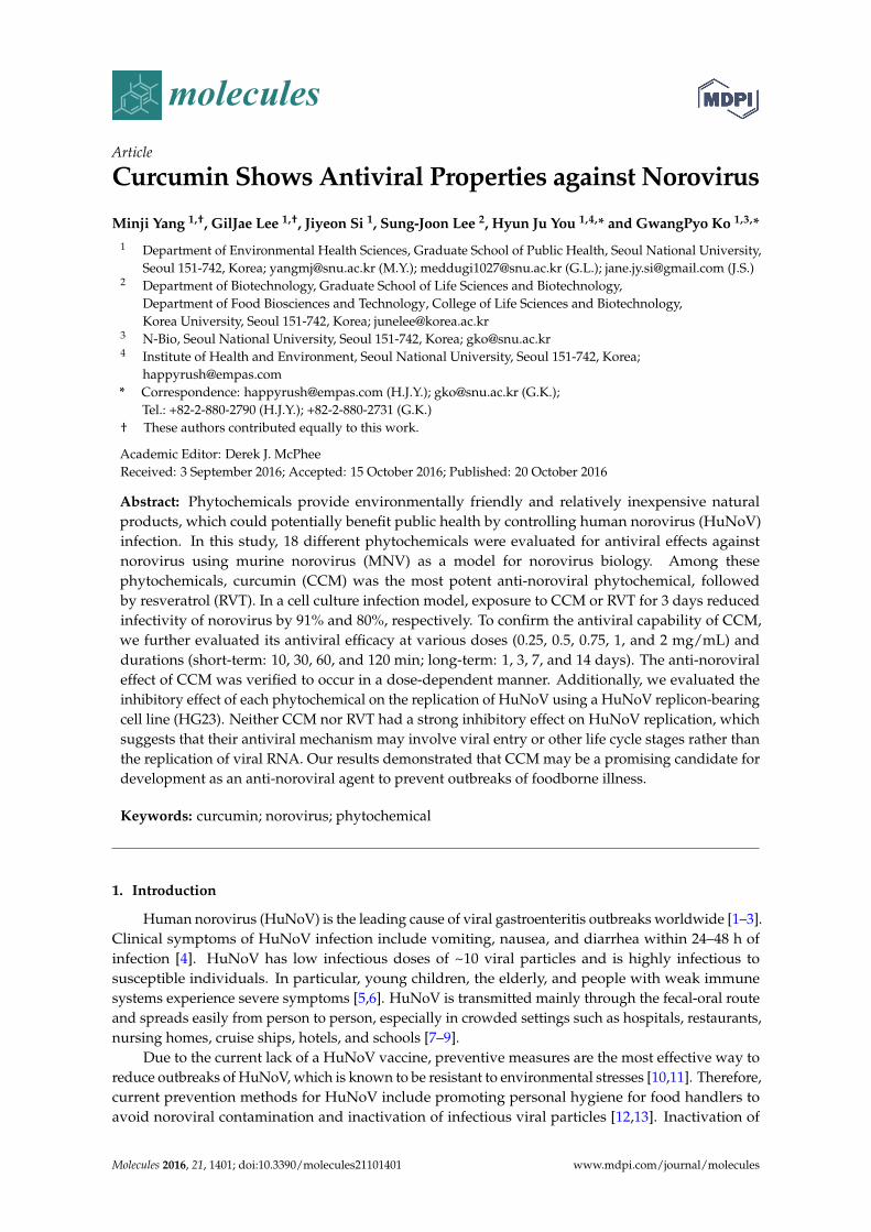
Abstract: Phytochemicals provide environmentally friendly and relatively inexpensive naturalproducts, which could potentially benefit public health by controlling human norovirus (HuNoV)infection. In this study, 18 different phytochemicals were evaluated for antiviral effects againstnorovirus using murine norovirus (MNV) as a model for norovirus biology. Among thesephytochemicals, curcumin (CCM) was the most potent anti-noroviral phytochemical, followedby resveratrol (RVT). In a cell culture infection model, exposure to CCM or RVT for 3 days reducedinfectivity of norovirus by 91% and 80%, respectively. To confirm the antiviral capability of CCM,we further evaluated its antiviral efficacy at various doses (0.25, 0.5, 0.75, 1, and 2 mg/mL) anddurations (short-term: 10, 30, 60, and 120 min; long-term: 1, 3, 7, and 14 days). The anti-noroviraleffect of CCM was verified to occur in a dose-dependent manner. Additionally, we evaluated theinhibitory effect of each phytochemical on the replication of HuNoV using a HuNoV replicon-bearingcell line (HG23). Neither CCM nor RVT had a strong inhibitory effect on HuNoV replication, whichsuggests that their antiviral mechanism may involve viral entry or other life cycle stages rather thanthe replication of viral RNA. Our results demonstrated that CCM may be a promising candidate fordevelopment as an anti-noroviral agent to prevent outbreaks of foodborne illness.
Keywords: curcumin; norovirus; phytochemical
1. Introduction
Human norovirus (HuNoV) is the leading cause of viral gastroenteritis outbreaks worldwide [1–3].Clinical symptoms of HuNoV infection include vomiting, nausea, and diarrhea within 24–48 h ofinfection [4]. HuNoV has low infectious doses of ~10 viral particles and is highly infectious tosusceptible individuals. In particular, young children, the elderly, and people with weak immunesystems experience severe symptoms [5,6]. HuNoV is transmitted mainly through the fecal-oral routeand spreads easily from person to person, especially in crowded settings such as hospitals, restaurants,nursing homes, cruise ships, hotels, and schools [7–9].
Due to the current lack of a HuNoV vaccine, preventive measures are the most effective way toreduce outbreaks of HuNoV, which is known to be resistant to environmental stresses [10,11]. Therefore,current prevention methods for HuNoV include promoting personal hygiene for food handlers toavoid noroviral contamination and inactivation of infectious viral particles [12,13]. Inactivation of
Molecules 2016, 21, 1401; doi:10.3390/molecules21101401 www.mdpi.com/journal/molecules
Molecules 2016, 21, 1401 2 of 14
HuNoV relies on physical methods, such as heating or radiation, and chemical methods such assodium hypochlorite or titanium dioxide [10,14–16]. However, the currently available methods havedisadvantages for direct application to food. These include (1) increased cost of equipment andmanagement; (2) potential acute and chronic toxic effects from chemical exposure; and (3) spoiling offood flavor or texture.
Therefore, environmentally friendly substances that might substitute for the established physicaland chemical methods would be beneficial and directly applicable to food products such as oysters.Phytochemicals can be obtained from various natural plant extracts and most of them are “generallyrecognized as safe” (GRAS) [17]. This has led many researchers to study natural substanceswith antimicrobial properties [18,19]. Although previous studies have demonstrated that variousphytochemicals possess antimicrobial effects, little research has been conducted to evaluate the efficacyof phytochemicals in reducing the infectiousness of norovirus [20].
Cultivation methods for HuNoV using B cells [21] or isolated enterocytes [22] were reportedrecently. Although those findings are very meaningful and can be a milestone to decipher themechanisms of norovirus infection at a molecular level, it is a bit early to apply the methods toscreening various antiviral compounds. The replication of HuNoV in a B-cell culture system didnot appear to be robust, as observed in murine norovirus (MNV) [23], and the methods need to bevalidated with different types of HuNoV in other laboratories. Therefore, many previous studies haveused viral models using MNV, feline calicivirus (FCV), and other in vitro models of HuNoV replication(e.g., HG23 cells) to evaluate the anti-noroviral activities of compounds [24,25]. In this study, we usedMNV and HG23 cells to investigate the ability of several phytochemicals to inactivate norovirus.
We screened the anti-noroviral effects of 18 different phytochemicals using a viral model to studynorovirus biology. Dose- and time-dependent trends were evaluated for the phytochemical with thegreatest efficacy. Subsequently, the results of inactivation after long-term phytochemical incubationwere fitted to three different mathematical models. Inhibitory effects on norovirus replication weremeasured using a HuNoV replicon-bearing cell (HG23) model.
2. Results
2.1. Evaluation of Phytochemical Cytotoxicity
Phytochemicals that had been previously reported to have antimicrobial effects were selected foranalysis. Previously validated antiviral phytochemicals such as resveratrol (RVT) were included aspositive controls [26]. The common names, abbreviations, and molecular weights of the phytochemicalstested in this study are summarized in Table A1. The cells were treated with phytochemicals andincubated for 1 h (RAW 264.7 cells) and 72 h (HG23 cells) at 37 ◦C with 5% CO2. DMSO was used asa vehicle control. The viability of cells (RAW 264.7 and HG23 cells) after treatment with phytochemicalswas evaluated using the water-soluble tetrazolium salt (WST) assay and was compared to a vehiclecontrol. In RAW 264.7 cells, phytochemicals showed no cytotoxicity at a concentration of 0.1 mg/mL(Table 1). On the other hand, the viability of HG23 cells fell to below 80% after treatment withmost phytochemicals at this concentration (data not shown). Because of the susceptibility of this cellline, HG23 cells were exposed to various concentrations of phytochemicals to find the maximumconcentration with no significant cytotoxicity. We found that for most phytochemicals, a concentrationof 20 µg/mL was optimal to assess antiviral activity without significant cytotoxicity in HG23 cells.Among the 18 tested phytochemicals, ellagic acid (ELG), curcumin (CCM), RVT, and compound K(CK) showed cytotoxicity at 20 µg/mL, so these four compounds were tested at 2 µg/mL in the assayof anti-noroviral activity.
Molecules 2016, 21, 1401 3 of 14
Table 1. Evaluation of cytotoxicity of phytochemicals in two different cell lines.
Common Name AbbreviationRAW 264.7 a HG23 b
% Cell Viability
Caffeic acid CFA 101.14 ± 6.00 92.62 ± 3.87Capsaicin CSC 119.18 ± 2.83 82.92 ± 0.58
Cinnamic acid CNA 102.25 ± 4.11 93.87 ± 6.75Compound K CK 91.89 ± 1.10 80.67 ± 3.07 †
Curcumin CCM 102.14 ± 1.89 92.57 ± 2.41 †
Ellagic acid ELG 80.32 ± 1.68 89.91 ± 7.50 †
Epigallocatechin gallate EGC 94.44 ± 5.55 87.44 ± 3.0910-Gingerol GGR 83.68 ± 3.03 86.07 ± 7.02
Ginsenoside F2 F2 88.30 ± 3.65 101.52 ± 4.22Ginsenoside Rb1 Rb1 95.27 ± 5.31 101.48 ± 4.61Ginsenoside Rd Rd 87.17 ± 0.79 102.11 ± 2.80Ginsenoside Rg3 Rg3 84.75 ± 2.10 95.29 ± 3.47Ginsenoside Rh1 Rh1 98.54 ± 2.94 94.38 ± 4.51Ginsenoside Rh2 Rh2 95.32 ± 2.00 94.18 ± 4.86Proanthocyanidin PAC 87.47 ± 7.31 102.40 ± 0.57Protopanaxadiol PPD 97.57 ± 1.80 99.03 ± 3.17
Quercetin QCT 127.26 ± 3.65 88.50 ± 7.23Resveratrol RVT 84.46 ± 2.03 80.07 ± 5.94 †
a RAW 264.7 cells were treated with 100 µg/mL of phytochemical and incubated for 1 h and b HG23 cells weretreated with 20 µg/mL and incubated for 72 h at 37 ◦C with 5% CO2. † Treatment concentration is 2 µg/mL dueto cytotoxicity at 20 µg/mL. DMSO was used as a vehicle control. All data (mean ± SEM) were derived fromtriplicate determination.
2.2. Effects of Phytochemicals on Neutralization of MNV
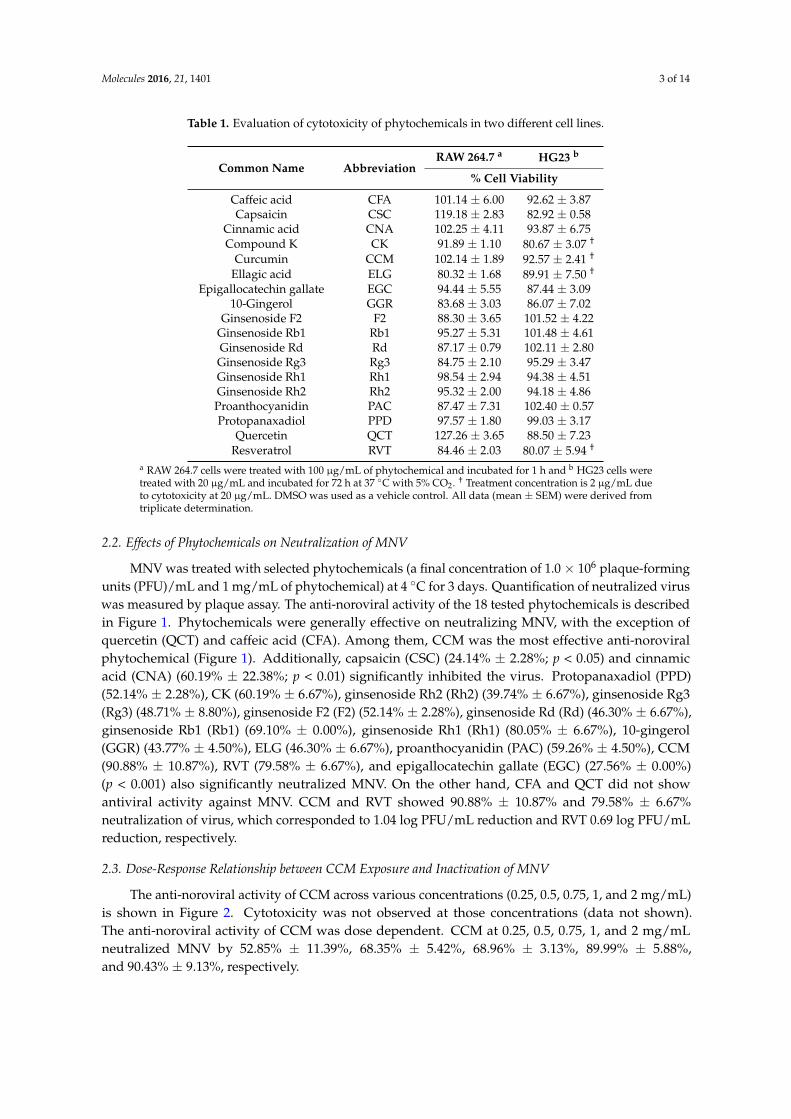
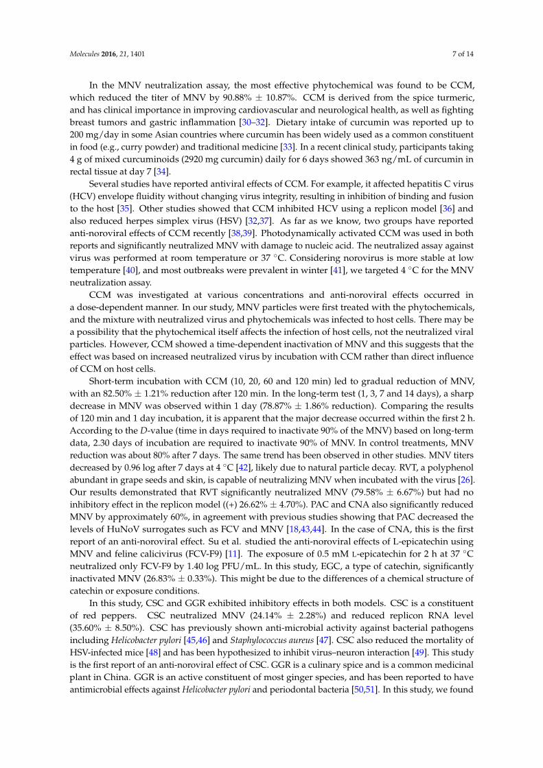
MNV was treated with selected phytochemicals (a final concentration of 1.0× 10⁶ plaque-formingunits (PFU)/mL and 1 mg/mL of phytochemical) at 4 ◦C for 3 days. Quantification of neutralized viruswas measured by plaque assay. The anti-noroviral activity of the 18 tested phytochemicals is describedin Figure 1. Phytochemicals were generally effective on neutralizing MNV, with the exception ofquercetin (QCT) and caffeic acid (CFA). Among them, CCM was the most effective anti-noroviralphytochemical (Figure 1). Additionally, capsaicin (CSC) (24.14% ± 2.28%; p < 0.05) and cinnamicacid (CNA) (60.19% ± 22.38%; p < 0.01) significantly inhibited the virus. Protopanaxadiol (PPD)(52.14% ± 2.28%), CK (60.19% ± 6.67%), ginsenoside Rh2 (Rh2) (39.74% ± 6.67%), ginsenoside Rg3(Rg3) (48.71% ± 8.80%), ginsenoside F2 (F2) (52.14% ± 2.28%), ginsenoside Rd (Rd) (46.30% ± 6.67%),ginsenoside Rb1 (Rb1) (69.10% ± 0.00%), ginsenoside Rh1 (Rh1) (80.05% ± 6.67%), 10-gingerol(GGR) (43.77% ± 4.50%), ELG (46.30% ± 6.67%), proanthocyanidin (PAC) (59.26% ± 4.50%), CCM(90.88% ± 10.87%), RVT (79.58% ± 6.67%), and epigallocatechin gallate (EGC) (27.56% ± 0.00%)(p < 0.001) also significantly neutralized MNV. On the other hand, CFA and QCT did not showantiviral activity against MNV. CCM and RVT showed 90.88% ± 10.87% and 79.58% ± 6.67%neutralization of virus, which corresponded to 1.04 log PFU/mL reduction and RVT 0.69 log PFU/mLreduction, respectively.
2.3. Dose-Response Relationship between CCM Exposure and Inactivation of MNV
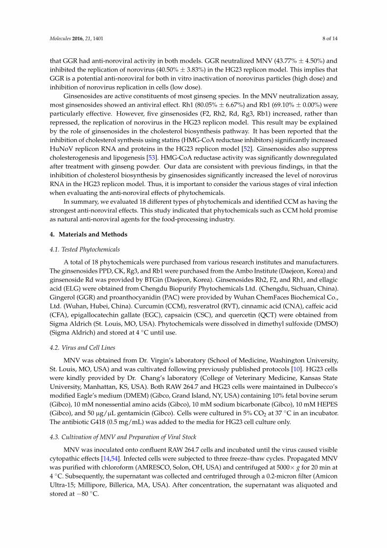
The anti-noroviral activity of CCM across various concentrations (0.25, 0.5, 0.75, 1, and 2 mg/mL)is shown in Figure 2. Cytotoxicity was not observed at those concentrations (data not shown).The anti-noroviral activity of CCM was dose dependent. CCM at 0.25, 0.5, 0.75, 1, and 2 mg/mLneutralized MNV by 52.85% ± 11.39%, 68.35% ± 5.42%, 68.96% ± 3.13%, 89.99% ± 5.88%,and 90.43% ± 9.13%, respectively.
Molecules 2016, 21, 1401 4 of 14
Molecules 2016, 21, 1401 4 of 14
Figure 1. The effect of phytochemicals on murine norovirus (MNV) neutralization in RAW 264.7 cells. MNV was mixed with 1 mg/mL of 18 phytochemicals and stored for 72 h at 4 °C. DMSO was used as a vehicle control and virus titer was measured by plaque assay. The mean diameter of plaques was 1 mm, and visible plaques were counted 72 h after infection. Each point was derived from triplicate determinations. Results are expressed as percentage neutralized virus compared to a vehicle control (mean ± SEM). Statistical significance was determined based on ANOVA with Bonferroni post-tests. * p < 0.05; ** p < 0.01; *** p < 0.001 compared to vehicle control.
Figure 2. Anti-noroviral effect of curcumin (CCM) at various concentrations in RAW 264.7 cells. MNV was mixed with 0.25, 0.5, 0.75, 1 or 2 mg/mL CCM and stored 72 h at 4 °C. Virus titers were measured by plaque assay and percentage was compared to a vehicle control (DMSO treated). Statistical significance was determined based on ANOVA with Bonferroni post-tests. Each point was derived from triplicate determinations (mean ± SEM). *** p < 0.001 comparing to vehicle control.
2.4. Short- and Long-Term Effects of Curcumin on Viral Neutralization
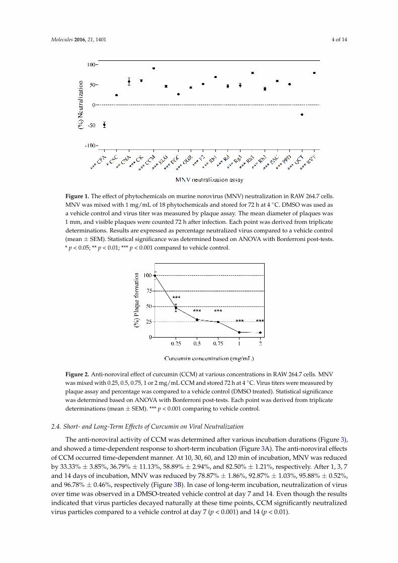
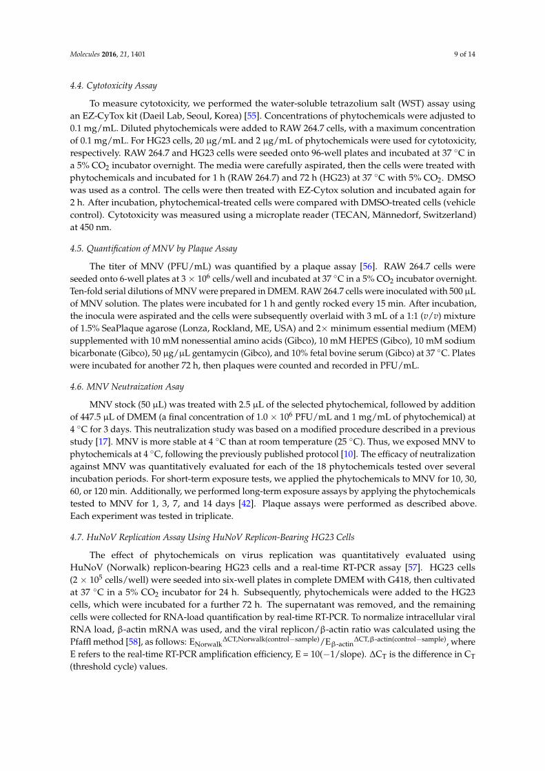
The anti-noroviral activity of CCM was determined after various incubation durations (Figure 3), and showed a time-dependent response to short-term incubation (Figure 3A). The anti-noroviral effects of CCM occurred time-dependent manner. At 10, 30, 60, and 120 min of incubation, MNV was reduced by 33.33% ± 3.85%, 36.79% ± 11.13%, 58.89% ± 2.94%, and 82.50% ± 1.21%, respectively. After 1, 3, 7 and 14 days of incubation, MNV was reduced by 78.87% ± 1.86%, 92.87% ± 1.03%, 95.88% ± 0.52%, and 96.78% ± 0.46%, respectively (Figure 3B). In case of long-term incubation, neutralization of virus over time was observed in a DMSO-treated vehicle control at day 7 and 14. Even though the results indicated that virus particles decayed naturally at these time points, CCM significantly neutralized virus particles compared to a vehicle control at day 7 (p < 0.001) and 14 (p < 0.01).
Figure 1. The effect of phytochemicals on murine norovirus (MNV) neutralization in RAW 264.7 cells.MNV was mixed with 1 mg/mL of 18 phytochemicals and stored for 72 h at 4 ◦C. DMSO was used asa vehicle control and virus titer was measured by plaque assay. The mean diameter of plaques was1 mm, and visible plaques were counted 72 h after infection. Each point was derived from triplicatedeterminations. Results are expressed as percentage neutralized virus compared to a vehicle control(mean ± SEM). Statistical significance was determined based on ANOVA with Bonferroni post-tests.* p < 0.05; ** p < 0.01; *** p < 0.001 compared to vehicle control.
Molecules 2016, 21, 1401 4 of 14
Figure 1. The effect of phytochemicals on murine norovirus (MNV) neutralization in RAW 264.7 cells. MNV was mixed with 1 mg/mL of 18 phytochemicals and stored for 72 h at 4 °C. DMSO was used as a vehicle control and virus titer was measured by plaque assay. The mean diameter of plaques was 1 mm, and visible plaques were counted 72 h after infection. Each point was derived from triplicate determinations. Results are expressed as percentage neutralized virus compared to a vehicle control (mean ± SEM). Statistical significance was determined based on ANOVA with Bonferroni post-tests. * p < 0.05; ** p < 0.01; *** p < 0.001 compared to vehicle control.
Figure 2. Anti-noroviral effect of curcumin (CCM) at various concentrations in RAW 264.7 cells. MNV was mixed with 0.25, 0.5, 0.75, 1 or 2 mg/mL CCM and stored 72 h at 4 °C. Virus titers were measured by plaque assay and percentage was compared to a vehicle control (DMSO treated). Statistical significance was determined based on ANOVA with Bonferroni post-tests. Each point was derived from triplicate determinations (mean ± SEM). *** p < 0.001 comparing to vehicle control.
2.4. Short- and Long-Term Effects of Curcumin on Viral Neutralization
The anti-noroviral activity of CCM was determined after various incubation durations (Figure 3), and showed a time-dependent response to short-term incubation (Figure 3A). The anti-noroviral effects of CCM occurred time-dependent manner. At 10, 30, 60, and 120 min of incubation, MNV was reduced by 33.33% ± 3.85%, 36.79% ± 11.13%, 58.89% ± 2.94%, and 82.50% ± 1.21%, respectively. After 1, 3, 7 and 14 days of incubation, MNV was reduced by 78.87% ± 1.86%, 92.87% ± 1.03%, 95.88% ± 0.52%, and 96.78% ± 0.46%, respectively (Figure 3B). In case of long-term incubation, neutralization of virus over time was observed in a DMSO-treated vehicle control at day 7 and 14. Even though the results indicated that virus particles decayed naturally at these time points, CCM significantly neutralized virus particles compared to a vehicle control at day 7 (p < 0.001) and 14 (p < 0.01).
Figure 2. Anti-noroviral effect of curcumin (CCM) at various concentrations in RAW 264.7 cells. MNVwas mixed with 0.25, 0.5, 0.75, 1 or 2 mg/mL CCM and stored 72 h at 4 ◦C. Virus titers were measured byplaque assay and percentage was compared to a vehicle control (DMSO treated). Statistical significancewas determined based on ANOVA with Bonferroni post-tests. Each point was derived from triplicatedeterminations (mean ± SEM). *** p < 0.001 comparing to vehicle control.
2.4. Short- and Long-Term Effects of Curcumin on Viral Neutralization
The anti-noroviral activity of CCM was determined after various incubation durations (Figure 3),and showed a time-dependent response to short-term incubation (Figure 3A). The anti-noroviral effectsof CCM occurred time-dependent manner. At 10, 30, 60, and 120 min of incubation, MNV was reducedby 33.33% ± 3.85%, 36.79% ± 11.13%, 58.89% ± 2.94%, and 82.50% ± 1.21%, respectively. After 1, 3, 7and 14 days of incubation, MNV was reduced by 78.87% ± 1.86%, 92.87% ± 1.03%, 95.88% ± 0.52%,and 96.78% ± 0.46%, respectively (Figure 3B). In case of long-term incubation, neutralization of virusover time was observed in a DMSO-treated vehicle control at day 7 and 14. Even though the resultsindicated that virus particles decayed naturally at these time points, CCM significantly neutralizedvirus particles compared to a vehicle control at day 7 (p < 0.001) and 14 (p < 0.01).

Molecules 2016, 21, 1401 5 of 14
Molecules 2016, 21, 1401 5 of 14
(A) (B)
Figure 3. Anti-noroviral efficacy of CCM under short- and long-term incubation in RAW 264.7 cells. MNV was mixed with 1 mg/mL of CCM and stored for (A) short (10, 30, 60, and 120 min) and (B) long periods (1, 3, 7 and 14 days) at 4 °C. Virus titers were measured by plaque assay (DMSO as a vehicle control). Percentage of plaque formation was compared to the initial time (day 0). Statistical significance was determined based on ANOVA with Bonferroni post-tests. Each point represents the mean ± SEM of triplicate determination. * p < 0.05; *** p < 0.001 comparing to vehicle control.
2.5. Model Evaluation with Experimental Data
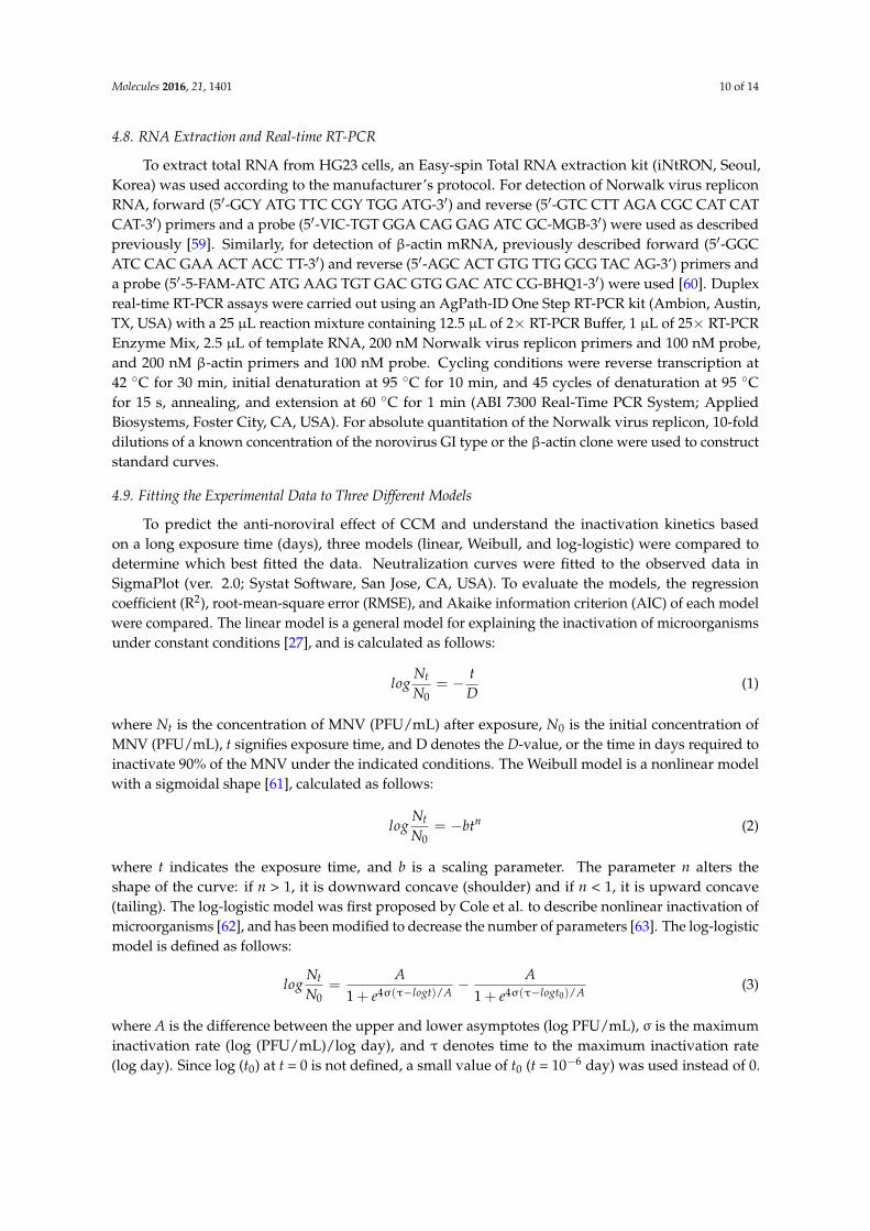
To assess the anti-noroviral behavior of CCM over a long period (days) (Figure 3B), three different models were evaluated: (i) the linear model, (ii) the Weibull model, and (iii) the log-logistic model (Figure 4). To assess the fit of the models, regression coefficient (R2), root-mean-square error (RMSE), and Akaike information criterion (AIC) were compared, and are summarized in Table 2. Higher R2 and lower RMSE values indicate a better-fitting model [27]. The log-logistic model showed the best fit to CCM data, with the highest R2 (0.99) and lowest RMSE (0.02) values. In contrast, the linear model appeared to be inappropriate for the observed data.
Figure 4. Data fitting of the inactivation curve for MNV under long-term incubation. MNV was mixed with 1 mg/mL of CCM and stored for 1, 3, 7 or 14 days at 4 °C. Data were fitted to several different models.
Table 2. Comparison of linear, Weibull, and log-logistic models in terms of long-term anti-noroviral data for curcumin.
Fitting Model Curcumin Inactivation
R2 RMSE AIC D-value Linear distribution 0.25 0.53 2.40 7.39
Weibull distribution 0.98 0.09 −10.94 2.72 Log-logistic distribution 0.99 0.02 −22.05 2.30
R2 is the correlation coefficient, RMSE is the root mean square error, AIC is the Akaike information criterion, and D-value is the time in days required to inactivate 90% of the MNV.
Figure 3. Anti-noroviral efficacy of CCM under short- and long-term incubation in RAW 264.7 cells.MNV was mixed with 1 mg/mL of CCM and stored for (A) short (10, 30, 60, and 120 min) and (B) longperiods (1, 3, 7 and 14 days) at 4 ◦C. Virus titers were measured by plaque assay (DMSO as a vehiclecontrol). Percentage of plaque formation was compared to the initial time (day 0). Statistical significancewas determined based on ANOVA with Bonferroni post-tests. Each point represents the mean ± SEMof triplicate determination. * p < 0.05; *** p < 0.001 comparing to vehicle control.
2.5. Model Evaluation with Experimental Data
To assess the anti-noroviral behavior of CCM over a long period (days) (Figure 3B), three differentmodels were evaluated: (i) the linear model; (ii) the Weibull model; and (iii) the log-logistic model(Figure 4). To assess the fit of the models, regression coefficient (R2), root-mean-square error (RMSE),and Akaike information criterion (AIC) were compared, and are summarized in Table 2. Higher R2
and lower RMSE values indicate a better-fitting model [27]. The log-logistic model showed the best fitto CCM data, with the highest R2 (0.99) and lowest RMSE (0.02) values. In contrast, the linear modelappeared to be inappropriate for the observed data.
Molecules 2016, 21, 1401 5 of 14
(A) (B)
Figure 3. Anti-noroviral efficacy of CCM under short- and long-term incubation in RAW 264.7 cells. MNV was mixed with 1 mg/mL of CCM and stored for (A) short (10, 30, 60, and 120 min) and (B) long periods (1, 3, 7 and 14 days) at 4 °C. Virus titers were measured by plaque assay (DMSO as a vehicle control). Percentage of plaque formation was compared to the initial time (day 0). Statistical significance was determined based on ANOVA with Bonferroni post-tests. Each point represents the mean ± SEM of triplicate determination. * p < 0.05; *** p < 0.001 comparing to vehicle control.
2.5. Model Evaluation with Experimental Data
To assess the anti-noroviral behavior of CCM over a long period (days) (Figure 3B), three different models were evaluated: (i) the linear model, (ii) the Weibull model, and (iii) the log-logistic model (Figure 4). To assess the fit of the models, regression coefficient (R2), root-mean-square error (RMSE), and Akaike information criterion (AIC) were compared, and are summarized in Table 2. Higher R2 and lower RMSE values indicate a better-fitting model [27]. The log-logistic model showed the best fit to CCM data, with the highest R2 (0.99) and lowest RMSE (0.02) values. In contrast, the linear model appeared to be inappropriate for the observed data.
Figure 4. Data fitting of the inactivation curve for MNV under long-term incubation. MNV was mixed with 1 mg/mL of CCM and stored for 1, 3, 7 or 14 days at 4 °C. Data were fitted to several different models.
Table 2. Comparison of linear, Weibull, and log-logistic models in terms of long-term anti-noroviral data for curcumin.
Fitting Model Curcumin Inactivation
R2 RMSE AIC D-value Linear distribution 0.25 0.53 2.40 7.39
Weibull distribution 0.98 0.09 −10.94 2.72 Log-logistic distribution 0.99 0.02 −22.05 2.30
R2 is the correlation coefficient, RMSE is the root mean square error, AIC is the Akaike information criterion, and D-value is the time in days required to inactivate 90% of the MNV.
Figure 4. Data fitting of the inactivation curve for MNV under long-term incubation. MNV wasmixed with 1 mg/mL of CCM and stored for 1, 3, 7 or 14 days at 4 ◦C. Data were fitted to severaldifferent models.
Table 2. Comparison of linear, Weibull, and log-logistic models in terms of long-term anti-noroviraldata for curcumin.
Fitting ModelCurcumin Inactivation
R2 RMSE AIC D-value
Linear distribution 0.25 0.53 2.40 7.39Weibull distribution 0.98 0.09 −10.94 2.72
Log-logistic distribution 0.99 0.02 −22.05 2.30
R2 is the correlation coefficient, RMSE is the root mean square error, AIC is the Akaike information criterion,and D-value is the time in days required to inactivate 90% of the MNV.
Molecules 2016, 21, 1401 6 of 14
2.6. Effects of Phytochemicals on Replication of HuNoV in Replicon-Bearing Cells
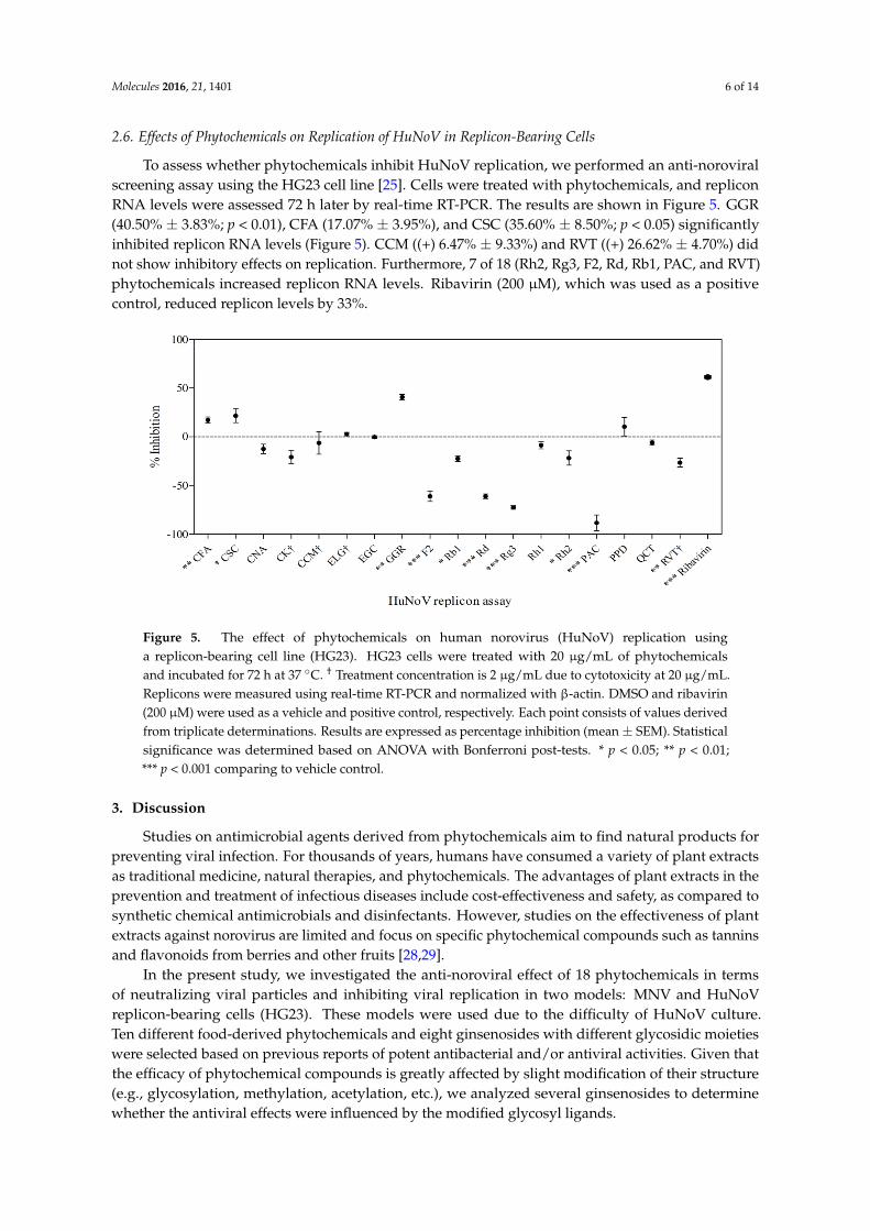
To assess whether phytochemicals inhibit HuNoV replication, we performed an anti-noroviralscreening assay using the HG23 cell line [25]. Cells were treated with phytochemicals, and repliconRNA levels were assessed 72 h later by real-time RT-PCR. The results are shown in Figure 5. GGR(40.50% ± 3.83%; p < 0.01), CFA (17.07% ± 3.95%), and CSC (35.60% ± 8.50%; p < 0.05) significantlyinhibited replicon RNA levels (Figure 5). CCM ((+) 6.47% ± 9.33%) and RVT ((+) 26.62% ± 4.70%) didnot show inhibitory effects on replication. Furthermore, 7 of 18 (Rh2, Rg3, F2, Rd, Rb1, PAC, and RVT)phytochemicals increased replicon RNA levels. Ribavirin (200 µM), which was used as a positivecontrol, reduced replicon levels by 33%.
Molecules 2016, 21, 1401 6 of 14
2.6. Effects of Phytochemicals on Replication of HuNoV in Replicon-Bearing Cells
To assess whether phytochemicals inhibit HuNoV replication, we performed an anti-noroviral screening assay using the HG23 cell line [25]. Cells were treated with phytochemicals, and replicon RNA levels were assessed 72 h later by real-time RT-PCR. The results are shown in Figure 5. GGR (40.50% ± 3.83%; p < 0.01), CFA (17.07% ± 3.95%), and CSC (35.60% ± 8.50%; p < 0.05) significantly inhibited replicon RNA levels (Figure 5). CCM ((+) 6.47% ± 9.33%) and RVT ((+) 26.62% ± 4.70%) did not show inhibitory effects on replication. Furthermore, 7 of 18 (Rh2, Rg3, F2, Rd, Rb1, PAC, and RVT) phytochemicals increased replicon RNA levels. Ribavirin (200 μM), which was used as a positive control, reduced replicon levels by 33%.
Figure 5. The effect of phytochemicals on human norovirus (HuNoV) replication using a replicon-bearing cell line (HG23). HG23 cells were treated with 20 μg/mL of phytochemicals and incubated for 72 h at 37 °C. † Treatment concentration is 2 μg/mL due to cytotoxicity at 20 μg/mL. Replicons were measured using real-time RT-PCR and normalized with β-actin. DMSO and ribavirin (200 μM) were used as a vehicle and positive control, respectively. Each point consists of values derived from triplicate determinations. Results are expressed as percentage inhibition (mean ± SEM). Statistical significance was determined based on ANOVA with Bonferroni post-tests. * p < 0.05; ** p < 0.01; *** p < 0.001 comparing to vehicle control.
3. Discussion
Studies on antimicrobial agents derived from phytochemicals aim to find natural products for preventing viral infection. For thousands of years, humans have consumed a variety of plant extracts as traditional medicine, natural therapies, and phytochemicals. The advantages of plant extracts in the prevention and treatment of infectious diseases include cost-effectiveness and safety, as compared to synthetic chemical antimicrobials and disinfectants. However, studies on the effectiveness of plant extracts against norovirus are limited and focus on specific phytochemical compounds such as tannins and flavonoids from berries and other fruits [28,29].
In the present study, we investigated the anti-noroviral effect of 18 phytochemicals in terms of neutralizing viral particles and inhibiting viral replication in two models: MNV and HuNoV replicon-bearing cells (HG23). These models were used due to the difficulty of HuNoV culture. Ten different food-derived phytochemicals and eight ginsenosides with different glycosidic moieties were selected based on previous reports of potent antibacterial and/or antiviral activities. Given that the efficacy of phytochemical compounds is greatly affected by slight modification of their structure (e.g., glycosylation, methylation, acetylation, etc.), we analyzed several ginsenosides to determine whether the antiviral effects were influenced by the modified glycosyl ligands.
Figure 5. The effect of phytochemicals on human norovirus (HuNoV) replication usinga replicon-bearing cell line (HG23). HG23 cells were treated with 20 µg/mL of phytochemicalsand incubated for 72 h at 37 ◦C. † Treatment concentration is 2 µg/mL due to cytotoxicity at 20 µg/mL.Replicons were measured using real-time RT-PCR and normalized with β-actin. DMSO and ribavirin(200 µM) were used as a vehicle and positive control, respectively. Each point consists of values derivedfrom triplicate determinations. Results are expressed as percentage inhibition (mean± SEM). Statisticalsignificance was determined based on ANOVA with Bonferroni post-tests. * p < 0.05; ** p < 0.01;*** p < 0.001 comparing to vehicle control.
3. Discussion
Studies on antimicrobial agents derived from phytochemicals aim to find natural products forpreventing viral infection. For thousands of years, humans have consumed a variety of plant extractsas traditional medicine, natural therapies, and phytochemicals. The advantages of plant extracts in theprevention and treatment of infectious diseases include cost-effectiveness and safety, as compared tosynthetic chemical antimicrobials and disinfectants. However, studies on the effectiveness of plantextracts against norovirus are limited and focus on specific phytochemical compounds such as tanninsand flavonoids from berries and other fruits [28,29].
In the present study, we investigated the anti-noroviral effect of 18 phytochemicals in termsof neutralizing viral particles and inhibiting viral replication in two models: MNV and HuNoVreplicon-bearing cells (HG23). These models were used due to the difficulty of HuNoV culture.Ten different food-derived phytochemicals and eight ginsenosides with different glycosidic moietieswere selected based on previous reports of potent antibacterial and/or antiviral activities. Given thatthe efficacy of phytochemical compounds is greatly affected by slight modification of their structure(e.g., glycosylation, methylation, acetylation, etc.), we analyzed several ginsenosides to determinewhether the antiviral effects were influenced by the modified glycosyl ligands.
Molecules 2016, 21, 1401 7 of 14
In the MNV neutralization assay, the most effective phytochemical was found to be CCM,which reduced the titer of MNV by 90.88% ± 10.87%. CCM is derived from the spice turmeric,and has clinical importance in improving cardiovascular and neurological health, as well as fightingbreast tumors and gastric inflammation [30–32]. Dietary intake of curcumin was reported up to200 mg/day in some Asian countries where curcumin has been widely used as a common constituentin food (e.g., curry powder) and traditional medicine [33]. In a recent clinical study, participants taking4 g of mixed curcuminoids (2920 mg curcumin) daily for 6 days showed 363 ng/mL of curcumin inrectal tissue at day 7 [34].
Several studies have reported antiviral effects of CCM. For example, it affected hepatitis C virus(HCV) envelope fluidity without changing virus integrity, resulting in inhibition of binding and fusionto the host [35]. Other studies showed that CCM inhibited HCV using a replicon model [36] andalso reduced herpes simplex virus (HSV) [32,37]. As far as we know, two groups have reportedanti-noroviral effects of CCM recently [38,39]. Photodynamically activated CCM was used in bothreports and significantly neutralized MNV with damage to nucleic acid. The neutralized assay againstvirus was performed at room temperature or 37 ◦C. Considering norovirus is more stable at lowtemperature [40], and most outbreaks were prevalent in winter [41], we targeted 4 ◦C for the MNVneutralization assay.
CCM was investigated at various concentrations and anti-noroviral effects occurred ina dose-dependent manner. In our study, MNV particles were first treated with the phytochemicals,and the mixture with neutralized virus and phytochemicals was infected to host cells. There may bea possibility that the phytochemical itself affects the infection of host cells, not the neutralized viralparticles. However, CCM showed a time-dependent inactivation of MNV and this suggests that theeffect was based on increased neutralized virus by incubation with CCM rather than direct influenceof CCM on host cells.
Short-term incubation with CCM (10, 20, 60 and 120 min) led to gradual reduction of MNV,with an 82.50% ± 1.21% reduction after 120 min. In the long-term test (1, 3, 7 and 14 days), a sharpdecrease in MNV was observed within 1 day (78.87% ± 1.86% reduction). Comparing the resultsof 120 min and 1 day incubation, it is apparent that the major decrease occurred within the first 2 h.According to the D-value (time in days required to inactivate 90% of the MNV) based on long-termdata, 2.30 days of incubation are required to inactivate 90% of MNV. In control treatments, MNVreduction was about 80% after 7 days. The same trend has been observed in other studies. MNV titersdecreased by 0.96 log after 7 days at 4 ◦C [42], likely due to natural particle decay. RVT, a polyphenolabundant in grape seeds and skin, is capable of neutralizing MNV when incubated with the virus [26].Our results demonstrated that RVT significantly neutralized MNV (79.58% ± 6.67%) but had noinhibitory effect in the replicon model ((+) 26.62% ± 4.70%). PAC and CNA also significantly reducedMNV by approximately 60%, in agreement with previous studies showing that PAC decreased thelevels of HuNoV surrogates such as FCV and MNV [18,43,44]. In the case of CNA, this is the firstreport of an anti-noroviral effect. Su et al. studied the anti-noroviral effects of L-epicatechin usingMNV and feline calicivirus (FCV-F9) [11]. The exposure of 0.5 mM L-epicatechin for 2 h at 37 ◦Cneutralized only FCV-F9 by 1.40 log PFU/mL. In this study, EGC, a type of catechin, significantlyinactivated MNV (26.83% ± 0.33%). This might be due to the differences of a chemical structure ofcatechin or exposure conditions.
In this study, CSC and GGR exhibited inhibitory effects in both models. CSC is a constituentof red peppers. CSC neutralized MNV (24.14% ± 2.28%) and reduced replicon RNA level(35.60% ± 8.50%). CSC has previously shown anti-microbial activity against bacterial pathogensincluding Helicobacter pylori [45,46] and Staphylococcus aureus [47]. CSC also reduced the mortality ofHSV-infected mice [48] and has been hypothesized to inhibit virus–neuron interaction [49]. This studyis the first report of an anti-noroviral effect of CSC. GGR is a culinary spice and is a common medicinalplant in China. GGR is an active constituent of most ginger species, and has been reported to haveantimicrobial effects against Helicobacter pylori and periodontal bacteria [50,51]. In this study, we found
Molecules 2016, 21, 1401 8 of 14
that GGR had anti-noroviral activity in both models. GGR neutralized MNV (43.77% ± 4.50%) andinhibited the replication of norovirus (40.50% ± 3.83%) in the HG23 replicon model. This implies thatGGR is a potential anti-noroviral for both in vitro inactivation of norovirus particles (high dose) andinhibition of norovirus replication in cells (low dose).
Ginsenosides are active constituents of most ginseng species. In the MNV neutralization assay,most ginsenosides showed an antiviral effect. Rh1 (80.05% ± 6.67%) and Rb1 (69.10% ± 0.00%) wereparticularly effective. However, five ginsenosides (F2, Rh2, Rd, Rg3, Rb1) increased, rather thanrepressed, the replication of norovirus in the HG23 replicon model. This result may be explainedby the role of ginsenosides in the cholesterol biosynthesis pathway. It has been reported that theinhibition of cholesterol synthesis using statins (HMG-CoA reductase inhibitors) significantly increasedHuNoV replicon RNA and proteins in the HG23 replicon model [52]. Ginsenosides also suppresscholesterogenesis and lipogenesis [53]. HMG-CoA reductase activity was significantly downregulatedafter treatment with ginseng powder. Our data are consistent with previous findings, in that theinhibition of cholesterol biosynthesis by ginsenosides significantly increased the level of norovirusRNA in the HG23 replicon model. Thus, it is important to consider the various stages of viral infectionwhen evaluating the anti-noroviral effects of phytochemicals.
In summary, we evaluated 18 different types of phytochemicals and identified CCM as having thestrongest anti-noroviral effects. This study indicated that phytochemicals such as CCM hold promiseas natural anti-noroviral agents for the food-processing industry.
4. Materials and Methods
4.1. Tested Phytochemicals
A total of 18 phytochemicals were purchased from various research institutes and manufacturers.The ginsenosides PPD, CK, Rg3, and Rb1 were purchased from the Ambo Institute (Daejeon, Korea) andginsenoside Rd was provided by BTGin (Daejeon, Korea). Ginsenosides Rh2, F2, and Rh1, and ellagicacid (ELG) were obtained from Chengdu Biopurify Phytochemicals Ltd. (Chengdu, Sichuan, China).Gingerol (GGR) and proanthocyanidin (PAC) were provided by Wuhan ChemFaces Biochemical Co.,Ltd. (Wuhan, Hubei, China). Curcumin (CCM), resveratrol (RVT), cinnamic acid (CNA), caffeic acid(CFA), epigallocatechin gallate (EGC), capsaicin (CSC), and quercetin (QCT) were obtained fromSigma Aldrich (St. Louis, MO, USA). Phytochemicals were dissolved in dimethyl sulfoxide (DMSO)(Sigma Aldrich) and stored at 4 ◦C until use.
4.2. Virus and Cell Lines
MNV was obtained from Dr. Virgin’s laboratory (School of Medicine, Washington University,St. Louis, MO, USA) and was cultivated following previously published protocols [10]. HG23 cellswere kindly provided by Dr. Chang’s laboratory (College of Veterinary Medicine, Kansas StateUniversity, Manhattan, KS, USA). Both RAW 264.7 and HG23 cells were maintained in Dulbecco’smodified Eagle’s medium (DMEM) (Gibco, Grand Island, NY, USA) containing 10% fetal bovine serum(Gibco), 10 mM nonessential amino acids (Gibco), 10 mM sodium bicarbonate (Gibco), 10 mM HEPES(Gibco), and 50 µg/µL gentamicin (Gibco). Cells were cultured in 5% CO2 at 37 ◦C in an incubator.The antibiotic G418 (0.5 mg/mL) was added to the media for HG23 cell culture only.
4.3. Cultivation of MNV and Preparation of Viral Stock
MNV was inoculated onto confluent RAW 264.7 cells and incubated until the virus caused visiblecytopathic effects [14,54]. Infected cells were subjected to three freeze–thaw cycles. Propagated MNVwas purified with chloroform (AMRESCO, Solon, OH, USA) and centrifuged at 5000× g for 20 min at4 ◦C. Subsequently, the supernatant was collected and centrifuged through a 0.2-micron filter (AmiconUltra-15; Millipore, Billerica, MA, USA). After concentration, the supernatant was aliquoted andstored at −80 ◦C.
Molecules 2016, 21, 1401 9 of 14
4.4. Cytotoxicity Assay
To measure cytotoxicity, we performed the water-soluble tetrazolium salt (WST) assay usingan EZ-CyTox kit (Daeil Lab, Seoul, Korea) [55]. Concentrations of phytochemicals were adjusted to0.1 mg/mL. Diluted phytochemicals were added to RAW 264.7 cells, with a maximum concentrationof 0.1 mg/mL. For HG23 cells, 20 µg/mL and 2 µg/mL of phytochemicals were used for cytotoxicity,respectively. RAW 264.7 and HG23 cells were seeded onto 96-well plates and incubated at 37 ◦C ina 5% CO2 incubator overnight. The media were carefully aspirated, then the cells were treated withphytochemicals and incubated for 1 h (RAW 264.7) and 72 h (HG23) at 37 ◦C with 5% CO2. DMSOwas used as a control. The cells were then treated with EZ-Cytox solution and incubated again for2 h. After incubation, phytochemical-treated cells were compared with DMSO-treated cells (vehiclecontrol). Cytotoxicity was measured using a microplate reader (TECAN, Männedorf, Switzerland)at 450 nm.
4.5. Quantification of MNV by Plaque Assay
The titer of MNV (PFU/mL) was quantified by a plaque assay [56]. RAW 264.7 cells wereseeded onto 6-well plates at 3× 10⁶ cells/well and incubated at 37 ◦C in a 5% CO2 incubator overnight.Ten-fold serial dilutions of MNV were prepared in DMEM. RAW 264.7 cells were inoculated with 500 µLof MNV solution. The plates were incubated for 1 h and gently rocked every 15 min. After incubation,the inocula were aspirated and the cells were subsequently overlaid with 3 mL of a 1:1 (v/v) mixtureof 1.5% SeaPlaque agarose (Lonza, Rockland, ME, USA) and 2×minimum essential medium (MEM)supplemented with 10 mM nonessential amino acids (Gibco), 10 mM HEPES (Gibco), 10 mM sodiumbicarbonate (Gibco), 50 µg/µL gentamycin (Gibco), and 10% fetal bovine serum (Gibco) at 37 ◦C. Plateswere incubated for another 72 h, then plaques were counted and recorded in PFU/mL.
4.6. MNV Neutraization Asay
MNV stock (50 µL) was treated with 2.5 µL of the selected phytochemical, followed by additionof 447.5 µL of DMEM (a final concentration of 1.0 × 10⁶ PFU/mL and 1 mg/mL of phytochemical) at4 ◦C for 3 days. This neutralization study was based on a modified procedure described in a previousstudy [17]. MNV is more stable at 4 ◦C than at room temperature (25 ◦C). Thus, we exposed MNV tophytochemicals at 4 ◦C, following the previously published protocol [10]. The efficacy of neutralizationagainst MNV was quantitatively evaluated for each of the 18 phytochemicals tested over severalincubation periods. For short-term exposure tests, we applied the phytochemicals to MNV for 10, 30,60, or 120 min. Additionally, we performed long-term exposure assays by applying the phytochemicalstested to MNV for 1, 3, 7, and 14 days [42]. Plaque assays were performed as described above.Each experiment was tested in triplicate.
4.7. HuNoV Replication Assay Using HuNoV Replicon-Bearing HG23 Cells
The effect of phytochemicals on virus replication was quantitatively evaluated usingHuNoV (Norwalk) replicon-bearing HG23 cells and a real-time RT-PCR assay [57]. HG23 cells(2 × 10⁵ cells/well) were seeded into six-well plates in complete DMEM with G418, then cultivatedat 37 ◦C in a 5% CO2 incubator for 24 h. Subsequently, phytochemicals were added to the HG23cells, which were incubated for a further 72 h. The supernatant was removed, and the remainingcells were collected for RNA-load quantification by real-time RT-PCR. To normalize intracellular viralRNA load, β-actin mRNA was used, and the viral replicon/β-actin ratio was calculated using thePfaffl method [58], as follows: ENorwalk
∆CT,Norwalk(control−sample)/Eβ-actin∆CT,β-actin(control−sample), where
E refers to the real-time RT-PCR amplification efficiency, E = 10(−1/slope). ∆CT is the difference in CT
(threshold cycle) values.
Molecules 2016, 21, 1401 10 of 14
4.8. RNA Extraction and Real-time RT-PCR
To extract total RNA from HG23 cells, an Easy-spin Total RNA extraction kit (iNtRON, Seoul,Korea) was used according to the manufacturer’s protocol. For detection of Norwalk virus repliconRNA, forward (5′-GCY ATG TTC CGY TGG ATG-3′) and reverse (5′-GTC CTT AGA CGC CAT CATCAT-3′) primers and a probe (5′-VIC-TGT GGA CAG GAG ATC GC-MGB-3′) were used as describedpreviously [59]. Similarly, for detection of β-actin mRNA, previously described forward (5′-GGCATC CAC GAA ACT ACC TT-3′) and reverse (5′-AGC ACT GTG TTG GCG TAC AG-3’) primers anda probe (5′-5-FAM-ATC ATG AAG TGT GAC GTG GAC ATC CG-BHQ1-3′) were used [60]. Duplexreal-time RT-PCR assays were carried out using an AgPath-ID One Step RT-PCR kit (Ambion, Austin,TX, USA) with a 25 µL reaction mixture containing 12.5 µL of 2× RT-PCR Buffer, 1 µL of 25× RT-PCREnzyme Mix, 2.5 µL of template RNA, 200 nM Norwalk virus replicon primers and 100 nM probe,and 200 nM β-actin primers and 100 nM probe. Cycling conditions were reverse transcription at42 ◦C for 30 min, initial denaturation at 95 ◦C for 10 min, and 45 cycles of denaturation at 95 ◦Cfor 15 s, annealing, and extension at 60 ◦C for 1 min (ABI 7300 Real-Time PCR System; AppliedBiosystems, Foster City, CA, USA). For absolute quantitation of the Norwalk virus replicon, 10-folddilutions of a known concentration of the norovirus GI type or the β-actin clone were used to constructstandard curves.
4.9. Fitting the Experimental Data to Three Different Models
To predict the anti-noroviral effect of CCM and understand the inactivation kinetics basedon a long exposure time (days), three models (linear, Weibull, and log-logistic) were compared todetermine which best fitted the data. Neutralization curves were fitted to the observed data inSigmaPlot (ver. 2.0; Systat Software, San Jose, CA, USA). To evaluate the models, the regressioncoefficient (R2), root-mean-square error (RMSE), and Akaike information criterion (AIC) of each modelwere compared. The linear model is a general model for explaining the inactivation of microorganismsunder constant conditions [27], and is calculated as follows:
logNt
N0= − t
D(1)
where Nt is the concentration of MNV (PFU/mL) after exposure, N0 is the initial concentration ofMNV (PFU/mL), t signifies exposure time, and D denotes the D-value, or the time in days required toinactivate 90% of the MNV under the indicated conditions. The Weibull model is a nonlinear modelwith a sigmoidal shape [61], calculated as follows:
logNt
N0= −btn (2)
where t indicates the exposure time, and b is a scaling parameter. The parameter n alters theshape of the curve: if n > 1, it is downward concave (shoulder) and if n < 1, it is upward concave(tailing). The log-logistic model was first proposed by Cole et al. to describe nonlinear inactivation ofmicroorganisms [62], and has been modified to decrease the number of parameters [63]. The log-logisticmodel is defined as follows:
logNt
N0=
A1 + e4σ(τ−logt)/A
− A1 + e4σ(τ−logt0)/A
(3)
where A is the difference between the upper and lower asymptotes (log PFU/mL), σ is the maximuminactivation rate (log (PFU/mL)/log day), and τ denotes time to the maximum inactivation rate(log day). Since log (t0) at t = 0 is not defined, a small value of t0 (t = 10−6 day) was used instead of 0.
Molecules 2016, 21, 1401 11 of 14
4.10. Statistical Analysis
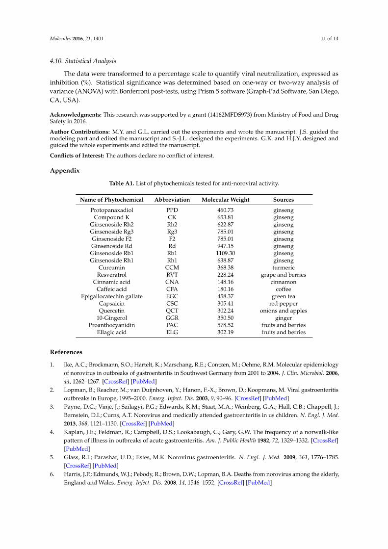
The data were transformed to a percentage scale to quantify viral neutralization, expressed asinhibition (%). Statistical significance was determined based on one-way or two-way analysis ofvariance (ANOVA) with Bonferroni post-tests, using Prism 5 software (Graph-Pad Software, San Diego,CA, USA).
Acknowledgments: This research was supported by a grant (14162MFDS973) from Ministry of Food and DrugSafety in 2016.
Author Contributions: M.Y. and G.L. carried out the experiments and wrote the manuscript. J.S. guided themodeling part and edited the manuscript and S.-J.L. designed the experiments. G.K. and H.J.Y. designed andguided the whole experiments and edited the manuscript.
Conflicts of Interest: The authors declare no conflict of interest.
Appendix
Table A1. List of phytochemicals tested for anti-noroviral activity.
Name of Phytochemical Abbreviation Molecular Weight Sources
Protopanaxadiol PPD 460.73 ginsengCompound K CK 653.81 ginseng
Ginsenoside Rh2 Rh2 622.87 ginsengGinsenoside Rg3 Rg3 785.01 ginsengGinsenoside F2 F2 785.01 ginsengGinsenoside Rd Rd 947.15 ginsengGinsenoside Rb1 Rb1 1109.30 ginsengGinsenoside Rh1 Rh1 638.87 ginseng
Curcumin CCM 368.38 turmericResveratrol RVT 228.24 grape and berries
Cinnamic acid CNA 148.16 cinnamonCaffeic acid CFA 180.16 coffee
Epigallocatechin gallate EGC 458.37 green teaCapsaicin CSC 305.41 red pepperQuercetin QCT 302.24 onions and apples
10-Gingerol GGR 350.50 gingerProanthocyanidin PAC 578.52 fruits and berries
Ellagic acid ELG 302.19 fruits and berries
References
1. Ike, A.C.; Brockmann, S.O.; Hartelt, K.; Marschang, R.E.; Contzen, M.; Oehme, R.M. Molecular epidemiologyof norovirus in outbreaks of gastroenteritis in Southwest Germany from 2001 to 2004. J. Clin. Microbiol. 2006,44, 1262–1267. [CrossRef] [PubMed]
2. Lopman, B.; Reacher, M.; van Duijnhoven, Y.; Hanon, F.-X.; Brown, D.; Koopmans, M. Viral gastroenteritisoutbreaks in Europe, 1995–2000. Emerg. Infect. Dis. 2003, 9, 90–96. [CrossRef] [PubMed]
3. Payne, D.C.; Vinjé, J.; Szilagyi, P.G.; Edwards, K.M.; Staat, M.A.; Weinberg, G.A.; Hall, C.B.; Chappell, J.;Bernstein, D.I.; Curns, A.T. Norovirus and medically attended gastroenteritis in us children. N. Engl. J. Med.2013, 368, 1121–1130. [CrossRef] [PubMed]
4. Kaplan, J.E.; Feldman, R.; Campbell, D.S.; Lookabaugh, C.; Gary, G.W. The frequency of a norwalk-likepattern of illness in outbreaks of acute gastroenteritis. Am. J. Public Health 1982, 72, 1329–1332. [CrossRef][PubMed]
5. Glass, R.I.; Parashar, U.D.; Estes, M.K. Norovirus gastroenteritis. N. Engl. J. Med. 2009, 361, 1776–1785.[CrossRef] [PubMed]
6. Harris, J.P.; Edmunds, W.J.; Pebody, R.; Brown, D.W.; Lopman, B.A. Deaths from norovirus among the elderly,England and Wales. Emerg. Infect. Dis. 2008, 14, 1546–1552. [CrossRef] [PubMed]
Molecules 2016, 21, 1401 12 of 14
7. Isakbaeva, E.T.; Widdowson, M.-A.; Beard, R.S.; Bulens, S.N.; Mullins, J.; Monroe, S.S.; Bresee, J.; Sassano, P.;Cramer, E.H.; Glass, R.I. Norovirus transmission on cruise ship. Emerg. Infect. Dis. 2005, 11, 154–158.[CrossRef] [PubMed]
8. Lopman, B.A.; Adak, G.K.; Reacher, M.H.; Brown, D. Two epidemiologic patterns of norovirus outbreaks:Surveillance in England and Wales, 1992–2000. Emerg. Infect. Dis. 2003, 9, 71–77. [CrossRef] [PubMed]
9. Centers for Disease Control and Prevention. Norovirus outbreak in an elementary school—District ofColumbia, February 2007. Morb. Mortal. Wkly. Rep. 2008, 56, 1340.
10. Lee, J.; Zoh, K.; Ko, G. Inactivation and UV disinfection of murine norovirus with TiO2 under variousenvironmental conditions. Appl. Environ. Microbiol. 2008, 74, 2111–2117. [CrossRef] [PubMed]
11. Su, X.; D’Souza, D.H. Naturally occurring flavonoids against human norovirus surrogates.Food Environ. Virol. 2013, 5, 97–102. [CrossRef] [PubMed]
12. Fino, V.R.; Kniel, K.E. UV light inactivation of hepatitis A virus, Aichi virus, and feline calicivirus onstrawberries, green onions, and lettuce. J. Food Prot. 2008, 71, 908–913. [PubMed]
13. Li, D.; Baert, L.; Uyttendaele, M. Inactivation of food-borne viruses using natural biochemical substances.Food Microbiol. 2013, 35, 1–9. [CrossRef] [PubMed]
14. Hewitt, J.; Rivera-Aban, M.; Greening, G. Evaluation of murine norovirus as a surrogate for human norovirusand hepatitis a virus in heat inactivation studies. J. Appl. Microbiol. 2009, 107, 65–71. [CrossRef] [PubMed]
15. Liu, P.; Yuen, Y.; Hsiao, H.-M.; Jaykus, L.-A.; Moe, C. Effectiveness of liquid soap and hand sanitizer againstnorwalk virus on contaminated hands. Appl. Environ. Microbiol. 2010, 76, 394–399. [CrossRef] [PubMed]
16. Park, G.; Linden, K.; Sobsey, M. Inactivation of murine norovirus, feline calicivirus and echovirus 12 assurrogates for human norovirus (nov) and coliphage (F+) MS2 by ultraviolet light (254 nm) and the effect ofcell association on UV inactivation. Lett. Appl. Microbiol. 2011, 52, 162–167. [CrossRef] [PubMed]
17. Gilling, D.H.; Kitajima, M.; Torrey, J.R.; Bright, K.R. Antiviral efficacy and mechanisms of action of oreganoessential oil and its primary component carvacrol against murine norovirus. J. Appl. Microbiol. 2014, 116,1149–1163. [CrossRef] [PubMed]
18. Iwasawa, A.; Niwano, Y.; Mokudai, T.; Kohno, M. Antiviral activity of proanthocyanidin against felinecalicivirus used as a surrogate for noroviruses, and coxsackievirus used as a representative enteric virus.Biocontrol Sci. 2009, 14, 107–111. [CrossRef] [PubMed]
19. Liu, Y.; Zhang, X.; Wang, Y.; Chen, F.; Yu, Z.; Wang, L.; Chen, S.; Guo, M. Effect of citrus lemon oil on growthand adherence of streptococcus mutans. World J. Microbiol. Biotechnol. 2013, 29, 1161–1167. [CrossRef][PubMed]
20. Gilling, D.H.; Kitajima, M.; Torrey, J.R.; Bright, K.R. Mechanisms of antiviral action of plant antimicrobialsagainst murine norovirus. Appl. Environ. Microbiol. 2014, 80, 4898–4910. [CrossRef] [PubMed]
21. Jones, M.K.; Watanabe, M.; Zhu, S.; Graves, C.L.; Keyes, L.R.; Grau, K.R.; Gonzalez-Hernandez, M.B.;Iovine, N.M.; Wobus, C.E.; Vinje, J.; et al. Enteric bacteria promote human and mouse norovirus infection ofB cells. Science 2014, 346, 755–759. [CrossRef] [PubMed]
22. Ettayebi, K.; Crawford, S.E.; Murakami, K.; Broughman, J.R.; Karandikar, U.; Tenge, V.R.; Neill, F.H.;Blutt, S.E.; Zeng, X.-L.; Qu, L. Replication of human noroviruses in stem cell-derived human enteroids.Science 2016, 353, 1387–1393. [CrossRef] [PubMed]
23. Kolawole, A.O.; Rocha-Pereira, J.; Elftman, M.D.; Neyts, J.; Wobus, C.E. Inhibition of human norovirus bya viral polymerase inhibitor in the b cell culture system and in the mouse model. Antiviral Res. 2016, 132,46–49. [CrossRef] [PubMed]
24. Wobus, C.E.; Thackray, L.B.; Virgin, H.W. Murine norovirus: A model system to study norovirus biologyand pathogenesis. J. Virol. 2006, 80, 5104–5112. [CrossRef] [PubMed]
25. Chang, K.O.; Sosnovtsev, S.V.; Belliot, G.; King, A.D.; Green, K.Y. Stable expression of a Norwalk virus RNAreplicon in a human hepatoma cell line. Virology 2006, 353, 463–473. [CrossRef] [PubMed]
26. Oh, M.; Lee, J.-H.; Bae, S.Y.; Seok, J.H.; Kim, S.; Chung, Y.B.; Han, K.R.; Kim, K.H.; Chung, M.S. Protectiveeffects of red wine and resveratrol for foodborne virus surrogates. Food Control 2015, 47, 502–509. [CrossRef]
27. Corbo, M.R.; Bevilacqua, A.; Campaniello, D.; D’Amato, D.; Speranza, B.; Sinigaglia, M. Prolonging microbialshelf life of foods through the use of natural compounds and non-thermal approaches—A review. Int. J.Food Sci. Technol. 2009, 44, 223–241. [CrossRef]
28. D’Souza, D.H. Phytocompounds for the control of human enteric viruses. Curr. Opin. Virol. 2014, 4, 44–49.[CrossRef] [PubMed]
Molecules 2016, 21, 1401 13 of 14
29. Zhang, X.-F.; Dai, Y.-C.; Zhong, W.; Tan, M.; Lv, Z.-P.; Zhou, Y.-C.; Jiang, X. Tannic acid inhibited norovirusbinding to HBGA receptors, a study of 50 Chinese medicinal herbs. Bioorg. Med. Chem. 2012, 20, 1616–1623.[CrossRef] [PubMed]
30. Prasad, S.; Gupta, S.C.; Tyagi, A.K.; Aggarwal, B.B. Curcumin, a component of golden spice: From bedsideto bench and back. Biotechnol. Adv. 2014, 32, 1053–1064. [CrossRef] [PubMed]
31. Santos, A.M.; Lopes, T.; Oleastro, M.; Gato, I.V.; Floch, P.; Benejat, L.; Chaves, P.; Pereira, T.; Seixas, E.;Machado, J.; et al. Curcumin inhibits gastric inflammation induced by helicobacter pylori infection ina mouse model. Nutrients 2015, 7, 306–320. [CrossRef] [PubMed]
32. Zandi, K.; Ramedani, E.; Mohammadi, K.; Tajbakhsh, S.; Deilami, I.; Rastian, Z.; Fouladvand, M.; Yousefi, F.;Farshadpour, F. Evaluation of antiviral activities of curcumin derivatives against HSV-1 in vero cell line.Nat. Prod. Commun. 2010, 5, 1935–1938. [PubMed]
33. Commandeur, J.; Vermeulen, N. Cytotoxicity and cytoprotective activities of natural compounds. The caseof curcumin. Xenobiotica 1996, 26, 667–680. [CrossRef] [PubMed]
34. Asher, G.N.; Xie, Y.; Moaddel, R.; Sanghvi, M.; Dossou, K.S.; Kashuba, A.D.M.; Sandler, R.S.; Hawke, R.L.Randomized pharmacokinetic crossover study comparing 2 curcumin preparations in plasma and rectaltissue of healthy human volunteers. J. Clin. Pharmacol. 2016. [CrossRef] [PubMed]
35. Colpitts, C.C.; Schang, L.M.; Rachmawati, H.; Frentzen, A.; Pfaender, S.; Behrendt, P.; Brown, R.J.;Bankwitz, D.; Steinmann, J.; Ott, M. Turmeric curcumin inhibits entry of all hepatitis c virus genotypes intohuman liver cells. Gut 2014. [CrossRef]
36. Kim, K.; Kim, K.H.; Kim, H.Y.; Cho, H.K.; Sakamoto, N.; Cheong, J. Curcumin inhibits hepatitis c virusreplication via suppressing the akt-srebp-1 pathway. FEBS Lett. 2010, 584, 707–712. [CrossRef] [PubMed]
37. Zorofchian Moghadamtousi, S.; Abdul Kadir, H.; Hassandarvish, P.; Tajik, H.; Abubakar, S.; Zandi, K.A review on antibacterial, antiviral, and antifungal activity of curcumin. BioMed Res. Int. 2014, 2014, 186864.[CrossRef] [PubMed]
38. Randazzo, W.; Aznar, R.; Sánchez, G. Curcumin-mediated photodynamic inactivation of norovirus surrogates.Food Environ. Virol. 2016. [CrossRef] [PubMed]
39. Wu, J.; Hou, W.; Cao, B.; Zuo, T.; Xue, C.; Leung, A.W.; Xu, C.; Tang, Q.-J. Virucidal efficacy oftreatment with photodynamically activated curcumin on murine norovirus bio-accumulated in oysters.Photodiagn. Photodyn. Ther. 2015, 12, 385–392. [CrossRef] [PubMed]
40. Lee, S.J.; Si, J.; Yun, H.S.; Ko, G. Effect of temperature and relative humidity on the survival of foodborneviruses during food storage. Appl. Environ. Microbiol. 2015, 81, 2075–2081. [CrossRef] [PubMed]
41. Ahmed, S.M.; Lopman, B.A.; Levy, K. A systematic review and meta-analysis of the global seasonality ofnorovirus. PLoS ONE 2013, 8, e75922. [CrossRef] [PubMed]
42. Horm, K.M.; D’Souza, D.H. Survival of human norovirus surrogates in milk, orange, and pomegranate juice,and juice blends at refrigeration (4 ◦C). Food Microbiol. 2011, 28, 1054–1061. [CrossRef] [PubMed]
43. Su, X.; Howell, A.B.; D’Souza, D.H. The effect of cranberry juice and cranberry proanthocyanidins on theinfectivity of human enteric viral surrogates. Food Microbiol. 2010, 27, 535–540. [CrossRef] [PubMed]
44. Su, X.; Howell, A.B.; D’Souza, D.H. Antiviral effects of cranberry juice and cranberry proanthocyanidins onfoodborne viral surrogates—A time dependence study in vitro. Food Microbiol. 2010, 27, 985–991. [CrossRef][PubMed]
45. Jones, N.L.; Shabib, S.; Sherman, P.M. Capsaicin as an inhibitor of the growth of the gastric pathogenhelicobacter pylori. FEMS Microbiol. Lett. 1997, 146, 223–227. [CrossRef] [PubMed]
46. Molina-Torres, J.; Garcia-Chavez, A.; Ramirez-Chavez, E. Antimicrobial properties of alkamides presentin flavouring plants traditionally used in Mesoamerica: Affinin and capsaicin. J. Ethnopharmacol. 1999, 64,241–248. [CrossRef]
47. Kalia, N.P.; Mahajan, P.; Mehra, R.; Nargotra, A.; Sharma, J.P.; Koul, S.; Khan, I.A. Capsaicin, a novel inhibitorof the nora efflux pump, reduces the intracellular invasion of staphylococcus aureus. J. Antimicrob. Chemother.2012, 67, 2401–2408. [CrossRef] [PubMed]
48. Ljungdahl, A.; Kristensson, K.; Lundberg, J.M.; Lycke, E.; Svennerholm, B.; Ziegler, R. Herpes simplex virusinfection in capsaicin-treated mice. J. Neurol. Sci. 1986, 72, 223–230. [CrossRef]
49. Stanberry, L.R. Capsaicin interferes with the centrifugal spread of virus in primary and recurrent genitalherpes simplex virus infections. J. Infect. Dis. 1990, 162, 29–34. [CrossRef] [PubMed]
Molecules 2016, 21, 1401 14 of 14
50. Mahady, G.B.; Pendland, S.L.; Yun, G.S.; Lu, Z.Z.; Stoia, A. Ginger (zingiber officinale roscoe) and thegingerols inhibit the growth of cag a+ strains of helicobacter pylori. Anticancer Res. 2003, 23, 3699–3702.[PubMed]
51. Park, M.; Bae, J.; Lee, D.S. Antibacterial activity of [10]-gingerol and [12]-gingerol isolated from gingerrhizome against periodontal bacteria. Phytother. Res. 2008, 22, 1446–1449. [CrossRef] [PubMed]
52. Chang, K.O. Role of cholesterol pathways in norovirus replication. J. Virol. 2009, 83, 8587–8595. [CrossRef][PubMed]
53. Qureshi, A.A.; Abuirmeileh, N.; Din, Z.Z.; Ahmad, Y.; Burger, W.C.; Elson, C.E. Suppression ofcholesterogenesis and reduction of LDL cholesterol by dietary ginseng and its fractions in chicken liver.Atherosclerosis 1983, 48, 81–94. [CrossRef]
54. Wobus, C.E.; Karst, S.M.; Thackray, L.B.; Chang, K.-O.; Sosnovtsev, S.V.; Belliot, G.; Krug, A.; Mackenzie, J.M.;Green, K.Y.; Virgin, H.W. Replication of norovirus in cell culture reveals a tropism for dendritic cells andmacrophages. PLoS Biol. 2004, 2, e432. [CrossRef] [PubMed]
55. Kim, Y.-J.; Shin, Y.; Lee, K.H.; Kim, T.-J. Anethum graveloens flower extracts inhibited a lipopolysaccharide-induced inflammatory response by blocking inos expression and NF-κb activity in macrophages.Biosci. Biotechnol. Biochem. 2012, 76, 1122–1127. [CrossRef] [PubMed]
56. Gonzalez-Hernandez, M.B.; Cunha, J.B.; Wobus, C.E. Plaque assay for murine norovirus. J. Vis. Exp. 2012, 22,e4297. [CrossRef] [PubMed]
57. Chang, K.-O.; George, D.W. Interferons and ribavirin effectively inhibit norwalk virus replication inreplicon-bearing cells. J. Virol. 2007, 81, 12111–12118. [CrossRef] [PubMed]
58. Pfaffl, M.W. A new mathematical model for relative quantification in real-time RT-PCR. Nucleic Acids Res.2001, 29. [CrossRef]
59. Park, Y.; Cho, Y.-H.; Ko, G. A duplex real-time RT-PCR assay for the simultaneous genogroup-specificdetection of noroviruses in both clinical and environmental specimens. Virus Genes 2011, 43, 192–200.[CrossRef] [PubMed]
60. Spann, K.M.; Tran, K.-C.; Chi, B.; Rabin, R.L.; Collins, P.L. Suppression of the induction of alpha, beta, andlambda interferons by the ns1 and ns2 proteins of human respiratory syncytial virus in human epithelialcells and macrophages. J. Virol. 2004, 78, 4363–4369. [CrossRef] [PubMed]
61. Peleg, M.; Cole, M.B. Reinterpretation of microbial survival curves. Crit. Rev. Food Sci. Nutr. 1998, 38,353–380. [CrossRef] [PubMed]
62. Cole, M.B.; Davies, K.W.; Munro, G.; Holyoak, C.D.; Kilsby, D.C. A vitalistic model to describe the thermalinactivation of Listeria monocytogenes. J. Ind. Microbiol. 1993, 12, 232–239. [CrossRef]
63. Chen, H.; Hoover, D.G. Modeling the combined effect of high hydrostatic pressure and mild heat on theinactivation kinetics of listeria monocytogenes scott a in whole milk. Innov. Food Sci. Emerg. Technol. 2003, 4,25–34. [CrossRef]
Sample Availability: Not Available.
© 2016 by the authors; licensee MDPI, Basel, Switzerland. This article is an open accessarticle distributed under the terms and conditions of the Creative Commons Attribution(CC-BY) license (http://creativecommons.org/licenses/by/4.0/).