CTV, MRV, Venography and IVUS for CVD - · PDF fileCTV, MRV, Venography and IVUS for CVD...
30
CTV, MRV, Venography and IVUS for CVD Peter Neglén, MD, PhD Vascular Surgeon Cyprus
Transcript of CTV, MRV, Venography and IVUS for CVD - · PDF fileCTV, MRV, Venography and IVUS for CVD...
CTV, MRV, Venography and
IVUS for CVD
Peter Neglén, MD, PhDVascular Surgeon
Cyprus

Disclosure
Peter Neglen, M.D., PhD, FACS
I disclose the following financial relationship(s):
•Ownership Interest: Veniti Ltd;
•Consultant/Advisory Board: Angiodynamics
Stockholder of Veniti, Inc.
Member of SAB, AngioDynamics
Wallstents and nitenol stents are used “off-label,” e.g.,
the use for iliac venous stenting is not described on the
product’s label.
Faculty DisclosurePeter Neglén, M.D., Ph.D
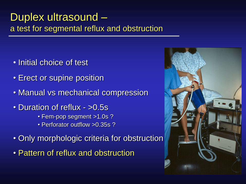
Duplex ultrasound –a test for segmental reflux and obstruction
• Initial choice of test
• Erect or supine position
• Manual vs mechanical compression
• Duration of reflux - >0.5s• Fem-pop segment >1.0s ?
• Perforator outflow >0.35s ?
• Only morphologic criteria for obstruction
• Pattern of reflux and obstruction
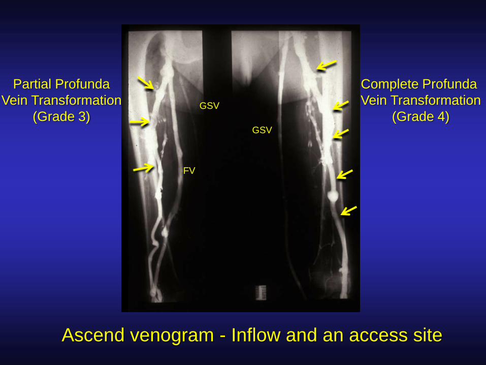
Complete Profunda
Vein Transformation
(Grade 4)
Partial Profunda
Vein Transformation
(Grade 3)
FV
GSV
GSV
Ascend venogram - Inflow and an access site
How do I find patients with femoro-ilio-
caval obstruction?
• At what degree a venous obstruction is hemodynamically significant is not defined!
• Not possible to hemodynamically quantify venous outflow obstruction!
• No reliable non-invasive study is available!
• Invasive pressure tests are insensitive!• hand/foot pressure differential
• reactive hyperemia pressure increase
• femoral vein pressure
• femoro-caval vein gradient
Morphologic Diagnosis
• Dx is morphologic, not hemodynamic
• Ultrasound scanning of the lower extremity has to
be complemented by transfemoral venography,
MRV, CT-V or IVUS in C3-6 cases
• IVUS is the standard for all other imaging
Transfemoral venogram
Multi-plane venograms
MRV
CTV
IVUS-verified area stenosis of >50% is considered
significant
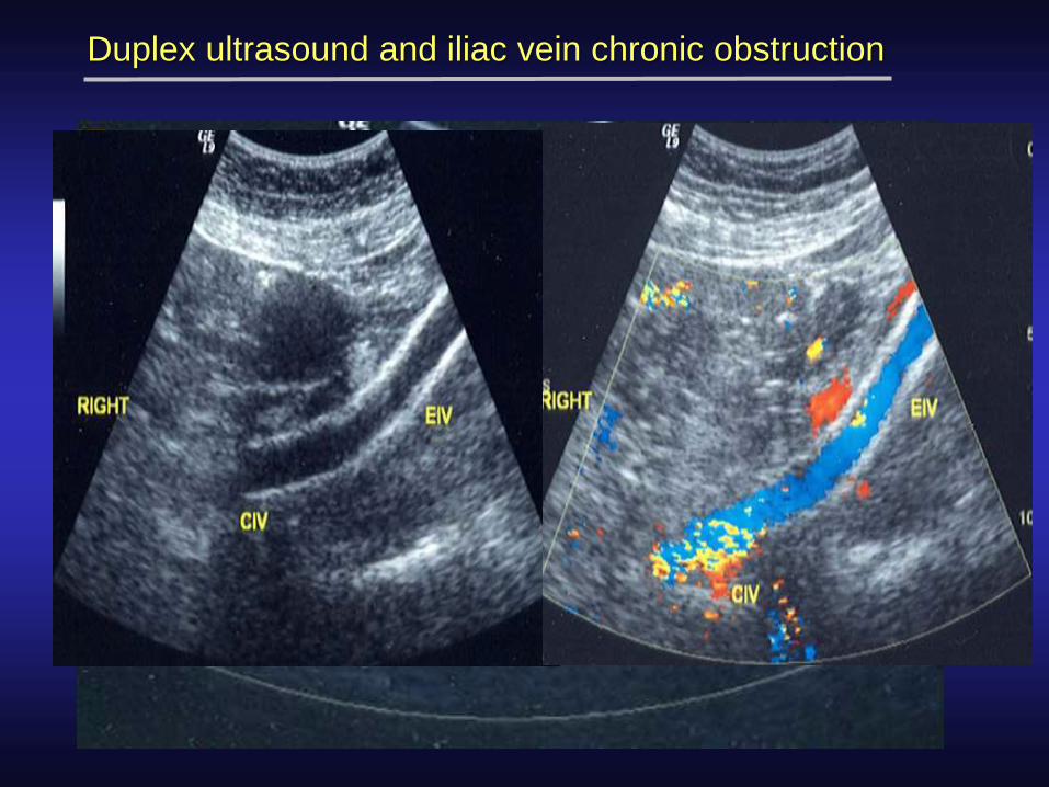
Duplex ultrasound and iliac vein chronic obstruction
>50% stenosis = post/pre stenotic peak velocity ratio >2.5[Labropoulos et al, J Vasc Surg 2007;46:101-7]

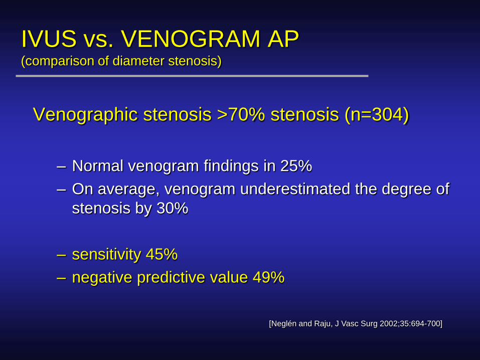
IVUS vs. VENOGRAM AP(comparison of diameter stenosis)
Venographic stenosis >70% stenosis (n=304)
– Normal venogram findings in 25%
– On average, venogram underestimated the degree of
stenosis by 30%
– sensitivity 45%
– negative predictive value 49%
[Neglén and Raju, J Vasc Surg 2002;35:694-700]
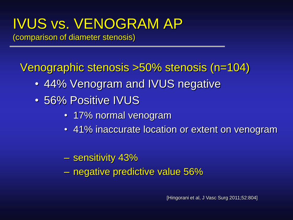
IVUS vs. VENOGRAM AP(comparison of diameter stenosis)
Venographic stenosis >50% stenosis (n=104)
• 44% Venogram and IVUS negative
• 56% Positive IVUS
• 17% normal venogram
• 41% inaccurate location or extent on venogram
– sensitivity 43%
– negative predictive value 56%
[Hingorani et al, J Vasc Surg 2011;52:804]

Transfemoral Venogram
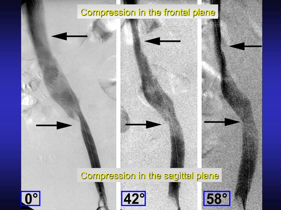
Compression in the frontal plane
Compression in the sagittal plane
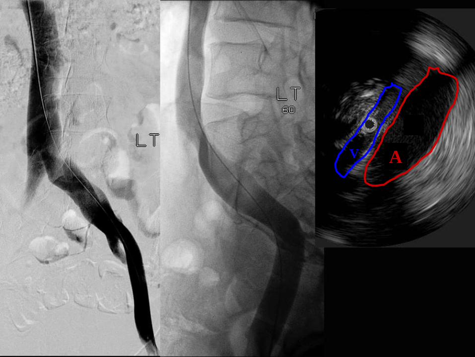
AV
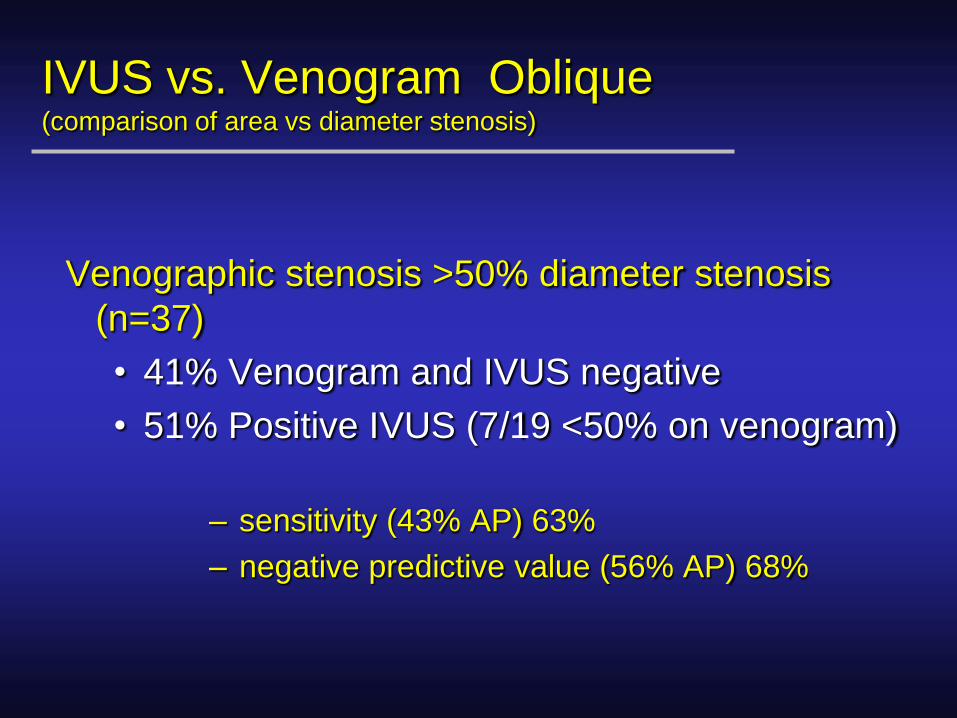
IVUS vs. Venogram Oblique(comparison of area vs diameter stenosis)
Venographic stenosis >50% diameter stenosis
(n=37)
• 41% Venogram and IVUS negative
• 51% Positive IVUS (7/19 <50% on venogram)
– sensitivity (43% AP) 63%
– negative predictive value (56% AP) 68%
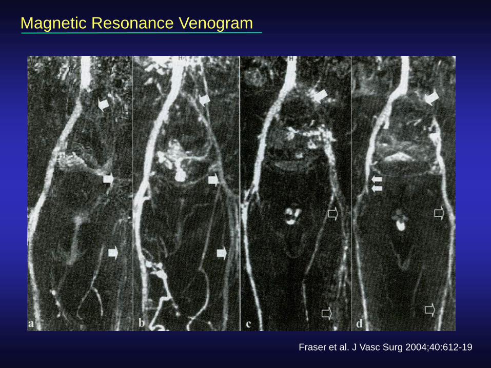
Magnetic Resonance Venogram
Fraser et al. J Vasc Surg 2004;40:612-19

Occluded iliac vein
Profunda vein
Profunda Transformation
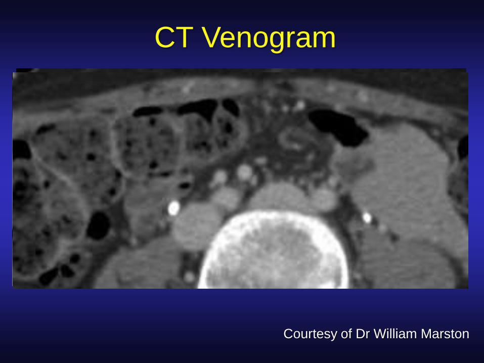
CT Venogram
Courtesy of Dr William Marston

CT – Venogram 3D reconstruction
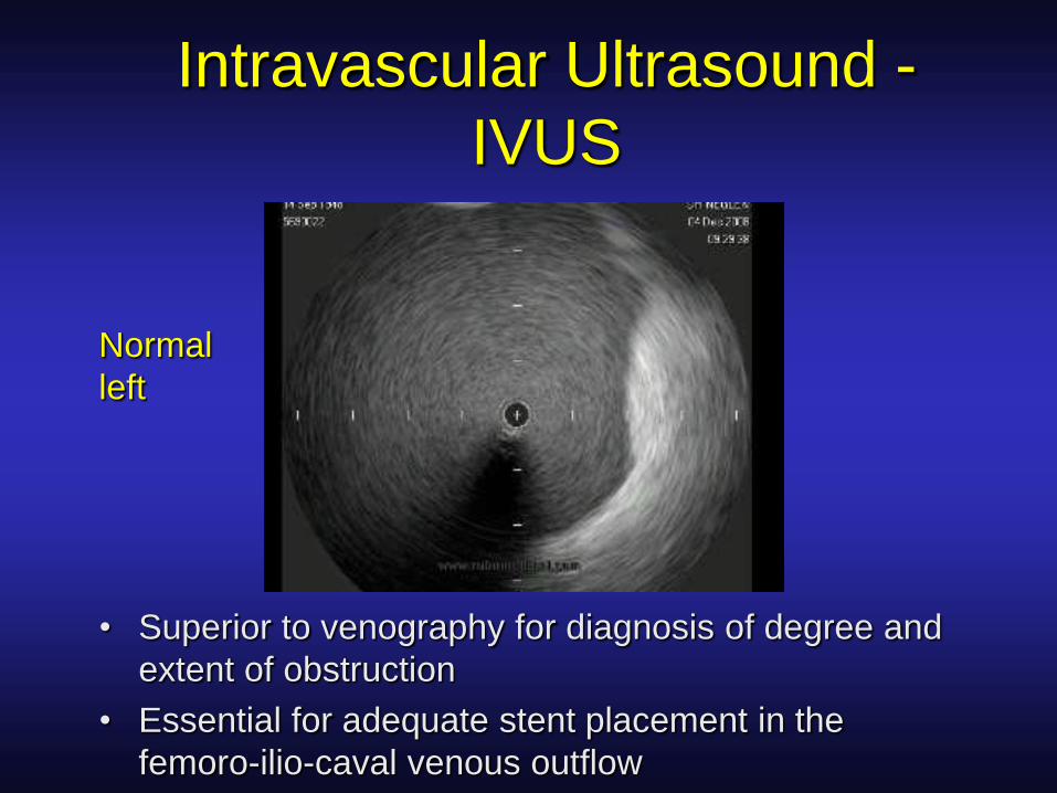
Intravascular Ultrasound -
IVUS
• Superior to venography for diagnosis of degree and
extent of obstruction
• Essential for adequate stent placement in the
femoro-ilio-caval venous outflow
Normal
left

Non-occlusive Non-thrombotic Obstruction(NIVL = nonthrombotic iliac vein lesion)
Pre-stenting
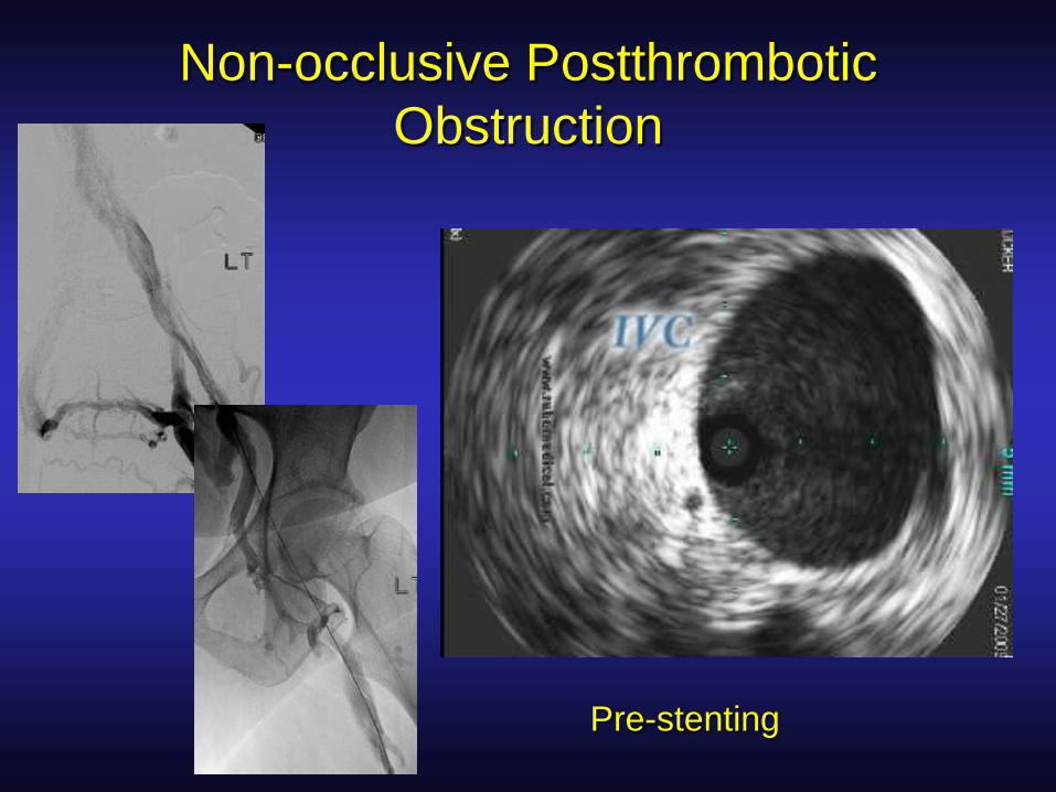
Non-occlusive Postthrombotic
Obstruction
Pre-stenting
Role of IVUS in Venous Interventions
• Standard for imaging venous obstruction
• Premier diagnostic tool
• Decreases use of contrast
Mainly used in
– Femoro–ilio–caval stenting
– Placement of IVC filters
– Adjuvant to surgical TE/thrombolysis of
iliofemoral DVT
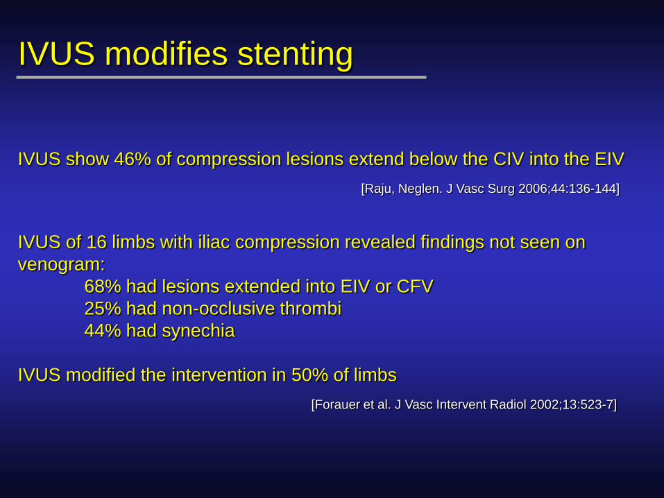
IVUS modifies stenting
IVUS show 46% of compression lesions extend below the CIV into the EIV
[Raju, Neglen. J Vasc Surg 2006;44:136-144]
IVUS of 16 limbs with iliac compression revealed findings not seen on
venogram:
68% had lesions extended into EIV or CFV
25% had non-occlusive thrombi
44% had synechia
IVUS modified the intervention in 50% of limbs
[Forauer et al. J Vasc Intervent Radiol 2002;13:523-7]

Practical Implications for Management of
Chronic Venous Disease
• A comprehensive workup and classification is mandatory prior to treatment
• CVI (C3-6) – think ilio-femoral vein obstruction!
• Complement ultrasound scanning of the lower extremity with transfemoral venography, MRV, CT-V or IVUS depending on local accessibility


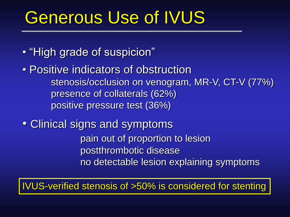
Generous Use of IVUS
• “High grade of suspicion”
• Positive indicators of obstructionstenosis/occlusion on venogram, MR-V, CT-V (77%)
presence of collaterals (62%)
positive pressure test (36%)
• Clinical signs and symptoms
pain out of proportion to lesion
postthrombotic disease
no detectable lesion explaining symptoms
IVUS-verified stenosis of >50% is considered for stenting
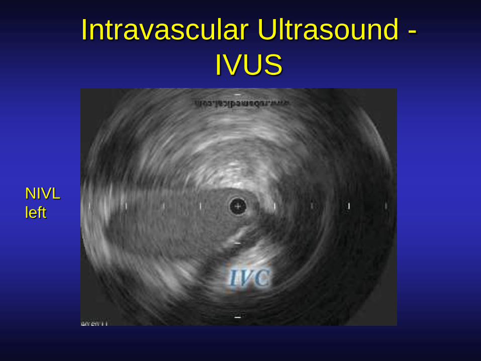
Intravascular Ultrasound -
IVUS
NIVL
left
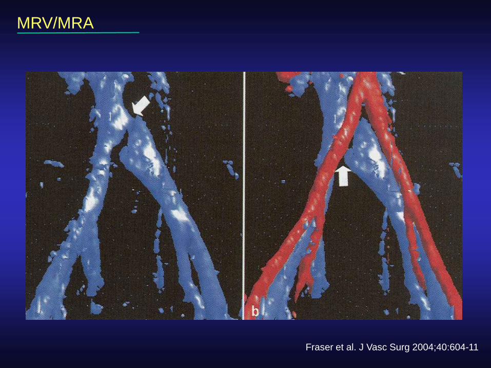
MRV/MRA
Fraser et al. J Vasc Surg 2004;40:604-11