CT of Brainstem Injury - AJNR · We reviewed our experience in CT diagnosis of brainstem injury and...
7
Fang Y. Tsai' James S. Teal' Michael F. Quinn' Hidea H. Itabashi2 James E. Huprich' · 3 Jamshid Ahmadi' Hervey D. Segall' Received April 10 , 1979; accepled after revi- sion September 7, 1979 . This manusc ri pt appears in January / February AJNR and April AJR. , Department of Neuroradiology, Los Angeles County-University of Southern California Medical Cenler, Box 766, 1200 N. State St., Los Angele s, CA 90033. Address reprint requests to F. Y. Tsai. 2 Department of Chief Medical Examiner-Cor- oner, County of Los Angeles, Los Angeles, CA 90033. 3 Present address: Holl ywood-Presbyterian Medical Center, Los Angeles, CA 90027 . AJNR 1 :23-29, January / February 1980 0195 -610 8/ 80 / 0011-0023 $00.00 © American Roentgen Ray Society 23 CT of Brainstem Injury • , '" , ." " ' .... 1 ,-, i- • -. • I. 1 ':. • _ I • I, II,., Cranial computed tomography (CT) scans of 1,600 head trauma patients, 67 of which demonstrated evidence of brainstem injury , were reviewed. CT diagnosis of brainstem injury was based on direct and indirect evidence . Direct signs , which in, elude focal hemorrhage , significant intraparenchymal contrast enhancement , hemorrhagic contu- sion , and edema of the brainstem , appear as areas of high density, mixed density, and low density on the CT scan. Indirect signs are obliteration of the pontine , cerebella- pontine angle , and peri mesencephalic cisterns . Mortality and morbidity rates after brainstem injury are 2-3 times greater than for head trauma with descending transten- torial herniation , but without brainstem injury. The clinical manifestations and pathologic findings of brainstem injury have been well described . Head trauma carries a much graver prognosis when brainstem involvement is present [1-10]. Sin ce severe head trauma is often characterized by injury to several sites, both intra- and extraaxial , there may be no clear-cut clinical evidence of a specific brainstem lesion. The most significant lesion may not be suspected until the patient fails to exhibit normal signs of re co very or it may be an unex pected autopsy finding [1-5, 11-16]. Because of its ability to demonstrate the nature, extent, sites, and multiplicity of brain injury, computed tomography (CTl is now the primary diagnostic method for head trauma. Although there have been numerous report s on the CT findings of most types of intracranial injury , the findings in brainstem injury have not been well described [1 7, 18]. We reviewed our experience in CT diagnosis of brainstem injury and conclude that the diagnosis and prognosis ca n often be app r eciated at the time of the initial evaluation. Materials and Methods During a 2 year period more than 1,600 patients with head trauma were exa mined in our hospital with an EMI CT-1 01 0 head scanne r. In most patient s a noncont rast sca n was foll owed immediately by a co ntr ast-enhanced scan with infusion of 300 ml of 30 % meglu- mine diatrizoate (Reno-M-DIP). Brainstem injury was diagnosed pr osp ec tively and retro- spec tively in 67 cases, and CT findings wer e corre lated with clini ca l manifestations and pathologic findings. A suppl ementary gro up of 100 patients having head tr auma with downward transtentorial herniat ion (based on clini ca l and CT finding s) but no CT evidence of brainstem injury were also studied. Of th e 67 br ain stem cases with seco ndary injury (resulting from downward transtentorial herniation or posterior fossa hematoma), 21 were au top sy proved. Of th e 67 who showed primary brainstem injury (a dir ec t injury without signifi cant additiona l trauma) , 11 were proved at autopsy.
Transcript of CT of Brainstem Injury - AJNR · We reviewed our experience in CT diagnosis of brainstem injury and...

Fang Y. Tsai' James S. Teal'
Michael F. Quinn' Hidea H. Itabashi2
James E. Huprich' · 3
Jamshid Ahmadi' Hervey D. Segall'
Received April 10, 1979; accepled after revision September 7, 1979.
This manuscri pt appears in January / February AJNR and April AJR.
, Department of Neuroradiology, Los Angeles County-University of Southern California Medical Cenler , Box 766, 1200 N. State St., Los Angeles, CA 90033. Address reprint requests to F. Y. Tsai.
2 Department of Chief Medical Examiner-Coroner, County o f Los Angeles, Los Angeles, CA 90033.
3 Present address: Hollywood-Presbyterian Medical Center, Los Angeles, CA 90027 . AJNR 1 :23-29, January / February 1980 0195-6108 / 80 / 0011-0023 $00.00 © American Roentgen Ray Society
23
CT of Brainstem Injury
• , '" , ." " ' .... 1 ,-, i- ~ ~ • -. •
I. 1 ':. ~ • _ ~ :~., I
• I, II,.,
Cranial computed tomography (CT) scans of 1,600 head trauma patients, 67 of which demonstrated evidence of brainstem injury, were reviewed. CT diagnosis of brainstem injury was based on direct and indirect evidence. Direct signs, which in,elude focal hemorrhage, significant intraparenchymal contrast enhancement, hemorrhagic contusion, and edema of the brainstem, appear as areas of high density, mixed density, and low density on the CT scan. Indirect signs are obliteration of the pontine, cerebellapontine angle, and peri mesencephalic cisterns. Mortality and morbidity rates after brainstem injury are 2-3 times greater than for head trauma with descending transtentorial herniation, but without brainstem injury.
The clinical manifestations and pathologic findings of brainstem injury have been well described . Head trauma carries a much graver prognosis when brainstem involvement is present [1-10]. Since severe head trauma is often characterized by injury to several sites, both intra- and extraaxial , there may be no clear-cut clinical evidence of a specific brainstem lesion. The most significant lesion may not be suspected until the patient fails to exhibit normal signs of recovery or it may be an unexpected autopsy finding [1-5, 11-16].
Because of its ability to demonstrate the nature, extent, sites, and multiplic ity of brain injury, computed tomography (CTl is now the primary diagnostic method for head trauma. Although there have been numerous reports on the CT finding s of most types of intracranial injury, the findings in brainstem injury have not been well described [1 7 , 18]. We reviewed our experience in CT diagnosis of brainstem injury and conclude that the diagnosis and prognosis can often be appreciated at the time of the initial evaluation.
Materials and Methods
During a 2 year period more th an 1,600 patients with head trauma were examined in our hospital with an EMI CT-1 01 0 head scanner. In most patients a noncontrast scan was foll owed immediately by a contrast-enhanced scan with infusion of 300 ml of 30% meg lumine diatrizoate (Reno-M-DIP). Brainstem injury was diagnosed prospectively and retrospectively in 67 cases, and CT findings were corre lated with c linica l manifestations and pathologic findings. A supplementary group of 100 patients having head trauma with downward transtentorial herniat ion (based on c linica l and CT finding s) but no CT evidence of brainstem injury were also studied. Of th e 67 brain stem cases with secondary injury (resulting from downward transtentorial herniation or posterior fossa hematoma), 21 were autopsy proved. Of the 67 who showed primary brain stem injury (a direct injury without significant additional trauma), 11 were proved at autopsy.

24 TSAI ET AL. AJNR:1 , January/February 1980
A B
Fig. 1.-Case 1. A, Hemorrhage at pons and ve rmi s. Right ce rebellopontine angle cistern visible. B , Hemorrhage at upper pons; perimesencephalic c isterns visible (primary brainstem injury with intact surrounding cistern s).
Fig . 2. - Case 2. A , Hemorrh ag ic density, central posterior fossa , possibly at vermi s. Fourth ventricle not seen. B , Enhanced scan. Enhanc ing contusion at brainstem (a rrow). C, Superior slice of enhanced scan. Obliteration of c istern al spaces.
Representative Case Reports
Case 1
A 38-year-old male intoxicated driver involved in an automobile accident was brought to the emergency room. He was unresponsive and exh ibited decerebrate posturing. The corneal and occulocephalic reflexes were absent . Retinal hemorrhage and papilledema were noted on the right . After a respiratory arrest, he was maintained on a respirator. CT demonstrated several traumatic hemorrhages of the cerebrum, cerebellum , and brain stem (fig. 1). He died shortl y after admission.
Case 2
A 44-year-o ld man with head trauma was comatose when treated by paramed ics. On arrival at the hospital , his pupils were asymmetri ca l (5 mm on the left and 6 mm on the right) and sluggishly reactive to light. Corneal and occulocephalic reflexes were absent . CT demonstrated small bilateral subdural hematomas (fig . 2) . Th ere was a small hemorrhag ic density in the center of the posterior fossa, and the pontine and perimesencephalic c isterns were not visible. Since CT finding s were incompatible with the clinical manifestations, a contrast-enhanced study was recommended . After infusion of contrast material, enhancing contusions in the pons, midbrain ,
AJNR: 1 , January / February 1980 CT OF BRAINSTEM INJURY 25
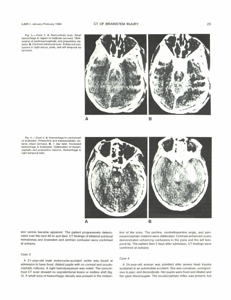
Fig . 3. -Case 3. A, Noncontrast scan. Smal! hemorrhage in region of midbrain (arrows). Obliteration of peri mesencephalic and prepontine cistern s. B, Contrast-enhanced scan. Enhanced contusions in right uncus, pons, and left temporal tip (arrows) .
Fig . 4.-Case 4. A, Hemorrhage in central part of brainstem . Prepontine and mesencephalic c isterns intact (arrows). B, 1 day later . Inc reased hemorrhage in brainstem. Obliteration of mesencephalic and prepontine cisterns. Hemorrhage in right temporal lobe.
A
A
and vermis became apparent. The patient progressively deteriorated over the next 48 hr and died . CT findings of bilateral subdural hematomas and brainstem and vermian contusion were confirmed at autopsy.
Case 3
A 21-year-old male motorcycle-accident victim was found at admission to have fixed, dilated pupils with no corneal and occulocephalic reflexes. A right hemotympanum was noted. The noncontrast CT scan showed no supratentorial lesion or midline shift (fig. 3). A small area of hemorrhagic density was present in the midpor-
B
B
lion of the pons. The pontine, cerebellopontine angle, and perimesencephalic c isterns were obli terated. Contrast-enhanced scans demonstrated enhancing con tusions in the pons and the left temporal tip. The patient died 2 days after admission. CT findings were con firmed at autopsy .
Case 4
A 24-year-old woman was admitted after severe head trauma sustained in an automobile accident. She was comatose , unresponsive to pain, and decerebrate . Her pupils were fi xed and dilated and her gaze disconjugate. The occulocephalic refl ex was present, but
26 TSAI ET AL. AJNR: 1 . January/ February 1980
the corneal refl ex was absent. Her Glasgow coma score was 5. A CT scan was negative with the exception of a small hemorrhagic density in the mid port ion of the upper pons (fig. 4) . The pontine . cerebellopontine angle. and perimesencephalic c isterns were intact. The patient' s clinical status remained unchanged for 24 hr. then deteriorated . Repeat CT disc losed an apparent increase in the size of the brainstem hemorrhage. as well as a hemorrhage within the right basal ganglia and obliteration of the same cisterns. The patient died 2 weeks later. CT findings were confirmed at autopsy.
Case 5
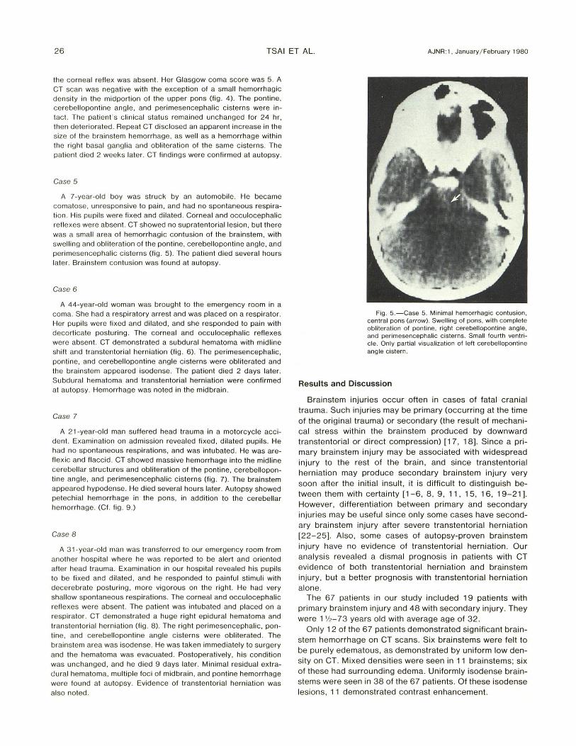
A 7-year-old boy was struck by an automobile. He became comatose. unresponsive to pain . and had no spontaneous respiration. His pupils were fi xed and d ilated . Corneal and occulocephalic refl exes were absent. CT showed no supratentorial lesion. but there was a small area of hemorrhag ic contusion of the brainstem. with swelling and obliteration of the pontine. cerebellopontine angle. and perimesencephalic c isterns (fig . 5). The patient died several hours later. Bra instem con tusion was found at autopsy.
Case 6
A 44-year-old woman was brought to the emergency room in a coma. She had a respiratory arrest and was placed on a respirator. Her pupils were fi xed and dil ated. and she responded to pain with decorti cate posturing. Th e corneal and occulocephalic refl exes were absent. CT demonstrated a subdural hematoma with midline shift and transtentorial herniation (fig . 6). The perimesencephalic . pontine. and cerebellopontine angle c istern s were obliterated and the brainstem appeared isodense . The patient died 2 days later. Subdural hematoma and transtentorial herniation were confirmed at autopsy . Hemorrhage was noted in the midbrain .
Case 7
A 21-year-old man suffered head trauma in a motorcycle accident. Examination on admission revealed fi xed. dilated pupils. He had no spontaneous respirations, and was intubated . He was aref1exic and flaccid . CT showed massive hemorrhage into the midline cerebellar struc tures and obliteration of the pontine , cerebellopontine angle , and perimesencephalic c isterns (fig . 7). The brainstem appeared hypodense. He died several hours later. Autopsy showed petechial hemorrhage in the pons, in addition to the cerebellar hemorrhage. (CI. fig . 9 .)
Case 8
A 3 1-year-old man was transferred to our emergency room from another hospital where he was reported to be alert and oriented after head trauma. Examination in our hospital revealed his pupils to be fi xed and dilated , and he responded to painful stimuli with decerebrate posturing , more vigorous on the right . He had very shallow spontaneous respirations. The corn eal and occulocephalic refl exes were absent. The patient was intubated and placed on a respirator . CT demonstrated a huge right epidural hematoma and transtentorial herniation (fig . 8 ). The right perimesencephalic , ponti ne, and cerebellopontine angle c isterns were obliterated. The brainstem area was isodense . He was taken immediately to surgery and the hematoma was evacuated. Postoperatively , his condition was unchanged, and he died 9 days later. Minimal residual extradural hematoma, multiple foc i of midbrain , and pontine hemorrhage were found at autopsy . Evidence of transtentorial herniation was also noted .
Fig. S.-Case 5. Minimal hemorrh agic contusion, cen tral pons (arrow). Swelling of pons. wi th complete ob literation of pontine, right cerebellopontine angle. and peri mesencephalic cisterns. Small fourth ventricle. Only partial visualization of left cerebellopontine angle cistern .
Results and Discussion
Brainstem injuries occur often in cases of fatal cranial trauma. Such injuries may be primary (occurring at the time of the original trauma) or secondary (the result of mechanical stress within the brainstem produced by downward transtentorial or direct compression) [17, 18]. Since a primary brainstem injury may be associated with widespread injury to the rest of the brain, and since transtentorial herniation may produce secondary brainstem injury very soon after the initial insult, it is difficult to distinguish between them with certainty [1-6, 8, 9 , 11 , 15, 16, 19-21]. However, differentiation between primary and secondary injuries may be useful since only some cases have secondary brainstem injury after severe transtentorial herniation [22-25]. Also , some cases of autopsy-proven brainstem injury have no evidence of transtentorial herniation . Our analysis revealed a dismal prognosis in patients with CT evidence of both transtentorial herniation and brainstem injury, but a better prognosis with transtentorial herniation alone.
The 67 patients in our study included 19 patients with primary brainstem injury and 48 with secondary injury . They were 1 '/2-73 years old with average age of 32 .
Only 1 2 of the 67 patients demonstrated significant brainstem hemorrhage on CT scans. Six brainstems were felt to be purely edematous, as demonstrated by uniform low density on CT. Mixed densities were seen in 11 brainstems; si x of these had surrounding edema. Uniformly isodense brainstems were seen in 38 of the 67 patients. Of these isodense lesions, 11 demonstrated contrast enhancement.

AJNR: 1, January / February 1980 CT OF BRAINSTEM INJURY 27
Fig. 6.-Case 6. A , Partial oblileration of prepontine c istern and fourth ventric le. B , Isodense brainstem, bul oblileralion of preponline and peri mesencephalic cistern s. Neither lemporal horn dilated . C , Subdural hematoma along left anlerior and middle c ranial fossa, with displace men I of midline slruc lures toward left side.
Fig . 7. -Case 7. Nonconlrast scan. Multiple hemorrhage al vermis and right hippocampal region and subarachnoid hemorrhage. Deformation and foreshorlening of edemalous pons (low density) . Temporal horns d ilaled.
Correlation of CT and autopsy findings ind icates that injury to the brainstem may include hemorrhage, contusion, and / or edema. Hemorrhage may be unifocal or multifocal, and the extent of hemorrhage ranges from petechial to massive bleeds involving much of the brainstem cross section , These brainstem lesions may occur alone or in association with other cran ial injuries. CT manifestations of brainstem injury are similar to those of cerebral injury. They may be hyperdense, isodense , or hypodense depending on the degree of hemorrhage and edema [18].
The CT diagnosis of primary brainstem injury requires demonstration of di rect evidence, such as hemorrhage, edema, or contusion, or indirect evidence, such as complete obliteration of the pontine, cerebellopontine angle, and peri mesencephalic cisterns, We believe the indirect findings constitute reliable signs of brainstem injury in head trauma patients, and can be seen in either primary or secondary brainstem injuries. Similar findings have been noted in brainstem neoplasms, but these are easily differentiated by clinical history. CT signs for downward transtentorial herniation have been previously described as contralateral temporalhorn di latation , oblite ration of the suprasel lar cistern, and widening of the ipsilateral cerebeliopontine angle c istern [22-24, 26].
Recognition of the type of brainstem injury may have some value in the management of severe head trauma, Among 48 patients with secondary brainstem injury , the mortality rate was 67% (32 / 48), and the rate of minimal recovery with resultant vegetative state was 29% (14 / 48) . The other 4% (2 / 48) survived with moderate but sig nificant deficits, Of our supplementary group of 100 patients with transtentorial herniation alone, the mortality rate was only 27 % , and the rate of minimal recovery was 12%. The other 61 % had mild to moderate defic its. In addition, of the 27 deaths, four were the result of intercurrent infection after a period of good recovery, not the direct result of the head injury , Thus, the survival rate in transtentorial herniation alone was at least 73% .
The mortality rate for secondary brainstem injury was 2-3 times greater than for patients with transtentorial herniation alone. Of the 19 patients with primary brainstem injuries, five survived , but in a permanent vegetative state. The mortality rate was 73.7% (14 / 19), compared with 67% in the secondary brainstem injury group , The rate of minimal

28 TSAI ET AL. AJNR : 1 , January / February 19aO
A
Fig . 9 .-Hematoma at ve rmis and deformed brainstem, but prepontine, peri mesencephalic, and ce rebello pontine angle c isterns intact (arrows); artifact at pons (a rrowhead) . Patient survived with moderate defic it.
B
recovery is very similar to that of secondary injury . During the survival period , follow-up scans often showed low attenuation in the region of the brainstem, with widening of the surrounding cisterns . This was most likely due to evolution of the hemorrhage and to posttraumatic atrophy as the result of contusion [20, 21, 26].
Fig . a. -Case a . A , Partial visualization of left ce rebellopontine angle c istern , but obliteration of prepontine and right-sided cerebellopontine angle and perimesencephalic cisterns. Contusion , right temporal lobe. B , Epidural hematoma (arrow) . Obliterated peri mesencephalic c isterns.
Among those patients with complete obliteration of the pontine, cerebellopontine angle , and peri mesencephalic cisterns or with enhanced contusion and partial obliteration of the cisterns, about one-third had autopsy proof of midbrain and / or pontine hemorrhage . In those patients without complete obliteration of the cisterns or enhanced contusion who received autopsies , no brainstem hemorrhage was identified . Therefore , we feel that complete obliteration of the c isterns or enhanced contusion is a valuable CT sign of brainstem injury in head trauma patients. The level of brainstem injury may be correlated with the parts of the cisterns that are obliterated. The peri mesencephalic cisterns are obliterated with injury at the level of the midbrain , and pontine lesions may result in obliteration of the pontine and cerebellopontine angle cisterns.
Although CT scanning has an excellent capability for evaluation of brainstem injury, artifacts often obstruct our efforts. In addition , obliteration of the cisterns may be due to small amounts of subarachnoid hemorrhage in the cisterns, which appear isodense against adjacent brain. In this situation , careful evaluation of the size of the fourth ventricle may be helpful. A small fourth ventricle may also indicate brainstem swelling (case 5) . If there is still a question , a contrast-enhanced scan may provide further evidence of brainstem injury (cases 2 and 3).
ACKNOWLEDGMENTS
We thank Dean W. Wiseley, Los Angeles County Medical Examiner' S Office, for providing autopsy specimens and Ellen Duncan for sec retarial assistance.

AJNR :1, January / Febru ary 1980 CT OF BRAINSTEM INJURY 29
REFERENCES
1. Klintworth GH . Paratentorial grooving of human brains with particular reference to transtentorial herniation and the pathogenesis of secondary brain stem hemorrhage. Am J Pathol 1968;53: 391 -408
2. Lindenberg R, Freytag E. Brainstem lesions characteristic of traumatic hyperextension of the head. Arch Pathol 1970;90 : 509-515
3. Lind enberg R. Pathology of c raniocerebral injuries. In : Newton TH , Potts DG , eds. Radiology of the skull and brain , vol 3, Ana tomy and pathology. SI. Loui s: Mosby , 1977;3079-3081
4 . Merianos P. Treatment of frac tures of the long bone in brainstem injury . Br Med J 1975; 2: 316
5. Mitchell DE , Adams JH . Primary focal impact damage to th e brainstem in blunt head injuries: does it exist? Lancet 1973; 1 :215-21 8
6. Stewart WA , Litten SP, Sheehe PR. A prognostic model for brain stem injury . Surg Neuro/1973 ;1 :303 - 310
7. Tandon PN. Brainstem hemorrhage in c raniocerebral trauma. Acta Neuro l Scand 1964 ;40 :375-385
8. Tomlinson BF. Brainstem lesions after head injury. J Clin Pathol [Supplj 1970;23: 154-165
9. Turazzi S, Alexandre A, Brico lo A. Inc idence and significance of c linica l signs of brainstem traumatic lesions: a study of 2600 head injury patients. J Neurosurg Sci 1975;19 :215-222
10. Turazzi S, Brico lo A. Acute pontine syndrome following head injury. Lancet 1977;2: 62-64
11 . Adams H, Mitchell DE, Graham 01 , Doyle D. Diffused brain damage of immed iate impact type: its re lationship to primary brainstem damage in head injury. Brain 1977;100 : 489-502
1 2. Brisman R, Harrington B. Military missile injury to pon s and survival. Surg NeuroI1973 ;1 : 171-172
13. Britt RH , Herri ck MK, Hamilton RD. Traumatic locked-in syndrome. Ann Neurol 1977; 1 : 590-592
14 . Caplan LR, Zervas NT. Surviva l with permanent midbrain dysfunction after su rgical treatmen t of traumatic subdural hematoma: the c linica l picture of a Duret hemorrhage. Ann Neurol 1977; 1 : 587 -589
15. Crompton MR. Brainstem lesions due to c losed head injury . Lancet 1971 ;1 :669-67 3
16. Friede RL, Roessmann U. The pathogenesis of secondary midbrain hemorrhage . Neurology 1966;16 : 1 210- 1216
17 . Tsai FY, Huprich JE, Itabashi H, Segall HD, Teal JS. Computed tomographic diagnosis of posterior fossa traumatic lesions. Presented at the annual meeting of the Western Neuroradiolog ica l Society, Newport Beach , Cal., October 1978
18. Tsai FY, Quinn MF, Itabashi H, Ahmadi J , Teal JS, Segall HD. The role of computed tomography in the evaluation of head trauma. Excerpta Medica CT 1979;6: 1 - 1 3
19. Gilbert JJ , Deonna T . Unusual brainstem findings following c losed head injury. J Pedia tr 1972;8 1 : 343-345
20. Gruszkiewicz J, Doron Y, Payser E. Recovery from severe craniocerebral injury with brainstem lesions in childhood . Surg Neurol 1973; 1 : 1 97 -201
21. Jellinger K, Seitelberger F. Prot racted post-traumatic encephalopathy : pathology, pathogenesis, and c linica l implications. J Neurol Sci 1970; 10: 51 -94
22. Azambuja N, Lindgren E, Sjogren SE. Tentorial herniation. Acta Radiol (Stockh) 1956;46 : 224-231
23. Naidich TP , Leeds NE, Kricheff II , Pudlowski RM , Naidich JB , Zimmerman RD . The tentorium in axial sect ion, I and II. Radiology 1977; 123 : 631 -648
24 . Osborn AG . Diagnosis of descending tentorial herniation by computed tomography. Radiology 1977 ; 123: 93- 96
25. Sunderland S. The tentorial notch and complications produced by herniation of the brain through that aperture. Br J Surg 1958;45: 422-438
26. Stovring J. Descending tentorial herniation: findings on computed tomography. Neuroradiology 1977; 14 : 101 -105