CT Findings of Granulomatous Pneumocystis jiroveci
6
1 Copyrights © 2021 The Korean Society of Radiology Case Report J Korean Soc Radiol https://doi.org/10.3348/jksr.2021.0043 eISSN 2288-2928 CT Findings of Granulomatous Pneumocystis jiroveci Pneumonia in a Patient with Multiple Myeloma 다발성 골수종 환자에서 발생한 육아종성 폐포자충 폐렴의 컴퓨터단층촬영 소견 So Ra Shin, MD 1 , Tae Sung Kim, MD 1 * , Joungho Han, MD 2 Departments of 1 Radiology and Center for Imaging Science and 2 Pathology, Samsung Medical Center, Sungkyunkwan University School of Medicine, Seoul, Korea Although the typical CT findings of Pneumocystis jiroveci pneumonia (PJP) include diffuse or multifocal areas of ground-glass opacities in both lungs, it can also rarely manifest as multiple pulmonary nodules. We report a rare case of atypical PJP in an immunocompromised patient with multiple myeloma, presenting as widespread ground-glass opacities and multiple necrot- ic subpleural nodules in both lungs on CT, which proved to be granulomatous PJP on percuta- neous transthoracic needle biopsy. Index terms Pneumonia; Pneumocystis; Multiple Myeloma INTRODUCTION Pneumocystis jiroveci pneumonia (PJP), formerly known as Pneumocystis carinii pneumonia, is an important opportunistic infection in the immunocompromised hosts. It has been a well-known cause of pulmonary infection in human immunodefi- ciency virus (HIV)/acquired immune deficiency syndrome (AIDS) patients (1). Non-HIV immunocompromised hosts at risk for PJP include some patients with immunodefi- ciency such as hematological malignancies and solid tumors, solid organ and bone marrow transplant recipients, and patients with collagen-vascular diseases (1). The characteristic CT finding of AIDS-related PJP is known to be extensive ground- glass opacity in both lungs (2), which can show a central distribution with relative pe- ripheral sparing, a mosaic pattern or a diffuse distribution with some predilection for the upper lobes (2, 3). In patients without HIV infection, the extent of ground-glass opac- ity is often greater (4). However, there have also been reports of atypical PJP which Received March 10, 2021 Revised April 16, 2021 Accepted April 17, 2021 *Corresponding author Tae Sung Kim, MD Department of Radiology, Samsung Medical Center, Sungkyunkwan University School of Medicine, 81 Irwon-ro, Gangnam-gu, Seoul 06351, Korea. Tel 82-2-3410-2518 Fax 82-2-3410-2559 E-mail [email protected] This is an Open Access article distributed under the terms of the Creative Commons Attribu- tion Non-Commercial License (https://creativecommons.org/ licenses/by-nc/4.0) which permits unrestricted non-commercial use, distribution, and reproduc- tion in any medium, provided the original work is properly cited. ORCID iDs So Ra Shin https:// orcid.org/0000-0002-5752-8178 Tae Sung Kim https:// orcid.org/0000-0001-7512-0283 Joungho Han https:// orcid.org/0000-0003-4424-7008
Transcript of CT Findings of Granulomatous Pneumocystis jiroveci

1Copyrights © 2021 The Korean Society of Radiology
Case ReportJ Korean Soc Radiolhttps://doi.org/10.3348/jksr.2021.0043eISSN 2288-2928
CT Findings of Granulomatous Pneumocystis jiroveci Pneumonia in a Patient with Multiple Myeloma다발성 골수종 환자에서 발생한 육아종성 폐포자충 폐렴의 컴퓨터단층촬영 소견
So Ra Shin, MD1 , Tae Sung Kim, MD1* , Joungho Han, MD2 Departments of 1Radiology and Center for Imaging Science and 2Pathology, Samsung Medical Center, Sungkyunkwan University School of Medicine, Seoul, Korea
Although the typical CT findings of Pneumocystis jiroveci pneumonia (PJP) include diffuse or multifocal areas of ground-glass opacities in both lungs, it can also rarely manifest as multiple pulmonary nodules. We report a rare case of atypical PJP in an immunocompromised patient with multiple myeloma, presenting as widespread ground-glass opacities and multiple necrot-ic subpleural nodules in both lungs on CT, which proved to be granulomatous PJP on percuta-neous transthoracic needle biopsy.
Index terms Pneumonia; Pneumocystis; Multiple Myeloma
INTRODUCTION
Pneumocystis jiroveci pneumonia (PJP), formerly known as Pneumocystis carinii pneumonia, is an important opportunistic infection in the immunocompromised hosts. It has been a well-known cause of pulmonary infection in human immunodefi-ciency virus (HIV)/acquired immune deficiency syndrome (AIDS) patients (1). Non-HIV immunocompromised hosts at risk for PJP include some patients with immunodefi-ciency such as hematological malignancies and solid tumors, solid organ and bone marrow transplant recipients, and patients with collagen-vascular diseases (1).
The characteristic CT finding of AIDS-related PJP is known to be extensive ground-glass opacity in both lungs (2), which can show a central distribution with relative pe-ripheral sparing, a mosaic pattern or a diffuse distribution with some predilection for the upper lobes (2, 3). In patients without HIV infection, the extent of ground-glass opac-ity is often greater (4). However, there have also been reports of atypical PJP which
Received March 10, 2021Revised April 16, 2021Accepted April 17, 2021
*Corresponding author Tae Sung Kim, MDDepartment of Radiology, Samsung Medical Center, Sungkyunkwan University School of Medicine, 81 Irwon-ro, Gangnam-gu, Seoul 06351, Korea.
Tel 82-2-3410-2518Fax 82-2-3410-2559E-mail [email protected]
This is an Open Access article distributed under the terms of the Creative Commons Attribu-tion Non-Commercial License (https://creativecommons.org/licenses/by-nc/4.0) which permits unrestricted non-commercial use, distribution, and reproduc-tion in any medium, provided the original work is properly cited.
ORCID iDsSo Ra Shin https:// orcid.org/0000-0002-5752-8178Tae Sung Kim https:// orcid.org/0000-0001-7512-0283Joungho Han https:// orcid.org/0000-0003-4424-7008
jksronline.org2
Granulomatous Pneumocystis jiroveci Pneumonia
manifested as multiple pulmonary nodules, which is termed as granulomatous PJP (5). To date, little has been reported on granulomatous PJP associated with multiple myeloma (4).
Herein, we report a case of granulomatous PJP in an immunocompromised patient with multiple myeloma, manifesting as multiple subpleural necrotic nodules in addition to wide-spread ground-glass opacities in both lungs at CT.
CASE REPORT
This study was approved by the Institutional Review Board of our institution and the re-quirement for informed consent was waived (IRB No. SMC 2021-03-149).
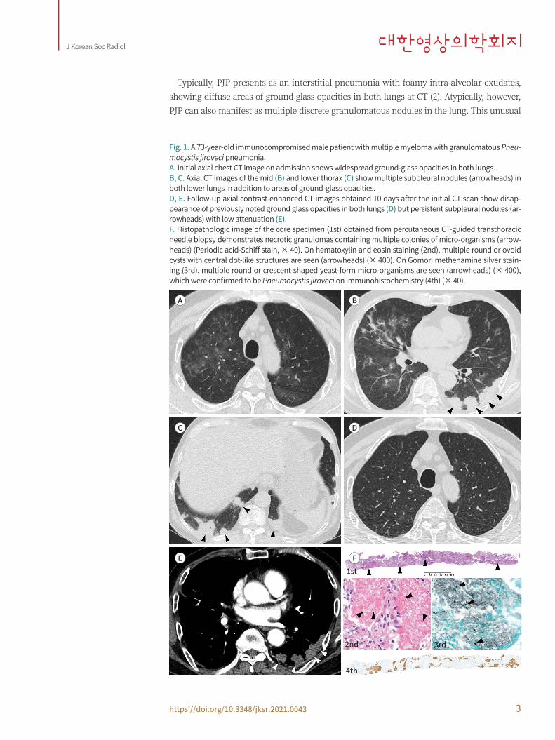
A 73-year-old male came to our hospital due to worsening dyspnea and pleuritic chest pain for 1 month. He had received chemotherapy (vincristine, melphalan, lenalidomide, pomalid-omide) and steroid therapy over 10 months for multiple myeloma until 2 weeks before. An initial chest X-ray reported ill-defined opacities in both lungs. A non-contrast high-resolution CT revealed a mosaic pattern of multifocal ground-glass opacities in both upper and mid lungs (Fig. 1A). In addition to it, there were also noted multiple subpleural nodules (size 1–3 cm in diameter) in both lower lungs (Fig. 1B, C). These nodular lesions had an irregularly mar-ginated but well-defined border. At this time, the initial clinical suspicion was an atypical pneumonia. Laboratory testing revealed leukocytopenia. The patient received a 10 day-course of antibiotics (imipenem/cilastatin and levofloxacin), which did not result in any significant clinical improvement. Follow-up contrast-enhanced chest CT taken in 10 days after the initial CT scan showed disappearance of multifocal ground-glass opacities (Fig. 1D) but persistent existence of multiple subpleural nodules without any change in size and extent. These nod-ules showed homogenous low attenuation showing an average of 40 Hounsfield unit (HU) on post-contrast CT with 20 HU net contrast enhancement (Fig. 1E). There was no significantly enlarged mediastinal lymphadenopathy. Percutaneous CT-guided transthoracic needle biop-sy was done and the core biopsy specimen showed well-formed necrotic granulomas con-taining Pneumocystis jiroveci micro-organisms (Fig. 1F). No other organisms were found. The patient was initiated on trimethoprim/sulfamethoxazole (TMP-SMX) therapy for 3 weeks. After 10 days since the patient started on TMP-SMX therapy, follow-up chest radiographs showed gradual decrease in extent of nodules in both lungs with improvement of symptoms. The last follow-up chest radiograph obtained 16 days later showed near-complete disappear-ance of nodules, and he was uneventfully discharged in stable condition.
DISCUSSION
Pneumocystis jiroveci, a type of fungus specific to humans and an opportunistic pathogen of ubiquitous distribution and low pathogenicity, can result in pneumonia in immunocom-promised individuals. Although Pneumocystis jiroveci has been an eminent entity of pulmo-nary infection in HIV/AIDS patients, it can also present in other immunocompromised hosts under cytotoxic and immunosuppressive therapies for hematologic malignancies, solid tu-mors, and transplant recipients (1). Incidence of PJP infection in these non-HIV immuno-compromised hosts was shown to be on the rise.
https://doi.org/10.3348/jksr.2021.0043 3
J Korean Soc Radiol
A
C
E
B
D
F1st
2nd
4th
3rd
Typically, PJP presents as an interstitial pneumonia with foamy intra-alveolar exudates, showing diffuse areas of ground-glass opacities in both lungs at CT (2). Atypically, however, PJP can also manifest as multiple discrete granulomatous nodules in the lung. This unusual
Fig. 1. A 73-year-old immunocompromised male patient with multiple myeloma with granulomatous Pneu-mocystis jiroveci pneumonia. A. Initial axial chest CT image on admission shows widespread ground-glass opacities in both lungs.B, C. Axial CT images of the mid (B) and lower thorax (C) show multiple subpleural nodules (arrowheads) in both lower lungs in addition to areas of ground-glass opacities.D, E. Follow-up axial contrast-enhanced CT images obtained 10 days after the initial CT scan show disap-pearance of previously noted ground glass opacities in both lungs (D) but persistent subpleural nodules (ar-rowheads) with low attenuation (E).F. Histopathologic image of the core specimen (1st) obtained from percutaneous CT-guided transthoracic needle biopsy demonstrates necrotic granulomas containing multiple colonies of micro-organisms (arrow-heads) (Periodic acid-Schiff stain, × 40). On hematoxylin and eosin staining (2nd), multiple round or ovoid cysts with central dot-like structures are seen (arrowheads) (× 400). On Gomori methenamine silver stain-ing (3rd), multiple round or crescent-shaped yeast-form micro-organisms are seen (arrowheads) (× 400), which were confirmed to be Pneumocystis jiroveci on immunohistochemistry (4th) (× 40).
jksronline.org4
Granulomatous Pneumocystis jiroveci Pneumonia
entity of granulomatous PJP has been previously reported to occur in up to 5% of PJP pa-tients, mostly HIV positive individuals (5).
The host’s defense against Pneumocystis jiroveci is known to be seriously dependent on CD4+ helper T cells, as seen in that the incidence of PJP in HIV/AIDS patients is high when the level of circulating CD4 cells falls below 200/μL (6). Granulomatous PJP is an emerging concept, and the mechanism of granuloma formation for PJP remains unclear yet (7). How-ever, it has been reported that a defect in B lymphocytes can also induce high susceptibility to Pneumocystis jiroveci and subsequent development of granulomatous PJP. B cells may be essential for the clearance of Pneumocystis jiroveci, and therefore, a deficiency of B cells fol-lowing immunosuppressive therapy potentially results in attenuated immunoprotection against Pneumocystis jiroveci and subsequently induces a granulomatous reaction by mac-rophages that substitute for the clearance of pathogens (6).
Clinical symptoms of patients with granulomatous PJP are reported to be dyspnea, cough, and fever, which are similar to those of typical PJP infection. Rarely, patients can present with pleuritic chest pain from involvement of the pleura or chest wall like our case (8).
Well-known CT findings of typical PJP are diffuse ground-glass opacities and consolidation with or without cyst formation (2). The atypical granulomatous form of PJP manifests as pul-monary nodules, which have been reported to be usually multiple in number and well-de-fined in margin, ranging from a few millimeters to 5 cm in diameter (2, 5, 9, 10). The granulo-matous nodules showed upper lobe predominance but randomly distributed from the hilar, parenchymal to subpleural location, showing necrotic low attenuation. Rarely, the necrotic nodules can show cavitation. Unlike a strong predilection of granulomas for an upper lung zone location in the previous literature, subpleural nodules in our case were seen only in mid and lower lungs. However, other CT findings including the size, morphology and attenu-ation of the nodules were quite similar to those of the previous reports. Additional atypical features of PJP infection have been described, consisting of lobar consolidation, bronchiol-itis, bronchiolitis obliterans, regional lymphadenopathy and pleural effusions (2), which were not noted in our case. As for disappearance of ground-glass opacity in both lungs at fol-low-up CT, it was not clear whether it really represented acute PJP infection or the ground-glass opacity of PJP infection was treated with antibiotics.
For differential diagnosis of pulmonary nodules seen at CT, such granulomatous manifes-tation of PJP in immune-compromised hosts is rare and so under-recognized that it can be easily misinterpreted as various diseases including subacute/chronic bacterial and other fun-gal infections, pulmonary metastasis, and lymphoproliferative disorders.
The definitive histological diagnosis of a granulomatous PJP requires the identification of Pneumocystis organisms within granulomas along with the absence of other pathogens such as bacteria or other fungi (7, 9). Pneumocystis organisms appear as thin-walled, spherical non-budding cysts on Gomori methenamine silver stained sections (7). Bronchoalveolar la-vage and transbronchial biopsy are sufficient for the diagnosis of typical PJP, in which the or-ganisms are found in abundance within the alveolar spaces. However, these traditional diag-nostic procedures tend to fail to detect Pneumocystis organisms in granulomatous PJP in which organisms are encapsulated within the granulomas. Therefore, the final diagnosis is usually made on percutaneous transthoracic needle biopsy or open lung biopsy of the pulmo-
https://doi.org/10.3348/jksr.2021.0043 5
J Korean Soc Radiol
nary nodules (7). Biopsy specimens typically reveal Pneumocystis organisms within necrotiz-ing hyalinized granulomas.
In treatment for granulomatous PJP, TMP-SMX therapy is usually used like in typical PJP. However, a more prolonged course of therapy and higher treatment failures are often report-ed when compared to typical PJP (9).
In conclusion, we report a rare case of granulomatous PJP in an immunosuppressed pa-tient with multiple myeloma, in which multiple low-attenuating subpleural nodules were seen along with diffuse areas of ground-glass opacities in both lungs at CT. Since this atypical manifestation of granulomatous PJP infection is increasingly encountered in immunocom-promised hosts, awareness of such atypical CT findings can lead to early consideration of this unique disease entity, prompting optimal treatment and a better clinical outcome in im-munocompromised patients with concomitant pneumonia.
Author ContributionsConceptualization, K.T.S.; data curation, all authors; formal analysis, all authors; investigation, all
authors; methodology, K.T.S., H.J.; project administration, K.T.S.; resources, all authors; supervision, K.T.S.; validation, K.T.S., H.J.; visualization, all authors; writing—original draft, S.S.R.; and writing—re-view & editing, K.T.S., H.J.
Conflicts of InterestThe authors have no potential conflicts of interest to disclose.
FundingNone
REFERENCES
1. Catherinot E, Lanternier F, Bougnoux ME, Lecuit M, Couderc LJ, Lortholary O. Pneumocystis jirovecii pneu-monia. Infect Dis Clin North Am 2010;24:107-138
2. Kanne JP, Yandow DR, Meyer CA. Pneumocystis jiroveci pneumonia: high-resolution CT findings in pa-tients with and without HIV infection. AJR Am J Roentgenol 2012;198:W555-W561
3. Fujii T, Nakamura T, Iwamoto A. Pneumocystis pneumonia in patients with HIV infection: clinical manifes-tations, laboratory findings, and radiological features. J Infect Chemother 2007;13:1-7
4. Hardak E, Brook O, Yigla M. Radiological features of Pneumocystis jirovecii pneumonia in immunocom-promised patients with and without AIDS. Lung 2010;188:159-163
5. Travis WD, Pittaluga S, Lipschik GY, Ognibene FP, Suffredini AF, Masur H, et al. Atypical pathologic manifes-tations of Pneumocystis carinii pneumonia in the acquired immune deficiency syndrome. Review of 123 lung biopsies from 76 patients with emphasis on cysts, vascular invasion, vasculitis, and granulomas. Am J Surg Pathol 1990;14:615-625
6. Lund FE, Hollifield M, Schuer K, Lines JL, Randall TD, Garvy BA. B cells are required for generation of pro-tective effector and memory CD4 cells in response to Pneumocystis lung infection. J Immunol 2006;176: 6147-6154
7. Hartel PH, Shilo K, Klassen-Fischer M, Neafie RC, Ozbudak IH, Galvin JR, et al. Granulomatous reaction to pneumocystis jirovecii: clinicopathologic review of 20 cases. Am J Surg Pathol 2010;34:730-734
8. Lauffer L, Kini JA, Costello P, Godleski J. Granulomatous Pneumocystis carinii pneumonia in a non-AIDS patient: an atypical presentation. J Thorac Imaging 2004;19:196-199
9. Ullmer E, Mayr M, Binet I, Ebnöther-Staub C, Dalquen P, Solèr M, et al. Granulomatous Pneumocystis carinii pneumonia in Wegener’s granulomatosis. Eur Respir J 2000;15:213-216
10. Kim HS, Shin KE, Lee JH. Single nodular opacity of granulomatous pneystis jirovecii pneumonia in an as-ymptomatic lymphoma patient. Korean J Radiol 2015;16:440-443
jksronline.org6
Granulomatous Pneumocystis jiroveci Pneumonia
다발성 골수종 환자에서 발생한 육아종성 폐포자충 폐렴의 컴퓨터단층촬영 소견
신소라1 · 김태성1* · 한정호2
폐포자충 폐렴의 전형적인 전산화단층촬영 소견은 양측 폐에 미만성 혹은 다발성의 간유리
음영으로 잘 알려져 있지만, 드물게 다발성 폐 결절 형태로 나타날 수도 있다. 본 저자들은 다
발성 골수종 치료로 인해 면역력이 저하된 환자의 양측 폐에 생긴 광범위한 간유리음영 및
이와 동반된 다발성 괴사성 늑막하 결절들이 경피적 폐생검을 통해 육아종성 폐포자충 폐렴
으로 확진된 증례를 보고한다.
성균관대학교 의과대학 삼성서울병원 1영상의학과, 2병리과