CT and MRI of Acute Thoracic Cardiovascular …...CT and MRI of Acute Thoracic Cardiovascular...
19
CT and MRI of Acute Thoracic Cardiovascular Emergencies Aamer Chughtai, FRCR a, * , Ella A. Kazerooni, MD, MS b a Department of Radiology, University of Michigan Medical Center, 1500 East Medical Center Drive, Ann Arbor, MI 48109-0326, USA b Department of Radiology, University of Michigan Health System, Cardiovascular Center #5482, 1500 East Medical Center Drive, Ann Arbor, MI 48109-5868, USA A wide spectrum of acute cardiovascular disorders is seen in patients who are hospitalized in a critical care setting. These consist of several acquired conditions, including aortic dissection, venous thromboembolism, pericar- dial compromise, myocardial infarction, and acute coronary syndrome. Im- aging plays a central role in the diagnosis and management of these conditions. The most frequently used imaging remains chest radiography; however, more advanced modalities, including coronary angiography, echo- cardiography, and radioisotope scintigraphy, have well established roles in the assessment of patients in the critical care setting. More recently, multi- detector row CT (MDCT) and MRI are being used increasingly for evalua- tion of coronary artery disease (CAD), cardiac structure and function, coronary artery anomalies, cardiac masses, pericardial disease, valvular disease, postoperative cardiovascular abnormalities, venous thromboembo- lism, and acute aortic syndromes, often with other ancillary findings that can provide important clinical information [1]. Cardiac MRI can evaluate cardiac function accurately by cine gradient echo imaging of the ventricles and flow analysis across cardiac valves and the great vessels and evaluation of cardiac wall motion, ventricular volumes, and ventricular mass [2]. Although MR angiography techniques are well established for evaluating the aorta, CT is preferred in unstable patients. MDCT is readily available in most places around the clock, often with in-house CT technologists, and provides rapid imaging assessment of cardiovascular structures in the thorax. * Corresponding author. E-mail address: [email protected] (A. Chughtai). 0749-0704/07/$ - see front matter. Published by Elsevier Inc. doi:10.1016/j.ccc.2007.08.002 criticalcare.theclinics.com Crit Care Clin 23 (2007) 835–853
Transcript of CT and MRI of Acute Thoracic Cardiovascular …...CT and MRI of Acute Thoracic Cardiovascular...

Crit Care Clin 23 (2007) 835–853
CT and MRI of Acute ThoracicCardiovascular Emergencies
Aamer Chughtai, FRCRa,*,Ella A. Kazerooni, MD, MSb
aDepartment of Radiology, University of Michigan Medical Center, 1500 East Medical Center
Drive, Ann Arbor, MI 48109-0326, USAbDepartment of Radiology, University of Michigan Health System, Cardiovascular Center
#5482, 1500 East Medical Center Drive, Ann Arbor, MI 48109-5868, USA
A wide spectrum of acute cardiovascular disorders is seen in patients whoare hospitalized in a critical care setting. These consist of several acquiredconditions, including aortic dissection, venous thromboembolism, pericar-dial compromise, myocardial infarction, and acute coronary syndrome. Im-aging plays a central role in the diagnosis and management of theseconditions. The most frequently used imaging remains chest radiography;however, more advanced modalities, including coronary angiography, echo-cardiography, and radioisotope scintigraphy, have well established roles inthe assessment of patients in the critical care setting. More recently, multi-detector row CT (MDCT) and MRI are being used increasingly for evalua-tion of coronary artery disease (CAD), cardiac structure and function,coronary artery anomalies, cardiac masses, pericardial disease, valvulardisease, postoperative cardiovascular abnormalities, venous thromboembo-lism, and acute aortic syndromes, often with other ancillary findings thatcan provide important clinical information [1]. Cardiac MRI can evaluatecardiac function accurately by cine gradient echo imaging of the ventriclesand flow analysis across cardiac valves and the great vessels and evaluationof cardiac wall motion, ventricular volumes, and ventricular mass [2].Although MR angiography techniques are well established for evaluatingthe aorta, CT is preferred in unstable patients. MDCT is readily availablein most places around the clock, often with in-house CT technologists,and provides rapid imaging assessment of cardiovascular structures in thethorax.
* Corresponding author.
E-mail address: [email protected] (A. Chughtai).
0749-0704/07/$ - see front matter. Published by Elsevier Inc.
doi:10.1016/j.ccc.2007.08.002 criticalcare.theclinics.com

836 CHUGHTAI & KAZEROONI
The three most common life-threatening cardiovascular processes inwhich advanced imaging plays a role, particularly CT, are discussed,including pulmonary embolism (PE), aortic dissection, and CAD.
Acute pulmonary embolism
Acute PE is associated with high morbidity and mortality, particularly inthe acute care setting. It is the third most common cause of cardiovasculardeath after myocardial infarction and stroke [3]. At postmortem examina-tion, PE is found in 7% to 27% of patients who had been in the ICU andcontributes to or is the cause of mortality in up to 12% of patients [4].
The incidence of PE has remained constant, with age- and sex-adjustedrates of 117 cases per 100,000 person-years [5]. The incidence increasessharply after age 60 years in men and women [6]. The mortality associatedwith PE is highest in the first 3 months following the event and exceeds 15%[7]. The initial clinical manifestation is sudden death in almost one fourth ofpatients who have acute PE [5].
Although there are a myriad of risk factors associated with acute PE,many of them are common in an acute care or intensive care setting,some predating the ICU admission and others developing over the courseof the ICU stay. These include prolonged immobilization, increased age,surgery, trauma, shock, stroke, malignancy, pancreatitis, and coagulationabnormalities, such as polycythemia, platelet abnormalities, and history ofvenous thrombosis. Pregnancy, oral contraceptive use, and smoking alsoare associated with a higher risk for PE. Patients in the ICU have more base-line risk factors for PE than do patients who are not in the ICU. These riskfactors include age older than 70 years, bed rest for 5 days or longer, anda diagnosis of cancer, chronic obstructive pulmonary disease, or congestivecardiac failure [8,9]. The prevalence of deep vein thrombosis (DVT), at 13%to 33%, also is higher in patients who are admitted to the ICU than in pa-tients who are not, regardless of whether they are receiving DVT prophy-laxis [4,10,11]. In one study, a DVT rate of 33% was reported, despiteDVT prophylaxis in 61% of the patients [10], whereas in another study of102 patients in the ICU who specifically were defined as high risk forDVT and all were receiving prophylaxis, the rate of DVT was 12% [12].
Diagnosis of acute pulmonary embolism in the critical care setting
The diagnosis of acute PE in patients in the ICU can be challenging formany reasons and requires an integrated approach using clinical history,physical examination, laboratory data, and imaging. The clinical signsand symptoms are nonspecific and may be absent or masked by otherdisease processes. The diagnosis is complicated by coexisting diseases.Patients commonly present with dyspnea or tachypnea, often associatedwith pleuritic pain. Nonproductive cough and hemoptysis can occur if there

837ACUTE THORACIC CARDIOVASCULAR EMERGENCIES
has been pulmonary infarction; however, this is uncommon. Syncope mayoccur with massive PE, but also with a lesser extent of PE in patients whohave impaired cardiopulmonary reserve. On physical examination, tachyp-nea is a common finding. If cyanosis is present, it usually indicates massivePE. With smaller emboli, pleural effusions, pleural rub with wheeze, andcrackles may be present. Lower extremity edema is found in only a thirdof patients who have acute PE. The major differential diagnoses to considerin this setting include acute myocardial infarction, heart failure, pneumonia,pneumothorax, and an acute aortic syndrome.
When normal, the D-dimer assay has a high negative predictive value of95.6% to 96.7% for the absence of venous thromboembolism (VTE) [13].An elevated D-dimer has a low specificity for VTE, ranging from 35% to77% [14]. Elevated D-dimer can be seen in many acute systemic conditionsthat may be present in patients in the ICU, including myocardial infarction,pneumonia, sepsis, cancer, and after surgery [14,15]. Chest radiographydal-though the most frequently performed imaging examination in patients inthe ICUdis of little value in the diagnosis of PE with its low specificity,and it often is confounded by coexisting infection, edema, or acute respira-tory distress syndrome (ARDS) [16].
Traditionally, ventilation/perfusion (V/Q) scanning has been the mainstayof evaluation, with catheter pulmonary angiography serving as the gold stan-dard or reference test. The presence of pulmonary disease in most critically illpatients makes V/Q scanning limited in its diagnostic value. In the Prospec-tive Investigation of Pulmonary Embolism Diagnosis (PIOPED) study, mostpatients (73%) had indeterminate (34%) or low (39%) probability V/Q scans,of which 33% and 12%, respectively, had PE. Only 13% of patients hada high probability scan result, in which the prevalence of PE was 88% [17].From the abstract of that publication, ‘‘Almost all patients with pulmonaryembolism had abnormal scans of high, intermediate, or low probability, butso did most without pulmonary embolism (sensitivity, 98%; specificity,10%).’’ Most of these patients were not patients in the ICU. Coexistinglung disease increases the likelihood of an indeterminate test by virtue ofthe interpretation criteria. When a perfusion defect is present in the settingof a radiographic opacity, a low probability test result is converted into anintermediate result. In the ICU setting, only a combination of a low clinicaland a low or very low scintigraphic probability renders the diagnosis of PEhighly unlikely [18]; the only advantage is that it is possible to perform scin-tigraphy at the bedside of unstable patients. Catheter angiography has beenrecognized to be an imperfect gold standard, with considerable interobservervariability at the small artery level [19].
Over the last decade, intravenous contrast-enhanced CT pulmonary angi-ography (CTPA) has emerged as the single most important imaging modalityfor the diagnosis of acute PE. CTPA is readily available, and the images areavailable for review in a matter of minutes. This reduces the time to makethe diagnosis and management. The sensitivity and specificity of MDCT
838 CHUGHTAI & KAZEROONI
pulmonary angiography combined with indirect lower extremity CT venogra-phy, as reported recently in the PIOPED 2 studydthe largest study ofMDCTaccuracy for PEdare 90% and 95%, respectively [20]. Several other studiesshowed a high sensitivity and specificity for CTPA of 90% to 100% and89% to 94%, respectively, and a high negative predictive value of 98% to99% [21–23]. Baile and colleagues [24] compared CT and catheter angiogra-phy in a porcine model for detecting subsegmental emboli, finding no differ-ence in the sensitivity and specificity of the two modalities for detecting PE.
It is important to consider the specificity of CTPA (95%–97%) [22,23]when PE is found, which allows treatment with a high degree of confidencein the diagnosis, as well as the high negative predictive value and the clinicaloutcome after a normal CT result. Patel and Kazerooni [25] summarized18 studies in which 4233 patients with a normal CTPA examination werefollowed from 3 to 12 months. The weighted average occurrence of venousthromboembolic disease was 1.3%, and the weighted average of fatal PEwas 0.4%. By comparison, the rate of PE after a normal catheter pulmonaryangiogram is 1.6% to 1.7% [26]. Many thoracic radiologists considerCTPA, not catheter angiography, to be the reference standard for evaluat-ing the pulmonary arteries. This is because catheter angiography is a projec-tional technique in which a limited number of views are obtained because ofthe contrast volume required for each injection and radiation concerns,small filling defects are difficult to detect, and even with expert readers, thereis considerable interobserver variation when interpreting the subsegmentaland smaller arteries [14,19]. In one porcine model study, catheter angiogra-phy had a false negative rate of 20%, attributed in many cases to partiallyoccluding thrombi [27].
The use of CTPA in the ICU setting has been questioned [28,29], as hasthe accuracy of CTPA when there is coexisting lung disease, such as pneu-monia, edema, or ARDS. Imaging is complicated further by factors such astubes and lines, metallic hardware, and impaired cardiopulmonary function,causing streak artifacts and suboptimal contrast delivery. Remy-Jardin andcolleagues [30] demonstrated that CTPA performed equally well in patientswho did and did not have coexisting lung disease. In a study by Kelly andcolleagues [11] specifically of patients in the ICU undergoing CTPA using4-row MDCT scanners, diagnostic quality images were obtained in most pa-tients (76%); images in the remaining 24% were nondiagnostic, highlightingthe challenges in this population. Advances in scanner technology since thattime, particularly 16- and 64-row scanners that allow the examination in beacquired in as little as 5 seconds, improve image quality by reducing respi-ratory motion. Additional strengths of CT are that it can evaluate the lungand pleural disease, which often coexists in patients in the ICU and may bethe actual cause of an acute clinical deterioration, as well as the aorta andheart in the same acquisition.
In these high-risk patients, a normal CTPA effectively rules out an acutethromboembolic event. Bourriot and colleagues [31] evaluated the clinical
839ACUTE THORACIC CARDIOVASCULAR EMERGENCIES
outcomes following a normal CTPA in 117 patients: 70% had a known car-diopulmonary disease and 36% had impaired cardiopulmonary reserve. Therate of recurrent PE in these patients was 1.8% to 4.9%, depending on the de-fining criteria used. This low recurrence confirms the usefulness of CTPA inexcluding PE in patients who are being managed in the critical care setting.
CT pulmonary angiography: technique, image reconstruction,and interpretation
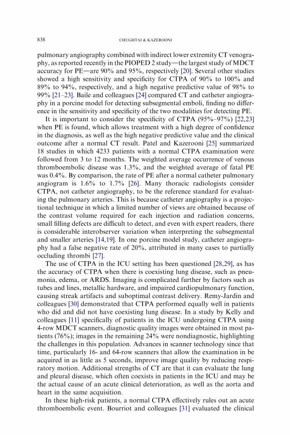
CT angiography of the pulmonary arteries is performed with 80 to 130mL of iodinated contrast material injected through an antecubital vein ata rate of 4 mL per second. Using a 16-detector row CT scanner, this takes10 to 12 seconds; with a 64-detector row scanner, it takes less than 5 secondsto complete a high-resolution examination of the entire thorax with collima-tion of approximately 1 mm. This means that the examination can be per-formed in a single breath hold, minimizing respiratory motion artifact.For an intubated patient, this minimizes the time that the ventilator issuspended for the image acquisition. With optimal enhancement of thepulmonary arteries, emboli in the main trunk down to subsegmental arteriescan be visualized easily (Fig. 1). In situations in which the visualization ofpulmonary artery filling defects is doubtful or difficult because of breathingor streak artifacts, multiplanar reformats can be generated on the worksta-tion to review the artery in any desired plane, which may enhance diagnosticconfidence (Fig. 2).
After the thoracic part of the examination, the veins of the pelvis andthighs are scanned after an additional 2 to 3 minutes as an indirect CTvenogram (CTV) to identify DVT (Fig. 3). Scans are obtained from the iliaccrests to the tibial plateaus as a contiguous acquisition using 5- or 7.5-mm
Fig. 1. A 55-year-old man who had sudden-onset chest pain. CTPA demonstrates emboli in
right upper lobe lobar, segmental, and subsegmental arteries (arrow).

840 CHUGHTAI & KAZEROONI
collimation. This one-stop CTPA combined with CTV essentially eliminatesthe need for a separate ultrasound of lower extremities, reducing the costand time for the diagnostic workup. Furthermore, it increases the diagnosticyield of the CT examination for disease. In the PIOPED II study, the sensi-tivity for VTE increased from 83% to 90% when CTV was considered withCTPA [20]. Several other studies showed an excellent correlation betweenindirect CTV and ultrasound in studies in which patients prospectivelyunderwent both tests, with sensitivity and specificity ranging from 89% to100% and 94% to 100%, respectively, and a negative predictive value of97% to 100% [32–38]. The reported interobserver agreement also is goodto excellent, with kappa values of 0.59 to 0.88 [35,37,39]. Therefore,combining CTV with CTPA increases confidence in the diagnosis of venousthromboembolic disease. This is particularly useful in patients in the ICU
Fig. 2. Multiplanar reformatted images in the coronal (A) and sagittal (B) planes demonstrate
a large embolus in the left lower lobar pulmonary artery and distal branches (arrow).
Fig. 3. Indirect CT venography in a patient who recently underwent abdominal surgery and
developed lower extremity swelling demonstrates a thrombus in the right superficial femoral
vein (arrow).

841ACUTE THORACIC CARDIOVASCULAR EMERGENCIES
who may not need anticoagulation treatment when results for thepulmonary arteries and leg veins are normal.
Aortic dissection
Aortic dissection occurs most commonly in adults between the ages of40 and 70 years, with an incidence of 1 to 6 cases per 100,000 per year. Itis two to five times more common in men. Risk factors for aortic dissectioninclude hypertension, pregnancy, coarctation of the aorta, bicuspid aorticvalve, Marfan syndrome, Ehlers-Danlos syndrome, Bechet’s disease, andprior cardiac/aortic surgery [40]. Given the 1% to 2% per hour mortalityafter symptom onset for the first 24 hours for type A dissection and a30-day mortality of 10% for type B dissection, early diagnosis is imperativeto avoid significant morbidity and mortality.
An aortic dissection is produced when there is penetration of circulatingblood into the wall of aorta, through a tear of the intima, for a varying de-gree. Any mechanism that weakens the media of the aorta may result in aor-tic dilatation and aneurysm formation and, eventually, intramuralhemorrhage, aortic dissection, or rupture [40]. The vessel walls can be af-fected by congenital connective tissue disorders, such as Marfan’s syndromeand Ehlers-Danlos syndrome. Acquired conditions, such as chronic hyper-tension, may cause aortic aneurysm and dissection [41,42]. Inflammatoryprocesses of the aortic wall or autoimmune processes involving the vasavasorum supplying the aortic wall, such as Takayasu arteritis, giant cellarteritis, syphilis, and Behcet’s disease, lead to weakening, expansion, anddissection, [40]. Iatrogenic aortic dissection can be caused by valve surgery,graft anastomosis, and the cannulation sites, as well as catheter placements[43]. Deceleration trauma, like car accidents and fall from height, can causeaortic dissection, pseudoaneurysm, and rupture, usually at the distal aorticarch just beyond the origin of left subclavian artery. Intramural hematomasof the aorta can lead to a secondary tear on the intima and communicatewith the aortic lumen [44].
Aortic dissections generally are classified with respect to what part of theaorta is involved. In the DeBakey classification, type I involves the ascend-ing and descending aorta, type II involves the ascending aorta only, andtype III involves the descending aorta only, distal to left subclavian artery[45]. In the more commonly used Stanford classification, a type A dissectionis defined as involving the ascending aorta, and type B dissection spares theascending aorta.
Clinical presentation
Acute-onset chest pain in the midline, radiating to the back, is the mostcommon presenting complaint. The onset usually is sudden and reaches max-imal intensity immediately. This abruptness is the most specific characteristic

842 CHUGHTAI & KAZEROONI
of the pain. The pain is characteristically described as ripping, tearing, chok-ing, or stabbing; it does not commonly radiate to the neck, shoulder, or armand may be absent in 5% to 10% of cases. Many patients are hypertensivebecause of preexisting hypertensive disease or increased sympathetic drive.
Clinical findings vary depending on branch artery involvement of differ-ent organ systems, due to ischemia secondary to obstruction of branches ofaorta, direct compression of organ by expanding false lumen, or leak or rup-ture of false lumen into surrounding structures. The most common findingsare due to cardiovascular and neurologic involvement when coronary ar-teries or aortic arch branches are involved. Cerebral ischemia and strokeis the most common feature. Syncope and myocardial infarction may beseen with coronary artery involvement. Spinal cord lesions are more com-mon with distal dissections and can cause paraplegia. There may be pulseand blood pressure differential between the two arms when the dissection ex-tends into or obliterates the arch vessels [46]. Similarly, acute renal failurecan occur with renal artery involvement, and mesenteric ischemia can occurwith celiac axis and mesenteric arterial involvement.
Differential diagnosis mainly includes acute myocardial infarction andPE. These can be evaluated easily with a single MDCT scan. Other condi-tions to consider in the differential include mesenteric arterial or venousthrombosis, peptic ulcer, acute appendicitis, intestinal obstruction, pancre-atic/peritoneal cyst, and acute cholecystitis. Conditions associated with aor-tic dissection, such as hypertension and connective tissue disorders, can behelpful in narrowing down the diagnosis.
Diagnosis and advanced imaging
Aortic dissection should be considered in any patient presenting withsudden-onset severe chest pain. A chest radiograph may show a widenedmediastinum, irregular aortic contour, deviation of the trachea or the naso-gastric tube in the esophagus, and displacement of calcified intima; however,chest radiograph alone does not confirm the diagnosis and is nonspecific[47]. Historically, invasive catheter aortography was the definitive diagnosticmodality. With advances in CT imaging, CT has replaced catheter angiog-raphy in the diagnostic evaluation of the aorta; it is best performed as anECG-gated CT on a 16- or more detector row scanner, which eliminatesaortic pulsation artifact. CT can quickly and noninvasively evaluate thetrue and false lumens and the intimal flap, including entry and reentry tears,as well complications, such as pericardial and pleural effusions and branchartery involvement. MDCT also is helpful in identifying causes of mediasti-nal widening as seen on chest radiograph other than dissection, such asmediastinal hematomas secondary to central line placement, mediastinalmasses, and aortic aneurysms.
Several studies demonstrated that CT is a highly accurate and reliableimaging modality for aortic dissection. In a study by Yoshida and colleagues

843ACUTE THORACIC CARDIOVASCULAR EMERGENCIES
[48], the accuracy of CT for the detection of aortic dissection or intramuralhematoma of the thoracic aorta was 100%. The sensitivity, specificity, andaccuracy, respectively, were 82%, 100%, and 84% for locating the entrytear; 95%, 100%, and 98% for arch branch vessel involvement; and83%, 100%, and 91% for pericardial effusion. All of these values were100% for aortic arch anomalies. In a more recent study, Hayter andcolleagues [49] evaluated 373 patients who had suspected aortic dissectionwith MDCT. There were no false positives, 1 false negative, 76 truepositives, and 304 true negatives, yielding a sensitivity, specificity, positivepredictive value (PPV), negative predictive value (NPV), and accuracy of99% (67 of 68), 100% (304 of 304), 100% (67 of 67), 99.7% (304 of 305),and 99.5% (371 of 373), respectively. Other studies have reported 100% sen-sitivity and specificity of MDCT to detect aortic dissections [50,51].
On CT, the primary finding in aortic dissection is the presence of two dis-tinct lumens with a visible intimal flap, which is seen in most cases (Fig. 4);in other cases, the two lumens are identified only by their differing rates ofopacification with contrast material or the low attenuation of the falselumen if it is completely thrombosed. An intramural hematoma is a variantof a classic dissection in which only a thickened wall is present, and there areno entry or reentry tears (Fig. 5). One explanation for this is rupture of thevaso vasora that supply the aortic wall. CT examinations done for acuteaortic syndromes routinely include noncontrast images first, because acuteblood in the aorta appears higher in attenuation than does the blood inthe aortic lumen, whereas long-standing hematoma will not (Fig. 6). Indirectsigns of dissection include compression of the true lumen by the false lumen,spiraling of a thrombosed false lumen, displaced intimal calcification, wid-ening of the aortic lumen, and ulcerlike projections of contrast material[52]. Three-dimensional reconstructions of the CT data are routine in eval-uating the morphology of the dissection and relation to branch vessels
Fig. 4. Type A aortic dissection with intimal separating the true and false lumens at the ascend-
ing aorta (A) and at the aortic arch (B).

844 CHUGHTAI & KAZEROONI
(Fig. 7), which are critical in clinical decision making, particularly whenopen or endovascular repair is necessary.
Patients who are unable to receive intravenous iodinated contrast for CTcan be evaluated with MRI. It also is an accurate noninvasive technique forexamining patients who are suspected of having aortic dissection, aorticintramural hematoma, or penetrating aortic ulcer; however, it generally isreserved for stable patients or follow-up imaging, because of the longerexamination time and the logistics of doing an MRI examination in a patientwho is unstable and requires close monitoring by the health care team. Al-though MRI has high sensitivity (95%–100%) and specificity (94%–98%)for the detection of aortic dissection, a sensitivity of 100% for detectionof aortic intramural hematoma, and a sensitivity of 86% for detection ofpenetrating aortic ulcer, it has serious limitations [53–55]. Most importantly,
Fig. 5. Acute intramural hematoma manifests as thickening of the ascending and descending
thoracic aortic wall (arrow), as shown on axial (A) and oblique coronal reformatted (B) images.
Fig. 6. Intramural hematoma of the aortic arch appears as high attenuation on a noncontrast
enhanced image (A, arrow) relative to blood in the aortic lumen, indicating that it is acute, and
as low attenuation thickening on a contrast-enhanced image (B, arrow).

845ACUTE THORACIC CARDIOVASCULAR EMERGENCIES
the MRI examination requires approximately 30 minutes or more, com-pared with the 10 to 30 seconds to acquire the CT data on an MDCT scan-ner, for which more time is spent moving the patient in and out of the roomthan on the scan itself. CT scan data are reconstructed so quickly on modernconsoles that a physician can review the images and make an assessment be-fore a patient from the ICU and medical team are ready to leave the CTsuite. The time it takes for an MRI scan (30 minutes) is a serious limitation,particularly in patients from the ICU who may be unstable, ventilated, orneed constant monitoring and who may have MRI incompatible hardware.Also, MRI is less readily available and commonly is at a site at great dis-tance from the ICU or in a remote area of a medical complex, which furtherlimits its role in the diagnosis of an aortic syndrome in the acute setting [49].
Coronary disease and advanced imaging in the critical care setting
A wide variety of cardiac disorders can be found in patients in the ICU;however, CAD with myocardial infarction is among the most commonacute conditions and contributes to a significant proportion of the mortalityin these patients. The clinical signs and symptoms of an acute cardiac event
Fig. 7. Acute aortic dissection involving the branches of the aortic arch, as demonstrated on an
axial image with flap extending into all three great vessels (A, arrows), flap extending into the
innominate artery (B), and on left common carotid artery on oblique sagittal reconstructions (C).

846 CHUGHTAI & KAZEROONI
can be nonspecific (eg, atypical chest pain, nausea, shortness of breath, fa-tigue, cough, and diaphoresis) and masked by other disease processes. In pa-tients in the ICU who frequently have a complicated clinical presentation,accurate and rapid diagnosis of an acute cardiac event can be challenging.Historically, the diagnosis of an acute coronary syndrome has been basedon the ECG and cardiac enzymes, often with a noninvasive stress modality(eg, echocardiography or radionuclide single photon emission computed to-mography [SPECT] imaging), with catheter coronary angiography as the fi-nal arbiter when the noninvasive test results are not conclusive or discordantwith the pretest clinical probability of disease.
The clinical applications of cardiovascular nuclear imaging techniques inthe intensive care setting have been well established. These include thal-lium201 and Tc99m-sestamibi SPECT and Multigated (MUGA) studies toprovide quantitative information concerning myocardial perfusion, acutemyocardial ischemia, and left ventricular function. These techniques provideobjective guidelines for therapy and prognosis [56]. The reported sensitivityand specificity of Tc99m-sestamibi SPECT to predict acute coronary ische-mia are 94% and 84%, respectively [57,58]; however, the lack of on-site,around-the-clock availability and long examination times are real concernsfor its use in patients from the ICU [59].
Rapid advances in MDCT technology over the last few years have greatlyfacilitated the accurate and rapid evaluation of the coronary arteries using64-detector MDCT scanners to perform CT coronary angiography. Severalstudies reported high sensitivity and specificity of MDCT for detecting cor-onary artery stenoses of 50% or greater, ranging from 90% to 95% and82% to 98%, respectively [60–65]. Perhaps the most important characteristicof CT coronary angiography is its consistently high NPV of 97% to 99%[22,23,61,62,66]. Mollet and colleagues [66], using vessel-based analysiswith 64-slice computerized tomographic angiography (CTA) to detect steno-ses of 50% or greater, reported sensitivity, specificity, PPV, and NPV rang-ing from 97% to 100%, 92% to 99%, 78% to 80%, and 99% to 100%,respectively, depending on the calcium score. The sensitivity for detectingsignificant disease in the left anterior descending was 96%, whereas in othermain coronary arteries the sensitivity was 100%. There was good correlationbetween CTA and coronary angiography, with a kappa value of 0.85. Eharaand colleagues [67] evaluated 64-slice MDCT for detecting angiographicallysignificant coronary artery stenosis in an unselected consecutive patientpopulation and compared it with conventional invasive angiography.Fifty-seven percent of the patients already had coronary artery stents.They reported that sensitivity for diagnosing significant stenosis (R50%)was 90%, specificity was 94%, PPV was 89%, and NPV was 95%. For thestented arteries, the sensitivity, specificity, PPV, and NPV were 93%, 96%,87%, and 98%, respectively.
Raff and colleagues [23] evaluated the diagnostic accuracy of 64-slicecoronary CT angiography in 70 consecutive patients undergoing invasive

847ACUTE THORACIC CARDIOVASCULAR EMERGENCIES
coronary angiography, including patients with high heart rates (23%O70beats per minute [bpm], range up to 96 bpm), obesity (50% with bodymass indexO30 kg/m2), and coronary calcification (26% had AgatstonscoreO400 with range up to 1804), reflecting a more ‘‘real world’’ groupof patients. They demonstrated a high NPV of 98% by segment and 97%by artery. They also observed improved image quality with smaller voxelsize provided by the 64-slice scanner, which reduced, but did not eliminate,the calcium blooming and beam hardening artifacts. The sensitivity andspecificity were 97% and 95%, respectively, with heart rate of less than70 bpm and 88% and 71%, respectively, with heart rate of 71 to 85 bpm,reinforcing the need to pharmacologically reduce the heart rate during theCT examination. This is done routinely with b-blocker and sometimescalcium channel blockers if there is a contraindication to the former. Ahigh or irregular heart rate decreases the image quality of coronary CT an-giography. Achenbach and colleagues [69] evaluated a new dual-sourceMDCT with the advantage of higher temporal resolution than other 64-detector MDCT scanners, which has made it possible to obtain good qualityimages at higher heart rates and has reduced, but not eliminated, the need topremedicate patients completely. They reported visualization of 98% of cor-onary artery segments free of cardiac motion artifacts.
There has been preliminary work on the use of coronary CT angiogra-phy in patients who have chest pain presenting to emergency rooms, whoare stable clinically, low risk for CAD, and have a normal ECG and car-diac enzymes for at least 4 hours. Patients with a normal scan can be dis-charged early, leading to a reduction in length of stay and cost of care;unfortunately, the sample sizes have been too small to determine the im-pact of this strategy on coronary event rates, such as myocardial infarctionand intervention [57,68,69]. In a recent study, Rubinshtein and colleagues[70] demonstrated emergency department (ED) MDCT sensitivity of100%, specificity of 92%, PPV of 87%, and NPV of 100% in a cohortof 58 patients. They concluded that 64-slice cardiac MDCT representsa valuable diagnostic tool in patients in the ED who have chest pain of un-certain origin, providing early direct noninvasive visualization of coronaryanatomy.
Technique
Cardiac imaging using CT is a technically demanding procedure, requir-ing high temporal and spatial resolution to visualize small coronary arterieswhile the heart is beating continuously. This is achieved using retrospectiveECG gating, segmentation, and tailored reconstruction algorithms. Respira-tory motion also must be eliminated for cardiac imaging, so scanning isoptimally performed in a single breath hold, easily achievable with the 5-to 10-second acquisition times for the examination using 64-slice MDCTscanners.
848 CHUGHTAI & KAZEROONI
A stable heart rate of 65 bpm or less is important to obtain diagnosticimage quality and to decrease radiation dose and shorten the time for imageprocessing and evaluation. For example, a study of 94 patients reported aninverse correlation between the number of analyzable vessel segments andheart rate [71]. Vessel visibility was highest when the heart rate was lessthan 65 bpm [71]. If the heart rate is more than 65 bpm or is irregular, b-blocker medication can be administered orally or intravenously before thescan. A single puff (0.4 mg) of sublingual nitroglycerin also is given a fewminutes before the scan to dilate the coronary arteries and exaggerate thedifference between normal and abnormal segments, as is done beforecatheter coronary angiography.
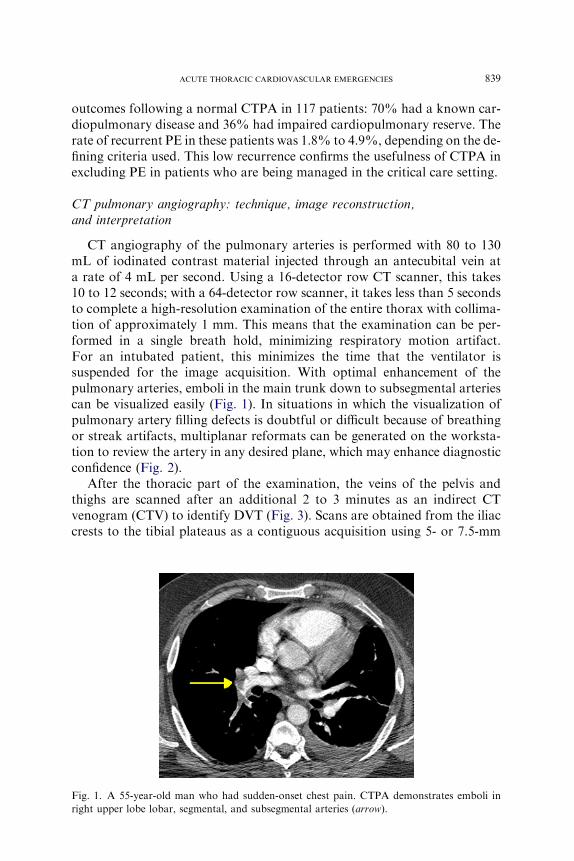
A noncontrast enhanced scan using prospective ECG gating is performedthrough the heart, from which the calcium score is generated and the loca-tion of the coronary arteries confirmed. The coronary calcium score is a sen-sitive marker of CAD; the higher the score, the greater the likelihood ofsignificant coronary event. A large coronary calcium load can potentiallydegrade the image quality of CTA, however. A timing bolus of 15 to 20mL of intravenous contrast agent is used to determine the optimumtime of arterial peak enhancement for the specific patient, by placing a regionof interest in the aortic root. Following this, the contrast-enhanced CTA ofthe coronary arteries is performed with 70 to 80 mL of low osmolar iodin-ated contrast material injected intravenously at a rate of 5 mL/s through an
Fig. 8. Volume-rendered images demonstrate excellent visualization of normal coronary
anatomy, including the right coronary artery (arrow), acute marginal artery (short arrow),
and left anterior descending coronary artery (arrowhead) (A) as well as the left main, left ante-
rior descending, and left circumflex coronary arteries and their branches (B).
849ACUTE THORACIC CARDIOVASCULAR EMERGENCIES
antecubital vein. During the later part of the contrast injection, the contrastis mixed with saline using a dual-headed power injector, so that the contrastin the right cardiac chambers is not so bright as to cause artifacts in the deepatrioventricular groove where the right coronary artery resides. With64-slice scanners, the collimation used ranges from 0.5 to 0.625 mm; gantryrotation time is less than 500 milliseconds. The typical scan time is 5 to 10seconds, short enough to complete the study in a single breath hold.
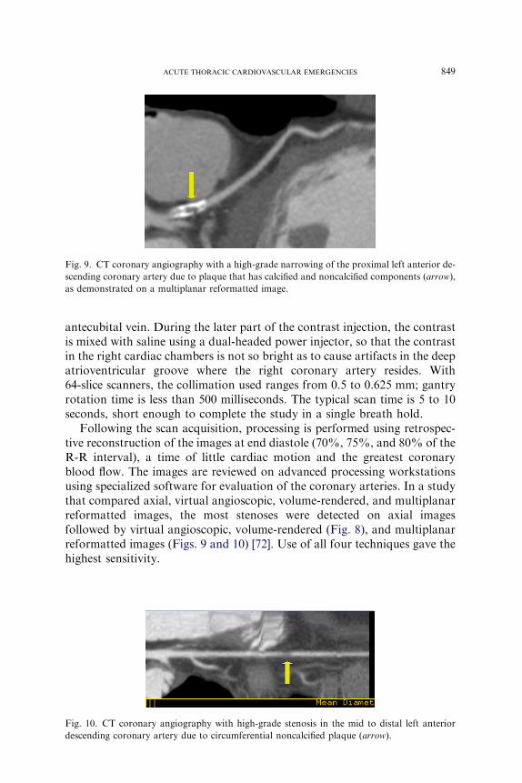
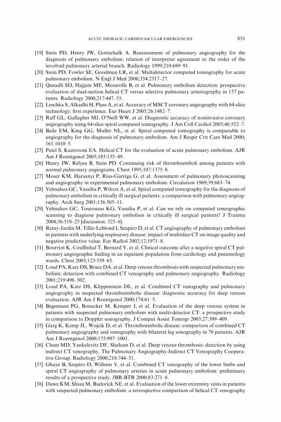
Following the scan acquisition, processing is performed using retrospec-tive reconstruction of the images at end diastole (70%, 75%, and 80% of theR-R interval), a time of little cardiac motion and the greatest coronaryblood flow. The images are reviewed on advanced processing workstationsusing specialized software for evaluation of the coronary arteries. In a studythat compared axial, virtual angioscopic, volume-rendered, and multiplanarreformatted images, the most stenoses were detected on axial imagesfollowed by virtual angioscopic, volume-rendered (Fig. 8), and multiplanarreformatted images (Figs. 9 and 10) [72]. Use of all four techniques gave thehighest sensitivity.
Fig. 10. CT coronary angiography with high-grade stenosis in the mid to distal left anterior
descending coronary artery due to circumferential noncalcified plaque (arrow).
Fig. 9. CT coronary angiography with a high-grade narrowing of the proximal left anterior de-
scending coronary artery due to plaque that has calcified and noncalcified components (arrow),
as demonstrated on a multiplanar reformatted image.

850 CHUGHTAI & KAZEROONI
Summary
ED MDCT is a rapid and accurate test for evaluation of patients whohave chest pain in the ED setting. Our understanding about the usefulnessand limitations of ED MDCT will improve as more data are made availablefrom ongoing studies.
References
[1] CocheEE,MullerNL,KimKI, et al. Acute pulmonary embolism: ancillary findings at spiral
CT. Radiology 1998;207:753–8.
[2] Greenberg SB. Assessment of cardiac function: magnetic resonance and computed tomogra-
phy. J Thorac Imaging 2000;15:243–51.
[3] White RH. The epidemiology of venous thromboembolism. Circulation 2003;107:I4–8.
[4] Geerts W, Selby R. Prevention of venous thromboembolism in the ICU. Chest 2003;124:
357S–63S.
[5] Heit JA. The epidemiology of venous thromboembolism in the community: implications for
prevention and management. J Thromb Thrombolysis 2006;21:23–9.
[6] Silverstein MD, Heit JA, Mohr DN, et al. Trends in the incidence of deep vein thrombosis
and pulmonary embolism: a 25-year population-based study. Arch Intern Med 1998;158:
585–93.
[7] Piazza G, Goldhaber SZ. Acute pulmonary embolism: Part II: treatment and prophylaxis.
Circulation 2006;114:e42–7.
[8] Goldhaber SZ, Visani L, De Rosa M. Acute pulmonary embolism: clinical outcomes in the
International Cooperative Pulmonary Embolism Registry (ICOPER). Lancet 1999;353:
1386–9.
[9] Flordal PA, Berggvist D, Burmark US, et al. Risk factors for major thromboembolism and
bleeding tendency after elective general surgical operations. The FragminMulticentre Study
group. Eur J Surg 1996;162:783–9.
[10] Hirsch DR, Ingenito EP, Goldhaber SZ. Prevalence of deep venous thrombosis among
patients in medical intensive care. JAMA 1995;274:335–7.
[11] Kelly AM, Patel S, Carlos RC, et al. Multidetector row CT pulmonary angiography and
indirect venography for the diagnosis of venous thromboembolic disease in intensive care
unit patients. Acad Radiol 2006;13:486–95.
[12] Marik PE, Andrews L, Maini B. The incidence of deep venous thrombosis in ICU patients.
Chest 1997;111:661–4.
[13] Palareti G, Legnani C, Cosmi B, et al. Risk of venous thromboembolism recurrence: high
negative predictive value of D-dimer performed after oral anticoagulation is stopped.
Thromb Haemost 2002;87:7–12.
[14] Sadosty AT, Goyal DG, Boie ET, et al. Emergency department D-dimer testing. J Emerg
Med 2001;21:423–9.
[15] Goldhaber SZ. The perils of D-dimer in the medical intensive care unit. Crit CareMed 2000;
28:583–4.
[16] Worsley DF, Alavi A, Aronchick JM, et al. Chest radiographic findings in patients with
acute pulmonary embolism: observations from the PIOPED Study. Radiology 1993;189:
133–6.
[17] Value of the ventilation/perfusion scan in acute pulmonary embolism. Results of the pro-
spective investigation of pulmonary embolism diagnosis (PIOPED). The PIOPED investiga-
tors. JAMA 1990;263:2753–9.
[18] Jolliet P, SlosmanDO,RicouB, et al. Pulmonary scintigraphy at the bedside in intensive care
patients with suspected pulmonary embolism. Intensive Care Med 1995;21:723–8.
851ACUTE THORACIC CARDIOVASCULAR EMERGENCIES
[19] Stein PD, Henry JW, Gottschalk A. Reassessment of pulmonary angiography for the
diagnosis of pulmonary embolism: relation of interpreter agreement to the order of the
involved pulmonary arterial branch. Radiology 1999;210:689–91.
[20] Stein PD, Fowler SE, Goodman LR, et al. Multidetector computed tomography for acute
pulmonary embolism. N Engl J Med 2006;354:2317–27.
[21] Qanadli SD, Hajjam ME, Mesurolle B, et al. Pulmonary embolism detection: prospective
evaluation of dual-section helical CT versus selective pulmonary arteriography in 157 pa-
tients. Radiology 2000;217:447–55.
[22] Leschka S, AlkadhiH, PlassA, et al. Accuracy ofMSCT coronary angiographywith 64-slice
technology: first experience. Eur Heart J 2005;26:1482–7.
[23] Raff GL, Gallagher MJ, O’Neill WW, et al. Diagnostic accuracy of noninvasive coronary
angiography using 64-slice spiral computed tomography. J Am Coll Cardiol 2005;46:552–7.
[24] Baile EM, King GG, Muller NL, et al. Spiral computed tomography is comparable to
angiography for the diagnosis of pulmonary embolism. Am J Respir Crit Care Med 2000;
161:1010–5.
[25] Patel S, Kazerooni EA. Helical CT for the evaluation of acute pulmonary embolism. AJR
Am J Roentgenol 2005;185:135–49.
[26] Henry JW, Relyea B, Stein PD. Continuing risk of thromboemboli among patients with
normal pulmonary angiograms. Chest 1995;107:1375–8.
[27] Moser KM, Harsanyi P, Rius-Garriga G, et al. Assessment of pulmonary photoscanning
and angiography in experimental pulmonary embolism. Circulation 1969;39:663–74.
[28] Velmahos GC, Vassiliu P, Wilcox A, et al. Spiral computed tomography for the diagnosis of
pulmonary embolism in critically ill surgical patients: a comparison with pulmonary angiog-
raphy. Arch Surg 2001;136:505–11.
[29] Velmahos GC, Toutouzas KG, Vassiliu P, et al. Can we rely on computed tomographic
scanning to diagnose pulmonary embolism in critically ill surgical patients? J Trauma
2004;56:518–25 [discussion: 525–6].
[30] Remy-JardinM, Tillie-Leblond I, Szapiro D, et al. CT angiography of pulmonary embolism
in patients with underlying respiratory disease: impact of multislice CT on image quality and
negative predictive value. Eur Radiol 2002;12:1971–8.
[31] Bourriot K, Couffinhal T, Bernard V, et al. Clinical outcome after a negative spiral CT pul-
monary angiographic finding in an inpatient population from cardiology and pneumology
wards. Chest 2003;123:359–65.
[32] Loud PA,KatzDS, BruceDA, et al. Deep venous thrombosiswith suspected pulmonary em-
bolism: detection with combined CT venography and pulmonary angiography. Radiology
2001;219:498–502.
[33] Loud PA, Katz DS, Klippenstein DL, et al. Combined CT venography and pulmonary
angiography in suspected thromboembolic disease: diagnostic accuracy for deep venous
evaluation. AJR Am J Roentgenol 2000;174:61–5.
[34] Begemann PG, Bonacker M, Kemper J, et al. Evaluation of the deep venous system in
patients with suspected pulmonary embolism with multi-detector CT: a prospective study
in comparison to Doppler sonography. J Comput Assist Tomogr 2003;27:399–409.
[35] Garg K, Kemp JL, Wojcik D, et al. Thromboembolic disease: comparison of combined CT
pulmonary angiography and venography with bilateral leg sonography in 70 patients. AJR
Am J Roentgenol 2000;175:997–1001.
[36] ChamMD, Yankelevitz DF, Shaham D, et al. Deep venous thrombosis: detection by using
indirect CT venography. The Pulmonary Angiography-Indirect CT Venography Coopera-
tive Group. Radiology 2000;216:744–51.
[37] Ghaye B, Szapiro D, Willems V, et al. Combined CT venography of the lower limbs and
spiral CT angiography of pulmonary arteries in acute pulmonary embolism: preliminary
results of a prospective study. JBR-BTR 2000;83:271–8.
[38] DuweKM, ShiauM, Budorick NE, et al. Evaluation of the lower extremity veins in patients
with suspected pulmonary embolism: a retrospective comparison of helical CT venography

852 CHUGHTAI & KAZEROONI
and sonography. 2000 ARRS Executive Council Award I. American Roentgen Ray Society.
AJR Am J Roentgenol 2000;175:1525–31.
[39] Coche EE, Hamoir XL, Hammer FD, et al. Using dual-detector helical CT angiography to
detect deep venous thrombosis in patientswith suspicion of pulmonary embolism: diagnostic
value and additional findings. AJR Am J Roentgenol 2001;176:1035–9.
[40] NienaberCA,EagleKA.Aortic dissection: new frontiers in diagnosis andmanagement: Part
I: from etiology to diagnostic strategies. Circulation 2003;108:628–35.
[41] Reed D, Reed C, Stemmermann G, et al. Are aortic aneurysms caused by atherosclerosis?
Circulation 1992;85:205–11.
[42] Larson EW, Edwards WD. Risk factors for aortic dissection: a necropsy study of 161 cases.
Am J Cardiol 1984;53:849–55.
[43] Januzzi JL, Sabatine MS, Eagle KA, et al. Iatrogenic aortic dissection. Am J Cardiol 2002;
89:623–6.
[44] Nienaber CA, Sievers HH. Intramural hematoma in acute aortic syndrome: more than one
variant of dissection? Circulation 2002;106:284–5.
[45] Chen K, Varon J, Wenker OC, et al. Acute thoracic aortic dissection: the basics. J Emerg
Med 1997;15:859–67.
[46] GanahaF,Miller DC, SugimotoK, et al. Prognosis of aortic intramural hematomawith and
without penetrating atherosclerotic ulcer: a clinical and radiological analysis. Circulation
2002;106:342–8.
[47] Jagannath AS, Sos TA, Lockhart SH, et al. Aortic dissection: a statistical analysis of the
usefulness of plain chest radiographic findings. AJR Am J Roentgenol 1986;147:1123–6.
[48] Yoshida S, Akiba H, Tamakawa M, et al. Thoracic involvement of type A aortic dissection
and intramural hematoma: diagnostic accuracy–comparison of emergency helical CT and
surgical findings. Radiology 2003;228:430–5.
[49] Hayter RG, Rhea JT, Small A, et al. Suspected aortic dissection and other aortic disorders:
multi-detector row CT in 373 cases in the emergency setting. Radiology 2006;238:841–52.
[50] Sommer T, Fehske W, Holzknecht N, et al. Aortic dissection: a comparative study of diag-
nosis with spiral CT, multiplanar transesophageal echocardiography, andMR imaging. Ra-
diology 1996;199:347–52.
[51] Sebastia C, Pallisa E, Quiroga S, et al. Aortic dissection: diagnosis and follow-upwith helical
CT. Radiographics 1999;19:45–60, quiz 149–50.
[52] Tisnado J, Cho SR, BeachleyMC, et al. Ulcerlike projections: a precursor angiographic sign
to thoracic aortic dissection. AJR Am J Roentgenol 1980;135:719–22.
[53] Nienaber CA, von Kodolitsch Y, Petersen B, et al. Intramural hemorrhage of the thoracic
aorta. Diagnostic and therapeutic implications. Circulation 1995;92:1465–72.
[54] Nienaber CA, vonKodolitsch Y, Nicolas V, et al. The diagnosis of thoracic aortic dissection
by noninvasive imaging procedures. N Engl J Med 1993;328:1–9.
[55] Nienaber CA, Spielmann RP, von Kodolitsch Y, et al. Diagnosis of thoracic aortic dissec-
tion. Magnetic resonance imaging versus transesophageal echocardiography. Circulation
1992;85:434–47.
[56] Ell PJ, Donaldson RM. Cardiovascular nuclear medicine and intensive care. Intensive Care
Med 1978;4:119–22.
[57] Hoffmann U, Nagurney JT, Moselewski F, et al. Coronary multidetector computed tomog-
raphy in the assessment of patients with acute chest pain. Circulation 2006;114:2251–60.
[58] Hilton TC, Thompson RC, Williams HJ, et al. Technetium-99m sestamibi myocardial per-
fusion imaging in the emergency room evaluation of chest pain. J Am Coll Cardiol 1994;23:
1016–22.
[59] White C, Read K, Kuo D. Assessment of chest pain in the emergency room: what is the role
of multidetector CT? Eur J Radiol 2006;57:368–72.
[60] Nieman K, Cademartiri F, Lemos PA, et al. Reliable noninvasive coronary angiography
with fast submillimeter multislice spiral computed tomography. Circulation 2002;106:
2051–4.

853ACUTE THORACIC CARDIOVASCULAR EMERGENCIES
[61] Ropers D, Baum U, Pohle K, et al. Detection of coronary artery stenoses with thin-slice
multi-detector row spiral computed tomography and multiplanar reconstruction. Circula-
tion 2003;107:664–6.
[62] HoffmannU,Moselewski F, Cury RC, et al. Predictive value of 16-slice multidetector spiral
computed tomography to detect significant obstructive coronary artery disease in patients at
high risk for coronary artery disease: patient- versus segment-based analysis. Circulation
2004;110:2638–43.
[63] Achenbach S, Ropers D, Hoffmann U, et al. Assessment of coronary remodeling in stenotic
and nonstenotic coronary atherosclerotic lesions bymultidetector spiral computed tomogra-
phy. J Am Coll Cardiol 2004;43:842–7.
[64] Kuettner A, Beck T, Drosch T, et al. Diagnostic accuracy of noninvasive coronary imaging
using 16-detector slice spiral computed tomography with 188 ms temporal resolution. J Am
Coll Cardiol 2005;45:123–7.
[65] Mollet NR, Cademartiri F, Krestin GP, et al. Improved diagnostic accuracy with 16-row
multi-slice computed tomography coronary angiography. J Am Coll Cardiol 2005;45:
128–32.
[66] Mollet NR, Cademartiri F, van Mieghem CA, et al. High-resolution spiral computed
tomography coronary angiography in patients referred for diagnostic conventional coronary
angiography. Circulation 2005;112:2318–23.
[67] Ehara M, Surmely JF, Kawai M, et al. Diagnostic accuracy of 64-slice computed
tomography for detecting angiographically significant coronary artery stenosis in an unse-
lected consecutive patient population: comparison with conventional invasive angiography.
Circ J 2006;70:564–71.
[68] Raff GL, Gallagher MJ, O’Neill WW, et al. Immediate coronary CTA rapidly and
definitively excludes CAD in low-risk acute chest pain. Presented at the American College
of Cardiology Annual Scientific Session. Atlanta (GA); 2006. p. 807–8.
[69] Achenbach S, Ropers D, Kuettner A, et al. Contrast-enhanced coronary artery visualization
by dual-source computed tomography–initial experience. Eur J Radiol 2006;57:331–5.
[70] Rubinshtein R, Halon DA, Gaspar T, et al. Usefulness of 64-slice cardiac computed
tomographic angiography for diagnosing acute coronary syndromes and predicting clinical
outcome in emergency department patients with chest pain of uncertain origin. Circulation
2007;115:1762–8.
[71] Schroeder S, Kopp AF, Kuettner A, et al. Influence of heart rate on vessel visibility in
noninvasive coronary angiography using new multislice computed tomography: experience
in 94 patients. Clin Imaging 2002;26:106–11.
[72] Vogl TJ, Abolmaali ND, Diebold T, et al. Techniques for the detection of coronary athero-
sclerosis: multi-detector row CT coronary angiography. Radiology 2002;223:212–20.