CrystalStructuresofHumanTBC1D1andTBC1D4(AS160) … · (TBC1D4) mg/ml by centrifugation (Amicon...
10
Crystal Structures of Human TBC1D1 and TBC1D4 (AS160) RabGTPase-activating Protein (RabGAP) Domains Reveal Critical Elements for GLUT4 Translocation * Received for publication, December 29, 2010, and in revised form, February 11, 2011 Published, JBC Papers in Press, March 23, 2011, DOI 10.1074/jbc.M110.217323 Sang-Youn Park 1 , Wanzhu Jin, Ju Rang Woo, and Steven E. Shoelson 2 From the Joslin Diabetes Center and Department of Medicine, Harvard Medical School, Boston, Massachusetts 02215 We have solved the x-ray crystal structures of the RabGAP domains of human TBC1D1 and human TBC1D4 (AS160), at 2.2 and 3.5 A ˚ resolution, respectively. Like the yeast Gyp1p RabGAP domain, whose structure was solved previously in com- plex with mouse Rab33B, the human TBC1D1 and TBC1D4 domains both have 16 -helices and no -sheet elements. We expected the yeast Gyp1p RabGAP/mouse Rab33B structure to predict the corresponding interfaces between cognate mamma- lian RabGAPs and Rabs, but found that residues were poorly conserved. We further tested the relevance of this model by Ala- scanning mutagenesis, but only one of five substitutions within the inferred binding site of the TBC1D1 RabGAP significantly perturbed catalytic efficiency. In addition, substitution of TBC1D1 residues with corresponding residues from Gyp1p did not enhance catalytic efficiency. We hypothesized that biologi- cally relevant RabGAP/Rab partners utilize additional contacts not described in the yeast Gyp1p/mouse Rab33B structure, which we predicted using our two new human TBC1D1 and TBC1D4 structures. Ala substitution of TBC1D1 Met 930 , corre- sponding to a residue outside of the Gyp1p/Rab33B contact, substantially reduced catalytic activity. GLUT4 translocation assays confirmed the biological relevance of our findings. Sub- stitutions with lowest RabGAP activity, including catalytically dead RK and Met 930 and Leu 1019 predicted to perturb Rab bind- ing, confirmed that biological activity requires contacts between cognate RabGAPs and Rabs beyond those in the yeast Gyp1p RabGAP/mouse Rab33B structure. The trafficking of intracellular vesicles is regulated by Rab GTPases (Rab), which participate in Rab-mediated vesicle bud- ding, uncoating, docking, and fusion (1, 2). Insulin-stimulated glucose uptake utilizes these processes during translocation of glucose transporter protein (e.g. GLUT4) 3 vesicles from intra- cellular pools to the cell surface (3). TBC1D1 and TBC1D4 (also known as AS160) are Rab GTPase-activating proteins (RabGAPs) in skeletal myocytes and adipocytes, respectively, with functions in GLUT4 vesicle trafficking (4, 5). TBC1D1- and TBC1D4-catalyzed hydrolysis of Rab-bound GTP (active) to GDP (inactive) leaves GLUT4 vesicles sequestered in an intracellular compartment. Insulin-stimulated Akt phosphor- ylation of TBC1D1 and TBC1D4 decreases GTP hydrolysis, which increases RabGTP concentrations, GLUT4 vesicle translocation, and glucose uptake. Of 60 Rab proteins in mammalian genomes (1, 2), 20 associate with GLUT4 vesi- cles, and among them only Rabs 2A, 8A, 10, and 14 have been shown to be potential substrates for TBC1D1 or TBC1D4 (6 – 8). TBC1D1 and TBC1D4 each contain 1300 residues. Besides catalytic RabGAP domains at their carboxyl termini, each has two putative amino-terminal phosphotyrosine binding domains whose functions are under investigation. The second phosphotyrosine binding domain has been shown to bind insu- lin-regulated aminopeptidase, a marker for GLUT4 vesicle (9). TBC1D1 and TBC1D4 are closely related paralogs, with 47% overall identity and 76% identity within their RabGAP domains, but they are expressed in different tissues. To further under- score their important roles in normal metabolic function, nat- urally occurring loss-of-function mutations in TBC1D1 and TBC1D4 RabGAP domains have been genetically linked to pro- tection against obesity in mice (10) and defective insulin signal- ing in humans (11), respectively. The structure of the yeast Gyp1p RabGAP in complex with mouse Rab33B revealed an interaction surface as well as cat- alytic roles for specific Arg and Gln residues (12). However, it has been difficult to use this structure to generalize details about Rab/RabGAP specificity, first because the proteins used were from such evolutionarily divergent organisms, and second because RabGAP structures are also variable. We have solved the x-ray crystal structures of the human TBC1D1 and TBC1D4 RabGAP domains and used the struc- tures to probe Rab/RabGAP interactions relevant to GLUT4 translocation. * This work was supported, in whole or in part, by National Institutes of Health Grants R01 DK43123 and R01 DK51729 (to S. E. S.), F32 DK77485 (to S.-Y. P.), and P30 DK36836 (to the Joslin Diabetes Center). Data were collected at beams X29A and X12C of National Synchrotron Light Source, Brookhaven National Laboratory, which is supported by the United States Department of Energy, Division of Materials Sciences and Division of Chemical Sciences, Contract DE-AC02-98CH10886. The atomic coordinates and structure factors (codes 3QYB and 3QYE) have been deposited in the Protein Data Bank, Research Collaboratory for Structural Bioinformatics, Rutgers University, New Brunswick, NJ (http://www.rcsb.org/). 1 Present address: School of Systems Biomedical Science, College of Natural Sciences, Soongsil University, 511 Sangdo-Dong, Dongjak-Gu, Seoul, Korea. 2 To whom correspondence should be addressed: Joslin Diabetes Center, One Joslin Place, Boston, MA 02215. Tel.: 617-732-2528; Fax: 617-732-2407; E-mail: [email protected]. 3 The abbreviations used are: GLUT4, glucose transporter 4; RabGAP, RabGTPase-activating protein; r.m.s.d., root mean square deviation; TBC1D, Tre-2, Bub2, and Cdc16-1 domain. THE JOURNAL OF BIOLOGICAL CHEMISTRY VOL. 286, NO. 20, pp. 18130 –18138, May 20, 2011 © 2011 by The American Society for Biochemistry and Molecular Biology, Inc. Printed in the U.S.A. 18130 JOURNAL OF BIOLOGICAL CHEMISTRY VOLUME 286 • NUMBER 20 • MAY 20, 2011 by guest on September 19, 2018 http://www.jbc.org/ Downloaded from
Transcript of CrystalStructuresofHumanTBC1D1andTBC1D4(AS160) … · (TBC1D4) mg/ml by centrifugation (Amicon...

Crystal Structures of Human TBC1D1 and TBC1D4 (AS160)RabGTPase-activating Protein (RabGAP) Domains RevealCritical Elements for GLUT4 Translocation*
Received for publication, December 29, 2010, and in revised form, February 11, 2011 Published, JBC Papers in Press, March 23, 2011, DOI 10.1074/jbc.M110.217323
Sang-Youn Park1, Wanzhu Jin, Ju Rang Woo, and Steven E. Shoelson2
From the Joslin Diabetes Center and Department of Medicine, Harvard Medical School, Boston, Massachusetts 02215
We have solved the x-ray crystal structures of the RabGAPdomains of human TBC1D1 and human TBC1D4 (AS160), at2.2 and 3.5 A resolution, respectively. Like the yeast Gyp1pRabGAPdomain,whose structurewas solvedpreviously in com-plex with mouse Rab33B, the human TBC1D1 and TBC1D4domains both have 16 �-helices and no �-sheet elements. Weexpected the yeast Gyp1p RabGAP/mouse Rab33B structure topredict the corresponding interfaces between cognate mamma-lian RabGAPs and Rabs, but found that residues were poorlyconserved.We further tested the relevance of thismodel byAla-scanning mutagenesis, but only one of five substitutions withinthe inferred binding site of the TBC1D1 RabGAP significantlyperturbed catalytic efficiency. In addition, substitution ofTBC1D1 residues with corresponding residues from Gyp1p didnot enhance catalytic efficiency. We hypothesized that biologi-cally relevant RabGAP/Rab partners utilize additional contactsnot described in the yeast Gyp1p/mouse Rab33B structure,which we predicted using our two new human TBC1D1 andTBC1D4 structures. Ala substitution of TBC1D1Met930, corre-sponding to a residue outside of the Gyp1p/Rab33B contact,substantially reduced catalytic activity. GLUT4 translocationassays confirmed the biological relevance of our findings. Sub-stitutions with lowest RabGAP activity, including catalyticallydead RK andMet930 and Leu1019 predicted to perturb Rab bind-ing, confirmed that biological activity requires contacts betweencognate RabGAPs and Rabs beyond those in the yeast Gyp1pRabGAP/mouse Rab33B structure.
The trafficking of intracellular vesicles is regulated by RabGTPases (Rab), which participate in Rab-mediated vesicle bud-ding, uncoating, docking, and fusion (1, 2). Insulin-stimulated
glucose uptake utilizes these processes during translocation ofglucose transporter protein (e.g. GLUT4)3 vesicles from intra-cellular pools to the cell surface (3). TBC1D1 and TBC1D4(also known as AS160) are Rab GTPase-activating proteins(RabGAPs) in skeletal myocytes and adipocytes, respectively,with functions in GLUT4 vesicle trafficking (4, 5). TBC1D1-and TBC1D4-catalyzed hydrolysis of Rab-bound GTP (active)to GDP (inactive) leaves GLUT4 vesicles sequestered in anintracellular compartment. Insulin-stimulated Akt phosphor-ylation of TBC1D1 and TBC1D4 decreases GTP hydrolysis,which increases Rab�GTP concentrations, GLUT4 vesicletranslocation, and glucose uptake. Of �60 Rab proteins inmammalian genomes (1, 2), �20 associate with GLUT4 vesi-cles, and among them only Rabs 2A, 8A, 10, and 14 have beenshown to be potential substrates for TBC1D1 or TBC1D4(6–8).TBC1D1 andTBC1D4 each contain�1300 residues. Besides
catalytic RabGAP domains at their carboxyl termini, eachhas two putative amino-terminal phosphotyrosine bindingdomains whose functions are under investigation. The secondphosphotyrosine binding domain has been shown to bind insu-lin-regulated aminopeptidase, a marker for GLUT4 vesicle (9).TBC1D1 and TBC1D4 are closely related paralogs, with 47%overall identity and 76% identitywithin their RabGAPdomains,but they are expressed in different tissues. To further under-score their important roles in normal metabolic function, nat-urally occurring loss-of-function mutations in TBC1D1 andTBC1D4RabGAPdomains have been genetically linked to pro-tection against obesity inmice (10) and defective insulin signal-ing in humans (11), respectively.The structure of the yeast Gyp1p RabGAP in complex with
mouse Rab33B revealed an interaction surface as well as cat-alytic roles for specific Arg and Gln residues (12). However,it has been difficult to use this structure to generalize detailsabout Rab/RabGAP specificity, first because the proteinsused were from such evolutionarily divergent organisms,and second because RabGAP structures are also variable.Wehave solved the x-ray crystal structures of the humanTBC1D1 and TBC1D4 RabGAP domains and used the struc-tures to probe Rab/RabGAP interactions relevant to GLUT4translocation.
* This work was supported, in whole or in part, by National Institutes of HealthGrants R01 DK43123 and R01 DK51729 (to S. E. S.), F32 DK77485 (to S.-Y. P.),and P30 DK36836 (to the Joslin Diabetes Center). Data were collected atbeams X29A and X12C of National Synchrotron Light Source, BrookhavenNational Laboratory, which is supported by the United States Departmentof Energy, Division of Materials Sciences and Division of Chemical Sciences,Contract DE-AC02-98CH10886.
The atomic coordinates and structure factors (codes 3QYB and 3QYE) have beendeposited in the Protein Data Bank, Research Collaboratory for StructuralBioinformatics, Rutgers University, New Brunswick, NJ (http://www.rcsb.org/).
1 Present address: School of Systems Biomedical Science, College of NaturalSciences, Soongsil University, 511 Sangdo-Dong, Dongjak-Gu, Seoul,Korea.
2 To whom correspondence should be addressed: Joslin Diabetes Center,One Joslin Place, Boston, MA 02215. Tel.: 617-732-2528; Fax: 617-732-2407;E-mail: [email protected].
3 The abbreviations used are: GLUT4, glucose transporter 4; RabGAP,RabGTPase-activating protein; r.m.s.d., root mean square deviation;TBC1D, Tre-2, Bub2, and Cdc16-1 domain.
THE JOURNAL OF BIOLOGICAL CHEMISTRY VOL. 286, NO. 20, pp. 18130 –18138, May 20, 2011© 2011 by The American Society for Biochemistry and Molecular Biology, Inc. Printed in the U.S.A.
18130 JOURNAL OF BIOLOGICAL CHEMISTRY VOLUME 286 • NUMBER 20 • MAY 20, 2011
by guest on September 19, 2018
http://ww
w.jbc.org/
Dow
nloaded from

EXPERIMENTAL PROCEDURES
Protein Expression and Purification—Fragments of cDNAencoding human TBC1D1 (residues 746–1072; DNA fromOpen Biosystem) and human TBC1D4 (residues 873–1172;DNA from Takahiro Nagase, Kazusa DNA Research Institute)RabGAP domains and mouse Rab14 (residues 1–175; DNAfrom Gus Lienhard, Dartmouth University) were PCR-clonedinto pET28a (Novagen) vectors. The proteins were expressedwithHis6 tags inEscherichia coli strain BL21 (DE3) (Stratagene)using kanamycin selection (25 �g/ml) and isolated on Ni2�-nitrilotriacetic acid columns. His tags were removed withthrombin. The proteins were further purified using a Superdex200 sizing column (Pharmacia) in 50 mM Tris, pH 7.5, 150 mM
NaCl, and concentrated to 80–100 (TBC1D1) or 15–30(TBC1D4) mg/ml by centrifugation (Amicon Centriprep).Rab14 was loaded with GTP by incubating 4mg of protein witha 10-fold molar excess of GTP at 4 °C for 2–3 h in 50 mM Tris,pH 7.5, 150 mM NaCl, 5 mM EDTA, and 5 mM DTT. Free GTPwas removed with a desalting column (Bio-Rad) preequili-brated with 50 mM Tris, pH 7.5, and 150 mM NaCl. Rab14 ali-quots were stored at �80 °C for enzyme assays.Crystallization and Data Collection—Conditions for crystal-
lizing the TBC1D1 and TBC1D4 RabGAP domains were foundusing commercial screening solutions (Hampton Research).Optimized TBC1D1 RabGAP domain crystals appeared over-night under vapor diffusion conditions in 2-�l hanging dropscontaining 1:1mixtures of protein and reservoir solutions: 2.0–3.2 M ammonium formate, 0.1 HEPES, pH 7.5, and 20–25%ethylene glycol. Osmium-derivatized crystals were obtained bysoaking the crystals in 10 mM ammonium hexabromoosmate(IV) or 10 mM potassium osmate (VI) dissolved in the reservoirsolution. Because prolonged exposure to osmium damaged the
crystals, they were frozen in liquid nitrogen after a 2-h soaking.Diffraction data for both native (2.2 Å resolution) and osmium-soaked (3.2 Å resolution) TBC1D1 RabGAP domain crystalswere collected under a 100 K nitrogen stream at the NationalSynchrotron Light Source at Brookhaven National Laboratoryin New York (NSLS X29/X12C) on a CCD detector (ADSCQuantum 315/210). Diffraction data were processed using theHKL (13) programpackage (Table 1). The crystals belong to theP43212 space group; Patterson analysis on the osmium-soakedcrystal dataset revealed two osmium atoms/asymmetric unit.Optimized TBC1D4 (AS160) RabGAP domain crystals
appeared overnight in 5–8% PEG 8000, 0.1 Tris, pH 8.5, and35% glycerol and had a space group of P3221, 3.5 Å diffraction,and one molecule in the asymmetric unit (151 Å � 151 Å � 53Å). Diffraction data were collected at NSLS (X29) on a CCDdetector (ADSCQuantum315) andprocessed as described pre-viously (Table 1).Structure Determination and Refinement—The initial C�
trace of the TBC1D1RabGAPdomainwas builtmanually usingthe XFIT (14) program from a 3.2 Å map that had been gener-ated with SOLVE (15); this was further modified by solventflattening using the programDM (16). The C� traces were con-verted into polyalanine chains using the program COOT (17).The initial model and the native 2.2 Å resolution dataset wereanalyzed together using the program ARP/wARP (18) to regis-ter side chain positions. The ARP/wARP model (initial R-fac-tor � 0.280/Rfree � 0.280) was iteratively modified using XFITand refined using CNS software packages (19). The final modelcontains two molecules of TBC1D1 RabGAP domain (residue750–1066) with R-factor � 0.197/Rfree � 0.236 (Table 2).
The TBC1D4 RabGAP domain structure was determinedby molecular replacement using the program PHASER (20)
TABLE 1Data collection and phasing statistics
ParametersTBC1D1
TBC1D4 (AS160)Native Os-peak Os-inflection Os-remote
Wavelength (Å) 1.0809 1.1394 1.1399 1.0876 1.0809Resolution (Å) 50-2.2 50-3.20 50-3.20 50-3.20 50-3.50Highest shell 2.28-2.20 3.31-3.20 3.31-3.20 3.31-3.20 3.63-3.50Completeness (%) 100 (100) 100 (100) 100 (100) 100 (100) 99.7 (99.7)Rmerge
a 0.115 (0.580) 0.105 (0.546) 0.107 (0.561) 0.112 (0.570) 0.083 (0.554)I/� (I) 28 (5.0) 22 (4.1) 22 (4.0) 21 (3.9) 20 (3.0)Redundancy 13.0 (11.9) 7.8 (7.8) 7.8 (7.8) 7.8 (7.8) 5.3 (4.9)Beamline NSLS (X29) NSLS (X12C) NSLS (X12C) NSLS (X12C) NSLS (X29)Figure of merit 0.52 (25-3.48 Å)
aRmerge � ��j�Ij � I�/��j Ij.
TABLE 2Refinement statistics
Parameters TBC1D1 TBC1D4
Space group unit cell dimensions P43212 (a � b � 117.36 Å, c � 141.27 Å) P3221 (a � b � 151.21 Å, c � 53.16 Å)Resolution range (Å) 50–2.2 50–3.5Unique reflections (test set) 50,676 (4,876) 8,683 (920)Wilson B (Å2) 38.61 143.87R-factora (Rfree) 0.199 (0.239) 0.246 (0.300)No. of scatters (no. of residues) A 750–821/829–1,066 (310) B 752–1,067 (316) 875–1,169 (295)No. of water molecules 409 9r.m.s.d. bonds (Å) 0.0106 0.0133r.m.s.d. angles (°) 1.606 1.572Average B-factor (main chain) (Å2) 44.49 101.31Average B-factor (side chain) (Å2) 44.95 105.58Average B-factor (water) (Å2) 51.00 62.17
aR-factor � �(�Fobs� � �Fcalc�)/��Fobs�.
Structures of TBC1D1 and TBC1D4 RabGAP Domains
MAY 20, 2011 • VOLUME 286 • NUMBER 20 JOURNAL OF BIOLOGICAL CHEMISTRY 18131
by guest on September 19, 2018
http://ww
w.jbc.org/
Dow
nloaded from

and the previously solved TBC1D1 structure as a searchmodel. An automated search routine found one molecule ofTBC1D4. The initial model (R-factor � 0.357/Rfree � 0.324)was iteratively modified using XFIT and refined using CNSprograms; the final model contains one molecule of TBC1D4RabGAP domain (residue 875–1169) with R-factor � 0.246/Rfree � 0.300 (Table 2). Figures were generated using PyMOL(21).GTPase Assays—Substitutions introduced into the human
TBC1D1 RabGAP domain using QuikChange mutagenesis(Stratagene) were verified by DNA sequencing. The alteredproteins were expressed and purified as described above;concentrations were quantified by absorption at � � 280 nmand further confirmed by SDS-PAGE. Kinetics of TBC1D1RabGAP-catalyzed Rab14�GTP hydrolysis were measured
using EnzChek Phosphate Assay kit (Invitrogen). Briefly, solu-tions containing 50 mM Tris, pH 7.5, 11 mM MgCl2, 0.2 mM
2-amino-6-mercapto-7-methylpurine riboside (MESG), 1unit/ml purine nucleoside phosphorylase, and 30 �M GTP-loaded mouse Rab14 were mixed with various concentrationsof TBC1D1 RabGAP domain variants in 96-well microplates(Corning). Absorbance changes at 360 nm were monitoredwith a microplate reader. Time course data at nine concentra-tions ranging from 20 nM to 2 �M wild-type TBC1D1 RabGAPdomain were fitted to a pseudo first-order Michaelis-Mentenmodel to obtain kcat/Km values (12). Initial velocities obtained atconstant 2�M concentrationwere also used tomeasure relativekcat/Km values for the TBC1D1 RabGAP mutants.GLUT4 Translocation Assays—M1017A and L1106A sub-
stitutions in mouse TBC1D1, corresponding to residuesMet930 and Leu1019 of human TBC1D1, were introduced byQuikChange mutagenesis into the corresponding wild-typeTBC1D1 in a pCAGGS vector (mouse TBC1D1 DNA wasprovided by Gus Lienhard). The wild-type and the substi-tuted full-length proteins were expressed with amino-termi-nal HA (hemagglutinin) tags. DNA sequences spanning the�3800-bp coding sequence were verified by PCR; the cata-lytically inactive R941K protein and the empty pCAGGS vec-tor were provided by An Ding and Laurie Goodyear (JoslinDiabetes Center).Cultured mouse L6 muscle cells were co-transfected with
plasmids encoding myc-GLUT4-GFP (construct from PeterSchjerling) and the TBC1D1 proteins using DharmaFECTDuo Transfection Reagent (Thermo Fisher Scientific). After24 h the transfected cells were serum-deprived overnightand stimulated or not with 100 nM insulin for 10 min. Morethan 80% of the cells expressed both myc-GLUT4-GFP andHA-TBC1D1 as verified by co-localization (see Fig. 8B) ofGFP and anti-HA fluorescence (mouse anti-HA, Cell Signal-ing; Cy3-conjugated anti-mouse, Jackson ImmunoResearch).Separate sets of cells were fixed with 3.6% formaldehyde, andthe myc-GLUT4-GFP at the cell surface was stained withmouse anti-myc (Santa Cruz Biotechnology) and Cy3-conju-gated anti-mouse antibodies. Because themyc epitope is withinan extracellular loop of GLUT4, Cy3 fluorescence at the cellperiphery signifies translocation; �100 cells were counted foreach TBC1D1 construct.
RESULTS
Structures of the RabGAP Domains of Human TBC1D1 andTBC1D4—The structure of the human TBC1D1 RabGAPdomain was solved at 2.2-Å resolution using multiwavelengthanomalous diffraction phasing. Using this structure as a searchmodel we subsequently solved the structure of the humanTBC1D4 RabGAP domain at 3.5-Å resolution by molecularreplacement. The crystals of TBC1D1 have two molecules inthe asymmetric unit forming a nonsymmetrical dimer, whereasthe crystals of TBC1D4 have only one molecule in the asym-metric unit. The TBC1D1 and TBC1D4 RabGAP domainsshare 76% identity, and their structures are accordingly similar(C� r.m.s.d. � 0.865 Å; Fig. 1A). Like the Gyp1p RabGAPdomain (22), the human TBC1D1 and TBC1D4 RabGAPdomains have 16�-helices and no�-sheet elements. The�-hel-
FIGURE 1. Superimposed structures of TBC1D1, TBC1D4, and Gyp1pRabGAP domains. A, schematic ribbon diagrams of the superimposedTBC1D1 (orange) and TBC1D4 (gray) RabGAP domain structures are shownwith catalytic residues and �-helices labeled. The structures are highly similar(C� r.m.s.d. � 0.865 Å), with minor differences within the loops connectingthe amino-terminal three helices. B, structures of the yeast Gyp1p (lavender),mouse Rab33B (green), GDP (yellow) complex (PDB 2G77), with TBC1D1(orange) are superimposed on Gyp1p.
Structures of TBC1D1 and TBC1D4 RabGAP Domains
18132 JOURNAL OF BIOLOGICAL CHEMISTRY VOLUME 286 • NUMBER 20 • MAY 20, 2011
by guest on September 19, 2018
http://ww
w.jbc.org/
Dow
nloaded from
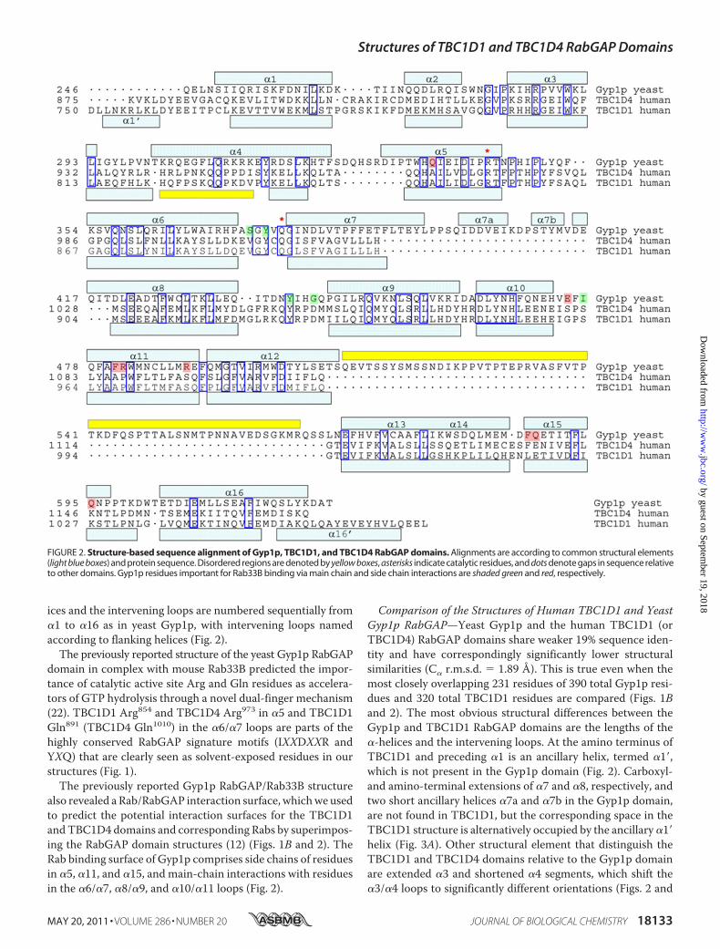
ices and the intervening loops are numbered sequentially from�1 to �16 as in yeast Gyp1p, with intervening loops namedaccording to flanking helices (Fig. 2).The previously reported structure of the yeast Gyp1p RabGAP
domain in complex with mouse Rab33B predicted the impor-tance of catalytic active site Arg and Gln residues as accelera-tors of GTP hydrolysis through a novel dual-finger mechanism(22). TBC1D1 Arg854 and TBC1D4 Arg973 in �5 and TBC1D1Gln891 (TBC1D4 Gln1010) in the �6/�7 loops are parts of thehighly conserved RabGAP signature motifs (IXXDXXR andYXQ) that are clearly seen as solvent-exposed residues in ourstructures (Fig. 1).The previously reported Gyp1p RabGAP/Rab33B structure
also revealed a Rab/RabGAP interaction surface, whichwe usedto predict the potential interaction surfaces for the TBC1D1andTBC1D4 domains and corresponding Rabs by superimpos-ing the RabGAP domain structures (12) (Figs. 1B and 2). TheRab binding surface of Gyp1p comprises side chains of residuesin �5, �11, and �15, and main-chain interactions with residuesin the �6/�7, �8/�9, and �10/�11 loops (Fig. 2).
Comparison of the Structures of Human TBC1D1 and YeastGyp1p RabGAP—Yeast Gyp1p and the human TBC1D1 (orTBC1D4) RabGAP domains share weaker 19% sequence iden-tity and have correspondingly significantly lower structuralsimilarities (C� r.m.s.d. � 1.89 Å). This is true even when themost closely overlapping 231 residues of 390 total Gyp1p resi-dues and 320 total TBC1D1 residues are compared (Figs. 1Band 2). The most obvious structural differences between theGyp1p and TBC1D1 RabGAP domains are the lengths of the�-helices and the intervening loops. At the amino terminus ofTBC1D1 and preceding �1 is an ancillary helix, termed �1�,which is not present in the Gyp1p domain (Fig. 2). Carboxyl-and amino-terminal extensions of �7 and �8, respectively, andtwo short ancillary helices �7a and �7b in the Gyp1p domain,are not found in TBC1D1, but the corresponding space in theTBC1D1 structure is alternatively occupied by the ancillary �1�helix (Fig. 3A). Other structural element that distinguish theTBC1D1 and TBC1D4 domains relative to the Gyp1p domainare extended �3 and shortened �4 segments, which shift the�3/�4 loops to significantly different orientations (Figs. 2 and
FIGURE 2. Structure-based sequence alignment of Gyp1p, TBC1D1, and TBC1D4 RabGAP domains. Alignments are according to common structural elements(light blue boxes) and protein sequence. Disordered regions are denoted by yellow boxes, asterisks indicate catalytic residues, and dots denote gaps in sequence relativeto other domains. Gyp1p residues important for Rab33B binding via main chain and side chain interactions are shaded green and red, respectively.
Structures of TBC1D1 and TBC1D4 RabGAP Domains
MAY 20, 2011 • VOLUME 286 • NUMBER 20 JOURNAL OF BIOLOGICAL CHEMISTRY 18133
by guest on September 19, 2018
http://ww
w.jbc.org/
Dow
nloaded from

3B). The �3/�4 loops in both TBC1D1 and TBC1D4 also havehigh temperature factors, indicating substantial motion andflexibility in these regions of the structures.In addition, �16 at the carboxyl terminus of TBC1D1 and
TBC1D4 is followed by an ancillary helix, termed�16�, which isnot present inGyp1p. The longitudinal axes of�16 and�16� areat �110° angles relative to one another. TBC1D1 moleculesform nonsymmetrical dimers in the crystals through interac-tions of�16� fromone subunit with�16 of the other subunit viaan antiparallel �-helix bundle. This likely represents crystallo-graphic packing, and not a biologically relevant interaction, asTBC1D1 and TBC1D4 RabGAP domains appear to be mono-meric in solution.4RabGAP Activities of Human TBC1D1 and TBC1D4—
GTPase hydrolysis kinetics were determined for humanTBC1D1 and TBC1D4 RabGAP domains using mouseRab14�GTP as the substrate. Rab14 was chosen because only itand Rabs 2A, 8A, and 10, of �20 total Rabs in mammalianproteomes that co-purify with GLUT4 vesicles, have beenshown to be in vitro substrates for TBC1D1 or TBC1D4 (6).We
also attempted to use Rab10 as a substrate for TBC1D4 (9), butwere unable to generate the quantities of purified recombinantRab10 needed for these assays.Free phosphate generated in the assays by TBC1D1 and
TBC1D4 RabGAP-catalyzed GTP hydrolysis was detectedspectrophotometrically (Fig. 4A). With Rab14�GTP as the sub-strate, catalytic efficiency parameters kcat/Km for TBC1D1 andTBC1D4 RabGAP domains were 5300 and 2800 M�1s�1,respectively (Fig. 4B). As anticipated, RabGAP activity wasabolished by substitution of invariant residues R854A, Q891A,and D851A within the catalytic pocket (see Fig. 7A).Catalytic Activities of Substituted TBC1D1 Proteins—Direct
binding assays would have been useful to assess the importanceof residues at the Rab/RabGAP interfaces, but neither we norothers in the field have successfully detected binding (resultsnot shown).5 We therefore used the GTPase assay as a very
4 S.-Y. Park, unpublished results. 5 Gus Lienhard, unpublished results.
FIGURE 3. Structural comparisons among TBC1D1, TBC1D4, and Gyp1pRabGAP domains. A, significant differences between Gyp1p and TBC1D1 orTBC1D4 are the residues between Gyp1p �7 and �8, creating �7a and �7b,which are absent in TBC1D1 and TBC1D4. The space occupied by these shorthelices in Gyp1p is occupied in TBC1D1 and TBC1D4 by the alternative ancil-lary helix �1�. B, �4 in Gyp1p is also much longer than in the TBC1D domains.Much of this region in TBC1D1 and TBC1D4 is a disordered loop. FIGURE 4. Catalytic activities of TBC1D1 RabGAP domains. A, TBC1D1 and
TBC1D4 RabGAP domains were used to catalyze hydrolysis of Rab14-loadedGTP. For each RabGAP domain concentration, kobs was fitted using a pseudofirst-order Michaelis-Menten model (A(t) � (A� � A0) (1 � exp(�kobst)) � A0).B, the catalytic efficiency parameter, Kcat/Km, was calculated by plotting kobsagainst TBC1D1 concentration, and the slopes of the best fit lines providekcat/Km values.
Structures of TBC1D1 and TBC1D4 RabGAP Domains
18134 JOURNAL OF BIOLOGICAL CHEMISTRY VOLUME 286 • NUMBER 20 • MAY 20, 2011
by guest on September 19, 2018
http://ww
w.jbc.org/
Dow
nloaded from

useful measure that integrates binding with catalysis. From thestructure of the yeast Gyp1p RabGAP�mouse Rab33B complex,we predicted potential interaction surfaces between Rabs andTBC1D1 and TBC1D4 (Figs. 1B and 2). We expected that rele-vant residues along �5, �11, and �15, where side chain interac-tions with Rab are observed, to be conserved between Gyp1p
and both TBC1D1/TBC1D4. However, our structures showedthat the side chains of Gyp1p that participate in Rab33B bind-ing through either hydrogen bonds (Gln336, Glu475, Arg482,Arg490, and Gln603) or hydrophobic clusters (Phe481, Phe595,and Gln596) are not conserved in TBC1D1 and TBC1D4(Fig. 5A, Gyp1p/TBC1D1/TBC1D4: Gln336/Ala847/Ala866,Glu475/Gly961/Ser980, Arg482/Pro968/Pro987, Arg490/Ser976/Ser995, Gln603/Lys1027/Lys1046, Phe481/Ala967/Ala986, Phe595/Leu1019/Phe1038, and Gln596/Glu1020/Glu1039). On the otherhand, the side chains ofmouse Rab33B interactingwith the sidechains of Gyp1p residues are well conserved in putativeTBC1D1 and TBC1D4 Rab partners, including mammalianRab10 and Rab14 (Fig. 6). This also suggests that side chainresidues different from those predicted by the Gyp1p/Rab33Bstructure may participate in forming TBC1D1 RabGAP/Rabinterfaces.Although there was little sequence conservation at the rele-
vant RabGAP surface, we constructed a series of TBC1D1domains containing Ala residues in place of the residues ofGyp1p that interact with Rab33B (P968A, S976A, L1019A,E1020A, and K1027A) in the crystal structure (12). The Ala-substituted TBC1D1 RabGAPs were tested for catalytic activi-ties against Rab14�GTP (attempts to pull down Rab14 witheither TBC1D1 or TBC1D4, or to induce complex formationusing either a nonhydrolyzable GTP analog or the transitionstatemimetic, GDP-AlFx, have consistently failed). Of note, thebacterial expression efficiencies, protein solubilities, and aggre-gation states of the substituted proteins were similar to those ofthe wild-type protein (results not shown), suggesting properprotein folding. Nevertheless, two of the five substituted pro-teins (P968A, K1027A) had near-normal catalytic efficiencies,two were reduced �3-fold (S976A, E1020A), and only one, theL1019A substitution, diminished GTP hydrolysis �5-fold (Fig.7A). Leu1019 of TBC1D1 aligns with Phe595 of Gyp1p, whoseside chain participates in a hydrophobic cluster at the Gyp1p/Rab33B interface. The corresponding residue of TBC1D4 isPhe1038, suggesting that either Leu or Phe may be accommo-dated at this site. The fact that two of the substitutions lackedeffects suggests that side chains Pro968 and Lys1027 of TBC1D1are unimportant for binding and catalysis, which counters pre-dictions from the Gyp1p/Rab33B structure.Because the residues are well conserved among mammalian
Rabs (Fig. 6), we also askedwhether changingTBC1D1 residuesto the corresponding Gyp1p residues improved TBC1D1 cata-lytic efficiency toward Rab14�GTP. None of the mutants
FIGURE 5. Lack of sequence similarity at Rab binding surfaces of TBC1D1and Gyp1p. A, Rab binding surfaces predicted by the Gyp1p/Rab33B struc-ture are poorly conserved between TBC1D1 and Gyp1p. B, because thesequence conservation is so low, we predicted additional solvent-exposedresidues of TBC1D1 (Pro928, Met930, and Glu959) that might participate in Rabbinding.
FIGURE 6. Sequence alignment of mammalian Rab proteins at the predicted RabGAP interface. The buried region of mouse Rab33B at the yeast Gyp1pinterface was aligned with other human Rab proteins reported potentially to interact with TBC1D proteins during GLUT4 trafficking. Rab residues important forGyp1p RabGAP binding via main chain and side chain interactions are colored green and red, respectively.
Structures of TBC1D1 and TBC1D4 RabGAP Domains
MAY 20, 2011 • VOLUME 286 • NUMBER 20 JOURNAL OF BIOLOGICAL CHEMISTRY 18135
by guest on September 19, 2018
http://ww
w.jbc.org/
Dow
nloaded from

(P968R, S976R, K1027Q, A847Q, G961E, and A967F) showed�2-fold increase in the efficiency of GTP hydrolysis (Fig. 7A).These results suggest that although general modes of Rab bind-ingmay be conserved betweenTBC1D1 andGyp1p, the contactresidues between the TBC1D1 and Rab14 likely differ signifi-cantly from those at the yeast Gyp1p/mouse Rab33B interface.To further test whether the Gyp1p:Rab33B structure pre-
dicts mammalian RabGAP/Rab binding in general, andTBC1D1/Rab14 binding in particular, we substituted residuesat the periphery of the defined Gyp1p and Rab33B interface(Fig. 5B). The three selected residues of TBC1D1, Pro928 andMet930 in the �8/�9 loop and Glu959 in the �10/�11 loop, cor-respond to residues ofGyp1p thatmakemain chain but not sidechain contacts with Rab33B. M930A in particular resulted in�5-fold decrease in catalytic efficiency, indicating that thehydrophobic side chain of Met930 in the �8/�9 loop is impor-tant in TBC1D1/Rab14 interactions (Fig. 7A).To summarize, we compared the previously solved structure
of a yeastGyp1pRabGAP�mouseRab33B complexwith the newstructures of human TBC1D1 and TBC1D4 RabGAP domainsreported here to predict residues potentially involved inTBC1D1 or TBC1D4 RabGAP�Rab binding. Because it has notbeen possible to study binding directly, we used enzymaticassays as a surrogate method for assessing binding. Based on
diminished activity for TBC1D1 substitutions L1019A andM930A, we conclude that these residues are involved at re-levant mammalian TBC1D1 and TBC1D4 RabGAP/Rabinterfaces.TBC1D1/Endogenous Rab Interactions inGLUT4Transloca-
tion Assays—Subsequent experiments determined whether thepredictions and conclusions from the aforementioned struc-ture-function studies apply to interactions between TBC1D1proteins and endogenous Rabs in living cells. To accomplishthis we expressed wild-type and alanine-substituted TBC1D1proteins in cultured mouse L6 muscle cells that had been engi-neered to monitor GLUT4 translocation through the stableexpression of labeled GLUT4. The substituted residues thatreduced human TBC1D1 RabGAP activity in the enzymaticGTPase assays (M930A and L1019A) were incorporated intothe conserved sites of full-lengthmouseTBC1D1 (M1017A andL1106A) and expressed in the L6 myocytes (Fig. 8A). Cellsexpressing wild-type or catalytically inactive mouse R941K(human R854K) TBC1D1 served as positive and dominant neg-ative controls, respectively, and numbers of cells positive forinsulin-stimulatedGLUT4 translocationwere determined (23).Insulin stimulated a 3–4-fold increase in the number of cells
with GLUT4 at the surface in cells transfected with the emptyvector control. By contrast, the expression of catalytically activewild-type TBC1D1, which converts active RabGTP to inactiveRabGDP, blocked insulin-stimulated GLUT4 translocation tothe cell surface (Figs. 7B and 8C) (23). The expression of R854KTBC1D1, with a catalytically inactive RabGAP domain, mildlystimulated GLUT4 translocation (Figs. 7B and 8C). This is con-sistent with a dominant inhibitory effect of the R854K mutant,which retains Rab binding but lacks catalytic activity, thus sup-pressing normal insulin signalingmediated by endogenous pro-teins. Expression of the L1019A- and M930A-substitutedTBC1D1 proteins had no effect on GLUT4 translocation. Theresults from the myocyte GLUT4 translocation assays agreewith results from the in vitro TBC1D1/Rab14 GTPase assays,which showed that these substitutions rendered TBC1D1 cat-alytically inactive. However, our structures predicted that theL1019A and M930A substitutions would inhibit TBC1D1/Rabbinding, which therefore inhibits catalysis through a distinctmechanism that is not dominant inhibitory, compared with theR854K mutant protein. These data confirm the biological rele-vance of Leu1019 and Met930 on interactions of TBC1D1 withendogenous Rab partner proteins.
DISCUSSION
Wehave solved structures of the RabGAPdomains of humanTBC1D1 and TBC1D4, two proteins directly involved in thetrafficking and translocation ofGLUT4-containing vesicles andinsulin-stimulated glucose uptake into cells. Although the twoRabGAP structures resemble the previously determined yeastGyp1p RabGAP domain, as each has 16 �-helices and the sameprotein fold, the length of the helical elements and the loopsconnecting the helices differ. For example, the two short ancil-lary helices (�7a and �7b) between �7 and �8 of yeast Gyp1pare not present in TBC1D1 or TBC1D4. The correspondingspace in TBC1D1 and TBC1D4 is occupied instead by an ancil-lary helix (�1�) unique among RabGAPs. The �7/�8 loops of
FIGURE 7. Assay results for catalytic activity and GLUT4 translocation.A, relative kcat/Km values for RabGAPs against GTP-loaded Rab14 are plottedrelative to WT protein kcat/Km. B, L6 muscle cells expressing a myc-GLUT4-GFPreporter were transfected with plasmids expressing WT or substituted mouseTBC1D1 proteins (numbering for the human protein provided for compari-son). *, p � 0.05; ***, p � 0.0005.
Structures of TBC1D1 and TBC1D4 RabGAP Domains
18136 JOURNAL OF BIOLOGICAL CHEMISTRY VOLUME 286 • NUMBER 20 • MAY 20, 2011
by guest on September 19, 2018
http://ww
w.jbc.org/
Dow
nloaded from

RabGAPs vary significantly in both sequence and length, evenamong yeast Gyp RabGAP homologs, and the function of thisregion is unknown (21). Another significant difference betweenTBC1D1 (and TBC1D4) and Gyp1p is in �3 and �4, whichchanges the orientation of the �3/�4 loop. However, these dif-ferences may not affect Rab binding affinity or specificitybecause they are opposite the catalytic active site. Determiningthe functional relevance of these structural differences requiresfurther experimentation.We measured catalytic activities for TBC1D1 and TBC1D4
RabGAPs using Rab14�GTP as the substrate. For TBC1D1,kcat/Km � 5300 M�1s�1 whereas for TBC1D4, kcat/Km � 2800M�1s�1. These are lower than the kcat/Km value of 100,000
M�1s�1 reported for yeast Gyp1p RabGAP toward mouseRab33B�GTP (12), but within a typical range for yeast Gyp1ptoward native yeast substrates (Sec4p kcat/Km � 2000 M�1s�1;Ypt1p kcat/Km � 26,000 M�1s�1) (12). The relatively high Kmvalue (low affinity) for TBC1D1 toward Rab14�GTP helps toexplain why we and others have been unsuccessful at co-pre-cipitating either GLUT4 vesicle-associating Rabs or recombi-nant Rab14 using TBC1D1 or TBC1D4 RabGAP proteins. Thishas been especially problematic for studies aimed at identifyingbiologically relevant Rab partners for TBC1D1 and TBC1D4,which remain unknown. It is possible that our reported kcat/Kmvalues underestimate in vivo catalytic efficiencies, as the recom-binant RabGAP proteins used for these measurements are
FIGURE 8. GLUT4 translocation assay. A, equivalent expression of the TBC1D1 proteins was verified by Western blotting lysates from the transfected L6myocytes using an anti-HA antibody. B, single-cell fluorescence assay shows co-expression of myc-GLUT4-GFP and HA-TBC1D1. C, differential effects of theHA-TBC1D1 proteins on insulin-induced myc-GLUT4-GFP translocation in L6 myocytes are shown. DAPI stains nuclei blue; Myc-GLUT4-GFP fluorescence is reddue to binding of Cy3-conjugated secondary antibody.
Structures of TBC1D1 and TBC1D4 RabGAP Domains
MAY 20, 2011 • VOLUME 286 • NUMBER 20 JOURNAL OF BIOLOGICAL CHEMISTRY 18137
by guest on September 19, 2018
http://ww
w.jbc.org/
Dow
nloaded from

taken out of the context of the intact TBC1D1 and TBC1D4proteins. Other regions likely participate in protein/membraneor protein/protein interactions, including the phosphotyrosinebinding domains of TBC1D1 and TBC1D4. Their phosphoty-rosine binding domains may interact with insulin-regulatedaminopeptidase in GLUT4 vesicles (11), which would increasethe local concentrations of TBC1D1 and TBC1D4 near mem-brane-bound Rabs.We had hoped that the structure of yeast Gyp1p RabGAP in
complexwithmouseRab33B (12)would accurately predict cor-responding interactions between TBC1D1 and TBC1D4 Rab-GAPs and biologically relevant, same-species Rab partners.This is partly true, but there also appear to be differences. Theresidues of Gyp1p that form hydrogen bonds and hydrophobicclusters at the Rab33B interface are not conserved in TBC1D1and TBC1D4, suggesting that TBC1D1 and TBC1D4 recognizebiologically relevant Rab partners differently. We tested thisusing an alanine-scanning approach to probe the functionalrelevance of corresponding residues of TBC1D1 and TBC1D4.We were surprised to find that two substitutions (P968A andK1027A) had little effect, two others hadmodest effects (S976Aand E1020A), and only one substitution (L1019A) of the fivetested showed �5-fold effect on catalytic efficiency. Based onthese results, we asked whether additional residues of TBC1D1corresponding to the buried surface of Gyp1p, but not actuallypresent in Gyp1p, might also participate. Using this approachwe found that substitution of Met930, in the �8/�9 loop ofTBC1D1, reduced RabGAP activity. Side chains of residues inthe corresponding loop of Gyp1p are not at the Gyp1p/Rab33Binterface and thus do not participate in binding. The cellularGLUT4 translocation assays provided a very important confir-mation that Leu1019 and Met930 are critical for TBC1D1 inter-actions with endogenous Rabs, as either substitution alone wassufficient to abrogate GLUT4 translocation.In conclusion, althoughTBC1D1 and TBC1D4 share general
modes of RabGAP/Rab binding with Gyp1p/Rab33B, addi-tional structural elements (e.g. side chains of residues in the�8/�9 loop, includingMet930) not identified in the yeast/mousehybrid structure contribute to the molecular contact surfacebetween corresponding mammalian RabGAP/Rab proteins.The perfect complementarity of proteins within biologicallyrelevant complexes results during and requires co-evolution,the fundamental driver of biological specificity. It is thereforenot surprising that the Gyp1p/Rab33B structure provides anincomplete picture of mammalian RabGAP/Rab interactions,given up to a billion years of evolutionary distance betweenyeast andmammals.Gyp1p andTBC1D1/4 are alsomore struc-turally divergent than Gyp1p and certain other mammalianRabGAP proteins, such as TBC1D22B, which may furtherdiminish our capacity to extrapolate from the Gyp1p/Rab33B
structure. Although we had aimed to crystallize TBC1D1 andTBC1D4 RabGAPs in complex with relevant same-speciesRabs, to date this has not been successful. The Gyp1p/Rab33Bstructure remains the only high resolution structure of anyRabGAP/Rab complex. A more complete picture TBC1D1 orTBC1D4 RabGAP/Rab binding thus awaits the successful solu-tions of such same-species protein complexes.
REFERENCES1. Zerial, M., and McBride, H. (2001) Nat. Rev. Mol. Cell Biol. 2, 107–1172. Stenmark, H. (2009) Nat. Rev. Mol. Cell. Biol. 10, 513–5253. Watson, R. T., and Pessin, J. E. (2006) Trends Biochem. Sci. 31, 215–2224. Kane, S., Sano, H., Liu, S. C., Asara, J. M., Lane, W. S., Garner, C. C., and
Lienhard, G. E. (2002) J. Biol. Chem. 277, 22115–221185. Sano, H., Kane, S., Sano, E., Mîinea, C. P., Asara, J. M., Lane,W. S., Garner,
C. W., and Lienhard, G. E. (2003) J. Biol. Chem. 278, 14599–146026. Mîinea, C. P., Sano, H., Kane, S., Sano, E., Fukuda, M., Peranen, J., Lane,
W. S., and Lienhard, G. E. (2005) Biochem. J. 391, 87–937. Sano,H., Eguez, L., Teruel,M.N., Fukuda,M., Chuang, T.D., Chavez, J. A.,
Lienhard, G. E., and McGraw, T. E. (2007) Cell Metab. 5, 293–3038. Sano, H., Roach,W.G., Peck, G. R., Fukuda,M., and Lienhard, G. E. (2008)
Biochem. J. 411, 89–959. Peck, G. R., Ye, S., Pham, V., Fernando, R. N., Macaulay, S. L., Chai, S. Y.,
and Albiston, A. L. (2006)Mol. Endocrinol. 20, 2576–258310. Chadt A., Leicht, K., Deshmukh, A., Jiang, L. Q., Scherneck, S., Bernhardt,
U., Dreja, T., Vogel, H., Schmolz, K., Kluge, R., Zierath, J. R., Hultschig, C.,Hoeben, R. C., Schurmann, A., Joost, H. G., and Al-Hasani, H. (2008)Nat.Genet. 40, 1354–1359
11. Dash, S., Sano, H., Rochford, J. J., Semple, R. K., Yeo, G., Hyden, C. S., Soos,M. A., Clark, J., Rodin, A., Langenberg, C., Druet, C., Fawcett, K. A., Tung,Y. C.,Wareham,N. J., Barroso, I., Lienhard, G. E., O’Rahilly, S., and Savage,D. B. (2009) Proc. Natl. Acad. Sci. U.S.A. 106, 9350–9355
12. Pan, X., Eathiraj, S.,Munson,M., and Lambright, D. G. (2006)Nature 442,303–306
13. Otwinowski, Z., and Minor, W. (1997)Methods Enzymol. 276, 307–32614. McRee, D. E. (1999) J. Struct. Biol. 125, 156–16515. Terwilliger, T. C., and Berendzen, J. (1999) Acta Crystallogr. D 55,
849–86116. Cowtan, K. (1994) Joint CCP4 and ESF-EACBM Newsletter on Protein
Crystallography 31, 34–3817. Emsley, P., and Cowtan, K. (2004)Acta Crystallogr. D Biol. Crystallogr. 60,
2126–213218. Perrakis, A., Morris, R., and Lamzin, V. S. (1999) Nat. Struct. Biol. 6,
458–46319. Brunger, A. T., Adams, P. D., Clore, G. M., DeLano, W. L., Gros, P.,
Grosse-Kunstleve, R.W., Jiang, J. S., Kuszewski, J., Nilges,M., Pannu,N. S.,Read, R. J., Rice L. M., Simonson, T., andWarren, G. L. (1998) Acta Crys-tallogr. D 54, 905–921
20. McCoy, A. J., Grosse-Kunstleve, R.W., Storoni, L. C., andRead, R. J. (2005)Acta Crystallogr. D 61, 458–464
21. DeLano, W. L. (2010) The PyMOL Molecular Graphics System, version1.3r1, Schrodinger, LLC, New York
22. Rak, A., Fedorov, R., Alexandrov, K., Albert, S., Goody, R. S., Gallwitz, D.,and Scheidig, A. J. (2000) EMBO J. 19, 5105–5113
23. Roach, W. G., Chavez, J. A., Mîinea, C. P., and Lienhard, G. E. (2007)Biochem. J. 403, 353–358
Structures of TBC1D1 and TBC1D4 RabGAP Domains
18138 JOURNAL OF BIOLOGICAL CHEMISTRY VOLUME 286 • NUMBER 20 • MAY 20, 2011
by guest on September 19, 2018
http://ww
w.jbc.org/
Dow
nloaded from

Sang-Youn Park, Wanzhu Jin, Ju Rang Woo and Steven E. ShoelsonGLUT4 Translocation
RabGTPase-activating Protein (RabGAP) Domains Reveal Critical Elements for Crystal Structures of Human TBC1D1 and TBC1D4 (AS160)
doi: 10.1074/jbc.M110.217323 originally published online March 23, 20112011, 286:18130-18138.J. Biol. Chem.
10.1074/jbc.M110.217323Access the most updated version of this article at doi:
Alerts:
When a correction for this article is posted•
When this article is cited•
to choose from all of JBC's e-mail alertsClick here
http://www.jbc.org/content/286/20/18130.full.html#ref-list-1
This article cites 22 references, 6 of which can be accessed free at
by guest on September 19, 2018
http://ww
w.jbc.org/
Dow
nloaded from