CrystalStructureoftheCatalyticDomainof Drosophila 1,4 ... · native data set was collected on the...
9
Crystal Structure of the Catalytic Domain of Drosophila 1,4-Galactosyltransferase-7 * □ S Received for publication, December 28, 2009, and in revised form, February 24, 2010 Published, JBC Papers in Press, March 17, 2010, DOI 10.1074/jbc.M109.099564 Boopathy Ramakrishnan ‡§ and Pradman K. Qasba ‡1 From the ‡ Structural Glycobiology Section and § Basic Research Program, SAIC-Frederick, Inc., Center for Cancer Research Nanobiology Program, Center for Cancer Research, NCI, National Institutes of Health, Frederick, Maryland 21702 The 1,4-galactosyltransferase-7 (4Gal-T7) enzyme, one of seven members of the 4Gal-T family, transfers in the presence of manganese Gal from UDP-Gal to an acceptor sugar (xylose) that is attached to a side chain hydroxyl group of Ser/Thr resi- dues of proteoglycan proteins. It exhibits the least protein sequence similarity with the other family members, including the well studied family member 4Gal-T1, which, in the pres- ence of manganese, transfers Gal from UDP-Gal to GlcNAc. We report here the crystal structure of the catalytic domain of 4Gal-T7 from Drosophila in the presence of manganese and UDP at 1.81 A ˚ resolution. In the crystal structure, a new manga- nese ion-binding motif (HXH) has been observed. Superposition of the crystal structures of 4Gal-T7 and 4Gal-T1 shows that the catalytic pocket and the substrate-binding sites in these pro- teins are similar. Compared with GlcNAc, xylose has a hydroxyl group (instead of an N-acetyl group) at C2 and lacks the CH 2 OH group at C5; thus, these protein structures show significant dif- ferences in their acceptor-binding site. Modeling of xylose in the acceptor-binding site of the 4Gal-T7 crystal structure shows that the aromatic side chain of Tyr 177 interacts strongly with the C5 atom of xylose, causing steric hindrance to any additional group at C5. Because Drosophila Cd7 has a 73% protein sequence similarity to human Cd7, the present crystal structure offers a structure-based explanation for the mutations in human Cd7 that have been linked to Ehlers-Danlos syndrome. Proteoglycans such as heparin/heparan sulfate, chondroitin, and dermatan sulfates are found widely on the cell surface and in the extracellular matrix of various tissues and are known to play important roles in several cellular functions such as cell growth and differentiation (1, 2). These glycosaminoglycans are biosynthesized as extended oligosaccharides on a linker tet- rasaccharide (GlcUA1,3Gal1,3Gal1,4Xyl-O-) 2 that is attached to a Ser residue on a core protein (3). Of the glycosyl- transferases that are involved in the linker saccharide synthesis, the crystal structure of 1,3-glucuronyltransferase I, which transfers GlcUA to Gal1,3Gal1,4Xyl-O, and 1,4-N- acetylhexosaminyltransferase (EXTL2), which transfers either GlcNAc or GalNAc to the terminal GlcUA residue of the linker tetrasaccharide, are available (4, 5). The Gal1,4Xyl di- saccharide moiety in the linker saccharide is synthesized by the enzyme 1,4-galactosyltransferase-7 (4Gal-T7), which trans- fers Gal to Xyl in the presence of manganese (6 –9). In humans, 4Gal-T7 is one of seven members of the 4Gal-T family, 4Gal-T1 to 4Gal-T7. Its homolog is present in all vertebrates and invertebrate species (10). It has been shown that 4Gal-T7 is an essential enzyme for the species viability, and mutation in the 4Gal-T7 gene has been linked to Ehlers-Danlos syndrome (9, 11). The 4Gal-T7 protein shows a 36% sequence similarity to its family member 4Gal-T1, whose structure and function are well known (12, 13). Briefly, in the presence of manganese, the 4Gal-T1 enzyme transfers Gal from UDP-Gal to GlcNAc present at the nonreducing end of an oligosaccharide acceptor (14). Also, in mammals, during lactation in the mammary gland, its acceptor specificity is altered from GlcNAc to Glc by the mammary gland-specific protein -lactalbumin, thus syn- thesizing lactose present in milk (15, 16). The structure and function studies on 4Gal-T1 have shown that the apoenzyme exists in an open conformation with its catalytic pocket exposed to the solvent environment to facilitate the binding of manga- nese and UDP-Gal (13). Upon manganese and UDP-Gal bind- ing, it undergoes conformational changes involving two flexible loops: 1) a short flexible loop where the side chain of a Trp residue moves from outside to inside the catalytic pocket bind- ing to UDP-Gal, and 2) a long flexible loop that moves to cover the bound UDP-Gal by forming a manganese ion coordination bond with its N-terminal His residue while its C-terminal resi- dues undergo loop-to-helix transition, creating the binding site for the acceptor sugar substrate and -lactalbumin (12, 13, 17). Upon binding of the acceptor substrate GlcNAc, Asp 318 in the bovine 4Gal-T1 enzyme (or Asp 314 in human) acts as the cat- alytic base, enabling the O4 atom of the GlcNAc to initiate a nucleophilic attack on the C1 atom of the galactose moiety of the UDP-Gal following the S n 2 catalytic mechanism (18, 19). After catalysis, the product disaccharide leaves the enzyme, * This work was supported, in whole or in part, by National Institutes of Health Contract HHSN261200800001E from NCI and by the Intramural Research Program of the National Institutes of Health, NCI, Center for Cancer Research. Use of beam line 22-ID/BM of the Southeast Regional Collabora- tive Access Team (SER-CAT), located at the Advanced Photon Source, Argonne National Laboratory, was supported by Contract W-31-109- Eng-38 from the United States Department of Energy, Office of Science, Office of Basic Energy Sciences. □ S The on-line version of this article (available at http://www.jbc.org) contains supplemental Figs. S1–S5. The atomic coordinates and structure factors (code 3LW6) have been deposited in the Protein Data Bank, Research Collaboratory for Structural Bioinformat- ics, Rutgers University, New Brunswick, NJ (http://www.rcsb.org/). 1 To whom correspondence should be addressed: Structural Glycobiology Section, CCRNP, CCR, NCI-Frederick, Bldg. 469, Rm. 221, Frederick, MD 21702. Tel.: 301-846-1934; Fax: 301-846-7149; E-mail: [email protected]. 2 The abbreviations used are: GlcUA, glucuronic acid; Xyl, xylose; 4Gal-T, 1,4-galactosyltransferase; MPD, 2-methyl-2,4-pentanediol. THE JOURNAL OF BIOLOGICAL CHEMISTRY VOL. 285, NO. 20, pp. 15619 –15626, May 14, 2010 Printed in the U.S.A. MAY 14, 2010 • VOLUME 285 • NUMBER 20 JOURNAL OF BIOLOGICAL CHEMISTRY 15619 by guest on June 4, 2019 http://www.jbc.org/ Downloaded from
Transcript of CrystalStructureoftheCatalyticDomainof Drosophila 1,4 ... · native data set was collected on the...

Crystal Structure of the Catalytic Domain of Drosophila�1,4-Galactosyltransferase-7*□S
Received for publication, December 28, 2009, and in revised form, February 24, 2010 Published, JBC Papers in Press, March 17, 2010, DOI 10.1074/jbc.M109.099564
Boopathy Ramakrishnan‡§ and Pradman K. Qasba‡1
From the ‡Structural Glycobiology Section and §Basic Research Program, SAIC-Frederick, Inc., Center for Cancer ResearchNanobiology Program, Center for Cancer Research, NCI, National Institutes of Health, Frederick, Maryland 21702
The �1,4-galactosyltransferase-7 (�4Gal-T7) enzyme, one ofsevenmembers of the �4Gal-T family, transfers in the presenceof manganese Gal from UDP-Gal to an acceptor sugar (xylose)that is attached to a side chain hydroxyl group of Ser/Thr resi-dues of proteoglycan proteins. It exhibits the least proteinsequence similarity with the other family members, includingthe well studied family member �4Gal-T1, which, in the pres-ence ofmanganese, transfers Gal fromUDP-Gal to GlcNAc.Wereport here the crystal structure of the catalytic domain of�4Gal-T7 from Drosophila in the presence of manganese andUDP at 1.81 A resolution. In the crystal structure, a newmanga-nese ion-bindingmotif (HXH)has beenobserved. Superpositionof the crystal structures of �4Gal-T7 and �4Gal-T1 shows thatthe catalytic pocket and the substrate-binding sites in these pro-teins are similar. Compared with GlcNAc, xylose has a hydroxylgroup (instead of anN-acetyl group) at C2 and lacks the CH2OHgroup at C5; thus, these protein structures show significant dif-ferences in their acceptor-binding site.Modeling of xylose in theacceptor-binding site of the �4Gal-T7 crystal structure showsthat the aromatic side chain of Tyr177 interacts strongly with theC5 atom of xylose, causing steric hindrance to any additionalgroup at C5. Because Drosophila Cd7 has a 73% proteinsequence similarity to human Cd7, the present crystal structureoffers a structure-based explanation for themutations in humanCd7 that have been linked to Ehlers-Danlos syndrome.
Proteoglycans such as heparin/heparan sulfate, chondroitin,and dermatan sulfates are found widely on the cell surface andin the extracellular matrix of various tissues and are known toplay important roles in several cellular functions such as cellgrowth and differentiation (1, 2). These glycosaminoglycans arebiosynthesized as extended oligosaccharides on a linker tet-
rasaccharide (GlcUA�1,3Gal�1,3Gal�1,4Xyl�-O-)2 that isattached to a Ser residue on a core protein (3). Of the glycosyl-transferases that are involved in the linker saccharide synthesis,the crystal structure of �1,3-glucuronyltransferase I, whichtransfers �GlcUA to Gal�1,3Gal�1,4Xyl�-O, and �1,4-N-acetylhexosaminyltransferase (EXTL2), which transfers either�GlcNAc or �GalNAc to the terminal �GlcUA residue of thelinker tetrasaccharide, are available (4, 5). The Gal�1,4Xyl di-saccharide moiety in the linker saccharide is synthesized by theenzyme �1,4-galactosyltransferase-7 (�4Gal-T7), which trans-fers Gal to Xyl in the presence of manganese (6–9). In humans,�4Gal-T7 is one of seven members of the �4Gal-T family,�4Gal-T1 to�4Gal-T7. Its homolog is present in all vertebratesand invertebrate species (10). It has been shown that �4Gal-T7is an essential enzyme for the species viability, and mutation inthe �4Gal-T7 gene has been linked to Ehlers-Danlos syndrome(9, 11).The �4Gal-T7 protein shows a 36% sequence similarity to its
family member �4Gal-T1, whose structure and function arewell known (12, 13). Briefly, in the presence of manganese, the�4Gal-T1 enzyme transfers Gal from UDP-Gal to GlcNAcpresent at the nonreducing end of an oligosaccharide acceptor(14). Also, in mammals, during lactation in the mammarygland, its acceptor specificity is altered from GlcNAc to Glc bythe mammary gland-specific protein �-lactalbumin, thus syn-thesizing lactose present in milk (15, 16). The structure andfunction studies on �4Gal-T1 have shown that the apoenzymeexists in an open conformationwith its catalytic pocket exposedto the solvent environment to facilitate the binding of manga-nese and UDP-Gal (13). Upon manganese and UDP-Gal bind-ing, it undergoes conformational changes involving two flexibleloops: 1) a short flexible loop where the side chain of a Trpresidue moves from outside to inside the catalytic pocket bind-ing to UDP-Gal, and 2) a long flexible loop that moves to coverthe bound UDP-Gal by forming a manganese ion coordinationbond with its N-terminal His residue while its C-terminal resi-dues undergo loop-to-helix transition, creating the binding sitefor the acceptor sugar substrate and �-lactalbumin (12, 13, 17).Upon binding of the acceptor substrate GlcNAc, Asp318 in thebovine �4Gal-T1 enzyme (or Asp314 in human) acts as the cat-alytic base, enabling the O4 atom of the GlcNAc to initiate anucleophilic attack on the C1 atom of the galactose moiety ofthe UDP-Gal following the Sn2 catalytic mechanism (18, 19).After catalysis, the product disaccharide leaves the enzyme,
* This work was supported, in whole or in part, by National Institutes of HealthContract HHSN261200800001E from NCI and by the Intramural ResearchProgram of the National Institutes of Health, NCI, Center for CancerResearch. Use of beam line 22-ID/BM of the Southeast Regional Collabora-tive Access Team (SER-CAT), located at the Advanced Photon Source,Argonne National Laboratory, was supported by Contract W-31-109-Eng-38 from the United States Department of Energy, Office of Science,Office of Basic Energy Sciences.
□S The on-line version of this article (available at http://www.jbc.org) containssupplemental Figs. S1–S5.
The atomic coordinates and structure factors (code 3LW6) have been depositedin the Protein Data Bank, Research Collaboratory for Structural Bioinformat-ics, Rutgers University, New Brunswick, NJ (http://www.rcsb.org/).
1 To whom correspondence should be addressed: Structural GlycobiologySection, CCRNP, CCR, NCI-Frederick, Bldg. 469, Rm. 221, Frederick, MD21702. Tel.: 301-846-1934; Fax: 301-846-7149; E-mail: [email protected].
2 The abbreviations used are: GlcUA, glucuronic acid; Xyl, xylose; �4Gal-T,�1,4-galactosyltransferase; MPD, 2-methyl-2,4-pentanediol.
THE JOURNAL OF BIOLOGICAL CHEMISTRY VOL. 285, NO. 20, pp. 15619 –15626, May 14, 2010Printed in the U.S.A.
MAY 14, 2010 • VOLUME 285 • NUMBER 20 JOURNAL OF BIOLOGICAL CHEMISTRY 15619
by guest on June 4, 2019http://w
ww
.jbc.org/D
ownloaded from

allowing it to revert back to the open conformation to exchangethe bound UDP for UDP-Gal to start a new catalytic cycle(12, 13).Although the �4Gal-T7 molecule shares protein sequence
similarity with the�4Gal-T1molecule and is expected to have asimilar overall three-dimensional structure, it exhibits distinctdifferences from �4Gal-T1, including its acceptor sugar speci-ficity. For example, it has been shown that the binding of man-ganese and UDP alone can introduce conformational changesin the �4Gal-T7 molecule, whereas manganese and UDP-Galare necessary in the �4Gal-T1 molecule (20–23). Also, the�4Gal-T1 enzyme exhibits high catalytic activity above pH 7.0,whereas the �4Gal-T7 enzyme exhibits maximum catalyticactivity at pH 6.5 (7). Therefore, the three-dimensional struc-ture of the �4Gal-T7 molecule is essential to understand itsstructure and function.We present here the crystal structure ofthe catalytic domain of �4Gal-T7 from Drosophila in the pres-ence of manganese and UDP at 1.81 Å resolution.
EXPERIMENTAL PROCEDURES
The Drosophila melanogaster �4Gal-T7 cDNA (AY094665)was purchased from Open Biosystems. It was found to have asingle amino acidmutation, Asp212 to Gly, whichwas correctedwhile cloning. TheDNA fragments coding the catalytic domain(Cd7) sequence (residues 71–322) and the C-terminal 11-amino acid deletion (Cd7�C) sequence (residues 71–311) werecloned into a pET23a vector between the restriction enzymesites BamHI and EcoRI (Fig. 1A). The N-terminal fusion pro-teins P-Cd7�C and P1-Cd7�C, containing a 46- and a 33-amino acid peptide (P and P1), respectively, from bovine�4Gal-T1 (residues 130–175 and 143–175, respectively), wereconstructed by inserting the DNA fragments corresponding tothe fusion peptides P and P1 at the N terminus of the Cd7�Cprotein between the restriction enzyme sites BamHI and NcoI(see Fig. 1A). The fusion constructs P-Cd7�C and P1-Cd7�Chave the DNA sequence corresponding to the C-terminaldeleted catalytic domain of Drosophila �4Gal-T7 (Cd7�C)inserted between the restriction enzyme sites NcoI and EcoRI.The four plasmids containing cDNA sequences of Cd7, Cd7�C,P-Cd7�C, and P1-Cd7�C were first confirmed by DNAsequencing and then transfected into a Rosetta(DE3)pLysS cellfor protein expression.Protein Expression and Refolding—The four Rosetta(DE3)-
pLysS cells, each containing one of the four forms ofDrosophilaCd7 cDNA sequences, Cd7, Cd7�C, P-Cd7�C, and P1-Cd7�C,in the pET23a vector described above, were grown to anabsorbance of 0.7–0.8 and then induced with 1 mM isopropyl�-D-thiogalactopyranoside. The inclusion bodies were purifiedfrom the bacterial pellet as described (24, 25). From 1 liter ofbacterial culture, 60–70 mg of protein were obtained as inclu-sion bodies. The in vitro folding of Cd7 was carried out in a waysimilar to that of�4Gal-T1 (25). Typically, 100mgof sulfonatedprotein were folded for 48 h in 1 liter of folding solution con-taining oxido-shuffling agents and 500 mM arginine HCl. Afterrefolding the protein, the folding solution was extensively dia-lyzed in water. During dialysis, the misfolded protein precipi-tated out, whereas the folded protein remained soluble. Thesoluble active protein was concentrated on an Amicon stirred
cell using a YM-10 membrane, and no further purification wasnecessary. Nearly 20 mg of folded and active Drosophila Cd7protein were obtained from 1 liter of folding solution. TheC-terminal deletion (Cd7�C) or the addition of N-terminalfusion peptide P (P-Cd7�C) or P1 (P1-Cd7�C) did not affectthe folding efficiency.Crystallization and Structure Determination—The crystals
of P1-Cd7�C were grown by the hanging drop vapor diffusionmethod by mixing equal amounts of P1-Cd7�C protein solu-tion containing 10–20 mg/ml protein, 33 mMUDP, and 66 mM
MnCl2with a solution of precipitating agent containing 100mM
Tris-HCl (pH 8.0), 1 M NaCl, 15% (v/v) MPD, and 5% (w/v)polyethylene glycol 6000. The tetragonal bipyramidal crystalsgrew in 1–2 days to a size of 0.2–0.4 mm3. A 1.81 Å resolutionnative data set was collected on the 22-BM beam line with anx-ray beam at a 1.0-Å wavelength. The data collection statisticsare given in Table 1. Crystals grown in the presence of NaBrinstead ofNaClwere used for solving the crystal structure usinganomalous dispersion arising from bromine atoms (26). Single-wavelength anomalous dispersion diffraction data up to 2 Åresolution were collected on the crystals grown with 1 M NaBrat a 0.919-Å wavelength on the 22-ID beam line. All frameswere processed with HKL3000 (27). The protein structure wassolved by single-wavelength anomalous dispersion methodsusing the program HKL2MAP (28). In the crystal structure,there is one P1-Cd7�Cmolecule in the asymmetric unit. Of thetotal 240 residues in the Cd7�C protein, 230 residues wereautomatically fitted into the solvent-flattened electron densitymaps generated by HKL2MAP by the web-based ARP/wARPprogram (29). At this stage, the model was refined using thenative data up to 1.81 Å resolution. The missing 10 residueswere better visible and were built based on the difference elec-tron density maps using the program Coot (30). In addition toone manganese ion, UDP, and one MPD molecule, the solventwater molecules located using the program Coot were alsoincluded in the refinement. All refinements were carried outusing Refmac5.5, which is part of the CCP4i package (31, 32).The final refinement statistics are given in Table 1. The finalprotein model contains all 240 residues from the Cd7�C pro-tein. The N-terminal fusion peptide P1 could not be located inthe electron density maps. Although the backbone conforma-tion for Leu263 is in a generously allowed region of the Ram-achandran plot, it has awell defined electron density. All figureswere drawn using the PyMOL molecular graphics program.The structure factors and coordinates have been deposited inthe Protein Data Bank (code 3LW6).
RESULTS AND DISCUSSION
Catalytic Domain of Drosophila �4Gal-T7 and Its Crys-tallization—Aprotein sequence comparison of the human andDrosophila �4Gal-T7 enzymes shows that a strong similarityexists, beginningwithHis93 of the human sequence andHis75 ofthe Drosophila sequence, suggesting that the catalytic domainmay start from this residue (Fig. 1B). This was further con-firmed by comparingmore known�4Gal-T7 protein sequencesfromother species (supplemental Fig. S1). Thus, theDrosophilaCd7 expression construct contained Gly71–Thr322. The in vitrorefolded active Cd7 protein exhibits characteristic catalytic
Crystal Structure of �4Gal-T7
15620 JOURNAL OF BIOLOGICAL CHEMISTRY VOLUME 285 • NUMBER 20 • MAY 14, 2010
by guest on June 4, 2019http://w
ww
.jbc.org/D
ownloaded from
activity shown for Drosophila �4Gal-T7 (supplemental Fig.S2) (7). Crystallization of this refolded Drosophila Cd7 pro-tein has not been successful. Because only the �4Gal-T7enzymes from flying insects have the C-terminal extendedpeptide (Fig. 1B and supplemental Fig. S1), it was hypothe-sized that this extended peptide might interfere with crystalpacking. Therefore, the C-terminal 11-amino acid extension inDrosophila Cd7 (residues 312–322) was deleted. The refoldingof the C-terminal deletion Drosophila protein Cd7�C alsogenerated soluble and active protein without any loss of cat-alytic activity (supplemental Fig. S2) or folding efficiency.However, so far, crystallization of this protein has also failed.Because �4Gal-T7 is amember of the �4Gal-T family, we com-pared the protein sequences of�4Gal-T7 fromhuman andDro-sophila with the catalytic domain of bovine �4Gal-T1 (Cd1)(Fig. 1B). Such a comparison shows that the Cd7 protein exhib-its sequence similarity to the bovine�4Gal-T1 catalytic domain(Cd1), beginning with residue 176. The Cd7 protein lacks asimilar N-terminal sequence corresponding to bovine Cd1 res-idues 129–175. Because bovine Cd1 crystallizes readily, it washypothesized that the absence of the similar N-terminal resi-dues inDrosophilaCd7may be responsible for the difficulties incrystallizing it. Therefore, theN-terminal peptides from bovineCd1 (residues 129–175 (P) or residues 143–175 (P1)) werefused as an N-terminal fusion peptide with the Cd7�C protein(Fig. 1A). The presence of theN-terminal fusion peptide P or P1affected neither the catalytic activity nor the folding efficiency.The fusion protein P-Cd7�C could not be crystallized. In con-trast, the P1-Cd7�Cprotein readily crystallized in the presenceof MnCl2 and UDP or UDP-Gal. Inclusion of its own stemregion (residues 34–70) with the catalytic domain decreasedthe folding efficiency to �1% only, and this soluble domainprotein has not been crystallized so far.
Overall Crystal Structure of Drosophila P1-Cd7�C—In thecrystal structure of P1-Cd7�C, all 240 residues in the Cd7�Cmolecule have been located, although the N-terminal fusionpeptide P1 from bovine Cd1 could not be clearly located fromelectron density mapping (Fig. 2A). However, SDS-PAGE anal-ysis of the protein crystals showed the presence of the intactfusion protein (P1-Cd7�C) in the crystals. Therefore, thefusion peptide in the crystal is considered to be disordered. Inthe crystal structure of Cd7�C, there are two disulfide bondsfound at the C terminus between Cys255 and Cys310 andbetween Cys300 and Cys308 (Fig. 2A). The cysteine residues ofthe former disulfide bond are present only in the �4Gal-T7protein from flying insects, whereas the latter are conserved inthe �4Gal-T7 protein from all of the species (supple-mental Fig. S1). This is in contrast to the crystal structure of thebovine �4Gal-T1 catalytic domain Cd1 (Fig. 2B) (22), in whichthe two disulfide bonds found are in the N-terminal region andare conserved in all six familymembers,�4Gal-T1 to�4Gal-T6,from all of the species.The crystal structure of the Cd7�C protein (Fig. 2A) forms
the conventional GT-A fold similar to bovine Cd1 (Fig. 2B).Superposition of these two crystal structures (Fig. 2C) usingcombinatorial extension methods (33) shows a root meansquare deviation of 1.8Å between theDrosophilaCd7 structure(His75–Cys300) and the bovine Cd1 structure (His180–Thr400)(22, 23). A total of 209 residues were used in the alignment; theZ-score is 6.8. In this superposition, there are five regions show-ing significant differences (labeled 1–5 in Fig. 2C). The N-ter-minal residues Cys134–Cys176 of bovine �4Gal-T1 and theirinteractionswith its C terminus are absent in theCd7�Ccrystalstructure (labeled 1 and 2, respectively, in Fig. 2C). The C-ter-minal region of the Cd7�C molecule has two disulfide bonds(Fig. 2A) and exhibits a significant difference from the�4Gal-T1 structure (labeled 3 in Fig. 2C). The acceptor sub-strate-binding site in these structures, particularly theN2-acetyl andC6H2OHgroups of theGlcNAc-binding regions,shows significant differences (labeled 4 and 5, respectively,in Fig. 2C). It has been shown that the catalytic domain ofbovine �4Gal-T1 begins with Cys134 and that its catalyticactivity is lost either upon mutation of Cys134 to Ser or upondeletion of 9 amino acids at the N terminus, suggesting thatthese residues in the N-terminal region of bovine Cd1 playan important role in catalytic activity (24). In the crystalstructure of bovine �4Gal-T1, Cys134 forms a disulfide bondwith Cys176 (Fig. 2B), and part of this N-terminal regioninteracts with the C-terminal residues (17, 22, 23). The lackof this region in �4Gal-T7 (labeled 1 in Fig. 2C) may havebeen compensated by the longer C-terminal extension with adisulfide bond (labeled 3 in Fig. 2C) and extended C-terminalsheet structure (labeled 2 in Fig. 2C).Manganese and UDP Binding to Drosophila Cd7�C—In the
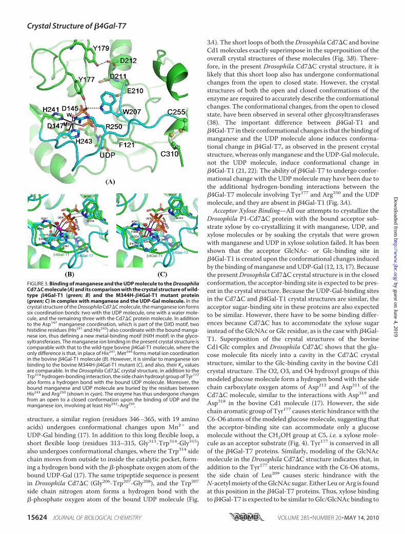
Drosophila Cd7�C crystal structure, one manganese ion andone UDP molecule have been located from electron densitymapping (Fig. 3A). The manganese ion exhibits six coordina-tion bonds: one with the side chain carboxylate oxygen atomof Asp147; two with the side chain nitrogen atoms N�2 and N�1of His241 andHis243, respectively; two with the oxygen atoms ofeach of the twophosphate groups of theUDPmolecule; and one
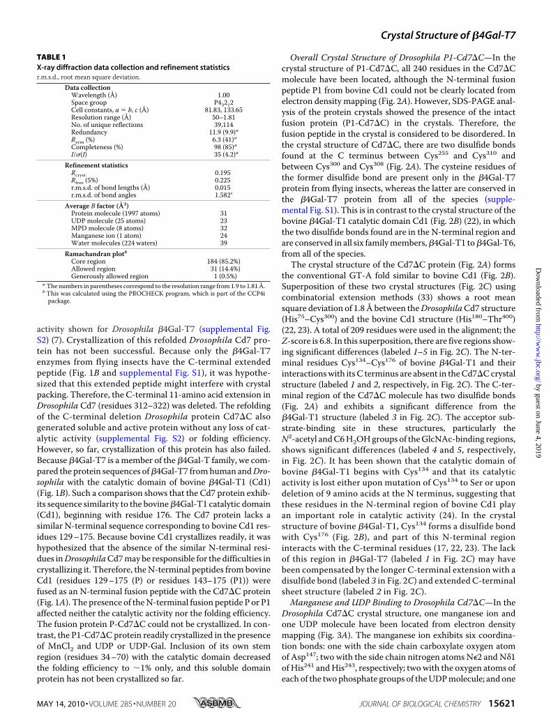
TABLE 1X-ray diffraction data collection and refinement statisticsr.m.s.d., root mean square deviation.
Data collectionWavelength (Å) 1.00Space group P43212Cell constants, a � b, c (Å) 81.83, 133.65Resolution range (Å) 50–1.81No. of unique reflections 39,114Redundancy 11.9 (9.9)aRsym (%) 6.3 (41)aCompleteness (%) 98 (85)aI/�(I) 35 (4.2)a
Refinement statisticsRcryst 0.195Rfree (5%) 0.225r.m.s.d. of bond lengths (Å) 0.015r.m.s.d. of bond angles 1.582°
Average B factor (Å2)Protein molecule (1997 atoms) 31UDP molecule (25 atoms) 23MPD molecule (8 atoms) 32Manganese ion (1 atom) 24Water molecules (224 waters) 39
Ramachandran plotbCore region 184 (85.2%)Allowed region 31 (14.4%)Generously allowed region 1 (0.5%)
a The numbers in parentheses correspond to the resolution range from1.9 to 1.81Å.b This was calculated using the PROCHECK program, which is part of the CCP4ipackage.
Crystal Structure of �4Gal-T7
MAY 14, 2010 • VOLUME 285 • NUMBER 20 JOURNAL OF BIOLOGICAL CHEMISTRY 15621
by guest on June 4, 2019http://w
ww
.jbc.org/D
ownloaded from

with awatermolecule (Fig. 3A). Asp147 is part of thewell knownmetal-binding DXD motif that is found in most glycosyltrans-ferases (34). However, in the present structure, a new metal-binding motif, 241HXH243, has been observed for the first timein the crystal structure of a glycosyltransferase. TheHXHmotifis conserved in the �4Gal-T7 proteins from all species. Super-position of the crystal structure of the DrosophilaCd7�C�Mn2��UDP complex with the crystal structure of thewild-type bovine Cd1�Mn2��UDP-Gal complex shows that thebinding of the manganese ion and UDP molecule in these pro-
teins is very similar (22, 23). However, the Kd value for manga-nese binding to theDrosophilaCd7�C enzyme (Kd � 1.5 � 0.2�M) (supplemental Fig. S3) is 20 times lower than the Kd valuefor its binding to the wild-type bovine Cd1 enzyme (Kd � 30�M), whereas it is comparablewith theKd value for theM344H-�4Gal-T1 mutant enzyme (Kd � 1.4 �M) (35). The higher Kdvalue for manganese binding to wild-type bovine Cd1 may bedue to the coordination of the side chain S� atomofMet344withthe manganese ion (22, 23). When Met344 in bovine Cd1 ismutated to His, as in the M344H-�4Gal-T1 enzyme, the N�2
FIGURE 1. A, schematic diagram showing the bovine catalytic domain of �4Gal-T1 (Cd1) and various protein constructs of the catalytic domain of the Drosophila�4Gal-T7 molecule (Cd7, Cd7�C, P-Cd7�C, and P1-Cd7�C). B, protein sequence comparison of human and Drosophila �4Gal-T7 proteins with the catalyticdomain of the bovine �4Gal-T1 (Cd1) protein. The protein sequence similarity and identity between the human and Drosophila �4Gal-T7 enzymes are 58 and43%, respectively, and the similarity between bovine Cd1 and the human and Drosophila �4Gal-T7 enzymes is 33 and 43% respectively. The �4Gal-T7 proteinsshow high similarity, starting with His93 in human and His75 in Drosophila, suggesting that their catalytic domains might start with these residues. The catalyticdomains of �4Gal-T7 proteins lack the N-terminal region corresponding residues 130 –175 of the bovine Cd1 protein. However, the functional residues(underlined) in �4Gal-T1, such as the metal-binding motif DVD and the catalytic pocket residues WGGEDDD, are conserved in the �4Gal-T7 proteins. Thedisulfide bond-forming residues in the human and Drosophila �4Gal-T7 proteins are shown by arrows. The three individual amino acid mutations (A186D,L206P, and R270C) that have been linked to Ehlers-Danlos syndrome in human �4Gal-T7 are indicated (#) (11, 42, 43).
Crystal Structure of �4Gal-T7
15622 JOURNAL OF BIOLOGICAL CHEMISTRY VOLUME 285 • NUMBER 20 • MAY 14, 2010
by guest on June 4, 2019http://w
ww
.jbc.org/D
ownloaded from

atom of His344 coordinates with the manganese ion (35), simi-lar to the situation in the Drosophila Cd7�C crystal structure(Fig. 3A). Interestingly, the bovine M344H-�4Gal-T1 mutantenzyme exhibits better catalytic activity with magnesium thanwith manganese (35), whereas the Drosophila Cd7 enzymeexhibits only very low catalytic activity with magnesium. In theM344H-�4Gal-T1�Mn2��UDP-Gal complex crystal structure,the N�2 atoms of both His344 and His347 form a coordinationbond with the manganese ion (35); this is in contrast to thepresent crystal structure of Drosophila Cd7�C, in which theN�2 and N�1 atoms of His241 and His243, respectively, form acoordination bond with the manganese ion. In solution, thetautomers of the His residue (N�2-H and N�1-H) exist in equi-librium, and the ratio of these two tautomers depends on thepH of the solution. It has been suggested that, due to the stericeffect, the N�1-H tautomer is often found to coordinate withthe metal ion rather than with the N�2-H tautomer (36). Thismay explain why these two factors might be responsible for theobserved high catalytic activity at pH 6.5 and lowerKd value forthe manganese ion for Drosophila Cd7�C compared with�4Gal-T1, where a maximum catalytic activity has beenobserved above the neutral pH values.
Although we grew crystals of theDrosophila P1-Cd7�C protein inthe presence of UDP-Gal and man-ganese, in the crystal structure, thegalactose moiety was not observed(Fig. 3A), suggesting that the UDP-Gal might have hydrolyzed. TheDrosophila Cd7 protein naturallyexhibits hydrolysis activity withUDP-Gal in the absence of theacceptor substrate, and this maybe responsible for the absence of theGal moiety in the crystals of theP1-Cd7�C protein grown withUDP-Gal and manganese. This issimilar to the bovine �3Gal-Tenzyme, which also exhibits UDP-Gal hydrolysis activity and whosecrystal structure with bound UDP-Gal could be determined only with amutant enzyme that had very lowcatalytic activity (37). In the presentcrystal structure (Fig. 3A), the bind-ing of UDP is similar to the bindingof UDP-Gal to the bovine Cd1 pro-tein (Fig. 3B). The uridine basestacks on the side chain phenylgroup of Phe121, similar to thebovine �4Gal-T1 enzyme, in whichthe uridine base stacks on the sidechain of Phe226 (22). The �-phos-phate oxygen atoms form twohydrogen bonds with the Cd7�Cprotein, where the side chain aro-matic nitrogen atom of Trp207 andthe side chain hydroxyl group of
Tyr177 form hydrogen bonds with the �-phosphate oxygenatom (Fig. 3A). Only the former hydrogen bond is observed inthe crystal structure of bovine Cd1, where the side chain nitro-gen atom of Trp314 forms a hydrogen bond with the oxygenatom of the�-phosphate group (17). The binding of UDP in thecrystal structure of Cd7�C is very similar to the binding ofUDP-Gal to the bovine �4Gal-T1molecule, where the residuesthat bind to galactose in bovine �4Gal-T1, such as Asp252,Glu317, and Asp318, are structurally conserved in the presentDrosophila Cd7�C protein. Thus, the binding of Gal to Cd7 isexpected to bindAsp145, Glu210, andAsp211, whichwill be quitesimilar to Gal binding to �4Gal-T1.DrosophilaCd7�CProtein Is in theClosedConformation—In
the present crystal structure, residues 242–251 cover the boundMn2� and the UDP molecule (Fig. 3A). This suggests that theobserved Drosophila Cd7�C crystal structure has undergoneconformational changes uponMn2� and UDP binding, involv-ing at least 9 amino acids, residues 242–251 or up to Cys255,which forms a disulfide bond with Cys310. This is substantiatedby earlier calorimetric studies onhuman�4Gal-T7 that showedthat the protein molecule undergoes conformational changeupon Mn2� and UDP binding (20). In the bovine Cd1 crystal
FIGURE 2. Shown are the crystal structures of the catalytic domain of the Drosophila Cd7�C molecule with abound manganese ion and UDP molecule (A) and the catalytic domain of the bovine �4Gal-T1 molecule in theclosed conformation (B), shown as a composite picture with the bound manganese, UDP-Gal, and GlcNAcmolecules (generated from individual structures with bound manganese and UDP-Gal and GlcNAc molecules,Protein Data Bank codes 1O0R and 1NQI, respectively). The coloring of the ribbon diagram of the �4Gal-T1molecule (B) progressively changes from blue for the N-terminal residues to red for the C-terminal residues; asimilar region in the Drosophila Cd7�C molecule (B) is colored likewise. In the ribbon diagram, the disulfidebond-forming Cys residues in both protein molecules are shown (yellow). Superposition of C� atoms of Cd7�C(in red) and bovine �4Gal-T1 (in blue) in stereo is shown in C. As predicted from the sequence comparison, theN-terminal polypeptide region (residues 134 –176; shown in blue) in bovine �4Gal-T1 is absent in the crystalstructure of the Drosophila Cd7�C molecule. Superposition of the C� atoms of these two molecules (C) showsfive surface regions with significant differences (labeled 1–5 in Fig. 2C). The differences around the acceptorsubstrate-binding site (labeled 2 and 3 in Fig. 2C) may be possibly due to the different sugar acceptor specificityof the Cd7�C molecule, whereas the lack of the region corresponding to the N-terminal region in bovine�4Gal-T1 (residues 134 –176) (labeled 1 in Fig. 2C) may have been structurally compensated by the differencein the C-terminal region of Cd7�C (labeled 4 and 5 in Fig. 2C).
Crystal Structure of �4Gal-T7
MAY 14, 2010 • VOLUME 285 • NUMBER 20 JOURNAL OF BIOLOGICAL CHEMISTRY 15623
by guest on June 4, 2019http://w
ww
.jbc.org/D
ownloaded from
structure, a similar region (residues 346–365, with 19 aminoacids) undergoes conformational changes upon Mn2� andUDP-Gal binding (17). In addition to this long flexible loop, ashort flexible loop (residues 313–315, Gly313-Trp314-Gly315)also undergoes conformational changes, where the Trp314 sidechain moves from outside to inside the catalytic pocket, form-ing a hydrogen bond with the �-phosphate oxygen atom of thebound UDP-Gal (17). The same tripeptide sequence is presentin Drosophila Cd7�C (Gly206-Trp207-Gly208), and the Trp207side chain nitrogen atom forms a hydrogen bond with the�-phosphate oxygen atom of the bound UDP molecule (Fig.
3A). The short loops of both theDrosophilaCd7�C and bovineCd1 molecules exactly superimpose in the superposition of theoverall crystal structures of these molecules (Fig. 3B). There-fore, in the present Drosophila Cd7�C crystal structure, it islikely that this short loop also has undergone conformationalchanges from the open to closed state. However, the crystalstructures of both the open and closed conformations of theenzyme are required to accurately describe the conformationalchanges. The conformational changes, from the open to closedstate, have been observed in several other glycosyltransferases(38). The important difference between �4Gal-T1 and�4Gal-T7 in their conformational changes is that the binding ofmanganese and the UDP molecule alone induces conforma-tional change in �4Gal-T7, as observed in the present crystalstructure, whereas onlymanganese and theUDP-Galmolecule,not the UDP molecule, induce conformational change in�4Gal-T1 (21, 22). The ability of �4Gal-T7 to undergo confor-mational change with the UDPmolecule may have been due tothe additional hydrogen-bonding interactions between the�4Gal-T7 molecule involving Tyr177 and Arg250 and the UDPmolecule, and they are absent in �4Gal-T1 (Fig. 3A).Acceptor Xylose Binding—All our attempts to crystallize the
Drosophila P1-Cd7�C protein with the bound acceptor sub-strate xylose by co-crystallizing it with manganese, UDP, andxylose molecules or by soaking the crystals that were grownwith manganese and UDP in xylose solution failed. It has beenshown that the acceptor GlcNAc- or Glc-binding site in�4Gal-T1 is created upon the conformational changes inducedby the binding ofmanganese andUDP-Gal (12, 13, 17). Becausethe presentDrosophilaCd7�C crystal structure is in the closedconformation, the acceptor-binding site is expected to be pres-ent in the crystal structure. Because the UDP-Gal-binding sitesin the Cd7�C and �4Gal-T1 crystal structures are similar, theacceptor sugar-binding site in these proteins are also expectedto be similar. However, there have to be some binding differ-ences because Cd7�C has to accommodate the xylose sugarinstead of the GlcNAc or Glc residue, as is the case with �4Gal-T1. Superposition of the crystal structures of the bovineCd1�Glc complex and Drosophila Cd7�C shows that the glu-cose molecule fits nicely into a cavity in the Cd7�C crystalstructure, similar to the Glc-binding cavity in the bovine Cd1crystal structure. The O2, O3, and O4 hydroxyl groups of thismodeled glucose molecule form a hydrogen bond with the sidechain carboxylate oxygen atoms of Asp212 and Asp211 of theCd7�C molecule, similar to the interactions with Asp319 andAsp318 in the bovine Cd1 molecule (17). However, the sidechain aromatic group of Tyr177 causes steric hindrancewith theC6-O6 atoms of themodeled glucosemolecule, suggesting thatthe acceptor-binding site can accommodate only a glucosemolecule without the CH2OH group at C5, i.e. a xylose mole-cule as an acceptor substrate (Fig. 4). Tyr177 is conserved in allof the �4Gal-T7 proteins. Similarly, modeling of the GlcNAcmolecule in the Drosophila Cd7�C structure indicates that, inaddition to the Tyr177 steric hindrance with the C6-O6 atoms,the side chain of Leu209 causes steric hindrance with theN-acetylmoiety of theGlcNAc sugar. Either Leu orArg is foundat this position in the �4Gal-T7 proteins. Thus, xylose bindingto�4Gal-T7 is expected to be similar toGlc/GlcNAc binding to
FIGURE 3. Binding of manganese and the UDP molecule to the DrosophilaCd7�C molecule (A) and its comparison with the crystal structure of wild-type �4Gal-T1 (green; B) and the M344H-�4Gal-T1 mutant protein(green; C) in complex with manganese and the UDP-Gal molecule. In thecrystal structure of the Drosophila Cd7�C molecule, the manganese ion formssix coordination bonds: two with the UDP molecule, one with a water mole-cule, and the remaining three with the Cd7�C protein molecule. In additionto the Asp147 manganese coordination, which is part of the DXD motif, twohistidine residues (His241 and His243) also coordinate with the bound manga-nese ion, thus defining a new metal-binding motif (HXH motif) in the glyco-syltransferases. The manganese ion binding in the present crystal structure iscomparable with that to the wild-type bovine �4Gal-T1 molecule, where theonly difference is that, in place of His241, Met344 forms metal ion coordinationin the bovine �4Gal-T1 molecule (B). However, it is similar to manganese ionbinding to the bovine M344H-�4Gal-T1 mutant (C), and also, their Kd valuesare comparable. In the Drosophila Cd7�C crystal structure, in addition to theTrp314 hydrogen-bonding interaction, the side chain hydroxyl group of Tyr177
also forms a hydrogen bond with the bound UDP molecule. Moreover, thebound manganese and UDP molecule are buried by the residues betweenHis243 and Arg250 (shown in cyan). The enzyme has thus undergone changesfrom an open to a closed conformation upon the binding of UDP and themanganese ion, involving at least His243–Arg250.
Crystal Structure of �4Gal-T7
15624 JOURNAL OF BIOLOGICAL CHEMISTRY VOLUME 285 • NUMBER 20 • MAY 14, 2010
by guest on June 4, 2019http://w
ww
.jbc.org/D
ownloaded from
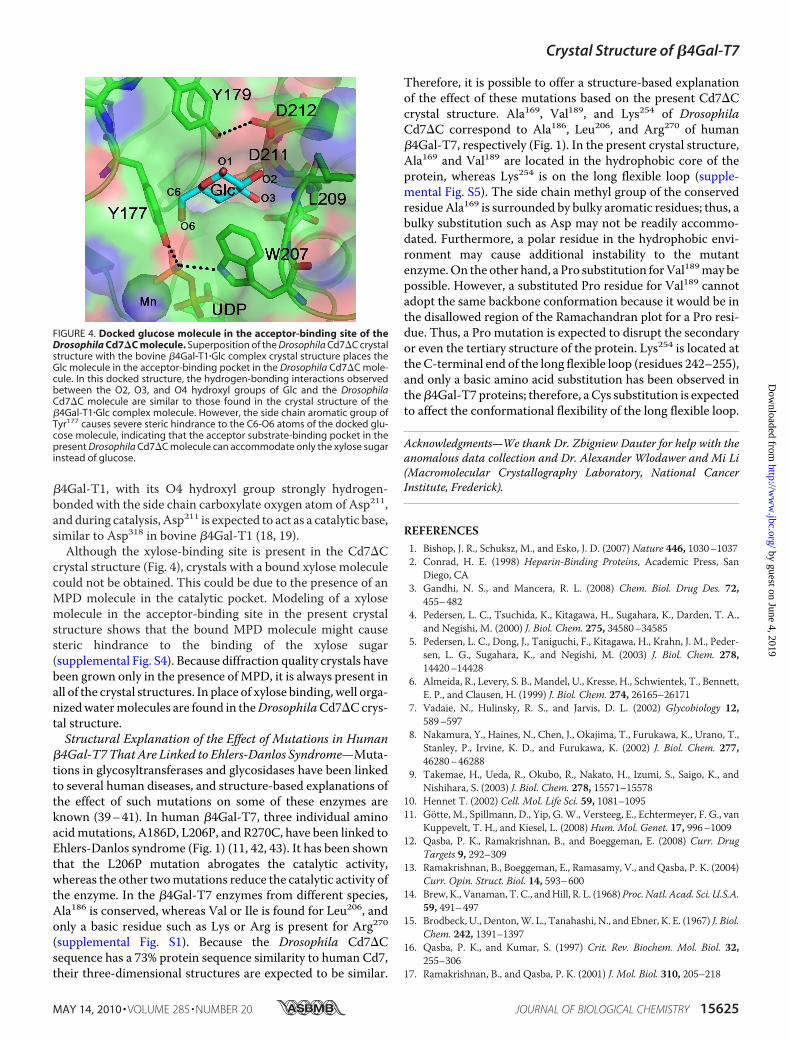
�4Gal-T1, with its O4 hydroxyl group strongly hydrogen-bonded with the side chain carboxylate oxygen atom of Asp211,and during catalysis, Asp211 is expected to act as a catalytic base,similar to Asp318 in bovine �4Gal-T1 (18, 19).Although the xylose-binding site is present in the Cd7�C
crystal structure (Fig. 4), crystals with a bound xylose moleculecould not be obtained. This could be due to the presence of anMPD molecule in the catalytic pocket. Modeling of a xylosemolecule in the acceptor-binding site in the present crystalstructure shows that the bound MPD molecule might causesteric hindrance to the binding of the xylose sugar(supplemental Fig. S4). Because diffraction quality crystals havebeen grown only in the presence ofMPD, it is always present inall of the crystal structures. In place of xylose binding, well orga-nizedwatermolecules are found in theDrosophilaCd7�Ccrys-tal structure.Structural Explanation of the Effect of Mutations in Human
�4Gal-T7 That Are Linked to Ehlers-Danlos Syndrome—Muta-tions in glycosyltransferases and glycosidases have been linkedto several human diseases, and structure-based explanations ofthe effect of such mutations on some of these enzymes areknown (39–41). In human �4Gal-T7, three individual aminoacidmutations, A186D, L206P, and R270C, have been linked toEhlers-Danlos syndrome (Fig. 1) (11, 42, 43). It has been shownthat the L206P mutation abrogates the catalytic activity,whereas the other twomutations reduce the catalytic activity ofthe enzyme. In the �4Gal-T7 enzymes from different species,Ala186 is conserved, whereas Val or Ile is found for Leu206, andonly a basic residue such as Lys or Arg is present for Arg270(supplemental Fig. S1). Because the Drosophila Cd7�Csequence has a 73% protein sequence similarity to human Cd7,their three-dimensional structures are expected to be similar.
Therefore, it is possible to offer a structure-based explanationof the effect of these mutations based on the present Cd7�Ccrystal structure. Ala169, Val189, and Lys254 of DrosophilaCd7�C correspond to Ala186, Leu206, and Arg270 of human�4Gal-T7, respectively (Fig. 1). In the present crystal structure,Ala169 and Val189 are located in the hydrophobic core of theprotein, whereas Lys254 is on the long flexible loop (supple-mental Fig. S5). The side chain methyl group of the conservedresidue Ala169 is surrounded by bulky aromatic residues; thus, abulky substitution such as Asp may not be readily accommo-dated. Furthermore, a polar residue in the hydrophobic envi-ronment may cause additional instability to the mutantenzyme.On the other hand, a Pro substitution forVal189may bepossible. However, a substituted Pro residue for Val189 cannotadopt the same backbone conformation because it would be inthe disallowed region of the Ramachandran plot for a Pro resi-due. Thus, a Pro mutation is expected to disrupt the secondaryor even the tertiary structure of the protein. Lys254 is located atthe C-terminal end of the long flexible loop (residues 242–255),and only a basic amino acid substitution has been observed inthe�4Gal-T7 proteins; therefore, a Cys substitution is expectedto affect the conformational flexibility of the long flexible loop.
Acknowledgments—We thank Dr. Zbigniew Dauter for help with theanomalous data collection and Dr. Alexander Wlodawer and Mi Li(Macromolecular Crystallography Laboratory, National CancerInstitute, Frederick).
REFERENCES1. Bishop, J. R., Schuksz, M., and Esko, J. D. (2007) Nature 446, 1030–10372. Conrad, H. E. (1998) Heparin-Binding Proteins, Academic Press, San
Diego, CA3. Gandhi, N. S., and Mancera, R. L. (2008) Chem. Biol. Drug Des. 72,
455–4824. Pedersen, L. C., Tsuchida, K., Kitagawa, H., Sugahara, K., Darden, T. A.,
and Negishi, M. (2000) J. Biol. Chem. 275, 34580–345855. Pedersen, L. C., Dong, J., Taniguchi, F., Kitagawa, H., Krahn, J. M., Peder-
sen, L. G., Sugahara, K., and Negishi, M. (2003) J. Biol. Chem. 278,14420–14428
6. Almeida, R., Levery, S. B., Mandel, U., Kresse, H., Schwientek, T., Bennett,E. P., and Clausen, H. (1999) J. Biol. Chem. 274, 26165–26171
7. Vadaie, N., Hulinsky, R. S., and Jarvis, D. L. (2002) Glycobiology 12,589–597
8. Nakamura, Y., Haines, N., Chen, J., Okajima, T., Furukawa, K., Urano, T.,Stanley, P., Irvine, K. D., and Furukawa, K. (2002) J. Biol. Chem. 277,46280–46288
9. Takemae, H., Ueda, R., Okubo, R., Nakato, H., Izumi, S., Saigo, K., andNishihara, S. (2003) J. Biol. Chem. 278, 15571–15578
10. Hennet T. (2002) Cell. Mol. Life Sci. 59, 1081–109511. Gotte, M., Spillmann, D., Yip, G. W., Versteeg, E., Echtermeyer, F. G., van
Kuppevelt, T. H., and Kiesel, L. (2008) Hum. Mol. Genet. 17, 996–100912. Qasba, P. K., Ramakrishnan, B., and Boeggeman, E. (2008) Curr. Drug
Targets 9, 292–30913. Ramakrishnan, B., Boeggeman, E., Ramasamy, V., and Qasba, P. K. (2004)
Curr. Opin. Struct. Biol. 14, 593–60014. Brew, K., Vanaman, T. C., andHill, R. L. (1968)Proc. Natl. Acad. Sci. U.S.A.
59, 491–49715. Brodbeck, U., Denton,W. L., Tanahashi, N., and Ebner, K. E. (1967) J. Biol.
Chem. 242, 1391–139716. Qasba, P. K., and Kumar, S. (1997) Crit. Rev. Biochem. Mol. Biol. 32,
255–30617. Ramakrishnan, B., and Qasba, P. K. (2001) J. Mol. Biol. 310, 205–218
FIGURE 4. Docked glucose molecule in the acceptor-binding site of theDrosophila Cd7�C molecule. Superposition of the Drosophila Cd7�C crystalstructure with the bovine �4Gal-T1�Glc complex crystal structure places theGlc molecule in the acceptor-binding pocket in the Drosophila Cd7�C mole-cule. In this docked structure, the hydrogen-bonding interactions observedbetween the O2, O3, and O4 hydroxyl groups of Glc and the DrosophilaCd7�C molecule are similar to those found in the crystal structure of the�4Gal-T1�Glc complex molecule. However, the side chain aromatic group ofTyr177 causes severe steric hindrance to the C6-O6 atoms of the docked glu-cose molecule, indicating that the acceptor substrate-binding pocket in thepresent Drosophila Cd7�C molecule can accommodate only the xylose sugarinstead of glucose.
Crystal Structure of �4Gal-T7
MAY 14, 2010 • VOLUME 285 • NUMBER 20 JOURNAL OF BIOLOGICAL CHEMISTRY 15625
by guest on June 4, 2019http://w
ww
.jbc.org/D
ownloaded from
18. Ramakrishnan, B., Ramasamy, V., andQasba, P. K. (2006) J.Mol. Biol. 357,1619–1633
19. Krupicka, M., and Tvaroska, I. (2009) J. Phys. Chem. B 113, 11314–1131920. Daligault, F., Rahuel-Clermont, S., Gulberti, S., Cung, M. T., Branlant, G.,
Netter, P., Magdalou, J., and Lattard, V. (2009) Biochem. J. 418, 605–61421. Geren, C. R., Magee, S. C., and Ebner, K. E. (1975) Biochemistry 14,
1461–146322. Ramakrishnan, B., Balaji, P. V., and Qasba, P. K. (2002) J. Mol. Biol. 318,
491–50223. Ramakrishnan, B., and Qasba, P. K. (2003) J. Biomol. Struct. Dyn. 21, 1–824. Boeggeman, E. E., Balaji, P. V., Sethi, N., Masibay, A. S., and Qasba, P. K.
(1993) Protein Eng. 6, 779–78525. Boeggeman, E. E., Ramakrishnan, B., andQasba, P. K. (2003) Protein Expr.
Purif. 30, 219–22926. Dauter, Z. (2002) Curr. Opin. Struct. Biol. 12, 674–67827. Otwinowski, Z., and Minor, W. (1997)Methods Enzymol. 276, 307–32628. Pape, T., and Schneider, T. R. (2004) J. Appl. Crystallogr. 37, 843–84429. Morris, R. J., Perrakis, A., and Lamzin, V. S. (2002)ActaCrystallogr. Sect. D
Biol. Crystallogr. 58, 968–97530. Emsley, P., and Cowtan, K. (2004) Acta Crystallogr. Sect. D Biol. Crystal-
logr. 60, 2126–2132
31. Potterton, E., Briggs, P., Turkenburg,M., and Dodson E. (2003)Acta Crys-tallogr. Sect. D Biol. Crystallogr. 59, 1131–1137
32. Murshudov, G. N., Vagin, A. A., and Dodson, E. J. (1997)Acta Crystallogr.Sect. D Biol. Crystallogr. 53, 240–255
33. Shindyalov, I. N., and Bourne, P. E. (1998) Protein Eng. 11, 739–74734. Wiggins, C. A., and Munro, S. (1998) Proc. Natl. Acad. Sci. U.S.A. 95,
7945–795035. Ramakrishnan, B., Boeggeman, E., and Qasba, P. K. (2004) Biochemistry
43, 12513–1252236. Chakrabarti, P. (1990) Protein Eng. 4, 57–6337. Tumbale, P., Jamaluddin, H., Thiyagarajan, N., Brew, K., and Acharya,
K. R. (2008) Biochemistry 47, 8711–871838. Qasba, P. K., Ramakrishnan, B., and Boeggeman, E. (2005) Trends Bio-
chem. Sci. 30, 53–6239. Zhao, H., and Grabowski, G. A. (2002) Cell. Mol. Life Sci. 59, 694–70740. Garman, S. C., and Garboczi, D. N. (2004) J. Mol. Biol. 337, 319–33541. Ju, T., and Cummings, R. D. (2005) Nature 437, 125242. Okajima, T., Fukumoto, S., Furukawa, K., and Urano, T. (1999) J. Biol.
Chem. 274, 28841–2884443. Seidler, D. G., Faiyaz-Ul-Haque, M., Hansen, U., Yip, G. W., Zaidi, S. H.,
Teebi, A. S., Kiesel, L., and Gotte, M. (2006) J. Mol. Med. 84, 583–594
Crystal Structure of �4Gal-T7
15626 JOURNAL OF BIOLOGICAL CHEMISTRY VOLUME 285 • NUMBER 20 • MAY 14, 2010
by guest on June 4, 2019http://w
ww
.jbc.org/D
ownloaded from
Boopathy Ramakrishnan and Pradman K. Qasba1,4-Galactosyltransferase-7
β DrosophilaCrystal Structure of the Catalytic Domain of
doi: 10.1074/jbc.M109.099564 originally published online March 17, 20102010, 285:15619-15626.J. Biol. Chem.
10.1074/jbc.M109.099564Access the most updated version of this article at doi:
Alerts:
When a correction for this article is posted•
When this article is cited•
to choose from all of JBC's e-mail alertsClick here
Supplemental material:
http://www.jbc.org/content/suppl/2010/03/17/M109.099564.DC1
http://www.jbc.org/content/285/20/15619.full.html#ref-list-1
This article cites 42 references, 10 of which can be accessed free at
by guest on June 4, 2019http://w
ww
.jbc.org/D
ownloaded from

![Untitled-1 [] · 2018. 3. 27. · Hatve Yüksekligi 1,4/ / l, 4/ / l, 4/ 1,6/ / l, 4/ / 1,4/ / l, 4/ 1,611 / 1,4/ 1,4/ / Kod 55300500 55300600 55300700 55300800 55300900 55301000](https://static.fdocuments.us/doc/165x107/60c24b180629fe1226743c36/untitled-1-2018-3-27-hatve-yksekligi-14-l-4-l-4-16-l-4.jpg)