Cryptogonimidae (Trematoda, Digenea) from Fishes of ...
19
This paper deals with 18 species of the family Cryptogonimidae (Trematoda, Digenea) from fishes of Japanese and adjacent waters. Dige- neans obtained were washed in saline, fixed in AFA under slight pressure, stained with Heiden- hain’s hematoxylin and mounted in Canada bal- sam. The specimens are deposited in the National Museum of Nature and Science, Tokyo (NSMT) and the Meguro Parasitological Museum (MPM). Measurements are given in millimeters unless otherwise indicated. Latuterus karemlal sp. nov. (Fig. 1) Type host. Lutjanus gibbus (Forsskål) (Lu- tjanidae). Site. Upper intestine. Type locality. Palau, western Caroline Is., 11-VIII-1994. Specimens. Holotype and 6 paratypes, NSMT-Pl 4671. Etymology. The specific name karemlal is from the Palauan local name of the host. Description. Based on seven specimens. Body oval, 0.87–1.38 long by 0.70–1.16 wide; length/ width ratio 1.1–1.3. Tegument with fine spines. Oral sucker subterminal, 0.09–0.180.16–0.27, without circumoral spines; prepharynx 0.04–0.10 long; pharynx well-developed, 0.10–0.130.12– 0.22; esophagus 0.01–0.09 long, bifurcating mid- way between suckers or closer to acetabulum; caeca voluminous, arcuate, terminating near pos- terior end of body. Acetabulum 0.07–0.110.08– 0.14, embedded in ventrogenital sac. Ventrogeni- tal sac surrounded by circular muscle and provid- ed anteriorly with glandular cells. Sucker ratio 1 : 0.45–0.51. Forebody 38–45% of body length. Cryptogonimidae (Trematoda, Digenea) from Fishes of Japanese and Adjacent Waters Masaaki Machida Department of Zoology, National Museum of Nature and Science, 3–23–1 Hyakunincho, Shinjuku-ku, Tokyo, 169–0073 Japan Meguro Parasitological Museum, 4–1–1 Shimomeguro, Meguro-ku, Tokyo, 153–0064 Japan Abstract Eighteen species of cryptogonimid digeneans (Trematoda) were obtained from fishes of Japanese and adjacent waters. Two new genera are proposed: Mehravermis gen. nov. for M. velasquezae sp. nov. and Opistognathotrema gen. nov. for O. philippinense sp. nov. Ten new species are described: Latuterus karemlal sp. nov. from Lutjanus gibbus (Lutjanidae), Metadena plectorhinchi sp. nov. from Plectorhinchus gibbosus (Haemulidae), M. pterocaesionis sp. nov. from Pterocaesio diagramma (Caesionidae), Mehravermis velasquezae sp. nov. from Opistognathus sp. (Opistognathidae), Opistognathotrema philippinense sp. nov. from Opistognathus sp., Siphoderina akamachi sp. nov. from Etelis coruscans (Lutjanidae) and Pristipomoides sieboldii (Lutjanidae), S. marangsi sp. nov. from Lutjanus quinquelineatus, S. nemipteri sp. nov. from Nemipterus virgatus (Nemipteridae), S. ryukyuensis sp. nov. from Lutjanus kasmira and L. quinquelineatus, and S. xenocephali sp. nov. from Xenocephlus elongatus (Uranoscopidae). Eight previously known species are recorded: Metadena lutiani (Yamaguti, 1942), M. rooseveltiae (Yamaguti, 1970), M. sheni Miller et Cribb, 2008, Perlevilobus platycephali (Shen, 1989), Neometadena ovata (Yama- guti, 1952), Siphoderina elongata (Gu et Shen, 1979), S. onaga (Yamaguti, 1970) and S. ulaula (Yamaguti, 1970). Key words : Digenea, Cryptogonimidae, new genus, new species, marine fish, Japan, Palau, Philippines, Indonesia. Bull. Natl. Mus. Nat. Sci., Ser. A, 35(2), pp. 137–155, June 22, 2009
Transcript of Cryptogonimidae (Trematoda, Digenea) from Fishes of ...
This paper deals with 18 species of the familyCryptogonimidae (Trematoda, Digenea) fromfishes of Japanese and adjacent waters. Dige-neans obtained were washed in saline, fixed inAFA under slight pressure, stained with Heiden-hain’s hematoxylin and mounted in Canada bal-sam. The specimens are deposited in the NationalMuseum of Nature and Science, Tokyo (NSMT)and the Meguro Parasitological Museum (MPM).Measurements are given in millimeters unlessotherwise indicated.
Latuterus karemlal sp. nov.(Fig. 1)
Type host. Lutjanus gibbus (Forsskål) (Lu-tjanidae).
Site. Upper intestine.Type locality. Palau, western Caroline Is.,
11-VIII-1994.Specimens. Holotype and 6 paratypes,
NSMT-Pl 4671.Etymology. The specific name karemlal is
from the Palauan local name of the host.Description. Based on seven specimens. Body
oval, 0.87–1.38 long by 0.70–1.16 wide; length/width ratio 1.1–1.3. Tegument with fine spines.Oral sucker subterminal, 0.09–0.18�0.16–0.27,without circumoral spines; prepharynx 0.04–0.10long; pharynx well-developed, 0.10–0.13�0.12–0.22; esophagus 0.01–0.09 long, bifurcating mid-way between suckers or closer to acetabulum;caeca voluminous, arcuate, terminating near pos-terior end of body. Acetabulum 0.07–0.11�0.08–0.14, embedded in ventrogenital sac. Ventrogeni-tal sac surrounded by circular muscle and provid-ed anteriorly with glandular cells. Sucker ratio 1 :0.45–0.51. Forebody 38–45% of body length.
Cryptogonimidae (Trematoda, Digenea) from Fishes of Japanese and Adjacent Waters
Masaaki Machida
Department of Zoology, National Museum of Nature and Science, 3–23–1 Hyakunincho, Shinjuku-ku, Tokyo, 169–0073 Japan
Meguro Parasitological Museum, 4–1–1 Shimomeguro, Meguro-ku, Tokyo, 153–0064 Japan
Abstract Eighteen species of cryptogonimid digeneans (Trematoda) were obtained from fishesof Japanese and adjacent waters. Two new genera are proposed: Mehravermis gen. nov. for M. velasquezae sp. nov. and Opistognathotrema gen. nov. for O. philippinense sp. nov. Ten newspecies are described: Latuterus karemlal sp. nov. from Lutjanus gibbus (Lutjanidae), Metadenaplectorhinchi sp. nov. from Plectorhinchus gibbosus (Haemulidae), M. pterocaesionis sp. nov. fromPterocaesio diagramma (Caesionidae), Mehravermis velasquezae sp. nov. from Opistognathus sp.(Opistognathidae), Opistognathotrema philippinense sp. nov. from Opistognathus sp., Siphoderinaakamachi sp. nov. from Etelis coruscans (Lutjanidae) and Pristipomoides sieboldii (Lutjanidae), S.marangsi sp. nov. from Lutjanus quinquelineatus, S. nemipteri sp. nov. from Nemipterus virgatus(Nemipteridae), S. ryukyuensis sp. nov. from Lutjanus kasmira and L. quinquelineatus, and S.xenocephali sp. nov. from Xenocephlus elongatus (Uranoscopidae). Eight previously knownspecies are recorded: Metadena lutiani (Yamaguti, 1942), M. rooseveltiae (Yamaguti, 1970), M.sheni Miller et Cribb, 2008, Perlevilobus platycephali (Shen, 1989), Neometadena ovata (Yama-guti, 1952), Siphoderina elongata (Gu et Shen, 1979), S. onaga (Yamaguti, 1970) and S. ulaula(Yamaguti, 1970).Key words : Digenea, Cryptogonimidae, new genus, new species, marine fish, Japan, Palau,Philippines, Indonesia.
Bull. Natl. Mus. Nat. Sci., Ser. A, 35(2), pp. 137–155, June 22, 2009

Testes consisting of nine spherical or ovoidlobes; in holotype four lobes along right caecum,partly overlapping it, from postacetabular level tonear posterior end of caecum, three lobes alongleft caecum, and the remaining two lobes situatedone behind another between right and left lobes;in paratypes nine lobes gather together, withoutcolumn, intercaecal, from postacetabular level tolevel of caecal termination. Seminal vesicle tubu-lar, anterior or antero-lateral to acetabulum; parsprostatica 15–25 mm long. Ventrogenital sac sur-rounded by circular muscle and provided anteri-orly with glandular cells. Genital pore median,on anterior border of acetabulum.
Ovary with ten or so oval lobes, 0.13–0.24�
0.21–0.41 as a whole, usually posterior to acetab-ulum with seminal receptacle in between. Oviductarising from mid-anterior edge of ovary, receiv-ing short duct from seminal receptacle, giving offLaurer’s canal, then joining vitelline reservoir be-fore entering Mehlis’ gland. Laurer’s canal wind-ing, opening dorsally near posterior border of acetabulum. Mehlis’ gland submedian, just ante-rior to ovary. Seminal receptacle saccular, 0.11–0.20�0.06–0.13, immediately posterior, postero-lateral or occasionally dorsal to acetabulum.Uterus filling whole body except anterior, centraland posterolateral region, extending posteriorlyalmost to end of body, connecting genital atriumby short metraterm. Eggs thin-shelled, 16–18�
9–10 mm. Vitelline follicles in available space anterior to bifurcal level. Vitelline reservoir between acetabulum and ovary. Excretory vesicleY-shaped; stem broad, bifurcating dorsal to ovary;arms wide, extending to prepharyngeal level; poreterminal.
Remarks. Two other species of Latuterushave been described from Lutjanus bohar:Latuterus tkachi Miller et Cribb, 2007 from theGreat Barrier Reef and L. maldivensis Miller etCribb, 2007 from Maldives. The present newspecies differs from both in that: an esophagus isdistinct, the posterior extent of the testes lies atthe level of the caecal termination, an ovary con-sists of ten or so lobes, and a uterus almostreaches the posterior end of the body.
Metadena lutiani (Yamaguti, 1942)(Fig. 2)
Siphoderina lutiani Yamaguti, 1942: 375–376, fig. 24.Pseudosiphoderoides lutiani: Yamaguti 1971: 238.Metadena lutiani: Miller and Cribb 2008a: 63–64.
Material. Four specimens from the intestineof Lutjanus bohar (Forsskål) (Lutjanidae), Palau,western Caroline Is., 1-VIII-1980, NSMT-Pl 2395;and 3 specimens from the intestine of L. bohar,Koniya, Kagoshima Pref., 3-III-1991, NSMT-Pl4128.
Description of NSMT-Pl 2395. Body ovoid,1.67–1.98 long by 1.10–1.28 wide; length/widthratio 1.4–1.6. Tegument spinose. Dermal glandscattered all over, densely in anterior half ofbody. Oral sucker 0.14–0.19�0.20–0.22, withoutcircumoral spines; prepharynx 0.05 long; phar-ynx 0.11–0.13�0.12–0.15; esophagus 0.06–0.09long; caeca passing ventral to testes, terminatingnear middle of posttesticular region. Acetabulum0.11–0.13�0.13–0.15, embedded in ventrogeni-tal sac, whose mouth is usually covered withglandular cells. Sucker ratio 1 : 0.65–0.70. Fore-body 25–30% of body length. Testes globular orsubglobular, symmetrical, near middle of hind-body; right testis 0.31–0.43�0.32–0.49; lefttestis 0.32–0.42�0.29–0.54. Posttesticular space21–30% of body length. Seminal vesicle broadtubular, almost straight or curved, bipartite, ex-tending posteriorly near anterior border of ovary.Pars prostatica very small, whose distal end con-nects metraterm to form short genital atrium.Genital pore median, on anterior edge of ventro-genital sac. Ovary consisting of a dozen or morelobes, 0.27–0.34�0.35–0.40, equatorial or slight-ly postequatorial. Oviduct arising from centralregion of ovary, running forward, receiving shortduct from seminal receptacle near anterior mar-gin of ovary, branching Laurer’s canal at thesame point where the duct from seminal recepta-cle is received, then turning backward to joinvitelline reservoir, entering Mehlis’ gland. Semi-nal receptacle saccular, tapering posteriorly, 0.25�
0.11–0.15, between acetabulum and ovary. Lau-rer’s canal opening middorsally near posteriormargin of ovary. Uterus reaching posteriorly to
138 Masaaki Machida

level of caecal termination. Eggs 16–18�10–11mm. Vitelline follicles filling most area fromesophageal level to ovarian level. Excretory vesi-cle Y-shaped, stem very wide, bifurcating in ovar-ian zone; arms extending to oral sucker; pore ter-minal.
Measurements of NSMT-Pl 4128. Body 2.55–2.83 long by 1.30–1.54 wide; length/width ratio 1.7–2.1. Oral sucker 0.21–0.23�0.25–0.30;prepharynx 0.07–0.10 long; pharynx 0.13–0.15�
0.14–0.21; esophagus 0.06–0.07 long; caeca ter-minating near or slightly beyond middle ofposttesticular space. Acetabulum 0.14–0.21�
0.15–0.17. Sucker ratio 1 : 0.55–0.62. Forebody26–30% of body length. Right testis 0.55–0.65�
0.47–0.50; left testis 0.57–0.62�0.40–0.50.Ovary 0.32–0.40�0.42–0.55. Seminal receptacle0.26–0.34�0.21–0.27. Eggs 18–21�9–11 mm.
Remarks. Yamaguti (1942) described thisspecies based on a single macerated specimenfrom Lutjanus vaigiensis of Japan; I therefore re-described the species as above on my non-macer-ated specimens. My specimens differ somewhatfrom the original description. Yamaguti (1942)stated that his specimen 1.0 long, had an irregu-larly indented ovary, and an excretory vesicle
branching just behind the intestinal bifurcation.My specimens are much larger, have a lobedovary, and excretory vesicles bifurcating in theovarian zone. Because of his only specimen(MPM Coll. No. 23196) in poor condition, shapeof the ovary and branching position of the excre-tory vesicle cannot be confirmed. My specimensshow that the distinctive features of Metadena lutiani are: the body is ovoid; the testes are glob-ular, arranged symmetrically; the ovary is lobate;and the vitelline follicles fill most area from theesophageal level to the ovarian level.
Metadena rooseveltiae (Yamaguti, 1970)Pseudosiphoderoides rooseveltiae Yamaguti, 1970: 105,
fig. 147.Metadena rooseveltiae: Miller and Cribb 2008a: 63–64.
Material. Three specimens from the pyloriccaeca of Pristipomoides argyrogrammicus (Va-lenciennes) (Lutjanidae), Ishigaki-jima, OkinawaPref., 15-III-1975, NSMT-Pl 1372a; 1 specimenfrom the pyloric caeca of P. argyrogrammicus,Nago, Okinawa Pref., 8-VI-1991, NSMT-Pl4178; and 6 specimens from the pyloric caeca ofP. argyrogrammicus, Palau, western Caroline Is.,
Cryptogonimidae from Fishes 139
Fig. 1. Latuterus karemlal sp. nov. Entire worm, ventral view (holotype, NSMT-Pl 4671).Fig. 2. Metadena lutiani (Yamaguti, 1942). Entire worm, ventral view (NSMT-Pl 2395).

16-VIII-1994, NSMT-Pl 4694.Brief description and remarks. Body 2.82–
3.98 long by 1.32–1.78 wide; length/width ratio 2.0–2.5. Oral sucker 0.19–0.25�0.23–0.33.Prepharynx 0.04–0.09 long. Pharynx 0.12–0.21�
0.12–0.19. Esophagus 0.10–0.24 long. Acetabu-lum 0.16–0.23�0.17–0.27. Sucker ratio 1 : 0.74–0.96. Forebody 31–38% of body length. A longi-tudinal series of two or three horizontally elon-gate slits (0.08–0.12 long) is observed in front ofthe acetabulum. Many glandular cells are at-tached to the slits, whose anterior or posteriormargin is provided with irregular-shaped protu-berances. Testes slightly diagonal. Right testis0.32–0.52�0.45–0.56; left testis 0.33–0.55�
0.35–0.56. Posttesticular space 22–30% of bodylength. Ovary with 2–11 incisions, 0.17–0.29�
0.46–0.59. Seminal receptacle 0.26–0.59�0.12–0.33. Eggs 19–22�9–10 mm. This species wasinitially described by Yamaguti (1970) from Roo-seveltia brighami (�Pristipomoides zonatus) of
Hawaii. Yamaguti (1970) stated that “in front ofthe acetabulum a longitudinal series of 3–4 (usu-ally 3) transversely elongated cuticular pads,whose anterior and posterior margins are incisedinto 3–5 minute rounded papillae” Actually the“pads” are protuberances on the margins of theslits as described above. The slits seem to be se-cretory in function.
Metadena sheni Miller et Cribb, 2008(Figs. 3–4)
Siphoderoides lutiani Shen, 1990: 88–89, fig. 83.Metadena lutiani (Shen, 1990), preoccupied by M. lutiani
(Yamaguti, 1942).Metadena sheni Miller et Cribb, 2008a: 63–64.
Material. Ten specimens from the pyloriccaeca and upper intestine of Lutjanus vitta (Quoyet Gaimard) (Lutjanidae), Nago, Okinawa Pref.,19-V-1993, NSMT-Pl 4395.
Description. Body fusiform with rounded
140 Masaaki Machida
Figs. 3–4. Metadena sheni Miller et Cribb, 2008.—3. Entire worm, ventral view (NSMT-Pl 4395). 4. Terminalgenitalia, ventral view. Abbreviations: A, acetabulum; E, egg; G, genital pore; M, metraterm; P, pad; PP, parsprostatica; S, seminal vesicle; V, ventrogenital sac.

ends, 2.22–3.23 long by 0.91–1.28 wide; length/width 2.3–3.1. Tegument spinose. Dermal glanddeveloped in anterior half of body and exterior totesticular region. Oral sucker subterminal, 0.14–0.21�0.16–0.26, without circumoral spines.Prepharyux 0.05–0.15 long. Pharynx 0.08–0.13�
0.08–0.14. Esophagus 0.02–0.10 long, bifurcat-ing midway between suckers. Caeca passing al-most along interior sides of testes, terminatingnear posterior end of body. Acetabulum 0.09–0.12�0.11–0.14, embedded in ventrogenital sac,which is covered anteriorly with bowl-shapedpad 53–56�89–128 mm. The pad surrounded an-teriorly by glandular cells. Sucker ratio 1 : 0.44–0.72. Forebody 21–30% of body length. Testeslongitudinally elongate, diagonal, sometimes an-terior edge of fore testis in contact with ovary;right testis 0.55–0.79�0.32–0.50; left testis 0.48–0.85�0.30–0.38. Seminal vesicle swollen, bipar-tite, almost straight or curved, extending to ovary.Pars prostatica very small, joining metraterm toform genital atrium. Genital pore median, nearanterior edge of acetabulum. Ovary consisting of15–30 lobes, 0.27–0.50�0.42–0.57 as a whole,39–49% of body length from anterior end.Oviduct arising from central region of ovary, run-ning forward, receiving short duct from seminalreceptacle, giving off Laurer’s canal at the samepoint where the duct from seminal receptacle isreceived, then turning backward, joining vitellinereservoir, entering Mehlis’ gland. Seminal recep-tacle saccate, 0.25–0.35�0.12–0.25, immediatelyanterior to ovary. Laurer’s canal opening middor-sally near posterior margin of ovary. Uterus ex-tending near posterior end of body. Metratermvery short. Eggs 17–19�10–11 mm. Vitellinefollicles predominantly exterior and dorsal tocaeca, extending from midway between intestinalbifurcation and acetabulum or nearer acetabulumto anterior edge of posterior testis. Excretoryvesicle Y-shaped, bifurcating near level of poste-rior border of ovary; arms extending nearprepharynx or oral sucker; pore terminal.
Remarks. This species resembles the above-mentioned Metadena lutiani, but differs from itby having a fusiform body; testes longitudinally
elongate, extracaecal, arranged diagonally; andvitelline follicles predominantly lying dorsal andexterior to caeca, not extending inward. Shen(1990) initially described this species from Lu-tjanus erythopterus of Hainan Island, China. Hisdescription was somewhat insufficient; I there-fore redescribe it based on my specimens.
Metadena plectorhinchi sp. nov.(Figs. 5–6)
Type host. Plectorhinchus gibbosus (Lacepède)(Haemulidae).
Site. Intestine.Type locality. Nago, Okinawa Pref., 2-XII-
1996.Specimens. Holotype and 19 paratypes,
NSMT-Pl 4993.Etymology. The specific name plectorhinchi
is derived from the generic name of the host.Description. Based on 20 specimens. Body
oval, 0.99–1.57 long by 0.53–0.79 wide; length/width ratio 1.6–2.2. Tegument spinose. Dermalgland developed in forebody. Oral sucker 71–107�122–166 mm; prepharynx 25–64 mmlong; pharynx subglobular, 89–122�87–115 mm;esophagus short, 25–64 mm long, bifurcatingnear anterior end of middle third of forebody;caeca relatively wide, passing ventral to testes,terminating near posterior end of body. Acetabu-lum 64–92�66–89 mm, embedded in shallowventrogenital sac. Forebody 53–62% of bodylength. Sucker ratio 1 : 0.48–0.62. In front of theacetabulum is a longitudinal series of three smallglandular pits, whose orifice is 10–25 mm wide.Anteriormost pid at a distance of 92–192 mmfrom anterior edge of acetabulum.
Testes spherical to ovoid, symmetrical, a shortdistance posterior to acetabulum; right testis0.15–0.21�0.12–0.19; left testis 0.12–0.22�
0.13–0.21. Posttesticular space 15–32% of bodylength. Seminal vesicle tubular, largely preac-etabular, beginning from acetabular level, firstrunning forward, straight, swollen, then turningbackward, gradually slender, highly convoluted,finally uniting pars prostatica 25–50�32–52 mm.
Cryptogonimidae from Fishes 141

Genital pore median, on anterior edge of ventro-genital sac.
Ovary median, 4–8 lobes, 0.11–0.23�0.18–0.29, partly overlapping testes ventrally. Oviductarising from near center of ovary, giving off Lau-rer’s canal just before connecting seminal recep-tacle, joining vitelline reservoir, entering Mehlis’gland. Laurer’s canal opening dorsally near mid-anterior border of ovary. Seminal receptacle 65–148�60–115 mm, immediately anterodextral toacetabulum. Uterus filling available space ofhindbody, then passing sinistral to acetabulum,joining genital atrium by short metraterm15–18 mm long. Eggs thick-shelled, 19–23�10–12 mm. Vitelline follicles rather ramified, be-tween some distance posterior to intestinal bifur-cation and postacetabular level. Excretory vesicleY-shaped, bifurcating in ovarian zone to formarms extending to near oral sucker; pore termi-nal.
Remarks. This species differs from all othersin Metadena by having an acetabulum consistent-ly posterior to the midbody, and a midventral lon-gitudinal row of three small glandular pits infront of the acetabulum.
Metadena pterocaesionis sp. nov.(Figs. 7–8)
Type host. Pterocaesio diagramma (Bleeker)(Caesionidae).
Site. Pyloric caeca and upper intestine.Type locality. Nago, Okinawa Pref., 26-XI-
1996.Other locality. Koniya, Kagoshima Pref., 20-
XI-1989.Specimens. Holotype and 5 paratypes, NSMT-
Pl 4964; 1 paratype, NSMT-Pl 3806.Etymology. The specific name pterocaesionis
is derived from the generic name of the host.
142 Masaaki Machida
Figs. 5–6. Metadena plectorhinchi sp. nov. —5. Entire worm, ventral view (holotype, NSMT-Pl 4993). 6. Termi-nal genitalia, ventral view.
Figs. 7–8. Metadena pterocaesionis sp. nov. —7. Entire worm, ventral view (holotype, NSMT-Pl 4964). 8. Ter-minal genitalia, ventral view. Abbreviations: A, acetabulum; E, mouth of embedded acetabulum; G, genitalpore; GP, glandular pit; M, metraterm; P, pad; PP, pars prostatica; S, seminal vesicle; V, ventrogenital sac.

Description. Based on seven specimens.Body elongate, 4.19–5.65 long by 0.78–1.14wide; length/width ratio 4.4–5.8. Tegument withfine spines. Dermal gland scattered all over. Oralsucker 0.20–0.24�0.29–0.33, without circumoralspines; prepharynx 0.05–0.10 long; pharynx0.11–0.15�0.11–0.14; esophagus 0.07–0.15 long,bifurcating midway between suckers; caeca nar-row, passing ventral to testes, terminating nearmiddle of posttesticular space. Acetabulum0.09–0.13�0.10–0.16, embedded in ventrogeni-tal sac. Sucker ratio 1 : 0.36–0.49. Forebody15–19% of body length.
Testes globular, diagonal in middle of posteri-or half of body; right testis 0.19–0.50�0.32–0.66; left testis 0.25–0.49�0.33–0.68. It is notfixed which side of testis is anterior. Posttesticu-lar space 15–24% of body length. Seminal vesi-cle long slender, slightly winding, maybe bipar-tite, beginning from some distance anterior toovary; pars prostatica 50–64�22–33 mm; unitingmetraterm to form short genital atrium. Mouth ofembedded acetabulum provided anteriorly withsmall pad. The pad enclosed anteriorly by glan-dular cells. Genital pore opening dorsal to thepad.
Ovary with 13–20 lobes, 0.29–0.46�0.34–0.60 as a whole, median, equatorial. Oviductarising from center of ovary, running anterodiag-onally, receiving short duct from seminal recep-tacle, giving off Laurer’s canal at the same pointwhere the duct from seminal receptacle is re-ceived, turning posterodiagonally, joiningvitelline reservoir, then entering Mehlis’ gland.Seminal receptacle saccular, 0.12–0.56�0.06–0.30, posterodorsal to ovary. Laurer’s canal open-ing middorsally near posterior margin of ovary.Uterine coils descending to near posterior end ofbody, then ascending, passing ventral to seminalvesicle. Eggs 17–20�9–10 mm. Vitellaria con-sisting of 7–10 bunches of follicles on each side,sometimes partly confluent, extending from alevel between acetabulum and ovary to a levelbetween ovary and anterior testis. Excretory vesi-cle Y-shaped, bifurcating posterior to ovary; armsreaching pharyngeal or prepharyngeal level; pore
terminal.Remarks. Oshmarin et al. (1961) described
Disacanthus longus as a new genus and speciesfrom Pristipomoides thypus (typus?) (Lutjanidae)in the Gulf of Tonkin. Yamaguti (1971) reducedDisacanthus to a junior synonym of Pseu-dosiphoderoides and transferred D. longus to the genus Pseudosiphoderoides as P. longus.Miller and Cribb (2008a) proposed Pseu-dosiphoderoides to a junior synonym of Meta-dena and placed P. longus in Metadena as M.longa. Yamaguti (1970) described Pseu-dosiphoderoides opakapaka (now Metadenaopakapaka) from Pristipomoides microlepis ofHawaii. He did not compare his species with M.longa. Both species are from fishes of Pristipo-moides and there is no significant difference be-tween the two. I consider M. opakapaka synony-mous with M. longa. M. longa is the only one inMetadena to possess an elongate body. The pre-sent new species is the second with an elongatebody, but differs from M. longa in having a moreanterior acetabulum, longer distance between theacetabulum and ovary, caeca passing ventral tothe testes, a seminal receptacle dorsoposterior tothe ovary, the anterior extent of the vitelline folli-cles lying midway between the acetabulum andovary, and smaller eggs.
Perlevilobus platycephali (Shen, 1989)Paraisocoelium platycephali Shen, 1989: 153–154, fig. 1.Perlevilobus platycephali: Miller and Cribb 2008a: 67.
Material. Three specimens from the pyloriccaeca of Inegocia guttata (Cuvier) (Platycephali-dae), Palawan, the Philippines, 12-XI-1988,NSMT-Pl 3564.
Measurements and remarks. Body 2.64–3.79 long by 0.45–0.53 wide; length/width ratio5.5–8.3. Oral sucker 0.14–0.15�0.13–0.16;prepharynx 0.03–0.05 long; pharynx 0.05–0.06�0.06–0.07; esophagus 0.19–0.32 long. Acetabulum 0.07–0.09�0.10. Sucker ratio1 : 0.65–0.77. Forebody 36–40% of body length.Anterior testis 0.18–0.31�0.24–0.26; posteriortestis 0.23–0.35�0.22–0.27. Posttesticular space
Cryptogonimidae from Fishes 143

22–30% of body length. Ovary 0.16–0.20�
0.21–0.22. Seminal receptacle 0.10–0.14�0.16–0.24. Eggs 16–19�9–10 mm. This species wasoriginally described by Shen (1989) from Platy-cephalus indicus (Platycephalidae) in JiaozhouBay, China. My specimens agree fairly well withthe original description. They have an ovoid tofunnel-shaped oral sucker, and a slightly lobed todistinctly trilobed ovary. The anterior extent ofthe vitelline follicles lies between the intestinalbifurcation and acetabulum.
Genus Mehravermis nov.
Diagnosis. Body linguiform, spinose. Der-mal gland densely developed in forebody. Eye-spot pigment present. Oral sucker subterminal,without circumoral spines. Prepharynx relativelyshort. Pharynx moderately developed. Esophagusshort. Caeca reaching near posterior extremity.Acetabulum smaller than oral sucker, a little an-terior to midbody. Testes globular, smooth, diag-onal, partly overlapping caeca, anterior to middleof hindbody. Seminal vesicle long, tubular, con-voluted anterior to acetabulum; pars prostaticadistinct. Distal end of pars prostatica uniting me-traterm to form short genital atrium. Genital poremedian, near anterior edge of acetabulum. Ovaryacinous, almost midbody, partly overlapping an-terior testis. Egg without filament. Seminal re-ceptacle saccate, between acetabulum and ovary.Laurer’s canal opening dorsally in ovarian zone.Uterus extending to near posterior end of body.Vitelline follicles almost confluent, extendingfrom near intestinal bifurcation to posteriortestis. Excretory vesicle Y-shaped, arms reachingnear intestinal bifurcation. Parasitic in marineteleosts.
Type and only species: Mehravermis ve-lasquezae sp. nov.
Remarks. Mehravermis closely resemblesMehrailla Srivastava 1939, but differs from it byhaving globular testes arranged diagonally, aseminal vesicle largely anterior to the acetabu-lum, vitelline follicles that extend from near the
intestinal bifurcation to the posterior margin ofthe rear testis, and eggs without a filament.Mehrailla is found in a stromateid fish off India,whereas Mehravermis occurs in an opistognathidoff the Philippines.
Mehravermis velasquezae sp. nov.(Figs. 9–10)
Type host. Opistognathus sp. (Opistognathi-dae).
Site. Intestine.Type locality. Palawan, the Philippines, 12-
XI-1988.Specimens. Holotype and 9 paratypes, NSMT-
Pl 3558a.Etymology. The name Mehravermis indicates
the close relationship of this genus with MehraillaSrivastava, 1939, and velasquezae is named inhonor of the late Prof. Carmen C. Velasquez inrecognition of her contributions to the field oftrematodology.
Description. Based on 10 specimens. Bodylinguiform, rounded anteriorly, tapering posteri-orly, 1.95–3.25 long by 0.95–1.23 wide; length/width ratio 2.1–2.8. Tegument spinose. Dermalgland densely developed in forebody. Eyespotpigment present. Oral sucker subterminal, round-ed, 0.20–0.28�0.27–0.32, without spines; prephar-ynx 0.03–0.13 long; pharynx 0.08–0.12�0.13–0.22; esophagus short, 0.01–0.08 long, bifurcat-ing nearer oral sucker than acetabulum; caecawide, reaching near posterior end of body. Ac-etabulum small, 0.09–0.11�0.10–0.13, embed-ded in weakly developed ventrogenital sac. Suck-er ratio 1 : 0.36–0.42. Forebody 35–44% of bodylength.
Testes globular, diagonal, anterior to midlineof hindbody; right testis usually posterior to lefttestis, 0.20–0.33�0.30–0.36; left testis 0.23–0.30�0.24–0.34. Posttesticular space 25–38% ofbody length. Seminal vesicle tubular, long, ex-tending from near or well into acetabular level tomidway between intestinal bifurcation and ac-etabulum; largely preacetabular; sinuous; con-necting pars prostatica 0.07–0.18�0.07–0.18,
144 Masaaki Machida

immediately anterior to acetabulum. Distal endof pars prostatica joining metraterm to formshort genital atrium. Genital pore median, nearanterior edge of acetabulum.
Ovary acinous, 0.16–0.33�0.44–0.55, a shortdistance posterior to acetabulum, partly overlap-ping anterior testis. Oviduct arising from centerof ovary, running forward, giving off Laurer’scanal just before uniting short duct from seminalreceptacle, turning backward, joining vitellinereservoir, entering Mehlis’ gland. Seminal recep-tacle saccular, 0.12–0.18�0.15–0.21, betweenacetabulum and ovary. Laurer’s canal short,curved, opening dorsally near center of ovary.Uterus forming transverse coils, reaching nearposterior end of body, then running forward dex-tral or sinistral to ovary. Metraterm very short.Eggs 21–23�12–13 mm. Vitellaria ramiform, almost confluent from midway between intestinalbifurcation and acetabulum to posterior marginof rear testis. Excretory vesicle Y-shaped; armsreaching near intestinal bifurcation; pore terminal.
Remarks. This species closely resemblesMehrailla ovocaudatum Srivastava, 1939 (typeand only species of the genus), but differs from itby having round testes that are diagonal, a semi-nal vesicle largely anterior to the acetabulum,vitelline follicles intruding to the median line,and eggs without a filament.
Genus Opistognathotrema nov.
Diagnosis. Body linguiform, spinose. Der-mal gland developed in anterior half of body.Eyespot pigment present. Oral sucker large; ori-fice surrounded by sphincter latero-posteriorly.Prepharynx short. Pharynx well-developed.Esophagus short. Caeca passing outside of testes,opening through separate ani near posterior endof body. Acetabulum smaller than oral sucker, atanterior 1/3 of body length. Testes globular, diag-onal, almost middle of hindbody. Seminal vesicletubular, bipartite, largely in acetabular zone. Parsprostatica distinct, immediately anterior to ac-etabulum. Distal end of pars prostatica joining
metraterm to form short genital atrium. Genitalpore median, near anterior edge of acetabulum.Ovary acinous, near midbody. Uterus extendingfrom acetabulum to posterior end of body. Semi-nal receptacle saccate, between acetabulum andovary. Laurer’s canal opening near center ofovary. Vitelline follicles extending along caecafrom midway between acetabulum and ovary tomiddle of posttesticular space. Excretory vesicleY-shaped, arms reaching near intestinal bifurca-tion. Parasitic in marine teleosts.
Type and only species: Opistognathotremaphilippinense sp. nov.
Remarks. Opistognathotrema differs fromclosely related Mehravermis by possessing anoral sucker with sphincter, caeca opening throughseparate ani at the posterior end of the body, aseminal vesicle largely lateral or dorsal to the ac-etabulum, and vitelline follicles in lateral fieldsextending from a level midway between the ac-etabulum and ovary to the midlevel of theposttesticular space.
Opistognathotrema philippinense sp. nov.(Figs. 11–12)
Type host. Opistognathus sp. (Opistognathi-dae).
Site. Intestine and rectum.Type locality. Palawan, the Philippines, 12-
XI-1988 and 22-VIII-1990.Specimens. Holotype, NSMT-Pl 3558b and 9
paratypes, NSMT-Pl 3558b and 3960.Etymology. The name Opistognathotrema is
derived from the generic name of the host, andphilippinense indicates the type locality.
Description. Based on 10 specimens. Bodylinguiform with rounded ends, 1.87–2.95 long by 0.80–1.05 wide; length/width ratio 2.2–2.8.Tegument spinose. Dermal gland developed inanterior half of body. Eyespot pigment present.Oral sucker subterminal, 0.27–0.35�0.33–0.44,orifice surrounded by well-developed sphincterlatero-posteriorly. Prepharynx 0.03–0.10 long;pharynx 0.10–0.15�0.14–0.21; esophagus short,up to 0.06 long; caeca broad, passing outsides of
Cryptogonimidae from Fishes 145

testes, opening through separate ani at posteriorend of body. Acetabulum 0.14–0.19�0.16–0.21,embedded in weakly developed ventrogenital sac.Sucker ratio 1 : 0.41–0.52. Forebody 28–39% ofbody length.
Testes globular, diagonal; right testis usuallybehind left one, 0.16–0.32�0.18–0.33; left testis0.15–0.28�0.17–0.31. Posttesticular space 28–35% of body length. Seminal vesicle tubular, usu-ally constricted into two portions, beginning frompostacetabular level, passing lateral or dorsal toacetabulum, then connecting saccate pars prostat-ica 55–90�30–50 mm, which lies in front of acetabulum. Distal end of pars prostatica joiningmetraterm to form short genital atrium. Genitalpore median, near anterior edge of acetabulum.
Ovary acinous, 0.17–0.29�0.30–0.46, nearmidbody, attached to or partly overlapping anteri-or testis. Oviduct arising from center of ovary,
running forward, joining short duct from seminalreceptacle, giving off Laurer’s canal, turningbackward, uniting vitelline reservoir, enteringMehlis’ gland. Seminal receptacle saccular, 0.12–0.29�0.09–0.20, between acetabulum and ovary.Laurer’s canal slightly curved, opening dorsallynear center of ovary. Uterus descending betweentestes, reaching near posterior end of body, thenascending between testes again, ventral to ovaryand seminal receptacle, finally lateral to acetabu-lum. Metraterm up to 40 mm long. Eggs 19–22�11–12 mm. Vitelline follicles extending alongcaeca from about midway between acetabulumand ovary to middle of posttesticular space. Ex-cretory vesicle Y-shaped; arms extending to nearintestinal bifurcation; pore terminal.
Remarks. This species was found with theabove-mentioned Mehravermis velasquezae froman unidentified Opistognathus species. It differs
146 Masaaki Machida
Figs. 9–10. Mehravermis velasquezae gen. et sp. nov. — 9. Entire worm, ventral view (holotype, NSMT-Pl3558a). 10. Terminal genitalia, ventral view.
Figs. 11–12. Opistognathotrema philippinense gen. et sp. nov. — 11. Entire worm, ventral view (holotype,NSMT-Pl 3558b). 12. Terminal genitalia, ventral view. Abbreviations: A, acetabulum; G, genital pore; M,metraterm; P, pars prostatica; S, seminal vesicle; U, uterus.

from the related M. velasquezae by possessing an oral sucker with sphincter, caeca openingthrough separate ani at the posterior end of thebody, an acetabulum lying a short distance poste-rior to the intestinal bifurcation, a seminal vesiclelargely lateral or dorsal to the acetabulum, andvitelline follicles extending into the lateral fieldsfrom a level midway between the acetabulum andovary to the midlevel of the posttesticular space.
Neometadena ovata (Yamaguti, 1952)Paracryptogonimus ovatus Yamaguti, 1952: 173–175, fig. 4.Neoparacryptogonimus ovatus: Hafeezullah 1975: 54.Neometadena ovata: Miller and Cribb 2008a: 65–66.
Material. One specimen from the intestine ofLutjanus monostigma (Cuvier) (Lutjanidae),Palawan, the Philippines, 8-XI-1988, NSMT-Pl3539; 1 specimen from the intestine of L. argen-timaculatus (Forsskål), Palawan, 11-XI-1988,NSMT-Pl 3554; and 8 specimens from the intes-tine of L. argentimaculatus, Palawan, 12-XI-1988, NSMT-Pl 3561.
Measurements. Body 1.52–2.63 long by 1.02–1.55 wide; length/width ratio 1.5–1.8. Oral suck-er 0.17–0.24�0.23–0.36 with 64–83 circumoralspines. Circumoral spines 14–16 mm long. Phar-ynx 0.14–0.21�0.16–0.28. Acetabulum 0.19–0.28�0.20–0.30. Sucker ratio 1 : 0.80–1.10. Fore-body 24–33% of body length. Right testis 0.23–0.40�0.16–0.36; left testis 0.17–0.37�0.17–0.34. Posttesticular space 25–36% of bodylength. Ovary 0.13–0.26�0.37–0.55. Eggs 16–19�9–10 mm. This species was originally de-scribed by Yamaguti (1952) from an unknownmarine fish of Macassar (=Ujung Pandang), In-donesia.
Siphoderina elongata (Gu et Shen, 1979)(Fig. 13)
Paracryptogonimus elongatus Gu et Shen, 1979: 347–348, fig. 6.
Siphoderina elongata: Miller and Cribb 2008a: 68–69.
Material. Five Specimens from the intestineof Nemipterus sp. (Nemipteridae), Palawan, the
Philippines, 8-XI-1988, NSMT-Pl 3535; 4 speci-mens from the pyloric caeca of Nemipterus sp.,Lombok, Indonesia, 20-I-1994, NSMT-Pl 5174;and 1 specimen from the pyloric caeca of Ne-mipterus sp., Lombok, 23-I-1994, NSMT-Pl 5187.
Brief description of NSMT-Pl 3535. Body2.77–3.17 long by 0.55–0.66 wide; length/widthratio 4.4–5.8. Tegument spinose. Eyespot pig-ment present. Oral sucker 0.17–0.21�0.22–0.24,with 25–27 circumoral spines. Prepharynx 0.06–0.14 long. Pharynx 0.10–0.11�0.10–0.13. Esoph-agus 0.02–0.09 long. Caeca passing exterior totestes, opening through separate ani near posteri-or end of body. Acetabulum 0.10–0.12�0.11–0.13, embedded in ventrogenital sac. Mouth ofventrogenital sac provided with semicircular padanteriorly. Glandular cells attached to the pad.Sucker ratio 1 : 0.48–0.53. Forebody 20–24% ofbody length. Right testis 0.33–0.40�0.15–0.21;left testis 0.33–0.39�0.15–0.20. Posttesticularspace 15–17% of body length. Seminal vesicletubular, bipartite. Ovary 0.22–0.29�0.25–0.33,41–46% of body length from anterior end. Semi-nal receptacle 0.22–0.30�0.13–0.16, anterior toovary. Eggs 16–18�10–11 mm.
Measurements of NSMT-Pl 5174 and 5187.Body 3.45–5.49 long by 0.72–1.03 wide; length/width ratio 4.4–5.4. Oral sucker 0.21–0.26�
0.19–0.24, with prominent 25–27 circumoralspines. Prepharynx up to 75 mm long. Pharynx0.08–0.13�0.09–0.10. Esophagus 0.02–0.09 long.Acetabulum 0.12–0.15�0.13–0.16, embedded inventrogenital sac. Sucker ratio 1 : 0.60–0.71.
Cryptogonimidae from Fishes 147
Fig. 13. Siphoderina elongata (Gu et Shen,1979). Posterior end of body, showing ani oneither side of excretory pore, ventral view(NSMT-Pl 5187). Abbreviations: A, anus; E,excretory pore; U, uterus.

Forebody 12–17% of body length. Right testis0.56–0.90�0.32–0.40; left testis 0.52–0.70�
0.30–0.39, anterior to right testis. Posttesticularspace 17–22% of body length. Ovary 0.28–0.35�0.56–0.75, 32–41% of body length fromanterior end. Seminal receptacle 0.24–0.58�
0.13–0.43. Eggs 17–19�9–10 mm.Remarks. Gu and Shen (1979) originally de-
scribed this species from Nemipterus virgatus ofHainan Island, China. My specimens were foundfrom the same genus of host, but somewhat differfrom the original description. They have a largersucker ratio, the posterior extent of the vitellinefollicles lying between the level of anterior andmiddle of the fore testis, and caeca openingthrough separate ani near the posterior end of thebody. Gu and Shen (1979) did not mention ani intheir description. The presence of ani is a distinc-tive feature of this species.
Siphoderina onaga (Yamaguti, 1970)Paracryptogonimus onaga Yamaguti, 1970: 102–103, fig.
143.Siphoderina onaga: Miller and Cribb 2008a: 68–69.
Material. Two specimens from the pyloriccaeca of Etelis coruscans Valenciennes (Lu-tjanidae), Nago, Okinawa Pref., 9-VI-1988,NSMT-Pl 3448.
Brief description and remarks. Body 11.2–12.5 long by 3.55–3.65 wide; length/width ratio3.2–3.4. Dermal glands developed in anteriorpart of body. Oral sucker 0.37–0.49�0.49–0.52,with 49 circumoral spines; prepharynx 0.12–0.24long; pharynx 0.18–0.21�0.22–0.23; esophagus0.40–0.55 long, bifurcating midway betweenpharynx and acetabulum; caeca passing alongouter margins of testes, ending a short distancefrom posterior end of body. Acetabulum 0.39–0.45�0.44–0.48. Sucker ratio 1 : 0.90–0.91.Forebody 19–22% of body length. Testes ellipti-cal, with lateral depressions, situated diagonallyin middle of hindbody; right testis anterior,2.18–2.63�0.78–0.88; left testis 2.45–2.83�
0.75–0.95. Posttesticular space 20–24% of bodylength. Ovary acinous, 0.84–0.97�0.92–1.14,
38–41% of body length from anterior end ofbody. Seminal receptacle 1.03–1.18�0.50–0.63.Eggs 21–26�8–10 mm. Vitellaria consisting ofseveral rosette-shaped clusters, extending alongcaeca from a short distance posterior to acetabu-lum to anterior border of front testis. Excretoryarms reaching near posterior margin of oral suck-er. This species was initially described by Yama-guti (1970) from Etelis carbunculus of Hawaii.His specimens were 2.2 to 6.9 long by 0.9 to 2.0wide. With the exception of being considerablylarger worms with correspondingly larger organs,my specimens agree well with the original de-scription such as the number of circumoralspines, an acetabulum slightly smaller than theoral sucker, elongate testes, the distribution ofvitellaria, egg size, etc.
Siphoderina ulaula (Yamaguti, 1970)Paracryptogonimus ulaula Yamaguti, 1970: 103, fig. 144.Siphoderina ulaula: Miller and Cribb 2008a: 68–69.
Material. Six specimens from the pyloriccaeca of Pristipomoides argyrogrammicus (Va-lenciennes) (Lutjanidae), Nago, Okinawa Pref.,8-VI-1991, NSMT-Pl 4178; and 7 specimensfrom the pyloric caeca of P. argyrogrammicus,Nago, 5-X-1994, NSMT-Pl 4740.
Measurements. Body 3.40–5.25 long by1.65–2.30 wide; length/width ratio 1.9–2.8. Oralsucker 0.11–0.24�0.18–0.36, with 44–57 circum-oral spines. Prepharyx 0.04–0.13 long. Pharynx0.10–0.13�0.11–0.18. Esophagus 0.11–0.27 long.Acetabulum 0.15–0.21�0.18–0.26. Sucker ratio1 : 0.56–1.16. Forebody 19–25% of body length.Right testis 0.64–0.84�0.53–0.70. Left testis0.65–0.99�0.53–0.75. Posttesticular space 30–40% of body length. Ovary 0.41–0.62�0.65–0.88. Eggs 20–24�10–12 mm. This species wasinitially described by Yamaguti (1970) fromEtelis marshi (Lutjanidae) of Hawaii.
Siphoderina akamachi sp. nov.(Fig. 14)
Type host. Etelis coruscans Valenciennes
148 Masaaki Machida

(Lutjanidae).Other host. Pristipomoides sieboldii (Bleek-
er) (Lutjanidae).Site. Pyloric caeca and upper intestine.Type locality. Koniya, Kagoshima Pref., 8-
III-1991.Other locality. Nago, Okinawa Pref., 7-XII-
1995; 29-XI-1996.Specimens. Holotype and 6 paratypes, NSMT-
Pl 4153; 5 paratypes, NSMT-Pl 4804; 3 paratypes,NSMT-Pl 4983.
Etymology. The specific name akamachi isfrom the Japanese local name of the host.
Description. Based on 15 specimens. Bodyelongate, 3.75–5.70 long by 1.03–1.40 wide, broadanteriorly and almost rounded posteriorly;length/width ratio 3.4–4.9. Tegument thick, withfine spines. Dermal glands developed all over.Cephalic glands gathering around oral sucker andmidway between suckers. Oral sucker 0.15–0.27�0.26–0.41, with 62–68 circumoral spines;
prepharynx 0.03–0.15 long; pharynx 0.13–0.17�
0.12–0.17; esophagus 0.07–0.15 long, bifurcat-ing midway between pharynx and acetabulum;caeca narrow, passing ventral to testes, terminat-ing a short distance from posterior end of body.Acetabulum 0.13–0.18�0.13–0.19, embedded inventrogenital sac. Ventrogenital sac surroundedby glandular cells. Forebody 17–24% of bodylength. Sucker ratio 1 : 0.40–0.58.
Testes with shallow and deep incisions, usuallywider than long, tandem, slightly separated; ante-rior testis 0.36–0.67�0.79–1.14; posterior testis0.44–0.68�0.71–1.00. Posttesticular space 19–32% of body length. Seminal vesicle a wide sinu-ous tube, indistinctly bi- or tri-partite, extendingfrom anterior edge of ovary to a short distanceposterior to acetabulum; pars prostatica 25–50mm long, near posterior border of acetabulum.Genital pore immediately in front of acetabulum.
Ovary acinous, 0.34–0.60�0.54–0.75, pre-equatorial. Oviduct arising from center of ovary,ascending, uniting duct from seminal receptacle,dividing Laurer’s canal, turning backward, re-ceiving vitelline reservoir, entering Mehlis’gland. Seminal receptacle 0.35–0.68�0.15–0.29,tapering posteriorly to connect oviduct, immedi-ately anterior to ovary, in contact with posteriorportion of seminal vesicle. Laurer’s canal long,descending, opening mid-dorsally near posteriorborder of ovary. Uterus reaching near posteriorend of body, then ascending laterad to ovary andventrad to seminal vesicle; metraterm 50 mm long.Eggs thin-shelled, 18–21�8–10 mm. Vitelline fol-licles partly rosette-shaped, largely in extracaecalfields, from acetabular level to near anterior mar-gin of fore testis. Excretory vesicle Y-shaped, bi-furcating just posterior to ovary; arms reachingsides of oral sucker or prepharynx; pore terminal.
Remarks. Velasquez (1961) describedPseudallacanthochasmus grandispinus as a newgenus and species from an unidentified species ofLutjanus off the Philippines. Miller and Cribb(2008b) reduced Pseudallacanthochasmus to ajunior synonym of Siphoderina and P. grandispi-nus was placed in Siphoderina as S. grandispinus.The present new species is most similar to S.
Cryptogonimidae from Fishes 149
Fig. 14. Siphoderina akamachi sp. nov. Entireworm, ventral view (holotype, NSMT-Pl4153).

grandispinus, but differs from it by having anoral sucker with 62 to 68 circumoral spines,transversely elongate testes with incisions, anacinous ovary, vitelline follicles extending in thelateral fields from the acetabular level to the an-terior margin of the fore testis, and larger eggs.
Siphoderina marangsi sp. nov.(Figs. 15–16)
Type host. Lutjanus quinquelineatus (Bloch)(Lutjanidae).
Site. Pyloric caeca.Type locality. Palawan, the Philippines, 17-
XI-1988.Specimens. Holotype and 14 paratypes,
NSMT-Pl 3600.Etymology. The specific name marangsi is
from the Philippine local name of the host.Description. Based on 15 specimens. Body
oval, 0.80–1.17 long by 0.47–0.82 wide; length/width ratio 1.2–1.8. Tegument with fine spines.Dermal gland sparsely scattered in forebody.Eyespot pigment dispersed near pharyngeal level.Oral sucker usually cup-shaped, 0.11–0.19�
0.15–0.23, with 23–28 circumoral spines. Circum-oral spines prominent, slender, 33–39�7–9 mm.Prepharynx 0.02–0.08 long; pharynx globular,0.05–0.07�0.06–0.11; esophagus 0.02–0.10 long,bifurcating midway between pharynx and acetab-ulum; caeca passing ventral to testes, terminatingnear posterior end of body. Acetabulum 0.04–0.08�0.05–0.08, embedded in ventrogenital sac.Sucker ratio 1 : 0.31–0.47. Forebody 32–42% ofbody length.
Testes almost ovoid, symmetrical, near middleof hindbody; right testis 0.20–0.35�0.14–0.31;left testis 0.20–0.33�0.13–0.31. Posttesticularspace 14–25% of body length. Seminal vesicletubular, convoluted, extending from ovarian to bi-furcal level. At a short distance anterior to ac-etabulum, distal end of seminal vesicle unitingsmall bulbous pars prostatica 37–50�25–40 mm,which joins metraterm to form short genital atri-um. Ventrogenital sac provided anteriorly withseed leaf-like pad. Genital pore median, near an-terior edge of acetabulum.
Ovary with 5–8 lobes, 0.10–0.23�0.14–0.32,median, between anterior portions of testes.Oviduct arising from center of ovary, running
150 Masaaki Machida
Figs. 15–16. Siphoderina marangsi sp. nov. — 15. Entire worm, dorsal view (holotype, NSMT-Pl 3600). 16.Terminal genitalia, ventral view. Abbreviations: A, acetabulum; G, genital pore; M, metraterm; P, pad; PP,pars prostatica; S, seminal vesicle; U, uterus.
forward to receive duct from seminal receptacle,then backward, giving off Laurer’s canal, receiv-ing vitelline reservoir, entering Mehlis’ gland.Seminal receptacle subglobular, 0.07–0.17�0.05–0.13, submedian, between acetabulum and ovary.Laurer’s canal opening middorsally at mid- topostovarian level. Uterus filling most of availablespace of hindbody; metraterm 15–20 mm long.Vitelline follicles in lateral fields, extending frompharyngeal to preovarian level. Eggs 16–19�8–10 mm. Excretory vesicle Y-shaped, bifurcatingnear postovarian level; broad arms extending tonear prepharyngeal level; pore terminal.
Remarks. The present new species is distin-guishable from all others in Siphoderina by com-bination of the oval body, the oral sucker withprominent 23 to 28 circumoral spines, the ovaryconsisting of five to eight lobes, and the vitellinefollicles in the lateral fields, extending from thepharyngeal level to the preovarian level.
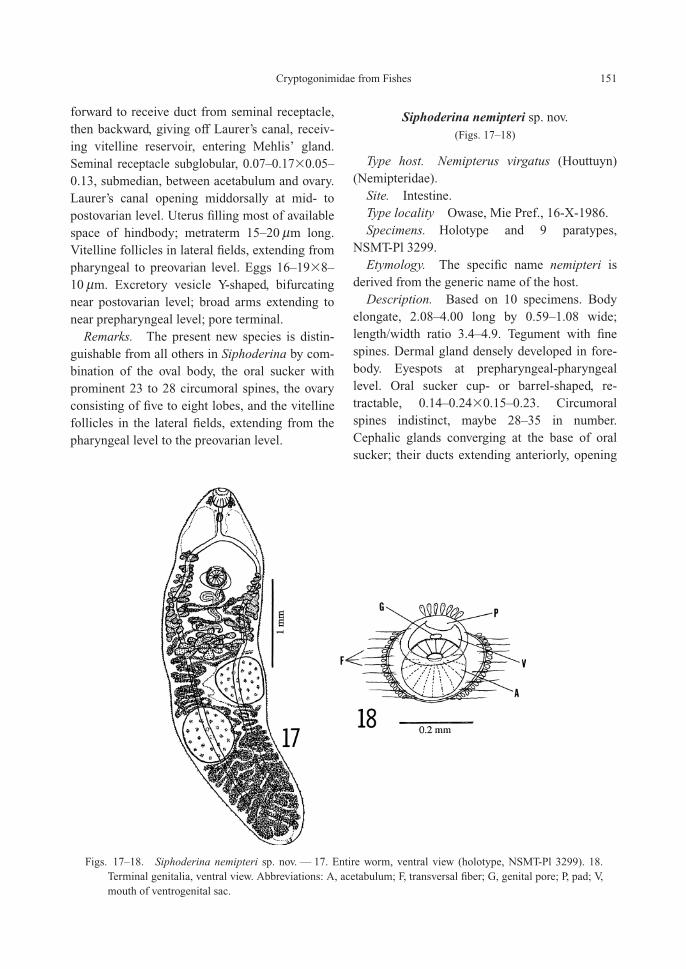
Siphoderina nemipteri sp. nov.(Figs. 17–18)
Type host. Nemipterus virgatus (Houttuyn)(Nemipteridae).
Site. Intestine.Type locality Owase, Mie Pref., 16-X-1986.Specimens. Holotype and 9 paratypes,
NSMT-Pl 3299.Etymology. The specific name nemipteri is
derived from the generic name of the host.Description. Based on 10 specimens. Body
elongate, 2.08–4.00 long by 0.59–1.08 wide;length/width ratio 3.4–4.9. Tegument with finespines. Dermal gland densely developed in fore-body. Eyespots at prepharyngeal-pharyngeallevel. Oral sucker cup- or barrel-shaped, re-tractable, 0.14–0.24�0.15–0.23. Circumoralspines indistinct, maybe 28–35 in number.Cephalic glands converging at the base of oralsucker; their ducts extending anteriorly, opening
Cryptogonimidae from Fishes 151
Figs. 17–18. Siphoderina nemipteri sp. nov. — 17. Entire worm, ventral view (holotype, NSMT-Pl 3299). 18.Terminal genitalia, ventral view. Abbreviations: A, acetabulum; F, transversal fiber; G, genital pore; P, pad; V,mouth of ventrogenital sac.
between circumoral spines. Prepharynx up to0.11 long. Pharynx 0.05–0.11�0.05–0.10. Esoph-agus 0.05–0.15 long. Intestinal bifurcation mid-way between suckers. Caeca narrow, passing ven-tral to testes, ending near middle of posttesticularspase. Acetabulum 0.11–0.17�0.12–0.20, em-bedded in ventrogenital sac. Ventrogenital sacwith semicircular pad anteriorly and thick walllatero-posteriorly. The pad provided anteriorlywith glandular cells. The wall 0.03–0.12 thick inlateral side, enclosed by glandular cells and con-necting lateral body margin by transversal fibers.Sucker ratio 1 : 0.81–0.95. Forebody 20–27% ofbody length.
Testes subglobular, diagonal or occasionallytandem; anterior testis 0.27–0.65�0.30–0.71,posterior testis 0.30–0.90�0.38–0.70, in middleof hindbody. Posttesticular space 18–29% ofbody length. Seminal vesicle tubular, convoluted,extending posteriorly near ovary; distal end ofseminal vesicle uniting small pars prostaticawhich joins metraterm to form short genital atri-um. Genital pore median, on anterior edge of ac-etabulum.
Ovary acinous, usually 20–30 small lobes sur-rounding a central large lobe, 0.19–0.38�0.42–0.78 as a whole, 29–46% of body length from an-terior end. Oviduct arising from anterior borderof central lobe, running anterosinistrally, branch-ing Laurer’s canal, receiving duct from seminalreceptacle at the same point where Laurer’s canalbranches, then passing anterodextrally, connect-ing vitelline reservoir, entering Mehlis’ gland.Laurer’s canal convoluted, opening middorsallyin ovarian zone. Seminal receptacle transverselyelongate, 0.20–0.38�0.05–0.15, immediately an-terior to ovary. Uterus filling available space ofhindbody. Eggs slender, thin-shelled, 18–22�8–10 mm; two specimens with larger eggs 26–28�
10–12 mm. Vitelline follicles usually in lateralfields, occasionally intruding inward, extendingfrom a level midway between intestinal bifurca-tion and acetabulum to ovarian level. Excretoryvesicle Y-shaped; arms reaching near prepharynxor oral sucker; pore terminal.
Remarks. The present new species is most
similar to Siphoderina elongata (Gu et Shen,1979) from the same genus of host Nemipterus,but differs from it by having an oral sucker with28 to 35 circumoral spines, a ventrogenital sacwith thick wall, caeca ending blindly near themiddle of the posttesticular space, and vitellinefollicles extending from a level midway betweenthe intestinal bifurcation and acetabulum to theovarian level.
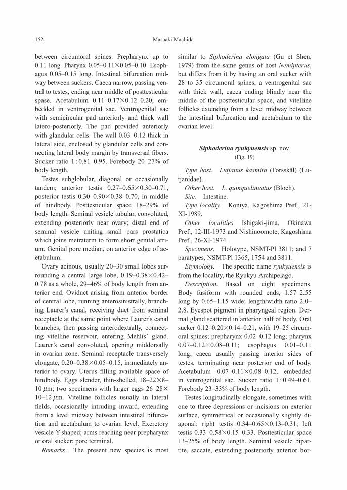
Siphoderina ryukyuensis sp. nov.(Fig. 19)
Type host. Lutjanus kasmira (Forsskål) (Lu-tjanidae).
Other host. L. quinquelineatus (Bloch).Site. Intestine.Type locality. Koniya, Kagoshima Pref., 21-
XI-1989.Other localities. Ishigaki-jima, Okinawa
Pref., 12-III-1973 and Nishinoomote, KagoshimaPref., 26-XI-1974.
Specimens. Holotype, NSMT-Pl 3811; and 7paratypes, NSMT-Pl 1365, 1754 and 3811.
Etymology. The specific name ryukyuensis isfrom the locality, the Ryukyu Archipelago.
Description. Based on eight specimens.Body fusiform with rounded ends, 1.57–2.55long by 0.65–1.15 wide; length/width ratio 2.0–2.8. Eyespot pigment in pharyngeal region. Der-mal gland scattered in anterior half of body. Oralsucker 0.12–0.20�0.14–0.21, with 19–25 circum-oral spines; prepharynx 0.02–0.12 long; pharynx0.07–0.12�0.08–0.11; esophagus 0.01–0.11long; caeca usually passing interior sides oftestes, terminating near posterior end of body.Acetabulum 0.07–0.11�0.08–0.12, embedded in ventrogenital sac. Sucker ratio 1 : 0.49–0.61.Forebody 23–33% of body length.
Testes longitudinally elongate, sometimes withone to three depressions or incisions on exteriorsurface, symmetrical or occasionally slightly di-agonal; right testis 0.34–0.65�0.13–0.31; lefttestis 0.33–0.58�0.15–0.33. Posttesticular space13–25% of body length. Seminal vesicle bipar-tite, saccate, extending posteriorly anterior bor-
152 Masaaki Machida
der of ovary; pars prostatica 28–40�22–30 mm;genital pore median, near anterior edge of ac-etabulum.
Ovary acinous, 0.22–0.35�0.25–0.44, nearmidbody. Oviduct arising from center of ovary,receiving duct from seminal receptacle, givingoff Laurer’s canal, joining vitelline reservoir, en-tering Mehlis’ gland. Seminal receptacle 0.12–0.34�0.03–0.14, between acetabulum and ovary.Laurer’s canal opening near posterior border of ovary. Uterus running posteriorly along leftcaecum, reaching near caecal ends, extending an-teriorly along right caecum, both sides of ovary,connecting metraterm. Eggs 17–21�9–10 mm.Vitelline follicles in lateral fields, extending froma level between intestinal bifurcation and acetab-ulum to near anterior edge of testes. Excretoryvesicle Y-shaped, bifurcating in ovarian region;arms reaching to oral sucker; pore terminal.
Remarks. The present new species can bedistinguished from all others in Siphoderina by
combination of the fusiform body; the oral suck-er with 18 to 25 circumoral spines; the longitudi-nally elongate testes arranged symmetrically oroccasionally a little diagonally; the acinousovary; and the vitelline follicles in the lateralfields, extending from a level between the intesti-nal bifurcation and acetabulum to near the anteri-or edge of the testes.
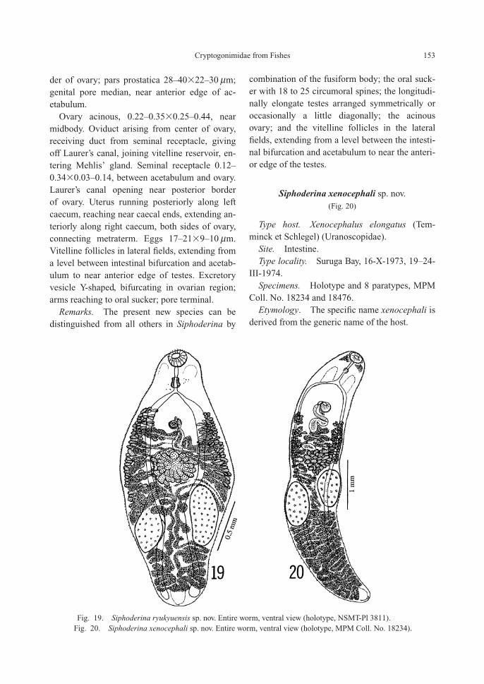
Siphoderina xenocephali sp. nov.(Fig. 20)
Type host. Xenocephalus elongatus (Tem-minck et Schlegel) (Uranoscopidae).
Site. Intestine.Type locality. Suruga Bay, 16-X-1973, 19–24-
III-1974.Specimens. Holotype and 8 paratypes, MPM
Coll. No. 18234 and 18476.Etymology. The specific name xenocephali is
derived from the generic name of the host.
Cryptogonimidae from Fishes 153
Fig. 19. Siphoderina ryukyuensis sp. nov. Entire worm, ventral view (holotype, NSMT-Pl 3811).Fig. 20. Siphoderina xenocephali sp. nov. Entire worm, ventral view (holotype, MPM Coll. No. 18234).

Description. Based on nine specimens. Bodyelongate, rounded anteriorly and tapering posteri-orly, 3.13–5.71 long by 0.87–1.25 wide; length/width ratio 3.2–5.4. Tegument spinose. Dermalgland densely developed in anterior half of body.Oral sucker subterminal, 0.14–0.24�0.21–0.29,with 44–51 circumoral spines. Cephalic glandssurrounding oral sucker, their ducts extendinganteriorly, opening between circumoral spines.Prepharynx 0.03–0.17 long; pharynx 0.07–0.09�
0.10–0.12; esophagus 0.06–0.29 long, bifurcat-ing midway between suckers or closer to acetab-ulum; caeca narrow, terminating near middle ofposttesticular space. Acetabulum 0.15–0.21�
0.15–0.22, embedded in ventrogenital sac. Ven-trogenital sac surrounded by glandular cells andcircular muscle. Sucker ratio 1 : 0.70–0.85. Fore-body 15–23% of body length.
Testes globular or subglobular, diagonal; righttestis 0.42–0.90�0.28–0.56; left testis 0.46–0.78�0.36–0.54. It is not fixed which side oftestis is anterior. Posttesticular space 31–43% ofbody length. Seminal vesicle tubular, sinuous, bipartite, extending posteriorly midway betweenacetabulum and ovary or near ovary. Pars prosta-tica 80–90 mm long, whose distal end unites me-traterm to form short genital atrium. Genital poremedian, on anterior edge of ventrogenital sac.
Ovary acinous, 0.30–0.46�0.53–0.74, attach-ing anterior edge of fore testis, lying 31–43% ofbody length from anterior end. Oviduct arisingfrom center of ovary, running forward, joiningshort duct from seminal receptacle and giving offLaurer’s canal, then curving backward to receivevitelline reservoir, entering Mehlis’ gland. Semi-nal receptacle saccate, 0.11–0.26�0.25–0.40,immediately anterior to ovary. Laurer’s canalopening middorsally near posterior border ofovary. Uterus forming transverse loops, descend-ing between testes, reaching near posterior end ofbody, then ascending between testes again, lateralto ovary, mid-region between ovary and acetabu-lum. Metraterm very short. Eggs 19–23�9–11 mm. Vitelline follicles in lateral fields, be-tween postacetabular level and midlevel of ovary.Excretory vesicle Y-shaped, bifurcating immedi-
ately behind ovary; arms extending pharyngeallevel; pore terminal.
Remarks. The present new species is mostlike S. onaga (Yamaguti, 1970) from Etelis cor-uscans, but differs from it by having caeca termi-nating near the middle of the posttesticularspace; a much longer posttesticular space; andvitelline follicles not forming bunch-like clusters.The figure of S. onaga illustrated by Yamaguti(1970, Fig. 143A) shows the posttesticular spaceat a ratio of 19% of the body length.
Acknowledgements
I am grateful to the following fishermen’s co-operative associations for my field work: Owase,Mie Pref.; Nishinoomote and Setouchi, Kagoshi-ma Pref.; Nago and Ishigaki, Okinawa Pref. Thisstudy was financially supported by the WatanabeMemorial Foundation for the Advancement ofTechnology (No. 19–139).
References
Gu, C. and J. Shen, 1979. Ten new species of digenetictrematodes of marine fishes. Acta Zootaxonomica Sini-ca, 4: 342–355. (In Chinese with English summary.)
Hafeezullah, M., 1975. A new cryptogonimid trematode(Digenea: Cryptogonimidae) of marine fish from OrissaCoast, with a brief review of the genus Paracryptogo-nimus Yamaguti, 1934. Journal of the Marine Associa-tion of India, 17: 49–55.
Miller, T. L. and T. H. Cribb, 2007. Two new cryptogo-nimid genera (Digenea, Cryptogonimidae) from Lut-janus bohar (Perciformes, Lutjanidae): analyses of ri-bosomal DNA reveals wide geographic distribution andpresence of cryptic species. Acta Parasitologica, 52:104–113.
Miller, T. L. and T. H. Cribb, 2008a. Family Cryptogo-nimidae Ward, 1917. In: Bray, R. A., D. I. Gibson andA. Jones (eds.), Keys to the Trematoda, Vol. 3, pp.51–112. CAB International and Natural History Muse-um, London.
Miller, T. L. and T. H. Cribb, 2008b. Eight new species ofSiphoderina Manter, 1934 (Digenea, Cryptogonimidae)infecting Lutjanidae and Haemulidae (Perciformes) offAustralia. Acta Parasitologica, 53: 344–364.
Oshmarin, P. G., Y. L. Mamaev and A. M. Parukhin,1961. Two new species and genera of the family Cryp-togonimidae from fishes of the North Viet-Nam bay(Tonking bay). Helminthologia, 3: 261–266. (In Rus-
154 Masaaki Machida

sian.)Shen, J., 1989. Studies on the digenetic trematodes of
fishes from Jiaozhou Bay. Studia Marina Sinica, (20):153–162. (In Chinese with English summary.)
Shen, J., 1990. Digenetic Trematodes of Marine Fishesfrom Hainan Island. 228 pp. Science Press, Beijing. (InChinese with English summary.)
Srivastava, H. D., 1939. The morphology and systematicrelationship of a new parasite, Mehrailla ovocaudatumgen. et sp. nov., (family Acanthostomidae) from an In-dian marine fish. Indian Journal of Veterinary Scienceand Animal Husbandry, 9: 209–212.
Velasquez, C. C., 1961. Cryptogonimidae (Digenea:
Trematoda) from Philippine food fishes. Journal ofParasitology, 47: 914–918.
Yamaguti, S., 1942. Studies on the helminth fauna ofJapan. Part 39. Trematodes of fishes mainly from Naha.Transactions of the Biogeographical Society of Japan,3: 329–398, Pl. XXIV.
Yamaguti, S., 1952. Parasitic worms mainly fromCelebes. Part 1. New digenetic trematodes of fishes.Acta Medicinae Okayama, 8: 146–198.
Yamaguti, S., 1970. Digenetic Trematodes of HawaiianFishes. 436 pp. Keigaku Publ., Tokyo.
Yamaguti, S., 1971. Synopsis of Digenetic Trematodes ofVertebrates. 1074 pp., 349 pls. Keigaku Publ., Tokyo.
Cryptogonimidae from Fishes 155