Cryptococcal chorioretinitis: a case report - bjo.bmj.com · British Journal of'Ophthalmology,...
3
British Journal of'Ophthalmology, 1977, 61, 411-413 Cryptococcal chorioretinitis: a case report J. S. CHAPMAN-SMITH From the Melbourne University Department of Ophthalmology, Royal Victorian Eye and Ear Hospital, Melbourne, Australia SUMMARY Chorioretinitis occurred in a young man whose long-standing cryptococcal meningitis had been identified and treated. In one eye spontaneous resolution had occurred. His case history and fluorescein angiograms are presented. Cryptococcus neoformans (or Torutla histolytica) is a yeast-like fungus producing systemic disease in man. Cryptococcosis (or torulosis) has a world-wide distribution. Infection is frequently by way of the respiratory tract. The organism is disseminated by the blood stream, particularly to the central nervous system, where a severe meningoencephalitis may develop. Before the introduction of amphotericin B in 1956 the disease was almost always fatal. Many reports of cryptococcosis have appeared, but surprisingly few of ocular involvement. The eye may suffer the secondary effects of meningitis and raised intracranial pressure-papilloedema, optic atrophy, sixth nerve palsy, photophobia, and nystagmus (Khodadoust and Payne, 1969). The eye may be affected secondarily to orbital invasion, or the eye may be involved directly. Hiles and Font (1968) reviewed the literature to find just 9 cases of proved cryptococcosis of the eye. They added a case of their own. Khodadoust and Payne (1969) updated the count to 13 cases. Cameron and Harrison (1970) described 1 further case in which a mousz-virulent form of C. neoformans was identified in the patient's cerebrospinal fluid. Case report A 27-year-old white male was referred to his local general hospital in October 1969. He described a steadily increasing frontal headache, which developed over 2 weeks. Over several days before his admission he suffered from photophobia, was nauseated, and had vomited. He worked as a commercial artist, and over several years had worked in a tower inhabited by pigeons. Address for reprints: Dr J. S. Chapman-Smith, Melbourne University Department of Ophthalmology, 32 Gisborne Street, East Melbourne, Victoria 3002, Australia On his first admission he was noted to be febrile (389°C) and to have neck stiffness. Cryptococcus neoformans was isolated from his lumbar cerebro- spinal fluid and cultured on Sabourad's agar. On 5 November 1969 the patient was transferred to Fairfield Hospital in Melbourne. Relevant laboratory findings are listed: Cerebrospinal fluid: 32 cmH2O pressure; C. neoformans present in Indian ink preparation. Leucocytes: 11-500 thousand/ mm3 (11 5x 10⁹/1)- 55% polymorphs, 32% lympho- cytes, 0 % oesinophils. Chest x-ray: a lesion present at the right lung base consistent with a toruloma. No bacteria or viruses were cultured from his throat, nasopharynx or blood-stream. No crypto- coccus grown from pigeon faeces. The patient began treatment on the day of his admission. Amphotericin B was administered as an intravenous infusion over 8 hours of each day. The dosage was 1 mg/kg per day, initially 5 days in 7, and later reducing to 3 days in 7. The patient was transferred to St. Vincent's Hospital on 17 December 1969, where a right lobectomy was performed. The pathology report described a lesion surrounded by damaged lung parenchyma, in which both granulation and mucoid tissue were present. 'Innumerable cryptococci' were present within the mucoid material, and some cryptococci lay in alveoli adjacent to the main lesion. The lesion was diagnosed as toruloma of the right lung. The patient convalesced at Fairfield Hospital until 26 February 1970, when he was discharged clinically well. He was subsequently readmitted for several days each month for review of his general health and cerebrospinal fluid. Flucytosine tablets (8 g/day) were introduced to his treatment pro- gramme. In March 1971 he had a generalised convulsion. He was prescribed phenytoin sodium tablets (100mg) 3 times daily, which he still takes. 411 on 2 March 2019 by guest. Protected by copyright. http://bjo.bmj.com/ Br J Ophthalmol: first published as 10.1136/bjo.61.6.411 on 1 June 1977. Downloaded from
Transcript of Cryptococcal chorioretinitis: a case report - bjo.bmj.com · British Journal of'Ophthalmology,...

British Journal of'Ophthalmology, 1977, 61, 411-413
Cryptococcal chorioretinitis: a case reportJ. S. CHAPMAN-SMITHFrom the Melbourne University Department of Ophthalmology, Royal Victorian Eye and Ear Hospital,Melbourne, Australia
SUMMARY Chorioretinitis occurred in a young man whose long-standing cryptococcal meningitishad been identified and treated. In one eye spontaneous resolution had occurred. His case historyand fluorescein angiograms are presented.
Cryptococcus neoformans (or Torutla histolytica) is ayeast-like fungus producing systemic disease in man.Cryptococcosis (or torulosis) has a world-widedistribution. Infection is frequently by way of therespiratory tract. The organism is disseminated bythe blood stream, particularly to the central nervoussystem, where a severe meningoencephalitis maydevelop. Before the introduction of amphotericin Bin 1956 the disease was almost always fatal.Many reports of cryptococcosis have appeared,
but surprisingly few of ocular involvement. The eyemay suffer the secondary effects of meningitis andraised intracranial pressure-papilloedema, opticatrophy, sixth nerve palsy, photophobia, andnystagmus (Khodadoust and Payne, 1969). The eyemay be affected secondarily to orbital invasion, orthe eye may be involved directly. Hiles and Font(1968) reviewed the literature to find just 9 cases ofproved cryptococcosis of the eye. They added a caseof their own. Khodadoust and Payne (1969) updatedthe count to 13 cases. Cameron and Harrison (1970)described 1 further case in which a mousz-virulentform of C. neoformans was identified in the patient'scerebrospinal fluid.
Case report
A 27-year-old white male was referred to his localgeneral hospital in October 1969. He described asteadily increasing frontal headache, which developedover 2 weeks. Over several days before his admissionhe suffered from photophobia, was nauseated, andhad vomited. He worked as a commercial artist,and over several years had worked in a towerinhabited by pigeons.
Address for reprints: Dr J. S. Chapman-Smith, MelbourneUniversity Department of Ophthalmology, 32 GisborneStreet, East Melbourne, Victoria 3002, Australia
On his first admission he was noted to be febrile(389°C) and to have neck stiffness. Cryptococcusneoformans was isolated from his lumbar cerebro-spinal fluid and cultured on Sabourad's agar.On 5 November 1969 the patient was transferred
to Fairfield Hospital in Melbourne. Relevantlaboratory findings are listed: Cerebrospinal fluid:32 cmH2O pressure; C. neoformans present inIndian ink preparation. Leucocytes: 11-500 thousand/mm3 (11 5x 10⁹/1)- 55% polymorphs, 32% lympho-cytes, 0% oesinophils. Chest x-ray: a lesion presentat the right lung base consistent with a toruloma.No bacteria or viruses were cultured from histhroat, nasopharynx or blood-stream. No crypto-coccus grown from pigeon faeces.The patient began treatment on the day of his
admission. Amphotericin B was administered as anintravenous infusion over 8 hours of each day. Thedosage was 1 mg/kg per day, initially 5 days in 7,and later reducing to 3 days in 7.The patient was transferred to St. Vincent's
Hospital on 17 December 1969, where a rightlobectomy was performed. The pathology reportdescribed a lesion surrounded by damaged lungparenchyma, in which both granulation and mucoidtissue were present. 'Innumerable cryptococci' werepresent within the mucoid material, and somecryptococci lay in alveoli adjacent to the main lesion.The lesion was diagnosed as toruloma of the rightlung. The patient convalesced at Fairfield Hospitaluntil 26 February 1970, when he was dischargedclinically well. He was subsequently readmitted forseveral days each month for review of his generalhealth and cerebrospinal fluid. Flucytosine tablets(8 g/day) were introduced to his treatment pro-gramme.
In March 1971 he had a generalised convulsion.He was prescribed phenytoin sodium tablets (100mg)3 times daily, which he still takes.
411
on 2 March 2019 by guest. P
rotected by copyright.http://bjo.bm
j.com/
Br J O
phthalmol: first published as 10.1136/bjo.61.6.411 on 1 June 1977. D
ownloaded from
J. S. Chapman-Smith
In November 1971 he was admitted to the AlfredHospital. His symptoms of headache and vomitinghad recurred, and he had lost weight. Although hiselectroencephalogram and brainscan appeared nor-mal, the air encephalogram showed dilatation ofboth lateral ventricles, suggesting moderate cerebralatrophy.
In early October 1972 he was admitted as anemergency to Fairfield Hospital and transferred tothe Alfred Hospital. His headaches and vomitingwere severe, he was wasted and unable to walk.Mentally he was vague and disorientated. Bothoptic discs had blurred margins, and peripapillaryhaemorrhages were seen. A large haemorrhagepartly obscured the right disc and inferonasalretina. C. neoformans was again isolated from thecerebrospinal fluid. An air encephalogram showedgrossly dilated lateral ventricles. Surgery wasperformed. A right atrioventricular shunt wasinserted and connected to a Rickham sub-aponeurotic reservoir, which permitted subsequentadministration of intrathecal amphotericin. Hislevel of consciousness improved rapidly. He wastransferred back to Fairfield Hospital, where hewas given a trial of clotrimazole to supplement hisdrug therapy. This ceased after 1 week followinggastrointestinal upset and inadequate blood andcerebrospinal fluid levels of the drug.
In November 1972 and June 1973 further obstruc-tions occurred, and additional shunts were installed.His drug treatment stopped in January 1974 as hisrenal function was disturbed.He remained symptom free until June 1976, when
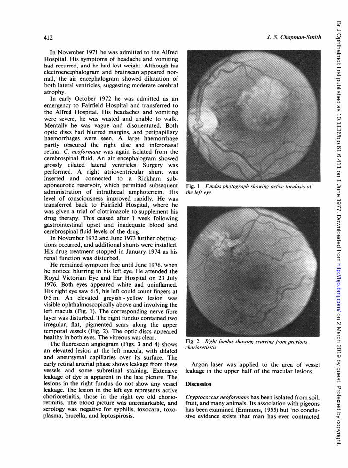
he noticed blurring in his left eye. He attended theRoyal Victorian Eye and Ear Hospital on 23 July1976. Both eyes appeared white and uninflamed.His right eye saw 6/5, his left could count fingers at0 5 m. An elevated greyish - yellow lesion wasvisible ophthalmoscopically above and involving theleft macula (Fig. 1). The corresponding nerve fibrelayer was disturbed. The right fundus contained twoirregular, flat, pigmented scars along the uppertemporal vessels (Fig. 2). The optic discs appearedhealthy in both eyes. The vitreous was clear.The fluorescein angiogram (Figs. 3 and 4) shows
an elevated lesion at the left macula, with dilatedand aneursymal capillaries over its surface. Theearly retinal arterial phase shows leakage from thesevessels and some subretinal staining. Extensiveleakage of dye is apparent in the late picture. Thelesions in the right fundus do not show any vesselleakage. The lesion in the left eye represents activechorioretinitis, those in the right eye old chorio-retinitis. The blood picture was unremarkable, andserology was negative for syphilis, toxocara, toxo-plasma, brucella, and leptospirosis.
Fig. 1 Fundus photograph showinig active torulosis ofthe left eye
Fig. 2 Right fundus showing scarring from previouschorioretinitis
Argon laser was applied to the area of vesselleakage in the upper half of the macular lesions.
Discussion
Cryptococcus neoformans has been isolated from soil,fruit, and many animals. Its association with pigeonshas been examined (Emmons, 1955) but 'no conclu-sive evidence exists that man has ever contracted
412
on 2 March 2019 by guest. P
rotected by copyright.http://bjo.bm
j.com/
Br J O
phthalmol: first published as 10.1136/bjo.61.6.411 on 1 June 1977. D
ownloaded from

413Cryptococcal chorioretinitis
4
Figs. 3, 4 Arteriovenous (Fig. 3) and klate (Fig. 4) fluorescein angiograms of the left fundus
the disease from infected birds' (Khodadoust andPayne, 1969).
In the cases reported so far chorioretinitis is thecommonest form of direct eye involvement. Hiles andFont (1968) recorded that spontaneous resolutionoccurred in I eye of their patient. Uveitis, endoph-thalmitis, vitreous invasion by C. neoformans, andneuroretinitis have been reported (Khodadoust andPayne, 1969). The organism spreads to the eye
either by the blood stream or in the meningealsheaths surrounding the optic nerve.A period of 64 years elapsed between the onset of
this patient's illness and the development of symp-
toms in his left eye. Hiles and Font (1969) noted a
2-year gap between meningitis and eye involvementin their reported case.
Treatment with amphotericin B has produced a
50% rate of cure in an American series (Okun andButler, 1964). The risk of renal damage isconsiderable. Oral flucystosine has proved effectivewithout significant adverse effects in some reports.Adequate cerebrospinal fluid levels can be achievedwith oral administration (Stanton and Sanderson,1974). Specific organism sensitivity to the drugshould be demonstrated in the laboratory.
This patient, though relatively symptom-free, hasregularly produced C. neoformans in his cerebro-spinal fluid until his last examination in June 1975.His cerebrospinal fluid glucose remains low, proteinhigh. Despite heroic treatment with amphotericin B,supplemented with flucystosine, the infection hasnot been eliminated. An uneasy truce exists betweenpatient and pathogen.
I thank Professor G. W. Crock for the opportunityto examine his patient; Dr H. Newton-John ofFairfield Hospital for access to the patient's pastrecords; and I acknowledge the photographicassistance of Mr J. Scrimgeour.
References
Cameron, M. E., and Harrison, A. (1970). Medical Journalof Australia, 1, 935.
Emmons, C. W. (1955). American Journal of Hygiene, 62, 227.Hiles, D. A., and Font, R. L. (1968). American Journal of
Ophthalmology, 65, 98.Khodadoust, A. A., and Payne, J. W. (1969). American
Journal of Ophthalmology, 67, 745.Okun, E., and Butler, W. T. (1964). Archives of Ophthal-
mology, 71, 52.Stanton, K. G., and Sanderson, C. R. (1974). Australian andNew Zealand Journal of Medicine, 4, 262.
3
on 2 March 2019 by guest. P
rotected by copyright.http://bjo.bm
j.com/
Br J O
phthalmol: first published as 10.1136/bjo.61.6.411 on 1 June 1977. D
ownloaded from