CRUSTACEAN ISSUES 6 - Decapoda
17
CRUSTACEAN ISSUES 6 General editor: FREDERICK R.SCHRAM San Diego Natural History Museum, California A.A.BALKEMA / ROTTERDAM / BROOKFIELD /1989
Transcript of CRUSTACEAN ISSUES 6 - Decapoda

CRUSTACEAN ISSUES
6
General editor:
FREDERICK R.SCHRAM San Diego Natural History Museum, California
A.A.BALKEMA / ROTTERDAM / BROOKFIELD /1989

FUNCTIONAL MORPHOLOGY OF FEEDING AND GROOMING IN CRUSTACEA Edited by
BRUCE E.FELGENHAUER Florida State University, Tallahassee
LES WATLING University of Maine, Walpole
ANNEB.THISTLE Florida State University, Tallahassee
A.A.BALKEMA / ROTTERDAM / BROOKFIELD /1989
CIP-DATA KONINKLIJKE BIBLIOTHEEK, DEN HAAG
Functional
Functional morphology of feeding and grooming in Crustacea / ed. by Bruce E.Felgenhauer. - Rotterdam [etc]: Balkema. - 111. - (Crustacean issues, ISSN 0168-6356; 6) ISBN 90 6191 777 8 bound SISO 597.6 UDC 591.4:595.3 Subject heading: crustacean
ISSN 0168-6356
ISBN90 6191777 8
© 1989 A.A.Balkema, P.O.Box 1675,3000 BR Rotterdam, Netherlands
Printed in the Netherlands
Table of contents
Preface VII
Introduction IX
The setal system of crustaceans: Types of setae, groupings, and functional morphology 1 Frangoise Jacques
A classification system for crustacean setae based on the homology concept 15 Les Watling
Physiology of sensory neurons in morphologically identified cuticular sensilla of crustaceans 27 Charles D.Derby
Decapod crustacean grooming: Functional morphology, adaptive value, and phylogenetic significance 49 Raymond TBauer
Gill and embryo grooming in lithodid crabs: Comparative functional morphology based on Lithodes maja 75 Gerhard Pohle
Grooming structure and function in some terrestrial Crustacea 95 Jeff G.Holmquist
Functional morphology of feeding in the Nectiopoda 115 Frederick R.Schram & Cynthia Arey Lewis
Morphology of feeding structures in the Conchostraca with special reference to Lynceus 123 JoelWMartin
Functional morphology of feeding appendages in calanoid copepods 137 Sigrid B.Schnack
Feeding mechanisms of the My sidacea 153 Y.Crouau
v

VI Table of contents
Functional morphology and feeding of euphausiids JMauchline
Development of the feeding apparatus in decapod crustaceans Jan Robert Factor
Evolution of the foregut in the lower Decapoda Bruce E.Felgenhauer & Lawrence GAbele
Index

I
A classification system for crustacean setae based on the homology concept
LES WATLING University of Maine - Darling Center, Walpole, USA
ABSTRACT
Setae are ubiquitous structures of crustaceans, but their classification has remained a significant problem. The schemes that do exist utilize a variety of morphological features, most of which have been arbitrarily chosen and emphasized. Further, they were developed for decapods and do not transfer well to other crustacean groups. In this paper, criteria established by Riedl (1978), and modified by Rieger and Tyler (1979) for determining the probability that similar structures are homologous, are applied. Structures considered to indicate homology, and which are therefore referred to as primary structures of setae, are presence or absence of an annulation, presence or absence of setules on the shaft, mode of articulation, and (possibly) the presence of a chemoreceptive tip. Features that are primarily mechanical or structural in nature, and which therefore have no value for indicating homology, are presence or absence of denticulations, strength of denticulations, degree of cuticularization of the shaft, diameter of setal lumen, features of the basal septum, and ringing of the distal part of the shaft. On the basis of the homologous structures, four fundamental types of setae were determined to exist: I, annulate, with setules; II, annulate, without setules; III, non-annulate, robust; IV, non-annulate, small, non-robust. Examples of all types and an expanded classification are given.
1 INTRODUCTION
Besides the multi-articulate appendage, setae are the single most distinguishing feature of crustaceans. They occur in a variety of styles and designs and are absolutely ubiquitous in the group. For the most part, however, setae have rarely been used in assessing the taxonomic status of an organism, though Menzies (1956) suggested that they could be of considerable use in Isopoda. No mention of setae, their diversity, or their use is made in most monographs dealing with specific crustacean groups (for example, the volumes by Bauchau 1966 and Warner 1977, dealing with crabs). Also, until relatively recently (Mauchline 1967, Farmer 1974, Bauer 1981, Holmquist 1982, Felgenhauer & Abele 1983, and the papers in this volume) setae have generally been ignored in studies of the ecology of a species. When setae have been discussed, it is usually in relation to either feeding or grooming. In these cases only the most general setal names are used, for example, plumose seta, comb seta, barbed seta
15
16 Les Watling
(Roberts 1968, Ivester & Coull 1975, Barker & Gibson 1978, Coombs & Allen 1978, Hindley & Alexander 1978, Alexander et al. 1980, Alexander & Hindley 1985).
It is likely that at least part of the reason for this omission of setae from crustacean studies has to do with the lack of a system for naming the myriad setal types that can be found on any given species. Although several early authors (e.g., Nordenstam 1933, Mauchline 1967) provided some names or type designations for the particular setae that occured on the organism they were studying, no comprehensive system of classification existed until the study of Thomas (1970) on the setae of the crayfish Austropotamobius pallipes. Later studies giving their own, somewhat different, systems were published by Fish (1972), Farmer (1974), and Drach & Jacques (1977,1979; see also the paper of Jacques, this volume). Minor variations on these themes have been put forward for individual species by Factor (1978), Kunze & Anderson (1979), Pohle & Telford (1981), and Schembri (1982).
It is the purpose of this paper to review these setal classificatory systems, to examine the morphological features that form their bases, and to show how a satisfactory classificatory system can be built using the criteria of homology. Such a system should provide a framework into which any seta can be put, regardless of the taxon on which it occurs, should be capable of including those not yet documented, and should reflect the ways in which setae can change during the course of development of an individual.
2 NEED FOR A UNIFORM SYSTEM OF SETAL CLASSIFICATION
Reading the crustacean literature, especially that dealing with taxonomy, one finds that many terms, such as seta, spine, scale, and setule, are used to indicate a wide variety of structures. This imprecision makes it very difficult for a person working with one group to identify easily an individual of another group. Because certain types of setae occur in specific positions depending on the function of the appendage or its parts, a standard terminology would help assess the function of a seta and thus of the portion of the appendage of which it is a part. In both of these cases, it is important to be able to recognize a seta that is of a fundamentally new type as opposed to one that represents a variant on an established pattern.
3 SOME FUNDAMENTAL TERMINOLOGY
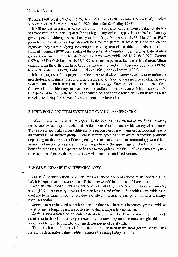
Because of the often varied use of the terms seta, spine, and scale, these are defined here (Fig. 1 a). It is hoped that all taxonomists will be more careful in their use of these terms.
Seta: an articulated cuticular extension of virtually any shape or size; may vary from very small (10-20 |im) to very large (> 1 mm in length) and robust, often with a very wide base; contrary to Thomas (1970), a seta does not always have an apical pore, nor does it always have an annulus.
Spine: a non-articulated cuticular extension that has a base that is generally not as wide as the structure is long; regardless of its size or shape, a spine has no socket.
Scale: a non-articulated cuticular extension of which the base is generally very wide relative to its length; microscopic secondary features may arm the outer margin; this term should not be used to describe very small extensions of setal shafts.
Terms such as 'hair', 'bristle', etc. should only be used in the most general sense. They have little descriptive value in either taxonomic or morphologic studies.

A classification system for crustacean setae 17
Figure 1. a. Diagrammatic representation of seta, spine, and scale, in side view on left and top view on right, b. Diagrammatic view of typical seta illustrating associated terminology.
The terms associated with the basic morphology of a seta are outlined below and illustrated in Figure lb:
Annulus: a faint ring circumscribing the shaft; may be located near the base or well along the shaft.
Denticule: a non-articulated extension of the shaft of a seta or spine. Lumen: the hollow canal extending the length of the setal shaft interior. Septum: a basal constriction of the lumen of the setal shaft. Setule: an extension of the shaft of a seta, usually of uniform width from base to tip, and
forming an articulated or flexible junction with the shaft.
4 REVIEW OF PREVIOUS SETAL CLASSIFICATION SCHEMES
The principal schemes that have been proposed for classifying setae are those of Thomas (1970), Fish (1972), Farmer (1974), and Drach & Jacques (1977 and paper by Jacques, this volume). These systems vary in their complexity and the degree to which they agree. Farmer's system seems the simplest; that of Drach and Jacques is easily the most complex.
Thomas (1970) offered the earliest comprehensive setal classificatory system. Although it was designed with a freshwater decapod as the source of the major setal types, it has been modified (Farmer 1974, Factor 1978) for use with other decapods. Thomas divided the setae of A.pallipes into two groups: group 1, setae with relatively thick walls, a narrow lumen, and no basal septum, and group 2, setae with relatively thin walls, a wide lumen, and a well-developed basal septum. Within group 1, setae were further divided into smooth and denticulate forms; in group 2 the subdivisions included septate denticulate setae, plumed setae, and plumodenticulate setae.
Studying the isopod Eurydicepulchra, Fish (1972) divided the setae she observed into two

18 LesWatling
major groups based on size: macrotrichs, which varied from 0.025 to 0.4 mm in length, and microtrichs, which were generally from 2 to 10 (im long. The macrotrichs were further subdivided into 4 groups: 1) simple, which included simple setae with blunt apices and aesthetascs; 2) setulose, which included serrated, brush, plumose seta, plumose bristle, hooked bristle, and brush spine; 3) denticulate, which included comb, serrated bristle, and serrated spine; and 4) non-denticulated and non-setulose, which was a heterogeneous group including forked spine, simple spine, blunt spine, and toothed spine. Fish used the terms seta, bristle, and spine to indicate degree of chitinization and thickening of the shaft wall, with setae the least, and spines the most, chitinized. No information was given about articulation; however, in most cases the presence of an annulation was noted. The microtrichs were of two types: 1) very small chitinous 'hairs' that arose in crescentic rows (probably from the edges of polygonal scales) and 2) single setae that arose from short sockets in the cuticle.
While acknowledging the previous work of Thomas (1970) and Fish (1972), Farmer (1974) divided the setae seen on the mouthparts and pereiopods ofNephrops norvegicus into three basic types: simple, plumose, and serrate. No plumodenticulate setae were found. Within this framework, all setae found were classified into one of 12 different types, coded 'a' through T . All of these were simply variations on one of the three basic types. Farmer suggested that the evolutionary development of setal types proceeded from the simple seta to either the plumose or the serrate seta.
The most complex of all systems is that proposed by Drach & Jacques (1977). Many of the features are covered in detail by Jacques (this volume), so only a cursory review will be provided here. Almost all aspects of setal morphology are considered, including mode of articulation, relative length of the seta, diameter of the inner canal, presence or absence of the septum, perforation of the septum, presence or absence of a terminal pore, whether the distal part of the seta is ringed or not, the type of surface relief features of the shaft, the type of circular outgrowths of the shaft, and the arrangement of these outgrowths. In all, nine pairs of outgrowth features are utilized. The result is a set of nine descriptors for a smooth seta and 18 descriptors for a seta with cuticular outgrowths. Drach and Jacques indicate that about 15 widely ranging types of setae (some of which are designated as sub-types 'a', 'b', etc.) are found on decapods. They leave the numbers 16-30 free for designating new types of setae that may be found in other orders.
5 THE APPLICATION OF HOMOLOGY TO SETAL MORPHOLOGY
There are many ways in which parts of organisms can be classified as similar. Principally, the major forms of similarity are homologies and analogies, but similarity can also take the form of homoiologies, homodynamies, isologies, homonomies, or symmetries (Riedl 1978). As several of these are special cases, the major problem lies in determining whether structures are homologues, analogues, or homoiologues. Analogous structures are those whose similarities have arisen by convergence; that is, they have different origins. Homologous structures are those with similar origins, although they may look very different. When structures contain both homologous and analogous substructures, or analogous ones on a homologous base, they are termed homoiologous (Riedl 1978).
Riedl (1978) noted that determining whether two features are homologous or analogous involved statements of probability, based on the use of several criteria advanced by Remane (1971) and since modified and extended by Rieger & Tyler (1979). As emended, Remane's

A classification system for crustacean setae 19
criteria for identifying homology are as follows (Rieger & Tyler 1979): 1) Similar structures may be considered homologues if they demonstrate similarity in a
positional hierarchy. Not only must the structures have similar position relative to other structures in the organism, but the component parts or substructures must also show positional similarity to one another, including the smallest component part.
2) Dissimilar structures may be considered to be homologues if a phylogenetic sequence can be determined that demonstrates the transformation of a series of structures. The more complete the sequence, the greater the probability of homology.
3) Similar structures may be considered homologues if they coincide in distribution with other homologues within a group of organisms. Riedl (1978) stressed the importance of biological order and noted that homologues are likely to co-occur; therefore, the greater the coincidence of homologues, the greater is the probability that each feature is, in fact, a homologue.
Before a structure can be considered a homologue, it must also be shown that it is not an analogue (Rieger & Tyler 1979). Several criteria were suggested by Rieger & Tyler (1979) for determining whether similar structures are analogues. These are: 'if 1) they are under the influence of common selective pressure; 2) they are composed of similar materials under the influence of similar environmental conditions; 3) they are likely to be the only possible means by which a particular function can be accomplished; 4) they develop in ontogeny from dissimilar origins; 5) they are under the influence of similarity-dependent selective pressure' (p.658). These criteria are designed to help determine the probability that observed similarity in structure is due to analogy rather than homology.
The problem here is to determine how these criteria can be applied to the substructures, or component parts, of setae. In order to develop a classification system for setae, we need to understand the kinds of similarities with which we are dealing, to know which features represent fundamental structures on which variations are built. In short, by determining which features are homologous and which are analogous, we can come to know which types of setae represent clear homologies and which represent homoiologies. It is obvious that a classificatory system for setae must be based on homologues; therefore, it is of paramount importance that the similarities based on homology be determined.
6 EVALUATION OF MORPHOLOGICAL FEATURES OF SETAE
In the different schemes used for classifying setae, various morphological features have been held to be more or less important. These features need to be evaluated if we are to determine which might represent homologues and therefore could be considered to be primary features of setae. In a sense, this process is similar to searching for 'Bauplane' of organisms. For each feature, arguments bearing on the probability that it represents a homologue or an analogue will be presented and discussed.
6.1 Presence ofannulation on shaft
Most setae form in invaginations of the epidermis, the innermost part of each invagination being delimited by the setal fold (Aiken 1973, Mills & Lake 1975, Reaka 1975, Longmuir 1983) (Fig. 2a). The setal fold leaves a circular indentation in the shaft of the seta after

20 Les Watling
Figure 2. a. Setae that form in pockets have annuli at various positions on shaft depending on depth of pocket. Short setae in most cases do not need as deep a pocket as many long plumose setae, b. Three setae from distal margin of maxillule 1 of unidentified Antarctic lysianassid amphipod (top) with newly forming setae (below). No pocket present, and no annulus found on setal shaft.
eversion. This is the annulus. Setae such as those that arm the distal margin of the maxilla of amphipods do not have annuli and are known to form without an invagination (Fig. 2b). Others, such as the cuspidate setae of Thomas (1970), have the annulus very near the base and can be seen to form in shallow pockets. Two very different modes of setal formation can thus be deduced from the occurrence of an annulus. Each of these modes constitutes a separate homology.
6.2 Cuticular expansions of the setal shaft
These are of two major types - setules and denticules. Many other terms have been applied in this context (e.g., barbules; Reaka 1975), but in all cases they are synonymous with, or are gradations of, one of these two types. Setae with setules are referred to as 'plumose', whereas setae with denticules may be referred to as 'serrate', 'toothed', 'combed', 'serrulate', etc., depending on the size of the denticulations.
Occasionally, a seta will possess both setules and denticules, in which case it is referred to as 'plumodenticulate'. In this case, the setules are invariably proximal to the annulus and the denticules distal. Most authors have considered setules and denticules to be,merely end-members of a gradational series. In my opinion, this view is not correct. In nearly all instances, setules are articulated at the setal shaft, whereas denticules are not.

A classification system for crustacean setae 21
Figure 3. Complexity of setal structures along shaft; a function of how seta forms. Portion of shaft forming adjacent to tissue in appendage, according to Reaka's model, more likely to support complexly innervated structures, such as setules; would consequently be mechano-receptive part of seta, whereas distal part of seta would contain only dendrites of chemore-ceptive nerve cells.
As noted by Reaka (1975), the possibility exists that the shaft outgrowths forming above the annulus may have differing innervation, or function, from those below, thus offering an explanation as to why plumodenticulate setae are always plumose proximally. Because the proximal portion of the setal shaft is surrounded by tissue, more complexly innervated structures could develop there, whereas it would be possible only to send dendrites up the lumen of the distal portion (Fig. 3). It is likely, at least in some cases, that the setules are connected by microfibrils in the setal shaft to the dendrites at the seta base (Crouau 1978). Plumose setae, therefore, are almost invariably mechanoreceptors with a rather complicated subcuticular structure (Crouau 1978, 1982, Felgenhauer & Abele 1983). The denticules of serrate setae are clearly used for functions such as grooming (Bauer 1981), but there is no evidence the denticules have mechanoreceptive capabilities; however, Derby (1982) indicated some mechanoreception for the field of serrate setae on the pereiopods of the lobster Homarus americanus. Shelton & Laverack (1970) ascribed chemosensory capability to a serrate seta even though it had no terminal pore, suggesting that chemical stimulants could pass through the untanned cuticle of the distal part of the seta. Thus, by the criteria outlined above, the probability seems very high that setae based on a plumose model, viz., bearing setules in any arrangement, are homologous structures, whereas the occurrence of denticules is an analogy that may or may not be related to chemoreception. At present there is insufficient evidence to determine whether a chemoreceptive capability is always found in denticulate setae.
6.3- Mode of articulation
Two types of articulation are typically seen - infracuticular, where the point of articulation occurs in a sunken socket, and supracuticular, where the cuticle is flexed outward to form the point of articulation (Fig. 4g). The former mode is typical of most setae, whereas the latter
I

22 Les Watling
Figure 4. Type I setae; annulate, with setules. a, c - plumose; b - pappose; d - forked (after Fish); e, f -plumodenticulate; g - plumose, with supracuticular pocket.
seems to be associated primarily with 'natatory' plumose setae. It is likely that there is some special functional adaptation for the supracuticular articulation that may or may not have other associated features. In any case, both types of articulation are considered to be homologies.
6.4 Length of seta vs. basal diameter
This is a feature whose importance has been stressed primarily by Drach & Jacques (1977). Similar setae vary in length and robustness, even along the edge of one or more articles of an appendage, and changes can be seen in both length and basal diameter during the ontogenetic sequence for any given species. Because there is a continuum in length vs. diameter, no particular ratio value can be singled out as indicating homology.
6.5 Diameter of lumen
The lumen diameter could be a function of the amount of innervation being carried by the seta, of the amount of resistance to bending being required of the seta, or of the presence of a duct from a small secretory gland. Because many setae are known that carry both mechano-and chemoreceptive dendrites and yet have very narrow lumens, it is unlikely that lumen diameter reflects the degree to which the seta is innervated. On the other hand, very large lumens are usually found in very robust setae. There are good biomechanical reasons for this to be the case. The overall weight of the seta is reduced, yet the ability to withstand deformation is maintained (Wainwright et al. 1982). In certain groups, such as the Ostracoda, setae are known to carry glandular secretions. It is not known whether this function occurs in the Decapoda, where some of the most robust setae have been found. I conclude that lumen diameter is a functional adaptation and has low significance for determination of homology.

A classification system for crustacean setae 23
6.6 Presence or absence of terminal pore
Thomas (1970) proposed that presence of a terminal pore was a universal feature of setae. We now know that this is not the case. However, it is likely that a terminal pore is associated with a chemoreceptive capability of the seta. On the other hand, setae without terminal pores are also known to have chemoreceptive functions. Whether a pore is present or not may have more to do with various specific aspects of chemoreception than with the complexities of setal structure. Because chemoreception without an external pore is known to occur (through a microporous cuticle (Derby, this volume)), presence of a pore may be merely an external manifestation of cuticular thinning and therefore has little probability of indicating homology.
6.7 Presence or absence of basal septum
Although the presence and degree of development of a basal septum has been discussed by both Thomas (1970) and Jacques (this volume), its correlation with other structural features of setae has not been investigated. Jacques assumed that if the septum is complete there must be a corresponding loss of innervation of the seta. Although this may be the case for chemoreceptive functions, it is not necessarily true for mechanoreceptive functions. Bauer (1981) and Felgenhauer & Abele (1983) illustrate mechanoreceptive setae with the dendrites proximal to what could be described as a basal septum. If a seta has only a minor function as a chemoreceptor, it could be partially occluded by a septum and still function as a mechanore-ceptor. The basal septum, in its various forms, then, has high probability of indicating analogy.
7 PRIMARY AND SECONDARY FEATURES OF SETAE
From the above discussion, it can be seen that there are very few morphological components of setae that can be used to determine homology of setal structure. Those that indicate homology with high probability are referred to here as primary features. They are:
Figure 5. Type II setae; aianulate, without setules. a, b - simple; c - cuspidate; d conate; e, f, g, h -various types of serrate; i - complex denticules of seriate seta; j - anvil-shaped denticules of seta from branchial epipod of crab.

24 Les Wading
- presence or absence of an annulation, - presence or absence of setules on shaft, - the mode of articulation of the seta, - (possibly) the presence of a chemoreceptive tip.
Secondary features of setal morphology are those that are primarily mechanical or structural and therefore probably indicate analogy. They are:
- presence or absence of denticulations, - strength of denticulations, - degree of cuticularization of the setal shaft, - diameter of setal lumen, - features of the basal septum, - ringing of distal part of the shaft.
These features help to distinguish particular variations of the major types of setae.
8 OUTLINE CLASSIFICATION OF CRUSTACEAN SETAE
All crustacean setae are homologous in that they are to be distinguished from scales or spines. In Riedl's (1978) terminology, the seta is a 'cadre homologue' within which are other cadre homologues of lower hierarchical order. There are probably only four fundamental types (= cadre homologies) of setae:
'i '•••'• A * r-:? * %Ck^ ^A^Atmukuei with setules: These are always mechanoreceptors; they always form in invaginated pockets, may be of a variety of sizes and shapes, may have the setules distributed along the shaft in many kinds of patterns, and may have denticules distally along the shaft.
„ v i njk *'=:v -&-Annulate^ without setules: These usually are chemoreceptive, but may be mechanore-ceptive in large groups; they always form in invaginated pockets and may be smooth or denticulate, elongate and stiff, or short and robust.
A/O/7-.0>'' J«- \0^lYt~N^n-6mmtktte, robust: These are often both mechano- and chemoreceptive; they usually occur in places where mechanical stress is high, such as along adjoining surfaces of chelae or on biting edges of amphipod and isopod mouthparts.
., , ^ V k f^/ rV- Non-annulate, small, non-robust: This may be a heterogeneous assemblage with several basic homologies represented and consequently will need re-evaluation; they are usually very small sensory setae found at numerous sites on the body surface.
Within each of these setal groups, there are numerous morphological variations. Those that have been pointed out by authors of earlier classification schemes will be considered below within the framework outlined above. It may be quite useful to retain the descriptive names for some of these, for example, the terms cuspidate, plumodenticulate, acuminate, etc., recognizing that there may be nearly continuous variation among several types.
Type I. Annulate, with setules (Fig. 4) A. with socket infracuticular
1. plumose (Thomas 1970) (syn: types 4,6, Drach & Jacques 1977) 2. pappose (Thomas 1970) (syn: ?brush spine, Fish 1972; types 7, 9, Drach &
Jacques 1977) 3. plumodenticulate (Thomas 1970) (syn: types 13,14, Drach & Jacques 1977) 4. forked seta (Fish 1972) (syn: forked spine, Fish 1972; type 15, Drach & Jacques
1977) B. with supracuticular socket

A classification system for crustacean setae 25
1. plumose (Thomas 1970) (syn: type 5, Drach & Jacques 1977; natatory setae of various authors)
Type II. Annulate, without setules (Fig. 5) A. with smooth shaft
1. acuminate (Thomas 1970) (syn: simple seta, Fish 1972; types 2, 3, Drach & Jacques 1977)
2. rod (Thomas 1970) (syn: seta with blunt apex, Fish 1972) 3. cuspidate (Thomas 1970) (syn: type 1, Drach & Jacques 1977) 4. conate (Thomas 1970) 5. papillate (Thomas 1970) (syn: blunt seta, Fish 1972) 6. simple (Fish 1972)
B. shaft with denticulae 1. serrate (Thomas 1970) (syn: comb seta, serrated bristle, serrated spine, Fish 1972;
types 10,11, Drach & Jacques 1977) a. serrulate (Thomas 1970) b. multidenticulate (Thomas 1970)
2. setobranch (Thomas) (syn: ?type 12, Drach & Jacques 1977) 3. teazel (Thomas 1970) 4. cincinnuli (Pohle & Telford 1981) (syn: soies a dents en double peigne, Jacques
1981) Type III. Non-annulate, robust
1. tooth seta (Thomas 1970, Fish 1972) Type IV Non-annulate, small, non-robust
A. Plumose 1. brush (Fish 1972) 2. scaled microseta (Drach & Jacques 1979)
9 ACKNOWLEDGMENTS
This paper was written after much discussion about its utility with Jan Factor and Bruce Felgenhauer. The continued encouragement of Frederick Schram is, as always, appreciated. Seth Tyler graciously read and offered important comments on the manuscript; his understanding of the homology concept helped clarify my sometimes muddled thoughts. Stephanie Staples prepared the final versions of the figures.
REFERENCES
Aiken, D.E. 1973. Proecdysis, setal development, and molt predictions in the American lobster (Homarus americanus). J. Fish. Res. BoardCanada 30:1337-1344.
Alexander, C.G. & J.P.R.Hindley 1985. The mechanism of food ingestion by the banana prawn, Penaeus merguiensis. Mar. Behav. Physiol. 12:33-46.
Alexander, C.G., J.P.R.Hindley & S.G.Jones 1980. Structure and function of the third maxillipeds of the banana prawn, Penaeus merguiensis. Mar. Biol. 58:245-249.
Barker, P.L. & R.Gibson 1978. Observations on the structure of the mouthparts, histology of the alimentary tract, and digestive physiology of the mud crab Scylla serrata (Forskal) (Decapoda: Portunidae). / . Exp. Mar. Biol. Ecol. 32:177-196.
Bauchau, A. 1966. La Vie des Crabes. Paris: Editions Paul Lechevalier.

26 Les Wading
Bauer, R.T. 1981. Grooming behavior and morphology in the decapod Crustacea. / . Crust, Biol. 1:153-173. Coombs, E.F. & J.A.Allen 1978. The functional morphology of the feeding appendages and gut of Hippolyte
varians (Crustacea: Natantia). Zool. J. Linn. Soc. 64:261-282. Crouau, Y. 1978. Infrastructure des phaneres spinules mecanorecepteurs d'un crustace mysidace souterrain
anophthalme. C. R.Acad. Sc. Paris 287:1215-1218. Crouau, Y. 1982. Primary stages in the sensory mechanism of the setulate sensilla, external mechanoreceptors of
acavernicolous Mysidacea. Biol. Cell. 44:45-56. Derby, CD. 1982. Structure and function of cuticular sensilla of the lobster Homarus americanus. J. Crust. Biol.
2:1-21. Drach, P. & F.Jacques 1977. Systeme setifere des crustaces decapodes. Principes d'une classification generate. C.
R. Acad. Sc. Paris 284:1995-1998. Drach, P. & F.Jacques 1977. Systeme setifre des crustaces decapodes. Le systeme microsetal. C. R. Acad. Sc.
Paris 284:1103-1105. Factor, J. 1978. Morphology of the mouthparts of larval lobsters, Homarus americanus (Decapoda:
Nephropidae), with special emphasis on their setae. Biol. Bull. 154:383-408. Farmer, A.S. 1974. The functional morphology of the mouthparts and pereiopods of Nephrops norvegicus (L.)
(Decapoda: Nephropidae). / . Nat. Hist. 8:121-142. Felgenhauer, B.E. &L.G.Abele 1983. Ultrastructure and functional morphology of feeding and associated
appendages in the tropical freshwater shrimp, Atya innocous (Herbst), with notes on its ecology. / . Crust. Biol. 3:336-363.
Fish, S. 1972. The setae of Eurydice pulehra (Crustacea: Isopoda). / . Zool., London, 166:163-177. Hindley, J.P.R. & C.G.Alexander 1978. Structure and function of the chelate pereiopods in the banana prawn,
Penaeus merguiensis. Mar. Biol. 48:153-160. Holmquist, J. 1982. The functional morphology of gnathopods: importance in grooming, and variation with regard
to habitat, in talitroidean amphipods. / . Crust. Biol. 2:159-179. Ivester, M.S. & B.C.Coull 1975. Comparative study of ultrastructural morphology of some mouthparts of our
haustoriid amphipods. Can. J. Zool. 53:408-417. Jacques, F. 1981. Systeme setifere des maxillipedes de Squilla mantis (Crustacea, Stomatopoda): morphologie
fonctionelle. Zoomorphology 98:233-239. Kunze, J. & D.T.Anderson 1979. Functional morphology of the mouthparts and gastric mill in the hermit crabs
Clibanarius taeniatus (Milne Edwards), Clibanarius virescens (Krauss), Paguristes squamosus McCulloch and Dardanus setifer (Milne-Edwards) (Anomura: Paguridae). Aust. J. Mar. Freshw. Res. 30:683-722.
Longmuir, E. 1983. Setal development, moult-staging and ecdysis in the banana prawn, Penaeus merguiensis. Mar. Biol. 77:183-190.
Mauchline, J. 1967. Feeding appendages of the Euphausiacea (Crustacea). / . Zool., London, 153:1-43. Menzies, R.J. 1956. A study of the microscopic structure of isopod setae. Ann. Mag. Nat. Hist., ser. 12,
9:698-700. Mills, B.J. & P.S.Lake 1975. Setal development and moult staging in the crayfish Parastacoides tasmanicus
(Erichson) (Decapoda, ParastacidaeXAiArt. / . Mar. Freshw. Res. 26:130-107. Nordenstam, A. 1933. Marine Isopoda of the families Serolidae, Idotheidae, Pseudidotheidae, Arcturidae,
Parasellidae and Stenetridae, mainly taken from the South Atlantic. Further Zool. Results Swed. Antarct. Exped. 3:1-384.
Pohle, G. & M.Telford 1981. Morphology and classification of decapod crustacean larval setae: a scanning electron microscope study of Dissodactylus crinitichelis Moreira, 1901 (Brachyura: Pinnotheridae). Bull. Mar. Sci. 31:736-752.
Reaka, M.L. 1975. Molting in stomatopod crustaceans. 1. Stages of the molt cycle, setagenesis, and morphology. / . Morph. 146:55-80.
Remane, A. 1971. Die Grundlagen des naturlichen Systems der vergleichenden Anatomie und der Phylogenetik (2 ed.). Koenigstein-Taunus: Koeltz.
Rieger, R. & S.Tyler 1979. The homology theorem in ultrastructural research. Amer. Zool. 19:655-664. Riedl, R. 1978. Order in Living Organisms. New York: Wiley & Sons. Roberts, M.H. 1968. Functional morphology of mouth parts of the hermit crabs, Pagurus longicarpus and Pagwus
policaris. Chesapeake Sci. 9:9-20. Schembri, P.J. 1982. The functional morphology of the feeding and grooming appendages of Ebalia tuberosa
(Pennant) (Crustacea: Decapoda: Leucosiidae). / . Nat. Hist. 16:467-480. Shelton, R.G.J. & M.S.Laverack 1970. Receptor hair structure and function in the lobster Homarus gammarus (L.).
/ . Exp. Mar. Biol. Ecol. 4:201-210. Thomas, W.J. 1970. The setae of Austropotamobius pallipes (Crustacea: Astacidae). / . Zool, London, 160:91-
142. Wainwright, S.A., W.D.Biggs, J.D.Currey & J.M.Gosline 1982. Mechanical Design in Organisms. Princeton:
Princeton University Press. Warner, G.F. 1977. The Biology of Crabs. New York: Van Nostrand Reinhold.