Crucial Conversations in Oncology Nursing: … · patient selection and nursing care ... bleeding,...
65
Crucial Conversations in Oncology Nursing: Management Considerations for the AML Patient
Transcript of Crucial Conversations in Oncology Nursing: … · patient selection and nursing care ... bleeding,...

Crucial Conversations in Oncology Nursing: Management Considerations for the AML Patient

Faculty
Laura Zitella, MS, RN, ACNP‐BC, AOCNHematology/Oncology Nurse Practitioner
Clinical Associate ProfessorUniversity of California, San Francisco

Commercial Support Disclosure
• This activity is supported through an educational grant from Pfizer.

Faculty Disclosures
• Advisory Board – for scientific information (Abbvie, Amgen, Array BioPharma Inc, Astra Zeneca, Gilead)
• Shareholder‐ Kite Pharmaceuticals

Learning Objectives
Upon completion of this educational activity, learners should be better able to:• Review recently approved agents for patients with newly
diagnosed and relapsed AML and their implications for patient selection and nursing care
• Evaluate patients with AML receiving treatment with recently approved agents in order to identify early indications of adverse events and implement appropriate interventions
• Outline key education points for the patient and family/caregiver to prevent infection, identify signs of bleeding, and perform other assessments to improve early identification and management of adverse events

AML At‐A‐Glance
Estimated New Cases in 2018
% of All New Cancer Cases
Estimated Deaths in 2018
% of All Cancer Deaths
% Surviving5 Years
19,520 1.1% 10,670 1.8% 28%
NCI Cancer Stat Facts: Acute Myeloid Leukemia https://seer.cancer.gov/statfacts/html/amyl.html
Median Age at Diagnosis:68 years
AML in older adults is a major
challenge

CD34+CD38+
CD34-
What is AML?Complex hematologic malignancy driven by multiple acquired genetic mutations that evolves over time
Normal hematopoiesis is driven by stem cells. AML is driven by leukemia stem cells rendered malignant by
driver mutations
Clonal expansion of leukemia cells leads to bone marrow failure and related
complications:severe infections,
anemia, andbleeding
Tan BT et al. et al. Lab Investig. 2006;86(12):1203‐1207.

Predisposing Factors in AML
Tamamyan G et al. et al. Crit Rev Oncology. 2017;110:20‐34.

AML is a Heterogenous Disease that Develops from Driver Mutations
Dohner H et al. et al. Blood. 2017;129:424‐447.
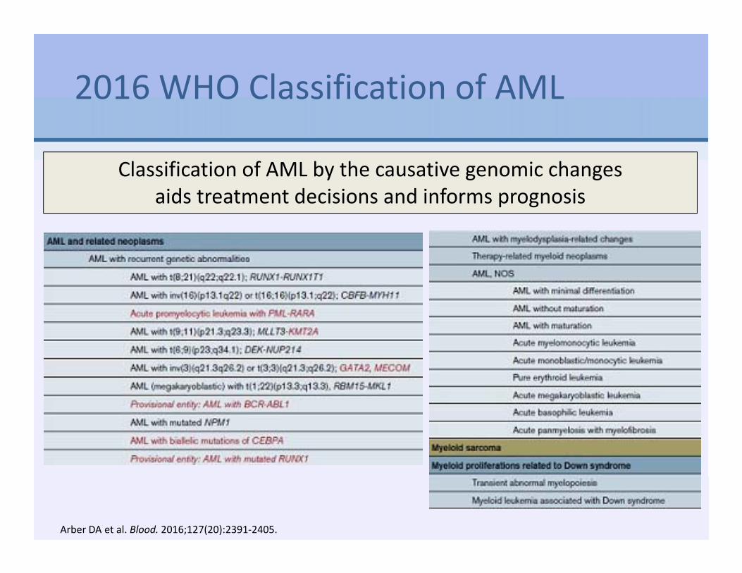
2016 WHO Classification of AML
Classification of AML by the causative genomic changes aids treatment decisions and informs prognosis
Arber DA et al. Blood. 2016;127(20):2391‐2405.

Genomic Changes and Age are the Most Important Predictors of Prognosis
2017 European Leukemia Network (ELN) Risk Stratification by Genetics
Dohner H et al. et al. Blood. 2017;129:424‐447.

Simplified Way to Think About AML
• Chemo‐sensitive AML – Core Binding Factor leukemias (without c‐KIT mutation)
• t(8;21), t(16;16), inv(16)– Diploid AML with NPM1 and CEBPmutation (without FLT3
mutation)– Dose intensification of chemotherapy may be helpful
• Chemo‐resistant AML– AML with adverse cytogenetics
• Complex karyotype, monosomal karyotype, TP53 gene mutation– AML with FLT3‐ITD– Secondary AMLs
• Treatment‐related AML• AML with myelodysplasia‐related changes
– Older patients– New agents are needed

How Do Patients with AML Present?
• Neutropenia– Infections
• Leukocytosis• Fever• Anemia
– Pallor, fatigue, weakness, palpitations, dyspnea on exertion• Thrombocytopenia
– Easy bruising, petechiae, epistaxis, gingival bleeding, conjunctival hemorrhages
• Hepatomegaly or splenomegaly
Symptoms due to impaired production of normal cells from leukemic infiltration of the bone marrow
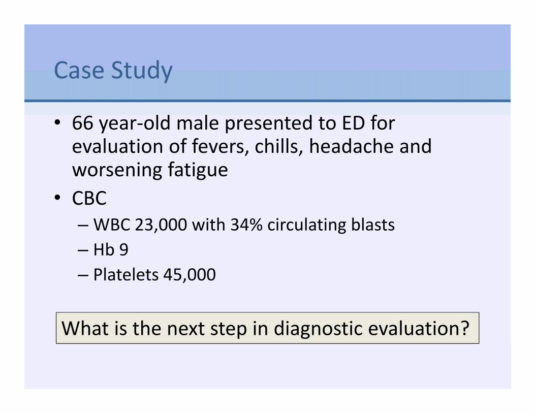
Case Study
• 66 year‐old male presented to ED for evaluation of fevers, chills, headache and worsening fatigue
• CBC– WBC 23,000 with 34% circulating blasts– Hb 9– Platelets 45,000
What is the next step in diagnostic evaluation?

Diagnostic Evaluation
Bone Marrow
Cytogenetics
Peripheral Blood
FISH
Molecular StudiesNPM1, CEBPA, RUNX1, FLT3, TP53, ASXL1, PML‐RARA, CBFB‐MYH11, RUNX1‐RUNX1T1, BCR‐ABL1, other fusion genes (if available)
Dohner H et al. et al. Blood. 2017;129:424‐447.
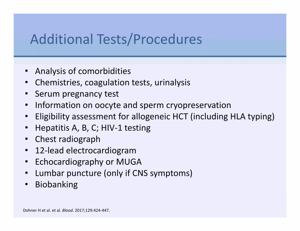
Additional Tests/Procedures
• Analysis of comorbidities• Chemistries, coagulation tests, urinalysis• Serum pregnancy test• Information on oocyte and sperm cryopreservation• Eligibility assessment for allogeneic HCT (including HLA typing)• Hepatitis A, B, C; HIV‐1 testing• Chest radiograph• 12‐lead electrocardiogram• Echocardiography or MUGA • Lumbar puncture (only if CNS symptoms)• Biobanking
Dohner H et al. et al. Blood. 2017;129:424‐447.

Case Study: Diagnosis = AML‐MRC
• AML‐MRC: AML with myelodysplasia‐related changes
What are the treatment options?

Pause and Reflect
How many patients with AML have you managed?• None• <5• 5‐10• >10

Considerations for Treatment
• Treatment initiation:– Delay of a week to complete
diagnostic testing doesn’t affect outcomes
– Exceptions are true emergencies such as:
• Coagulopathy, leukostasis with respiratory distress syndrome, or tumor lysis syndrome
• Majority of patients who achieve complete response will relapse and few are cured
• Assessing fitness for intensive therapy is major aspect of treatment planning– Transplant eligibility– Palliative chemotherapy
Intensive Treatment Paradigm
Allogeneic Transplantation
Potentially curative for medically fit patients
Consolidation
Achieve durable molecular remission with no minimal residual disease
Induction
Suppress malignant clone with induced hypoplasia
Brandwein JM et al. Am J Blood Res. 2017;7(4): 30–40.Sekeres MA et al. Blood. 2009;113(1):28‐36.Bertoli S et al. Blood. 2013;121(14):2618‐2626.
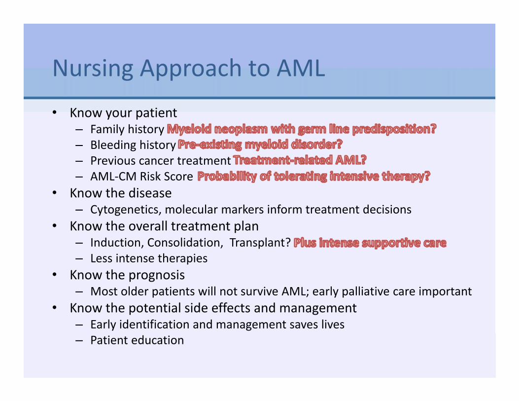
Nursing Approach to AML• Know your patient
– Family history– Bleeding history– Previous cancer treatment– AML‐CM Risk Score
• Know the disease– Cytogenetics, molecular markers inform treatment decisions
• Know the overall treatment plan– Induction, Consolidation, Transplant?– Less intense therapies
• Know the prognosis– Most older patients will not survive AML; early palliative care important
• Know the potential side effects and management– Early identification and management saves lives– Patient education

Factors that Significantly Decrease Chance of Survival
• Unfavorable disease‐related genetic factors: increased risk of AML resistance or recurrence
• Unfavorable patient‐related factors: increased risk of treatment‐associated toxicity and mortality– Physiologic age– Poor performance status– Complex or poorly controlled comorbidities
What tools are available to predict if your older patient can tolerate intense induction therapy?
Arber DA et al. Blood. 2016;127(20):2391‐2405. Tsai C‐H et al. Leukemia. 2016;30(7):1485‐1492.

Eligibility for Intensive Therapy:Hematopoietic Cell Transplantation‐Comorbidity Index (HCT‐CI) Predicts Survival for Older Patients with AML
HCT‐CI Score
CR Rate 30‐day Mortality
Median Survival
0 64% 3% 11 mos
1‐2 43% 11% 8 mos
≥ 3 42% 29% 5 mos
22%
30%
48%No comorbidity
Score 1‐2
Score ≥ 3
AML patients ≥60 y/o undergoing induction (n=177)
Michelis FV et al. Blood. 2015;126(23):3201; Giles FJ et al. Brit J Haematol. 2007;136(4):624‐627.

AML Composite Model (AML‐CM) can Guide Decision‐Making About Treatment
AML‐CMHCT‐CI PLUS Additional Factors
Score
Albumin <3.5 g/dL 1
Platelets <20,000/mcL 1
LDH >200‐1000 U/L 1
>1000 U/L 2
Age 50‐59 years old 1
≥60 years old 2
ELN cytogenetic/molecular risk
group
Intermediate 1
Adverse 2
AML Composite Model. Available at http://www.AMLCompositeModel.orgSorror ML et al. JAMA Oncology. 2017;3(12):1675‐1682.

AML Treatment Algorithm
Brandwein JM et al. Am J Blood Res. 2017;7(4): 30–40.
Unfit for inductionchemotherapy
Low‐Intensity therapiesClinical TrialsAzacitidineDecitabine

7+3 Induction Therapy
Daunorubicin 60‐90 mg/m2
IVP on days 1‐3Cytarabine 100‐200 mg/m2
CIV on days 1‐7
Await Count Recovery
<5% blasts
>5% blasts
Re‐induction Chemotherapy
Day 14 Bone Marrow Biopsy
Recovery Bone Marrow Biopsy to
determine complete
remission and MRD
• “7+3” regimen first described by Yates et al. in 1973• 44 years later, remains standard of care for medically fit patients
• CR rate in younger patients: 60‐80%• CR rate in older patients: 40‐60%• Yet, relapse inevitable in most patients
Döhner H et al. Blood. 2017;129:424–447; Yates JW et al. Cancer Chemother Rep. 1973;57:485–488.
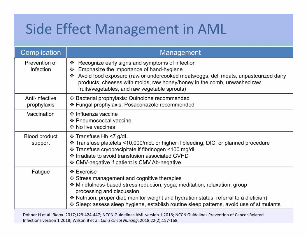
Side Effect Management in AML
Dohner H et al. Blood. 2017;129:424‐447; NCCN Guidelines AML version 1.2018; NCCN Guidelines Prevention of Cancer‐Related Infections version 1.2018; Wilson B et al. Clin J Oncol Nursing. 2018;22(2):157‐168.
Complication ManagementPrevention of
Infection Recognize early signs and symptoms of infection Emphasize the importance of hand-hygiene Avoid food exposure (raw or undercooked meats/eggs, deli meats, unpasteurized dairy
products, cheeses with molds, raw honey/honey in the comb, unwashed raw fruits/vegetables, and raw vegetable sprouts)
Anti-infective prophylaxis
Bacterial prophylaxis: Quinolone recommended Fungal prophylaxis: Posaconazole recommended
Vaccination Influenza vaccine Pneumococcal vaccine No live vaccines
Blood product support
Transfuse Hb <7 g/dL Transfuse platelets <10,000/mcL or higher if bleeding, DIC, or planned procedure Transfuse cryoprecipitate if fibrinogen <100 mg/dL Irradiate to avoid transfusion associated GVHD CMV-negative if patient is CMV Ab-negative
Fatigue Exercise Stress management and cognitive therapies Mindfulness-based stress reduction; yoga; meditation, relaxation, group
processing and discussion Nutrition: proper diet, monitor weight and hydration status, referral to a dietician) Sleep: assess sleep hygiene, establish routine sleep patterns, avoid use of stimulants
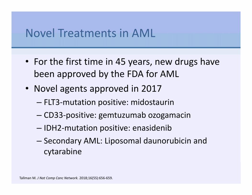
Novel Treatments in AML
• For the first time in 45 years, new drugs have been approved by the FDA for AML
• Novel agents approved in 2017– FLT3‐mutation positive: midostaurin– CD33‐positive: gemtuzumab ozogamacin– IDH2‐mutation positive: enasidenib– Secondary AML: Liposomal daunorubicin and cytarabine
Tallman M. J Nat Comp Canc Network. 2018;16(5S):656‐659.
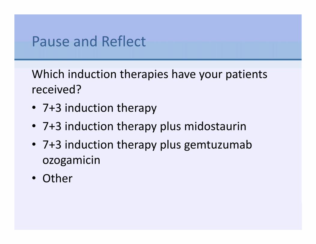
Pause and Reflect
Which induction therapies have your patients received?• 7+3 induction therapy• 7+3 induction therapy plus midostaurin• 7+3 induction therapy plus gemtuzumabozogamicin
• Other

Case Study
• 66 year‐old male with AML‐MRC: AML with myelodysplasia‐related changes
• Induction treatment selected: liposomal daunorubicin and cytarabine

Liposomal Daunorubicin and Cytarabine
CONSOLIDATION
Day 1 2 3
X X
Daunorubicin 29 mg/m2 and Cytarabine 65 mg/m2 liposome IV over 90 minutes on Days 1 and 3
Up to 2 cycles consolidation
Indicated for the treatment of adults with newly‐diagnosed therapy‐related acute myeloid leukemia (t‐AML) or AML with
myelodysplasia‐related changes (AML‐MRC)
INDUCTION
Day 1 2 3 4 5
X X X
Daunorubicin 44 mg/m2 and Cytarabine 100 mg/m2 liposome IV over 90 minutes on Days 1, 3, and 5
1‐2 cycles induction
Vyxeos PI. 8.2017.
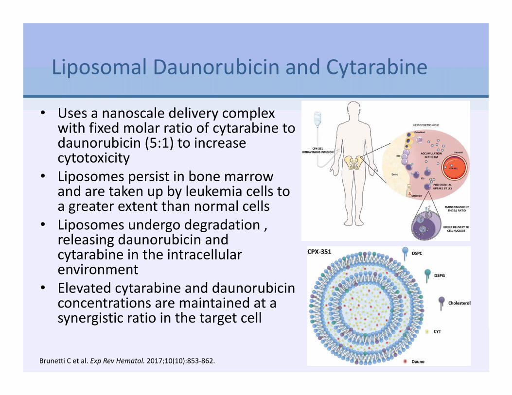
Liposomal Daunorubicin and Cytarabine
• Uses a nanoscale delivery complex with fixed molar ratio of cytarabine to daunorubicin (5:1) to increase cytotoxicity
• Liposomes persist in bone marrow and are taken up by leukemia cells to a greater extent than normal cells
• Liposomes undergo degradation , releasing daunorubicin and cytarabine in the intracellular environment
• Elevated cytarabine and daunorubicin concentrations are maintained at a synergistic ratio in the target cell
Brunetti C et al. Exp Rev Hematol. 2017;10(10):853‐862.
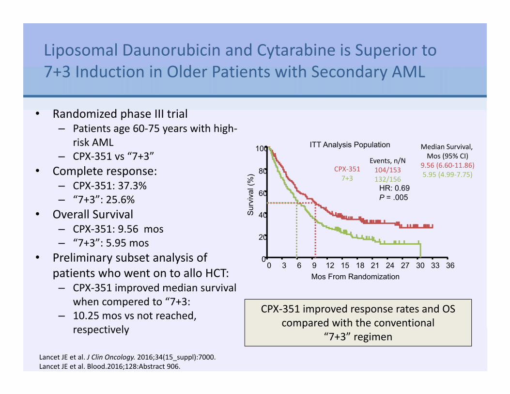
Liposomal Daunorubicin and Cytarabine is Superior to 7+3 Induction in Older Patients with Secondary AML
• Randomized phase III trial – Patients age 60‐75 years with high‐
risk AML– CPX‐351 vs “7+3”
• Complete response:– CPX‐351: 37.3% – “7+3”: 25.6%
• Overall Survival– CPX‐351: 9.56 mos– “7+3”: 5.95 mos
• Preliminary subset analysis of patients who went on to allo HCT:– CPX‐351 improved median survival
when compered to “7+3:– 10.25 mos vs not reached,
respectively
100
80
60
40
20
0Su
rviv
al (%
)
Mos From Randomization0 3 6 9 12 15 18 21 24 27 30 33 36
ITT Analysis Population
Events, n/N104/153132/156
Median Survival, Mos (95% CI)
9.56 (6.60‐11.86)5.95 (4.99‐7.75)
CPX‐3517+3
HR: 0.69P = .005
CPX‐351 improved response rates and OS compared with the conventional
“7+3” regimen
Lancet JE et al. J Clin Oncology. 2016;34(15_suppl):7000.Lancet JE et al. Blood.2016;128:Abstract 906.

Side Effects of Liposomal Daunorubicin/Cytarabine Similar to 7+3
Grade ≥3 AEs (≥5% Pts), n (%)
CPX‐351(n=153)
7+3(n=151)
Febrile neutropenia 104 (68) 107 (71)
Pneumonia 30 (20) 22 (15)
Hypoxia 20 (13) 23 (15)
Sepsis 14 (9) 11 (7)
Hypertension 16 (10) 8 (5)
Respiratory failure 11 (7) 10 (7)
Fatigue 11 (7) 9 (6)
Bacteremia 15 (10) 3 (2)
Reduced ejection fraction 8 (5) 8 (5)
Lancet JE et al. J Clin Oncology. 2016;34(15_suppl):7000.

Prolonged Cytopenias Associated with Liposomal Daunorubicin/Cytarabine
Induction Consolidation
CPX‐351(n=58)n (%)
7+3(n=34)n (%)
CPX‐351(n=48)n (%)
7+3(n=32)n (%)
Prolonged thrombocytopenia 16 (28) 4 (12) 12 (25) 5 (16)
Prolonged neutropenia 10 (17) 1 (3) 5 (10) 1 (3)
Lancet JE et al. J Clin Oncology. 2016;34(15_suppl):7000.

Considerations with Liposomal Daunorubicin/Cytarabine• Pre‐treatment:
– Assess cardiac function and obtain liver and renal function studies
• Major adverse events:– Myelosuppression, infections, bleeding– Cardiotoxicity
• Echo pre‐induction and pre‐consolidation and as indicated• Be mindful of cumulative anthracycline dose
– Copper toxicity• Contains copper and may cause copper overload in patients with Wilson’s disease or other copper‐related metabolic disorders
– Hypersensitivity reactions– Rash

Practical Tips
• Moderate emetic risk: Pre‐medication: ondansetron and dexamethasone
• Calculate cumulative anthracycline exposure• Do not interchange daunorubicin and cytarabine liposome with other
daunorubicin and/or cytarabine products• Daunorubicin and cytarabine liposome has unique preparation
instructions and takes close to an hour to prepare– Key steps include equilibration to room temperature, reconstitution
followed by swirling vial contents, and aseptically transferring the medication to an infusion bag
• Dose adjustments are not required for renal or hepatic insufficiency• If patients develop a hypersensitivity reaction, pre‐medicate patients
with an antihistamine and/or corticosteroid prior to subsequent doses
• Vesicant– Daunorubicin has been associated with necrosis where the drug leaks
into the skin and subcutaneous tissue during IV infusion. Vyxeos PI. 8.2017.

Pause and Reflect
What toxicities have you treated in patients with AML receiving liposomal daunorubicin and cytarabine?• Pancytopenia‐transfusion dependent• Neutropenic fevers• Mild headaches• Anorexia• Other

Case Study
• 66 year‐old male with AML‐MRC– AML with myelodysplasia‐related changes treated with liposomal daunorubicin and cytarabine
• Toxicities:– Pancytopenia‐transfusion dependent– Neutropenic fevers‐treated with broad spectrum antibiotics, no source identified
– Mild headaches– Anorexia

Case Study
On Day 10 after therapy, patient develops rash• Began as erythematous
papules that evolved into coalescing purpuric papules and plaques, particularly over trunk and extremities
• Intertriginous accentuation in the fold of the pannus
• Lower extremity shows scattered palpable purpura
• Dorsal right foot shows a striking purpuric plaque
Differential Diagnosis• Antibiotic‐induced rash• Viral exanthem• Cytarabine‐induced rash
Ruben BS et al. J Am Acad Dermatol. 2015;73(5):821‐828.

• Benign rash, but looks impressive• Occurs 1‐2 weeks after cytarabine exposure• May present as papular purpuric eruption or morbilliform eruption, particularly over trunk and extremities
• May also involve the axillae, groin, back of neck, ears, and scalp
• Pathology of skin biopsy typically spongiotic dermatitis• Treated with high potency topical steroid
• 0.1% triamcinolone ointment
Cytarabine‐Induced Rash
Ruben BS et al. J Am Acad Dermatol. 2015;73(5):821‐828.

Case Study
• 33 year‐old with newly diagnosed AML with FLT3‐ITD mutation
• Induction treatment:– Standard 7+3 induction therapy
• Daunorubicin 60 mg/m2 IVP on Days 1‐3• Cytarabine 200 mg /m2 CIV on Days 1‐7
– Midostaurin 50 mg PO BID on Days 8 to 21

FLT3 (fms‐related tyrosine kinase 3) Gene Mutations
• Occur in 30% of adults with newly diagnosed AML
• Two types of FLT3 mutations– FLT3 internal tandem duplication (ITD) mutations
• ~22% incidence• Associated with a poor prognosis owing to a high relapse rate
– FLT3 point mutation in the tyrosine kinase domain (TKD)
• ~8% incidence• Impact of TKD mutations on prognosis is uncertain
• FLT3 inhibitors prevent growth of leukemia cells
Stone RM et al. N Engl J Med. 2017;377:454‐464.

Midostaurin
• FDA approval 2017 :– Indicated for adults with FLT3 mutation‐positive AML in combination
with 7+3 induction and cytarabine consolidation therapy– Based on the RATIFY trial, which took 13 years to complete!
• Dose: – 50 mg PO BID with food on Days 8‐21 of each induction and
consolidation cycle• Results:
– 4‐year survival: 51.4% on midostaurin vs 44.2% on placebo (P=.0074)– 22% reduced risk of death in midostaurin arm– Benefit was most pronounced for patients with NPM1wt and FLT3‐
high• Well tolerated:
– Side effects mostly due to induction chemotherapy– Slightly more anemia and rash than placebo
Stone RM et al. N Engl J Med. 2017;377:454‐464.

Case Study: Day 10 of Induction Therapy
• Temperature 39 °C• HR 124• RR 24• BP 70/39
• WBC 0.2• ANC 0• Hb 8.5• Platelets 24, 000
What is your concern?
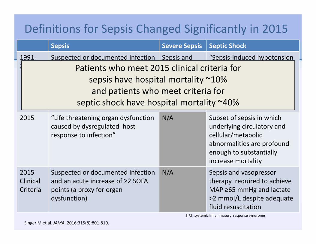
Definitions for Sepsis Changed Significantly in 2015Sepsis Severe Sepsis Septic Shock
1991‐2015
Suspected or documented infection PLUS 2 or more SIRS criteria:• Temperature >38 °C or <36 °C• Heart rate >90/min• Respiratory rate >20/min or PaCO2 <32
mm Hg (4.3 kPa)• White blood cell count >12 000/mm3 or
<4000/mm3 or >10% immature bands
Sepsis and organ dysfunction
“Sepsis‐induced hypotension persisting despite adequate fluid resuscitation.”
2015 “Life threatening organ dysfunction caused by dysregulated host response to infection”
N/A Subset of sepsis in which underlying circulatory and cellular/metabolic abnormalities are profound enough to substantially increase mortality
2015 Clinical Criteria
Suspected or documented infection and an acute increase of ≥2 SOFA points (a proxy for organ dysfunction)
N/A Sepsis and vasopressor therapy required to achieve MAP ≥65 mmHg and lactate >2 mmol/L despite adequate fluid resuscitation
SIRS, systemic inflammatory response syndrome
Singer M et al. JAMA. 2016;315(8):801‐810.
Patients who meet 2015 clinical criteria for sepsis have hospital mortality ~10% and patients who meet criteria for
septic shock have hospital mortality ~40%

How to Recognize Patients With Sepsis and Septic Shock: SOFA and qSOFA
MAP, mean arterial pressure; qSOFA, quick SOFA; SOFA, Sequential [Sepsis-related] Organ Failure Assessment
Singer M et al. JAMA. 2016;315(8):801‐810.

Elevated Lactate Predicts Mortality in Patients with Sepsis• Lactate
– Marker of cellular hypoxia and metabolic acidosis
– Normal level <2– Levels >4 associated with increased mortality
• With serial testing, decreasing lactate is a marker of improved perfusion, reduced mortality, and better prognosis – rising lactate levels suggests the opposite.
Mortality Risk Relative to Lactate Level
Howell MD et al. Intensive Care Med. 2007;33(11):1892‐1899.Shapiro NI et al. Ann Emerg Med. 2005;45(5):524‐528.

Management of Sepsis
Goal:MAP >65 mmHg, signs of improved perfusion
(normalization of lactate, normal organ function)
WITHIN 1 HOUR:
Levy MM et al. Crit Care Med. 2018;46(6):997‐1000.Rhodes A et al. Crit Care Med. 2017;45:486‐552.

Case Study
Blood cultures: 2 sets obtainedUA and urine culture obtainedCXR: no evidence of infection
Lactate: 4
After Fluid Resuscitation• Repeat Lactate: 1.7• BP 108/63• HR 84• RR 18• Temp: 38.2 C
Treatment• Started antibiotics: Piperacillin‐
tazobactam 3.375 g IV bolus then extended infusion over 4 hours q 8 hrs
• Fluids: weight 96.4 x 30 mL/kg = 2892 mL NS
• Given 3 liters NS

Case Study
Diagnosis: Sepsis due to e. coli bacteremia• Blood cultures (4/4
bottles): e. coli • Sensitive to piperacillin‐
tazobactam • Bacteremia resolved with
piperacillin‐tazobactam• Surveillance cultures
negative• ANC recovery at Day 32
• Achieved CR1– Bone marrow biopsy on
Day 14: hypoplasia– Bone marrow biopsy after
count recovery: CR1• Consolidation treatment:
– IDAC 1.5 g/m2 IV over 3 h on days 1‐3
– Midostaurin 50 mg PO BID on Days 8 to 21
• Allogeneic transplant

Clinical Pearls About Serious Infections
• Infection associated with hypotension or respiratory failure carries a poorer prognosis
• Infection with gram negative organisms has a greater risk of septic shock than gram positive organisms
• Abdominal source of sepsis is more fatal than any other source
• The longer a person is ill, hospitalized, or immune compromised, the greater their chance of developing significant sepsis
• Sepsis worsens other clinical complications (e.g. malnutrition, adrenal insufficiency)

Pause and Reflect
What protocols do you follow to prevent infection in patients with AML?• NCCN• Professional guideline (not NCCN)• Institutional protocol• Other

Gemtuzumab Ozogamicin (GO)
Indication: • Newly diagnosed
CD33‐positive AML in adults
• Relapsed or refractory CD33‐positive AML in adults and in pediatric patients 2 years and older
Mechanism of action:• Antibody‐drug conjugate targeted to CD33• CD33 present on AML blasts, not normal
hematopoietic stem cells
Jain, N., et al. Pharmaceutical Research, 32(11), 3526–40.
Appelbaum FR, Bernstein ID. Blood. 2017;130(22):2373‐2376.Castaigne S et al. Lancet. 2012;379(9825):1508‐1516.

Gemtuzumab Ozogamicin (GO):An Old Drug is New Again
• Originally approved by the FDA in 2000, withdrawn in 2010, and then reintroduced in 2017
• Based on ALFA‐0701 trial: Patients with AML ages 50‐70 years treated with GO plus standard 7+3– Improved event‐free survival in all patients– Improved overall survival in favorable‐risk > intermediate risk disease, not in poor‐
risk diseaseInduction Consolidation
Day 1 4 7 8 1
Newly diagnosed AML; in combination with daunorubicin/cytarabine
3 mg/m2
3 mg/m2
3 mg/m2
3 mg/m2
Newly diagnosed AML; single agent
6 mg/m2
3 mg/m2
2 mg/m2
Up to 8 cycles Q4W
Relapsed or refractory AML; single agent
3 mg/m2
3 mg/m2
3 mg/m2
Given in a fractionated schedule to reduce toxicity
Appelbaum FR, Bernstein ID. Blood. 2017;130(22):2373‐2376.Castaigne S et al. Lancet. 2012;379(9825):1508‐1516.

Gemtuzumab Ozogamicin:Significant Side Effects
• Infusion‐related toxicities (chills, fever and mild hypotension)
• Myelosuppression• Persistent thrombocytopenia• Hyperbilirubinemia• Veno‐occlusive disease (VOD) of the liver
Appelbaum FR, Bernstein ID. Blood. 2017;130(22):2373‐2376.
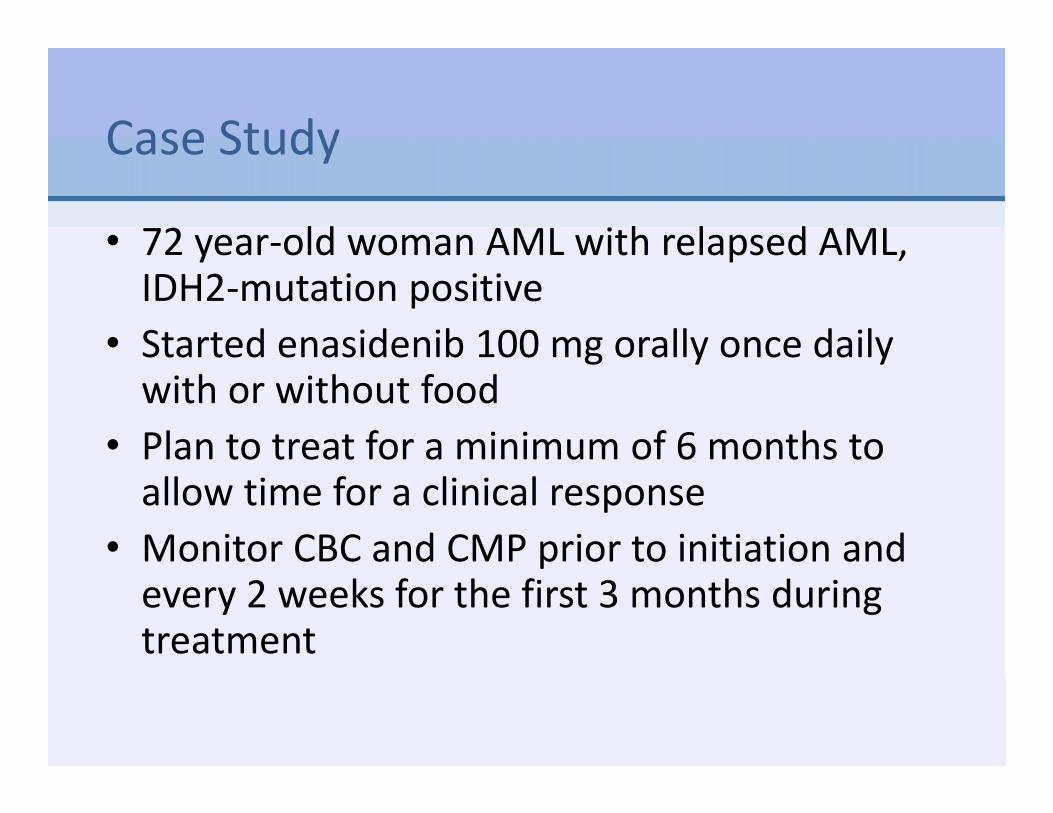
Case Study
• 72 year‐old woman AML with relapsed AML, IDH2‐mutation positive
• Started enasidenib 100 mg orally once daily with or without food
• Plan to treat for a minimum of 6 months to allow time for a clinical response
• Monitor CBC and CMP prior to initiation and every 2 weeks for the first 3 months during treatment

Pause and Reflect
When would you instruct the patient to call the immediately?• They are more fatigued than usual • They experience fever or chills• They experience nausea or vomiting• They experience tenderness or swelling in the abdomen

IDH1 and IDH2 in AML
• IDH: cellular enzyme• IDH1 and IDH2 mutations
– Alter DNA methylation blocking myeloid differentiation
– Prevent blasts in the bone marrow from differentiating into mature functioning blood cells
• Mutations in ~12% of AML
• Prognostic implication of the IDH1/2mutations remain unclear
αKG, alpha ketoglutarate; D‐2HG, D‐2‐hydroxyglutarate; IDH, isocitrate dehydrogenase; DNA, deoxyribonucleic acid; mut, mutated; NAD, nicotinamide adenine dinucleotide; NADP, nicotinamide adenine dinucleotide phosphate; TCA cycle, tricarboxylic acid cycle
Mondesir J. et al. J Blood Med. 2016;7:171‐180.Stein EM et al. Blood. 2017;130:722‐731.

Enasidenib: IDH2 inhibitor
38
23
1815 14 14
10 107 7 6 6 5
Adverse Events Attributed To EnasidenibResponses
• Overall response rate: 37%
• Complete morphologic remission: 20%
• Stable disease: 40% to 50%
Stein EM et al. Blood. 2017;130:722‐731.

Case Study
• Three months after starting enasidenib, presents with shortness of breath and fevers
• Temp 101.8 HR 106 BP 142/78 RR 24• PE: bibasilar rales and 1+ bilateral pedal edema
• Differential diagnosis:– Infection– Differentiation syndrome

IDH‐Inhibitor‐Associated Differentiation Syndrome (IDH‐DS)Signs/Symptoms Management
IDH‐DS and pulmonary or renal manifestations
Hospitalize for continued close observation
Severe pulmonary symptoms and/or renal dysfunction attributed to IDH‐DS persist for >48 hours after initiation of corticosteroids
Treatment with enasidenib should be interrupted. Note: Due to long the half‐life of enasidenib (~45 hours), treatment interruption may not immediately reverse IDH‐DS symptoms. Enasidenib may be reinitiated at the original dose after IDH‐DS improves to Grade 2 (moderate) or lower
Elevated WBC count (>30x109 /L or rapidly rising)
Prompt initiation of hydroxyurea is suggested. In cases of severe leukocytosis, leukapheresis may be appropriate
Substantial edema or weight gain Initiation of furosemide may be appropriate
Pericardial effusion (rare symptom of IDH‐DS, but can be life‐threatening)
Urgent intervention is required; patient should be managed, when appropriate, in consultation with a cardiac specialist.
Increasing serum creatinine Evaluate for concurrent tumor lysis syndrome (TLS)
Rapid increase in peripheral blood cells
Monitor for disseminated intravascular coagulopathy and related bleeding complications
Stein EM et al. Blood. 2017;130:722‐731.

Case Study
• Admitted to hospital• Started dexamethasone 10 mg IV q 12hr• Held enasidenib• Shortness of breath and fevers resolved within 24 hrs
• Started steroid taper and discharged from hospital
• Able to restart enasidenib

Case Study
• Continued drug and achieved CR at 5 months
• Enadesinib promotes bone marrow differentiation and maturation (not ablation), so it takes time to see effect
• Educate patient that it takes time to achieve response– Median time to first response
1.9 months (range, 0.5–9.4 months)
– 87.3% patients obtained a response by cycle 5
Stein EM et al. Blood. 2017;130:722‐731.
CR, complete response; CRi, CR with incomplete hematologic recovery; CRp, CR with incomplete platelet recovery; MLFS, morphologic leukemia‐free state; PD, progressive disease; PR, partial response; SD, stable disease.

Summary
• AML is a complex hematologic malignancy driven by multiple acquired genetic mutations that evolves over time
• Clonal expansion of leukemia cells leads to bone marrow failure and related complications: severe infections, anemia, and bleeding
• Age and genomic abnormalities are the most significant predictors of survival
• Most newly diagnosed patients are >60 years old and AML in older adults remains a major therapeutic challenge
• In 2017, 4 new agents approved for AML, bringing promise of better outcomes
• Future successes will depend on further refining risk categories and treatment algorithms for older patients with AML, and incorporation of novel treatments
Suggested Readings
• Brandwein JM, Zhu N, Kumar R et al. Treatment of older patients with acute myeloid leukemia (AML): revised Canadian consensus guidelines. American Journal of Blood Research. 2017;7(4):30–40.Available at http://www.ncbi.nlm.nih.gov/pubmed/28804680.
• Döhner H, Estey E, Grimwade D et al: Diagnosis and management of AML in adults: 2017 ELN recommendations from an international expert panel. Blood. 2017;129:424–447.
• NCCN Guidelines: Acute Myeloid Leukemia v 1.2018.Available at https://www.nccn.org/professionals/physician_gls/pdf/aml.pdf.
• Tallman M. (2018). Prognostic Significance of Molecular Markers and Targeted Regimens in the Management of Acute Myeloid Leukemia. Journal of the National Comprehensive Cancer Network. 2018;16(5S):656–659.Available at https://doi.org/10.6004/jnccn.2018.0050.