Cross-species transmission of canine distemper virus—an · PDF filelar dermatitis...
11
Cross-species transmission of canine distemper virus—an update Andreas Beineke a,b, ⁎, Wolfgang Baumgärtner a,b , Peter Wohlsein a a Department of Pathology, University of Veterinary Medicine Hannover, Bünteweg 17, D-30559 Hanover, Germany b Center for Systems Neuroscience, Hanover, Germany abstract article info Article history: Received 2 June 2015 Received in revised form 1 September 2015 Accepted 2 September 2015 Available online 13 September 2015 Keywords: Canine distemper virus Spillover H protein Carnivores Human health risk Canine distemper virus (CDV) is a pantropic morbillivirus with a worldwide distribution, which causes fatal dis- ease in dogs. Affected animals develop dyspnea, diarrhea, neurological signs and profound immunosuppression. Systemic CDV infection, resembling distemper in domestic dogs, can be found also in wild canids (e.g. wolves, foxes), procyonids (e.g. raccoons, kinkajous), ailurids (e.g. red pandas), ursids (e.g. black bears, giant pandas), mustelids (e.g. ferrets, minks), viverrids (e.g. civets, genets), hyaenids (e.g. spotted hyenas), and large felids (e.g. lions, tigers). Furthermore, besides infection with the closely related phocine distemper virus, seals can be- come infected by CDV. In some CDV outbreaks including the mass mortalities among Baikal and Caspian seals and large felids in the Serengeti Park, terrestrial carnivores including dogs and wolves have been suspected as vectors for the infectious agent. In addition, lethal infections have been described in non-carnivore species such as pec- caries and non-human primates demonstrating the remarkable ability of the pathogen to cross species barriers. Mutations affecting the CDV H protein required for virus attachment to host-cell receptors are associated with virulence and disease emergence in novel host species. The broad and expanding host range of CDV and its maintenance within wildlife reservoir hosts considerably hampers disease eradication. © 2015 The Authors. Published by Elsevier B.V. This is an open access article under the CC BY-NC-ND license (http://creativecommons.org/licenses/by-nc-nd/4.0/). Contents Introduction . . . . . . . . . . . . . . . . . . . . . . . . . . . . . . . . . . . . . . . . . . . . . . . . . . . . . . . . . . . . . . . . . 49 Distemper in carnivore species . . . . . . . . . . . . . . . . . . . . . . . . . . . . . . . . . . . . . . . . . . . . . . . . . . . . . . . . . 50 Domestic dogs . . . . . . . . . . . . . . . . . . . . . . . . . . . . . . . . . . . . . . . . . . . . . . . . . . . . . . . . . . . . . . 50 Wild canids . . . . . . . . . . . . . . . . . . . . . . . . . . . . . . . . . . . . . . . . . . . . . . . . . . . . . . . . . . . . . . . 50 Procyonids . . . . . . . . . . . . . . . . . . . . . . . . . . . . . . . . . . . . . . . . . . . . . . . . . . . . . . . . . . . . . . . 51 Ailurids . . . . . . . . . . . . . . . . . . . . . . . . . . . . . . . . . . . . . . . . . . . . . . . . . . . . . . . . . . . . . . . . . 51 Ursids . . . . . . . . . . . . . . . . . . . . . . . . . . . . . . . . . . . . . . . . . . . . . . . . . . . . . . . . . . . . . . . . . 51 Mustelids . . . . . . . . . . . . . . . . . . . . . . . . . . . . . . . . . . . . . . . . . . . . . . . . . . . . . . . . . . . . . . . . 51 Felids . . . . . . . . . . . . . . . . . . . . . . . . . . . . . . . . . . . . . . . . . . . . . . . . . . . . . . . . . . . . . . . . . . 51 Viverrids . . . . . . . . . . . . . . . . . . . . . . . . . . . . . . . . . . . . . . . . . . . . . . . . . . . . . . . . . . . . . . . . 53 Hyaenids . . . . . . . . . . . . . . . . . . . . . . . . . . . . . . . . . . . . . . . . . . . . . . . . . . . . . . . . . . . . . . . . 53 Distemper in non-carnivore species . . . . . . . . . . . . . . . . . . . . . . . . . . . . . . . . . . . . . . . . . . . . . . . . . . . . . . . 53 Canine distemper virus and other morbilliviruses in marine mammals . . . . . . . . . . . . . . . . . . . . . . . . . . . . . . . . . . . . . . . 54 Conclusion . . . . . . . . . . . . . . . . . . . . . . . . . . . . . . . . . . . . . . . . . . . . . . . . . . . . . . . . . . . . . . . . . . 55 Acknowledgment . . . . . . . . . . . . . . . . . . . . . . . . . . . . . . . . . . . . . . . . . . . . . . . . . . . . . . . . . . . . . . . 55 References . . . . . . . . . . . . . . . . . . . . . . . . . . . . . . . . . . . . . . . . . . . . . . . . . . . . . . . . . . . . . . . . . . 56 Introduction Morbilliviruses belong to the family Paramyxoviridae and include a number of highly pathogenic viruses, such as measles virus, rinderpest virus, canine distemper virus (CDV), and peste-des-petits-ruminants One Health 1 (2015) 49–59 ⁎ Corresponding author at: Department of Pathology, University of Veterinary Medicine Hannover, Bünteweg 17, D-30559 Hanover, Germany. Tel.: +49 511 953 8640; fax: +49 511 953 8675. E-mail address: [email protected] (A. Beineke). http://dx.doi.org/10.1016/j.onehlt.2015.09.002 2352-7714/© 2015 The Authors. Published by Elsevier B.V. This is an open access article under the CC BY-NC-ND license (http://creativecommons.org/licenses/by-nc-nd/4.0/). Contents lists available at ScienceDirect One Health journal homepage: http://www.journals.elsevier.com/one-health
Transcript of Cross-species transmission of canine distemper virus—an · PDF filelar dermatitis...

One Health 1 (2015) 49–59
Contents lists available at ScienceDirect
One Health
j ourna l homepage: ht tp : / /www. journa ls .e lsev ie r .com/one-hea l th
Cross-species transmission of canine distemper virus—an update
Andreas Beineke a,b,⁎, Wolfgang Baumgärtner a,b, Peter Wohlsein a
a Department of Pathology, University of Veterinary Medicine Hannover, Bünteweg 17, D-30559 Hanover, Germanyb Center for Systems Neuroscience, Hanover, Germany
⁎ Corresponding author at: Department of Pathology, UHannover, Bünteweg 17, D-30559 Hanover, Germany. Tel511 953 8675.
E-mail address: [email protected] (A
http://dx.doi.org/10.1016/j.onehlt.2015.09.0022352-7714/© 2015 The Authors. Published by Elsevier B.V
a b s t r a c t
a r t i c l e i n f oArticle history:Received 2 June 2015Received in revised form 1 September 2015Accepted 2 September 2015Available online 13 September 2015
Keywords:Canine distemper virusSpilloverH proteinCarnivoresHuman health risk
Canine distemper virus (CDV) is a pantropic morbillivirus with a worldwide distribution, which causes fatal dis-ease in dogs. Affected animals develop dyspnea, diarrhea, neurological signs and profound immunosuppression.Systemic CDV infection, resembling distemper in domestic dogs, can be found also in wild canids (e.g. wolves,foxes), procyonids (e.g. raccoons, kinkajous), ailurids (e.g. red pandas), ursids (e.g. black bears, giant pandas),mustelids (e.g. ferrets, minks), viverrids (e.g. civets, genets), hyaenids (e.g. spotted hyenas), and large felids(e.g. lions, tigers). Furthermore, besides infection with the closely related phocine distemper virus, seals can be-come infected by CDV. In someCDVoutbreaks including themassmortalities amongBaikal and Caspian seals andlarge felids in the Serengeti Park, terrestrial carnivores including dogs andwolves have been suspected as vectorsfor the infectious agent. In addition, lethal infections have been described in non-carnivore species such as pec-caries and non-human primates demonstrating the remarkable ability of the pathogen to cross species barriers.Mutations affecting the CDV H protein required for virus attachment to host-cell receptors are associated withvirulence and disease emergence in novel host species. The broad and expanding host range of CDV and itsmaintenance within wildlife reservoir hosts considerably hampers disease eradication.
© 2015 The Authors. Published by Elsevier B.V. This is an open access article under the CC BY-NC-ND license(http://creativecommons.org/licenses/by-nc-nd/4.0/).
Contents
Introduction . . . . . . . . . . . . . . . . . . . . . . . . . . . . . . . . . . . . . . . . . . . . . . . . . . . . . . . . . . . . . . . . . 49Distemper in carnivore species . . . . . . . . . . . . . . . . . . . . . . . . . . . . . . . . . . . . . . . . . . . . . . . . . . . . . . . . . 50
Domestic dogs . . . . . . . . . . . . . . . . . . . . . . . . . . . . . . . . . . . . . . . . . . . . . . . . . . . . . . . . . . . . . . 50Wild canids . . . . . . . . . . . . . . . . . . . . . . . . . . . . . . . . . . . . . . . . . . . . . . . . . . . . . . . . . . . . . . . 50Procyonids . . . . . . . . . . . . . . . . . . . . . . . . . . . . . . . . . . . . . . . . . . . . . . . . . . . . . . . . . . . . . . . 51Ailurids . . . . . . . . . . . . . . . . . . . . . . . . . . . . . . . . . . . . . . . . . . . . . . . . . . . . . . . . . . . . . . . . . 51Ursids . . . . . . . . . . . . . . . . . . . . . . . . . . . . . . . . . . . . . . . . . . . . . . . . . . . . . . . . . . . . . . . . . 51Mustelids . . . . . . . . . . . . . . . . . . . . . . . . . . . . . . . . . . . . . . . . . . . . . . . . . . . . . . . . . . . . . . . . 51Felids . . . . . . . . . . . . . . . . . . . . . . . . . . . . . . . . . . . . . . . . . . . . . . . . . . . . . . . . . . . . . . . . . . 51Viverrids . . . . . . . . . . . . . . . . . . . . . . . . . . . . . . . . . . . . . . . . . . . . . . . . . . . . . . . . . . . . . . . . 53Hyaenids . . . . . . . . . . . . . . . . . . . . . . . . . . . . . . . . . . . . . . . . . . . . . . . . . . . . . . . . . . . . . . . . 53
Distemper in non-carnivore species . . . . . . . . . . . . . . . . . . . . . . . . . . . . . . . . . . . . . . . . . . . . . . . . . . . . . . . 53Canine distemper virus and other morbilliviruses in marine mammals . . . . . . . . . . . . . . . . . . . . . . . . . . . . . . . . . . . . . . . 54Conclusion . . . . . . . . . . . . . . . . . . . . . . . . . . . . . . . . . . . . . . . . . . . . . . . . . . . . . . . . . . . . . . . . . . 55Acknowledgment . . . . . . . . . . . . . . . . . . . . . . . . . . . . . . . . . . . . . . . . . . . . . . . . . . . . . . . . . . . . . . . 55References . . . . . . . . . . . . . . . . . . . . . . . . . . . . . . . . . . . . . . . . . . . . . . . . . . . . . . . . . . . . . . . . . . 56
niversity of VeterinaryMedicine.: +49 511 953 8640; fax: +49
. Beineke).
. This is an open access article under
Introduction
Morbilliviruses belong to the family Paramyxoviridae and include anumber of highly pathogenic viruses, such as measles virus, rinderpestvirus, canine distemper virus (CDV), and peste-des-petits-ruminants
the CC BY-NC-ND license (http://creativecommons.org/licenses/by-nc-nd/4.0/).

50 A. Beineke et al. / One Health 1 (2015) 49–59
virus, which cause devastating diseases in humans and animals. In thelast decades, morbilliviruses emerged also as causative agents of severalmass-mortalities in marine mammals [1,2]. Canine distemper is a fataldisease of dogs with a worldwide distribution [3]. The causative agent,CDV, is an enveloped, negative-sense, single-stranded RNA virus. Similarto other paramyxoviruses the virus contains six structural proteins,termed nucleocapsid (N), phospho (P), large (L), matrix (M), hemagglu-tinin (H) and fusion (F) protein, and two accessory non-structural pro-teins (C and V) that were found as extratranscriptional units within theP gene [4]. Generally, CDV exhibits lympho-, neuro- and epitheliotropismresulting in systemic infection of almost all organ systems including re-spiratory, digestive, urinary, lymphatic, endocrine, cutaneous, skeletaland central nervous system (CNS) [5,6]. The disease course andpathogenesis in canine distemper resemble those of human measlesvirus infection including, fever, rash, respiratory signs, lymphopenia,and profound immunosuppression with generalized depletion oflymphoid organs during the acute disease phase [7]. In addition, CDVinfection shows a high incidence of neurological complications [5].
Unlike the related measles virus which is maintained by single hostspecies, CDV represents a rather promiscuous agent causing distemper-like pathology in a variety of different carnivorous and also non-carnivorous species [8–10]. Clinical findings and pathology resemblelargely the disease in dogs. However, morbidity and mortality mayvary greatly among animal species. Phylogenetic and molecularevolutionary analyses of CDV have revealed that mutations affectingthe binding site of theH protein for virus entry receptors (signaling lym-phocytic activationmolecule [SLAM, CD150] and nectin-4) are associatedwith the occurrence of disease emergence in novel host species [11–15].
The aim of the present article is to give an updated overview ofinterspecies transmission of CDV and the pathogenesis of distemper indifferent mammalian species.
Distemper in carnivore species
Domestic dogs
The pathogenesis of CDV infection in domestic dogs has been exten-sively reviewed previously [3,5]. In brief, disease duration and severityin domestic dogs depends mainly on the animal's age and immune sta-tus and strain virulence. The primary mode of infection is via inhalation[16]. Initially, CDV replicates in lymphoid tissue of the upper respiratorytract. Here, monocytes and macrophages are the first target cells whichpropagate the virus [17]. Following a variable incubation period (one tofour weeks), animals develop a characteristic biphasic fever [16,18].During the first viremic phase, generalized infection of lymphoid tissueswith lymphoid depletion, lymphopenia and transient fever is observed.Profound immunosuppression is a consequence of leukocyte necrosis,apoptosis and dysfunction [16,19,20]. Second viremia is associatedwith high fever and infection of parenchymal tissues such as the respi-ratory tract, digestive tract, skin, and CNS [16,17]. During this diseasestage, various clinical manifestations may be present such as conjuncti-vitis, nasal discharge, anorexia, respiratory signs, gastrointestinal signs,and neurological deficiencies [16]. Respiratory signs are a sequel ofvirus-induced rhinitis and interstitial pneumonia, while vomiting,diarrhea and dehydration are caused by gastrointestinal tract infection[21]. Often enteric and respiratory signs are worsened by secondarybacterial infections. Characteristic dermalmanifestations include pustu-lar dermatitis (distemper exanthema) and hyperkeratosis of foodpadsand nasal planum (hard pad disease). In young animals also enamel hy-poplasia andmetaphyseal osteosclerosis have been described followingCDV infection [22]. Neurologic signs depend on viral distribution in theCNS and include hyperesthesia, cervical rigidity, seizures, cerebellar andvestibular signs, as well as paraparesis or tetraparesis with sensoryataxia [9,23]. Histological manifestations include polioencephalitis anddemyelinating leukoencephalomyelitis [24,25]. Recovery depends onthe host immune response. Particularly, a strong and effective cellular
immune response can eliminate the virus prior to infection of parenchy-mal tissues, while weak and delayed cellular and humoral immuneresponses lead to virus spread and persistence, respectively [5,16,26].
Wild canids
Besides domesticated dogs natural and/or vaccine-induced CDV-associated disease has been reported in almost all genera of the tribustrue canids. Affected members of the genus Canis include Australiandingos (Canis dingo) [27], coyotes (Canis latrans) [28,29], black-backedjackals (Canis mesomelas) [30], golden jackals (Canis aureus) [31], Cana-dianwolves (Canis lupus) [32], American graywolves (Canis lupus) [33],Mexican wolves (Canis lupus baileyi) [34], Iberian wolves (Canis lupus)[35], and Apennine wolves (Canis lupus) [36]. Phylogenetic analysessuggest a CDV spillover from domestic dogs to free-ranging jackalsandwolves [30,35]. Referring to this, sequencing of CDV from Apenninewolves in Italy identified a strain belonging to the Arctic lineage, knownto circulate in European dog populations [36]. The Ethiopianwolf (Canissimensis) is recognized as the rarest canid species in the world and themost threatened carnivore in Africa. This species is almost extinct dueto combined effects of rabies and CDV infections [37]. Wolf-derivedCDV from the Ethiopian outbreak show sequence homologies to isolatesfromdomestic dogs in theUSA, Germany and Japan, suggestive of globalvirus spread [37]. There is serological evidence of CDV exposition tomaned wolves (Chrysocyon brachyurus) in Brazil. Natural clinicaldistemper has not been reported in this species [38], but vaccination-induced distemper may occur [39]. Similarly, there are no reportsabout cases of naturally occurring distemper in bush dogs (Speothosyenaticus), however, a possible vaccine-induced case has beendescribed [40].
Endangered African wild dogs (Lycaon pictus) have been reported tobe exposed to CDV and are highly susceptible to develop distemper [41,42]. Molecular analyses of isolates from African wild dogs suggest thatCDV is endemic inwildlife carnivore populations in Tanzania (Serengetiecosystem) [43,44]. Lethal lesions include interstitial pneumonia andsuppurative to necrotizing bronchopneumonia with viral inclusionbodies and syncytial cells [43,44]. Besides natural infection, Africanwild dogs in captivity may also succumb to vaccine-induced caninedistemper [45].
All genera of the tribus true foxes, i.e. Vulpes sp., including Vulpeslagopus (syn. Alopex lagopus), Urocyon and Otocyon, are susceptible toCDV infections and may develop clinical disease. CDV infections havebeen reported in red foxes (Vulpes vulpes) from various European coun-tries including Germany [46,47], Italy [48,49], Spain [50], and Portugal[51]. Disease has been reported also in swift foxes (Vulpes velox) [52],kit foxes (Vulpes macrotis) [52], Indian foxes (Vulpes bengalensis) [53],and fennec foxes (Vulpes zerda) [54]. Infected foxes show abnormalbehavior including loss of fear for humans, disorientation, and/orrespiratory distress.Morphologic findings comprisemainly conjunctivi-tis, pustular dermatitis, lymphohistiocytic polioencephalitis, andbronchointerstitial pneumonia with viral inclusion bodies and syncytia[14].
Recently, the emergence and spread of a single genetic clusterwithin the Europe-1 clade of CDV among foxes and other wild carni-vores in the Alpine region has been reported indicating the ability ofthis virus to replicate in a wider host range [55]. In gray foxes(Urocyon sp.), CDV outbreaks might have caused a dramatic populationdecline of Santa Catalina Island foxes (Urocyon littoralis catalinae).Sequence analyses indicate virus transmission from infected mainlandUSA raccoons unintendedly introduced to the island [56]. Mainlandgray foxes (Urocyon cinereoargenteus) are susceptible to natural distem-per and vaccine-induced distemper [57]. Crab-eating foxes (Cerdocyonthous) show neurological signs and succumb to CDV infection [58].Free-ranging culpeo (Dusicyon culpaeus) and South American grayfoxes (Dusicyon griseus) have been exposed to CDV [59]. Similarly, inthe Serengeti-Mara ecosystem of East Africa, bat-eared foxes (Otocyon

51A. Beineke et al. / One Health 1 (2015) 49–59
megalotis) have succumbed to CDV during epidemics [60]. Serologicalevidence of CDV infection or fatal CDV infection have been observedin various Pseudalopex sp. [61], Pampas gray foxes (Lycalopexgymnocercus) [62], and hoary foxes (Lycalopex vetulus) [63].
The raccoon dog (Nyctereutes procyonoides) originally distributed inEast Asia represents a recently established neozoon in Germany andneighboring countries [64]. This wild omnivore serves as host and vec-tor for parasites and other pathogens including CDV [64]. Raccoon dogsare highly susceptible to CDV infection [65] showing similar morpho-logical changes as infected domestic dogs including interstitial pneumo-nia, demyelinating encephalitis, lymphoid depletion in variouslymphoid tissues and catarrhal or necrotizing gastroenteritis [66]. Theemergence of CDV strains belonging to the Asia-1 genotype with twoamino acid substitutions in the H protein isolated from raccoon dogsand other carnivores in China resulted in clinical distemper even in vac-cinated animals [67].
Procyonids
The raccoon is native to North America and a neozoon in continentalEurope and Japan [68,69]. Serological surveys revealed CDV expositionof members of the family Procyonidae including predominantly rac-coons (Procyon lotor) [70], but also pygmy racoons (Procyon pygmaeus)[71]. Spontaneous clinical distemper has been reported in sylvatic andurban populations of raccoons [68,72], while vaccination-induced dis-temper is reported in kinkajous (Potos flavus) [73]. Clinical signs in rac-coons resemble those in dogs and must be differentiated from rabies incases with neurologic signs [72]. Pathology is characterized byblepharoconjunctivitis, rhinitis, occasional pigmentation of the muzzleand footpads with hyperkeratosis, interstitial pneumonia with syncytiaand viral inclusion bodies, and demyelinating cerebellar white matterdisease [68,72,73]. Lednicky et al. (2004) identified two differentAmerican CDV lineages causing raccoon distemper outbreaks in thesame area suggesting multiple reintroductions of the virus [74]. Phylo-genetic analyses of CDV isolates from an outbreak in free-ranging rac-coons in Germany from 2012 to 2013 revealed close relations withEuropean CDV lineages especially from foxes and domestic dogs sugges-tive of interspecies transmission [68]. In addition, raccoons might haveintensified transmission of Asia-1 lineage CDV during an epidemic inwildlife mammals in Japan (2007–2008) [65,69].
Ailurids
Red pandas (Ailurus fulgens) are susceptible to CDV infection. A fataldisease clinically similar to canine distemper occurred after vaccinationwith modified live distemper vaccine [75]. Giant cell pneumonia andviral inclusion bodies in pulmonary and digestive tract epitheliumwere found histologically [75].
Ursids
There is marked serological evidence that various species of bearshave been exposed to CDV including American black bears (Ursusamericanus) [76], Asian black bears (Ursus tibethanus) [66], polar bears(Ursus maritimus) [77], grizzly bears (Ursus arctos horribilis) [32] andMarsican brown bears (Ursus arctos marsicanus) [78]. However, clinico-pathological manifestation of distemper in ursids is rare. An Americanblack bear yearling showed loss of fear for humans, periods of somno-lence, sporadic tremors and seizures caused by nonsuppurativepolioencephalitis with eosinophilic intranuclear and cytoplasmic inclu-sion bodies in neurons. Additionally, hyperkeratotic thickened footpadswere recorded. Sequence homologies with a CDV vaccine strain(Rockborn strain) indicate the potential virus exchange between vacci-nated domestic animals and wildlife [79]. Furthermore, neonatal deathof polar bears (Ursus maritimus) and a spectacled bear (Tremarctosornatus) has been attributed to CDV infection [80]. The virus can be
transmitted to bears by dogs, mustelids, coyotes, and other carnivoresthat might be sympatric with bears. Besides serological evidence ofCDV infection in captive Giant pandas (Ailuropoda melanoleuca) [81],also fatal CDV infection was noted recently among these endangeredspecies in a wildlife rescue and breeding center in China [82].
Mustelids
Domestic ferrets (Mustela putorius furo) are highly susceptible toCDV infection with a mortality rate of up to 100% in non-vaccinatedpopulations. As a consequence of systemic infections, ferrets develophigh fever together with respiratory and intestinal signs [83]. Classicaldermal manifestations include reddening and crusting of chin andmouth and progressive hyperkeratosis of nose and footpads.Polioencephalitis leading to behavioral changes, lethargy and seizuresis a common cause of death or reasons for euthanasia. But CNSmanifes-tation varies among CDV isolates and is preferentially caused by strainsknown to be neurovirulent in dogs (e.g. Snyder Hill and Cornell A75-17)[84,85]. In addition, profound generalized lymphoid depletion can beobserved in affected animals [86].
First reports of CDV infection in farmedminks (Neovison vison) havebeen described in 1930 [87]. Similar to ferrets, young minks usually diesuddenly, while adult mink have an increased resistance and exhibitprotracteddisease courseswith neurological signs [88]. Recently, phylo-genetic analyses showed thatwildlife species in Denmark, such as foxes,potentially contribute to the transmission of CDV to farmed mink andthat the virus is able to be maintained in the wild animal reservoir be-tween outbreaks. Isolates from the Danish outbreak in 2012 clusteredin the European CDV lineage and were closely related to viruses circu-lating inwildlife populations fromGermany and Hungary [89]. Interest-ingly, identification of CDV in fleas collected from amink carcass has ledto speculations about vector-mediated transmission of viruses betweenmink and other species [89].
Systemic, often lethal disease has been observed in black-footed fer-rets (Mustela nigripes) following CDV infection representing a seriousthreat for wildlife and captive populations [90]. Noteworthy, severepruritus can be commonly observed as an initial clinical sign in affectedanimals, followed by hyperkeratosis and progressive loss of body condi-tion. The high susceptibility of black-footed ferrets is demonstrated alsoby their fatal response to modified-live CDV vaccines demonstrated tobe safe in domestic ferrets and Siberianpolecats [91].Mortalities follow-ing CDV infection have been described also in colonies andwildlife pop-ulations of other mustelids, including martens (Fig. 1), polecats,badgers, ferret-badger, otters, and weasels, leading to the assumptionthat all members of the family are susceptible [10,92]. At post mortemexamination interstitial pneumonia, enteritis, encephalitis (Fig. 2) andlymphoid depletion (Fig. 3) with intralesional virus antigen can befound. Common lethal complications in mustelids are secondaryparasitic or bacterial diseases (Figs. 4 and 5) as a consequence ofvirus-induced immunosuppression [86]. Mustelids are regarded as aCDV reservoir and potential source of transmission to other species, in-cluding domestic dogs. Phylogenetic analyses revealed a co-circulationof several contemporary CDV genotypes in carnivores of centralEurope with the occurrence of a distinct CDV lineage in ferrets, polecatsand martens, suggestive of mustelid-adapted strains [93]. As in othercarnivores distemper represents an important differential diagnosisfor other CNS infections and has to be discriminated especially from ra-bies [94]. Distemper has been described also in striped skunks (Mephitismephitis) belonging to the mustelid related Mephitidae family [95].
Felids
In felids, CDV can cause clinically silent infections or fatal disease. Al-though CDV antibodies have been detected in domestic cats (Felis catus)[96], there are no reports of naturally occurring systemic CDV infections,despite frequent contact with dogs. Experimental infection of domestic
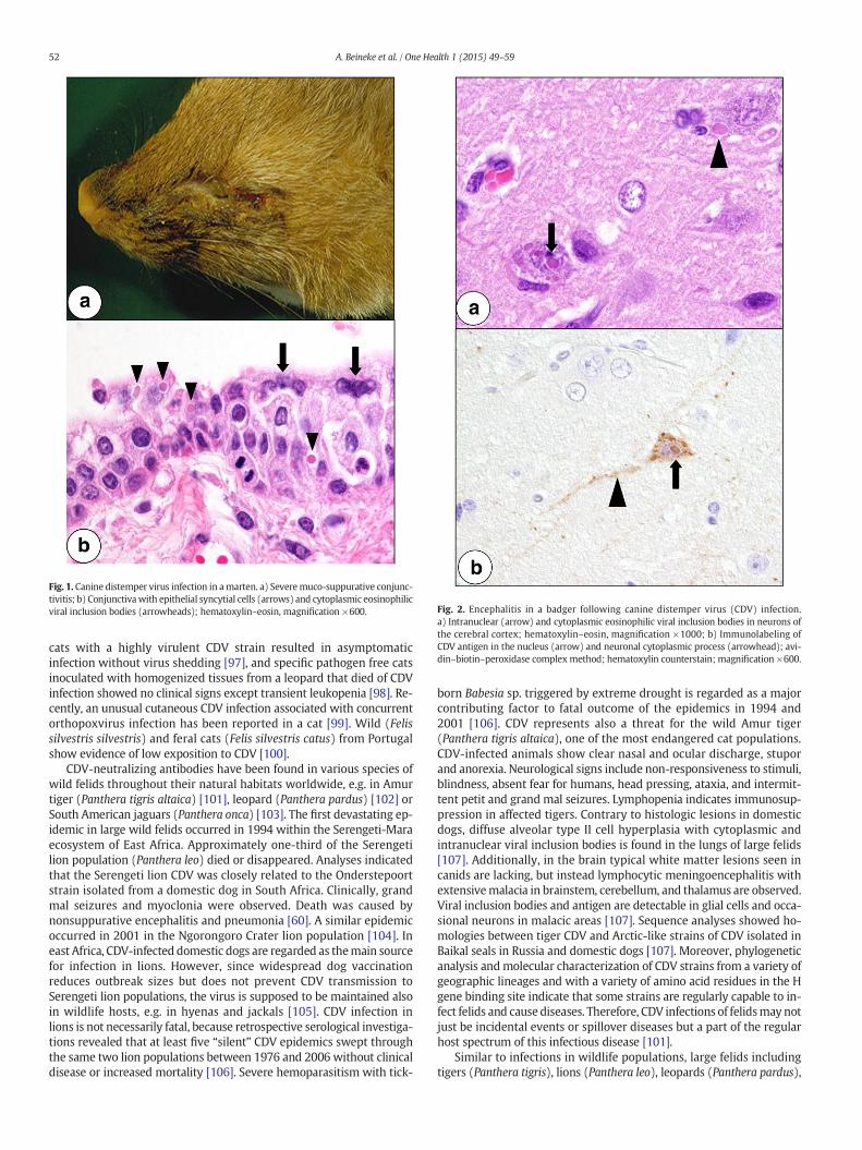
Fig. 1. Canine distemper virus infection in amarten. a) Severemuco-suppurative conjunc-tivitis; b) Conjunctivawith epithelial syncytial cells (arrows) and cytoplasmic eosinophilicviral inclusion bodies (arrowheads); hematoxylin–eosin, magnification ×600. Fig. 2. Encephalitis in a badger following canine distemper virus (CDV) infection.
a) Intranuclear (arrow) and cytoplasmic eosinophilic viral inclusion bodies in neurons ofthe cerebral cortex; hematoxylin–eosin, magnification ×1000; b) Immunolabeling ofCDV antigen in the nucleus (arrow) and neuronal cytoplasmic process (arrowhead); avi-din–biotin–peroxidase complex method; hematoxylin counterstain; magnification ×600.
52 A. Beineke et al. / One Health 1 (2015) 49–59
cats with a highly virulent CDV strain resulted in asymptomaticinfection without virus shedding [97], and specific pathogen free catsinoculated with homogenized tissues from a leopard that died of CDVinfection showed no clinical signs except transient leukopenia [98]. Re-cently, an unusual cutaneous CDV infection associated with concurrentorthopoxvirus infection has been reported in a cat [99]. Wild (Felissilvestris silvestris) and feral cats (Felis silvestris catus) from Portugalshow evidence of low exposition to CDV [100].
CDV-neutralizing antibodies have been found in various species ofwild felids throughout their natural habitats worldwide, e.g. in Amurtiger (Panthera tigris altaica) [101], leopard (Panthera pardus) [102] orSouth American jaguars (Panthera onca) [103]. The first devastating ep-idemic in large wild felids occurred in 1994 within the Serengeti-Maraecosystem of East Africa. Approximately one-third of the Serengetilion population (Panthera leo) died or disappeared. Analyses indicatedthat the Serengeti lion CDV was closely related to the Onderstepoortstrain isolated from a domestic dog in South Africa. Clinically, grandmal seizures and myoclonia were observed. Death was caused bynonsuppurative encephalitis and pneumonia [60]. A similar epidemicoccurred in 2001 in the Ngorongoro Crater lion population [104]. Ineast Africa, CDV-infected domestic dogs are regarded as themain sourcefor infection in lions. However, since widespread dog vaccinationreduces outbreak sizes but does not prevent CDV transmission toSerengeti lion populations, the virus is supposed to be maintained alsoin wildlife hosts, e.g. in hyenas and jackals [105]. CDV infection inlions is not necessarily fatal, because retrospective serological investiga-tions revealed that at least five “silent” CDV epidemics swept throughthe same two lion populations between 1976 and 2006 without clinicaldisease or increased mortality [106]. Severe hemoparasitism with tick-
born Babesia sp. triggered by extreme drought is regarded as a majorcontributing factor to fatal outcome of the epidemics in 1994 and2001 [106]. CDV represents also a threat for the wild Amur tiger(Panthera tigris altaica), one of the most endangered cat populations.CDV-infected animals show clear nasal and ocular discharge, stuporand anorexia. Neurological signs include non-responsiveness to stimuli,blindness, absent fear for humans, head pressing, ataxia, and intermit-tent petit and grand mal seizures. Lymphopenia indicates immunosup-pression in affected tigers. Contrary to histologic lesions in domesticdogs, diffuse alveolar type II cell hyperplasia with cytoplasmic andintranuclear viral inclusion bodies is found in the lungs of large felids[107]. Additionally, in the brain typical white matter lesions seen incanids are lacking, but instead lymphocytic meningoencephalitis withextensivemalacia in brainstem, cerebellum, and thalamus are observed.Viral inclusion bodies and antigen are detectable in glial cells and occa-sional neurons in malacic areas [107]. Sequence analyses showed ho-mologies between tiger CDV and Arctic-like strains of CDV isolated inBaikal seals in Russia and domestic dogs [107]. Moreover, phylogeneticanalysis andmolecular characterization of CDV strains from a variety ofgeographic lineages and with a variety of amino acid residues in the Hgene binding site indicate that some strains are regularly capable to in-fect felids and cause diseases. Therefore, CDV infections of felidsmay notjust be incidental events or spillover diseases but a part of the regularhost spectrum of this infectious disease [101].
Similar to infections in wildlife populations, large felids includingtigers (Panthera tigris), lions (Panthera leo), leopards (Panthera pardus),

Fig. 3. Lymphoid depletion in wildlife species following canine distemper virus (CDV) in-fection. a) Severe hypocellularity (asterisk) of the splenic white pulp in a CDV-infectedmarten; A = central artery; hematoxylin–eosin, magnification ×200; b) Detection ofCDV antigen in a splenic follicle of a raccoon by immunohistochemistry; A = central ar-tery; avidin–biotin–peroxidase complex method; hematoxylin counterstain; magnifica-tion ×400.
53A. Beineke et al. / One Health 1 (2015) 49–59
and jaguars (Panthera onca) in zoological collections may become in-fected with CDV. Animals often develop fatal disease with respiratoryand gastrointestinal signs followed by neurological manifestation [66,108]. Possible sources of the virus in zoo outbreaks are small carnivores,such as raccoons or raccoon dogs that may come in contact with captivecats [66,108]. Usually CDV spreads through aerosol droplets and contactwith infected body fluids, but felidsmay become also infected by preda-tion, e.g. exposure to unvaccinated and infected domestic dogs or otherwild susceptible hosts [66,108].
CDV infections have been also reported in members of the genusLynx including the highly endangered Iberian lynx (Lynx pardinus)[109], the Eurasian lynx (Lynx lynx) [14], the Canadian lynx (Lynxcanadensis) [110], and bobcats (Lynx rufus) [110]. CDV was reported asthe etiological agent of encephalitis in a Canadian lynx [110].
There is serological evidence of CDV infections in Namibian freeranging and captive cheetahs (Acinonyx jubatus) [102], Namibian cara-cals (Caracal caracal) [102], Argentinian Geoffroy's cats (Leopardusgeoffroyi) [111], Brazilian pumas (Puma concolor) [112], and Californianmountain lions (Puma concolor) [113].
Viverrids
Members of the family Viverridae including the Binturong (Arctictisbinturong) [114], masked palm civet (Paguma larvata) [115], Asianpalm civet (Paradoxurus hermaphroditus) [115], small Indian civet
(Viverricula indica) [115], and genet (Genetta genetta) [116] are suscep-tible to CDV and develop clinical disease. Infected animals show neuro-logical signs, dyspnea, oculonasal discharge, diarrhea, alopecia, andthickened hyperkeratotic and scaling footpads [114]. Morphological le-sions comprise bronchointerstitial pneumonia with syncytial cells,vesiculopustular dermatitis, hyperplastic pododermatitis with necrosis,lymphoid depletion as well as leuko- and polioencephalitis withintralesional viral antigen [114–116].
Hyaenids
Free-ranging Serengeti hyenas (Crocuta crocuta) and captivehyenas may succumb following CDV infection (Fig. 6). Sequencedata revealed closest homology to CDV strains causing high mortali-ty in sympatric lions [117]. Seropositivity of living animals indicatesthat Serengeti hyenas may also become subclinically infectedwithout overt disease or can recover from disease, respectively[118]. Similarly, CDV exposure has been reported from Zambianhyenas [41].
Distemper in non-carnivore species
The remarkable ability of CDV to cross species barriers is exemplifiedby its infection of non-carnivore species such as peccaries and non-human primates. In 1989, a CDV epizootic with fatal encephalitis wasobserved in collared peccaries (javelina; Pecari tajacu) in the desert ofsouthern Arizona (USA) [119]. Serological surveys suggest that CDV isenzootic in free-ranging peccaries of this area and that animals usuallyrecover from infection. Thus, increased fatality rate during the outbreakwas probably supported by high population densities and crowdingaround remaining water sources [120]. CDV-neutralizing antibodiessuggestive of subclinical infection have been detected also in wildboars and Sika deer during an epidemic in different wildlife mammalsin Japan [65].
In 1989, first cases of natural CDV infections in Japanese macaques(Macaca fuscata) with two fatalities were reported [121]. In 2006,large CDVoutbreaks occurred among rhesusmonkeys (Macacamulatta)in a breeding farm in Guangxi province (China) with death rates up to30% (about 4000 fatalities). Animals displayed measles-like signs, suchas respiratory distress, anorexia, fever, rash and conjunctivitis. Althoughthe exact source of infection could not be determined, virus transmis-sion by contact between farm monkeys with local wild monkeys or aspillover from a stray dog carrying CDV that became adapted to thenew host was discussed [122]. CDV infection of twenty rhesusmonkeysin an animal center in Beijing (China) was likely associated with thisoutbreak [123]. In a subsequent CDV outbreak in Japan in 2008, similarfatality numbers and febrile systemic diseases were observed incolonies of long-tailed macaques (Macaca fascicularis). Post mortemexamination revealed interstitial pneumonia, generalized lymphoid de-pletion and demyelination in the brain. Sequence analyses of the viralgenome revealed that Chinese and Japanese isolates are closely relatedwithin the Asia-1 clade, suggesting continuous chains of CDV infectionin monkeys [124].
Expansion of host species to include primates has raised concernsabout a potential risk of CDV infection in humans. It has been demon-strated in vitro that the monkey-adapted strain (CYN07-dV) has anintrinsic ability to use human nectin-4 for virus entry and easily becomeadapted to use the human CD150 following minimal amino acidchanges of the viral H protein [125]. Thus, species jumps to humanbeings, especially in people with a lack of cross-protective measles im-munity are proposed to happen in the future [12,126,127]. Moreover,the participation of CDV in the pathogenesis of Paget's disease of boneand multiple sclerosis in human beings has been speculated but lacksfinal verification [3,128].

Fig. 4. Concurrent toxoplasmosis in a marten infected with canine distemper virus (CDV). a) lympho-histiocytic encephalitis with numerous protozoal tachyzoites (arrows) andintranuclear eosinophilic viral inclusion bodies (arrowheads); hematoxylin–eosin, magnification ×400; b) Immunolabeling of CDV antigen in neurons and glial cells (arrows); note accu-mulation of protozoal tachyzoites (arrowhead); avidin–biotin–peroxidase complexmethod; hematoxylin counterstain; magnification ×400; c) Immunolabeling of Toxoplasma gondii an-tigen (arrows); avidin–biotin–peroxidase complex method; hematoxylin counterstain; magnification ×400.
54 A. Beineke et al. / One Health 1 (2015) 49–59
Canine distemper virus and other morbilliviruses in marinemammals
Several morbillivirus epidemics have been observed in differentmarine mammal species. Distemper in seals can be caused by CDV andthe closely related but genetically different phocine distempervirus (PDV) [129]. The devastating PDV epidemic among harbor seals(Phoca vitulina) and gray seals (Halichoerus grypus) in northwesternEuropean waters in 1988 represents the first documenteddisease manifestation of a morbillivirus infection in marine mammals[130]. At the same time, epidemics with CDV strains of theArctic group were observed among Baikal seals (Phoca sibirica) inSiberia [131]. CDV was isolated also from Caspian seals duringdisease outbreaks with high mortality rates in 1997, 2000 and 2001[132–134].
Experimental infection revealed duration of phocine distemperranging from two to three weeks with a mortality rate of 60% to 80%[135]. Similar to CDV, PDV infection of seals leads to interstitial pneumo-nia and catarrhal enteritis, causing fever, diarrhea, coughing, anddyspnea [135]. Other signs include nasal discharge, ocular discharge,anorexia, weight loss and abortion [136]. Common neurologicalmanifestations represent tremor, behavioral changes and lethargy[129]. Brain lesions in PDV-infected seals are similar to CDV-inducedacute polioencephalitis in dogs and measles virus inclusion body
polioencephalitis in human beings, respectively. With disease progres-sion also demyelination in the CNS can be observed [137,138]. Typicalfindings in PDV-infected seals include lymphoid depletion in spleenand lymph nodes with inclusion bodies and syncytial cells [136] andthymic atrophy, which renders the animals susceptible to developopportunistic infections. Interestingly, few harbor seals develop alsoepidermal hyperplasia and hyperkeratosis as a consequence of dermalinfection [139].
Surprisingly, PDV has been isolated only during the epidemics innorthwestern European waters in 1988 and 2002 [129,137]. In someCDV outbreaks including the mass mortalities among Baikal andCaspian seals, terrestrial carnivores including dogs and wolves havebeen suspected as vectors for the infectious agent [140]. Different hy-potheses concerning the origin of PDV and its geographical and chrono-logical dissemination pattern have been presented. These include virusspread from less susceptible marinemammals including Canadian harpseals (Phoca groenlandica) and Baltic gray seals (Halichoerus grypus), aswell as infection from diseased terrestrial animals including minks,wolves and polar bears. Debated predisposing factors for diseaseoutbreaks include malnutrition and immunosuppressive xenobiotics[141,142]. It still remains a possibility that PDV strains, with reducedvirulence for terrestrial mammals, are circulating in these species andcause mass die-offs in pinnipeds after crossing the species barrier[143,144].
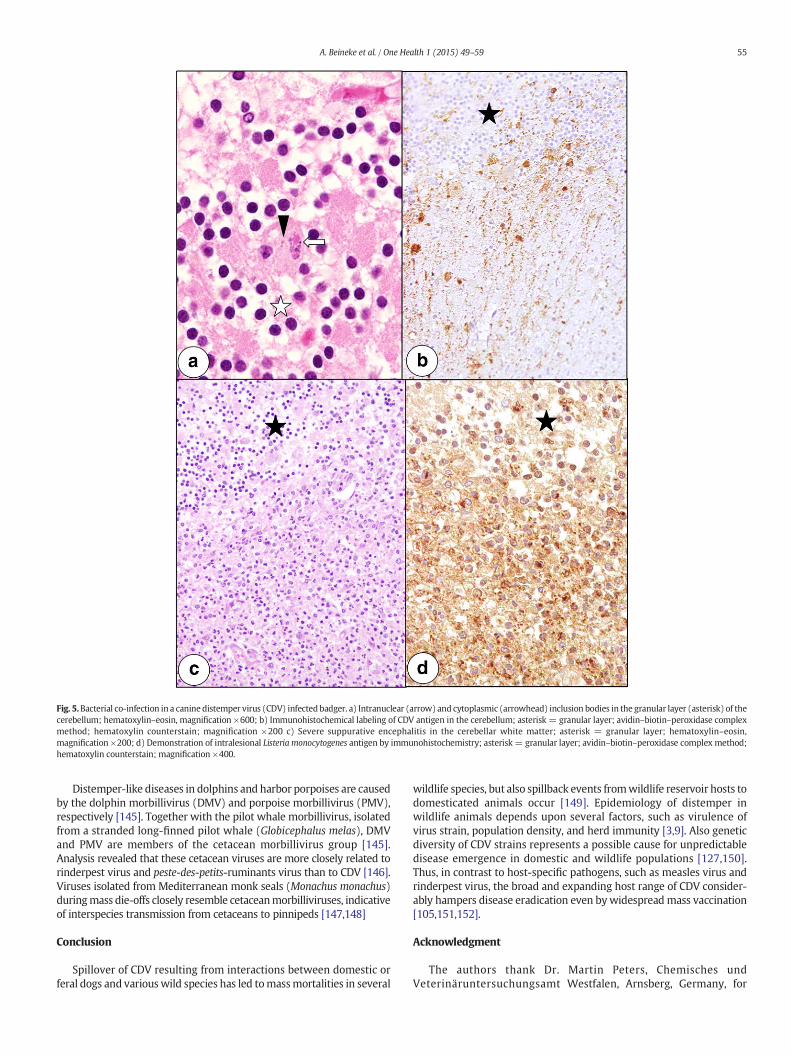
Fig. 5.Bacterial co-infection in a canine distemper virus (CDV) infected badger. a) Intranuclear (arrow) and cytoplasmic (arrowhead) inclusion bodies in the granular layer (asterisk) of thecerebellum; hematoxylin–eosin, magnification ×600; b) Immunohistochemical labeling of CDV antigen in the cerebellum; asterisk = granular layer; avidin–biotin–peroxidase complexmethod; hematoxylin counterstain; magnification ×200 c) Severe suppurative encephalitis in the cerebellar white matter; asterisk = granular layer; hematoxylin–eosin,magnification ×200; d) Demonstration of intralesional Listeria monocytogenes antigen by immunohistochemistry; asterisk = granular layer; avidin–biotin–peroxidase complex method;hematoxylin counterstain; magnification ×400.
55A. Beineke et al. / One Health 1 (2015) 49–59
Distemper-like diseases in dolphins and harbor porpoises are causedby the dolphin morbillivirus (DMV) and porpoise morbillivirus (PMV),respectively [145]. Together with the pilot whale morbillivirus, isolatedfrom a stranded long-finned pilot whale (Globicephalus melas), DMVand PMV are members of the cetacean morbillivirus group [145].Analysis revealed that these cetacean viruses are more closely related torinderpest virus and peste-des-petits-ruminants virus than to CDV [146].Viruses isolated from Mediterranean monk seals (Monachus monachus)duringmass die-offs closely resemble cetaceanmorbilliviruses, indicativeof interspecies transmission from cetaceans to pinnipeds [147,148]
Conclusion
Spillover of CDV resulting from interactions between domestic orferal dogs and various wild species has led tomassmortalities in several
wildlife species, but also spillback events fromwildlife reservoir hosts todomesticated animals occur [149]. Epidemiology of distemper inwildlife animals depends upon several factors, such as virulence ofvirus strain, population density, and herd immunity [3,9]. Also geneticdiversity of CDV strains represents a possible cause for unpredictabledisease emergence in domestic and wildlife populations [127,150].Thus, in contrast to host-specific pathogens, such as measles virus andrinderpest virus, the broad and expanding host range of CDV consider-ably hampers disease eradication even bywidespreadmass vaccination[105,151,152].
Acknowledgment
The authors thank Dr. Martin Peters, Chemisches undVeterinäruntersuchungsamt Westfalen, Arnsberg, Germany, for

Fig. 6. Canine distemper virus infection in a spotted hyena from the Serengeti NationalPark; demonstration of viral antigen in nuclei (arrows) and cytoplasm (arrowheads) ofneuronal and glial cells of the brain by immunohistochemistry. Peroxidase-antiperoxidasetechnique; hematoxylin counterstain; magnification ×600.
56 A. Beineke et al. / One Health 1 (2015) 49–59
providing histologic material. This study was in part supported byNiedersachsen-Research Network on Neuroinfectiology (N-RENNT) ofthe Ministry of Science and Culture of Lower Saxony, Germany. Thefunders had no role in decision to publish or preparation of themanuscript.
References
[1] T. Barrett, Morbillivirus infections, with special emphasis on morbilliviruses ofcarnivores, Vet. Microbiol. 69 (1999) 3–13.
[2] A.D. Osterhaus, J. Groen, H.E. Spijkers, H.W. Broeders, F.G. UytdeHaag, P. deVries, J.S. Teppema, I.K. Visser, M.W. van de Bildt, E.J. Vedder, Mass mortalityin seals caused by a newly discovered morbillivirus, Vet. Microbiol. 23 (1990)343–350.
[3] A. Beineke, C. Puff, F. Seehusen,W. Baumgärtner, Pathogenesis and immunopathol-ogy of systemic and nervous canine distemper, Vet. Immunol. Immunopathol. 127(2009) 1–18.
[4] C. Orvell, Structural polypeptides of canine distemper virus, Arch. Virol. 66 (1980)193–206.
[5] C. Lempp, I. Spitzbarth, C. Puff, A. Cana, K. Kegler, S. Techangamsuwan, W.Baumgärtner, F. Seehusen, New aspects of the pathogenesis of canine distemperleukoencephalitis, Viruses 6 (2014) 2571–2601.
[6] V. von Messling, D. Milosevic, R. Cattaneo, Tropism illuminated: lymphocyte-basedpathways blazed by lethal morbillivirus through the host immune system, Proc.Natl. Acad. Sci. U. S. A. 101 (2004) 14216–14221.
[7] V. von Messling, N. Svitek, R. Cattaneo, Receptor (SLAM [CD150]) recognition andthe V protein sustain swift lymphocyte-based invasion of mucosal tissue and lym-phatic organs by a morbillivirus, J. Virol. 80 (2006) 6084–6092.
[8] K. Frölich, O. Czupalla, L. Haas, J. Hentschke, J. Dedek, J. Fickel, Epizootiological in-vestigations of canine distemper virus in free-ranging carnivores from Germany,Vet. Microbiol. 74 (2000) 283–292.
[9] S.L. Deem, L.H. Spelman, R.A. Yates, R.J. Montali, Canine distemper in terrestrialcarnivores: a review, J. Zoo Wildl. Med. 31 (2000) 441–451.
[10] W. Baumgärtner, S. Alldinger, A. Beineke, S. Gröters, C. Herden, U. Kaim, G. Müller,F. Seeliger, P. Van Moll, P. Wohlsein, Das Staupevirus—Ein Erreger auf der Suchenach neuen Wirten, Dtsch. Tierarztl. Wochenschr. 110 (2003) 137–142.
[11] A.J. McCarthy, M.A. Shaw, S.J. Goodman, Pathogen evolution and diseaseemergence in carnivores, Proc. Biol. Sci. 274 (2007) 3165–3174.
[12] M. Bieringer, J.W. Han, S. Kendl, M. Khosravi, P. Plattet, J. Schneider-Schaulies, Ex-perimental adaptation of wild-type canine distemper virus (CDV) to the humanentry receptor CD150, PLoS One 8 (2013), e57488.
[13] V.M. Nikolin, G. Wibbelt, F.U. Michler, P. Wolf, M.L. East, Susceptibility of carnivorehosts to strains of canine distemper virus from distinct genetic lineages, Vet.Microbiol. 156 (2012) 45–53.
[14] F.C. Origgi, P. Plattet, U. Sattler, N. Robert, J. Casaubon, F. Mavrot, M. Pewsner, N.Wu, S. Giovannini, A. Oevermann, M.H. Stoffel, V. Gaschen, H. Segner, M.P. Ryser-Degiorgis, Emergence of canine distemper virus strains with modified molecularsignature and enhanced neuronal tropism leading to high mortality in wildcarnivores, Vet. Pathol. 49 (2012) 913–929.
[15] U. Sattler, M. Khosravi, M. Avila, P. Pilo, J.P. Langedijk, N. Ader-Ebert, L.A. Alves, P.Plattet, F.C. Origgi, Identification of amino acid substitutions with compensationaleffects in the attachment protein of canine distemper virus, J. Virol. 88 (2014)8057–8064.
[16] S. Krakowka, R.J. Higgins, A. Koestner, Canine distemper virus: review of structuraland functional modulations in lymphoid tissues, Am. J. Vet. Res. 41 (1980)284–292.
[17] Appel MJ. Distemper pathogenesis in dogs. J Am Vet Med Assoc. 197;156:1681–4.[18] N.G. Wright, H.J. Cornwell, H. Thompson, I.M. Lauder, Canine distemper: current
concepts in laboratory and clinical diagnosis, Vet. Rec. 94 (1974) 86–92.[19] M. Schobesberger, A. Summerfield, M.G. Doherr, A. Zurbriggen, C. Griot, Canine
distemper virus-induced depletion of uninfected lymphocytes is associated withapoptosis, Vet. Immunol. Immunopathol. 104 (2005) 33–44.
[20] V. Qeska, Y. Barthel, V. Herder, V.M. Stein, A. Tipold, C. Urhausen, A.R. Günzel-Apel,K. Rohn, W. Baumgärtner, A. Beineke, Canine distemper virus infection leads to aninhibitory phenotype of monocyte-derived dendritic cells in vitrowith reduced ex-pression of co-stimulatory molecules and increased interleukin-10 transcription,PLoS One 9 (2014), e96121.
[21] N. Decaro, M. Camero, G. Greco, N. Zizzo, A. Tinelli, M. Campolo, A. Pratelli, C.Buonavoglia, Canine distemper and related diseases: report of a severe outbreakin a kennel, New Microbiol. 27 (2004) 177–181.
[22] W. Baumgärtner, R.W. Boyce, S. Alldinger, M.K. Axthelm, S.E. Weisbrode, S.Krakowka, K. Gaedke, Metaphyseal bone lesions in young dogs with systemiccanine distemper virus infection, Vet. Microbiol. 44 (1995) 201–209.
[23] E.L. von Rüden, J. Avemary, C. Zellinger, D. Algermissen, P. Bock, A. Beineke, W.Baumgärtner, V.M. Stein, A. Tipold, H. Potschka, Distemper virus encephalitisexerts detrimental effects on hippocampal neurogenesis, Neuropathol. Appl.Neurobiol. 38 (2012) 426–442.
[24] A. Nesseler, W. Baumgärtner, A. Zurbriggen, C. Orvell, Restricted virus proteintranslation in canine distemper virus inclusion body polioencephalitis, Vet.Microbiol. 69 (1999) 23–28.
[25] G. Wyss-Fluehmann, A. Zurbriggen, M. Vandevelde, P. Plattet, Canine distempervirus persistence in demyelinating encephalitis by swift intracellular cell-to-cellspread in astrocytes is controlled by the viral attachment protein, ActaNeuropathol. 119 (2010) 617–630.
[26] M. Vandevelde, A. Zurbriggen, Demyelination in canine distemper virus infection:a review, Acta Neuropathol. 109 (2005) 56–68.
[27] W.H. Armstrong, C.H. Anthony, An epizootic of canine distemper in a zoologicalpark, Cornell Vet. 32 (1942) 286–288.
[28] B.L. Cypher, J.H. Scrivner, K.L. Hammer, T.P. O'Farrell, Viral antibodies in coyotesfrom California, J. Wildl. Dis. 34 (1998) 259–264.
[29] E.M. Gese, R.D. Schultz, M.R. Johnson, E.S. Williams, R.L. Crabtree, R.L. Ruff, Serolog-ical survey for diseases in free-ranging coyotes (Canis latrans) in Yellowstone Na-tional Park, Wyoming, J. Wildl. Dis. 33 (1997) 47–56.
[30] S. Gowtage-Sequeira, A.C. Banyard, T. Barrett, H. Buczkowski, S.M. Funk, S.Cleaveland, Epidemiology, pathology, and genetic analysis of a canine distemperepidemic in Namibia, J. Wildl. Dis. 45 (2009) 1008–1020.
[31] M. Shamir, B. Yakobson, G. Baneth, R. King, S. Dar-Verker, A. Markovics, I. Aroch,Antibodies to selected canine pathogens and infestation with intestinal helminthsin golden jackals (Canis aureus) in Israel, Vet. J. 162 (2001) 66–72.
[32] J.D. Philippa, F.A. Leighton, P.Y. Daoust, O. Nielsen, M. Pagliarulo, H. Schwantje, T.Shury, R. Van Herwijnen, B.E. Martina, T. Kuiken, M.W. Van de Bildt, A.D.Osterhaus, Antibodies to selected pathogens in free-ranging terrestrial carnivoresand marine mammals in Canada, Vet. Rec. 155 (2004) 135–140.
[33] E.S. Almberg, L.D. Mech, D.W. Smith, J.W. Sheldon, R.L. Crabtree, A serological sur-vey of infectious disease in Yellowstone National Park's canid community, PLoSOne 4 (2009) e7042.
[34] P.W. Hedrick, R.N. Lee, C. Buchanan, Canine parvovirus enteritis, canine distemper,and major histocompatibility complex genetic variation in Mexican wolves, J.Wildl. Dis. 39 (2003) 909–913.
[35] A. Müller, E. Silva, N. Santos, G. Thompson, Domestic dog origin of canine distem-per virus in free-ranging wolves in Portugal as revealed by hemagglutinin genecharacterization, J. Wildl. Dis. 47 (2011) 725–729.
[36] D. Di Sabatino, A. Lorusso, C.E. Di Francesco, L. Gentile, V. Di Pirro, A.L. Bellacicco, A.Giovannini, G. Di Francesco, G. Marruchella, F. Marsilio, G. Savini, Arctic lineage-canine distemper virus as a cause of death in Apennine wolves (Canis lupus) inItaly, PLoS One 9 (2014), e82356.
[37] C.H. Gordon, A.C. Banyard, A. Hussein, M.K. Laurenson, J.R. Malcolm, J. Marino, F.Regassa, A.M.E. Stewart, A.R. Fooks, C. Sillero-Zubiri, Canine distemper in endan-gered Ethiopian wolves, Em Inf Dis. 21 (2015) 824–831.
[38] N.H. de Almeida Curi, C.M. Coelho, M. de Campos Cordeiro Malta, E.M. Magni, M.A.Sábato, A.S. Araújo, Z.I. Lobato, J.L. Santos, H.A. Santos, A.A. Ragozo, S.L. de Souza,Pathogens of wild maned wolves (Chrysocyon brachyurus) in Brazil, J. Wildl. Dis.48 (2012) 1052–1056.
[39] B. Thomas-Baker, Vaccination-induced distemper in maned wolves, vaccination-induced corneal opacity in a maned wolf, Proceedings of the American Associationof Zoo Veterinarians. Annual Report, Scottsdale, Arizona 1985, p. 53.
[40] E.F. McInnes, R.E. Burroughs, N.M. Duncan, Possible vaccine-induced canine dis-temper in a South American bush dog (Speothos venaticus), J. Wildl. Dis. 28(1992) 614–617.
[41] A.R. Berentsen, M.R. Dunbar, M.S. Becker, J. M'soka, E. Droge, N.M. Sakuya, W.Matandiko, R. McRobb, C.A. Hanlon, Rabies, canine distemper, and canine parvovi-rus exposure in large carnivore communities from two Zambian ecosystems, Vec-tor Borne Zoonotic Dis. 13 (2013) 643–649.
[42] K.A. Alexander, M.J.G. Appel, African wild dogs (Lycaon pictus) endangered by acanine distemper epizootic among domestic dogs near the Masai Mara NationalReserve, J. Wildl. Dis. 30 (1994) 481–485.
[43] K.V. Goller, R.D. Fyumagwa, V. Nikolin, M.L. East, M. Kilewo, S. Speck, T. Müller, M.Matzke, G. Wibbelt, Fatal canine distemper infection in a pack of African wild dogsin the Serengeti ecosystem, Tanzania, Vet. Microbiol. 146 (2010) 245–252.

57A. Beineke et al. / One Health 1 (2015) 49–59
[44] M.W. van de Bildt, T. Kuiken, A.M. Visee, S. Lema, T.R. Fitzjohn, A.D. Osterhaus, Dis-temper outbreak and its effect on African wild dog conservation, Emerg. Infect. Dis.8 (2002) 211–213.
[45] B. Durchfeld,W. Baumgartner, W. Herbst, R. Brahm, Vaccine-associated canine dis-temper infection in a litter of African hunting dogs (Lycaon pictus), Zentralbl.Veterinarmed. 37 (1990) 203–212.
[46] K. Sekulin, A. Hafner-Marx, J. Kolodziejek, D. Janik, P. Schmidt, N. Nowotny, Emer-gence of canine distemper in Bavarian wildlife associated with a specific aminoacid exchange in the haemagglutinin protein, Vet. J. 187 (2011) 399–401.
[47] N. Denzin, V. Herwig, E. van der Grinten, Occurrence and geographical distributionof Canine Distemper Virus infection in red foxes (Vulpes vulpes) of Saxony-Anhalt,Germany, Vet. Microbiol. 162 (2013) 214–218.
[48] V. Martella, A. Bianchi, I. Bertoletti, L. Pedrotti, A. Gugiatti, A. Catella, P. Cordioli,M.S. Lucente, G. Elia, C. Buonavoglia, Canine distemper epizootic among redfoxes, Italy, 2009, Emerg. Infect. Dis. 16 (2010) 2007–2009.
[49] P. Nouvellet, C.A. Donnelly, M. De Nardi, C.J. Rhodes, P. De Benedictis, C. Citterio, F.Obber, M. Lorenzetto, M.D. Pozza, S. Cauchemez, G. Cattoli, Rabies and canine dis-temper virus epidemics in the red fox population of northern Italy (2006–2010),PLoS One 8 (2013), e61588.
[50] M. López-Peña,M.I. Quiroga, S. Vázquez, J.M.Nieto, Detection of caninedistemper viralantigen in foxes (Vulpes vulpes) in Northwestern Spain, J.Wildl. Dis. 30 (1994) 95–98.
[51] N. Santos, C. Almendra, L. Tavares, Serologic survey for canine distemper virus andcanine parvovirus in free-ranging wild carnivores from Portugal, J. Wildl. Dis. 45(2009) 221–226.
[52] D.S. Miller, D.F. Covell, R.G. McLean, W.J. Adrian, M. Niezgoda, J.M. Gustafson, O.J.Rongstad, R.D. Schultz, L.J. Kirk, T.J. Quan, Serologic survey for selected infectiousdisease agents in swift and kit foxes from the western United States, J. Wildl. Dis.36 (2000) 798–805.
[53] A.V. Belsare, A.T. Vanak, M.E. Gompper, Epidemiology of viral pathogens of free-ranging dogs and Indian foxes in a human-dominated landscape in central India,Transbound. Emerg. Dis. 61 (2014) 78–86.
[54] G.H. Woo, Y.S. Jho, E.J. Bak, Canine distemper virus infection in fennec fox (Vulpeszerda), J. Vet. Med. Sci. 72 (2010) 1075–1079.
[55] I. Monne, A. Fusaro, V. Valastro, C. Citterio, M. Dalla Pozza, F. Obber, K. Trevisiol, M.Cova, P. De Benedictis, M. Bregoli, I. Capua, G. Cattoli, A distinct CDV genotype caus-ing a major epidemic in Alpine wildlife, Vet. Microbiol. 150 (2011) 63–69.
[56] S.F. Timm, L. Munson, B.A. Summers, K.A. Terio, E.J. Dubovi, C.E. Rupprecht, S. Kapil,D.K. Garcelon, A suspected canine distemper epidemic as the cause of a catastroph-ic decline in Santa Catalina Island foxes (Urocyon littoralis catalinae), J. Wildl. Dis.45 (2009) 333–343.
[57] R.D. Halbrooks, L.J. Swango, R.P. Schurrenberger, F.E. Mitchell, E.P. Hill, Response ofgray foxes to modified-live virus canine distemper vaccine, J. Am. Vet. Med. Assoc.179 (1981) 1170–1174.
[58] H. Ferreyra, M.G. Calderón, D. Marticorena, C. Marull, B.C. Leonardo, Canine distem-per infection in crab-eating fox (Cerdocyon thous) from Argentina, J. Wildl. Dis. 45(2009) 1158–1162.
[59] P.E. Martino, J.L. Montenegro, J.A. Preziosi, C. Venturini, D. Bacigalupe, N.O. Stanchi,E.L. Bautista, Serological survey of selected pathogens of free-ranging foxes insouthern Argentina, 1998–2001, Rev. Sci. Tech. 23 (2004) 801–806.
[60] M.E. Roelke-Parker, L. Munson, C. Packer, R. Kock, S. Cleaveland, M. Carpenter, S.J.O'Brien, A. Pospischil, R. Hofmann-Lehmann, H. Lutz, G.L. Mwamengele, M.N.Mgasa, G.A. Machange, B.A. Summers, M.J. Appel, A canine distemper virus epi-demic in Serengeti lions (Panthera leo), Nature 379 (1996) 441–445.
[61] G. Acosta-Jamett, W.S. Chalmers, A.A. Cunningham, S. Cleaveland, I.G. Handel, B.M.Bronsvoort, Urban domestic dog populations as a source of canine distemper virusfor wild carnivores in the Coquimbo region of Chile, Vet. Microbiol. 152 (2011)247–257.
[62] F. Giannitti, S.S. Diab, F.A. Uzal, K. Fresneda, D. Rossi, D. Talmi-Frank, G. Baneth, In-fection with a Hepatozoon sp. closely related to Hepatozoon felis in a wild Pampasgray fox (Lycalopex–Pseudalopex-gymnocercus) co-infected with canine distempervirus, Vet. Parasitol. 186 (2012) 497–502.
[63] J. Megid, C.R. Teixeira, R.L. Amorin, A. Cortez, M.B. Heinemann, J.M. de PaulaAntunes, L.F. da Costa, F. Fornazari, J.R. Cipriano, A. Cremasco, L.J. Richtzenhain,First identification of canine distemper virus in hoary fox (Lycalopex vetulus): path-ologic aspects and virus phylogeny, J. Wildl. Dis. 46 (2010) 303–305.
[64] A. Sutor, S. Schwarz, F.J. Conraths, The raccoon dog (Nyctereutes procyonoides) inGermany—an established Neozoon as host and vector for parasites and other path-ogens, Berl. Munch. Tierarztl. Wochenschr. 124 (2011) 457–464.
[65] Y. Kameo, Y. Nagao, Y. Nishio, H. Shimoda, H. Nakano, K. Suzuki, Y. Une, H. Sato, M.Shimojima, K. Maeda, Epizootic canine distemper virus infection among wildmammals, Vet. Microbiol. 154 (2012) 222–229.
[66] Y. Nagao, Y. Nishio, H. Shiomoda, S. Tamaru, M. Shimojima, M. Goto, Y. Une, A. Sato,Y. Ikebe, K. Maeda, An outbreak of canine distemper virus in tigers (Panthera ti-gris): possible transmission from wild animals to zoo animals, J. Vet. Med. Sci. 74(2012) 699–705.
[67] J. Zhao, H. Zhang, X. Bai, V. Martella, B. Hu, Y. Sun, C. Zhu, L. Zhang, H. Liu, S. Xu, X.Shao, W.Wu, X. Yan, Emergence of canine distemper virus strains with two aminoacid substitutions in the haemagglutinin protein, detected from vaccinated carni-vores in North-Eastern China in 2012–2013, Vet. J. 200 (2014) 191–194.
[68] Z. Rentería-Solís, C. Förster, A. Aue, U. Wittstatt, G. Wibbelt, M. König, Canine dis-temper outbreak in raccoons suggests pathogen interspecies transmissionamongst alien and native carnivores in urban areas from Germany, Vet. Microbiol.174 (2014) 50–99.
[69] J. Suzuki, Y. Nishio, Y. Kameo, Y. Terada, R. Kuwata, H. Shimoda, K. Suzuki, K.Maeda, Canine distemper virus infection among wildlife before and after the
epidemic, J. Vet. Med. Sci. (2015) http://dx.doi.org/10.1292/jvms.15-0237 (Epubahead of print).
[70] H. Nakano, Y. Kameo, H. Sato, M. Mochizuki, M. Yokoyama, S. Uni, T. Shibasaki, K.Maeda, Detection of antibody to canine distemper virus in wild raccoons(Procyon lotor) in Japan, J. Vet. Med. Sci. 71 (2009) 1661–1663.
[71] K.W. McFadden, S.E. Wade, E.J. Dubovi, M.E. Gompper, A serological and fecal par-asitologic survey of the critically endangered pygmy raccoon (Procyon pygmaeus),J. Wildl. Dis. 41 (2005) 615–617.
[72] A.N. Hamir, B.A. Summers, C.E. Rupprecht, Concurrent rabies and canine distemperencephalitis in a raccoon (Procyon lotor), J. Vet. Diagn. Invest. 10 (1998) 194–196.
[73] K.R. Kazacos, H.L. Thacker, H.L. Shivaprasad, P.P. Burger, Vaccination-induced dis-temper in kinkajous, J. Am. Vet. Med. Assoc. 179 (1981) 1166–1169.
[74] J.A. Lednicky, J. Dubach, M.J. Kinsel, T.P. Meehan,M. Bocchetta, L.L. Hungerford, N.A.Sarich, K.E. Witecki, M.D. Braid, C. Pedrak, C.M. Houde, Genetically distantAmerican Canine distemper virus lineages have recently caused epizootics withsomewhat different characteristics in raccoons living around a large suburbanzoo in the USA, Virol. J. 1 (2004) 2.
[75] M. Bush, R.J. Montali, D. Brownstein, A.E. James, M.J. Appel, Vaccine-induced caninedistemper in a lesser panda, J. Am. Vet. Med. Assoc. 169 (1976) 959–960.
[76] N. Stephenson, J.M. Higley, J.L. Sajecki, B.B. Chomel, R.N. Brown, Foley JE demographiccharacteristics and infectious diseases of a population of American black bears inHumboldt County, California, Vector Borne Zoonotic Dis. 15 (2015) 116–123.
[77] C.M. Kirk, S. Amstrup, R. Swor, D. Holcomb, T.M. O'Hara, Morbillivirus and Toxo-plasma exposure and association with hematological parameters for southernBeaufort Sea polar bears: potential response to infectious agents in a sentinel spe-cies, Ecohealth 7 (2010) 321–331.
[78] C.E. Di Francesco, L. Gentile, V. Di Pirro, L. Ladiana, S. Tagliabue, F. Marsilio, Serolog-ic evidence for selected infectious diseases in Marsican brown bears (Ursus arctosmarsicanus) in Italy (2004–09), J. Wildl. Dis. 51 (2015) 209–213.
[79] W.O. Cottrell, M.K. Keel, J.W. Brooks, D.G. Mead, J.E. Phillips, First report of clinicaldisease associatedwith canine distemper virus infection in awild black bear (Ursusamericana), J. Wildl. Dis. 49 (2013) 1024–1027.
[80] M. Schönbauer, S. Kölbl, A. Schönbauer-Längle, Perinatale Staupeinfektion bei dreiEisbären (Ursus maritimus) und bei einem Brillenbären (Tremarctos ornatus),Verhandlungsbericht Int. Symp. Erkrankungen Zootiere. 26 (1984) 131–136.
[81] Q. Qin, D. Li, H. Zhang, R. Hou, Z. Zhang, C. Zhang, J. Zhang, F. Wei, Serosurvey ofselected viruses in captive giant pandas (Ailuropoda melanoleuca) in China, Vet.Microbiol. 142 (2010) 199–204.
[82] M. Hvistendahl, Endangered species. Captive pandas succumb to killer virus, Sci-ence 347 (2015) 700–701.
[83] D. Perpiñán, A. Ramis, A. Tomás, E. Carpintero, F. Bargalló, Outbreak of canine dis-temper in domestic ferrets (Mustela putorius furo), Vet. Rec. 163 (2008) 246–250.
[84] P.A. Rudd, R. Cattaneo, V. von Messling, Canine distemper virus uses both the an-terograde and the hematogenous pathway for neuroinvasion, J. Virol. 80 (2006)9361–9370.
[85] M. Ludlow, D.T. Nguyen, D. Silin, O. Lyubomska, R.D. de Vries, V. von Messling, S.McQuaid, R.L. De Swart, W.P. Duprex, Recombinant canine distemper virus strainSnyder Hill expressing green or red fluorescent proteins causes meningoencepha-litis in the ferret, J. Virol. 86 (2012) 7508–7519.
[86] J.F. Evermann, C.W. Leathers, J.R. Gorham, A.J. McKeirnan, M.J. Appel, Pathogenesisof two strains of lion (Panthera leo) morbillivirus in ferrets (Mustela putorius furo),Vet Pathol. 38 (2001) 311–316.
[87] J. Rudolf, Beitrag zur Staupe beim Silberfuchs, Nerz undWaschbär. Dtsch. Tierärztl.Wschr. 30 (1930) 728–732.
[88] E. Crook, J.R. Gorham, S.H. McNutt, Experimental distemper in mink and ferrets. I.Pathogenesis, Am. J. Vet. Res. 19 (1958) 955–957.
[89] R. Trebbien, M. Chriel, T. Struve, C.K. Hjulsager, G. Larsen, L.E. Larsen, Wildlife res-ervoirs of canine distemper virus resulted in a major outbreak in Danish farmedmink (Neovison vison), PLoS One 9 (2014), e85598.
[90] E.S. Williams, E.T. Thorne, M.J. Appel, D.W. Belitsky, Canine distemper in black-footed ferrets (Mustela nigripes) from Wyoming, J. Wildl. Dis. 24 (1988) 385–398.
[91] J.W. Carpenter, M.J. Appel, R.C. Erickson, M.N. Novilla, Fatal vaccine-induced caninedistemper virus infection in black-footed ferrets, J. Am. Vet. Med. Assoc. 169 (1976)961–964.
[92] P. van Moll, S. Alldinger, W. Baumgärtner, M. Adami, Distemper in wild carnivores:an epidemiological, histological and immunocytochemical study, Vet. Microbiol. 44(1995) 193–199.
[93] H. Liermann, T.C. Harder, M. Löchelt, V. vonMessling,W. Baumgärtner, V. Moennig,L. Haas, Genetic analysis of the central untranslated genome region and the prox-imal coding part of the F gene of wild-type and vaccine canine distempermorbilliviruses, Virus Genes 17 (1998) 259–270.
[94] M. Hewicker, S. Damsch, G. Trautwein, Detection of canine distemper viral antigenin formalin-fixed and paraffin-embedded tissue of a fitch (Mustela putorius), usingan immunoperoxidase technique, Dtsch. Tierarztl. Wochenschr. 97 (1990) 85–88.
[95] S.D. Gehrt, M.J. Kinsel, C. Anchor, Pathogen dynamics and morbidity of stripedskunks in the absence of rabies, J. Wildl. Dis. 46 (2010) 335–347.
[96] Y. Ikeda, K. Nakamura, T. Miyazawa, M.C. Chen, T.F. Kuo, J.A. Lin, T. Mikami, C. Kai, E.Takahashi, Seroprevalence of canine distemper virus in cats, Clin. Diagn. Lab.Immunol. 8 (2011) 641–644.
[97] M.J.G. Appel, B.E. Sheffy, D.H. Percy, J.M. Gaskin, Canine distemper virus in domes-ticated cats and pigs, Am. J. Vet. Res. 34 (1974) 1459–1463.
[98] T.C. Harder, M. Kenter, H. Vos, K. Siebelink, W. Huisman, G. van Amerongen, C.Orvell, T. Barrett, M.J. Appel, A.D. Osterhaus, Canine distemper virus from diseasedlarge felids: biological properties and phylogenetic relationships, J. Gen. Virol. 77(1996) 397–405.

58 A. Beineke et al. / One Health 1 (2015) 49–59
[99] D.J. Wiener, M.M. Welle, F.C. Origgi, Cutaneous lesions associated with dual infec-tion caused by canine distemper virus and orthopoxvirus in a domestic cat, Vet.Dermatol. 24 (2013) 543–546.
[100] A. Duarte, M. Fernandes, N. Santos, L. Tavares, Virological Survey in free-rangingwildcats (Felis silvestris) and feral domestic cats in Portugal, Vet. Microbiol. 158(2012) 400–404.
[101] K.A. Terio, M.E. Craft, Canine distemper virus (CDV) in another big cat: should CDVbe renamed carnivore distemper virus? mBio. 4 (2013) e00702–e00713.
[102] S. Thalwitzer, B. Wachter, N. Robert, G. Wibbelt, T. Müller, J. Lonzer, M.L. Meli, G.Bay, H. Hofer, H. Lutz, Seroprevalences to viral pathogens in free-ranging and cap-tive cheetahs (Acinonyx jubatus) on Namibian Farmland, Clin. Vaccine Immunol. 17(2010) 232–238.
[103] M.M. Furtado, J.D. de Ramos Filho, K.C. Scheffer, C.J. Coelho, P.S. Cruz, C.Y. Ikuta, A.T.Jácomo, G.E. Porfírio, L. Silveira, R. Sollmann, N.M. Tôrres, J.S. Ferreira Neto,Serosurvey for selected viral infections in free-ranging jaguars (Panthera onca)and domestic carnivores in Brazilian Cerrado, Pantanal, and Amazon, J. Wildl. Dis.49 (2013) 510–521.
[104] B.M. Kissui, C. Packer, Top-down population regulation of a top predator: lions inthe Ngorongoro Crater, Proc. Biol. Sci. 271 (2004) 1867–1874.
[105] M. Viana, S. Cleaveland, J. Matthiopoulos, J. Halliday, C. Packer, M.E. Craft, K.Hampson, A. Czupryna, A.P. Dobson, E.J. Dubovi, E. Ernest, R. Fyumagwa, R.Hoare, J.G. Hopcraft, D.L. Horton, M.T. Kaare, T. Kanellos, F. Lankester, C. Mentzel,T. Mlengeya, I. Mzimbiri, E. Takahashi, B. Willett, D.T. Haydon, T. Lembo, Dynamicsof a morbillivirus at the domestic-wildlife interface: canine distemper virus in do-mestic dogs and lions, Proc. Natl. Acad. Sci. U. S. A. 112 (2015) 1464–1469.
[106] L. Munson, K.A. Terio, R. Kock, T. Mlengeya, M.E. Roelke, E. Dubovi, B. Summers,A.R. Sinclair, C. Packer, Climate extremes promote fatal co-infections during caninedistemper epidemics in African lions, PLoS One 3 (2008), e2545.
[107] T.A. Seimon, D.G. Miquelle, T.Y. Chang, A.L. Newton, I. Korotkova, G. Ivanchuk, E.Lyubchenko, A. Tupikov, E. Slabe, D. McAloose, Canine distemper virus: an emerg-ing disease inwild endangered Amur tigers (Panthera tigris altaica), MBio. 4 (2013)e00410–e00413.
[108] M.J. Appel, R.A. Yates, G.L. Foley, J.J. Bernstein, S. Santinelli, L.H. Spelman, L.D. Miller,L.H. Arp, M. Anderson, M. Barr, et al., Canine distemper epizootic in lions, tigers,and leopards in North America, J. Vet. Diagn. Invest. 6 (1994) 277–288.
[109] M.L. Meli, P. Simmler, V. Cattori, F. Martínez, A. Vargas, F. Palomares, J.V. López-Bao,M.A. Simón, G. López, L. León-Vizcaino, R. Hofmann-Lehmann, H. Lutz, Importanceof canine distemper virus (CDV) infection in free-ranging Iberian lynxes (Lynxpardinus), Vet. Microbiol. 146 (2010) 132–137.
[110] P.Y. Daoust, S.R. McBurney, D.L. Godson, M.W. van de Bildt, A.D. Osterhaus, Canine-distemper virus-associated encephalitis in free-living lynx (Lynx canadensis) andbobcats (Lynx rufus) of eastern Canada, J. Wildl. Dis. 45 (2009) 611–624.
[111] M.M. Uhart, M.V. Rago, C.A. Marull, V. Ferreyra Hdel, J.A. Pereira, Exposure to se-lected pathogens in to selected pathogens in Geoffroy's cats and domestic carni-vores from central Argentina, J. Wildl. Dis. 48 (2012) 899–909.
[112] A.F. Nava, L. Cullen, D.A. Sana, M.S. Nardi, J.D. Filho, T.F. Lima, K.C. Abreu, F. Ferreira,First evidence of canine distemper in Brazilian free-ranging felids, EcoHealth 5(2008) 513–518.
[113] J.E. Foley, P. Swift, K.A. Fleer, S. Torres, Y.A. Girard, C.K. Johnson, Risk factors for ex-posure to feline pathogens in California mountain lions (Puma concolor), J. Wildl.Dis. 49 (2013) 279–293.
[114] A.M.S. Chandra, P.E. Ginn, S.P. Terrell, B. Ferguson, A. Adjirir-Awere, P. Dennis, B.L.Homer, Canine distemper virus infection in binturongs (Arctictis binturong), J. Vet.Diagn. Invest. 12 (2000) 88–91.
[115] S. Techangamsuwan, W. Banlunara, A. Radtanakatikanon, A. Sommanustweechai,B. Siriaroonrat, E.D. Lombardini, A. Rungsipipat, Pathologic and molecular virologiccharacterization of a canine distemper outbreak in farmed civets, Vet. Pathol. 24(2014) (pii: 0300985814551580. [Epub ahead of print] PubMed PMID: 25253065).
[116] M. López-Peña, S. Vázquez, N. Alemañ, A. López-Beceiro, F. Muñoz, J.L. Pereira, J.M.Nieto, Canine distemper in a genet (Gennetta gennetta), associated with endoge-nous lipid pneumonia, J. Comp. Pathol. 124 (2001) 207–211.
[117] L. Haas, H. Hofer, H. East, P. Wohlsein, B. Liess, T. Barrett, Canine distemper virusinfection in Serengeti spotted hyenas, Vet. Microbiol. 49 (1996) 147–152.
[118] T.M. Harrison, J.K. Mazet, K.E. Holekamp, E. Dubovi, A.L. Engh, K. Nelson, R.C.Van Horn, L. Munson, Antibodies to canine and feline viruses in spotted hyenas(Crocuta crocuta) in the Masai Mara National Reserve, J. Wildl. Dis. 40 (2004)1–10.
[119] M.J. Appel, C. Reggiardo, B.A. Summers, S. Pearce-Kelling, C.J. Maré, T.H. Noon, R.E.Reed, J.N. Shively, C. Orvell, Canine distemper virus infection and encephalitis in ja-velinas (Collared peccaries), Arch. Virol. 119 (1991) 147–152.
[120] T.H. Noon, J.R. Heffelfinger, R.J. Olding, S.L. Wesche, C. Reggiardo, Serologic surveyfor antibodies to canine distemper virus in collared peccary (Tayassu tajacu) pop-ulations in Arizona, J. Wildl. Dis. 39 (2003) 221–223.
[121] Y. Yoshikawa, F. Ochikubo, Y. Matsubara, H. Tsuruoka, M. Ishii, K. Shirota, Y.Nomura, M. Sugiyama, K. Yamanouchi, Natural infection with canine distempervirus in a Japanese monkey (Macaca fuscata), Vet. Microbiol. 20 (1989) 193–205.
[122] W. Qiu, Y. Zheng, S. Zhang, Q. Fan, H. Liu, F. Zhang, W. Wang, G. Liao, R. Hu, Caninedistemper outbreak in rhesus monkeys, China, Emerg Infect Dis. 17 (2011)1541–1543.
[123] Z. Sun, A. Li, H. Ye, Y. Shi, Z. Hu, L. Zeng, Natural infection with canine distempervirus in hand-feeding Rhesus monkeys in China, Vet. Microbiol. 141 (2010)374–378.
[124] K. Sakai, N. Nagata, Y. Ami, F. Seki, Y. Suzaki, N. Iwata-Yoshikawa, T. Suzuki, S.Fukushi, T. Mizutani, T. Yoshikawa, N. Otsuki, I. Kurane, K. Komase, R.Yamaguchi, H. Hasegawa, M. Saijo, M. Takeda, S. Morikawa, Lethal canine
distemper virus outbreak in cynomolgus monkeys in Japan in 2008, J. Virol. 87(2) (Jan 2013) 1105–1114.
[125] K. Sakai, T. Yoshikawa, F. Seki, S. Fukushi, M. Tahara, N. Nagata, Y. Ami, T. Mizutani,I. Kurane, R. Yamaguchi, H. Hasegawa,M. Saijo, K. Komase, S. Morikawa, M. Takeda,Canine distemper virus associated with a lethal outbreak in monkeys can readilyadapt to use human receptors, J. Virol. 87 (2013) 7170–7175.
[126] R.D. de Vries, M. Ludlow, R.J. Verburgh, G. van Amerongen, S. Yüksel, D.T. Nguyen,S. McQuaid, A.D. Osterhaus, W.P. Duprex, R.L. de Swart, Measles vaccination ofnonhuman primates provides partial protection against infection with canine dis-temper virus, J. Virol. 88 (2014) 4423–4433.
[127] M. Ludlow, L.J. Rennick, S. Nambulli, R.L. de Swart, W.P. Duprex, Using the ferretmodel to study morbillivirus entry, spread, transmission and cross-species infec-tion, Curr Opin Virol. 4 (2014) 15–23.
[128] A.P. Mee, J.A. Dixon, J.A. Hoyland, M. Davies, P.L. Selby, E.B. Mawer, Detection of ca-nine distemper virus in 100% of Paget's disease samples by in situ-reversetranscriptase-polymerase chain reaction, Bone 23 (1998) 171–175.
[129] P.J. Duignan, M.F. Van Bressem, J.D. Baker, M. Barbieri, K.M. Colegrove, S. DeGuise, R.L. de Swart, G. Di Guardo, A. Dobson, W.P. Duprex, G. Early, D.Fauquier, T. Goldstein, S.J. Goodman, B. Grenfell, K.R. Groch, F. Gulland, A.Hall, B.A. Jensen, K. Lamy, K. Matassa, S. Mazzariol, S.E. Morris, O. Nielsen, D.Rotstein, T.K. Rowles, J.T. Saliki, U. Siebert, T. Waltzek, J.F. Wellehan, Phocinedistemper virus: current knowledge and future directions, Viruses. 6 (2014)5093–5134.
[130] A.D. Osterhaus, E.J. Vedder, Identification of virus causing recent seal deaths, Na-ture 335 (1988) 20.
[131] T.V. Butina, N.N. Denikina, S.I. Belikov, Canine distemper virus diversity in LakeBaikal seal (Phoca sibirica) population, Vet. Microbiol. 144 (2010) 192–197.
[132] S.C. Wilson, T.M. Eybatov, M. Amano, P.D. Jepson, S.J. Goodman, The role of caninedistemper virus and persistent organic pollutants in mortality patterns of Caspianseals (Pusa caspica), PLoS One 9 (2014), e99265.
[133] T. Kuiken, S. Kennedy, T. Barrett, M.W. Van de Bildt, F.H. Borgsteede, S.D. Brew, G.A.Codd, C. Duck, R. Deaville, T. Eybatov, M.A. Forsyth, G. Foster, P.D. Jepson, A.Kydyrmanov, I. Mitrofanov, C.J. Ward, S. Wilson, A.D. Osterhaus, The 2000 caninedistemper epidemic in Caspian seals (Phoca caspica): pathology and analysis ofcontributory factors, Vet. Pathol. 43 (2006) 321–338.
[134] S. Kennedy, T. Kuiken, P.D. Jepson, R. Deaville, M. Forsyth, T. Barrett, M.W. van deBildt, A.D. Osterhaus, T. Eybatov, C. Duck, A. Kydyrmanov, I. Mitrofanov, S.Wilson, Mass die-Off of Caspian seals caused by canine distemper virus, Emerg. In-fect. Dis. 6 (2000) 637–639.
[135] G. Pohlmeyer, J. Pohlenz, P. Wohlsein, Intestinal lesions in experimental phocinedistemper: light microscopy, immunohistochemistry and electron microscopy, J.Comp. Pathol. 109 (1993) 57–69.
[136] T. Jauniaux, G. Boseret, M. Desmecht, J. Haelters, C. Manteca, J. Tavernier, J. vanGompel, F. Coignoul, Morbillivirus in common seals stranded on the coastsof Belgium and northern France during summer 1998, Vet. Rec. 148 (2001)587–591.
[137] G. Müller, P. Wohlsein, A. Beineke, L. Haas, I. Greiser-Wilke, U. Siebert, S. Fonfara, T.Harder, M. Stede, A.D. Gruber, W. Baumgärtner, Phocine distemper in Germanseals, 2002, Emerg. Infect. Dis. 10 (2004) 723–725.
[138] L. Stimmer, U. Siebert, P. Wohlsein, J.J. Fontaine, W. Baumgärtner, A. Beineke, Viralprotein expression and phenotyping of inflammatory responses in the central ner-vous system of phocine distemper virus-infected harbor seals (Phoca vitulina), Vet.Microbiol. 145 (2010) 23–33.
[139] T.P. Lipscomb, M.G. Mense, P.L. Habecker, J.K. Taubenberger, R. Schoelkopf,Morbilliviral dermatitis in seals, Vet. Pathol. 38 (2001) 724–726.
[140] M. Kreutzer, R. Kreutzer, U. Siebert, G. Müller, P. Reijnders, S. Brasseur, T. Härkönen,R. Dietz, C. Sonne, E.W. Born, W. Baumgärtner, In search of virus carriers of the1988 and 2002 phocine distemper virus outbreaks in European harbour seals,Arch. Virol. 153 (2008) 187–192.
[141] T. Härkönen, R. Dietz, P. Reijnders, J. Teilmann, K. Harding, A. Hall, S. Brasseur, U.Siebert, S.J. Goodman, P.D. Jepson, T. Dau Rasmussen, P. Thompson, The 1988and 2002 phocine distemper virus epidemics in European harbour seals, DisAquat Organ. 68 (2006) 115–130.
[142] S.C. Wilson, T.M. Eybatov, M. Amano, P.D. Jepson, S.J. Goodman, The role of caninedistemper virus and persistent organic pollutants in mortality patterns of Caspianseals (Pusa caspica), PLoS One 9 (2014), e99265.
[143] M. Blixenkrone-Møller, V. Svansson, P. Have, A. Botner, J. Nielsen, Infection studiesin mink with seal-derived morbillivirus, Arch. Virol. 106 (1989) 165–170.
[144] M. Blixenkrone-Møller, Detection of intracellular canine distemper virus antigen inmink inoculated with an attenuated or a virulent strain of canine distemper virus,Am. J. Vet. Res. 50 (1989) 1616–1620.
[145] M.F. Van Bressem, P.J. Duignan, A. Banyard, M. Barbieri, K.M. Colegrove, S. DeGuise, G. Di Guardo, A. Dobson, M. Domingo, D. Fauquier, A. Fernandez, T.Goldstein, B. Grenfell, K.R. Groch, F. Gulland, B.A. Jensen, P.D. Jepson, A. Hall,T. Kuiken, S. Mazzariol, S.E. Morris, O. Nielsen, J.A. Raga, T.K. Rowles, J. Saliki,E. Sierra, N. Stephens, B. Stone, I. Tomo, J. Wang, T. Waltzek, J.F. Wellehan,Cetacean morbillivirus: current knowledge and future directions, Viruses. 6(2014) 5145–5181.
[146] J.K. Taubenberger, M.M. Tsai, T.J. Atkin, T.G. Fanning, A.E. Krafft, R.B. Moeller, S.E.Kodsi, M.G. Mense, T.P. Lipscomb, Molecular genetic evidence of a novel morbilli-virus in a long-finned pilot whale (Globicephalus melas), Emerg. Infect. Dis. 6(2000) 42–45.
[147] A. Osterhaus, J. Groen, H. Niesters, M. van de Bildt, B. Martina, L. Vedder, J. Vos, H.van Egmond, B. Abou-Sidi, M.E. Barham,Morbillivirus inmonk seal mass mortality,Nature 388 (1997) 838–839.

59A. Beineke et al. / One Health 1 (2015) 49–59
[148] M.W. van de Bildt, E.J. Vedder, B.E. Martina, B.A. Sidi, A.B. Jiddou, M.E. Ould Barham,E. Androukaki, A. Komnenou, H.G. Niesters, A.D. Osterhaus, Morbilliviruses inMediterranean monk seals, Vet. Microbiol. 69 (1999) 19–21.
[149] S. Kapil, T.J. Yeary, Canine distemper spillover in domestic dogs from urbanwildlife, Vet Clin North Am Small Anim Pract. 41 (2011) 1069–1086.
[150] Y. Panzera, N. Sarute, G. Iraola, M. Hernández, R. Pérez, Molecular phylogeographyof canine distemper virus: geographic origin and global spreading, Mol.Phylogenet. Evol. 92 (2015) 147–154.
[151] R.L. de Swart, W.P. Duprex, A.D. Osterhaus, Rinderpest eradication: lessons formeasles eradication? Curr Opin Virol. 2 (2012) 330–334.
[152] W.J. Moss, P. Strebel, Biological feasibility of measles eradication, J. Infect. Dis. 204(2011) 47–53.