Critical testing of EarthÕ s oldest putati ve fossil ... · PDF filea Earth Sciences...
48
Precambrian Research xxx (2005) xxx–xxx Critical testing of Earth’s oldest putative fossil assemblage from the ∼3.5 Ga Apex chert, Chinaman Creek, Western Australia Martin D. Brasier a,* , Owen R. Green a , John F. Lindsay b , Nicola McLoughlin a , Andrew Steele c , Cris Stoakes d a Earth Sciences Department, University of Oxford, South Parks Road, Oxford OX1 3PR, UK b Lunar and Planetary Institute and Johnson Space Center, Houston, TX 77058, USA c Geophysical Laboratory, Carnegie Institution of Washington, 5251 Broad Branch Road NW, Washington, DC 20015-1305, USA d C. A Stoakes and Associates Pty Ltd., 3185 Victoria Road, Hovea, WA 6071, Australia Received 10 December 2004; received in revised form 10 June 2005; accepted 24 June 2005 Abstract Structures resembling cyanobacterial microfossils from the ca. 3465 Ma old Apex chert of the Warrawoona Group in Western Australia have until recently been accepted as providing the oldest morphological evidence for life on Earth, and have been taken to support an early beginning for oxygen-releasing photosynthesis. Eleven species of filamentous prokaryote, principally distinguished by shape and geometry, have been put forward as meeting the criteria required of authentic Archaean microfossils. They were contrasted with other microfossils that were dismissed as either unreliable or irreproducible. The aim of this paper is to provide a detailed account of research recently reported by us on the type and recollected material, involving optical and electron microscopy, digital image analysis and other techniques. All previously figured holotype materials are illustrated here, and the context for all the published materials is re-evaluated. The Apex chert ‘microfossils’ occur near the top of a 1.5-km long chert dyke complex associated with major synsedimentary growth faults. Highly localised, glassy felsic tuffs erupted explosively from this and other fissures during the early stages of volcanism, and were followed by the deposition of essentially hydrothermal black and white BaSO 4 rich cherts that infiltrated the feeder dykes, underplating and dilating adjacent stratiform cherts before the start of the next volcanic cycle. The Apex chert ‘microfossils’ occur within multiple generations of these metalliferous hydrothermal vein cherts some 100 m down the dyke system. Comparable structures occur in associated volcanic vent glass and in hydrothermal cherts at least 1 km deep. We find no supporting evidence for a primary biological origin. We reinterpret the purported microfossil-like structures as pseudofossils that formed from the reorganization of carbonaceous matter, mainly during recrystallization from amorphous to spherulitic silica. © 2005 Elsevier B.V. All rights reserved. Keywords: Apex chert; Archaean fossils; Early life; Warrawoona; Western Australia * Corresponding author. Tel.: +44 1865 272074; fax: +44 1865 272072. E-mail address: [email protected] (M.D. Brasier). 0301-9268/$ – see front matter © 2005 Elsevier B.V. All rights reserved. doi:10.1016/j.precamres.2005.06.008 PRECAM-2583; No. of Pages 48
Transcript of Critical testing of EarthÕ s oldest putati ve fossil ... · PDF filea Earth Sciences...

Precambrian Research xxx (2005) xxx–xxx
Critical testing of Earth’s oldest putative fossil assemblage fromthe !3.5Ga Apex chert, Chinaman Creek, Western AustraliaMartin D. Brasier a,", Owen R. Green a, John F. Lindsay b, Nicola McLoughlin a,
Andrew Steele c, Cris Stoakes d
a Earth Sciences Department, University of Oxford, South Parks Road, Oxford OX1 3PR, UKb Lunar and Planetary Institute and Johnson Space Center, Houston, TX 77058, USA
c Geophysical Laboratory, Carnegie Institution of Washington, 5251 Broad Branch Road NW, Washington, DC 20015-1305, USAd C. A Stoakes and Associates Pty Ltd., 3185 Victoria Road, Hovea, WA 6071, Australia
Received 10 December 2004; received in revised form 10 June 2005; accepted 24 June 2005
Abstract
Structures resembling cyanobacterial microfossils from the ca. 3465Ma old Apex chert of the Warrawoona Group in WesternAustralia have until recently been accepted as providing the oldest morphological evidence for life on Earth, and have beentaken to support an early beginning for oxygen-releasing photosynthesis. Eleven species of filamentous prokaryote, principallydistinguished by shape and geometry, have been put forward as meeting the criteria required of authentic Archaean microfossils.They were contrasted with other microfossils that were dismissed as either unreliable or irreproducible. The aim of this paperis to provide a detailed account of research recently reported by us on the type and recollected material, involving optical andelectron microscopy, digital image analysis and other techniques. All previously figured holotype materials are illustrated here,and the context for all the published materials is re-evaluated.The Apex chert ‘microfossils’ occur near the top of a 1.5-km long chert dyke complex associated with major synsedimentary
growth faults. Highly localised, glassy felsic tuffs erupted explosively from this and other fissures during the early stages ofvolcanism, and were followed by the deposition of essentially hydrothermal black and white BaSO4 rich cherts that infiltratedthe feeder dykes, underplating and dilating adjacent stratiform cherts before the start of the next volcanic cycle. The Apex chert‘microfossils’ occur within multiple generations of these metalliferous hydrothermal vein cherts some 100m down the dykesystem. Comparable structures occur in associated volcanic vent glass and in hydrothermal cherts at least 1 km deep. We find nosupporting evidence for a primary biological origin. We reinterpret the purported microfossil-like structures as pseudofossils thatformed from the reorganization of carbonaceous matter, mainly during recrystallization from amorphous to spherulitic silica.© 2005 Elsevier B.V. All rights reserved.
Keywords: Apex chert; Archaean fossils; Early life; Warrawoona; Western Australia
" Corresponding author. Tel.: +44 1865 272074; fax: +44 1865 272072.E-mail address: [email protected] (M.D. Brasier).
0301-9268/$ – see front matter © 2005 Elsevier B.V. All rights reserved.doi:10.1016/j.precamres.2005.06.008
PRECAM-2583; No. of Pages 48
2 M.D. Brasier et al. / Precambrian Research xxx (2005) xxx–xxx
1. Introduction
The extreme rarity of Archaean (3.8–2.5Ga) micro-fossils, in comparison with those from the Protero-zoic (2.5–0.54Ga), is surprising given the presence oftufa-like carbonates (Grotzinger, 1994) and the abun-dance of sedimentary cherts (Buick, 1990) at this time.One possible explanation for this paradox is that mostArchaean cherts had a lower preservation potential,being formed by hot, acidic, reducing and concentratedhydrothermal fluids, in contrast with the higher preser-vation potential of relatively cool, neutral and dilutedsurficial fluids ofmost Proterozoic cherts (Buick, 1988,1990). But this cannot explain the absence of bacterialmolds in tufa-like carbonates of this age.Another possi-bility, therefore, is that Archaean habitats were largelyvolcanogenic or hydrothermal and that true cyanobac-terial communities did not emerge until near the endof the era (Brasier et al., 2002). Whatever the rea-son, morphological evidence for life beyond 3.0Gais arguably of such great scientific significance, andnow appears to be so rare, that rigorous criteria areneeded for acceptance by the scientific community(Buick, 1990). As others have put it: extraordinaryclaims require extraordinary evidence. This caution-ary dictum, attributed to the late Carl Sagan, receivedwide public notice at the pressmeeting called byNASAon August 7th 1996 (Raeburn and Golombek, 1998, p.184) to announce and debate the putative microfos-sils from Martian meteorite ALH 84001 (McKay etal., 1996). It was here that the biogenicity of the Mar-tian ‘microfossils’ was first questioned and contrastedwith the oldest evidence of life on Earth—microfossilsfrom the 3.46 billion-year-old Apex chert at China-man Creek, in the Warrawoona Group, near MarbleBar in Western Australia (Schopf, 1999; Raeburn andGolombek, 1998, p. 184).These world famous Apex ‘microfossils’ have been
described in a series of papers (Schopf and Packer,1987; Schopf, 1992a,b, 1993, 1994; Schopf et al.,2002a,b). They have rightly held a key position inArchaean palaeobiology because of their supposedlygood state of preservation and their wide acceptanceby the scientific community (e.g. Buick, 1990; Knolland Walter, 1996; McClendon, 1999; Schopf, 1999).This contrasts with preliminary reports of other pre-sumed ‘microfossils’ from the Warrawoona Group,dismissed as either unreliable or irreproducible (Schopf
and Packer, 1987; Schopf, 1993; Buick et al., 1981;Buick, 1984, 1988, 1990). Eleven putative species ofmicrofossils from the Apex chert have, hitherto, pro-vided the oldest accepted morphological evidence forlife on Earth. These structures are nearly a billionyears older than putative cyanobacterial biomarkers(Summons et al., 1999), genomic arguments for datingthe appearance of cyanobacteria (Hedges et al., 2001)and an oxygenic atmosphere (Catling et al., 2001;Lindsay and Brasier, 2002) andmore than 1500millionyears older than any comparable suite of microfossilsso far described (Brasier et al., 2002; Knoll, 2003). Ifaccepted, this must imply that high levels of biologicaldiversity were achieved at a very early stage in Earthhistory (Schopf, 1993), remarkably soon after the endof massive meteoritic bombardment of the inner solarsystem at ca. 3.8–3.9Ga (cf. Kamber et al., 2001), withlittle evidence for further diversification in the fossilrecord until the emergence of widespread eukaryotesnearly two billion years later (Knoll, 1994, 2003). Fur-thermore, the size range of the supposed cells (<20!min diameter) has been taken to suggest that oxygen-releasing cyanobacteria may have been present at least3.45Ga (Schopf, 1992a,b, 1993, 1994, 1999), imply-ing an early start for the contribution of photosyntheticoxygen to the atmosphere.The security of these claims is nowopen to question.
This is in part becausemajor aspects of the preservationand context of this potentially important evolutionarybenchmark have received little independent or detailedstudy, and in part because new techniques of analysisare now available. We have therefore taken a fresh lookat Earth’s oldest ‘microfossils’, following an integratedand collaborative programme of research involvingfield mapping over three field seasons, multiple sam-pling, petrography, optical and electron microscopycoupled to computer-controlled digital image analy-sis plus geochemical techniques.We have re-examinedthe petrographic slices of all ‘microfossil’-bearing typematerial deposited at the Natural History Museum(NHM) in London (Schopf, 1993) and compared themwith new slices and thin sections of material recentlyre-collected from the same horizon (deposited at theNHM and the Geological Survey ofWestern Australia,GSWA). Fabrics and mineralogy in both the originaland the recollected samples are similar andboth containcomparablemicrofossil-like structures (seeBrasier andGreen, 2004, Fig. 1a–o). In this paper, we begin by re-

M.D. Brasier et al. / Precambrian Research xxx (2005) xxx–xxx 3
Fig. 1. Western Australia showing the Archean Pilbara and Yilgarn Cratons, the deformed suture zone of the Capricorn Orogen (Tyler andThorne, 1990) and the younger sedimentary basins that formed during suturing. The Chinaman Creek study area is located close to the town ofMarble Bar. Inset shows Australian crustal mega-elements (Shaw et al., 1996).
evaluating the evidence for these important materials.We then go on to falsify the claims for syngenicity andbiogenicity of the filamentous structures from theWar-rawoona Group, and question whether the Apex chertdyke provided a potential habitat for early life, devel-oping arguments laid out by us in Brasier et al. (2002,2004a,b). Our abiotic model for the generation of themicrofossil-like fabrics involves the recrystallizationof carbon-rich chert in a hydrothermal setting.We con-clude the paper with detailed petrographic descriptionsof rock slices containing the 11 holotypes.
2. Methods
Fieldmapping of the Apex chert at Chinaman Creek(see Figs. 1–6) was undertaken by us as part of awider programme with the Geological Survey ofWest-ern Australia, Perth (Van Kranendonk, in press), sup-plemented by a detailed collaborative programme ofmapping and sampling undertaken jointly by Oxfordand NASA, Houston across an area of about 12 km2.Multiple samples were collected from the microfos-sil locality around the site between 1999 and 2003,located by means of satellite images and Global Posi-
tioning Systems (GPS). Optical petrography and fabricmappingwas performed at Oxford on published and re-collected material with the use of 30, 150 and 240!mthick sections. Polished slices, hydrofluoric acid (HF)-etched rock chips and HF residues were examined in aJeol-840AandPhillipsXLS30Sfield-emission SEM’s,fitted with a light-element EDX system operating at1–15 kV. Rock slices, polished slices, hydrofluoric acid(HF)-etched rock chips and HF residues were alsoexamined at Oxford under epifluorescence, cathodolu-minescence, phase contrast and brightfield, polarizedtransmitted and incident (reflected) light using NikonOptiophot-2 (biological) and Optiophot-pol (polariz-ing) andWild-M8 microscopes. Images were obtainedusing a single-chip CCD camera, providing live imagesin full RGB colour, and processed using AcQuis andAuto-Montage image capturing software. AcQuis is asingle-frame image capturing, processing and archiv-ing systemdesigned specifically for opticalmicroscopywhen using brightfield, fluorescent or polarized illu-mination. The user can apply a scale-bar and anno-tation to the processed image, and store this infor-mation either engraved onto the original image oras a separate file that can be overlain on the orig-inal image. Auto-Montage is a more sophisticated

4 M.D. Brasier et al. / Precambrian Research xxx (2005) xxx–xxx
software package, processing and combining multi-ple source images, each obtained at a different focalplane within the section. Processing algorithms enablethe most sharply focussed areas of each source imageto be combined into a single well-focused montagedimage. This rendering facility is ideal for obtaininghigh-resolution images of 3D microscopical artefactsaligned obliquely to the z-axis within a slide. It is sim-ilar to the established technique of manual montage, inwhich images of a microfossil-like structure are col-lected by optical photomicrography and then manuallyspliced in the darkroom or laboratory (e.g. Schopf,1992b, 1993). It differs, however, in that selected focalplanes from the montaged image can be displayedon the screen or print. Single-source images can beviewed independently and, when combined, used toscan through the z-plane of the structure or to make amoving image through the structure. The latter allowsobservers to come to their own opinion about any
microfossil-like structure. All images are stored in theOxford digital database of Archaean microfossil-likestructures.
3. Context
3.1. Regional setting
The Pilbara Craton of Western Australia (Fig. 1)contains some of Earth’s oldest and best-preservedrocks. The craton is part of anArchean proto-continent,comprising granitoid complexes that were emplacedinto and are overlain by the Pilbara Supergroup, nowpreserved as greenstone belts (Fig. 1). The PilbaraSupergroup consists of five unconformity-boundstratigraphic intervals (or groups) that formed as avolcanogenic carapace contemporaneous with theintruding granitoid bodies between ca. 3.51 and
Fig. 2. Simplified stratigraphy of the Archean rocks exposed in the Chinaman Creek study area (adapted from Van Kranendonk, 1999, 2000)showing confirmed isochron ages and formations that contain putative microfossils and stromatolites. Arrows indicate dated period of activity ofthe granitoid complexes as distinct from the intrusion of smaller plutons that are shown in cartoon form. Major periods of hydrothermal activity(H) tend to coincide with periods of intrusion. Putative fossils (1) Awramik and Semikhatov (1979), Awramik et al. (1983); (2 and 3) Schopfand Packer (1987), Schopf (1993); (4) Buick (1984), Hoffman et al. (1999); (5) Rasmussen (2000).

M.D. Brasier et al. / Precambrian Research xxx (2005) xxx–xxx 5
2.94Ga. Components of the granitoid complexes arebroadly coeval with the felsic volcanic horizons in thePilbara Supergroup, with which they can have intrusiveor sheared intrusive contacts. The five groups weredeposited one above the other and they consistentlydip away from the domal granitoids. The dips of thebedding gradually decrease with time, suggesting thatthey were deposited as thickening wedges adjacent tothe growing granitoid diapirs (Hickman, 1975, 1983,1984; Van Kranendonk, 1999, 2000; Van Kranendonket al., 2002). This succession is believed to haveaccumulated in a series of grabens developed in anextensional setting that evolved above the intrudingand doming granitoid complexes.The well-preserved Warrawoona Group is
10–15 km thick and accumulated between ca. 3.5and 3.4Ga. It consists largely of extrusive volcanicrocks, with less common interstratified chert, barite,sulphide, carbonate and volcaniclastic units (Hickman,1983). It is these interstratified units that contain theputative microfossils and stromatolites that currentlyprovide the focus for much current research intoEarth’s earliest biosphere (Fig. 2).
This study focuses on the Apex chert of the ApexBasalt that lies in the middle of theWarrawoona Groupand is approximately 3.46Ga old (Van Kranendonk,2000). The Apex chert is geographically limited tothe Panorama Belt of the central Pilbara Craton. Theunit consists mainly of tholeiitic basalts and subordi-nate high-Mg basalts (komatiitic basalts) that showabundant evidence for subaqueous eruption includ-ing pillows, chilled margins and hyaloclastic breccias.Doleritic flows and intrusions are also common anddoleritic dykes are locally present (Van Kranendonk,2000). The formation includes several thin (<30mthick) chert horizons of which the Apex chert, closeto Marble Bar in the Chinaman Creek area, is the bestknown.
4. Results
4.1. Field mapping
Our mapping shows that the Apex Basalts and Apexchert in the Marble Bar area (Fig. 3) are distributed
Fig. 3. Detailed geological map of the Apex chert in the Chinaman Creek area. The area consists of three structural blocks, north, central andsouth, defined by growth faults. Dykes are numbered S1 to S4 in the south block and N1 to N4 in the north block. The microfossil site is locatedin the north block approximately 100m palaeodepth down the N1 dyke.
6 M.D. Brasier et al. / Precambrian Research xxx (2005) xxx–xxx
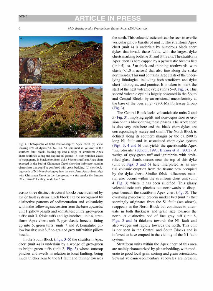
Fig. 4. Photographs of field relationship of Apex chert. (a) Viewlooking SW of dykes S1, S2, S3, S4 (outlined in yellow) in thesouthern fault block, feeding up into a ridge of stratiform Apexchert (outlined along the skyline in green); (b) sub-rounded clastsof megaquartz in black chert from dyke S4; (c) stratiform Apex chertexposed in the bed of Chinaman Creek showing imbricate, tabularchert clasts that could be confusedwith cross-bedding; (d) view look-ing south of N1 dyke feeding up into the stratiform Apex chert ridgewith Chinaman Creek in the foreground—a star marks the famous‘Microfossil’ locality; scale bar 5 cm.
across three distinct structural blocks, each defined bymajor fault systems. Each block can be recognised bydistinctive patterns of sedimentation and volcanicitywithin the following succession from the base upwards:unit 1, pillow basalts and komatiites; unit 2, grey-greentuffs; unit 3, felsic tuffs and ignimbrites; unit 4, strat-iform Apex chert; unit 5, pyroclastic breccia, finingup into 6, green tuffs; units 7 and 9, komatiitic pil-low basalts; unit 8, fine-grained grey tuff within pillowbasalts.In the South Block (Figs. 3–5) the stratiform Apex
chert (unit 4) is underlain by a wedge of grey-greento bright green tuffs (unit 2, Fig. 3) whose outcroppinches and swells in relation to local faulting, beingmuch thicker near to the S1 fault and thinner towards
the north. This volcaniclastic unit can be seen to overlievesicular pillow basalts of unit 1. The stratiform Apexchert (unit 4) is underlain by numerous black chertdykes that invade these faults, with the largest dykechertsmarking both the S1 andS4 faults. The stratiformApex chert is here capped by a pyroclastic breccia bed(unit 5), ca. 3m thick and thinning northwards, withclasts (<1.0m across) that also fine along the strikenorthwards. This unit contains large clasts of the under-lying lithologies, including both stratiform and dykechert lithologies, and pumice. It is taken to mark thestart of the next volcanic cycle (units 5–9, Fig. 3). Thissecond volcanic cycle is largely obscured in the Southand Central Blocks by an erosional unconformity atthe base of the overlying !2700Ma Fortescue Group(Fig. 3).The Central Block lacks volcaniclastic units 2 and
5 (Fig. 3), implying uplift and non-deposition or ero-sion on this block during these phases. The Apex chertis also very thin here and the black chert dykes arecorrespondingly scarce and small. The North Block isdefined along its southern margin by the ca.1500mlong N1 fault and its associated chert dyke system(Figs. 3, 4 and 6) that yields the questionable Apex‘microfossils’ (Schopf, 1993; Brasier et al., 2002). Awedge of grey-green tuff and ignimbrite with devit-rified glass shards occurs near the top of this dyke(unit 3, Figs. 3 and 6) here interpreted as an ini-tial volcanic eruption from the fissure now occupiedby the dyke chert. Similar felsic tuffaceous mate-rial also occurs within the stratiform chert unit (unit4, Fig. 3) where it has been silicified. This glassyvolcaniclastic unit pinches out northwards to disap-pear beneath the stratiform Apex chert (Fig. 3). Theoverlying pyroclastic breccia marker bed (unit 5) thatseemingly originates from the S1 fault (see above),reappears in the North Block but continues to atten-uate in both thickness and grain size towards thenorth. A distinctive bed of fine grey tuff (unit 8,Figs. 3 and 6) thickens towards the N1 fault andalso wedges out rapidly towards the north. This unitis not seen in the Central and South Blocks and isinferred to have erupted in the vicinity of the N1 faultsystem.Stratiform units within the Apex chert of this area
are mainly characterized by planar bedding, with mod-erate to good local grain sorting and grain orientation.Several volcanic-sedimentary subcycles are present,

M.D. Brasier et al. / Precambrian Research xxx (2005) xxx–xxx 7
Fig. 5. Detailed geological map of S4 dyke in the south block of the Chinaman Creek study area. Note the presence of rounded blocks ofstratified chert up to 450m palaeodepth down the dyke system.
showing rhythms from 1 to 5m thick that typicallybegin with grey-green, planar bedded siliceous tuffsplus red-brown jaspilitic banded chert (1–4 cm thicklayers), passing upwards into grey, black or black-white planar bedded chert (1–10 cm) and thence intoblack-white banded cherts with localised soft sedimentdeformation and breccias, indicating zones of lique-faction, cavity formation and collapse. A similar shift,from tuffaceous and jaspilitic chert near the base, toblack and brecciated chert, can be seen in the strati-form chert as a whole, especially to the north of N4.Earlier reports of cross-bedding from the bed of Chi-naman Creek (e.g. Schopf, 1993) are re-interpreted asimbricate slabs of chert that have slumped within azone of liquefaction (Figs. 4d and 5a). Such featuresare common. Even so, hummocky cross-stratificationfrom the possibly coeval Antarctic Creek cherts of theNorth Pole area, suggests that water depths were closeto storm wave base in parts of the basin (Van Kranen-donk, 2000).
Dyke cherts are abundant in both the South andNorth Blocks (Fig. 3). They are characterised by across-cutting geometry; by a lack of grain sorting orgrain orientation in the matrix; by multiple genera-tions of fracturing, spalling (with jigsaw puzzle fit ofclasts), fissure formation and fissure filling; by fault-rotated geopetal fabrics; by hydrocarbon-impregnatedbotryoidal and spherulitic chalcedony and chert; byassociated megaquartz veining. These dyke cherts tendto fan upwards, and to become both thicker and moremultiphase upwards. In the South Block, dyke chertsappear to fan outwards from a depth of about 500mbeneath the paleosurface. At greater depth are founddiscontinuous dykes and horizontal sills of grey chertplus sills of vein quartz (Fig. 3).Two black chert dyke systems have been studied by
us in detail (see Figs. 3–6). The S4 dyke complex hasa curved outcrop that extends for some 500m beneaththe stratiform cherts, extending along the line of theS4 fault that marks the northern boundary of the South
8 M.D. Brasier et al. / Precambrian Research xxx (2005) xxx–xxx
Block (Figs. 3 and 4a). The grey-green volcanoclasticsof unit 2 in the South Block cannot be traced across thisfault (Fig. 5), suggesting they were not deposited orwere later removed prior to deposition of the very thinand discontinuous stratiform Apex chert of the MiddleBlock. By way of contrast, there is a marked thicken-ing of the stratiform Apex chert around the mouth ofthe S4 dyke on the South Block. This thickening mayin part be due to greater accommodation close to thefault, and in part to the dilational effects of black chertsills that intrude southwards from the dyke (Fig. 5).Rotated and scattered blocks of black chert are alsofound around the upper part of S4, presumably dis-placed by faulting (Fig. 5). The upper part of the S4dyke complex comprises black cherts in which floatlarge (<1m), angular to rounded blocks of stratiformchert (Fig. 4b). These clasts appear to have fallen downthe dyke system from the palaeosurface, diminishingin size downwards, being inconspicuous in the bottom200m of the black chert dyke. At greater depth arefound a few small isolated chert dykes plus a seriesof north–south trending sills of white vein quartz andbrown carbonate-filled veins (see Fig. 3).
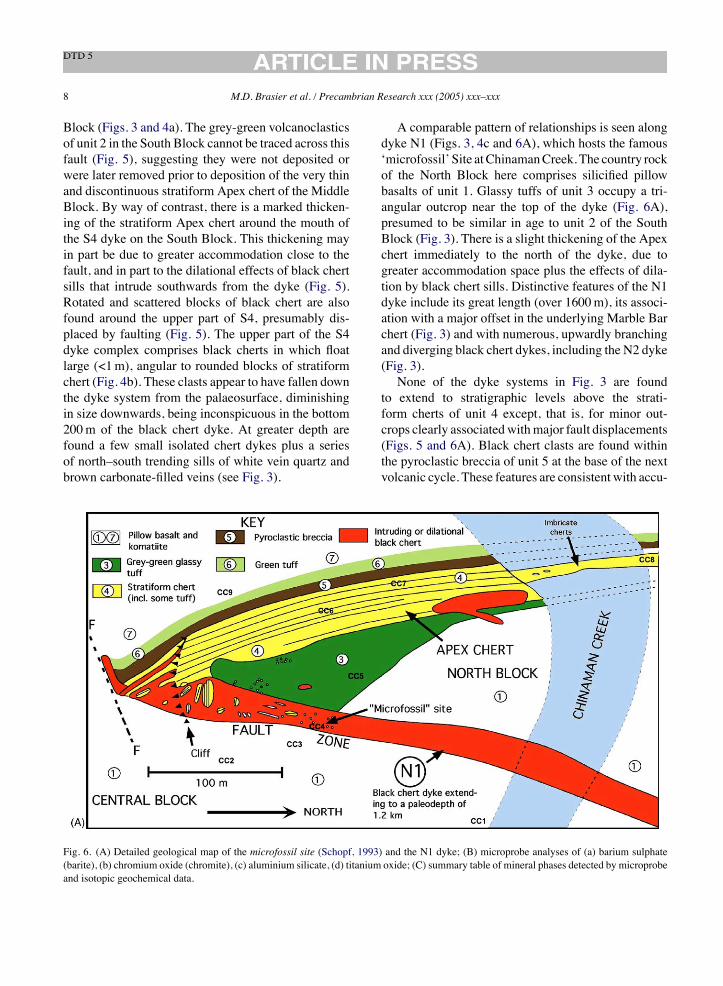
A comparable pattern of relationships is seen alongdyke N1 (Figs. 3, 4c and 6A), which hosts the famous‘microfossil’ Site atChinamanCreek.The country rockof the North Block here comprises silicified pillowbasalts of unit 1. Glassy tuffs of unit 3 occupy a tri-angular outcrop near the top of the dyke (Fig. 6A),presumed to be similar in age to unit 2 of the SouthBlock (Fig. 3). There is a slight thickening of the Apexchert immediately to the north of the dyke, due togreater accommodation space plus the effects of dila-tion by black chert sills. Distinctive features of the N1dyke include its great length (over 1600m), its associ-ation with a major offset in the underlying Marble Barchert (Fig. 3) and with numerous, upwardly branchingand diverging black chert dykes, including the N2 dyke(Fig. 3).None of the dyke systems in Fig. 3 are found
to extend to stratigraphic levels above the strati-form cherts of unit 4 except, that is, for minor out-crops clearly associatedwithmajor fault displacements(Figs. 5 and 6A). Black chert clasts are found withinthe pyroclastic breccia of unit 5 at the base of the nextvolcanic cycle. These features are consistent with accu-
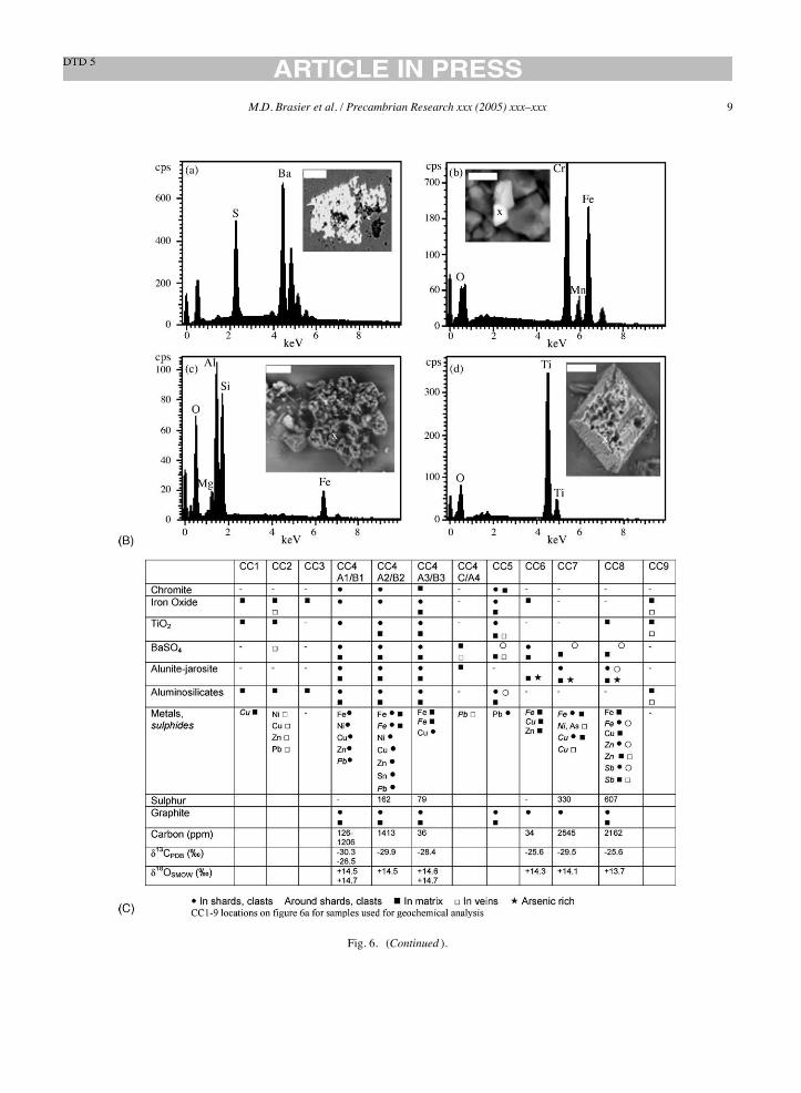
Fig. 6. (A) Detailed geological map of the microfossil site (Schopf, 1993) and the N1 dyke; (B) microprobe analyses of (a) barium sulphate(barite), (b) chromium oxide (chromite), (c) aluminium silicate, (d) titanium oxide; (C) summary table of mineral phases detected by microprobeand isotopic geochemical data.
M.D. Brasier et al. / Precambrian Research xxx (2005) xxx–xxx 9
Fig. 6. (Continued ).
10 M.D. Brasier et al. / Precambrian Research xxx (2005) xxx–xxx
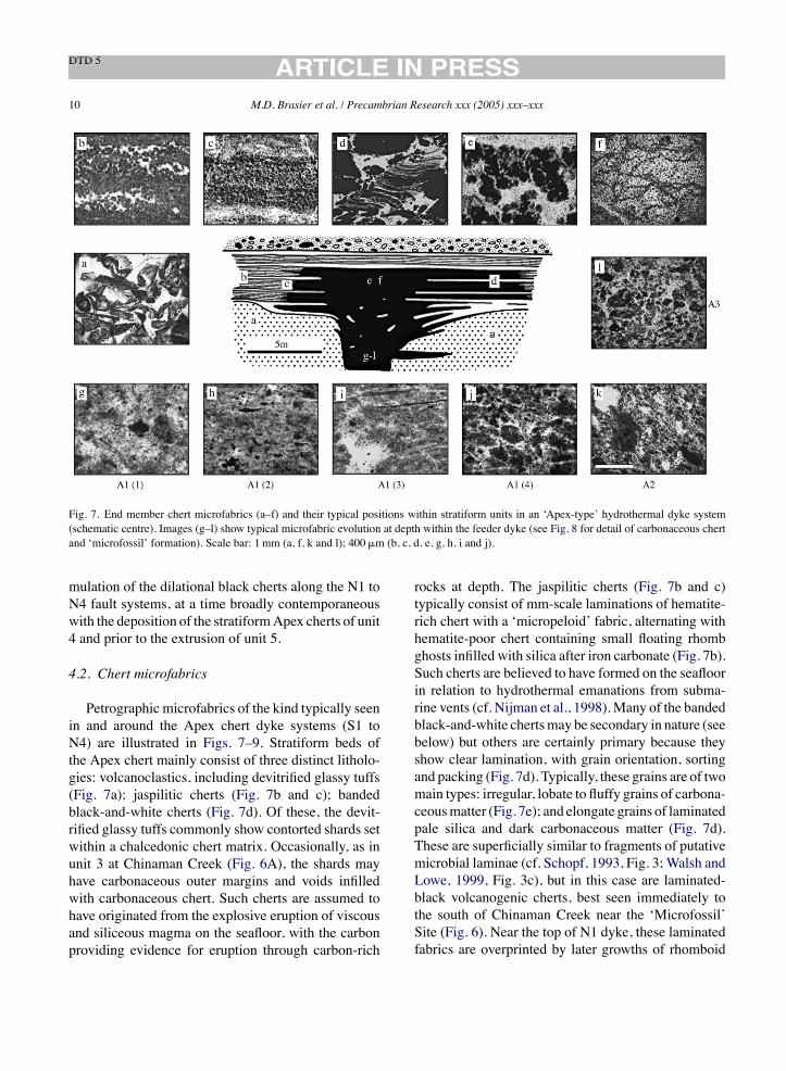
Fig. 7. End member chert microfabrics (a–f) and their typical positions within stratiform units in an ‘Apex-type’ hydrothermal dyke system(schematic centre). Images (g–l) show typical microfabric evolution at depth within the feeder dyke (see Fig. 8 for detail of carbonaceous chertand ‘microfossil’ formation). Scale bar: 1mm (a, f, k and l); 400!m (b, c, d, e, g, h, i and j).
mulation of the dilational black cherts along the N1 toN4 fault systems, at a time broadly contemporaneouswith the deposition of the stratiformApex cherts of unit4 and prior to the extrusion of unit 5.
4.2. Chert microfabrics
Petrographic microfabrics of the kind typically seenin and around the Apex chert dyke systems (S1 toN4) are illustrated in Figs. 7–9. Stratiform beds ofthe Apex chert mainly consist of three distinct litholo-gies: volcanoclastics, including devitrified glassy tuffs(Fig. 7a); jaspilitic cherts (Fig. 7b and c); bandedblack-and-white cherts (Fig. 7d). Of these, the devit-rified glassy tuffs commonly show contorted shards setwithin a chalcedonic chert matrix. Occasionally, as inunit 3 at Chinaman Creek (Fig. 6A), the shards mayhave carbonaceous outer margins and voids infilledwith carbonaceous chert. Such cherts are assumed tohave originated from the explosive eruption of viscousand siliceous magma on the seafloor, with the carbonproviding evidence for eruption through carbon-rich
rocks at depth. The jaspilitic cherts (Fig. 7b and c)typically consist of mm-scale laminations of hematite-rich chert with a ‘micropeloid’ fabric, alternating withhematite-poor chert containing small floating rhombghosts infilled with silica after iron carbonate (Fig. 7b).Such cherts are believed to have formed on the seafloorin relation to hydrothermal emanations from subma-rine vents (cf. Nijman et al., 1998).Many of the bandedblack-and-white chertsmay be secondary in nature (seebelow) but others are certainly primary because theyshow clear lamination, with grain orientation, sortingand packing (Fig. 7d). Typically, these grains are of twomain types: irregular, lobate to fluffy grains of carbona-ceousmatter (Fig. 7e); and elongate grains of laminatedpale silica and dark carbonaceous matter (Fig. 7d).These are superficially similar to fragments of putativemicrobial laminae (cf. Schopf, 1993, Fig. 3; Walsh andLowe, 1999, Fig. 3c), but in this case are laminated-black volcanogenic cherts, best seen immediately tothe south of Chinaman Creek near the ‘Microfossil’Site (Fig. 6). Near the top of N1 dyke, these laminatedfabrics are overprinted by later growths of rhomboid
M.D. Brasier et al. / Precambrian Research xxx (2005) xxx–xxx 11
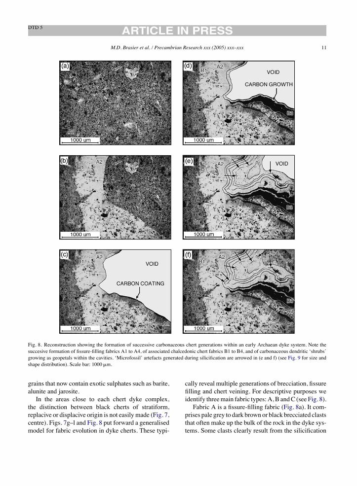
Fig. 8. Reconstruction showing the formation of successive carbonaceous chert generations within an early Archaean dyke system. Note thesuccesive formation of fissure-filling fabrics A1 to A4, of associated chalcedonic chert fabrics B1 to B4, and of carbonaceous dendritic ‘shrubs’growing as geopetals within the cavities. ‘Microfossil’ artefacts generated during silicification are arrowed in (e and f) (see Fig. 9 for size andshape distribution). Scale bar: 1000!m.
grains that now contain exotic sulphates such as barite,alunite and jarosite.In the areas close to each chert dyke complex,
the distinction between black cherts of stratiform,replacive or displacive origin is not easily made (Fig. 7,centre). Figs. 7g–l and Fig. 8 put forward a generalisedmodel for fabric evolution in dyke cherts. These typi-
cally reveal multiple generations of brecciation, fissurefilling and chert veining. For descriptive purposes weidentify threemain fabric types:A, B andC (see Fig. 8).Fabric A is a fissure-filling fabric (Fig. 8a). It com-
prises pale grey to dark brown or black brecciated claststhat often make up the bulk of the rock in the dyke sys-tems. Some clasts clearly result from the silicification
12 M.D. Brasier et al. / Precambrian Research xxx (2005) xxx–xxx
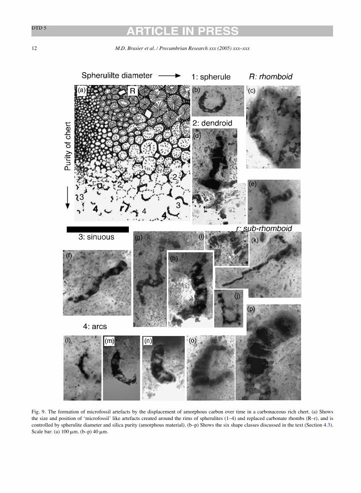
Fig. 9. The formation of microfossil artefacts by the displacement of amorphous carbon over time in a carbonaceous rich chert. (a) Showsthe size and position of ‘microfossil’ like artefacts created around the rims of spherulites (1–4) and replaced carbonate rhombs (R–r), and iscontrolled by spherulite diameter and silica purity (amorphous material). (b–p) Shows the six shape classes discussed in the text (Section 4.3).Scale bar: (a) 100!m, (b–p) 40!m.

M.D. Brasier et al. / Precambrian Research xxx (2005) xxx–xxx 13
and carbon-impregnation of the country rock. At leastfour successive generations (A1 to A4) can be usu-ally be identified in thin sections of such dyke cherts(Fig. 8a–f).FabricB is amicroquartz vein fabric (Fig. 8). It com-
prises chalcedonic microquartz that penetrates fabricsof generation A via small fissures, to infill irregularlyshaped vugs (Fig. 8). Within larger cavities, it formslaminated layers and botryoids of clear chert, includ-ing roof pendants (Figs. 8 and 13a and b). Inmost cases,each generation of fabric A was accompanied by vein-ing of fabric B (Fig. 8).Fabric C is a megaquartz vein fabric. It comprises
crystals of drusy quartz that infill the central zone oflarger veins (e.g. Fig. 18b) especially after the forma-tion of chalcedonic chert of generation B3. Severalphases of fabric C can be present. When studied ingreater detail, generation A1 is seen to comprise brec-ciated and angular to rounded fragments of microcrys-talline quartz containing carbonaceous microclasts,grains of iron oxide, rutile and chromite, native metals(Fe, Ni, Cu, Zn), pyrite, sericite and rhombic pseu-domorphs (Brasier et al., 2002). The pseudomorphsinclude relict grains of barite, plus dark reaction rimsof chromite and rutile (in EDX). The texture of gen-eration A1 tends to intergrade from rather homoge-nous and fine-grained (Figs. 7g and 8a), through chertswith carbon-impregnated cracks (Fig. 7h) and chert-filled vugs (Figs. 7i, 15b and 18c), and thence tomore mottled textures in which incipient grains aresurrounded by chert-filled cracks (Fig. 7j). These A1clasts are commonly traversed by small cracks andvugs now of chalcedonic microquartz (generation B1).Both B1 veins and linear fabric appears may have beeninduced by fault-related shearing. More often, how-ever, the chert-filled cracks have an arcuate to polyg-onal pattern (Figs. 7j and 19b), somewhat resemblingthe circum-granular-cracks of ‘caliche’ and ‘evapor-itic palaeosols’ (cf. Rettalack, 1997) and suggest-ing that a comparable, subsurface process of incip-ient precipitation and lithification has taken place.Generation A1 was clearly brecciated by successiveepisodes of fracturing in situ, to form fissures, cavi-ties and adjacent clasts with a ‘jigsaw puzzle’ fit (seeFig. 8a and b). Cavities are often lined with epitaxialrims of botryoidal, spherulitic and fan-shaped whitechalcedonic microquartz (Figs. 8c–f), as describedbelow.
Fissure-filling generation A2, which tends to bepaler with more distinct lithoclasts, was produced byspalling of A1 and B1, geopetally infilling voids withinthe breccia (Figs. 7j and k and 8b). Generation A2 hasquite clearly defined but poorly sorted microclasts ofA1 and B1 type, producing a muddy to poorly sortedappearance (see Figs. 7k, 8b, 15b, 16a, 17a and 19b).A comparable set of processes led to generations A3and B3 (see Figs. 71, 8d, 17i and 20d). Conspicuousrhombic crystals (containing barite in EDX) or voidsmay float in the outer margins of cross-cutting chal-cedonic veins of generation B3 (Fig. 20d). The latteralso penetrates the earlier generations A1 and A2 viasmall fissures and vugs, forming laminated layers andconspicuous botryoids (Fig. 8e and f).Each of these phases (A1 to B1, A2 to B2 and
A3 to B3) appears to have been accompanied by car-bonaceous growth or hydrocarbon migration. This car-bon has penetrated along microfissures to line or infillsmall vugs within A1 (Figs. 7 and 18c middle), coatand darken lithified clasts of A1 to A3 and B1 to B2(Fig. 8c), and accumulate widely as laminae and soot-like dendrites within botryoidal-spherulitic fabrics ofB2 to B4 (Figs. 7e, 8d and e and 13a–c). The centralzone of larger chalcedonic veins was later infilled bycrystals of drusy quartz (fabric C), especially in con-spicuous cross-cutting, linear fractures (Figs. 14a, 15aand b and 18a) and these tend to lack carbonaceousmatter. Late stage metallic oxides have also infiltratedsmall cracks and fissures and these may be associatedwith secondary oxidation and reddening. No carbonatehas yet been revealed by geochemistry at this level inthe hydrothermal system.Such fissure-filling black cherts are often seen to
both cross-cut and to modify the adjacent stratiformcherts. This phenomenon is especially well displayedin the Marble Bar chert (Van Kranendonk et al.,2001, Plate 2). Where black cherts cross-cut jaspiliticcherts (e.g. Fig. 7b), thin sections show progressiveremoval of the oxidised hematite and its replacementby fluffy black grains of carbonaceous chert (Fig. 7c).This replacement has often resulted in large cavitiesthat were later infilled by carbonaceous black andwhite cherts showing botryoidal growth of mainlyfibrous chalcedony around bushes and dendrites ofcarbonaceousmatter that grew either upwards or down-wards into the cavities (Figs. 7e and 8c–f). Alsoseen are pillow-shaped botryoids of fibrous chalcedony
14 M.D. Brasier et al. / Precambrian Research xxx (2005) xxx–xxx
containing thin drapes of carbon plus sparse carbona-ceous intraclasts—these pillows can also growupwardsand downwards into the voids (e.g. Fig. 7f).It is therefore possible to recognise at least three
kinds of carbonaceous chert around these dyke sys-tems. Stratiform chert that was originally laid downas bedded sediments (e.g. Fig. 7d). Chert that wasreplacive after stratiform chert, especially ferrugi-nous chert (e.g. Fig. 7e); and chert that was dis-placive, growing by successive episodes of fracturing,spalling, flocculation and lithification (Figs. 7g–l andFig. 8).
4.3. Microfossil filaments
All the illustrated ‘microfossils’ from theApex cherthave been re-examined by us, including holotypes andparatypes deposited at the Natural History Museumin London (Schopf and Packer, 1987; Schopf, 1992b,1993). These carbonaceous structures come from threeblocks of chert collected from the ‘Microfossil’ siteshown in Fig. 6, from a position some 100m beneaththe surface of the N1 dyke system. In Table 1, we sum-marise the characteristics of all of the figured ‘micro-fossils’ and list their enclosing fabrics (e.g. fabric gen-erations A1 to A3, B1 to B3 and C outlined in Section4.2 above). A detailed description of the petrographyand the context for each ‘microfossil holotype’ slide isgiven in the Appendix A.The majority of the filamentous microfossil-like
objects from the ‘Microfossil’ can be assigned to one ofsix shape classes shown in Fig. 9: spherules (Fig. 9b),rhomboids (Fig. 9c), dendroids (Fig. 9d and e), sin-uous forms (Fig. 9f–i), sub-rhomboids (Fig. 9j and k)and arcs (Fig. 9l–p). The frequency distribution of theseshape classes has beenmeasured along thin section tra-verses through brecciated shards (previously regardedas ‘microfossiliferous’ clasts, Schopf, 1992b, 1993)within the dyke chert. The results are as follows: arcs(ca. 50%), dendroids (ca. 30%), spherules (ca. 8%),sinuous forms (ca. 6%), rhomboids and sub-rhomboids(ca. 6%). Hence, the sinuous-shaped filaments imagedin previous studies (Schopf, 1992b, 1993) are found byus to comprise only a minor component of the morpho-logical population, with the majority of microfossil-like objects being arcuate. In Section 6.1, we combinethese results with our fabric observations to propose amodel for the formation of the pseudofossils.
The maximum diameter and the maximum lengthof all microfossil-like objects has been measured by usalong traverses across natural populations of structureswithin each of the previously illustrated ‘microfos-siliferous’ clasts (Schopf, 1992b, 1993). We find noevidence for the bimodal distribution of filament diam-eters reported by Schopf (1993, Fig. 6). Instead,we findthat the structures occupy a morphological continuumin which the filament diameters decrease (exponen-tially) in frequency as their diameters increase from1!m to at least 36!m.
5. Interpretation
Here, we address questions raised by re-mappingand re-analysis of the Apex ‘microfossil’ materials.These questions are assembled broadly in the order fol-lowed by Brasier et al. (2002).
5.1. Context viable for life—neptunean or deephydrothermal?
Does the context of the Apex chert point to apotentially viable habitat for life? Careful attention togeological context is certainly essential for correctlydecoding the signals for early life on Earth. Untilrecently, it has been accepted that layered siliceouscherts of the 3.4–3.5Ga old Warrawoona Group werelaid down in settings thatwere comparablewithmodernevaporitic and exposed lagoonal environments (Dunlopet al., 1978; Walter et al., 1980; Lowe, 1983; Awramiket al., 1983). Several programs of careful mappingand elemental analysis of the cherts, however, haveshown that the environmental setting of these rocksis largely volcanic, volcaniclastic and hydrothermalin nature, and that cross-cutting chert-barite veins aremore readily explained as hydrothermal feeder dykes(e.g. Nijman et al., 1998; Nakishima et al., 2001; VanKranendonk et al., 2001; Brasier et al., 2002;Lindsay et al., submitted for publication), althoughLowe (in Tenenbaum, 2003) has argued that some asso-ciated South African dykes are neptunean in origin.The Apex ‘microfossils’ are now known to have
been collected from the ‘Microfossil’ site at ChinamanCreek (Fig. 3; Dr. Bonnie Packer, written communi-cation to Brasier, 2002; J.W. Schopf, communicationto Van Kranendonk, 1999; see Brasier et al., 2002,

M.D.Brasieretal./Precam
brianResearch
xxx(2005)xxx–xxx
15
Table 1Characteristics of the previously figured Apex ‘microfossils’, clasts and fabrics
a b c d e f g h i j k
V.63164 [1] P. conicoterminatum holotype Schopf (1992b), Fig. 1.5.4C 64C2, Fig. 12a B B A1 sa 5.0 206 –V.63164 [2] P. conicoterminatum paratype Schopf (1992b), Fig. 1.5.4D 64C PlanM3 B B A1 r – –V.63164 [3] P. delicatulum paratype Schopf (1992b), Fig. 1.5.4F 64C PlanM32 B B A1 r 0.5 –V.63164 [4] P. delicatulum paratype Schopf (1993), Fig. 3K Not confirmedV.63164 [5] P. amoenum paratype Schopf (1992b), Fig. 1.5.5C 64C21 Fig. 12l B B A1 sa 5.0 46 –V.63164 [6] P. amoenum paratype Schopf (1992b), Fig. 1.5.5E 64C20, Fig. 12k B B A1 sa 5.0 144 –V.63164 [7] Un-named type A broad filament Schopf (1992b), Fig. 1.5.6A 64C1, Fig. 11g B B A1 sa 5.0 150 –V.63164 [8] Un-named type C narrow filament Schopf (1992b), Fig. 1.5.6D 64C30 B B A1 sa 5.0 330 –V.63164 [9] P. conicoterminatum Schopf (1993), Fig. 4G 64D1 B B A2 – – 380 –V.63164 [10] P. laticellulosum holotype Schopf (1993), Fig. 5A 64C19, Fig. 12f B B A1 sa 5.0 331 –V.63164 [11] P. laticellulosum Schopf (1993), Fig. 5B Not confirmedV.63164 [12] A. maxima holotype Schopf (1993), Fig. 5F 64B1, Fig. 10e R R A1 sa 2.4 7 –V.63165 [1] Un-named degraded cellular filament Schopf (1992b), Fig. 1.5.4B 65H12 Fig. 15m B B A1 sa 2.1 343 –V.63165 [2] P. delicatulum holotype Schopf (1992b), Fig. 1.5.4G 65H6, Fig. 15g B B A1 sa 2.1 248 –V.63165 [3] P. amoenum paratype Schopf (1992b), Fig. 1.5.5D 65P3 Fig. 16n B B A2 – – 10 –V.63165 [4] Un-named type B broad filament Schopf (1992b), Fig. 1.5.6B 65G11, Fig. 15l B B A1 sa 2.1 289 –V.63165 [5] Microfossiliferous clast Schopf (1993), Fig. 3A and B 65A Fig. 14a–c B B A1 r – –V.63165 [6] P. minutum Schopf (1993), Fig. 3H 65A53 Fig. 14i B B A1 r 1.8 160 –V.63165 [7] P. delicatulum Schopf (1993), Fig. 3J 65A18 Fig. 14p B B A1 r 1.8 133 –V.623165 [8] A. disciformis Schopf (1993), Fig. 3N 65P4 Fig. 16j–l B B A2 – – 88 –V.63165 [9] Un-named solitary unicell-like microfossil Schopf (1993), Fig. 5K 65P4 Fig. 16i B B A2 – – 113 –V.63165 [10] Microfossiliferous clast Schopf (1992b), Fig. 1.5.4A 65H Fig. 15b B B A1 sr – –V.63166 [1] P. amoenum holotype Schopf (1992b), Fig. 1.5.5A 66B1, Fig. 17g B B A2 – – 315 –V.63166 [2] Un-named type C narrow filament Schopf (1992b), Fig. 1.5.6C 66B5, Fig. 17h B B A2 – – 298 –V.63166 [3] A. septatum holotype Schopf (1993), Fig. 3D 66F5, Fig. 17f R R B3 – – – 5V.63727 [1] P. conicoterminatum Schopf (1993), Fig. 4F 27C8 B W A1 sa – –V.63727 [2] P. attenuatum holotype Schopf (1993), Fig. 5g 27D1, Fig. 18e B B A2 – – 266 –V.63728 [1] A. disciformis Schopf (1993), Fig. 3L 28A2 G G A2 – – 252 –V.63728 [2] P. amoenum Schopf (1993), Fig. 4C 28B1 B B A2 – – 75 –V.63729 [1] E. apex holotype Schopf, (1993), Fig. 3F 29B1, Fig. 19f B B A2 – – – 150V.63729 [2] P. minutum holotype Schopf (1993), Fig. 3G 29C2 Fig. 19e B B A2 – – 112 –V.63730 [1] A. disciformis holotype Schopf (1993), Fig. 3M 30A3, Fig. 20d B B A3 – – 74 –V.63731 [1] Stromatolite-like clast Schopf (1993), Fig. 3C 31A1 Fig. 21b B B A2 – – – –V.63732 [1] A. septatum Schopf (1993), Fig. 3E 32A2 B B A3 – – 18 27V.63732 [2] Solitary unicell-like microfossil Schopf (1993), Fig. 5L 32B1 B B A3 – – 75 –V.63733 [1] P. amoenum Schopf (1993), Fig. 4D 33B1 B W A1 sa 50 208 –V.63733 [2] A. grandis Schopf (1993), Fig. 5E 33A1 Y G A1 sa 50 373 –V.63734 [1] P. amoenum Schopf (1993), Fig. 4E 34A3 B W A2 – – 348 –V.63734 [2] P. conicoterminatum Schopf (1993), Fig. 4J 34B1 B W A2 – – 665 –V.63734 [3] Poorly preserved trichome Schopf (1993), Fig. 5H 34C1 B W A2 – – – 146V.63734 [4] P. amoenum Schopf (1992b), Fig. 1.5.5B 34C2 B W A1 sa – –
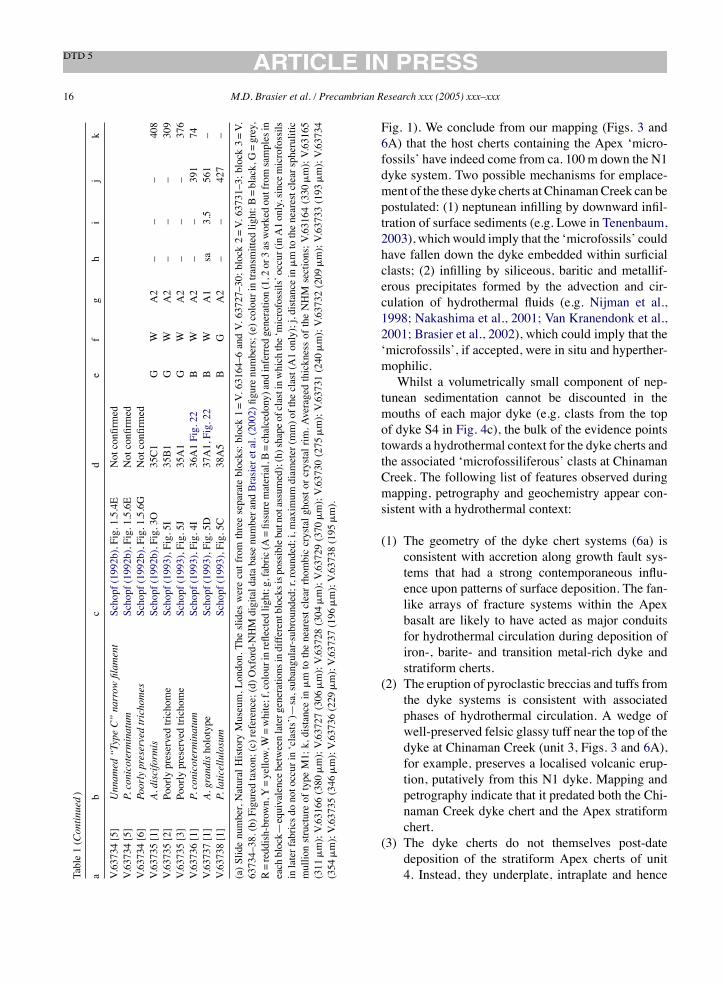
16 M.D. Brasier et al. / Precambrian Research xxx (2005) xxx–xxx
Table1(Continued)
ab
cd
ef
gh
ij
k
V.63734[5]
Unnam
ed“TypeC”narrow
filam
ent
Schopf(1992b),Fig.1.5.4E
Notconfirmed
V.63734[5]
P.conicoterminatum
Schopf(1992b),Fig.1.5.6E
Notconfirmed
V.63734[6]
Poorlypreservedtrichomes
Schopf(1992b),Fig.1.5.6G
Notconfirmed
V.63735[1]
A.disciformis
Schopf(1992b),Fig.3O
35C1
GW
A2
––
–408
V.63735[2]
Poorlypreservedtrichome
Schopf(1993),Fig.5I
35B1
GW
A2
––
–309
V.63735[3]
Poorlypreservedtrichome
Schopf(1993),Fig.5J
35A1
GW
A2
––
–376
V.63736[1]
P.conicoterminatum
Schopf(1993),Fig.4I
36A1Fig.22
BW
A2
––
391
74V.63737[1]
A.grandisholotype
Schopf(1993),Fig.5D
37A1,Fig.22
BW
A1
sa3.5
561
–V.63738[1]
P.laticellulosum
Schopf(1993),Fig.5C
38A5
BG
A2
––
427
–
(a)Slidenumber,NaturalHistoryMuseum,London.Theslideswerecutfromthreeseparateblocks:block1=V.63164–6andV.63727–30;block2=V.63731–3;block3=V.
63734–38.(b)Figuredtaxon;(c)reference;(d)Oxford-NHMdigitaldatabasenumberandBrasieretal.(2002)figurenumbers;(e)colourintransmittedlight:B=black,G=grey,
R=reddish-brown,Y=yellow,W=white;f,colourinreflectedlight;g,fabric(A=fissurematerial,B=chalcedony)andinferredgeneration(1,2or3asworkedoutfromsamplesin
eachblock—
equivalencebetweenlatergenerationsindifferentblocksispossiblebutnotassumed);(h)shapeofclastinwhichthe‘microfossils’occur(inA1only,sincemicrofossils
inlaterfabricsdonotoccurin‘clasts’)—
sa,subangular-subrounded;r,rounded;i,maximum
diameter(mm)oftheclast(A1only);j,distancein
!mtothenearestclearspherulitic
mullionstructureoftypeM1;k,distancein
!mtothenearestclearrhombiccrystalghostorcrystalrim.AveragedthicknessoftheNHMsections;V.63164(330
!m);V.63165
(311
!m);V.63166(380
!m);V.63727(306
!m);V.63728(304
!m);V.63729(370
!m);V.63730(275
!m);V.63731(240
!m);V.63732(209
!m);V.63733(193
!m);V.63734
(354
!m);V.63735(346
!m);V.63736(229
!m);V.63737(196
!m);V.63738(195
!m).
Fig. 1). We conclude from our mapping (Figs. 3 and6A) that the host cherts containing the Apex ‘micro-fossils’ have indeed come from ca. 100m down the N1dyke system. Two possible mechanisms for emplace-ment of the these dyke cherts at ChinamanCreek can bepostulated: (1) neptunean infilling by downward infil-tration of surface sediments (e.g. Lowe in Tenenbaum,2003), which would imply that the ‘microfossils’ couldhave fallen down the dyke embedded within surficialclasts; (2) infilling by siliceous, baritic and metallif-erous precipitates formed by the advection and cir-culation of hydrothermal fluids (e.g. Nijman et al.,1998; Nakashima et al., 2001; Van Kranendonk et al.,2001; Brasier et al., 2002), which could imply that the‘microfossils’, if accepted, were in situ and hyperther-mophilic.Whilst a volumetrically small component of nep-
tunean sedimentation cannot be discounted in themouths of each major dyke (e.g. clasts from the topof dyke S4 in Fig. 4c), the bulk of the evidence pointstowards a hydrothermal context for the dyke cherts andthe associated ‘microfossiliferous’ clasts at ChinamanCreek. The following list of features observed duringmapping, petrography and geochemistry appear con-sistent with a hydrothermal context:
(1) The geometry of the dyke chert systems (6a) isconsistent with accretion along growth fault sys-tems that had a strong contemporaneous influ-ence upon patterns of surface deposition. The fan-like arrays of fracture systems within the Apexbasalt are likely to have acted as major conduitsfor hydrothermal circulation during deposition ofiron-, barite- and transition metal-rich dyke andstratiform cherts.
(2) The eruption of pyroclastic breccias and tuffs fromthe dyke systems is consistent with associatedphases of hydrothermal circulation. A wedge ofwell-preserved felsic glassy tuff near the top of thedyke at Chinaman Creek (unit 3, Figs. 3 and 6A),for example, preserves a localised volcanic erup-tion, putatively from this N1 dyke. Mapping andpetrography indicate that it predated both the Chi-naman Creek dyke chert and the Apex stratiformchert.
(3) The dyke cherts do not themselves post-datedeposition of the stratiform Apex cherts of unit4. Instead, they underplate, intraplate and hence
M.D. Brasier et al. / Precambrian Research xxx (2005) xxx–xxx 17
dilate and thicken the stratiform Apex chert neareach ‘vent’, through emplacement of numerousblack chert sills (Figs. 3–6). Comparable chertsoccur as clasts within the pyroclastic breccias atthe base of overlying unit 5.
(4) Dyke cherts are rare in the Central Block wherestratiform Apex cherts of unit 4 are thin or absent.This is consistent with a model in which the thick-ness of the stratiform cherts is controlled by thevolumeof silica supplied at depth, including under-plating and intraplating.
(5) Mafic extrusives adjacent to the dyke (Figs. 3–6,unit 1) are bleached and fissured by numeroussmall barite- andmetal-bearing chalcedonic quartzveins, similar to those within the dyke cherts, con-sistent with hydrothermal alteration. Importantly,extrusives far beneath the dykes and above the bed-ded chert are much less affected by hydrothermalalteration.
(6) Energy dispersive X-ray (SEM–EDX) analysisshows that barite (BaSO4) is common in boththe dyke and stratiform chert samples, occur-ring together with other hydrothermally associatedminerals including native metals, sulphides, chal-cedonic quartz and megaquartz (Fig. 6B; Brasieret al., 2002). Both the dyke cherts and felsic vol-canics contain grains of chromite (FeCr2O4), ironoxide and TiO2 (in SEM–EDX, e.g. relict miner-als fromhydrothermal leachingof adjacent basalts)plus Al- and K-rich phyllosilicates (in SEM–EDX,e.g. from hydrothermally altered feldspars).
(7) The black stratiform cherts (Figs. 3 and 6A) yieldsulphates rich in Al, K, Fe and As (e.g. fromhydrothermal reactions involving sulphuric acid,feldspars, zeolites or sulphides). These sulphatesresemble both ‘alunite’ KAl3(SO4)2(OH)6 and‘jarosite’ KFe3(SO4)2(OH)6 (in SEM–EDX, e.g.from hydrothermal reactions involving sulphuricacid, feldspars, zeolites or sulphides). Beddingis overprinted near the dyke by rhombic crys-tal ghosts, now of chalcedonic quartz, containingrelict grains of barite and alunite, suggestive ofhydrothermal brines.
(8) Oxygen isotopes of +13.7–+14.7‰ from withinSiO2 are less 18O-enriched than many Archaeansedimentary cherts (Robert, 1988; Beaumont andRobert, 1999). Along with the inferred presenceof native metals (Fig. 5c) this might be taken to
imply that comparatively high (ca. 250–350 #C)hydrothermal temperatures were reached (Brasieret al., 2002, cf. Robert, 1988) but it may also reflectthe temperatures reached during low grade meta-morphism. Fluid inclusion work is being under-taken to resolve these two options.
(9) We find no evidence for surficial sedimentarystructures or textures (e.g. sorting, rounded quartz,grain sorting or sedimentary laminae) in the‘microfossiliferous’ clasts (see below).
We conclude from these studies that the China-man Creek chert that yielded the famous ‘microfos-sils’ (e.g. Schopf, 1993) does not have a depositionalsetting comparable with a wave-washed beach or themouth of a stream or river (Schopf, 1999). It has acontext consistent with a hydrothermal feeder dyke,much like that inferred for the barite-chert beds of sim-ilar age at nearby North Pole (Nijman et al., 1998).If estimated surface water temperatures of !70 #C arecorrect for the early Archaean ocean (e.g. Knauth andLowe, 2003), temperatures within the dyke system atthe level of the Apex ‘microfossils’ (some 100m downthe dyke system) are likely to have reached !70 #Cor higher, which would today be taken to indicatemarginal extremophile conditions.
5.2. Rounded grains
Are the microfossils found only in detrital clasts?Original reports describe the ‘microfossils’ as occur-ring ‘within clasts that were deposited in this unit priorto its lithification’ (Schopf and Packer, 1987) but laterinterpreted as rounded grains of chert transported agreat distance before redeposition in a bedded grain-stone conglomerate (Schopf, 1992b, 1993, 1994).Field mapping, sampling (Figs. 3–6) and fabric
mapping (Figs. 7–22) reveal that the ‘microfossilifer-ous’ chert at Chinaman Creek (Fig. 6, sample CC4),is not part of the bedded succession. It is part of achert breccia that infills one of a series of dykes thatfeed upwards into overlying stratiform cherts (Fig. 6,samples CC6–8). Fabric mapping also reveals that themajority of figured ‘microfossils’ do not occur exclu-sively in first generation fabrics, nor in rounded clastsas previously suggested (e.g. Schopf, 1992b, 1993,1999).Only40%occurwithin rounded to angular clastsof generation A1. The majority actually occur within

18 M.D. Brasier et al. / Precambrian Research xxx (2005) xxx–xxx
larger areas ofA2 andA3 (57%) and some clearly occurwithin later chalcedonic chert (B3; 3%, see Table 1).Comparable structures also occur within carbonaceousclasts and chalcedonic matrix of the stratiform cherts(Brasier et al., 2002, Fig. 4p), within the carbonaceousrims of the volcanic glass shards (ibid, Fig. 4l) andin the chalcedony cement between glass shards (ibid,Fig. 4f, j and z) thus calling their indigenous and bio-genic origin into question.We conclude that the microfossils do not occur in a
‘petrographically distinctive population of clasts’, norare they absent ‘from all other clasts or the surroundingmatrix’ (see also Section 5.4 below). This means thatthe inference that ‘filaments predate deposition of thechert unit and were initially preserved in older rocks,some part of which was eroded as a detrital componentof the bedded chert’ (Schopf, 1993) is not sustained.
5.3. ‘Stromatolite-like clasts’
Are ‘stromatolite-like clasts’ present in the‘microfossil’-bearing samples and could they supporta biogenic interpretation? Such an association ofputative Archaean microfossils with stromatoliticlaminae could be regarded as a mutually supportivecriterion for biogenicity. It is important to note,however, that the biogenicity of stromatolitic struc-tures cannot be assumed (see Buick et al., 1981;Buick, 1990; Grotzinger and Rothman, 1996) and thatabiogenic origins must be falsified. These challengesare especially great when only small fragments <1mmacross are preserved, as here.We have re-examined the siliceous structures
with undulose laminations (Fig. 21a–f) labelled as‘stromatolite-like clasts’ by Schopf (1993, Fig. 3). Ourobservations indicate that this structure is intergrownwith enclosing A1 to A3 and B1 to B3 fabrics, formingvoid filling drapes and overhangs (Fig. 21a–d). This,
and associated ‘stromatoloids’, contain lithoclasts,phyllosilicates, metallic oxides, pyrite and rhombicghosts exactly like those of the enclosing fabric. Wefind that these ‘stromatoloids’ are indistinguishablefrom other shards of chalcedony with laminae of uni-form thickness (B1 to B3) that grew and fragmentedwithin the dyke system (Figs. 8, 13a and 21f) and wereinterpret them as such. The biogenicity of these ‘stro-matoloids’ should not be assumed and from our studiesappears questionable.
5.4. The distribution of ‘microfossils’
Does the distribution of microfossil-like structures(at the micrometer to kilometre scale) reflect any signsof biogenic behaviour? Typical populations of Pro-terozoic to modern cyanobacteria reveal trichomesthat are commonly occur clustered together in layers,often with a distinctive orientation relative to bedding.That distribution pattern may be attributed to photo orchemotactic growth and is well known, for example, inthe 1900MaGunflint chert (Awramik and Semikhatov,1979) and has therefore been proposed as a biogenicitycriterion (Schopf and Walter, 1983). Our micrometerscale fabric studies of the Apex chert microfossilif-erous populations (Figs. 10–22) clearly confirm thatthe ‘microfossils’ occur as isolated, irregularly dis-tributed and randomly orientated solitary filaments (asacknowledged by Schopf, 1993).We also find that the ‘microfossil’-like structures
occur throughout the dyke system, and at depths of upto 1500m below the palaeosurface. These examplesof dendritic to arcuate filaments are indistinguishablefrom those observed at the ‘Microfossil’ site. Theyoccur in botryoidal chert of fabric B, regarded as inte-gral to the hydrothermal system at depth. We findno evidence that sediment or microfossils could havefallen some 1500m down the dyke system. We con-
Fig. 10. (a) Macroscopic view showing the core of NHM slide V.63164 showing area 64B discussed in the text, scale in mm; (b) closer view ofarea 64B in (a) showing the iron-stained, angular shard of fabric A1 that contains object 64B1, regarded as the holotype for A. maxima (Schopf,1993; Fig. 5F) surrounded by fabric A2, plus the objects illustrated in Figures ‘f’, ‘g’ and ‘h’ below; (c) closer view of the patch of iron stainedspherulitic mullion fabric in area 64B associated with object 64B1 (arrowed) plus the objects illustrated in figures ‘d’ below; (d) detailed view ofspherulitic mullions giving rise to two septate dendritic mullion pseudofossils (objects 64B2, arrowed); (e) detailed view of arcuate and septateobject 64B1, the holotype for A. maxima (Schopf, 1993; Fig. 5F); (f) detailed view of spherulitic mullions giving rise to three septate arcuatemullion pseudofossils (objects 64B3, arrowed); (g) detailed view of spherulitic mullions giving rise to three septate arcuate mullion pseudofossils(arrowed, objects 64B3); (h) objects 64B4 formed as artefacts where the ink of a marker pen has infiltrated into arcuate cracks on the surface ofthe slide, forming numerous pseudofossils. Labels are as described in the text. Scale bar: 40!m (d–h); 100!m (c); 400!m (b).
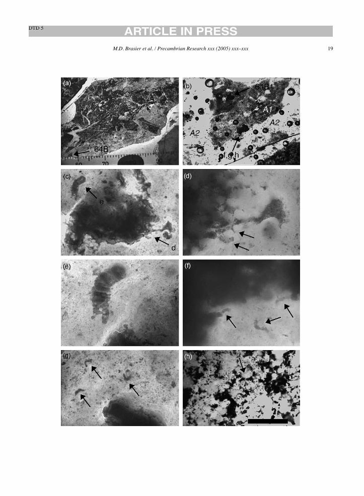
M.D. Brasier et al. / Precambrian Research xxx (2005) xxx–xxx 19

20 M.D. Brasier et al. / Precambrian Research xxx (2005) xxx–xxx
Fig. 11. (a) Macroscopic view showing the core of slide NHMV.63164 showing areas 64C and 64D discussed in the text, scale in mm; (b) closerview of area 64C (‘east’); (c) view of area 64C (‘mid east’); (d) closer view of area 64C (‘northeast)’; (e) view of area 64C (‘west’); (f) viewof area 64C (‘south’); (g) object 64C1, described as un-named ’Type A’ broad filament (Schopf, 1992b; Fig. 1.5.6A); (h) non-septate, arcuateobject 64C7; (i) septate and non-septate objects 64C8. Scale bar: 40!m (g–i); 100!m (d); 400!m (b, c, e and f).
M.D. Brasier et al. / Precambrian Research xxx (2005) xxx–xxx 21
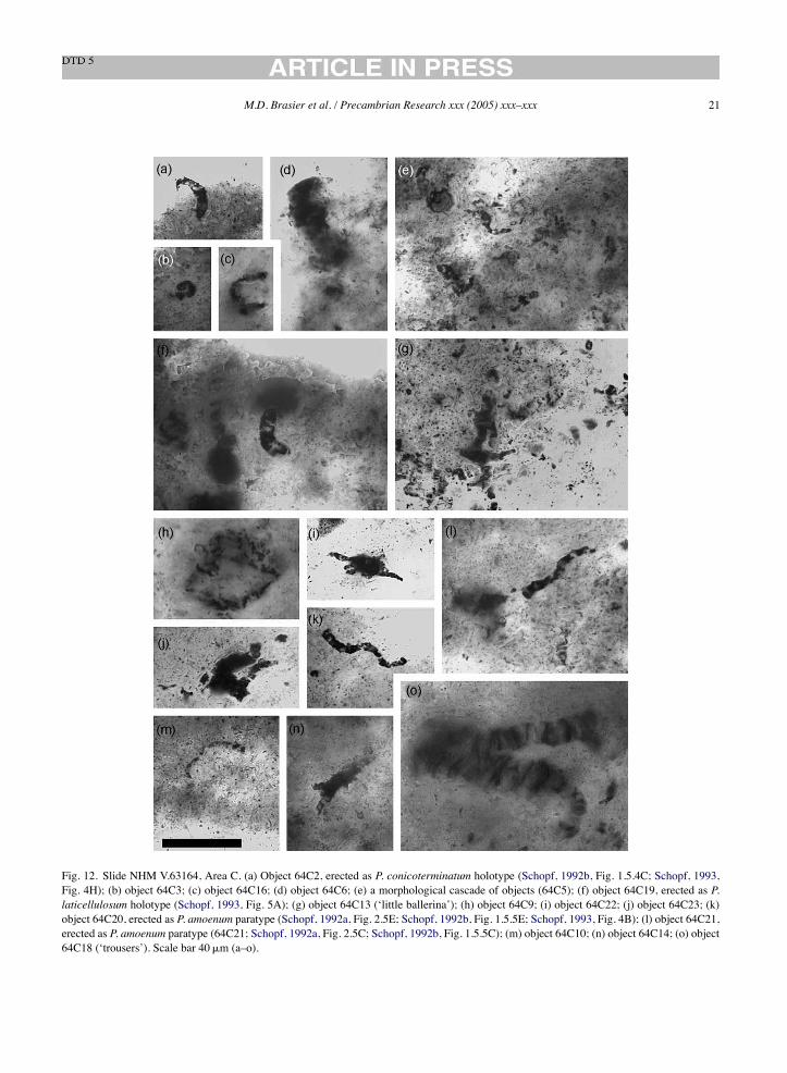
Fig. 12. Slide NHM V.63164, Area C. (a) Object 64C2, erected as P. conicoterminatum holotype (Schopf, 1992b, Fig. 1.5.4C; Schopf, 1993,Fig. 4H); (b) object 64C3; (c) object 64C16; (d) object 64C6; (e) a morphological cascade of objects (64C5); (f) object 64C19, erected as P.laticellulosum holotype (Schopf, 1993, Fig. 5A); (g) object 64C13 (‘little ballerina’); (h) object 64C9; (i) object 64C22; (j) object 64C23; (k)object 64C20, erected as P. amoenum paratype (Schopf, 1992a, Fig. 2.5E; Schopf, 1992b, Fig. 1.5.5E; Schopf, 1993, Fig. 4B); (l) object 64C21,erected as P. amoenum paratype (64C21; Schopf, 1992a, Fig. 2.5C; Schopf, 1992b, Fig. 1.5.5C); (m) object 64C10; (n) object 64C14; (o) object64C18 (‘trousers’). Scale bar 40!m (a–o).

22 M.D. Brasier et al. / Precambrian Research xxx (2005) xxx–xxx
Fig. 13. Slide NHM V.63164, Area C continued. Botryoidal and spherulitic chalcedony void infilling (from area shown in preceding Fig. 11f).(a) Botryoids of chalcedony fabric B3, growing downwards from a mandelbrot-like pendant of dark, fluffy fabric A1 into a void space (object64C27); (b) similar object 64C26 here showing carbonaceous reaction rims around the botryoid margins (arrowed); (c) cross-section of botryoid(object 64C24) showing carbonaceous reaction rim around the margin (arrowed); (d) fluffy fabric A1 containing a white-coloured angular clastof vein chalcedony (fabric B3, object 64C25). Scale bar: 100!m (a–c); 400!m (d).
clude from these studies that the distribution pattern ofthese microfossil-like structures does not conform topatterns consistent with biogenicity.
5.5. Filament branching
Are the filaments really simple and unbranched asdescribed (Schopf and Packer, 1987; Schopf, 1992b,1993, 1999)? We use the term ‘branched’ to describeany feature of variable diameter, visibly connected tothe main ‘microfossil’ axis and arising in some wayfrom it. We find that many of the previously figuredfilamentous structures are ‘branched’ or formed inways not evident in the original descriptions and illus-trations because of the choice of focal depth and/orillustrated field of view (see Schopf and Packer, 1987;Schopf, 1992b, 1993). We highlight here some con-spicuous examples from the holotypes. The holotypeofPrimaevifilumamoenum, foundwithin fabricA2 and
interpreted as a prokaryote cellular trichome (Schopf,1992b) is seen to have a small continuous side branch(Fig. 17g). The holotype of Primaevifilum delicatu-lum (Fig. 15d and g) compared with modern Oscil-latoriacea (Schopf, 1992b, Fig. 1.5.4G), is part of acomplex structure involving a continuous upper sidebranch. Such branching is inconsistent with an oscilla-toriacean affinity and not confirmed in the fossil recorduntil ca. 900–800Ma (Mendelson and Schopf, 1982).The tiny thread-like holotype of Archaeotrichion sep-tatum (Fig. 17k) is part of a larger branched structure,deflected along polygonal planes between quartz crys-tals in an area of pervasive iron staining within fabricB3. One end of the holotype of Primaevifilum attenu-atum (Fig. 18e) has been reconstructed (Schopf, 1993,Fig. 5g) to incorporate a contiguous iron-stained frac-ture.A complete,montaged image of this “microfossil”shows it to be a strongly arcuate structure within fabricA2 (Fig. 18e). We also find a continuum between such

M.D. Brasier et al. / Precambrian Research xxx (2005) xxx–xxx 23
filaments and comparable but unreported pseudosep-tate forms nearby with multiple branches of markedlyvarying diameter (e.g. Figs. 9d, 12g, j and o, 15h and18k) and with multiple strings that radiate from a bul-bous central body (e.g. Fig. 14y). The holotype ofEoleptonema apex (Figs. 9k and 19f) compared withmodern bacterium Beggiatoa (Schopf, 1993) is foundto be part of a larger, sharply angular, sheet-like struc-ture (Fig. 19c and d) consistent with formation aroundrhombic crystal ghosts within fabric A2.In summary, our re-imaging of the holotypes reveals
that many of the structures are more sinuous thanpreviously illustrated, and many have side branchingfeatures not previously described. Furthermore, rein-terpretation of some filaments as ‘folded’ (Schopf inDalton, 2002) would call into question the use of ‘ter-minal cell shape’ as a diagnostic criterion (Schopf,1992b, 1993) for each of the 11 ‘microfossil’ taxa.
5.6. Filament diameter
Can the criterion of filament diameter (Schopf,1993) be used to characterise 11 different speciesof bacterial microfossils in the Apex chert? Can thelarge range of microfossil diameters (<20!m at least)be used to infer the presence of oxygen-releasingcyanobacteria at 3.45Ga (Schopf, 1992a,b, 1993, 1994,1999), implying an early source of photosynthetic oxy-gen to the atmosphere? Or can a study of the sizedistribution of the supposed microfossils be used toreject evidence for their biogenic origin?Systematic re-measurement has been undertaken
on all the figured microfossils, together with mea-surements of populations of microfossil-like structureswithin previously illustrated ‘microfossiliferous’ clastsin the Apex chert (see Section 4.3 above). We findno evidence to support the previously reported poly-modal pattern of filament width distribution (Schopf,1993, Fig. 6). Nor do we find evidence for clusteringof the filament width data to support the distinctionbetween 11 separate ‘microfossil’ taxa. Our measure-ments reveal that natural populations of microfossil-like structures within the ‘microfossiliferous’ clastsproduces a smooth morphological continuum, withthe mode falling at ca. 4.1–6!m and decreasing log-arithmically with size. We also report an extensionto the unusually large range of Apex chert filamentdiameters (Schopf, 1993, Fig. 7), with branched pseu-
doseptate structures that range up to 36!m in diameter(Fig. 12o).For comparison, microfossil filament populations,
such as Gunflintia filaments from the 1900Ma oldGunflint chert, have well-defined modal widths andsmall standard deviations, as indeed, do our (unpub-lished) studies of degraded remains of Eoentophysaliscolonies from the ca. 900Ma old Boorthanna chert. Incontrast, abiotic pseudofossil populations formed bythe devitrification of glassy silica in the ProterozoicGwna Group hydrothermal cherts exhibit a logarith-mic pattern of maximum width attenuation, accom-panied by large standard deviations very much likethose for the Apex chert ‘microfossils’. These studiesshow that simple biometry (cf. Schopf, 1992a,b, 1993)cannot readily distinguish biological filaments fromnon-biological populations of spherulitic artefacts. Amathematicalmethodologywill be needed for themap-ping of morphospace of microbial and pseudofossilstructures.
5.7. Pseudoseptate filaments
Can the phenomenon of ‘septation’ and ‘bifurcatedcells’ (Schopf and Packer, 1987; Schopf, 1992b, 1993)be accepted as evidence in favour of the biogenicity ofthe Apex filaments? Those authors have interpreted thedarker mineral of these filaments to be kerogen and thetransecting paler areas to be of cellular origin, takentogether to reflect a trichomic organization like thatof Proterozoic to modern bacteria and cyanobacteria.Features interpreted as terminal and medial cell shape,cellular dimension and the degree of trichomic attenu-ation were then been used to distinguish the 11 taxa offilamentous ‘microfossils’ (Schopf, 1992b, 1993).Raman (Brasier et al., 2002) and thin section pet-
rography suggest that the septate appearance of thefilaments is caused by microcrystalline quartz grains(ca. 1–10!m) interspersedwith darker amorphous car-bon that makes up the bulk of the filament (e.g. Fig. 9),a featurewhich can also be seen in associated rhomboidghost reaction rims (e.g. Fig. 9c and j). The filamentsare not hollow but composed of solid to discontinuouscarbon (Fig. 9m). The appearance of numerous thinsepta (e.g. Fig. 9i, o and p) is caused by closely packedplates of carbon alternating with thin fibres of quartz,a feature which can be seen in associated abiogenicchalcedony structures (Fig. 9d) and even within asso-
24 M.D. Brasier et al. / Precambrian Research xxx (2005) xxx–xxx
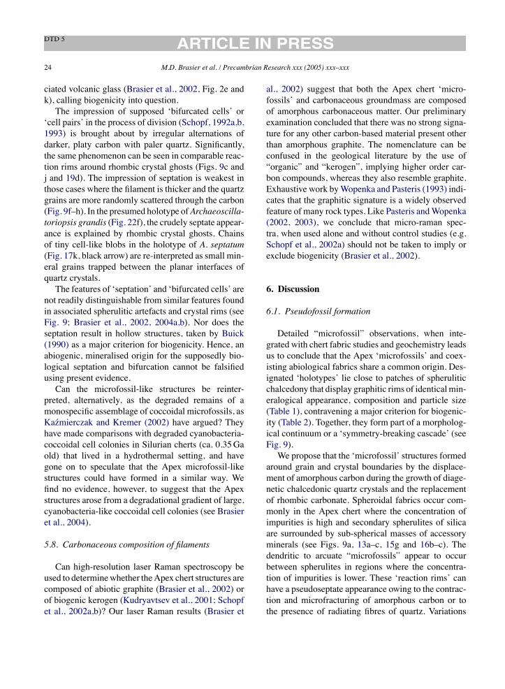
ciated volcanic glass (Brasier et al., 2002, Fig. 2e andk), calling biogenicity into question.The impression of supposed ‘bifurcated cells’ or
‘cell pairs’ in the process of division (Schopf, 1992a,b,1993) is brought about by irregular alternations ofdarker, platy carbon with paler quartz. Significantly,the same phenomenon can be seen in comparable reac-tion rims around rhombic crystal ghosts (Figs. 9c andj and 19d). The impression of septation is weakest inthose cases where the filament is thicker and the quartzgrains are more randomly scattered through the carbon(Fig. 9f–h). In the presumed holotype ofArchaeoscilla-toriopsis grandis (Fig. 22f), the crudely septate appear-ance is explained by rhombic crystal ghosts. Chainsof tiny cell-like blobs in the holotype of A. septatum(Fig. 17k, black arrow) are re-interpreted as small min-eral grains trapped between the planar interfaces ofquartz crystals.The features of ‘septation’ and ‘bifurcated cells’ are
not readily distinguishable from similar features foundin associated spherulitic artefacts and crystal rims (seeFig. 9; Brasier et al., 2002, 2004a,b). Nor does theseptation result in hollow structures, taken by Buick(1990) as a major criterion for biogenicity. Hence, anabiogenic, mineralised origin for the supposedly bio-logical septation and bifurcation cannot be falsifiedusing present evidence.Can the microfossil-like structures be reinter-
preted, alternatively, as the degraded remains of amonospecific assemblage of coccoidal microfossils, asKazmierczak and Kremer (2002) have argued? Theyhave made comparisons with degraded cyanobacteria-coccoidal cell colonies in Silurian cherts (ca. 0.35Gaold) that lived in a hydrothermal setting, and havegone on to speculate that the Apex microfossil-likestructures could have formed in a similar way. Wefind no evidence, however, to suggest that the Apexstructures arose from a degradational gradient of large,cyanobacteria-like coccoidal cell colonies (see Brasieret al., 2004).
5.8. Carbonaceous composition of filaments
Can high-resolution laser Raman spectroscopy beused to determinewhether theApex chert structures arecomposed of abiotic graphite (Brasier et al., 2002) orof biogenic kerogen (Kudryavtsev et al., 2001; Schopfet al., 2002a,b)? Our laser Raman results (Brasier et
al., 2002) suggest that both the Apex chert ‘micro-fossils’ and carbonaceous groundmass are composedof amorphous carbonaceous matter. Our preliminaryexamination concluded that there was no strong signa-ture for any other carbon-based material present otherthan amorphous graphite. The nomenclature can beconfused in the geological literature by the use of“organic” and “kerogen”, implying higher order car-bon compounds, whereas they also resemble graphite.Exhaustive work byWopenka and Pasteris (1993) indi-cates that the graphitic signature is a widely observedfeature of many rock types. Like Pasteris andWopenka(2002, 2003), we conclude that micro-raman spec-tra, when used alone and without control studies (e.g.Schopf et al., 2002a) should not be taken to imply orexclude biogenicity (Brasier et al., 2002).
6. Discussion
6.1. Pseudofossil formation
Detailed “microfossil” observations, when inte-grated with chert fabric studies and geochemistry leadsus to conclude that the Apex ‘microfossils’ and coex-isting abiological fabrics share a common origin. Des-ignated ‘holotypes’ lie close to patches of spheruliticchalcedony that display graphitic rims of identicalmin-eralogical appearance, composition and particle size(Table 1), contravening a major criterion for biogenic-ity (Table 2). Together, they form part of a morpholog-ical continuum or a ‘symmetry-breaking cascade’ (seeFig. 9).We propose that the ‘microfossil’ structures formed
around grain and crystal boundaries by the displace-ment of amorphous carbon during the growth of diage-netic chalcedonic quartz crystals and the replacementof rhombic carbonate. Spheroidal fabrics occur com-monly in the Apex chert where the concentration ofimpurities is high and secondary spherulites of silicaare surrounded by sub-spherical masses of accessoryminerals (see Figs. 9a, 13a–c, 15g and 16b–c). Thedendritic to arcuate “microfossils” appear to occurbetween spherulites in regions where the concentra-tion of impurities is lower. These ‘reaction rims’ canhave a pseudoseptate appearance owing to the contrac-tion and microfracturing of amorphous carbon or tothe presence of radiating fibres of quartz. Variations

M.D.Brasieretal./Precam
brianResearch
xxx(2005)xxx–xxx
25
Table 2Criteria previously used for determining the antiquity and biogenicity of putative microfossils
Antiquity Biogenicity
Contextural characteristics required byfeatures to be considered ‘ancient’
Present or absent—petrographic detailsexhibited by specific structures in opticalthin section
Contextural characteristics required byfeatures to be considered ‘biogenic’
Petrographic details exhibited by specificstructures in optical thin section
Occur in rocks of known provenance,confirmed by replicate sampling
Structures should not be enclosed inmetastable mineral phases (e.g.amorphous opaline silica)
In petrographic thin section“microfossil” like objects should be theprimary source of palaeontological data,with biogenic authenticity verified by useof independent analytical techniques
Relatively abundant occurrence andmem-bers of a multi-component biologicassemblage
Occur in rocks of established Archaeanage
Structures should occur in randomlyextinguishing micro-quartz crystals
In petrographic thin section Archaean“microfossil” like objects should exhibitall the characteristics listed right
Of carbonaceous composition or, if min-eral, be a result of biologically-mediatedmineral encrustation or a product of min-eral replacement
Demonstrably indigenous to the primarydeposition of the enclosing rock
Structures should not occur in second-generation void-filling minerals (e.g.botryoidal chalcedony), or radially extin-guishing micro-quartz after fibrous chal-cedony
Exhibit “biological morphology” -characterized by a range of (statisticallydemonstrable) variability (rather thanuniformity), including life cycle variants,comparable to that exhibited bymorphologically similar modern and/orfossil micro-organisms
Demonstrably syngenetic with theprimary deposition of the enclosingrock
“Microfossils” from planar,infilled-mineral veins, or concentrated atmineral grain boundaries rather thanalong primary depositional laminae, orexhibiting cross-cutting relationships inwhich they have disrupted primaryfeatures of the lithified rock fabric aresubject to question
Occur in a geological context plausible forlife
Precambrian “microfossils” shouldexhibit a significantly different colourfrom that of the particulate finelylaminated kerogenous component of therock matrix
Fit within a well-established evolutionarycontext
Filaments (or colonial unicells) shouldexhibit an orientation and distributionindicative of their role in the formation ofstromatolitic laminae
Be dissimilar from potentially coexistingabiologic organic bodies (e.g. proteinoidmicrospheres, carbonaceous “organizedelements”, and products of organicsyntheses)
Compiled from discussions in Awramik et al. (1983, 1988), Bridgewater et al. (1981), Buick (1988, 1990), Cloud andMorrison (1979), Marshall et al. (1964), Schopf (1975), Schopfand Walter (1983), Tyler and Barghoorn (1963).

26 M.D. Brasier et al. / Precambrian Research xxx (2005) xxx–xxx
in the size and shape of the filaments (Schopf, 1993)are attributed to localised differences in primary tex-ture, spherulite size and shape. We suggest the Apexcherts can be generated by the following ‘substrateneutral’ algorthythmic process: (1)mix one amorphousmineral within another amorphous mineral (e.g. amor-phous carbon within silica glass); (2) allow the second(host) mineral to crystallise out as spherules, vario-lites, rhombs or other crystal forms, around discretenuclei; (3) expel the first amorphous mineral (carbon)as filamentous reaction rims around the newly form-ing spherules or rhombs; (4) allow the first mineral tobecome interspersed with the second mineral, to formpseudoseptate reaction rims.A full taxonomic re-description and re-inter-
pretation of each of the figured ‘microfossil’ holo-types will be given by us elsewhere (Brasier et al., inpress). Of the 11 holotypes of prokaryotic ‘microfos-sils’ defined from the Apex chert (Schopf and Packer,1987; Schopf, 1992b, 1993), we regard those of E.apex (Figs. 9k and 19f) and A. septatum (Fig. 17k)to be mineral rims that formed around sub-rhombiccrystal margins, while the other nine can be explainedas arcuate, sinuous and branched mineral rims ofspherulitic origin. We point out that similar (but non-septate) filamentous pseudofossils are mimicked bypencil graphite found artificially infilling the arcuatefractures around chalcedony spherulites near figuredspecimens (Fig. 10h). The model we propose above isclose to those suggested for pseudomicrofossils fromother Archaean and Proterozoic horizons (Buick et al.,1981; Buick, 1988, 1992).
6.2. Criteria for biogenicity
While Proterozoic rocks are full of microfossilswhose biogenicity cannot be seriously challenged, thesituation in Archean rocks is less straightforward. InTable 2, we list major criteria previously suggestedfor testing both the age and biogenicity of putativeArchaeanmicrofossils. The comprehensive scheme putforward by Schopf and Walter (1983) rightly held amajor place in such discussions during the 1980’s.This scheme was abbreviated and adapted for a seriesof later papers describing putative microfossils fromthe Warrawoona Group (Awramik et al., 1983, 1988),though the biogenicity of these ‘microfossils’ washotly disputed by Buick (1984, 1988, 1990). In recent
years, these criteria and their associated debates havetended to slip from view. Many other early Archaean‘microfossils’ that have been published over the lastdecade (e.g. Walsh, 1992; Rasmussen, 2000; Westallet al., 2001; Schopf et al., 2002a; Furnes et al., 2004)are promising and worthy of re-examination (see alsoWestall and Folk, 2003). Here, we suggest that the test-ing of biological signals in very ancient rocks addressthree main criteria: geological context, biology-likemorphology and biology-like processing.
6.2.1. Geological contextThe context for potential biosignals should be
mapped and studied at a wide range of scales fromoutcrop to thin section. Evidence for, and interpreta-tion for, the context should be clearly separated. Anypotential biological signals should be referable to awell-defined history within this context, to show if theyare early or late, indigenous or exogenous. Plausibilityof the context for early life (e.g. temperature limits) canthen be assessed.
6.2.2. Biology-like morphologyIdeally, there should be evidence for biology-
like morphology in the form of cell aggregates, cellenvelopes, microborings or waste products. Theseshould occupy a restrictedmorphospace that pertains tobiology alone, and not occupy themorphospace for dis-tinct categories of abiological pseudofossils. The nullhypothesis (of an abiological origin for this morphol-ogy) should be falsified. While biology-like morphol-ogy of this kind is highly desirable, it overlooks thedifficulty of distinguishing the earliest true cells fromtheir prebiotic precursors, and should only be obliga-tory for microfossils themselves.
6.2.3. Biology-like processingEvidence for ancient, biology-like processing in
space and time should be put forward for testing. Morethan a single tier of biology-like processing ought tobe present. These steps may take the form of extracel-lular polymeric substances (EPS) plus organominerals(Perry et al., 2004) that show micro-tiering or zona-tion in morphology and geochemistry. Petrographic‘thin sections’ seem likely to remain fundamental tothe mapping of such potential bioproducts. We empha-size that these multiple lines of evidence should beavailable (as with the Schopf, 1993 material) for sci-
M.D. Brasier et al. / Precambrian Research xxx (2005) xxx–xxx 27
entific loan, for scientifically repeated tests, and forpublic scrutiny. The null hypothesis (of an abiogenicorigin) should be addressed and falsified in all casehistories.
7. Conclusions
Our study of the Apex chert ‘microfossils’ revealsthat the majority is part of a morphological contin-uum (a ‘symmetry-breaking cascade’) ranging fromspheroidal through bulbous and radiating to arcuate andbranching forms (cf. Buick, 1990). Credence has beengiven to the Apex filamentous ‘microfossils’ becausethey are claimed to meet all of the criteria requiredof bona fide authentic Archaean microfossils (Schopf,1992b, 1993, 1999). We do not accept this to be true.Nor do we accept that filamentous structures describedas ‘microfossils’ from the Apex chert are demonstra-bly biogenic. They fail to meet a wide range of cri-teria for biogenicity and their occurrence in a geo-logically plausible context (especially for cyanobac-teria) is doubtful. They intergrade morphologicallywith a range of coexisting abiological bodies, mainlyformed as reaction rims around chalcedony spherulitesor crystal (e.g. barite) margins during silification anddiagenesis.We do not accept that the filamentous structures
described from the Apex chert are biogenic micro-fossils, including cyanobacteria, for the followingreasons:
(1) Mapping (Figs. 3–6) shows that putative‘microfossil’-bearing clasts were formed by frac-turing and pulverization of earlier fissure-fillingdeposits and chert, and consistent with formationas hydrobreccia under hydrothermal conditions(Brasier et al., 2002).
(2) More than half of the previously illustrated spec-imens (Schopf, 1993) occur in fabrics mapped aslate stage fissure-filling and secondary chalcedony(Table 1).
(3) The filaments are distributed in a way that is con-sistent with their formation as secondary artefactsof crystal growth (Figs. 8 and 9).
(4) The filaments are not hollow but composed of solidto discontinuous carbonaceous material, wrappedaround quartz crystals.
(5) Arcuate and branched filamentous are reinter-preted as mineral ‘reaction rims’ formed by dis-placement and/or accretion of mineral impuritiesduring the devitrification and recrystallization ofhydrothermal silica (Fig. 9), and compare withother well-known pseudofossils from Archaeanand Proterozoic rocks (Horodyski, 1981; Buicket al., 1981; Buick, 1988, 1992). Septation andfeatures regarded as cells in the process of cell divi-sion (Schopf, 1992b, 1993) are both explained bythe interleaving of accessory minerals with quartzcrystallites within reaction rims.
Ancient filamentous structures should not beaccepted as of biological origin until all possibilitiesof their non-biological origin have been exhausted(Brasier et al., 2002, 2004a,b). Complex structurescan result from simple physical processes (Goodwin,1994). Reinterpretation of the Warrawoona structuresas pseudofossils raises serious questions about the pres-ence of a cyanobacterial biota at 3.46Ga. Instead, wefind evidence consistent with a hydrothermal settingfor the Apex cherts at Chinaman Creek. While carbonisotopic values (Fig. 6C) have traditionally been inter-preted as indicating a significant biological contribu-tion to the carbon cycle, perhaps from otherwise unpre-served hyperthermophile bacteria like those thoughtto occur as microfossils in younger Archean rocks(Rasmussen, 2000;Westall et al., 2001), estimated tem-peratures of ca. 250–350 #Ccould imply a lethal habitatat times. We suggest that redox buffers such as nativemetals, sulphides and iron oxides and present in abun-dance within the carbonaceous dyke cherts (Fig. 6B)could have encouraged the transformation of CO2 intoorganic compounds via Fischer-Tropsch type synthe-sis (cf. Holm and Charlou, 2001). Such compounds,together with freely available sulphur species and H2,conceivably provided the carbon and energy sourcesfor hyperthermophile bacteria (cf. Holm and Charlou,2001). This hypothesis is consistentwith evidence frombacterial rRNAsequencing,which indicates that hyper-thermophile methanogenic archaebacteria branchmostdeeply in the tree of life and are arguably of muchgreater antiquity than cyanobacteria (Hedges et al.,2001). The Apex chert may provide evidence for a cra-dle of life, nurtured not by the energy of sunlight but bythe thermal energy and mineral resources of the earlycrust.
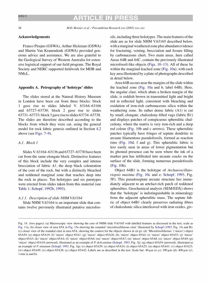
28 M.D. Brasier et al. / Precambrian Research xxx (2005) xxx–xxx
Acknowledgements
Franco Pirajno (GSWA), Arthur Hickman (GSWA)and Martin Van Kranendonk (GSWA) provided gen-erous advice and assistance. We are also grateful tothe Geological Survey of Western Australia for exten-sive logistical support of our field program. The RoyalSociety and NERC supported fieldwork for MDB andNMcL.
Appendix A. Petrography of ‘holotype’ slides
The slides stored at the Natural History Museumin London have been cut from three blocks: block1 gave rise to slides labeled V. 63164–63166and 63727–63730; block 2 gave rise to slides63731–63733; block3gave rise to slides 63734–63738.The slides are therefore described according to theblocks from which they were cut, using the generalmodel for rock fabric genesis outlined in Section 4.2above (see Figs. 7–9).
A.1. Block 1
SlidesV. 63164–63136 and 63727–63730 have beencut from the same elongate block. Distinctive featuresof this block include the very complex and intensebrecciation of fabric A1, the deep black colourationof the core of the rock, but with a distinctly bleachedand reddened marginal zone that reaches deep intothe rock in places. Ten holotypes and six paratypeswere erected from slides taken from this material (seeTable 1; Schopf, 1992b, 1993).
A.1.1. Description of slide NHM V.63164Slide NHM V.63164 is an important slide that con-
tains twelve previously illustrated putative microfos-
sils, including three holotypes. Themain features of theslide are as for slide NHM V.63165 described below,with amarginalweathered zoneplus abundant evidencefor fracturing, veining, brecciation and fissure fillingby carbonaceous chert. Two main areas, here calledAreas 64B and 64C, contain the previously illustratedmicrofossil-like objects (Figs. 10–13). All of these liewithin the marginal leached zone (Fig. 10a), with eachkey area illustrated by a plate of photographs describedin detail below.Area 64B occurs near themargins of the slidewithin
the leached zone (Fig. 10a and b, label 64B). Here,the angular clast, which abuts a broken margin of theslide, is reddish brown in transmitted light and brightred in reflected light, consistent with bleaching andoxidation of iron-rich carbonaceous silica within theweathering zone. Its rather dense fabric (A1) is cutby small, elongate, chalcedony-filled vugs (fabric B1)and displays patches of conspicuous spherulitic chal-cedony, where the matrix is very iron-rich and a deepred colour (Fig. 10b and c arrows). These spheruliticpatches typically have fringes of septate dendritic toarcuate filamentous pseudofossils, formed as reactionrims (Fig. 10d, f and g). This spherulitic fabric isless easily seen in areas of lower pigmentation butits ghosted presence can be seen where the ink of amarker pen has infiltrated into arcuate cracks on thesurface of the slide, forming numerous pseudofossils(Fig. 10h).Object 64B1 is the holotype of Archaeoscillato-
riopsis maxima (Fig. 10c and e; Schopf, 1993, Fig.5F). This pseudoseptate arcuate structure lies imme-diately adjacent to an artefact-rich patch of reddenedspherulites. Geochemical analysis (SEM/EDX) showsthat the ‘holotype’ is indistinguishable in mineralogyfrom the adjacent spherulitic mass. The septate fab-ric of object 64B1 clearly preserves radiating fibresof chalcedonic silica interleaved with iron oxides, giv-
Fig. 14. (two pages): (a) Macroscopic view showing the core of NHM slide V.63165 with labelled features as discussed in the text, scale asFig. 11a; (b) closer view of area 65A in Fig. 15a showing the rounded ‘microfossiliferous clast’ illustrated by Schopf (1993, Fig. 3A and B);(c) closer view of the rounded clast in area 65A, showing the context for the objects shown in (d–p). (d) ‘Microfossiliferous’ (‘micro’) object65A54; (e) object 65A19; (f) ‘micro’ object 65A1; (g) ‘micro’ object 65A26; (h) ‘micro’ object 65A4; (i) ‘micro’ object 65A53; (j) ‘micro’object 65A3; (k) ‘micro’ object 65A4; (l) ‘micro’ object 65A6; (m) ‘micro’ object 65A7; (n) ‘micro’ object 65A8; (o) ‘micro’ object 65A9; (p)‘micro’ object 65A18 (arrowed), illustrated as an example of P. delicatulum (Schopf, 1993, Fig. 3j); (q) object 65A54 (arrowed), illustrated asan example of P. minutum (Schopf, 1993, Fig. 3q); (r) object 65A29; (s) object 65A24; (t) object 65A25; (u) object 65A41; (v) object 65A23;(w) object 65A49; (x) object 65A38; (y) object 65A42. Labels are as described in the text. Scale bar: 40!m (e–y); 100!m (d); 400!m (c),1mm (a and b).
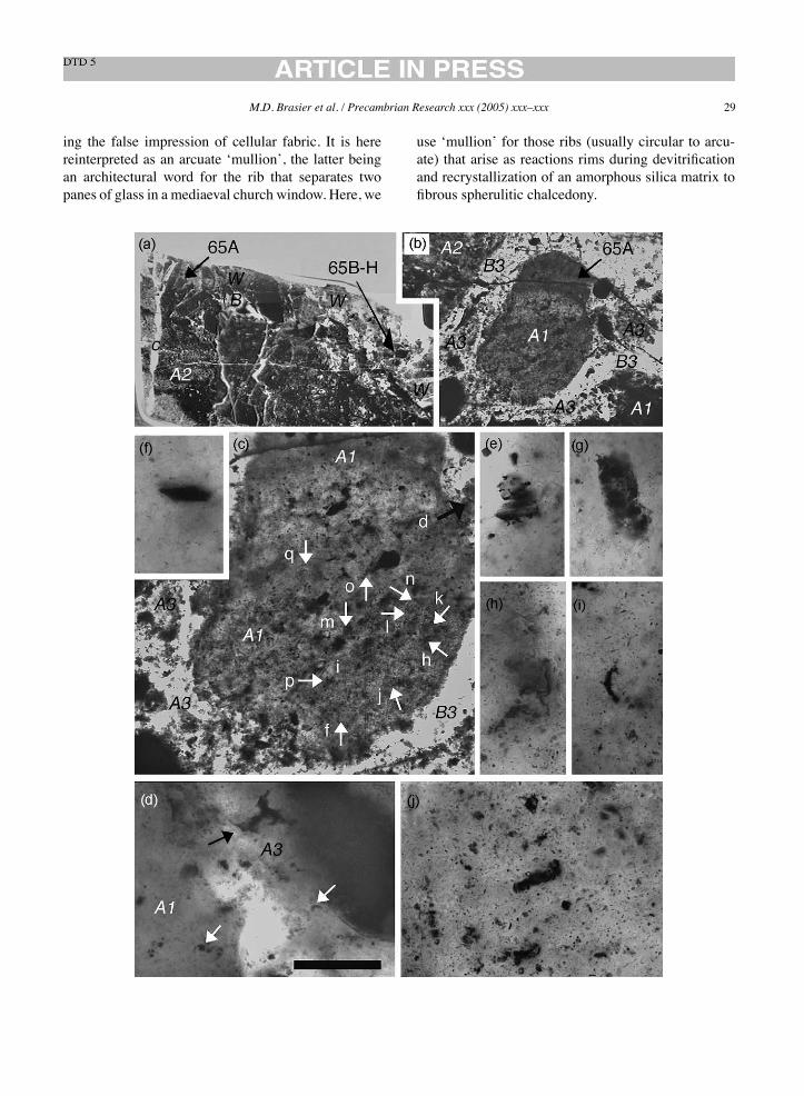
M.D. Brasier et al. / Precambrian Research xxx (2005) xxx–xxx 29
ing the false impression of cellular fabric. It is herereinterpreted as an arcuate ‘mullion’, the latter beingan architectural word for the rib that separates twopanes of glass in a mediaeval church window. Here, we
use ‘mullion’ for those ribs (usually circular to arcu-ate) that arise as reactions rims during devitrificationand recrystallization of an amorphous silica matrix tofibrous spherulitic chalcedony.
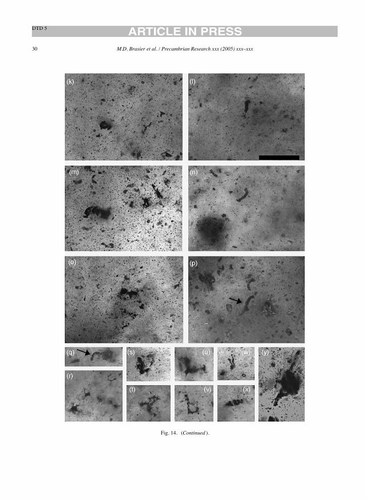
30 M.D. Brasier et al. / Precambrian Research xxx (2005) xxx–xxx
Fig. 14. (Continued ).
M.D. Brasier et al. / Precambrian Research xxx (2005) xxx–xxx 31
Area 64C within the slide (Fig. 11a) contains manyof the figured ‘microfossils’ including three holotypesand five paratypes, and comprises an angular, polygo-nal clast of fluffy fabric A1 (Fig. 11b–f). Onemargin ofthe clast (close to label f in Fig. 11a) comprises an espe-cially dark, carbonaceous fluffy fabric that can be seento spall into grainy fabrics A2 and A3, forming smallvoids between the grains that are infilled with botry-oidal chalcedony (Fig. 13a–d). These botryoids showclear arcuate reaction rims. Similar botryoidal cementsinfiltrate numerous irregular fractures within the mainbody of the clast and have helped to form the ubiq-uitous spheroidal to arcuate mullions of carbonaceousmatter, through the process of reaction rim growth dur-ing devitrification and recrystallization of a silicified,glassy protolith. It is this process which has given riseto the numerous pseudofossils described below.The range of microfossil-like structures from clast
C is shown in Figs. 11 and 12. Filamentous objects arescattered through the clast (Figs. 11d and 12e) and arecommonest near to patches of carbonaceous spheroidalchalcedony. The forms range widely in shape fromcomplex dendritic forms (Fig. 12g, j, l, n and o) throughserpentine forms (Fig. 12k and l) to common arcuateforms that may appear septate (Figs. 11g and i and12a–c), diffuse (Fig. 12d) or solid (Fig. 11h–i). Theappearance of septation is due to scattered crystals ofquartz within the carbonaceous matter that is fibrous(e.g. Fig. 12o) or equant and irregular (e.g. Figs. 11gand 12f,k and l). A smaller number of microfossil-likeobjects arise from incomplete carbonaceous reactionrims that have grown around the margins of rhombiccrystal ghosts (Fig. 12h). These artefacts include threeillustrated holotypes:Primaevifilum conicoterminatumholotype (64C2, Schopf, 1992b, Fig. 1.5.4C; Schopf,1993, Fig. 4H): Primaevifilum laticellulosum holotype(64C19; Schopf, 1993, Fig. 5A); five paratypes: P.amoenum paratype (64C21; Schopf, 1992a, Fig. 2.5C;1992b, Fig. 1.5.5C); P. amoenum paratype (64C20;Schopf, 1992a, Fig. 2.5E; Schopf, 1992b, Fig. 1.5.5E;Schopf, 1993, Fig. 4B), P. conicoterminatum paratype(64CplanM3; Schopf, 1992b, Fig. 1.5.4D), P. delicat-ulum paratype (Schopf, 1992b, Fig. 1.5.4H; Schopf,1993, Fig. 3K), P. delicatulum paratype (64CplanM32;Schopf, 1992b, Fig. 1.5.4F), plus figured examples ofP. conicoterminatum (64D1: Schopf, 1993, Fig. 4G;Schopf, 1993, Fig. 4D), P. laticellulosum (Schopf,1993, Fig. 5B), un-named ‘Type A’ broad filament
(64C1; Schopf, 1992b, Fig. 1.5.6A) and un-named‘Type C’ narrow filament (64C30; Schopf, 1992b, Fig.1.5.6D).
A.1.2. Description of slide NHM V.63165 (Area A)Slide NHM V.63165 contains nine previously illus-
trated putative microfossils, including one holotype (P.delicatulum), one paratype (P. amoenum) and threeclasts labeled as ‘microfossiliferous’ (Schopf, 1992b,Fig. 1.5.4A, 1993, Fig. 3A and B). On one side ofthe slide (nearest to the larger label, and at the top ofFig. 14a), there is a conspicuous weathered marginalzone that cuts across all fabrics (Fig. 14a, label W).This leached margin tends to be pale grey to white inreflected and there is conspicuously reddened, oxidisedzone where it abuts against the darker and more car-bonaceous rock light, consistent with bleaching of thecarbonaceous matter. All the ‘microfossil-like’ objectsdescribed below come from zones where this carbonhas been partly or largely leached. The core of the slide(Fig. 14a, lower edge) consists of brecciated and angu-lar shards of relatively compact grey to black fabricA1, often with jigsaw-puzzle fit, set within a matrixof paler, less compact, fissure-filling fabrics of gener-ations A2 and A3 (e.g. Fig. 14a–c, labels A1, A2, A3).Angular grains of laminated chert can also occurwithinfabric A1 and resemble microclasts of fractured veinchert. Less opaque areas within fabric A1 can revealgrey or brown chert with a mottled texture of scatteredsmall elliptical grains, layered sericite flakes and ironoxide wisps, suggestive of a volcanic or volcanoclas-tic protolith that has been secondarily silicified. Theserather homogenous clasts of fabric A1 can be black-ened (‘melanized’) around their margins and along thinfractures that penetrate into the interior of the clast. Themajority of the clasts of A1 fabric appear to have beenpervasively melanized in this way.In places, shards of fabric A1 are clearly seen to
spall at themargins via sheet-like or irregular (‘circum-granular’) cracks, to form a dense to loosely packed,unsorted microbreccia of fabric A2, comprising palegrey to black grains of angular to subrounded, lobateor elliptical shape, plus sparse angular to subroundedgrains of clear and laminated vein chert (fabric B1)and conspicuous grains of pyrite. These are set withina clear chert matrix that in places can be traced laterallyinto elongate vugs lined with layers of clear botry-oidal chalcedony (Fig. 15a and b, fabric B2). There
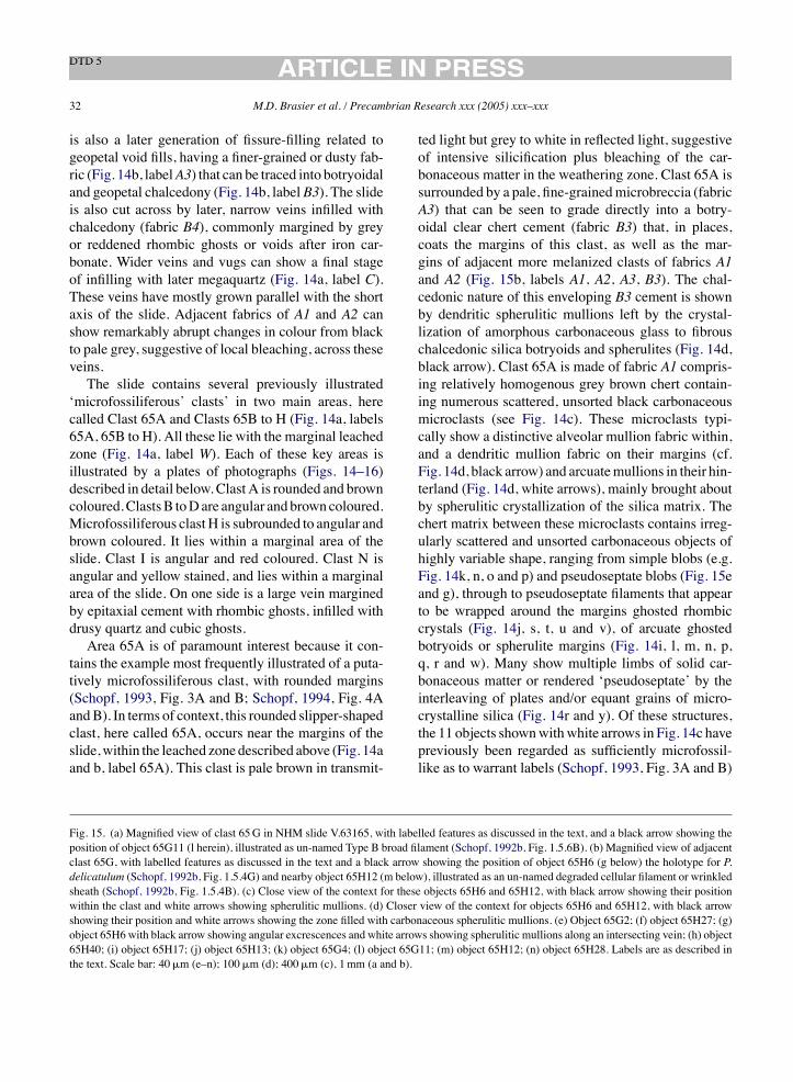
32 M.D. Brasier et al. / Precambrian Research xxx (2005) xxx–xxx
is also a later generation of fissure-filling related togeopetal void fills, having a finer-grained or dusty fab-ric (Fig. 14b, labelA3) that can be traced into botryoidaland geopetal chalcedony (Fig. 14b, label B3). The slideis also cut across by later, narrow veins infilled withchalcedony (fabric B4), commonly margined by greyor reddened rhombic ghosts or voids after iron car-bonate. Wider veins and vugs can show a final stageof infilling with later megaquartz (Fig. 14a, label C).These veins have mostly grown parallel with the shortaxis of the slide. Adjacent fabrics of A1 and A2 canshow remarkably abrupt changes in colour from blackto pale grey, suggestive of local bleaching, across theseveins.The slide contains several previously illustrated
‘microfossiliferous’ clasts’ in two main areas, herecalled Clast 65A and Clasts 65B to H (Fig. 14a, labels65A, 65B to H). All these lie with the marginal leachedzone (Fig. 14a, label W). Each of these key areas isillustrated by a plates of photographs (Figs. 14–16)described in detail below. Clast A is rounded and browncoloured.ClastsB toDare angular andbrowncoloured.Microfossiliferous clastH is subrounded to angular andbrown coloured. It lies within a marginal area of theslide. Clast I is angular and red coloured. Clast N isangular and yellow stained, and lies within a marginalarea of the slide. On one side is a large vein marginedby epitaxial cement with rhombic ghosts, infilled withdrusy quartz and cubic ghosts.Area 65A is of paramount interest because it con-
tains the example most frequently illustrated of a puta-tively microfossiliferous clast, with rounded margins(Schopf, 1993, Fig. 3A and B; Schopf, 1994, Fig. 4AandB). In terms of context, this rounded slipper-shapedclast, here called 65A, occurs near the margins of theslide,within the leached zone described above (Fig. 14aand b, label 65A). This clast is pale brown in transmit-
ted light but grey to white in reflected light, suggestiveof intensive silicification plus bleaching of the car-bonaceous matter in the weathering zone. Clast 65A issurrounded by a pale, fine-grainedmicrobreccia (fabricA3) that can be seen to grade directly into a botry-oidal clear chert cement (fabric B3) that, in places,coats the margins of this clast, as well as the mar-gins of adjacent more melanized clasts of fabrics A1and A2 (Fig. 15b, labels A1, A2, A3, B3). The chal-cedonic nature of this enveloping B3 cement is shownby dendritic spherulitic mullions left by the crystal-lization of amorphous carbonaceous glass to fibrouschalcedonic silica botryoids and spherulites (Fig. 14d,black arrow). Clast 65A is made of fabric A1 compris-ing relatively homogenous grey brown chert contain-ing numerous scattered, unsorted black carbonaceousmicroclasts (see Fig. 14c). These microclasts typi-cally show a distinctive alveolar mullion fabric within,and a dendritic mullion fabric on their margins (cf.Fig. 14d, black arrow) and arcuatemullions in their hin-terland (Fig. 14d, white arrows), mainly brought aboutby spherulitic crystallization of the silica matrix. Thechert matrix between these microclasts contains irreg-ularly scattered and unsorted carbonaceous objects ofhighly variable shape, ranging from simple blobs (e.g.Fig. 14k, n, o and p) and pseudoseptate blobs (Fig. 15eand g), through to pseudoseptate filaments that appearto be wrapped around the margins ghosted rhombiccrystals (Fig. 14j, s, t, u and v), of arcuate ghostedbotryoids or spherulite margins (Fig. 14i, l, m, n, p,q, r and w). Many show multiple limbs of solid car-bonaceous matter or rendered ‘pseudoseptate’ by theinterleaving of plates and/or equant grains of micro-crystalline silica (Fig. 14r and y). Of these structures,the 11 objects shownwithwhite arrows in Fig. 14c havepreviously been regarded as sufficiently microfossil-like as to warrant labels (Schopf, 1993, Fig. 3A and B)
Fig. 15. (a) Magnified view of clast 65G in NHM slide V.63165, with labelled features as discussed in the text, and a black arrow showing theposition of object 65G11 (l herein), illustrated as un-named Type B broad filament (Schopf, 1992b, Fig. 1.5.6B). (b) Magnified view of adjacentclast 65G, with labelled features as discussed in the text and a black arrow showing the position of object 65H6 (g below) the holotype for P.delicatulum (Schopf, 1992b, Fig. 1.5.4G) and nearby object 65H12 (m below), illustrated as an un-named degraded cellular filament or wrinkledsheath (Schopf, 1992b, Fig. 1.5.4B). (c) Close view of the context for these objects 65H6 and 65H12, with black arrow showing their positionwithin the clast and white arrows showing spherulitic mullions. (d) Closer view of the context for objects 65H6 and 65H12, with black arrowshowing their position and white arrows showing the zone filled with carbonaceous spherulitic mullions. (e) Object 65G2; (f) object 65H27; (g)object 65H6 with black arrow showing angular excrescences and white arrows showing spherulitic mullions along an intersecting vein; (h) object65H40; (i) object 65H17; (j) object 65H13; (k) object 65G4; (l) object 65G11; (m) object 65H12; (n) object 65H28. Labels are as described inthe text. Scale bar: 40!m (e–n); 100!m (d); 400!m (c), 1mm (a and b).
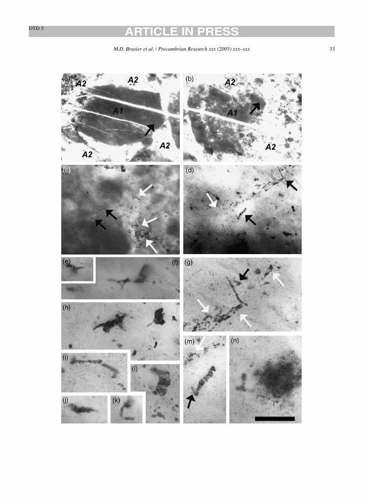
M.D. Brasier et al. / Precambrian Research xxx (2005) xxx–xxx 33
34 M.D. Brasier et al. / Precambrian Research xxx (2005) xxx–xxx
M.D. Brasier et al. / Precambrian Research xxx (2005) xxx–xxx 35
or Linnaean names. Objects 65A18 (Fig. 14p, arrowed)has been regarded as an example of P. delicatulum(Schopf, 1993, Fig. 3j) and occurs alongside an identi-cal arcuate structure. Object 65A54 (Fig. 14q, arrowed)has been regarded as an example ofPrimaevifilummin-utum (Schopf, 1993, Fig. 3q).
A.1.3. Description of slide NHM V.63165 (Areas Bto H)Areas B to H occurs towards the right of the slide,
againwithin themarginal bleached zone (Fig. 14a, label65B to H in area label W). Here is found a cluster ofmainly angular to subangular clasts of fabric A1, manyof which have been lithified in the process of beingfractured and broken apart, to be infilled by clear chertveins of fabric B2. These clasts are enclosed withinfissure-filling fabric A2 and/or associated botryoidalsilica of fabric B2, which has a later void-filling phaseofmegaquartz. Of these, two clasts here called 65G and65H have previously been illustrated as ‘microfossilif-erous’ (Fig. 15a and b; see Schopf, 1992b, Fig. 1.5.4A).These comprises subangular shard of grey-brown fab-ric A1, fractured by numerous small, straight, anasto-mosing and arcuate veinlets of fabric B2. The marginsof these chert veins can be preferentially coated withcarbonaceous matter (e.g. clast 65I). The wider exam-ples of these chert-filled fractures can contain floatingmasses of carbonaceousmatter that has crystallised intoa distinctive, alveolar mullion fabric (caused by thecrystallization of an amorphous glassy silica phase toform small spherulites of chalcedonic silica surroundedby reaction rims of carbon). Similar spherulitic carbonstructures are also seen around themargins of the veins,near the edges of the clasts and within the surround-ing fissure filling of fabric A2. It is notable that themicrofossil-like structures appear within these areas ofspherulitic mullion fabric.
Clast 65G is a subprismoidal, subangular grain ofrather homogenous, grey fabric A1 (Fig. 15a). It con-tains abundant scattered grains of sericite, suggestive ofa volcanic or volcanoclastic protolith, and is cut acrossby two parallel, late stage veins of clear chert. Its leftmargin appears to spall into smaller clasts of fabricA2 (Fig. 15a). The range of carbonaceous objects ismuch as in clast 65A described above, and includespseudoseptate dendritic mullions (Fig. 15e) and angu-lar crystal rims (Fig. 15k). Object 65G11 (Fig. 15l) isa grey-brown structure with an iron stained microfab-ric of mesh-like lamellae interleaved with larger quartzgrains, and has been described as an ‘un-named TypeB broad filament’ (Schopf, 1992b, Fig. 1.5.6B).Clast 65H occurs immediately to the right of clast
65G. This near-equant, subangular to subrounded grainof grey fabric A1 (Fig. 15b) is broken into smallerpatches by numerous curving ‘circum-granular’ cracksinfilled with clear chert, and its upper margin appearsto spall into the fissure filling of fabric A2 with scat-tered reaction rims of opaque iron oxide. Clast 65Halso contains scattered grains of sericite, suggestive ofa volcanic or volcanoclastic protolith, and is cut acrossby a late stage vein of clear chert. The carbonaceousstructures described below are sparsest within a zoneabout 200!mwide on either side of this late stage vein.The range of carbonaceous objects seen here is muchas seen in clast 65A described above, and includesblobs, angular crystal rims, arcuate, dendritic and alve-olar mullions (Fig. 15c–j, m, n). The context for twopreviously figured objects (65H6 and 65H12), takento be evidence for early life (Schopf, 1992b, 1993) isshown in Fig. 15c (black arrows). These objects occurwithin a region of the clast that has abundant spheruliticmullion structures (Fig. 15c, d, g, m, white arrows).Object 65H6 (Fig. 15g) is the holotype for P. delicat-ulum (Schopf, 1992b, Fig. 1.5.4G; Schopf, 1993, Fig.
Fig. 16. (a) Magnified view of clasts 65B, 65C and area 65Q in NHM slide V.63165, with labelled features as discussed in the text, and a blackarrow showing the position of (b). (b) Magnified view of area 65Q with spherulitic and botryoidal features discussed in the text and a whitearrow showing the position of Fig. 17c. (c) Closer view of the preceding figure, showing carbonaceous strings (black arrows). (d) Magnified viewclasts 65B, 65C and 65D, with white arrows showing the positions of the next two figures. (e) Object 65D4; (f) object 65B3. (g) Magnified viewof clast 65P and the adjacent area, with labelled features as discussed in the text, and black arrow showing the position of following (h–o). (h)Object 65P1. (i) Object 65P4, illustrated as an un-named unicell-like microfossil (Schopf, 1993, Fig. 5K). (j–l) Four successive images throughthe plane of focus for object 65P4, regarded as an example of A. disciformis (Schopf, 1993, Fig. 3N) but here seen to be much more complex.(n and o) Object 65P3, erected as a paratype for P. amoenum (Schopf, 1992b, Fig. 1.5.5D) showing the side branch and the associated galaxyof pseudoseptate carbonaceous strings and crystal rims. Labels are as described in the text. Scale bar: 40!m (c, e, f, h–o); 100!m (b); 400!m(a and d), 1mm (g).
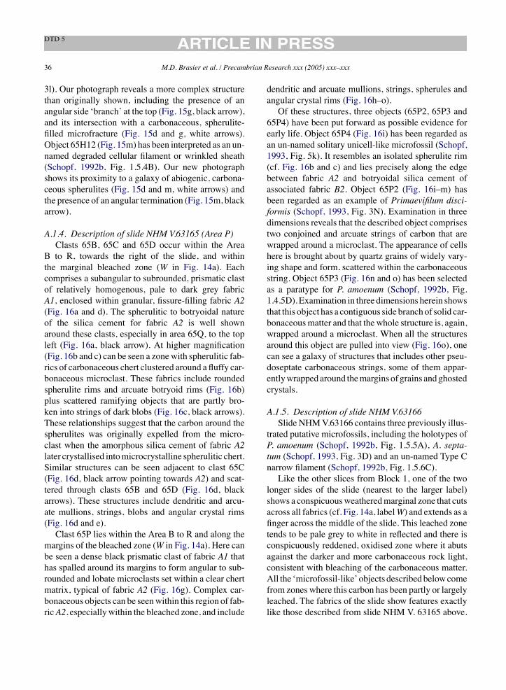
36 M.D. Brasier et al. / Precambrian Research xxx (2005) xxx–xxx
3l). Our photograph reveals a more complex structurethan originally shown, including the presence of anangular side ‘branch’ at the top (Fig. 15g, black arrow),and its intersection with a carbonaceous, spherulite-filled microfracture (Fig. 15d and g, white arrows).Object 65H12 (Fig. 15m) has been interpreted as an un-named degraded cellular filament or wrinkled sheath(Schopf, 1992b, Fig. 1.5.4B). Our new photographshows its proximity to a galaxy of abiogenic, carbona-ceous spherulites (Fig. 15d and m, white arrows) andthe presence of an angular termination (Fig. 15m, blackarrow).
A.1.4. Description of slide NHM V.63165 (Area P)Clasts 65B, 65C and 65D occur within the Area
B to R, towards the right of the slide, and withinthe marginal bleached zone (W in Fig. 14a). Eachcomprises a subangular to subrounded, prismatic clastof relatively homogenous, pale to dark grey fabricA1, enclosed within granular, fissure-filling fabric A2(Fig. 16a and d). The spherulitic to botryoidal natureof the silica cement for fabric A2 is well shownaround these clasts, especially in area 65Q, to the topleft (Fig. 16a, black arrow). At higher magnification(Fig. 16b and c) can be seen a zonewith spherulitic fab-rics of carbonaceous chert clustered around a fluffy car-bonaceous microclast. These fabrics include roundedspherulite rims and arcuate botryoid rims (Fig. 16b)plus scattered ramifying objects that are partly bro-ken into strings of dark blobs (Fig. 16c, black arrows).These relationships suggest that the carbon around thespherulites was originally expelled from the micro-clast when the amorphous silica cement of fabric A2later crystallised intomicrocrystalline spherulitic chert.Similar structures can be seen adjacent to clast 65C(Fig. 16d, black arrow pointing towards A2) and scat-tered through clasts 65B and 65D (Fig. 16d, blackarrows). These structures include dendritic and arcu-ate mullions, strings, blobs and angular crystal rims(Fig. 16d and e).Clast 65P lies within the Area B to R and along the
margins of the bleached zone (W in Fig. 14a). Here canbe seen a dense black prismatic clast of fabric A1 thathas spalled around its margins to form angular to sub-rounded and lobate microclasts set within a clear chertmatrix, typical of fabric A2 (Fig. 16g). Complex car-bonaceous objects can be seenwithin this region of fab-ricA2, especiallywithin the bleached zone, and include
dendritic and arcuate mullions, strings, spherules andangular crystal rims (Fig. 16h–o).Of these structures, three objects (65P2, 65P3 and
65P4) have been put forward as possible evidence forearly life. Object 65P4 (Fig. 16i) has been regarded asan un-named solitary unicell-like microfossil (Schopf,1993, Fig. 5k). It resembles an isolated spherulite rim(cf. Fig. 16b and c) and lies precisely along the edgebetween fabric A2 and botryoidal silica cement ofassociated fabric B2. Object 65P2 (Fig. 16i–m) hasbeen regarded as an example of Primaevifilum disci-formis (Schopf, 1993, Fig. 3N). Examination in threedimensions reveals that the described object comprisestwo conjoined and arcuate strings of carbon that arewrapped around a microclast. The appearance of cellshere is brought about by quartz grains of widely vary-ing shape and form, scattered within the carbonaceousstring. Object 65P3 (Fig. 16n and o) has been selectedas a paratype for P. amoenum (Schopf, 1992b, Fig.1.4.5D). Examination in three dimensions herein showsthat this object has a contiguous side branchof solid car-bonaceousmatter and that the whole structure is, again,wrapped around a microclast. When all the structuresaround this object are pulled into view (Fig. 16o), onecan see a galaxy of structures that includes other pseu-doseptate carbonaceous strings, some of them appar-entlywrapped around themargins of grains andghostedcrystals.
A.1.5. Description of slide NHM V.63166Slide NHMV.63166 contains three previously illus-
trated putative microfossils, including the holotypes ofP. amoenum (Schopf, 1992b, Fig. 1.5.5A), A. septa-tum (Schopf, 1993, Fig. 3D) and an un-named Type Cnarrow filament (Schopf, 1992b, Fig. 1.5.6C).Like the other slices from Block 1, one of the two
longer sides of the slide (nearest to the larger label)shows a conspicuousweatheredmarginal zone that cutsacross all fabrics (cf. Fig. 14a, labelW) and extends as afinger across the middle of the slide. This leached zonetends to be pale grey to white in reflected and there isconspicuously reddened, oxidised zone where it abutsagainst the darker and more carbonaceous rock light,consistent with bleaching of the carbonaceous matter.All the ‘microfossil-like’ objects describedbelowcomefrom zones where this carbon has been partly or largelyleached. The fabrics of the slide show features exactlylike those described from slide NHM V. 63165 above.

M.D. Brasier et al. / Precambrian Research xxx (2005) xxx–xxx 37
These include dense grey to black fabric A1, fracturingand spalling to form generations paler fissure-fillingmicrobreccias of fabrics A2 and A3, associated chertcements that infill fractures and voids (fabrics B1 toB3) plus late stage megaquartz (fabric C).Twomain areas, here called areas 66B and 66F, con-
tain the previously illustrated microfossil-like objectsand both lie with the leached zone. Area 66B liestowards the right hand margin of the slide, and shows aboundary between a subangular, dense black and car-bonaceous shard of fabric A1 which is fractured andspalled on its margins to form the paler, more fluffymicroclasts of fabric A2 (Fig. 17a, black arrow) setwithin a botryoidal silica cement of fabric B2. Thechert matrix between these microclasts contains irreg-ularly scattered and unsorted carbonaceous objects ofhighly variable shape, ranging from simple blobs (e.g.Fig. 17f) or pseudoseptate blobs (Fig. 17e), throughto pseudoseptate filaments that appear to be wrappedaround the margins ghosted botryoids or spherulitemargins to form single arcuate strings (Fig. 17c, d andh), sinuous strings (Fig. 17g) and even rare ‘W’ shapedstrings (Fig. 17c). The false appearance of ‘septation’in these carbonaceous strings is due to the interleavingof silica plates (e.g. Fig. 17d and e) and/or scatteredgrains of microcrystalline silica (e.g. Fig. 17b, c andf). Of these structures, object 66B1 (Fig. 17g) occursbetween microclasts in fabric A2within cement of fab-ric B2 (i.e. it does not occur within a clast). It has beenerected as the holotype forP. amoenum (Schopf, 1992b,Fig. 1.1.5A). It shows a hitherto undescribed solid sidebranch (Fig. 17g, white arrow). Arcuate object 66B5(Fig. 17h), with a similar setting, has been regarded asan example of an un-named Type C narrow filament(Schopf, 1992b, Fig. 1.5.6C).Area 66P is nearby and shows numerous compa-
rable arcuate and branched filaments that occur onlyupon the surface of the slide (Fig. 17l). These filamentshave been formed accidentally from the pencil graphiteused by previous researchers to mark this part of theslide. The graphite has here infilled the arcuate surfacedepressions within the chert, which are brought aboutby its recrystallized, spherulitic fabric. Immersion oilused by us for the high power ($100) microscopeobjectives has caused then these graphite molds to floatabout on the surface of the slide. Note the similarity insize and shape to the associated arcuate pseudofossilsdescribed above.
Area 66F lies near the bottom margin of the slide(Fig. 17i, black arrow), within a region of bleachedchert and very close to a strongly iron-stained crack(Fig. 17i, white arrow). The fabric here is dominated bypale microcrystalline chert of fabric B3 with a scatter-ing of iron oxide grains (Fig. 17j, black arrow) and tiny,reddish-brown, blob-like mineral growths arrangedinto strings. These strings commonly show hints ofa polygonal arrangement, reflecting the margins ofquartz crystal rims (Fig. 17j, white arrows). Of thesestructures, object 66F4 (Fig. 17k, black arrow) has beenerected as the holotype for A. septatum (Schopf, 1993,Fig. 3D). This extremely small object is reddish brown(iron oxide?) in both transmitted and reflected light.It appears to pass, at an angle, into two discontinu-ous strings (Fig. 17k, white arrow). The holotype isnot distinguishable in either form or colour from theassociated crystal rim mineral strings.
A.1.6. Description of slide NHM V.63727The core of NHM slide V. 63727 (Fig. 18a) con-
sists of dark, brecciated and angular shards of fabricA1, often with jigsaw-puzzle fit, set within a flocculentmatrix of fissure-filling of generationsA2 (e.g. Fig. 18b,label A2) and A3. On one side (nearest to the label, andat the top of Fig. 18a) there is a weathered marginalzone of leached, paler fabric A1 to A3 that extends asa distinctive pale ‘finger’ towards the right of the slide(Fig. 18a, labelW). This leached zone in places showsa reddened, oxidised margin where it abuts against thedarker-less-altered fabric. The slide contains numerousirregular vugs that are infilled with chalcedony (fabricB; e.g. Fig. 18a and b, label B). These tend to be elon-gated along the longer axis of the slide. The sample iscross-cut by a number of conspicuous veins filled withchalcedony (fabric B) and by later megaquartz (fabricC). These veins have mostly grown parallel with theshort axis of the slide (Fig. 18a, arrow).Fig. 18c shows a detail of Area C in Fig. 18a. It
shows a shard (ca. 2.86mm$ 2.13mm in diameter)of fabric !A1 (4L) that has highly irregular margins.The shard is deeply invaded by veins and vugs of chal-cedony (Fig. 18c, label B) that mainly lie parallel withthe long (E–W) axis of the slide. The paler colour in thelower part of the shard indicates the marginal zone ofleaching referred to above. Outside of the leached zone,spherulitic fabrics are hard to see because of the dark,almost black fabric. Within the leached zone, however,

38 M.D. Brasier et al. / Precambrian Research xxx (2005) xxx–xxx
Fig. 17. (a) Magnified view of area 66B (west) in slide NHMV.63166, with labelled features as discussed in the text and a black arrow showingthe position of objects in following (b–h). (b) Object 66B4; (c) objects 66B7; (d) objects 66B2; (e) object 66B3; (f) object 66B6; (g) object66B1, previously erected as the holotype for P. amoenum (Schopf, 1992b, Fig. 1.5.5A); (h) object 66B5, previously illustrtated as an exampleof an un-named Type C narrow filament (Schopf, 1992b, Fig. 1.5.6C); (i) magnified view of area 66F (west) with labelled features as discussedin the text, and a black arrow showing the position of objects in (j and k); (j) objects 66F4; (k) object 66F5, previously erected as the holotypefor A. septatum (Schopf, 1993, Fig. 3D). Labels are as described in the text. Scale bar: 40!m (b–h, j–k); 400!m (a and i).
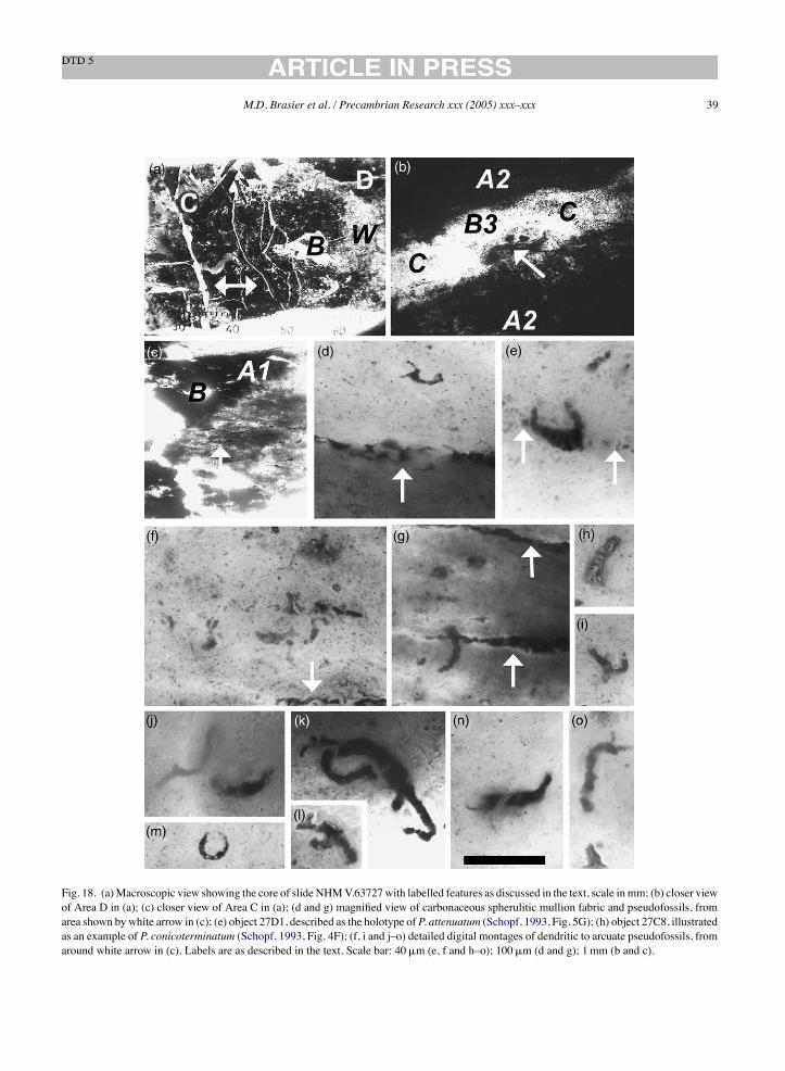
M.D. Brasier et al. / Precambrian Research xxx (2005) xxx–xxx 39
Fig. 18. (a)Macroscopic view showing the core of slide NHMV.63727with labelled features as discussed in the text, scale inmm; (b) closer viewof Area D in (a); (c) closer view of Area C in (a); (d and g) magnified view of carbonaceous spherulitic mullion fabric and pseudofossils, fromarea shown by white arrow in (c); (e) object 27D1, described as the holotype of P. attenuatum (Schopf, 1993, Fig. 5G); (h) object 27C8, illustratedas an example of P. conicoterminatum (Schopf, 1993, Fig. 4F); (f, i and j–o) detailed digital montages of dendritic to arcuate pseudofossils, fromaround white arrow in (c). Labels are as described in the text. Scale bar: 40!m (e, f and h–o); 100!m (d and g); 1mm (b and c).
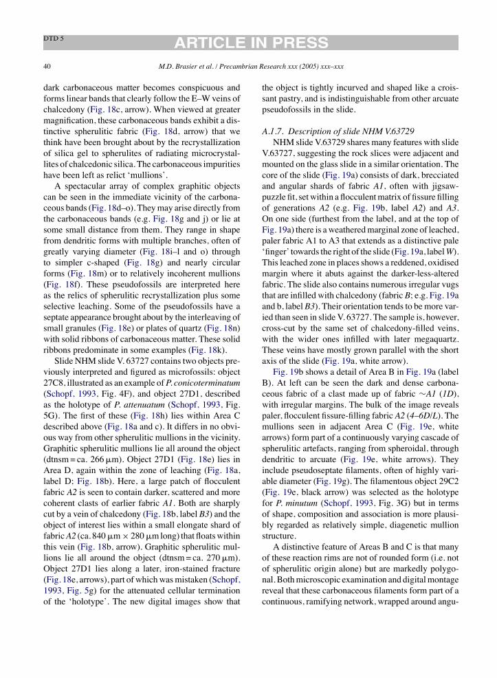
40 M.D. Brasier et al. / Precambrian Research xxx (2005) xxx–xxx
dark carbonaceous matter becomes conspicuous andforms linear bands that clearly follow the E–Wveins ofchalcedony (Fig. 18c, arrow). When viewed at greatermagnification, these carbonaceous bands exhibit a dis-tinctive spherulitic fabric (Fig. 18d, arrow) that wethink have been brought about by the recrystallizationof silica gel to spherulites of radiating microcrystal-lites of chalcedonic silica. The carbonaceous impuritieshave been left as relict ‘mullions’.A spectacular array of complex graphitic objects
can be seen in the immediate vicinity of the carbona-ceous bands (Fig. 18d–o). Theymay arise directly fromthe carbonaceous bands (e.g. Fig. 18g and j) or lie atsome small distance from them. They range in shapefrom dendritic forms with multiple branches, often ofgreatly varying diameter (Fig. 18i–l and o) throughto simpler c-shaped (Fig. 18g) and nearly circularforms (Fig. 18m) or to relatively incoherent mullions(Fig. 18f). These pseudofossils are interpreted hereas the relics of spherulitic recrystallization plus someselective leaching. Some of the pseudofossils have aseptate appearance brought about by the interleaving ofsmall granules (Fig. 18e) or plates of quartz (Fig. 18n)with solid ribbons of carbonaceous matter. These solidribbons predominate in some examples (Fig. 18k).Slide NHM slide V. 63727 contains two objects pre-
viously interpreted and figured as microfossils: object27C8, illustrated as an example ofP. conicoterminatum(Schopf, 1993, Fig. 4F), and object 27D1, describedas the holotype of P. attenuatum (Schopf, 1993, Fig.5G). The first of these (Fig. 18h) lies within Area Cdescribed above (Fig. 18a and c). It differs in no obvi-ous way from other spherulitic mullions in the vicinity.Graphitic spherulitic mullions lie all around the object(dtnsm= ca. 266!m). Object 27D1 (Fig. 18e) lies inArea D, again within the zone of leaching (Fig. 18a,label D; Fig. 18b). Here, a large patch of flocculentfabric A2 is seen to contain darker, scattered and morecoherent clasts of earlier fabric A1. Both are sharplycut by a vein of chalcedony (Fig. 18b, label B3) and theobject of interest lies within a small elongate shard offabricA2 (ca. 840!m$ 280!mlong) that floatswithinthis vein (Fig. 18b, arrow). Graphitic spherulitic mul-lions lie all around the object (dtnsm= ca. 270!m).Object 27D1 lies along a later, iron-stained fracture(Fig. 18e, arrows), part of whichwasmistaken (Schopf,1993, Fig. 5g) for the attenuated cellular terminationof the ‘holotype’. The new digital images show that
the object is tightly incurved and shaped like a crois-sant pastry, and is indistinguishable from other arcuatepseudofossils in the slide.
A.1.7. Description of slide NHM V.63729NHM slide V.63729 shares many features with slide
V.63727, suggesting the rock slices were adjacent andmounted on the glass slide in a similar orientation. Thecore of the slide (Fig. 19a) consists of dark, brecciatedand angular shards of fabric A1, often with jigsaw-puzzle fit, setwithin a flocculentmatrix of fissure fillingof generations A2 (e.g. Fig. 19b, label A2) and A3.On one side (furthest from the label, and at the top ofFig. 19a) there is aweatheredmarginal zone of leached,paler fabric A1 to A3 that extends as a distinctive pale‘finger’ towards the right of the slide (Fig. 19a, labelW).This leached zone in places shows a reddened, oxidisedmargin where it abuts against the darker-less-alteredfabric. The slide also contains numerous irregular vugsthat are infilled with chalcedony (fabric B; e.g. Fig. 19aand b, labelB3). Their orientation tends to bemore var-ied than seen in slide V. 63727. The sample is, however,cross-cut by the same set of chalcedony-filled veins,with the wider ones infilled with later megaquartz.These veins have mostly grown parallel with the shortaxis of the slide (Fig. 19a, white arrow).Fig. 19b shows a detail of Area B in Fig. 19a (label
B). At left can be seen the dark and dense carbona-ceous fabric of a clast made up of fabric !A1 (1D),with irregular margins. The bulk of the image revealspaler, flocculent fissure-filling fabric A2 (4–6D/L). Themullions seen in adjacent Area C (Fig. 19e, whitearrows) form part of a continuously varying cascade ofspherulitic artefacts, ranging from spheroidal, throughdendritic to arcuate (Fig. 19e, white arrows). Theyinclude pseudoseptate filaments, often of highly vari-able diameter (Fig. 19g). The filamentous object 29C2(Fig. 19e, black arrow) was selected as the holotypefor P. minutum (Schopf, 1993, Fig. 3G) but in termsof shape, composition and association is more plausi-bly regarded as relatively simple, diagenetic mullionstructure.A distinctive feature of Areas B and C is that many
of these reaction rims are not of rounded form (i.e. notof spherulitic origin alone) but are markedly polygo-nal. Bothmicroscopic examination and digitalmontagereveal that these carbonaceous filaments form part of acontinuous, ramifying network, wrapped around angu-
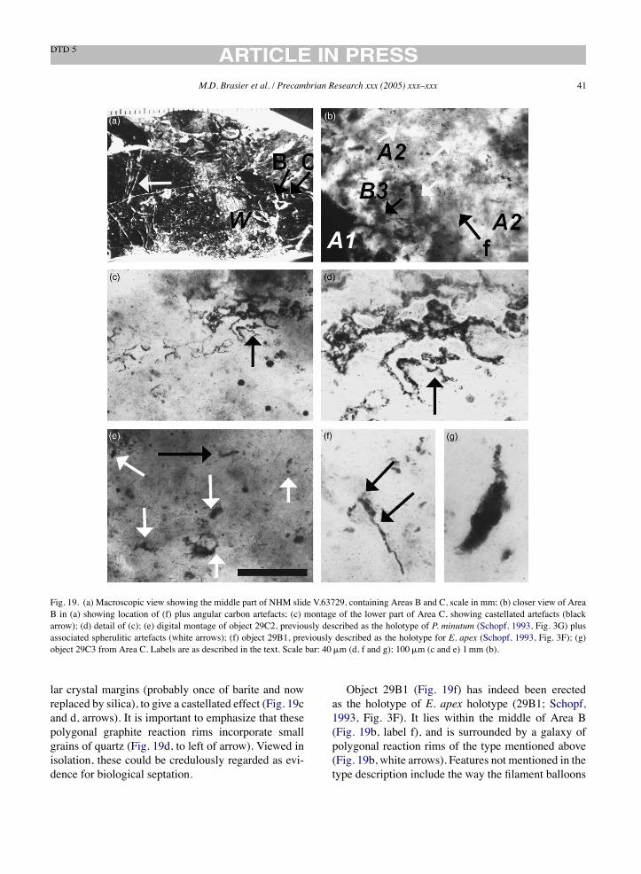
M.D. Brasier et al. / Precambrian Research xxx (2005) xxx–xxx 41
Fig. 19. (a) Macroscopic view showing the middle part of NHM slide V.63729, containing Areas B and C, scale in mm; (b) closer view of AreaB in (a) showing location of (f) plus angular carbon artefacts; (c) montage of the lower part of Area C, showing castellated artefacts (blackarrow); (d) detail of (c); (e) digital montage of object 29C2, previously described as the holotype of P. minutum (Schopf, 1993, Fig. 3G) plusassociated spherulitic artefacts (white arrows); (f) object 29B1, previously described as the holotype for E. apex (Schopf, 1993, Fig. 3F); (g)object 29C3 from Area C. Labels are as described in the text. Scale bar: 40!m (d, f and g); 100!m (c and e) 1mm (b).
lar crystal margins (probably once of barite and nowreplaced by silica), to give a castellated effect (Fig. 19cand d, arrows). It is important to emphasize that thesepolygonal graphite reaction rims incorporate smallgrains of quartz (Fig. 19d, to left of arrow). Viewed inisolation, these could be credulously regarded as evi-dence for biological septation.
Object 29B1 (Fig. 19f) has indeed been erectedas the holotype of E. apex holotype (29B1; Schopf,1993, Fig. 3F). It lies within the middle of Area B(Fig. 19b, label f), and is surrounded by a galaxy ofpolygonal reaction rims of the type mentioned above(Fig. 19b, white arrows). Features not mentioned in thetype description include the way the filament balloons
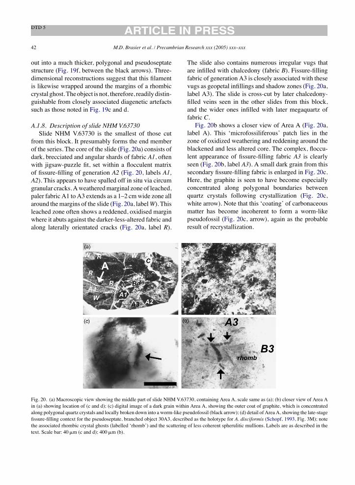
42 M.D. Brasier et al. / Precambrian Research xxx (2005) xxx–xxx
out into a much thicker, polygonal and pseudoseptatestructure (Fig. 19f, between the black arrows). Three-dimensional reconstructions suggest that this filamentis likewise wrapped around the margins of a rhombiccrystal ghost. Theobject is not, therefore, readily distin-guishable from closely associated diagenetic artefactssuch as those noted in Fig. 19c and d.
A.1.8. Description of slide NHM V.63730Slide NHM V.63730 is the smallest of those cut
from this block. It presumably forms the end memberof the series. The core of the slide (Fig. 20a) consists ofdark, brecciated and angular shards of fabric A1, oftenwith jigsaw-puzzle fit, set within a flocculent matrixof fissure-filling of generation A2 (Fig. 20, labels A1,A2). This appears to have spalled off in situ via circumgranular cracks.Aweatheredmarginal zone of leached,paler fabric A1 to A3 extends as a 1–2 cmwide zone allaround themargins of the slide (Fig. 20a, labelW). Thisleached zone often shows a reddened, oxidised marginwhere it abuts against the darker-less-altered fabric andalong laterally orientated cracks (Fig. 20a, label R).
The slide also contains numerous irregular vugs thatare infilled with chalcedony (fabric B). Fissure-fillingfabric of generation A3 is closely associated with thesevugs as geopetal infillings and shadow zones (Fig. 20a,label A3). The slide is cross-cut by later chalcedony-filled veins seen in the other slides from this block,and the wider ones infilled with later megaquartz offabric C.Fig. 20b shows a closer view of Area A (Fig. 20a,
label A). This ‘microfossiliferous’ patch lies in thezone of oxidized weathering and reddening around theblackened and less altered core. The complex, floccu-lent appearance of fissure-filling fabric A3 is clearlyseen (Fig. 20b, label A3). A small dark grain from thissecondary fissure-filling fabric is enlarged in Fig. 20c.Here, the graphite is seen to have become especiallyconcentrated along polygonal boundaries betweenquartz crystals following crystallization (Fig. 20c,white arrow). Note that this ‘coating’ of carbonaceousmatter has become incoherent to form a worm-likepseudofossil (Fig. 20c, arrow), again as the probableresult of recrystallization.
Fig. 20. (a) Macroscopic view showing the middle part of slide NHM V.63730, containing Area A, scale same as (a); (b) closer view of Area Ain (a) showing location of (c and d); (c) digital image of a dark grain within Area A, showing the outer coat of graphite, which is concentratedalong polygonal quartz crystals and locally broken down into a worm-like pseudofossil (black arrow); (d) detail of Area A, showing the late-stagefissure-filling context for the pseudoseptate, branched object 30A3, described as the holotype for A. disciformis (Schopf, 1993, Fig. 3M); notethe associated rhombic crystal ghosts (labelled ‘rhomb’) and the scattering of less coherent spherulitic mullions. Labels are as described in thetext. Scale bar: 40!m (c and d); 400!m (b).
M.D. Brasier et al. / Precambrian Research xxx (2005) xxx–xxx 43
The weathered zone of Area A (Fig. 20a, label A)is notable for containing the object 30A3 (Fig. 20d),erected as the holotype for the taxon Archaeoscillato-riopsis disciformis holotype (Schopf, 1993, Fig. 3M).It clearly lies within a very late and rather pale fissure-filling fabric of A3 type (Fig. 20b) in which float largerhombic ghosts (Fig. 20d, labelled ‘rhomb’), whichare especially concentrated near the junction of fabricA3 with void-infilling white chalcedony of generationB3 (Fig. 20d, label B3). Incoherent carbonaceousobjects float all around the object, which is found bymicroscopy to be much more complex than illustratedor described in the original type descriptions. Notonly does it have an incoherent extension at one end(Fig. 20d, lower arrow) but branches angularly continu-ously into a second structure at the other end (Fig. 20d,upper pair of arrows). This means that what had beenregarded as a diagnostic termination of the ‘holotype’is not in fact the ends of the structure at all, and iscertainly not diagnostic. Both the filamentous limbs ofobject 30A3 broadly follow the outline of the adjacentrhombic ghost, suggesting that the crystal (possiblyonce barite, and since replaced by quartz) was helpedto control its shape and hence, conceivably, its growth.Three-dimensional likewise confirms that the limbs arearranged along twin sides of a polygon. The structurecan be reinterpreted as a dendritic mullion structureformed as the result of the recrystallization of amor-phous, carbonaceous silica, at a late stage of fissurefilling.
A.2. Block 2
Slides NHM V.63731–63733 all appear to havebeen cut from the same elongate block. Distinctivefeatures of this block include brecciation and fissurefilling which are more uniform and somewhat easierto study than in block 1. The slides arte cross-cut byparallel, rather linear veins filled with fabric C. Previ-ously figured, supposedly fossiliferous material, fromthese slides includes three filamentous taxa, a solitaryunicell-like structure and a stromatolite-like structure(Schopf, 1993).
A.2.1. Description of slide NHM V.63731Slide NHM V. 63731 (overview not shown) con-
sists of dark, brecciated and angular shards of fabricA1,often with jigsaw-puzzle fit, set within themore floccu-
lent matrix of fissure-filling generations A2 to A4 (e.g.Fig. 21a, labels A1, A2). There is no obvious leachedzone around the slide. The bulk of the slide appears tocomprise paler fabric A3 but there are also quite largeareas of fabric A4 in the centre. Fabric A4 tends tohave a less fine-grained matrix, to be grain-supported,and to show a clear, geopetal relationship with the latestage chalcedony-filled vugs (of fabric B4). The slidecontains many irregular such vugs infilled with botry-oidal chalcedony (fabric B; e.g. Fig. 21a, label B2)which tend to be elongated along the longer axis ofthe slide. These botryoids can be coated with graphite.The sample is obliquely transected by three linear veinsinfilled with chalcedony (Fig. 21a and b, label B4)which are cross-cut by five linear veins parallel with theshort axis of the slide and filled with later megaquartz(fabric C).Fig. 21a shows the context for the ‘clast with
stromatolite-like laminae’ reported and figured bySchopf (1993, Fig. 3C). The darker fabric here con-sists of small grains of dense, and mostly very blackfabricA1, setwithin amore flocculentmatrix of fissure-filling generation A2 or A3 (Fig. 21a, labels A1, A2).The whispy, white areas comprise void-filling botry-oidal chalcedony of fabric B2 or B3. A faint linearvein, probably of B3, transects the lower half of theslide and the whole is cross-cut by a linear vein of latestage fabric B4. Closer inspection of the ‘stromatolite-like clast’ reveals that the basal laminae thin overflocculent grains of A2 (Fig. 21c, white arrow) butare essentially continuous across the asymmetricalpeaks and troughs (Fig. 21d). Most importantly, theycan be seen to wrap around the enclosing flocculentmatrix of fabric A2 (Fig. 21b, white arrow), imply-ing that they formed at roughly the same time, i.e.that they are part of a laminated cavity-fill of fab-ric B2. This interpretation is supported by the pres-ence within the laminae of a suite of minerals iden-tical with those of the enclosing matrix, includinghematite (Fig. 21c and e, black arrows), phyllosili-cates (Fig. 21d, black arrow) and flocculent grains offabric A2 (Fig. 21e, white arrow). Especially interest-ing is the formation of a dark filamentous structurealong the boundary between this grain and the chal-cedony. We regard this as a pseudofossil that formedas a reaction rim around the grain during crystal-lization of the silica, as noted elsewhere (cf. Fig. 8).By way of contrast, Fig. 21f shows an angular shard

44 M.D. Brasier et al. / Precambrian Research xxx (2005) xxx–xxx
Fig. 21. (a) Magnified view of Area A, showing the core of slide NHM V.63731 with labelled features as discussed in the text; (b) closer viewthe ‘stromatolite-like clast’ in Area A in (a) (taken from the reverse of the slide); (c) closer view of (b) showing pinching of laminae and ironoxide crystals; (d) closer view of (c) showing laminae continue across peaks and troughs; (e) detail of (b) showing enclosed grain of flocculentfabric A2 type; (f) angular shard of banded, clear and black carbonaceous chalcedony from Area B. Labels are as described in the text. Scalebar: 40!m (e); 100!m (c and d); 400!m (b), 1mm (a and f).
of fabric B, with undulating laminae and dark car-bonaceous layers. This has clearly been brecciatedand incorporated within flocculent fabric A3, whichwas later cut by veins chalcedony (fabric B4) andmegaquartz (fabric C). The undulating laminae resem-ble those reported from geyserites sinters (cf. Walter,1976, Fig. 19).
A.3. Block 3
Slides NHM V. 63734–63738 have been cut fromthe same rather triangular block. The features of brec-ciation and fissure filling are very clear and relativelysimple, showing a large, angular and fractured shardof darkish fabric A2, transected by a broad vein filled
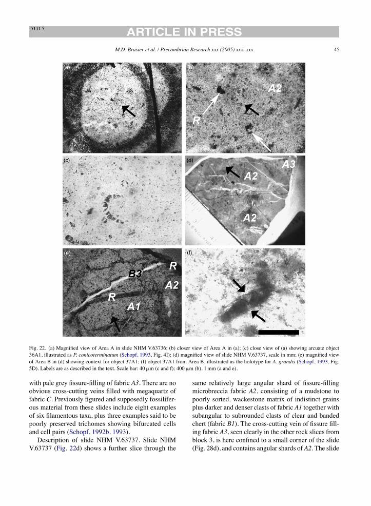
M.D. Brasier et al. / Precambrian Research xxx (2005) xxx–xxx 45
Fig. 22. (a) Magnified view of Area A in slide NHM V.63736; (b) closer view of Area A in (a); (c) close view of (a) showing arcuate object36A1, illustrated as P. conicoterminatum (Schopf, 1993, Fig. 4I); (d) magnified view of slide NHM V.63737, scale in mm; (e) magnified viewof Area B in (d) showing context for object 37A1; (f) object 37A1 from Area B, illustrated as the holotype for A. grandis (Schopf, 1993, Fig.5D). Labels are as described in the text. Scale bar: 40!m (c and f); 400!m (b), 1mm (a and e).
with pale grey fissure-filling of fabric A3. There are noobvious cross-cutting veins filled with megaquartz offabric C. Previously figured and supposedly fossilifer-ous material from these slides include eight examplesof six filamentous taxa, plus three examples said to bepoorly preserved trichomes showing bifurcated cellsand cell pairs (Schopf, 1992b, 1993).Description of slide NHM V.63737. Slide NHM
V.63737 (Fig. 22d) shows a further slice through the
same relatively large angular shard of fissure-fillingmicrobreccia fabric A2, consisting of a mudstone topoorly sorted, wackestone matrix of indistinct grainsplus darker and denser clasts of fabric A1 together withsubangular to subrounded clasts of clear and bandedchert (fabric B1). The cross-cutting vein of fissure fill-ing fabric A3, seen clearly in the other rock slices fromblock 3, is here confined to a small corner of the slide(Fig. 28d), and contains angular shards ofA2. The slide
46 M.D. Brasier et al. / Precambrian Research xxx (2005) xxx–xxx
is transected by several thin and rather irregular veinsinfilled with botryoidal chalcedony, some stained redwith iron oxide.Area A in slide NHM V.63737 lies within a small
subangular clast of fabric A1 set within fissure fillingfabricA2 (Fig. 22d black arrow). This area is transectedby later veins containing clear chert andbydarker zonescaused by localised pressure solution (Fig. 22e, labelsB3 andR, respectively) while the primary fabric is scat-tered with rhombic ghosts and carbonaceous reactionrims (Fig. 22e). Elongate object 37A1 lies within thisarea (Fig. 22e, black arrow) and is taken to be theholotype of A. grandis (Schopf, 1993, Fig. 5D). It isblack in transmitted light but white in reflected light.Septation here is brought about by the impingementof several small rhombic ghosts into the carbonaceousmatter (Fig. 22f, black arrows).
References
Awramik, S.M., Semikhatov, M.A., 1979. The relationship betweenmorphology, microstructure, and microbiota in three verticallyintergrading stromatolites from the Gunflint iron formation. Can.J. Earth Sci. 16, 484–495.
Awramik, S.M., Schopf, J.W.,Walter,M.R., 1983. Filamentous fossilbacteria from the Archean of Western Australia. PrecambrianRes. 20, 357–374.
Awramik, S.M., Schopf, J.W., Walter, M.R., 1988. Carbonaceousfilaments fromNorth Pole,WestAustralia: are they fossil bacteriain Archean stromatolites? A discussion. Precambrian Res. 39,303–309.
Beaumont, V., Robert, F., 1999. Nitrogen isotope ratios of kerogensin Precambrian cherts: a record of the evolution of atmospherechemistry. Precambrian Res. 96, 63–82.
Brasier, M.D., Green, O.R., 2004. Cyanobacterial fossils. In:McGraw-Hill Yearbook of Science and Technology, pp. 72–74.
Brasier, M.D., Green, O.R., Jephcoat, A.P., Kleppe, A.K., Van Kra-nendonk, M.J., Lindsay, J.F., Steele, A., Grassineau, N.V., 2002.Questioning the evidence for Earth’s oldest fossils. Nature 416,76–81.
Brasier, M.D., Green, O.R., Lindsay, J.F., Steele, A., 2004a. Earth’soldest (!3.5Ga) fossils and the ‘Early Eden’ hypothesis: ques-tioning the evidence. Origins Life Evol. Biosphere 34, 257–269.
Brasier, M.D., Green, O.R., McLoughlin, N., 2004b. Characteriza-tion and critical testing of potential microfossils from the earlyEarth: the Apex ‘microfossil debate’ and its lessons for Marssample return. Int. J. Astrobiol. 3, 139–150.
Brasier, M.D., Green, O.R., Lindsay, J.F., McLoughlin, N., Jephcoat,A., Kleppe, A.K., Steele, A., Stoakes, C., Wacey, D. Contexturalredescriptions of Earth’s oldest putative fossil assemblage fromthe !3.5Ga Apex chert, Chinaman Creek, Western Australia.Geological Survey of Western Australia Publication, in press.
Bridgewater, D., Allaart, J.H., Schopf, J.W., Klein, C., Walter, M.R.,Barghoorn, E.S., Strother, P., Knoll, A.H., Gorman, B.E., 1981.Microfossil-like objects from the Archaean of Greenland: a cau-tionary note. Nature 289, 51–53.
Buick, R., 1984. Carbonaceous filaments from North Pole, West-ern Australia: are they fossil bacteria in Archaean stromatolites?Precambrian Res. 24, 157–172.
Buick, R., 1988.Carbonaceous filaments fromNorth Pole,WestAus-tralia: are they fossil bacteria in Archean stromatolites? A reply.Precambrian Res. 39, 311–317.
Buick, R., 1990. Microfossil recognition in Archean rocks: anappraisal of spheroids and filaments from a 3500M.Y. old chert-barite unit at North Pole, Western Australia. Palaios 5, 441–459.
Buick, R., 1992. The antiquity of oxygenic photosynthesis: evidencefrom stromatolites in sulfate-deficient Archean lakes. Science255, 74–77.
Buick, R., Dunlop, J.S.R., Groves, D.I., 1981. Stromatolite recogni-tion in ancient rocks: an appraisal of irregular laminated struc-tures in an early Archaean chert-barite unit from North Pole,Western Australia. Alcheringa 5, 161–181.
Catling, D.C., Zahnle, K.J., McKay, C.P., 2001. Biogenic methane,hydrogen escape, and the irreversible oxidation of early Earth.Science 293, 839–843.
Cloud, P., Morrison, K., 1979. On microbial contaminants,micropseudofossils, and the oldest records of life. PrecambrianRes. 9, 81–91.
Dalton, R., 2002. Squaring up over ancient life. Nature 417, 782–784.Dunlop, J.S.R., Muir, M.D., Milne, V.A., Groves, D.I., 1978. A new
microfossil assemblage from the Archaean ofWestern Australia.Nature 274, 676–678.
Furnes, H., Banerjee, N.R., Muehlenbachs, K., Staudigel, H., deWit,M., 2004. Early life recorded in Archean pillow lavas. Science304, 578–581.
Goodwin, B., 1994. How the Leopard Changed Its Spots: The Evolu-tion of Complexity. Charles Scribner’s Sons, New York, p. 252.
Grotzinger, J.P., 1994. Trends in Precambrian carbonate sedimentsand their implications for understanding evolution. In: Bengtson,S. (Ed.), Early Life on Earth. Nobel Symposium 84. ColumbiaUniversity Press, New York, pp. 245–258.
Grotzinger, J.P., Rothman, D.H., 1996. An abiotic model for stroma-tolite morphogenesis. Nature 383, 423–425.
Hedges, S.B., Chen, H., Kumar, S.Y.-C., Wang, D., Thompson,A.S., Watanabe, H., 2001. A genomic timescale for the originof eukaryotes. BMC Evol. Biol., 1–4.
Hickman, A.H., 1975. Precambrian Structural Geology of Part ofthe Pilbara region: Western Australia Geological Survey. AnnualReport 1974, pp. 68–73.
Hickman, A.H., 1983. Geology of the Pilbara block and its environs.Western Aust. Geol. Survey Bull. 127, 268.
Hickman, A.H., 1984. Archean diapirism in the Pilbara Block,Western Australia. In: Kroner, A., Greiling, R. (Eds.), Precam-brian Tectonics. E. Schweizerbarts’che Verlagsbuchhandlung,Stuttgart, pp. 113–127.
Hoffman, H.J., Grey, K., Hickman, A.H., Thorpe, R., 1999. Originof 3.45Ga coniform stromatolites inWarawoonaGroup,WesternAustralia. Geol. Soc. Am. Bull. 111, 1256–1262.
M.D. Brasier et al. / Precambrian Research xxx (2005) xxx–xxx 47
Holm, N.G., Charlou, J.L., 2001. Initial indicators of abiotic for-mation of hydrocarbons in the rainbow ultramafic hydrothermalsystem, Mid-Atlantic Ridge. Earth Planetary Sci. Lett. 191, 1–8.
Horodyski, R.J., 1981. Pseudomicrofossils and altered microfossilsfrom amiddle Proterozoic shale, Belt Supergroup,Montana. Pre-cambrian Res. 16, 143–154.
Kamber, B.Z., Moorbath, S., Whitehouse, M.J., 2001. The oldestrocks on Earth: time constraints and geological controversies. In:Lewis, C.L.E., Knell, S.J. (Eds.), The Age of the Earth from 4004b.c. to 2002 a.d., vol. 290. Special Publication of the GeologicalSociety of London, pp. 177–203.
Kazmierczak, J., Kremer, B., 2002. Thermal alteration of Earth’soldest fossils. Nature 420, 477–478.
Knauth, L.P., Lowe, D.R., 2003. High Archean climatic temperatureinferred fromoxygen isotope geochemistry of cherts in the 3.5GaSwaziland Supergroup, South Africa. Bull. Geol. Soc. Am. 115,566–580.
Knoll, A.H., 1994. Proterozoic and early Cambrian protists: evi-dence for accelerating evolutionary tempo. In: Proceedings ofthe National Academy for Sciences USA, vol. 91, pp. 6743–6750.
Knoll, A.H., 2003. Life on a Young Planet: The First Three BillionYears of Evolution on Earth. Princeton University Press, p. 277.
Knoll, A.H., Walter, M.R., 1996. The limits of palaeontologicalknowledge: finding gold among the dross. In: Bock,G.R., Goode,J.A. (Eds.), Evolution ofHydrothermalEcosystems onEarth (andMars?). Wiley, Chichester, pp. 198–213.
Kudryavtsev,A.B., Schopf, J.W.,Agresti,D.G.,Wdowiak,T.J., 2001.In situ laser-Raman imagery of Precambrian microscopic fossils.In: Proceedings of the National Academy of Sciences, vol. 98,pp. 823–826.
Lindsay, J.F., Brasier, M.D., 2002. Did global tectonics drive earlybiosphere evolution? Carbon isotope record from 2.6 to 1.9Gacarbonates of Western Australian basins. Precambrian Res. 114,1–34.
Lindsay, J.F., Brasier, M.D., McLoughlin, N., Green, O.R., Fogel,M., Steele, A., Mertzman, S.A. The problem of deep carbon—anArchean paradox. Precambrian Res., submitted for publication.
Lowe, D.R., 1983. Restricted shallow-water sedimentation of earlyArchean stromatolitic and evaporitic strata of the Strelley Poolchert, Pilbara block, Western Australia. Precambrian Res. 19,239–283.
Marshall, C.G.A., May, J.W., Perret, C.J., 1964. Fossil microor-ganisms: possible presence in Precambrian shield of WesternAustralia. Science 144, 290–292.
McClendon, J.H., 1999. The origin of life. Earth Sci. Rev. 47, 71–93.McKay, D.S., Gibson, E.K., Thomas-Keprta, K.L., Vali, H.,
Romanek, C.S., Clemett, S.J., Chillier, X.D.F., Maechling, C.R.,Zare, R.N., 1996. Search for past life on Mars: possible relicbiogenic activity in Martian meteorite ALH84001. Science 273,924–930.
Mendelson, C.V., Schopf, J.W., 1982. Proterozoic microfossils fromthe Sukhaya Tunguska, Shorikha, and Yudoma formations of theSiberian platform. J. Plaeontol. 56, 42–83.
Nakashima, S.,Maruyama, S., Brack, A.,Windley, B.F. (Eds.), 2001.Geochemistry and the Origin of Life. Universal Academy PressInc., Tokyo, p. 335.
Nijman, W., De Bruin, K., Valkering, M., 1998. Growth fault controlof early Archaean cherts, barite mounds, and chert-barite veins,North Pole Dome, Eastern Pilbara, Western Australia. Precam-brian Res. 88, 25–52.
Pasteris, J.D., Wopenka, B., 2002. Images of the Earth’s earliest fos-sils? (Discussion and reply). Nature 420, 476–477.
Pasteris, J.D., Wopenka, B., 2003. Necessary, but not sufficient:Raman identification of disordered carbon as a signature ofancient life. Astrobiology 3, 727–738.
Perry, R.S., Brasier, M.D., Engel, M.H., Green, O.R., Gorbushina,A.A., Kolb, V.M., Krumbein, W.E., Perry, C.C., Campbell, K.,Staley, J.T., 2004. Should organic compounds found in associa-tion with minerals be called biominerals? GSA Annual Meeting(Denver), Abstract, pp. 29–3.
Raeburn, P., Golombek, M., 1998. Mars—Uncovering the Secretsof the Red Planet. National Geographic Society, Washington, p.232.
Rasmussen, B., 2000. Filamentous microfossils in a 3250-million-year-old volcanogenic massive sulphide deposit. Nature 405,676–679.
Rettalack, G.J., 1997. A Colour Guide to Paleosols. John Wiley andSons, Chichester, p. 175.
Robert, F., 1988. Carbon and oxygen isotope variations in Precam-brian cherts. Geochim. Cosmochim. Acta 52, 1473–1478.
Schopf, J.W., 1975. Precambian paleobiology: problems and per-spectives. Annu. Rev. Earth Planetary Sci. 3, 213–249.
Schopf, J.W., 1992a. The oldest fossils and what they mean. In:Schopf, J.W. (Ed.), Major Events in the History of Life. Johnand Bartlett, Chapter 2, 29–63, 190 pp.
Schopf, J.W., 1992b. Paleobiology of the Archean. In: Schopf, J.W.,Klein, C. (Eds.), The Proterozoic Biosphere: AMultidisciplinaryStudy. Cambridge University Press, pp. 25–39.
Schopf, J.W., 1993. Microfossils of the Early Archean Apex chert:new evidence of the antiquity of life. Science 260, 640–646.
Schopf, J.W., 1994. Disparate rates, differing fates: tempo and modeof evolution changed from the Precambrian to the Phanerozoic.In: Proceedings of the National Academy of Sciences, vol. 91,pp. 6735–6742.
Schopf, J.W., 1999. The Cradle of Life. Princeton University Press,New York, 367 pp.
Schopf, J.W., Packer, B.M., 1987. Early Archean (3.3 billion to3.5 billion-year-old) microfossils fromWarawoona Group, Aus-tralia. Science 237, 70–73.
Schopf, J.W., Walter, M.R., 1983. Archean microfossils: new evi-dence of ancient microbes. In: Schopf, J.W. (Ed.), Earth’sEarliest Biosphere. Princeton University Press, pp. 214–239.
Schopf, J.W., Kudryavtsev, A.B., Agresti, D.G., Wdowiak, T.J.,Czaja, A.D., 2002a. Laser-Raman imagery of Earth’s earliest fos-sils. Nature 416, 73–76.
Schopf, J.W., Kudryavtsev, A.B., Agresti, D.G., Wdowiak, T.J.,Czaja, A.D., 2002b. Images of the Earth’s earliest fossils? (Dis-cussion and reply). Nature 420, 476–477.
Shaw, R.D., Wellman, P., Gunn, P., Whitaker, A.J., Tarlowski, C.,Morse,M., 1996.Guide to usingAustralian crustal elementsmap.Aust. Geol. Survey Org. Record 30, 49.

48 M.D. Brasier et al. / Precambrian Research xxx (2005) xxx–xxx
Summons, R.E., Jahnke, L.L., Hope, M., Logan, G.A., 1999. 2-methulhopanoids as biomarkers for cyanobacterial oxygenicphotosynthesis. Nature 400, 554–557.
Tenenbaum, D., 2003. Earth’s oldest fossils reverse course. OnlineAstrobiology Magazine, Article 421, April 7.
Tyler, S.A., Barghoorn, E.S., 1963. Ambient pyrite grains in Precam-brian cherts. Am. J. Sci. 261, 424–432.
Tyler, I.M., Thorne, A.M., 1990. The northern margin of the Capri-corn Orogen, Western Australia—an example of an early Pro-terozoic collision zone. J. Struct. Geol. 12, 685–701.
Van Kranendonk, M.J., 1999. North Shaw: Western Australia Sheet2755:Western Australian Geological Survey, 1:100 000 Geolog-ical Series.
Van Kranendonk, M.J., 2000. Geology of the North Shaw 1:100,000sheet: Western Australia Geological Survey, 1:100,000 Geolog-ical Series Explanatory Notes, p. 86.
Van Kranendonk, M.J., Hickman, A.H., Williams, I.R., Nijman, W.,2001. Archaean Geology of the East Pilbara Granite-Greenstoneterrane Western Australia—a field guide. West. Aust. Geol. Sur-vey Record 9, 134.
VanKranendonk,M.,Hickman,A., Smithies,R.H.,Nelson,D., 2002.Geology and tectonic evolution of theArcheanNorth Pilbara Ter-rain, PilbaraCraton,WesternAustralia. Econ.Geol. 97, 695–732.
Van Kranendonk, M.J., 2001. Geology of the Marble Bar 1:100,000sheet: Western Australia Geological Survey.
Walsh, M.M., 1992. Microfossils and possible microfossils fromthe early Archean onverwacht group, Barberton mountain land,South Africa. Precambrian Res. 271, 271–293.
Walsh, M.M., Lowe, D.R., 1999. Modes of accumulation of car-bonaceous matter in the Early Archean: a petrographic andgeochemical study of the carbonaceous cherts of the SwazilandSupergroup: in: Lowe, D.R., Byerly, G., R., (Eds.), GeologicEvolution of the Barberton Greenstone Belt, South Africa, Geo-logical Society of America Special Paper 329, pp. 115–132.
Walter, M.R. (Ed.), 1976. Stromatolites: Developments in Sedimen-tology, vol. 20. Elsevier, Amsterdam, p. 790.
Walter, M.R., Buick, R., Dunlop, J.S.R., 1980. Stromatolites,3400–3500Myr old from the North Pole area,Western Australia.Nature 284, 443–445.
Westall, F., deWit,M.J., Dann, J., van derGaast, S., deRonde, C.E.J.,Gerneke, D., 2001. Early Archean fossil bacteria and biofilms inhydrothermally influenced sediments from the Barberton green-stone belt, South Africa. Precambrian Res. 106, 93–116.
Westall, F., Folk, R.L., 2003. Exogenous carbonaceous microstuc-tures in EarlyArchaean cherts andBIFs from the IsuaGreenstoneBelt: implications for the search for life in ancient rocks. Precam-brian Res. 126, 313–330.
Wopenka, B., Pasteris, J.D., 1993. Structural characterization ofkerogens to granulite-facies graphite: applicability of Ramanmicroprobe spectroscopy. Am. Mineral. 78, 533–557.