Critical roles of cytomegalovirus-induced Natural Killer ...
109
Critical roles of cytomegalovirus-induced Natural Killer cells in chronic hepatitis C virus infection and rituximab-mediated cancer therapy Jun Seok Oh A thesis submitted to the Faculty of Graduate and Postdoctoral Studies in partial fulfillment of the requirements for the MSc degree in Microbiology and Immunology Department of Biochemistry, Microbiology and Immunology Faculty of Medicine University of Ottawa © Jun Seok Oh, Ottawa, Canada, 2017
Transcript of Critical roles of cytomegalovirus-induced Natural Killer ...

Critical roles of cytomegalovirus-induced Natural
Killer cells in chronic hepatitis C virus infection and
rituximab-mediated cancer therapy
Jun Seok Oh
A thesis submitted to the Faculty of Graduate and Postdoctoral Studies in partial fulfillment of
the requirements for the MSc degree in Microbiology and Immunology
Department of Biochemistry, Microbiology and Immunology
Faculty of Medicine
University of Ottawa
© Jun Seok Oh, Ottawa, Canada, 2017

ii
ABSTRACT
Natural Killer (NK) cells, members of the innate lymphoid cells (ILCs), are known to play an
important role in the defense against foreign cells and abnormal host cells that have arisen due to
viral infection or cancer inducing mutations. The typical immune response of NK cells involves
the release of cytotoxic granules containing perforin and granzyme, and the secretion of immune-
regulatory cytokines such as interferon gamma (IFN-γ). Unlike the adaptive lymphocytes such as
T cells and B cells, NK cells do not require prior sensitization, enabling them to initiate an
immune response much faster. This unique feature of NK cells is made possible by the utilization
of an array of germline encoded receptors; but on the other hand, it limits NK cells ability to
respond against rapidly evolving pathogens. NK cells overcome this shortcoming with an
antibody-assisted process called antibody dependent cellular cytotoxicity (ADCC).
A novel subset of human NK cells, which displays potent and broad antiviral responsiveness in
concert with virus-specific antibodies, was recently discovered in cytomegalovirus positive
(CMV+) individuals. This NK cell subset, called g-NK cell, was characterized by a deficiency in
the expression of FcεRIγ, an adaptor protein that associates with CD16 which enables ADCC.
Surprisingly, despite this deficiency, g-NK cells displayed an enhanced ADCC as compared to
their conventional counterparts. Furthermore, having a long-lasting memory-like NK-cell
phenotype suggests a role for g-NK cells in chronic infections.
This study investigates the importance of g-NK-cells in clinical settings, first by investigating
whether the presence of g-NK cells is associated with the magnitude of liver disease during
chronic hepatitis C virus (HCV) infection. Analysis of g-NK cell proportions and function in the
peripheral blood mononuclear cells (PBMCs) of healthy controls and chronic HCV subjects
showed that chronic HCV subjects had slightly lower proportions of g-NK cells, while having
iii
similarly enhanced ADCC responses compared to conventional NK cells. Notably, among
CMV+ chronic HCV patients, lower levels of liver enzymes and fibrosis were found in those
possessing g-NK cells. g-NK cells were predominant among the CD56neg
NK cell population
often found in chronic HCV patients, suggesting their involvement in the immune response
against HCV.
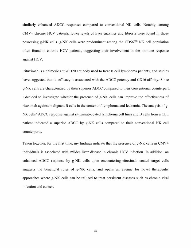
Rituximab is a chimeric anti-CD20 antibody used to treat B cell lymphoma patients; and studies
have suggested that its efficacy is associated with the ADCC potency and CD16 affinity. Since
g-NK cells are characterized by their superior ADCC compared to their conventional counterpart,
I decided to investigate whether the presence of g-NK cells can improve the effectiveness of
rituximab against malignant B cells in the context of lymphoma and leukemia. The analysis of g-
NK cells’ ADCC response against rituximab-coated lymphoma cell lines and B cells from a CLL
patient indicated a superior ADCC by g-NK cells compared to their conventional NK cell
counterparts.
Taken together, for the first time, my findings indicate that the presence of g-NK cells in CMV+
individuals is associated with milder liver disease in chronic HCV infection. In addition, an
enhanced ADCC response by g-NK cells upon encountering rituximab coated target cells
suggests the beneficial roles of g-NK cells, and opens an avenue for novel therapeutic
approaches where g-NK cells can be utilized to treat persistent diseases such as chronic viral
infection and cancer.
iv
ACKNOWLEDGEMENTS
I would first like to thank God for His unconditional love, and for guiding my life throughout
times of difficulties and happiness. I would also like to thank my supervisor Dr. Seung-Hwan
Lee for his guidance and patience, allowing me to become a better scientist and improve my
skills. I would like to thank my TAC committee members, Dr. Angela Crawley and Dr. Subash
Sad for their valuable time, and many advices. I would also like to thank Dr. Curtis Cooper for
his invaluable support and advice for my main HCV project. In addition, I would also like to
thank Dr. Daniel Corsi, and many nurses including Ms. Elyse Sevigny, and Ms. Lynda Theoret at
the Ottawa Hospital for their valuable time and advices. I would like to thank Dr. Vera Tang for
the maintenance of the flow cytometry core facility and training. I would also like to thank my
lab members, Alaa Ali, Dahn Hahm, Saeedah Almutairi, and Jennifer Park for their valuable help
and fun memories. I would also like to thank Ms. Fay Draper, Ms. Suzanne Surgeson, Ms.
Victoria Stewart, and Ms. Wendy Ip for their administrative support. Lastly, I would like to
thank my family and friends for their continuous prayers, support, and epic encouragement.

v
COPYRIGHT PERMISSION
The content of this paper is based on my published journal article entitled “NK cells lacking
FcεRIγ are associated with reduced liver damage in chronic hepatitis C virus infection”. The
permission to reuse this copyrighted material from the publisher, European Journal of
Immunology (Wiley-VCH Verlag GmbH & Co. KGaA), is acquired.
Jun S. Oh, Alaa K. Ali, Sungjin Kim, Daniel J. Corsi, Curtis L. Cooper, Seung-Hwan Lee. NK
cells lacking FcεRIγ are associated with reduced liver damage in chronic HCV infection. Eur J
Immunol. 2016; 46(4):1020-1029.
Contributions:
Jun S. Oh: Performed all experiments. Contributed to the writing and editing of the manuscript.
Alaa K. Ali: Assisted with the blood process, surface and intracellular staining, and ELISA.
Contributed to the editing of the manuscript.
Sungjin Kim: Provided guidance regarding g-NK cells.
Daniel J. Corsi: Provided guidance in regard to statistical analysis.
Curtis L. Cooper: A clinician and collaborator from The Ottawa Hospital who provided guidance
as well as patient data and samples.
Seung-Hwan Lee: Supervised, designed and participated in the execution of experiments.
Contributed to the writing and editing of the manuscript.

vi
Table of Contents ABSTRACT .................................................................................................................................................. ii
ACKNOWLEDGEMENTS ......................................................................................................................... iv
COPYRIGHT PERMISSION ....................................................................................................................... v
LIST OF ABBREVIATIONS .................................................................................................................... viii
LIST OF FIGURES AND TABLES ............................................................................................................ xi
1. INTRODUCTION .................................................................................................................................... 1
1.1 Natural Killer (NK) cell ...................................................................................................................... 1
1.2 Human Cytomegalovirus (HCMV) ..................................................................................................... 6
1.3 NK cell subset: g-NK cell ................................................................................................................. 10
1.4 Hepatitis C Virus (HCV) infection ................................................................................................... 11
1.5 Lymphoma and leukemia .................................................................................................................. 13
1.6 Monoclonal antibody-mediated cancer therapy ................................................................................ 14
1.7 Hypothesis and statement of objectives ............................................................................................ 16
AIM 1: Investigate whether the maintenance of g-NK cells is advantageous to resist against chronic
infection. ............................................................................................................................................. 16
AIM 2: Investigate the effectiveness of g-NK cells during monoclonal antibody-mediated therapy
against cancer such as lymphoma and CLL. ....................................................................................... 16
2. MATERIALS AND METHODS ............................................................................................................ 17
2.1 Study subjects ................................................................................................................................... 17
2.2 Extraction and storage of peripheral blood mononuclear cells and plasma from blood samples ..... 20
2.3 CMV serology and peptide stimulation ............................................................................................ 20
2.4 Flow cytometry analysis of NK cell phenotype and function ........................................................... 21
2.5 METAVIR stage system ................................................................................................................... 25
2.6 Analysis of NK cell response against rituximab coated target cells ................................................. 25
2.7 Statistical analysis ............................................................................................................................. 26
3. RESULTS ............................................................................................................................................... 27
3.1 Characterization of g-NK cells in healthy controls ........................................................................... 27
3.2 A strong association of g-NK cells with prior CMV infection is also found in chronic HCV patients
................................................................................................................................................................ 34
3.3 Superior ADCC of g-NK cells is maintained in chronic HCV infection .......................................... 47
3.4 Reduced liver damage is associated with the presence of g-NK cells in CMV-infected HCV+
individuals ............................................................................................................................................... 50
3.5 CD56neg
CD16+ NK cells predominantly contain g-NK cells ............................................................ 61

vii
3.6 g-NK cells show enhanced ADCC against rituximab coated target cells ......................................... 65
3.7 Evaluation of CD20 expression on target B cells ............................................................................. 74
4. DISCUSSION ......................................................................................................................................... 77
5. CONCLUDING REMARKS .................................................................................................................. 84
6. REFERENCES ....................................................................................................................................... 87
7. APPENDIX ............................................................................................................................................. 94
viii
LIST OF ABBREVIATIONS oC Celsius (temperature)
ADCC Antibody Dependent Cellular Cytotoxicity
ALL Acute Lymphoblastic Leukemia
ALT Alanine Transaminase
AML Acute Myeloid Leukemia
AST Aspartate Transaminase
CCL Chemokine C-C Motif Ligand
CD Cluster of Differentiation
CLL Chronic Lymphocytic Leukemia
CMV Cytomegalovirus
DAA Direct acting antiviral
DAB2 Disabled homolog 2
DMSO Dimethyl Sulfoxide
DNA Deoxyribonucleic Acid
EAT-2 Ewing’s sarcoma-associated transcript 2
ELISA Enzyme Linked Immunosorbent Assay
FBS Fetal Bovine Serum
FcεRIγ Fragment Crystallizable Epsilon Receptor I gamma
FcγRIIIα CD16a
FcR Fc Receptor
g-NK Gamma-chain negative Natural Killer
GGT Gamma Glutamyl Transpeptidase
GM-CSF Granulocyte Macrophage Colony Stimulating Factor
HBV Hepatitis B virus
HCMV Human Cytomegalovirus
HCV Hepatitis C Virus
HEPES 4-(2-hydroxyethyl)-1-piperazineethanesulfonic acid

ix
HIV Human Immunodeficiency Virus
HL Hodgkin Lymphoma
HSV Herpes Simplex Virus
IFN Interferon
IgG Immunoglobulin G
ILCs Innate Lymphoid Cells
iNOS Inducible Nitric Oxide Synthase
ITAM Immuno-receptor Tyrosine-based Activation Motifs
kb kilo base
LAMP Lysosome Associated Membrane Glycoprotein
mAb Monoclonal Antibody
METAVIR Meta-analysis of Histological Data in Viral Hepatitis
MHC Major Histocompatibility Complex
MIP Macrophage Inflammatory Protein
µg microgram
µL microlitre
NHL Non-Hodgkin Lymphoma
NK Natural Killer
NKG2 Natural Killer group 2
NO Nitric Oxide
PAMPs Pathogen Associated Molecular Patterns
PBMC Peripheral Blood Mononuclear Cell
PLZF Promyelocytic Leukaemia Zinc Finger
pp65 Phosphoprotein 65
RANTES Regulated on Activation Normal T cell Expressed and Secreted
RCF Relative centrifugal force
RNA Ribonucleic Acid

x
RPMI 1640 Roswell Park Memorial Institute 1640 medium
SVR Sustained virologic response
SYK Spleen Tyrosine Kinase
TNF Tumor Necrosis Factor
TRAIL TNF Related Apoptosis Inducing Ligand
ZAP70 Zeta-chain Associated Protein Kinase 70kDa (kilo Dalton)

xi
LIST OF FIGURES AND TABLES
Figure 1: The representative diagram of signaling molecules involved in ADCC ....................................... 4
Figure 2: The simplified diagram of CMV structure .................................................................................... 8
Figure 3: Characterization of g-NK cells in healthy control individuals .................................................... 30
Figure 4: Heterogeneity of g-NK cells population in healthy individuals .................................................. 32
Figure 5: A strong association of g-NK cells with prior CMV infection are also found in chronic HCV
patients ........................................................................................................................................................ 37
Figure 6: Detection of HCMV pp65 specific T cell response using peptide stimulation ........................... 39
Figure 7: No correlation between proportion of g-NK cells and the age of subjects .................................. 41
Figure 8: NKG2C expression of NK cells among healthy and chronic HCV patients ............................... 43
Figure 9: SYK expression of NK cells among healthy and chronic HCV patients .................................... 45
Figure 10: Superior ADCC of g-NK cell is maintained in individuals chronically infected with HCV ..... 48
Figure 11: Reduced liver damage is associated with the presence of g-NK cells in CMV+ HCV+
individuals ................................................................................................................................................... 53
Figure 12: No correlation between proportion of g-NK cells and the levels of liver enzymes ................... 55
Figure 13: GGT enzyme level does not correlate with stages of fibrosis ................................................... 57
Figure 14: Proportion of g-NK cells does not correlate with viral load level ............................................. 59
Figure 15: CD56neg CD16+ NK cells predominantly contain g-NK cells ................................................ 63
Figure 16: Proportion of NK cells producing IFN-g upon co-incubation with rituximab treated lymphoma
cell lines: Daudi, Raji and RAMOS ............................................................................................................ 68
Figure 17: Proportion of NK cells producing CD107a (perforin) upon co-incubation with rituximab
treated lymphoma cell line: RAMOS.......................................................................................................... 70
Figure 18: Proportion of NK cells producing IFN-g upon co-incubation with rituximab treated PBMC of
CLL patient ................................................................................................................................................. 72
Figure 19: Expression of surface CD20 on Daudi, Raji, and RAMOS lymphoma cell lines, and B cells
from CLL patient ........................................................................................................................................ 75
Figure 20: Summary diagram showing beneficial role of g-NK cells in chronic HCV patients ................ 85
Table 1: Table showing number and average age of study subjects ........................................................... 18
Table 2: Table containing list of antibodies used in flow cytometry .......................................................... 23

1
1. INTRODUCTION
1.1 Natural Killer (NK) cell
The Natural Killer (NK) cell, a large cell featuring a distinctive granular cytoplasm, is part of the
innate lymphoid cells (ILCs) that arise from common lymphoid progenitor cells found in the
bone marrow. The resulting NK precursor cell then traffics through the lymph nodes, spleen and
peripheral blood for further maturation and differentiation (Huntington et al., 2007). NK cells are
specialized in the rapid recognition and killing of abnormal target cells, such as virus-infected
cells and cancer cells, without prior sensitization (Biron et al., 1999; Lee et al., 2007). This
unique ability is achieved by utilizing a repertoire of germline-encoded natural cytotoxicity
receptors (NCR), as well as activating and inhibitory receptors that are specifically designed to
detect stress ligands, self-ligands, Pathogen-Associated Molecular Patterns (PAMPs), and
antibodies, exclusively Immunoglobulin G (IgG). Upon activation, NK cells induce the apoptosis
of abnormal cells through the release of cytotoxic granules containing perforin and granzyme.
Alternatively, NK cells can induce cell lysis through Fas/FasL interactions, Tumor necrosis
factor (TNF)-Related Apoptosis-Inducing Ligand (TRAIL), or TNF-α pathways (Biron et al.,
1999; Lee et al., 2007; Vivier et al., 2008; Warren and Smyth, 1999). NK cells also participate
in the immune regulation through their surface receptors and production of pro-inflammatory and
regulatory cytokines such as Interferon gamma (IFN-γ), TNF, Granulocyte-Macrophage Colony-
Stimulating Factor (GM-CSF), Macrophage Inflammatory Proteins (MIP-1α/CCL3), MIP-
1β/CCL4, and Regulated on Activation Normal T cell Expressed and Secreted (RANTES/CCL5)
(Biron et al., 1999). Human NK cells in healthy individuals are commonly identified by using
CD16 (Fc receptor) and CD56 (neural cell adhesion molecule) markers and divided into two

2
separate populations: CD16-CD56
bright, and CD16
+CD56
dim. The CD16
-CD56
bright NK cell
population, which is found at low proportions in the blood, is highly immune-regulatory;
whereas the CD16+CD56
dim NK cell population is highly cytotoxic and makes up the majority of
the NK cell population in the blood of healthy individuals (Caligiuri, 2008). Adaptive immune
cells such as T cells and B cells utilize gene rearrangement mechanisms to generate a near
limitless repertoire of antigen specific receptors. This mechanism involves the rearrangement of
variable (V), diversity (D), joining (J) gene segments, and constant (C) genes located in TCRα
locus (chromosome 14) and TCRβ locus (chromosome 7) for T cells, and the immunoglobulin
heavy, κ, and λ loci (located in chromosome 14, 2 and 22 respectively) for B cells. This feature,
although time consuming, enables them to recognize any known or unknown highly evolving
foreign target and mount an immune response such as antibody production. Unlike these
adaptive immune cells, NK cells lack such mechanism that would allow them to recognize
rapidly evolving pathogens. In order to overcome this limitation, NK cells utilize the Fc receptor
CD16, an activating receptor, to expand the range of targets that can be detected. The CD16
molecule interacts with IgG antibody coated target cells; and plays a crucial role in a mechanism
known as antibody dependent cellular cytotoxicity (ADCC) (Vivier et al., 2008). This allows NK
cells to enjoy the benefit of a faster response, and in addition, a limitless target recognition
capability driven by antibody.
Several immune cells possessing Fc receptors, such as NK cells, monocytes, macrophages, and
eosinophils can utilize ADCC. This critical mechanism is important for NK cells as it allows
them, with the help of target specific antibodies, to recognize highly evolving abnormal cells that
have managed to develop ways to evade direct recognition by NK cells through germ-line
encoded receptors. In NK cells, the process of ADCC begins from NK cells’ Fc receptor CD16

3
(FcγRIIIα) that binds to the Fc fragment of IgG (preferably to IgG1 and IgG3) bound to the
target cell (Takai, 2002). Upon binding, activated CD16 covalently interacts with hetero- or
homodimers of membrane-bound signalling adaptors such as FcεRIγ (γ chain of high affinity
tetrameric IgE receptor that can also interact with other Fc receptors) and CD3ζ that possess
Immunoreceptor Tyrosine-based Activation Motif (ITAM), which acts as signalling transducers
(Figure 1) (Hwang et al., 2012). The phosphorylation of ITAM leads to the recruitment of
Spleen Tyrosine Kinase (SYK) and Zeta-chain Associated Protein kinase 70kDa (ZAP70) which
subsequently activate Linker for Activation of T cells (LAT) (Maghazachi, 2005). The activation
of LAT leads to a change in the intracellular calcium ion concentration via Phospholipases C
gamma 1 and 2 (PLC-gamma 1 and 2) activation (Ting et al., 1992). The change in calcium ion
concentration activates calcineurin which is responsible for dephosphorylating Nuclear Factor of
Activated T-cell Cytoplasmic Calcineurin-dependent 2 (NFATC2). Dephosphorylated NFATC2
then travels to the nucleus in order to initiate the transcription of various cytokines, such as IFN-
γ, for NK cell (Sica et al., 1997). NK cells activated by CD16 rapidly internalize and/or shed the
receptor, leading to reduced CD16 expression on their surface (Masilamani et al., 2009).

4
Figure 1
Figure 1: The representative diagram of signaling molecules involved in ADCC. (A) Upon
encounter with antibody coated target cell, the Fc receptor, CD16, of NK cells binds to the Fc
region of target cell bound IgG antibody and releases cytotoxic granules and anti-viral immune-
regulatory cytokines. (B) Upon activation, CD16 associates with a heterodimer or homodimer of
intracellular adaptor proteins, FcεRIγ and CD3ζ, in order to transmit signals further downstream
via immunoreceptor tyrosine-based activation motif (ITAM). CMV associated NK cell subset, g-
NK cells, lost the FcεRIγ chain, hence it only has CD3ζ. It is also important to note that the
FcεRIγ chain contains a single (ITAM), whereas the CD3ζ chain contains three ITAMs,
implying that enhanced CD16-mediated signaling may be due to CD3ζ chain.
Figure 1: The representative diagram of signaling molecules involved in ADCC

5

6
1.2 Human Cytomegalovirus (HCMV)
The human cytomegalovirus (HCMV) belongs to the Herpesviridae family under the
Betaherpesvirinae subfamily. This virus shares common structural characteristics with other
herpesviruses. Its viral genome, which consists of 235 kilobase pair (kb) of double stranded
DNA, is contained within an icosahedral nucleocapsid. The nucleocapsid is surrounded by
amorphous tegument proteins within the outer lipid membrane that contains glycoprotein
complex I and II which facilitate membrane fusion with the host cell, thereby these glycoproteins
are critical in viral entry (Griffiths et al., 2015). Upon fusion of both viral and host cell
membranes, the nucleocapsid and tegument proteins are released into the cytoplasm of the host
cell. Upon entry, the expression of viral immediate-early (IE) genes produces IE proteins that are
necessary to replicate the viral DNA genome. The expression of late genes begins after DNA
replication and leads to the production of late proteins which mostly consist of structural proteins
necessary for the assembly and release of progeny virus. The tegument mainly consists of the
pp65 protein, which is also referred to as ppUL83 or Lower matrix protein (Figure 2). Although
pp65 is not critical for viral replication, this protein is critical in the formation of noninfectious
extracellular viral particle, so called dense bodies that lack a nucleocapsid. The pp65 protein is
also known to play a role in immune evasion (Kalejta, 2008). Firstly, pp65 interacts with NKp30,
which is an activating receptor on NK cells, to inhibit innate NK cell mediated cytotoxicity.
Secondly, pp65 can help HCMV evade the adaptive immune response by either preventing the
presentation of viral IE proteins derived peptides via major histocompatibility complex (MHC,
also known as HLA in human) class I molecules, or by causing the degradation of the alpha
chain in the MHC class II cell surface receptor HLA-DR. Thirdly, pp65 is also known to down-
modulate the expression of MHC class II, interferon, and chemokines. These properties indicate
7
that pp65 plays a significant role in immune evasion against NK cells and T cells (Kalejta, 2008;
Tomtishen, 2012). A study has found that pp65 is a major epitope that is commonly recognized
by CD4 and CD8 T cells (Sylwester et al., 2005); hence making it an ideal antigen to test
individuals’ prior HCMV encounter.
NK cells are known to play a critical role in the immune response against herpesvirus infections.
Deficiencies in NK cell population or function have been associated with an increased
susceptibility to herpesviruses, including cytomegalovirus (CMV), EBV, and varicella zoster
virus (Orange, 2013). Among herpesviruses, CMV is highly prevalent, with a 50-100%
seroprevalence rate depending on the geographical location and socioeconomic factors. On
average, the prevalence of CMV infection is low in modernized countries located in Europe,
Australia, and North America, while it is relatively high in developing countries located in Africa
and Southeast Asia. The prevalence of CMV infection also varies within the same country (Ho,
1990). HCMV infects people of all ages, gender, and races. This virus transmits via close contact
with infected bodily fluids such as saliva, blood, urine, breast milk, cervical and vaginal
excretions, and semen. In addition, it is also commonly known to be transmitted in utero, leading
to birth defects in infants (Gaytant et al., 2002). Once it enters a host, it can infect many cell
types, such as fibroblasts, smooth muscle cells, tissue macrophages, epithelial cells and
endothelial cells (Sinzger et al., 1995). The CMV infection is generally asymptomatic in an
immunocompetent host, while establishing a lifelong latent infection. However, reactivation
from a latent infection can occur in immunocompromised individuals including transplant
recipients, neonates, or human immunodeficiency virus (HIV) infected individuals, leading to
morbidity and mortality (Crough and Khanna, 2009; Dowd et al., 2009).

8
Figure 2
Figure 2. The representative diagram of CMV structure. HCMV, a member of the
herpesviridae family, consists of an icosahedral nucleopcapsid containing a double stranded
DNA viral genome. The nucleocapsid is surrounded by the tegument, and an outer lipid
membrane that contains glycoprotein complex I and II.
Figure 2: The simplified diagram of CMV structure

9

10
1.3 NK cell subset: g-NK cell
Interestingly, CMV infection was shown to induce the expansion of a unique subset of human
NK cells that highly express the CD94-NKG2C activating receptor which can interact with HLA,
and are sustained for many years (Lopez-Verges et al., 2011). This finding was surprising
because NK cells were initially thought of as short-lived cells, lasting about 10 days (Prlic et al.,
2003; Ranson et al., 2003). Interestingly, this unique NK cell subset can be activated upon
subsequent encounters with other viruses, including HIV-1, hantavirus, chikungunya virus,
hepatitis B virus (HBV), and HCV (Beziat et al., 2012; Bjorkstrom et al., 2011; Brunetta et al.,
2010; Guma et al., 2006; Petitdemange et al., 2011), suggesting the importance of the subset in a
broader range of infections. The NK cell subset was also marked by the expression of the cell
surface receptor CD57, a maturation marker (Lopez-Verges et al., 2011).
Recently, an NK cell subset that is highly associated with prior CMV infection and lacking the
FcεRIγ adaptor protein (hence called g-NK or gamma-negative NK cells) was identified. CD57
and NKG2C were largely coexpressed on the g-NK cells, suggesting that g-NK cells are highly
related to CD57+NKG2C+ NK cells (Hwang et al., 2012; Zhang et al., 2013). Upon binding of
CD16 with the Fc portion of a cell bound antibody, either a homodimer or heterodimer of
adaptor proteins, FcεRIγ and CD3ζ, will physically associate with CD16 in conventional NK
cells. This interaction induces ADCC by transmitting biochemical signals through ITAMs that
are present on the adaptor proteins (Figure 1) (Lanier, 2008). Upon phosphorylation, the ITAM-
bearing subunits bind the tyrosine kinases SYK and ZAP-70 that activate the functions of NK
cells such as cytokine secretion and cytotoxicity. Surprisingly, similar to the loss of FcεRIγ
expression, SYK downmodulation also occurred in g-NK cells (Hwang et al., 2012; Schlums et
al., 2015). In addition, despite the lack of both FcεRIγ and SYK proteins, which are critical in

11
transmitting biochemical signals, g-NK cells exhibited enhanced ADCC compared to
conventional NK cells. The expression of other critical proteins, CD3ζ adaptor and ZAP-70,
were intact in g-NK cells, suggesting that the CD3ζ-ZAP70 pathway might be predominantly
responsible for the signal transduction emanated from CD16 in g-NK cells (Hwang et al., 2012;
Lee et al., 2015; Zhang et al., 2013). Moreover, the downregulation of several signaling
molecules including EAT-2, PLZF, and DAB2 was found in varied manners, suggesting the
complex nature of g-NK cells (Lee et al., 2015; Schlums et al., 2015).
Currently, g-NK cells are known to be highly associated with prior CMV infection; but their
ability to respond to infection is not strictly limited to CMV infection. Interestingly, in addition
to an enhanced response against CMV infected cells, g-NK cells were also able to exhibit
enhanced ADCC capability against HSV and influenza infected cells in the presence of plasma
from infected individuals (Lee et al., 2015; Zhang et al., 2013). Therefore, it would be interesting
to determine the importance of g-NK cells in other chronic virus infections such as HBV, HCV,
and HIV.
1.4 Hepatitis C Virus (HCV) infection
Hepatitis C virus, a member of the Flaviviridae family, contains a positive sense, single stranded
RNA as its genome. Human is the only natural host for this virus and it is transmitted mainly via
contaminated blood, and unsafe usage of needles. Upon entering the host, HCV gains access to
the liver via the hepatic artery and the portal vein, where it primarily infects hepatocytes (Heim,
2013). HCV replicates very fast as it is capable to produce about 10 trillion viral particles per day.
Despite this rapid replication, it is suggested that the virus itself does not destroy the infected

12
hepatocytes, but instead it is the host’s own immune cells, such as cytotoxic T cells, that are
actually causing damage to the liver (Guidotti and Chisari, 2006). Destroyed hepatocytes will
release various enzymes such as alanine transaminase (ALT), aspartate transaminase (AST), and
gamma-glutamyl transpeptidase (GGT) into the blood; hence the measurement of these enzymes
in the serum is used to evaluate the liver function and allows the detection of liver injury. The
METAVIR stage, which indicates the level of liver fibrosis, is also used to assess the liver
condition (Gowda et al., 2009; Theise, 2007).
Currently, over 130-150 million people worldwide are infected with hepatitis C virus (HCV)
(World Health Organization). This corresponds to approximately 3% of the world’s population.
HCV is a major cause of chronic viral hepatitis that can lead to cirrhosis and eventually to
hepatocellular carcinoma (HCC). Not all HCV infections lead to chronic viral hepatitis as
spontaneous resolution can occur in a minority of cases (about 25%), with the majority (about
75%) progressing to chronic infection that can culminate in liver cirrhosis, liver failure (about
20%), and HCC (about 2% per year) (Post et al., 2009). A natural resistance to HCV infection
has been associated with a strong, multifaceted cellular immune response, which includes a rapid
and strong NK cell response early on during infection (Holder et al., 2014; Lloyd et al., 2007;
Rehermann, 2013). As IFNs are known to have immune-regulatory and antiviral features, IFN
based therapy, in combination with ribavirin, has been used in the past to treat HCV infection. A
strong NK cell-mediated cytotoxicity induced by the IFN treatment was highly correlated with
the virologic response in HCV infection (Ahlenstiel et al., 2011). Moreover, an association of
IFN-induced expression of TRAIL on NK cells with the control of HCV infection has been
reported (Stegmann et al., 2010). The recent development of direct acting antiviral (DAA)
treatment, such as Sofosbuvir that directly inhibits viral RNA synthesis via disabling the viral

13
RNA polymerase, has led to more success in achieving sustained virologic response (SVR)
(Gentile et al., 2015), where the SVR status is defined by absence of detectable HCV in the
blood 24 weeks after completing the therapy. This surprising benefit comes without any IFN
associated side effects, such as fever, headache, fatigue, arthralgia, and myalgia; making the
DAA treatment superior to the IFN based HCV treatment. Even though NK cells are known to
be important for the early immune control of HCV, it is unclear whether the NK cell response
also contributes to the protection from liver disease in chronic HCV infection (Park and
Rehermann, 2014).
1.5 Lymphoma and leukemia
Lymphoma is a type of cancer that begins from lymphocytes in the lymphatic system; whereas
leukemia is a type of cancer that begins from blood forming cells in the bone marrow.
Lymphoma is classified into either Hodgkin Lymphoma (HL) or Non-Hodgkin Lymphoma
(NHL) depending on the presence or absence of Reed-Sternberg cells (a large, abnormal
multinucleated cell derived from B cells) respectively (American Cancer Society). On the other
hand, leukemia is classified into four types depending on the type of blood forming cells that are
affected. The four types are Acute Lymphoblastic Leukemia (ALL), Acute Myeloid Leukemia
(AML), Chronic Lymphocytic Leukemia (CLL), and Chronic Myeloid Leukemia (CML).
Although CLL is type of leukemia, it is also considered as a NHL due to its shared disease
characteristics and treatment regime with one of the NHL subtype known as Small Lymphocytic
Lymphoma (SLL) (American Cancer Society). Among the various types of leukemia, CLL is the
most frequent form found in Western countries and appears more commonly in men aged on
average in the 60s (Rozman and Montserrat, 1995). It is characterized by the proliferation and

14
accumulation of abnormal B cells that expresses CD5, CD19, CD20, and CD23, but with
reduced levels of surface-membrane immunoglobulins, such as IgM, IgD, and CD79b. These
abnormal cells accumulate in the blood, bone marrow, lymph nodes, and spleen and are
commonly diagnosed via a blood smear test. The course of the disease varies between
individuals. While some patients can live many years without therapy, others rapidly progress
into a fatal outcome and die within five years after detection despite the intense therapy
(Chiorazzi et al., 2005; Rozman and Montserrat, 1995). Although the cause of disease remains
unknown, the severity of the disease was found to be determined by factors such as the presence
of V gene mutations, as well as CD38 and ZAP70 expression. For example, patients with clones
lacking V gene mutations or having many B cells expressing CD38 or ZAP70 were found to
carry an aggressive form of CLL; while patients with mutated clones or having few B cells
expressing CD38 or ZAP70 were found to have an indolent form of CLL (Chiorazzi et al., 2005).
1.6 Monoclonal antibody-mediated cancer therapy
Over the last few years, therapeutic monoclonal antibodies (mAbs) that recognize tumor-specific
antigens on the surface of tumor cells have been used successfully as cancer therapy (Scott et al.,
2012; Wang et al., 2015). These therapeutic mAbs include humanized anti-GD2 antibody for the
treatment of melanoma and neuroblastoma cells, trastuzumab (humanized anti-HER2 Ab) for
breast carcinoma, and cetuximab (humanized anti-EGFR Ab) for metastatic colorectal cancer
and head and neck cancer. In addition, humanized anti-CD20 antibody (rituximab) is being used
to treat B cell lymphoproliferative malignancies, particularly NHL (Alderson and Sondel, 2011;
Iannello and Ahmad, 2005). Various mechanisms have been suggested to explain the beneficial
effect of monoclonal antibodies, including blocking proliferation signals on tumor cells,

15
complement-dependent cytotoxicity, and ADCC by FcR+ cells (Wang et al., 2015; Weiner,
2010). Notably, the anti-tumor effect of rituximab was greatly reduced in FcR-deficient mice in
xenograft mouse models of B cell lymphoma (Clynes et al., 1998; Clynes et al., 2000),
suggesting that ADCC is a principal mechanism for the anti-tumor response. Approaches to
enhance ADCC appear to clinically translate to improved antitumor activity. For example,
patients with NHL harboring a high-affinity CD16 polymorphism have been shown to respond
more favorably to rituximab with better remission and overall survival rates (Bowles and Weiner,
2005; Cartron et al., 2002; Kim et al., 2006). As g-NK cells are known for their superior ADCC
capability, g-NK cells may have a great potential to enhance monoclonal-mediated cancer
therapy.

16
1.7 Hypothesis and statement of objectives
Considering the long lasting and enhanced ADCC characteristics that endow g-NK cells with a
broad range of immune surveillance capabilities, I reasoned that maintaining g-NK cells is
advantageous to fight against an array of chronic infections and cancer. Therefore, I investigated
the impact of g-NK cells in chronic HCV infection and in monoclonal antibody-mediated
immunotherapy against cancer (rituximab coated lymphoma cell lines and B cells from CLL
patients).
AIM 1: Investigate whether the maintenance of g-NK cells is advantageous to resist against
chronic viral infection.
I analyzed blood samples from chronic HCV patients to answer the following main questions:
1) Are g-NK cells also present in chronic HCV patients who are CMV seropositive?
2) Do g-NK cells in chronic HCV patients retain their superior ADCC compared to their
conventional counterparts?
3) Is the presence of g-NK cells in chronic HCV patients associated with a positive clinical
outcome?
AIM 2: Investigate the effectiveness of g-NK cells during monoclonal antibody-mediated
therapy against cancer such as lymphoma and CLL.
I analyzed lymphoma cell lines (Daudi, Raji, and RAMOS) and PBMC of CLL patient to answer
the following main questions:
1) Can g-NK cells exhibit superior ADCC upon encountering rituximab coated target cells?

17
2. MATERIALS AND METHODS
2.1 Study subjects
Blood samples from 34 healthy volunteers with an average age of 36 years and 47 chronically
infected HCV+ individuals (i.e. >6 months HCV RNA positive) with an average age of 49 years
who were antiviral naive were collected at The Ottawa Hospital in collaboration with Dr. Curtis
Cooper (Table 1). Heparin treated green capped tubes (Vacutainer; BD, Franklin Lakes, NJ,
USA) were used for the collection and transportation of the blood samples. The patients
information, such as age, sex, ethnicity, alcohol consumption status, HCV viral load level, ALT,
AST, GGT, and METAVIR, were provided by the hospital. A written informed consent was
obtained from all participants, and the study was approved by The Ottawa Health Science
Network Research Ethics Board (Ottawa Health Science Network REB 2012-0009, REB
attached in appendix).

18
Table 1
Table 1. Patient Characteristics. This study consists of 34 healthy volunteers with an average
age of 36 years, and 47 chronic HCV patients (i.e. >6 months HCV RNA positive) with an
average age of 49 years.
Table 1: Table showing number and average age of study subjects

19
Healthy
(n = 34)
HCV
(n = 47)
CLL
(n = 1)
Age 36±13 49±9 72
Sex M = 15; F = 19 M = 32; F = 15 M = 1
Ethnicity Asian 4 Black 1 Not given
Black 1 Caucasian 44
Caucasian 25 Methis 2
Hispanic 2
Mid East/Arab 2
EtOH consumption N/A Yes 29 N/A
No 15
Unknown 3
CMV serostatus (+/-) 15/19 27/20 Not given
METAVIR F0 ND 1 ND
METAVIR F1 ND 15 ND
METAVIR F2 ND 7 ND
METAVIR F3 ND 6 ND
METAVIR F4 ND 5 ND
ALT ND 39 ND
AST ND 39 ND
GGT ND 37 ND
Age values are represented as mean±SD.
ND = Not determined.

20
2.2 Extraction and storage of peripheral blood mononuclear cells and plasma from blood
samples
Plasma was isolated by centrifuging patient blood samples supplemented with heparin (Fisher
Scientific, Waltham, MA, USA). PBMCs were isolated by Ficoll gradient density centrifugation
(Ficoll-Paque PLUS; GE Healthcare, Mississauga, ON, Canada). This process was done by
carefully overlaying blood on Ficoll and centrifuging at 515 relative centrifugal force (RCF) for
35 minutes with the lowest acceleration and without any brakes. The collected PBMCs were then
aliquoted in freezing media consisting of 10% DMSO (Fisher BioReagents, Fisher Scientific)
and 90% FBS (Gibco; Life Technologies, Burlington, Ontario, Canada), and stored at -80oC for
later use. The cells were thawed and incubated in complete RP-10 media, consisting of RPMI
1640 (HyClone; GE Healthcare) supplemented with 10% FBS, 2 mM L-glutamine (HyClone),
10 mM HEPES buffer (HyClone), 1% penicillin/streptomycin (HyClone), and 55 μM 2-
mercaptoethanol (Gibco), overnight before assessing cell surface and intracellular proteins
expression and cell response upon stimulation.
2.3 CMV serology and peptide stimulation
Plasma samples were screened for CMV serological status using human CMV (HCMV) specific
ELISA kits (MP Biomedicals, Solon, OH, USA) according to the manufacturer’s instructions.
For the detection of CMV specific T cell response, PBMCs were incubated overnight in
complete RP-10 media. Subsequently, PBMCs were stimulated in sterile 96-well V-bottom
plates at 2 x 10⁵ cells/well with HCMV-specific pp65 peptide (NIH AIDS reagent program,
Germantown, MD, USA) at 0.5 μg/mL, for 1 hour followed by an additional 6 hours stimulation
in the presence of 5 μg/mL of Brefeldin A (Sigma-Aldrich, Oakville, Ontario, Canada). Positive

21
controls contained control CEF peptide pool, which is a pool of peptides containing MHC class I
restricted T cell epitopes from human CMV, Epstein Barr, and influenza virus (NIH AIDS
reagent program). Prior CMV infection was primarily determined by HCMV specific ELISA.
Subjects showing uncertain ELISA data, where the absorbance reading was very close to the
threshold value calculated in accordance with the kit, were further analyzed by measuring their
HCMV specific T cell response.
2.4 Flow cytometry analysis of NK cell phenotype and function
Multicolor flow cytometry was performed using the CyAn ADP 9 analyzer (Beckman Coulter,
Indianapolis, IN, USA) to analyze NK cell subpopulations and phenotypes. A complete list of
antibodies used for staining is available in Table 2. The gating strategy for the identification of
NK cells and the definition of g-NK cells is shown in Figure 5A. In order to determine g-NK
cells, CD14-CD19
- live cells were utilized to set a gate for FcεRIγ. Then, the same gate was
applied to the CD56dim
CD16+ NK cell population. If two distinctive peaks representing FcεRI-
and FcεRIγ+ NK cells were observed, the FcεRIγ- population was considered as g-NK cells. In
situations where two distinctive peaks were not observed due to overlap, the subject was
considered to have g-NK cells only if the proportion of NK cells within the FcεRIγ- gate was
above the threshold of 14.5%. To evaluate the ADCC function, a sterile ELISA plate was first
coated for at least 90 minutes with 2 μg/mL of anti-human CD16 antibody in PBS. After the
removal of unbound antibody by thorough rinses, NK cells were stimulated in complete RP-10
medium on the plate-bound anti-human CD16 antibody (BD Pharmingen, BD Bioscience, San
Jose, CA, USA) for 1 hour followed by an additional 6 hours stimulation in the presence of
Brefeldin A at a final concentration of 5μg/mL. NK cells were analyzed for their IFN-γ

22
intracellular staining via flow cytometry. LIVE/DEAD fixable dead cell stain kit (Invitrogen;
Life Technologies) was used to exclude dead cells. The frequency and phenotype of NK cells
were analyzed using Kaluza 1.2 software (Beckman Coulter).

23
Table 2
Table 2. List of antibodies used in flow cytometry. For NK cell analysis, CD3, CD14, and
CD19 were utilized to exclude T cells, monocytes and B cells from PBMC. CD16 and CD56
were used to define NK cells population. NKG2C, FcεRIγ, SYK were used for NK cell
phenotype study, and IFN-γ and CD107a were used for NK cell function. CD4 and CD8a were
used to examine T cells. Lastly, CD20 was used to examine CD20 expression on lymphoma cell
lines and B cells of a CLL patient.
Table 2: Table containing list of antibodies used in flow cytometry

24
Antigen Reacts to Conjugated Supplier Cat. No
FcεRIγ Human FITC Millipore FCABS400F
NKG2C Human PE R&D Systems FAB138P-100
NKG2C Human APC R&D Systems FAB138A-100
CD16 Human PE-Cy7 BioLegend 302016
CD56 Human APC Miltenyi Biotec 130-090-843
CD56 Human PE BioLegend 318306
CD56 Human Brilliant Violet 421 BioLegend 318327
CD56 Human eFluor 450 eBioscience 48-0566-42
CD3 Human Brilliant Violet 510 BioLegend 317332
CD3 Human eFluor 450 eBioscience 48-0038-82
SYK Human APC eBioscience 17-6696-42
CD14 Human APC-Cy7 BioLegend 325620
CD19 Human APC-Cy7 eBioscience 47-0199-42
CD19 Human Brilliant Violet 786 BD 563325
IFN-γ Human PE BioLegend 506507
CD4 Human PE-Cy eBioscience 25-0049-42
CD8a Human eFluor 450 eBioscience 48-0086-42
CD107a Human APC BD 560664
CD20 Human PE BD 560961
Live/Dead Human Near-IR Life Technologies L10119
25
2.5 METAVIR stage system
HCV patients develop fibrosis on their liver, which is a formation of scar tissue resulting from
the constant damage and healing process of the liver. This leads to a gradual loss of liver
function and eventually a complete loss of liver function, known as cirrhosis, as the majority of
the functioning hepatocytes are replaced with scar tissue. The fibrosis level of the liver were
determined by transient elastography (FibroScan; Echosens) and/or liver biopsy, and grouped in
accordance with the METAVIR stages, a scoring system used to assess the degree of scarring.
The METAVIR stages are divided into five groups as follows (Dhingra et al., 2016): F0
represents no fibrosis; F1 represents minimal fibrosis; F2 represents spreading of fibrosis to other
areas of the liver including blood vessels; F3 represents spreading and presence of a fibrosis
network in the liver; and lastly F4 which represents cirrhosis, the total loss of liver function. The
fibrosis evaluation was performed at The Ottawa Hospital Viral Hepatitis Clinic.
2.6 Analysis of NK cell response against rituximab coated target cells
Prior to the co-culture of NK cells with target cells, NK cells were enriched from PBMCs of
healthy individuals using the human NK cell enrichment kit via negative selection (affymetrix
eBioscience, San Diego, CA, USA); and 1x10⁶ cells/mL of lymphoma cell lines or PBMC of
CLL patients were treated with final concentration of 10 μg/mL of either IgG antibody (Hizentra;
CSL Behring, Ottawa, ON, Canada) or rituximab (Rituxan; Roche, Mississauga, ON, Canada)
diluted in RP-10 media for 1 hour at room temperature. The target cells were washed thoroughly
with RP-10 after the antibody treatment in order to remove any unbound antibodies. To examine
the ADCC response of NK cells against antibody coated B cells, enriched NK cells from healthy
individuals, who possesses g-NK cells, were co-cultured with antibody treated B cell lymphoma
26
cell lines (Daudi, Raji and RAMOS, which are originated from patients with Burkitt lymphoma,
a type of NHL) or antibody treated PMBC from a CLL patient at a 1 to 1 cell number ratio on
flat-bottom 96 well plate for 1 hour, followed by an additional 6 hours stimulation in the
presence of Brefeldin A at a final concentration of 5μg/mL, and CD107a antibody. Upon
completion, cells were stained and the proportion of NK cells expressing IFN-γ and the
degranulation marker, CD107a, were measured using flow cytometry.
2.7 Statistical analysis
One-way ANOVA followed by unpaired parametric Student’s t-test with Welch’s correction was
used for NK cell phenotype analysis between defined groups. Ratio paired parametric t-test was
used to compare between conventional NK cell and g-NK cell in terms of phenotype and
function. Pearson correlation was applied for the statistical analysis of correlational studies.
Differences were considered significant when p ≤ 0.05 (GraphPad Prism version 6.05).
27
3. RESULTS
3.1 Characterization of g-NK cells in healthy controls
CMV infection is known to induce the expansion of NK cells expressing NKG2C that are
sustained for many years. These cells are also marked by CD57 expression (Lopez-Verges et al.,
2011). Furthermore, other studies have found that g-NK cells, one of the NK cell subsets that are
highly associated with prior CMV infection and lacking FcεRIγ adaptor, also co-express CD57
and NKG2C while lacking SYK expression. However, the expression of CD3ζ and ZAP-70 in g-
NK cells remained intact (Hwang et al., 2012; Zhang et al., 2013). Therefore, these surface and
intracellular markers, NKG2C, CD57, FcεRIγ, SYK, CD3ζ and ZAP-70 provide a good starting
point to confirm the identification of g-NK cells prior to the examination of actual samples from
chronic HCV patients. I first examined these phenotypic markers on NK cells from a small group
of healthy individuals.
Upon examination, the presence of NKG2C+ CD57+ NK cell population (indicated by an arrow)
was observed in a healthy individual who had prior exposure to CMV. However, no such distinct
NK cell population was observed in a healthy individual who is CMV seronegative (Figure 3A,
left panels). In addition, NK cells lacking FcεRIγ (g-NK cells as indicated by arrows) are present
in the individual with CMV; and those g-NK cells express NKG2C, yet again, no such
population was observed in the CMV- individual (Figure 3A, middle panels). An NK cell
population lacking Syk was only found among g-NK cells in the CMV+ individual (Figure 3A,
right panels).
The expression of membrane-bound signaling adaptor proteins (FcεRIγ and CD3ζ), and their
immediate downstream signaling proteins (Syk and ZAP70) on NK cells, gated using CD16 and

28
CD56, were also examined via flow cytometry. Consistent with Figure 3A, NK cells lacking
FcεRIγ and Syk were only present in the individual with prior CMV infection. However, CMV
infection had no effect on the expression of CD3ζ and ZAP-70 (Figure 3B). These results are
consistent with the literature where differences in NKG2C and Syk expressions were observed in
g-NK cells (Hwang et al., 2012; Zhang et al., 2013). These findings are also consistent with a
report associating CMV infection with CD57 expression in NK cells (Lopez-Verges et al., 2011).
These preliminary results suggest that NKG2C, CD57, FcεRIγ, SYK are the ideal markers to
further examine in my study. Hence, in order to ensure that these patterns are consistent among a
variety of individuals, I expanded my sample pool and examined these markers in multiple
healthy individuals.
The group of healthy individuals who were examined consisted of two CMV- and four CMV+
individuals. It was evident that a similar trend could be seen in a variety of individuals. As
shown in Figure 4, none of the two individuals who are CMV- (Control 4 and 11) have an NK
cell population lacking FcεRIγ, clearly indicating that these individuals do not have g-NK cells.
Furthermore, it was evident that their NK cell population neither expressed NKG2C nor lacked
Syk (although there seems to be a very tiny population of NK cells lacking Syk in control 11). In
regard to CD57 expression, their NK cell population could be divided into two subsets, one that
expresses CD57, and another that lacks CD57. Previously, CD57 has been used as a maturation
marker (Nielsen et al., 2013). This indicates that CD57 is not an ideal marker to distinguish
between conventional NK cells and g-NK cells.
When four individuals who are CMV seropositive (Control 3, 7, 18, and 19) were examined, the
results were somewhat diverse. In terms of FcεRIγ, two out of four individuals have a clear g-
NK cell population that lacks the adaptor protein, indicating that not all CMV infection gives rise

29
to a g-NK cell population. Among CMV+ individuals, a distinct NK cell population that lacks
Syk was only found in individuals who have g-NK cells (Control 7 and 19, indicated by arrows).
In addition, a portion of the g-NK cell population in control 19 also expresses Syk. A pattern
similar to that of Syk was observed in regard to NKG2C expression. An NK cell population that
expresses NKG2C was only found in CMV+ individuals who possess g-NK cells (Control 7 and
19, indicated by arrows). In addition, there is a portion of the g-NK cell population in control 19
that does not express NKG2C. In regard to CD57 expression, NK cells, including g-NK cells, of
all CMV+ individuals consist of a population that expresses CD57 and another population that
lacks CD57.
In summary, I have looked at NKG2C, CD57, FcεRIγ, SYK, CD3ζ and ZAP-70 expressions in
NK cells of healthy individuals in order to determine ideal markers to investigate g-NK cells.
Consistent with the literature, my preliminary data showed that an NK cell population that lacks
FcεRIγ, representing g-NK cells, was only found among CMV+ individuals. The NK cell
population in all individuals consists of a subset that expresses CD57 and another subset that
does not express CD57 regardless of CMV infection; indicating that CD57 is not an ideal marker
to study g-NK cells. Despite the heterogeneous expression of Syk and NKG2C that was observed
in g-NK cells, the NK cell population that lacks Syk and expresses NKG2C was specific to only
g-NK cells. These results confirm that FcεRIγ, NKG2C and SYK are the ideal markers to
investigate the phenotype of g-NK cells, providing us with a solid background to investigate g-
NK cells in chronic HCV patients.

30
Figure 3
Figure 3. Characterization of g-NK cells in healthy control individuals. PBMCs of
representative healthy individuals who are CMV- (n = 1) or CMV+ (n = 1) were analyzed using
flow cytometry. Live cells that do not express CD3, CD14, and CD19 were further gated for
cells expressing CD56 and CD16. (A) Expression of CD57, NKG2C, FcεRIγ, and SYK were
examined and presented on plots. Arrows indicate the NK cell population of interest. (B)
Expression of FcεRIγ and CD3ζ adaptor proteins, and SYK and ZAP70 downstream signaling
molecules were examined and presented on histograms. The red dash lines indicate a positive
peak.
Figure 3: Characterization of g-NK cells in healthy control individuals

31

32
Figure 4
Figure 4. Heterogeneity of g-NK cell population in six healthy individuals. PBMCs of
multiple healthy individuals who are CMV- (total two individuals) or CMV+ (total four
individuals) were analyzed using flow cytometry. Live cells that do not express CD3, CD14, and
CD19 were further gated for cells expressing CD56 and CD16. Expression of CD57, NKG2C,
FcεRIγ, and SYK were examined and presented on histograms. Arrows indicate a unique NK
cell population that is highly associated with prior CMV infection.
Figure 4: Heterogeneity of g-NK cells population in healthy individuals

33
34
3.2 A strong association of g-NK cells with prior CMV infection is also found in chronic
HCV patients
The strong association of the g-NK cell subset with prior CMV infection has been shown in
independent studies involving healthy individuals (Beziat et al., 2012; Lee et al., 2015; Lopez-
Verges et al., 2011; Schlums et al., 2015; Zhang et al., 2013). Due to the stable existence of g-
NK cell population after CMV infection and their possible roles in a broad range of infections, I
decided to investigate the phenotype and function of the g-NK cell subset in individuals
chronically infected with HCV. Since g-NK cells were reported to be restricted to the CD56dim
population (Hwang et al., 2012; Lee et al., 2015; Schlums et al., 2015; Zhang et al., 2013), the
expression of FcεRIγ, NKG2C and SYK were comparatively analyzed on the CD56dim
CD16+
NK cell population of chronically HCV-infected and healthy individuals. The characteristics of
the study subjects are summarized in Table 1. Using multicolor flow cytometry, g-NK cells were
identified as FcεRIγ-negative cells among CD56dim
CD16+ NK cells after gating on lymphocytes
based on their characteristics of cell size and granularity, followed by the exclusion of CD14+
monocytes, CD19+ B cells, dead cells, and CD3
+ T cells. Upon separation of g-NK cells and
conventional NK cell population based on FcεRIγ expression, each NK cell population was
analyzed and compared for their NKG2C and Syk expression (Figure 5A). As previously
demonstrated with the small pool of samples from the healthy individuals, g-NK cells were
largely overlapped with cells expressing NKG2C and lacking SYK expression, with
heterogeneous patterns in both CMV+ healthy and chronic HCV patients (Figure 5B, control 29
and HCV13) (Hwang et al., 2012; Lee et al., 2015; Lopez-Verges et al., 2011; Schlums et al.,
2015). Furthermore, none of their conventional NK cells had a clear distinct population
expressing NKG2C or lacking Syk (Figure 5B, control 11 and HCV41).

35
Consistent with other reports, the association between prior CMV infection and the presence of
g-NK cells was observed among health individuals (Figure 5C) (Zhang et al., 2013). But among
the group of chronic HCV patients, the comparison of g-NK cell frequencies between CMV- and
CMV+ individuals failed to show a statistical significance (Figure 5C). However, this analysis
was influenced by an outlying data from a lone CMV-seronegative individual, HCV37 (indicated
with an arrow), with a high proportion of the g-NK cell subset. As the presence of g-NK cells is
known to be associated with prior CMV infection, this high proportion of g-NK cells in HCV37,
who is CMV seronegative, was unusual. Measuring this individual’s CMV-specific T cell
response (based on the IFN- producing T cells) upon stimulation with CMV peptide pp65, a
major epitope that is commonly recognized by CD4 and CD8 T cells, resulted in the absence of a
CMV-specific T cell population in this specific individual compared to others who are CMV
seropositive, further supporting that individual HCV37 was never subjected to CMV infection
(Figure 6). The exclusion of this outlying individual, justified by his/her possession of non-
CMV associated g-NK cells, resulted in a significant association (p = 0.003). Interestingly, when
the proportions of g-NK cells in CMV+ individuals are compared between healthy and chronic
HCV patients, there is a trend where the proportions of g-NK cells are found lower in the HCV+
group than the control group (Figure 5C), even though the difference was not statistically
significant. The average age of the HCV+ group was higher than that of the control group
(averaging 47 years old and 34 years old respectively) (Table 1), raising the possibility that the
reduced proportion of g-NK cells in the HCV+ group is age-related. To determine whether this
difference in the proportions of the g-NK cell subset is due to the age difference between the two
groups, I looked for a correlation between the age and NK cell proportions in each group.

36
However, I could not find any correlation between g-NK cell proportions and the subjects’ ages
in both groups (Figure 7).
Earlier, I have observed a heterogeneous expression of NKG2C in g-NK cells from CMV+
healthy individuals, where cells expressing NKG2C were only found among g-NK cells.
Therefore, I also looked for a correlation between the proportion of NKG2C expressing NK cells
and CMV infection. The NKG2C+ NK cell population was associated with prior CMV infection
among the healthy group, but no such correlation was found among the HCV patient group
(Figure 8A). When I compared NKG2C expression between conventional NK cells and g-NK
cells within each individual, I was not able to observe any difference (Figure 8B). Similar to the
result with NKG2C, I found a correlation between the presence of an NK cell population lacking
Syk and prior CMV infection among the healthy group, but no such correlation was found
among the patient group (Figure 9A). However, unlike the result with NKG2C, there was a clear
difference between conventional NK cells and g-NK cells in each individual when considering
the proportion of NK cells lacking Syk (Figure 9B).
Taken together, I showed that among FcεRIγ, NKG2C and SYK markers that I tested, FcεRIγ
shows the most association with prior CMV infection in the group of chronic HCV individuals.
Furthermore, the novel g-NK cell subset, represented by an NK cell population that lacks FcεRIγ,
observed in prior CMV exposed individuals was found at slightly reduced proportions in chronic
HCV patients compared to the healthy group. No correlation between the age and the proportion
of g-NK cells was found, suggesting that the g-NK cell proportion is marginally modulated
during chronic HCV infection.

37
Figure 5
Figure 5. A strong association of g-NK cells with prior CMV infection is also found in
chronic HCV patients. (A-C) Human PBMCs were analyzed using flow cytometry. (A) (Upper
panels) Live cells that do not express CD3, CD14, and CD19 were further gated for cells
expressing CD56 and CD16. (Lower panels) Among those gated, NK cells were further defined
in accordance with the expression of FcεRIγ. Those that express FcεRIγ were classified as
conventional NK cells, and those that do not express FcεRIγ as g-NK cells. Expression of
NKG2C and SYK was then examined on conventional NK and g-NK cells. (B) Plots of NKG2C,
FcεRIγ and SYK expression on CD56dim
CD16+ NK cells from PBMCs of healthy and HCV
individuals, each divided based on their CMV status. (A and B) Flow cytometry plots are
representative of at least two independent experiments. (C) The percentage of g-NK cells was
measured by flow cytometry. (healthy group, CMV- (n=19) and CMV+ (n=15); HCV group,
CMV- (n=20) and CMV+ (n=27)). Data are shown as mean ± SEM and are representative
of/pooled from at least two independent experiments. Data shown are representative of/pooled
from at least two independent experiments. *p ≤ 0.05; **p ≤ 0.01; ***p ≤ 0.001. The p† value in
(C) indicates the statistical result by excluding patient HCV37 (CMV- with g-NK cells). (C)
Unpaired parametric Student’s t-test with Welch’s correction was used for the analysis of g-NK
cell proportion between the defined groups.
Figure 5: A strong association of g-NK cells with prior CMV infection are also found in
chronic HCV patients

38

39
Figure 6
Figure 6. Detection of HCMV pp65 specific T cell response using peptide stimulation. Along with the CMV specific IgG immunoassay on the serum of patient HCV37 who has
developed a high percentage of g-NK cell population, the patient’s PBMC was also incubated
overnight and stimulated with HCMV specific pp65 peptide for 1 hour followed by an additional
6 hours stimulation in the presence of Brefeldin A. Positive controls contained control CEF
peptides. Stimulation using CMV specific pp65 peptide on PBMC was performed in order to
measure CMV specific memory T cell response. For comparison purposes, CMV-specific
peptide stimulation was also performed on PMBCs of two controls who are CMV- and CMV+
and a HCV patient who is CMV+. The numerical values on each dot plot indicate the proportions
of IFN-γ producing CD4 T cells (left) or CD8 T cells (right) from the total T cell population.
Figure 6: Detection of HCMV pp65 specific T cell response using peptide stimulation

40

41
Figure 7
Figure 7. No correlation between the proportions of g-NK cells and the age of subjects. The
percentages of g-NK cells in healthy and HCV subjects’ PBMC were compared with their ages.
Pearson correlation was applied for statistical analysis. Controls on the top (n = 9). HCV patients
on the bottom (n = 9).
Figure 7: No correlation between proportion of g-NK cells and the age of subjects

42

43
Figure 8
Figure 8. NKG2C expression on NK cells from healthy and chronic HCV patients. (A-B) Human PBMCs were analyzed using flow cytometry. (A) The percentage of NK cells expressing
NKG2C (healthy group, CMV- (n=13) and CMV+ (n=14); HCV group, CMV- (n=15) and
CMV+ (n=22)). Data are shown as mean ± SEM and are representative of/pooled from at least
two independent experiments. (B) CMV+ healthy and HCV individuals were subdivided in
respect to the presence of g-NK cells, and expression of NKG2C (healthy (n=8); HCV (n=6)) in
each individual’s NK cell population was compared. Data shown are representative of/pooled
from at least two independent experiments. *p ≤ 0.05; **p ≤ 0.01; ***p ≤ 0.001. (A) Unpaired
parametric Student’s t-test with Welch’s correction was used for NK cell phenotype analysis
between the defined groups. (B) Ratio paired parametric t-test was used to compare conventional
NK and g-NK cell phenotypes.
Figure 8: NKG2C expression of NK cells among healthy and chronic HCV patients

44

45
Figure 9
Figure 9. SYK expression on NK cells from healthy and chronic HCV patients. (A-B) Human PBMCs were analyzed using flow cytometry. (A) NK cells lacking SYK were measured
by flow cytometry (healthy group, CMV- (n=19) and CMV+ (n=15); HCV group, CMV- (n=19)
and CMV+ (n=26)). Data are shown as mean ± SEM and are representative of/pooled from at
least two independent experiments. (B) CMV+ healthy and HCV individuals were subdivided in
respect to the presence of g-NK cells, and lack of SYK expression (n=9 for each of healthy and
HCV groups) in each individual’s NK cell population was compared. Data shown are
representative of/pooled from at least two independent experiments. *p ≤ 0.05; **p ≤ 0.01; ***p
≤ 0.001. (A) Unpaired parametric Student’s t-test with Welch’s correction was used for NK cell
phenotype analysis between the defined groups. (B) Ratio paired parametric t-test was used to
compare conventional NK and g-NK cell phenotypes.
Figure 9: SYK expression of NK cells among healthy and chronic HCV patients

46

47
3.3 Superior ADCC of g-NK cells is maintained in chronic HCV infection
Despite that the patients have a chronic HCV infection, the presence of g-NK cells in these
individuals is still associated with prior CMV infection; although the proportion of g-NK cells
was slightly reduced compared to the healthy group (Figure 5C). A key functional characteristic
of the g-NK cell subset is its enhanced ADCC activity (Hwang et al., 2012; Lee et al., 2015;
Zhang et al., 2013). While the results showed that chronic HCV patients maintain their g-NK cell
population, it is also important for those g-NK cells to remain functionally capable. Hence, in
order to determine whether g-NK cells found in HCV+ individuals retain this superior ADCC
capability, IFN-γ production by conventional NK cells (expressing FcεRIγ) and g-NK cells upon
stimulation with plate-bound α-CD16 was measured (Figure 10A). This assay has been used to
evaluate the ADCC function in previous studies (Hwang et al., 2012; Lee et al., 2015). In both
healthy and chronic HCV patients groups, g-NK cells showed superior ADCC than conventional
NK cells (Figure 10B). The proportion of IFN-γ producing cells among g-NK cells was
comparable in both control and HCV+ groups; while the proportion of IFN-γ producing cells
among conventional NK cells was slightly higher in HCV+ individuals (Figure 10C).
Taken together, similar to that in the healthy control group, the g-NK cell subset in the HCV+
patients group retained its superior ADCC activity compared to conventional NK cells. Hence, it
is possible that the maintenance of a functionally capable g-NK cell population in CMV+ chronic
HCV patients may potentially have an effect on the patients’ disease progression.

48
Figure 10
Figure 10. Superior ADCC of g-NK cell subset is maintained in individuals chronically
infected with HCV. (A) The PBMCs of healthy and HCV individuals possessing g-NK cells
were incubated in α-CD16 antibody coated plate in order to stimulate both the conventional and
g-NK cells and compare their ADCC functions by measuring the IFN-γ producing cells via flow
cytometry. Plot shows IFN-γ production in conventional and g-NK cells. Representative flow
cytometry plot from at least two independent experiments is shown. (B) ADCC capabilities of
conventional and g-NK cells in healthy and HCV individuals were compared. (healthy (n=6);
HCV (n=8)). (C) The proportion of IFN-γ+ cells among conventional NK cells from healthy and
HCV individuals were compared in regard to their ADCC capability. The same was done for the
g-NK cell subset. (healthy (n=6); HCV (n=8)). Data are shown as mean ± SEM. (B and C) Data
shown are representative of/pooled from at least two independent experiments. *p ≤ 0.05. (B)
Ratio paired parametric t-test was used to compare conventional NK and g-NK cell functions. (C)
Unpaired parametric Student’s t-test with Welch’s correction was used to compare conventional
NK and g-NK cell functions in healthy and HCV groups.
Figure 10: Superior ADCC of g-NK cell is maintained in individuals chronically infected
with HCV

49

50
3.4 Reduced liver damage is associated with the presence of g-NK cells in CMV-infected
HCV+ individuals
Although the patients have a chronic HCV infection, I showed that these patients still maintain
the superior ADCC capability of g-NK cells compared to their conventional counterparts. It
would be interesting to know whether these CMV-induced g-NK cells can lead to improved
clinical outcomes in chronic HCV patients who maintain this unique subset of NK cells.
Therefore, in order to first determine whether there is an association between prior CMV
infection and the extent of hepatocellular injury in individuals chronically infected with HCV,
the levels of ALT, AST, and GGT, measured at the Ottawa General Hospital, were compared
between CMV+ and CMV- among chronic HCV patients. The levels of these liver enzymes are
directly related to extent of hepatocellular injury. Based on my initial analysis, no correlations
were observed between the liver enzyme levels and the status of CMV infection (Figure 11A-C).
Nonetheless, it is noteworthy that not all CMV+ individuals developed the g-NK cell population
(Figure 5C). Therefore, in order to determine whether the presence of g-NK cells influences the
extent of liver damage in CMV+ individuals, the CMV+ HCV+ individuals were further
subdivided into two groups based on either the presence or absence of g-NK cells. Upon
comparison of the liver enzyme levels between these two subgroups, no statistically significant
correlation was observed in regards to ALT, AST levels, although there is a trend of reduced
levels of these enzymes in individuals possessing g-NK cells (Figure 11A-B). Notably, the GGT
levels were found to be significantly lower in CMV+ HCV+ individuals possessing g-NK cells
(Figure 11C).
Liver fibrosis was evaluated by liver biopsy or transient elastography (i.e. Fibroscan) which was
done at the Ottawa General Hospital. The METAVIR staging system is commonly used for

51
evaluating liver fibrosis in patients with HCV (Goodman, 2007). Since the presence of g-NK
cells is associated with a reduction of the GGT levels, it would be interesting to know whether a
similar trend can be found in regards to the liver fibrosis. In fact, I found that the presence of g-
NK cells was associated with lower METAVIR stages and all individuals showing severe liver
fibrosis (stage 3 or 4) were only found in the group lacking g-NK cells (Figure 11D).
Next, I investigated whether the extent of g-NK cell proportion has an impact on the protection
from liver damage. Even though there was slightly less GGT levels in individuals possessing
high proportions of g-NK cells, no significant correlation was found between g-NK cell
proportions and the levels of liver enzymes and/or liver fibrosis (Figure 12A-D). Since the
presence of g-NK cells had an effect on the GGT level and the METAVIR stage (Figure 11C-D),
I sought to find a relationship between the GGT level and the METAVIR stage. When we
compared the GGT level with the METAVIR stage among all HCV patients, I did not find
correlation between those two factors (Figure 13A). In addition, further subdividing the HCV
patients into two groups based on CMV failed to show a correlation between the GGT level and
the METAVIR stage in those groups (Figure 13B-C). Moreover, I did not observe a correlation
between the METAVIR stages and the GGT levels with respect to either the presence or absence
of g-NK cell population, indicating that the beneficial roles of g-NK cells is exclusively
associated with either the fibrosis stages or the GGT levels (Figure 13D-E). It is also important
to note that the difference was rather difficult to determine due to low values of GGT levels
(Figure 13E).
Since I observed a reduction of the GGT level and the METAVIR stage among chronic HCV
patients possessing g-NK cells, I questioned whether this protective effect is a result of g-NK
cells’ antiviral capability to control HCV replication. Upon analyzing the viral loads between

52
HCMV- g-NK-, HCMV+ g-NK-, and HCMV+ g-NK+ patient groups, I did not find any
statistical difference in the viral loads between these groups; indicating that the presence of g-NK
cells has no effect on controlling the viral loads in chronic HCV patients (Figure 14A). Because
I was interested to know whether the proportion of g-NK cells has an effect on the viral load, I
analyzed the viral loads among chronic HCV patients who possess g-NK cells. I was unable to
identify a correlation between the proportion of g-NK cells and the viral load (Figure 14B),
indicating that having increased numbers of g-NK cells does not impact the viral load.
In summary, I observed that chronic HCV patients, who possess g-NK cells, have a reduced
GGT concentration in their blood and a lower METAVIR stage. This indicates that the patients
with g-NK cells have reduced liver damage, potentially leading to a positive clinical outcome,
although I was not able to determine the mechanism. I also found that neither the presence nor
quantity of g-NK cells necessarily control the HCV viral load in the patients.

53
Figure 11
Figure 11. Reduced liver damage is associated with the presence of g-NK cells in CMV+
HCV+ individuals. Liver enzyme levels such as ALT (n = 15 and 24 for CMV- and CMV+
patients respectively. Among CMV+ group, 16 and 8 patients were g-NK- and g-NK+
respectively) (A), AST (n = 15 and 24 for CMV- and CMV+ patients respectively. Among
CMV+ group, 16 and 8 patients were g-NK- and g-NK+ respectively) (B), GGT (n = 15 and 22
for CMV- and CMV+ patients respectively. Among CMV+ group, 14 and 8 patients were g-NK-
and g-NK+ respectively) (C) as well as the METAVIR stage (n = 12 and 22 for CMV- and
CMV+ patients respectively. Among CMV+ group, 16 and 6 patients were g-NK- and g-NK+
respectively) (D) were examined in HCV patients who are CMV- and CMV+. HCV patients who
are CMV+ were further divided based on the presence of g-NK cells. (A-D) Data are shown as
mean ± SEM and are representative of/pooled from at least two independent experiments. *p ≤
0.05. Unpaired parametric Student’s t-test with Welch’s correction was used for liver enzymes
and METAVIR analysis between the defined groups.
Figure 11: Reduced liver damage is associated with the presence of g-NK cells in CMV+
HCV+ individuals

54

55
Figure 12
Figure 12. No correlation between the proportions of g-NK cells and the levels of liver
enzymes and/or liver fibrosis. The percentages of g-NK cells in HCV patients’ PBMC were
compared with ALT (n = 8) (A), AST (n = 8) (B), GGT (n = 8) (C), and METAVIR stage (n = 6)
(D). Pearson correlation was applied for statistical analysis.
Figure 12: No correlation between proportion of g-NK cells and the levels of liver enzymes

56

57
Figure 13
Figure 13. GGT enzyme level does not correlate with the stage of fibrosis. (A) GGT
concentrations in HCV subjects were compared with the METAVIR stage (n = 31). (B, C) HCV
subjects were subdivided based on their CMV status for comparison. (n = 12 and 19 for B and C
respectively). (D, E) CMV+ HCV subjects were further subdivided based on the presence of g-
NK cells. (n = 13 and 6 for D and E respectively). Pearson correlation was applied for statistical
analysis.
Figure 13: GGT enzyme level does not correlate with stages of fibrosis

58

59
Figure 14
Figure 14. Proportion of g-NK cells does not correlate with the viral load. (A) The
proportion of g-NK cells in HCV subjects, divided based on HCMV serology and further
subdivided based on absence or presence of g-NK cells, were compared with the viral load (n =
14, 17, and 6 for HCMV- g-NK-, HCMV+ g-NK- and HCMV+ g-NK+ respectively). (B) The
proportion of g-NK cells in HCV patients’ PBMC were compared with the viral load of HCV (n
= 6). (A) Unpaired parametric Student’s t-test with Welch’s correction was used for statistical
analysis. (B) Pearson correlation was applied for statistical analysis.
Figure 14: Proportion of g-NK cells does not correlate with viral load level

60

61
3.5 CD56neg
CD16+ NK cells predominantly contain g-NK cells
Human NK cells in PBMC can generally be divided into two subsets based on their expression of
CD56 and CD16, a major CD56dim
CD16+ NK cell population and a minor CD56
brightCD16
- NK
cells. As previously reported (Hwang et al., 2012; Lee et al., 2015; Schlums et al., 2015; Zhang
et al., 2013), CD56bright
CD16- NK cell population contains few g-NK cells (Figure 15, second
column); while the majority of them are found among CD56dim
CD16+ NK cells (Figure 15,
third column). Interestingly, another CD16+ NK cell population characterized by the absence of
CD56 expression (CD56neg
CD16+) was often observed in HIV-1 or HCV infected individuals
(Bjorkstrom et al., 2010), and was suggested to be an exhausted NK cell population due to its
persistent stimulation during the chronic state of infection (Gonzalez et al., 2009; Gonzalez et al.,
2008). Since the association of g-NK cells with liver fibrosis and the GGT liver enzyme levels
suggested beneficial roles of the g-NK cells in the immune response against chronic HCV
infection, this led me to examine whether more g-NK cells are found among the CD56neg
CD16+
NK cell population.
Among all HCV+ individuals, the CD56neg
CD16+ NK cell population was present in two HCV+
individuals, HCV13, and HCV37, whereas none among the healthy control group possessed the
population. Importantly, compared to CD56dim
CD16+ NK population, the CD56
negCD16
+ NK
population is primarily composed of g-NK cells (Figure 15, fourth column). The NK cell
phenotype of the HCV37 individual was particularly interesting due to the high proportion of
CD56neg
CD16+ NK cells. It is also noteworthy that the HCV37 individual is the outlier who
possesses a high proportion of the g-NK cell subset without a sign of prior CMV exposure
(Figure 5C). Consistently, the CD56neg
CD16+ NK population did not exhibit CMV-associated
NKG2C expression (Lopez-Verges et al., 2011). Instead, in agreement with the previously

62
demonstrated characteristics of the CD56neg
CD16+ NK population (Bjorkstrom et al., 2010; Eller
et al., 2009; Gonzalez et al., 2009; Mavilio et al., 2005), those NK cells in the HCV37 individual
expressed NKG2D and showed IFN-γ production as well as a sign of degranulation via CD107a
upon CD16 stimulation (data not shown), supporting the identity of this subset of cells as bona
fide NK cells. Hence, these evidences identify the majority of CD56neg
CD16+ population in two
HCV+ individuals as g-NK cells and further support the active involvement of g-NK cells in
chronic HCV infection.

63
Figure 15
Figure 15. CD56neg
CD16+ NK cells predominantly contain g-NK cells.
Flow cytometric analysis was performed on the PBMCs of one healthy and two HCV individuals
in order to determine the proportion of g-NK cells in CD56neg
CD16+, CD56dim
CD16+, and
CD56bright
CD16- populations. Indicated numbers represent g-NK cell proportion. HCV13 patient
is a CMV+ individual with g-NK cells; and HCV37 patient is a unique CMV- individual with g-
NK cells. Representative flow cytometry plots from at least two independent experiments are
shown.
Figure 15: CD56neg CD16+ NK cells predominantly contain g-NK cells

64

65
3.6 g-NK cells show enhanced ADCC against rituximab coated target cells
I was able to observe reduced GGT enzyme and fibrosis level in chronic HCV patients who
possess g-NK cells (Figure 11). This indicates that the presence of g-NK cells brings a positive
clinical outcome in chronic viral infection. I observed a superior ADCC response by g-NK cells
upon cross-linking their surface CD16 with α-CD16 antibody (Figure 10). Furthermore,
although g-NK cells are known to be highly associated with prior CMV infection, others have
shown that g-NK cells can also respond to herpes simplex virus (HSV) infected or influenza
virus infected cells in the presence of antibody specific to the respective viruses (Lee et al., 2015;
Zhang et al., 2013). This suggests that the ADCC response by g-NK cells is not exclusive to
CMV infection alone, and could be utilized to enhance NK cell based therapy in other diseases.
Rituximab monoclonal antibody therapy is being used to treat B lymphoproliferative
malignancies, such as NHL (Alderson and Sondel, 2011; Iannello and Ahmad, 2005); and ADCC
has been suggested to be the principal mechanism of rituximab in its anti-tumor response (Clynes
et al., 1998; Clynes et al., 2000). Therefore, I wanted to test whether g-NK cells can exhibit a
superior anti-tumor response via ADCC compared to their conventional counterparts. For my
preliminary study, the ADCC response of g-NK cells from healthy individuals was tested in vitro
by stimulating NK cells with rituximab coated lymphoma cell lines and B cells from a CLL
patient.
When enriched NK cells from healthy individuals were co-incubated with target lymphoma cell
lines originated from patients with Burkitt lymphoma (type of NHL), such as Daudi, Raji, and
RAMOS cells, no NK cells response, in the form of IFN-γ production, was observed in the
absence of antibody (Figure 16, first column). In the presence of IgG antibody, no NK cell
response was observed as well (Figure 16, second column). However, in the presence of

66
rituximab, the response by both conventional NK cells and g-NK cells was observed (Figure 16,
third column). In addition, I observed that unlike conventional NK cells, g-NK cells responded
more vigorously to all rituximab-coated CD20+ B lymphoma cell lines tested and predominantly
produce IFN-γ (Figure 16, third column). It is also interesting to note that out of all three
lymphoma cell lines tested, Daudi and RAMOS cells elicited the strongest g-NK cells response
(Figure 16).
NK cells are one of the major producers of IFN-γ which can modulate the immune response of
other cells. In addition, NK cells are also known for their cytotoxic function via the release of
cytotoxic molecules such as granzyme and perforin. Hence, CD107a, a marker used to measure
the exocytosis of granules by NK cells, was also measured upon stimulation by RAMOS cells, a
lymphoma cell line that elicited the strongest IFN-γ response in g-NK cells. The degranulation
by NK cells from two different healthy subjects was weak compared to the IFN-γ response
(Figure 17). Interestingly, the NK cells from each individual showed a different response. While
g-NK cells from eC041 responded better than conventional NK cells (Figure 17A), both NK
cells from eC054 responded similarly (Figure 17B).
So far, I have tested the NK cells response against lymphoma cell lines. But the measurement of
NK cell response against cancer cells from an actual CLL patient would provide better insights
toward potential clinical outcome as it would be more clinically relevant. When NK cells were
stimulated with the patient sample in the absence of antibody, no response by NK cells was
observed (Figure 18, first column). Similarly, no NK cell response was observed when IgG
antibody was added (Figure 18, second column). However, when rituximab was added, only g-
NK cells showed a meaningful response while conventional NK cells showed a negligible
response (Figure 18, third column). It is also interesting to note that the NK cell response,

67
based on IFN-γ production, elicited against B cells from the CLL patient was dramatically lower
than the one elicited by lymphoma cell lines (Figure 16 and Figure 18).
In summary, I observed an ADCC response by NK cells only when they were stimulated by
rituximab coated lymphoma cell lines and CD20+ B cells from CLL patient. The ADCC
response was characterized by strong IFN-γ production but weak granule exocytosis. Among
lymphoma cell lines, RAMOS and Daudi cells elicited a NK cell response stronger than that by
Raji. Furthermore, the ADCC response by g-NK cells was substantially better than the
conventional NK cells. A similar trend was observed when NK cells were stimulated with a CLL
patient sample, but the response was dramatically lower than the one elicited by lymphoma cell
lines. These results suggest a new therapeutic strategy where we could take advantage of the
stronger CD16 mediated ADCC response by g-NK cells and enhance the rituximab treatment in
CLL patients.

68
Figure 16
Figure 16. Proportion of NK cells producing IFN-γ upon co-incubation with rituximab
treated lymphoma cell lines: Daudi, Raji and RAMOS cells. Enriched NK cells from healthy
individual, who possesses g-NK cells, were co-cultured for 7 hours with rituximab treated B cell
lymphoma cell lines Daudi, Raji and RAMOS at a 1 to 1 cell number ratio. Upon completion,
cells were stained and the proportion of NK cells expressing IFN-γ was measured using flow
cytometry. The numerical values on the dot plots indicate proportions of IFN-γ producing NK
cells from the total NK cell population. The numerical values on the histograms indicate the
proportion of IFN-γ producing NK cells from either conventional NK cells or g-NK cells.
Figure 16: Proportion of NK cells producing IFN-g upon co-incubation with rituximab
treated lymphoma cell lines: Daudi, Raji and RAMOS

69

70
Figure 17
Figure 17. Proportion of NK cells producing CD107a (degranulation marker) upon co-
incubation with rituximab treated RAMOS lymphoma cell line. Enriched NK cells from
healthy individuals, who possess g-NK cells, were co-cultured for 7 hours with rituximab treated
B cell lymphoma cell line RAMOS at a 1 to 1 cell number ratio. Upon completion, cells were
stained and the proportion of NK cells expressing CD107a was measured using flow cytometry.
The numerical values on the histograms indicate the proportions of CD107a expressing NK cells
from either conventional NK cells or g-NK cells. The numerical values on the dot plots indicate
the proportion of IFN-γ producing NK cells from total NK cells population.
Figure 17: Proportion of NK cells producing CD107a (perforin) upon co-incubation with
rituximab treated lymphoma cell line: RAMOS

71

72
Figure 18
Figure 18. Proportion of NK cells producing IFN-γ upon co-incubation with rituximab
treated PBMC of CLL patient. Enriched NK cells from a healthy individual, who possesses g-
NK cells, were co-cultured for 7 hours with rituximab treated PBMC of a CLL patient at a 1 to 1
cell number ratio. Upon completion, cells were stained and the proportion of NK cells expressing
IFN-γ was measured using flow cytometry. The numerical values on the dot plots indicate the
proportions of IFN-γ producing NK cells from total NK cells population. The numerical values
on the histograms indicate the proportion of IFN-γ producing NK cells from either conventional
NK cells or g-NK cells.
Figure 18: Proportion of NK cells producing IFN-g upon co-incubation with rituximab
treated PBMC of CLL patient

73

74
3.7 Evaluation of CD20 expression on target B cells
Previously, I observed the ADCC response by NK cells upon stimulation with rituximab coated
lymphoma cell lines (Daudi, Raji and RAMOS) and CD20+ B cells from a CLL patient. It was
interesting to note that the proportion of g-NK cells expressing IFN-γ differed between each of
lymphoma cell lines and CLL despite using the same concentration of rituximab, a monoclonal
antibody against CD20, in each test. RAMOS and Daudi cell lines were able to elicit the
strongest ADCC response by g-NK cells followed by Raji. B cells from the CLL patient elicited
a dramatically lower response (Figure 16 and Figure 18). Since rituximab is an anti-CD20
antibody, I suspected that the varied response of g-NK cells against rituximab coated target cells
might be due to the differences in the expression of CD20 on each target cell. Therefore, in order
to test whether this difference is a result of the differential expression of CD20 on each target
cell, CD20 expression level was analyzed on all lymphoma cell lines, and B cells of CLL patient.
Using multicolour flow cytometry, B cells were isolated as CD19-positive CD3-negative cells
after gating on lymphocytes based on their characteristics of cell size and granularity, followed
by the exclusion of CD14+ monocytes, and dead cells (Figure 19A). Consistent with the NK cell
ADCC response against antibody coated target cells, it was observed that Daudi and RAMOS
cell lines showed the highest expression of CD20, followed by Raji then the B cells from CLL
patient (Figure 19B).
This result strongly suggests that the NK cells’ ADCC response against rituximab coated target
cells is dependent on the level of CD20 surface expression on the target cells.

75
Figure 19
Figure 19. Expression of surface CD20 on Daudi, Raji, and RAMOS lymphoma cell lines,
and B cells from CLL patient. The lymphoma cell lines Daudi, Raji and RAMOS and PBMC
of CLL patient were stained using CD3, CD14, CD16, CD56, CD19 and CD20 antibodies.
Following exclusion of T cells, monocytes, and NK cells, expression of CD20 was measured on
CD19+ B cells. The numerical values on the histograms indicate mean intensity values of CD20
expression on B cells (from lymphoma cell lines or CLL patient).
Figure 19: Expression of surface CD20 on Daudi, Raji, and RAMOS lymphoma cell lines,
and B cells from CLL patient

76

77
4. DISCUSSION
CMV has coevolved with humans for millions of years and expresses various encoded proteins,
such as the pp65 tegument protein, which is directed to modulate both the innate and adaptive
immune responses and allows the virus to establish a life-long latency (Engel and Angulo, 2012;
Sun and Lanier, 2009). In particular, CMV infection is known to shape the phenotype and
function of NK cells exemplified by the expansion and persistence of an NK cell subset
displaying high surface levels of the NKG2C receptor (Muntasell et al., 2013). g-NK cells
characterized by the lack in expression of the FcεRIγ adaptor protein are a newly identified NK
cell subset, the expansion of which is also highly associated with prior CMV infection (Lee et al.,
2015; Schlums et al., 2015; Zhang et al., 2013). Interestingly, the population is found to be stable
for many years (Beziat et al., 2013; Lee et al., 2015; Schlums et al., 2015; Zhang et al., 2013),
reminiscent of memory cell populations. Strong co-expression of NKG2C in the g-NK cell
population was reported (Schlums et al., 2015; Zhang et al., 2013), suggesting that g-NK cells
largely overlap with NKG2C+ NK cells. Considering the high and various prevalence of CMV
infection in human populations (50-100%, depending on the geography) (Gaytant et al., 2002), it
suggests that CMV can dramatically influence the immunological makeup of specific
populations, and studying this modulation is important to understand the geographic or ethnic
specific immune response.
NK cells exhibit a restricted recognition of target cells via their germline-encoded activating
receptors (Lanier, 2008; Lee et al., 2007). This limitation could be exacerbated in cases of
recognition of target cells infected with RNA viruses that show higher rates of mutations. ADCC
function might represent an adaptation by which NK cells can recognize a broader range of
target cells with the aid of antibodies. Interestingly, an in vitro study has revealed that g-NK cells

78
can respond to herpes simplex virus (HSV) infected or influenza virus infected cells in the
presence of plasma (Lee et al., 2015; Zhang et al., 2013). The reactivity of g-NK cells against a
broad range of targets is similar to the in vivo NKG2C+ NK cell expansion upon HIV-1,
hantavirus, HBV, and HCV as previously reported (Beziat et al., 2012; Bjorkstrom et al., 2011;
Brunetta et al., 2010; Guma et al., 2006; Petitdemange et al., 2011). Considering their long-
lasting presence and the broad range of target cell recognition supported by antibodies, I
hypothesized that g-NK cells are implicated in chronic infections such as HCV. In my analysis,
slightly reduced proportions of the g-NK cell subset were found in chronic HCV subjects
compared to healthy controls. Although the precise mechanism for the reduced g-NK cell
proportions has yet to be identified, NK cells from chronically infected individuals showed
diminished expression and/or function of NK cell activating receptors in several studies (Holder
et al., 2014; Mela et al., 2005; Nattermann et al., 2005). To understand the interaction between g-
NK cells and HCV infection, it would be very interesting to study the ADCC response of g-NK
cells upon encountering antibody coated HCV infected hepatocytes. Unfortunately, the
acquisition of HCV replicons in order to produce infectious HCV proved to be challenging; and
in addition, my PBMC stocks of chronic HCV patients were depleted.
A mechanism by which g-NK cells are generated has recently been demonstrated, involving the
epigenetic downregulation of FcεRIγ and SYK (Lee et al., 2015; Schlums et al., 2015). The
altered phenotypes in NK cells are maintained for many years without a major fluctuation in
healthy individuals. Thus far, CMV is the only identified driving force leading to the generation
of the g-NK cell population, although only a few viruses such as HSV-1/2 have been assessed
(Zhang et al., 2013). My results suggest that alternative pathways other than CMV infection
might generate g-NK cells. One CMV seronegative patient who possesses a high g-NK cell

79
proportion was negative for both the CMV serology and the CMV pp65 specific T-cell response,
suggesting that the patient had never been exposed to CMV. In addition, the g-NK cells of this
patient did not express NKG2C (data not shown), supporting the possibility that the generation
of g-NK cells is not due to CMV infection. Interestingly, the individual presented a high
proportion of the CD56neg
NK cells (Figure 15). The CD56neg
NK cells were known to express
lower levels of the natural cytotoxicity receptor NKp30 and NKp46 (Eller et al., 2009; Mavilio et
al., 2005), consistent with the dramatically reduced expression of those receptors on g-NK cells.
Therefore, in order to elucidate the mechanism of g-NK cell generation during chronic infections,
investigating the relationship between g-NK cells and the CD56neg
NK cells should be warranted.
Possible driving forces for the generation of g-NK cells could be the virus infection itself or the
inflammatory environment accompanied by the infection.
Although the roles of NK cell subsets expanded upon CMV infection are not fully resolved,
several lines of evidence suggest a role in infection and cancer. In a T-cell deficient patient, a
protective role for NKG2C+ NK cells has been proposed in the resolution of CMV infection
(Kuijpers et al., 2008). Notably, a recent study reported a correlation between early CMV
reactivation and a decrease in leukemia relapse after allogenic hematopoietic stem cell
transplantation in adult patients (Elmaagacli et al., 2011; Green et al., 2013; Manjappa et al.,
2014). Furthermore, Kim and colleagues demonstrated that g-NK cells exhibit enhanced ADCC
capability through CD16 and can respond to not only CMV-infected cells but also HSV-infected
and influenza virus infected cells in the presence of plasma from infected individuals, suggesting
its potential to control a variety of virus-infected cells (Lee et al., 2015; Zhang et al., 2013). Thus
I investigated whether the CMV-induced g-NK cell subset is associated with beneficial
consequences in chronic HCV infection.

80
ALT, AST, and GGT are enzymes metabolically required by hepatocytes. Upon hepatocyte lysis,
these enzymes are released into the serum and therefore the increased levels of the enzymes in
the serum are considered as critical indicators of hepatocellular injury caused by hepatotrophic
virus infections such as HBV and HCV (Guidotti and Chisari, 2006). Among chronic HCV+
individuals, no differences in the levels of liver enzymes or fibrosis between CMV+ and CMV-
were identified in my study (Figure 11). These observations preclude us from drawing a simple
conclusion regarding a beneficial effect of previous CMV exposure on the NK cell response to
subsequent viral infections. Remarkably, my results showed that there was a trend of reduced
liver enzyme levels in the g-NK+ group among CMV+ chronic HCV individuals. The most
significant reduction was observed in the GGT levels of the g-NK+ group. Although these liver
enzymes are routinely used to evaluate liver inflammation, less is known about the association
between GGT levels and chronic HCV infection outcomes. Currently, the reason why a stronger
correlation with g-NK cell population was found with GGT levels compared to ALT or AST
levels remains unclear. One explanation could be that elevated GGT levels have been associated
with staging 3 and 4 fibrosis, suggesting that the GGT level could be a marker of more advanced
liver disease in chronic HCV (Silva et al., 2004). ALT or AST is considered to be more specific
to HCV-induced hepatocellular liver disease (Afdhal and Nunes, 2004; Guidotti and Chisari,
2006). Furthermore, the associations between g-NK cell populations and liver fibrosis were
primarily found in CMV+ chronic HCV subjects. In spite of the ameliorated liver damage
observed in the presence of g-NK cells, I could not correlate the extent of g-NK cell proportions
with the levels of liver enzymes (Figure 12), viral load (Figure 14) and/or HCV-RNA levels
(data not shown). Previous studies demonstrated that CMV associated NK cells subset is
expanded upon encounter with other viruses (Beziat et al., 2012; Bjorkstrom et al., 2011;

81
Brunetta et al., 2010; Guma et al., 2006; Petitdemange et al., 2011). In addition, the antibody
dependent expansion of g-NK cells was also observed in response to CMV and influenza
infected cells in the presence of plasma of infected individuals (Lee et al., 2015). Thus, the lack
of expansion of g-NK cells in CMV+ chronic HCV subjects seems counterintuitive. One
possible explanation for this discrepancy could be that PBMCs used in my study have been
obtained from chronically infected HCV+ individuals (i.e. >6 months HCV RNA-positive). Even
though stable maintenance of g-NK cells has been demonstrated in healthy controls (Lee et al.,
2015; Schlums et al., 2015; Zhou et al., 2015), it is plausible that the proportion of the g-NK cell
population might fluctuate in pathological conditions of chronic infections that cause liver
damage during decades. In my cross-sectional study, it might be difficult to detect the peak
responses of g-NK cells. Therefore, the definitive parameters that I mainly considered in this
study were those of the presence of g-NK cells and liver damage. So far, the peak responses of
NK cell expansion have only been observed in acute infections such as hantavirus and
chikungunya infections (Bjorkstrom et al., 2011; Petitdemange et al., 2011). Another possible
scenario could be that g-NK cells are required to protect the host from CMV reactivation during
chronic HCV infection. Higher levels of CMV reactivation have been observed in chronic HCV
subjects than self-resolving subjects (Tabll et al., 2011) and higher fibrosis was found in patients
with reactivated CMV (Bader el-Din et al., 2011). In addition, the impact of heavy alcohol
consumption on the differential levels of GGT or liver fibrosis between CMV+ g-NK- and
CMV+ g-NK+ groups is expected to be minimal since both groups have similar ratios of
individuals who daily consume alcohol excessively (62.5-64.3% and 50.0%, respectively). Even
though the definitive role of g-NK cells during chronic HCV infection remains elusive, my
results demonstrated that the presence of g-NK cells is associated with less hepatocellular injury

82
and liver fibrosis in chronic HCV patients. I believe that these data add novel information to the
function of the newly discovered g-NK cells during pathological settings as shown here for HCV
infection.
Several limitations of this study need to be considered. First, the direct killing of HCV-infected
hepatoma cells by g-NK cell mediated ADCC in the presence of plasma from HCV+ individuals
is plausible. However, due to limited resource, my current study did not directly test it. For an
alternative study, I used lymphoma cell lines and B cells from CLL patient in order to test the
ADCC capability of g-NK cells. As the results showed enhanced ADCC response by g-NK cells
to rituximab coated target cells, including actual clinical patient sample (Figure 16 and Figure
18), it is plausible to hypothesize that ADCC by g-NK cells against infected hepatocytes could
have also occurred in CMV+ g-NK+ chronic HCV patients. Interestingly, a report showed an
association between the proportions of peripheral blood CD56dim
CD16neg
NK cells and SVR
(Nattermann et al., 2005; Oliviero et al., 2013). Since CD16 is downregulated on cells that have
recently degranulated (Bowles and Weiner, 2005), it might suggest an active role of NK cell
mediated ADCC in the killing of HCV-infected hepatocytes. In addition, ADCC against HCV-
infected cells via antibody binding to a surface-expressed HCV envelope protein has been
suggested (Nattermann et al., 2005). Second, I could not study the intrahepatic NK cells during
chronic HCV infection because a noninvasive method for assessing liver fibrosis (i.e. Fibroscan)
was utilized in most patients evaluated in this study. It is noteworthy that NK cells are enriched
in the liver and therefore the investigation of the g-NK cell population in liver tissues is likely
important to determine whether the g-NK cell population is found only in peripheral blood or
also in tissues. Third, the presence of the g-NK cell population within the CD56neg
NK cells
questions the maturation and exhaustion status of g-NK cells. Further studies of the phenotype

83
and function of this subset of cells are required to understand the maturation process of g-NK
cells during HCV pathogenesis.
The ADCC response by g-NK cells is not exclusive to CMV infection. Other reports have shown
that g-NK cells can also respond to HSV infected or influenza virus infected cells in the presence
of plasma (Lee et al., 2015; Zhang et al., 2013). Furthermore, I have shown that the presence of
g-NK cells increases the resistance against chronic HCV induced liver damage. As ADCC has
been suggested to be the principal mechanism of rituximab in its anti-tumor response (Clynes et
al., 1998; Clynes et al., 2000), this raises an interesting question as to whether g-NK cells could
be utilized to enhance the efficacy of rituximab in cancer therapy. My preliminary results
showed a superior ADCC response by g-NK cells upon encountering rituximab coated
lymphoma cell lines and B cells from a CLL patient. More tests with a larger sample pool are
needed to confirm this observation, but the superior response was only limited to IFN-γ
production and not degranulation (indicated by CD107a marker); which may affect NK cells’
direct cytotoxicity. IFN-γ is known for its diverse immunomodulatory feature (Zaidi and Merlino,
2011). IFN-γ induces the differentiation and modulates the function of many types of immune
cells. It regulates the differentiation, activation, and homeostasis of Th1-mediated immune
responses and this regulation is mainly mediated by the activation of the dendritic cells’ antigen
presenting capability. It also activates macrophages and induces the production of chemokines,
which recruit specific effector cells to the site of inflammation. In addition, IFN-γ is a potent
antiviral cytokine capable of inducing nitric oxide (NO) production in infected cells. IFN-γ is the
key cytokine for the induction of iNOS (inducible nitric oxide synthase) in activated
macrophages and the generation of NO from the amino acid L-arginine, and thereby contributing
to the control of replication or killing of intracellular microbial pathogens (Bogdan et al., 2000).

84
These activations are all associated with the anti-tumor mechanism during cell mediated adaptive
immune response.
5. CONCLUDING REMARKS
In conclusion, I provided evidence that functionally capable g-NK cells are also present in
chronic HCV patients, and their presence is associated with reduced liver damage. Based on this
result, I propose a model where evolution plays an important role in the continual enhancement
of the immune-evasive ability of CMV, and the antiviral ability of NK cells. In a way, this
rivalry can be viewed as a symbiotic relationship, because in the end, they bring mutual benefit
to each other. From the perspective of NK cells, an enhancement of NK cells is the generation of
g-NK cells, a unique subset of NK cells with an extraordinary ADCC capability; and the
presence of this unique subset of NK cells leads to mild liver fibrosis in chronic HCV patients,
bringing a positive clinical outcome (Figure 20).
Thanks to the modular design of the mechanism of ADCC and the limitless antigen specificity of
antibodies, the response of g-NK cells against infection is not strictly limited to CMV, but to a
broad range of infections in the presence of effective antibodies. This was clearly seen in my
experiment involving lymphoma cell lines, such Daudi, Raji and RAMOS cells, and CD20+ B
cells from CLL patient, where a superior ADCC response by g-NK cells against rituximab
coated lymphoma and leukemia target cells was observed.
For the first time, my data suggest that CMV influences the immune response by NK cells
against chronic HCV infection and CLL. Hence, these results will provide insight in our
understanding of the role of the long-lasting NK cell population in the context of a variety of
pathological conditions such as chronic viral infections and cancers.

85
Figure 20
Figure 20. Summary diagram showing the beneficial role of g-NK cells in chronic HCV
patients. The study has shown that similar to healthy individuals, prior encounter with CMV
leads to development of g-NK cell population, and this population is maintained in chronic HCV
patients. I observed that chronic HCV patients who possess this unique subset of NK cells have
reduced liver fibrosis compared to those who do not; indicating that g-NK cells increase the host
resistance against HCV induced liver damage.
Figure 20: Summary diagram showing beneficial role of g-NK cells in chronic HCV
patients

86
87
6. REFERENCES
Afdhal, N.H., and D. Nunes. 2004. Evaluation of liver fibrosis: a concise review. Am J Gastroenterol
99:1160-1174.
Ahlenstiel, G., B. Edlich, L.J. Hogdal, Y. Rotman, M. Noureddin, J.J. Feld, L.E. Holz, R.H. Titerence, T.J.
Liang, and B. Rehermann. 2011. Early changes in natural killer cell function indicate virologic
response to interferon therapy for hepatitis C. Gastroenterology 141:1231-1239, 1239 e1231-
1232.
Alderson, K.L., and P.M. Sondel. 2011. Clinical cancer therapy by NK cells via antibody-dependent cell-
mediated cytotoxicity. J Biomed Biotechnol 2011:379123.
American Cancer Society. Learn About Cancer. In.
Bader el-Din, N.G., M. Abd el-Meguid, A.A. Tabll, M.A. Anany, G. Esmat, N. Zayed, A. Helmy, A.R.
el-Zayady, A. Barakat, and M.K. el-Awady. 2011. Human cytomegalovirus infection inhibits
response of chronic hepatitis-C-virus-infected patients to interferon-based therapy. J
Gastroenterol Hepatol 26:55-62.
Beziat, V., O. Dalgard, T. Asselah, P. Halfon, P. Bedossa, A. Boudifa, B. Hervier, I. Theodorou, M.
Martinot, P. Debre, N.K. Bjorkstrom, K.J. Malmberg, P. Marcellin, and V. Vieillard. 2012. CMV
drives clonal expansion of NKG2C+ NK cells expressing self-specific KIRs in chronic hepatitis
patients. Eur J Immunol 42:447-457.
Beziat, V., L.L. Liu, J.A. Malmberg, M.A. Ivarsson, E. Sohlberg, A.T. Bjorklund, C. Retiere, E.
Sverremark-Ekstrom, J. Traherne, P. Ljungman, M. Schaffer, D.A. Price, J. Trowsdale, J.
Michaelsson, H.G. Ljunggren, and K.J. Malmberg. 2013. NK cell responses to cytomegalovirus
infection lead to stable imprints in the human KIR repertoire and involve activating KIRs. Blood
121:2678-2688.
Biron, C.A., K.B. Nguyen, G.C. Pien, L.P. Cousens, and T.P. Salazar-Mather. 1999. Natural killer cells in
antiviral defense: function and regulation by innate cytokines. Annual review of immunology
17:189-220.
Bjorkstrom, N.K., T. Lindgren, M. Stoltz, C. Fauriat, M. Braun, M. Evander, J. Michaelsson, K.J.
Malmberg, J. Klingstrom, C. Ahlm, and H.G. Ljunggren. 2011. Rapid expansion and long-term
persistence of elevated NK cell numbers in humans infected with hantavirus. J Exp Med 208:13-
21.
Bjorkstrom, N.K., H.G. Ljunggren, and J.K. Sandberg. 2010. CD56 negative NK cells: origin, function,
and role in chronic viral disease. Trends Immunol 31:401-406.
Bogdan, C., M. Rollinghoff, and A. Diefenbach. 2000. The role of nitric oxide in innate immunity.
Immunol Rev 173:17-26.
Bowles, J.A., and G.J. Weiner. 2005. CD16 polymorphisms and NK activation induced by monoclonal
antibody-coated target cells. J Immunol Methods 304:88-99.

88
Brunetta, E., M. Fogli, S. Varchetta, L. Bozzo, K.L. Hudspeth, E. Marcenaro, A. Moretta, and D. Mavilio.
2010. Chronic HIV-1 viremia reverses NKG2A/NKG2C ratio on natural killer cells in patients
with human cytomegalovirus co-infection. AIDS 24:27-34.
Caligiuri, M.A. 2008. Human natural killer cells. Blood 112:461-469.
Cartron, G., L. Dacheux, G. Salles, P. Solal-Celigny, P. Bardos, P. Colombat, and H. Watier. 2002.
Therapeutic activity of humanized anti-CD20 monoclonal antibody and polymorphism in IgG Fc
receptor FcgammaRIIIa gene. Blood 99:754-758.
Chiorazzi, N., K.R. Rai, and M. Ferrarini. 2005. Chronic lymphocytic leukemia. The New England
journal of medicine 352:804-815.
Clynes, R., Y. Takechi, Y. Moroi, A. Houghton, and J.V. Ravetch. 1998. Fc receptors are required in
passive and active immunity to melanoma. Proc Natl Acad Sci U S A 95:652-656.
Clynes, R.A., T.L. Towers, L.G. Presta, and J.V. Ravetch. 2000. Inhibitory Fc receptors modulate in vivo
cytotoxicity against tumor targets. Nat Med 6:443-446.
Crough, T., and R. Khanna. 2009. Immunobiology of human cytomegalovirus: from bench to bedside.
Clin Microbiol Rev 22:76-98, Table of Contents.
Dhingra, S., S.C. Ward, and S.N. Thung. 2016. Liver pathology of hepatitis C, beyond grading and
staging of the disease. World J Gastroenterol 22:1357-1366.
Dowd, J.B., A.E. Aiello, and D.E. Alley. 2009. Socioeconomic disparities in the seroprevalence of
cytomegalovirus infection in the US population: NHANES III. Epidemiol Infect 137:58-65.
Eller, M.A., L.A. Eller, B.J. Ouma, D. Thelian, V.D. Gonzalez, D. Guwatudde, F.E. McCutchan, M.A.
Marovich, N.L. Michael, M.S. de Souza, F. Wabwire-Mangen, M.L. Robb, J.R. Currier, and J.K.
Sandberg. 2009. Elevated natural killer cell activity despite altered functional and phenotypic
profile in Ugandans with HIV-1 clade A or clade D infection. Journal of acquired immune
deficiency syndromes (1999) 51:380-389.
Elmaagacli, A.H., N.K. Steckel, M. Koldehoff, Y. Hegerfeldt, R. Trenschel, M. Ditschkowski, S.
Christoph, T. Gromke, L. Kordelas, H.D. Ottinger, R.S. Ross, P.A. Horn, S. Schnittger, and D.W.
Beelen. 2011. Early human cytomegalovirus replication after transplantation is associated with a
decreased relapse risk: evidence for a putative virus-versus-leukemia effect in acute myeloid
leukemia patients. Blood 118:1402-1412.
Engel, P., and A. Angulo. 2012. Viral immunomodulatory proteins: usurping host genes as a survival
strategy. Advances in experimental medicine and biology 738:256-276.
Gaytant, M.A., E.A. Steegers, B.A. Semmekrot, H.M. Merkus, and J.M. Galama. 2002. Congenital
cytomegalovirus infection: review of the epidemiology and outcome. Obstetrical &
gynecological survey 57:245-256.
Gentile, I., A.E. Maraolo, A.R. Buonomo, E. Zappulo, and G. Borgia. 2015. The discovery of sofosbuvir:
a revolution for therapy of chronic hepatitis C. Expert Opin Drug Discov 10:1363-1377.

89
Gonzalez, V.D., K. Falconer, N.K. Bjorkstrom, K.G. Blom, O. Weiland, H.G. Ljunggren, A. Alaeus, and
J.K. Sandberg. 2009. Expansion of functionally skewed CD56-negative NK cells in chronic
hepatitis C virus infection: correlation with outcome of pegylated IFN-alpha and ribavirin
treatment. J Immunol 183:6612-6618.
Gonzalez, V.D., K. Falconer, J. Michaelsson, M. Moll, O. Reichard, A. Alaeus, and J.K. Sandberg. 2008.
Expansion of CD56- NK cells in chronic HCV/HIV-1 co-infection: reversion by antiviral
treatment with pegylated IFNalpha and ribavirin. Clin Immunol 128:46-56.
Goodman, Z.D. 2007. Grading and staging systems for inflammation and fibrosis in chronic liver diseases.
J Hepatol 47:598-607.
Gowda, S., P.B. Desai, V.V. Hull, A.A. Math, S.N. Vernekar, and S.S. Kulkarni. 2009. A review on
laboratory liver function tests. The Pan African medical journal 3:17.
Green, M.L., W.M. Leisenring, H. Xie, R.B. Walter, M. Mielcarek, B.M. Sandmaier, S.R. Riddell, and M.
Boeckh. 2013. CMV reactivation after allogeneic HCT and relapse risk: evidence for early
protection in acute myeloid leukemia. Blood 122:1316-1324.
Griffiths, P., I. Baraniak, and M. Reeves. 2015. The pathogenesis of human cytomegalovirus. J Pathol
235:288-297.
Guidotti, L.G., and F.V. Chisari. 2006. Immunobiology and pathogenesis of viral hepatitis. Annual review
of pathology 1:23-61.
Guma, M., C. Cabrera, I. Erkizia, M. Bofill, B. Clotet, L. Ruiz, and M. Lopez-Botet. 2006. Human
cytomegalovirus infection is associated with increased proportions of NK cells that express the
CD94/NKG2C receptor in aviremic HIV-1-positive patients. The Journal of infectious diseases
194:38-41.
Heim, M.H. 2013. 25 years of interferon-based treatment of chronic hepatitis C: an epoch coming to an
end. Nat Rev Immunol 13:535-542.
Ho, M. 1990. Epidemiology of cytomegalovirus infections. Reviews of infectious diseases 12 Suppl
7:S701-710.
Holder, K.A., R.S. Russell, and M.D. Grant. 2014. Natural killer cell function and dysfunction in hepatitis
C virus infection. Biomed Res Int 2014:903764.
Huntington, N.D., C.A. Vosshenrich, and J.P. Di Santo. 2007. Developmental pathways that generate
natural-killer-cell diversity in mice and humans. Nat Rev Immunol 7:703-714.
Hwang, I., T. Zhang, J.M. Scott, A.R. Kim, T. Lee, T. Kakarla, A. Kim, J.B. Sunwoo, and S. Kim. 2012.
Identification of human NK cells that are deficient for signaling adaptor FcRgamma and
specialized for antibody-dependent immune functions. Int Immunol 24:793-802.
Iannello, A., and A. Ahmad. 2005. Role of antibody-dependent cell-mediated cytotoxicity in the efficacy
of therapeutic anti-cancer monoclonal antibodies. Cancer metastasis reviews 24:487-499.
Kalejta, R.F. 2008. Tegument proteins of human cytomegalovirus. Microbiol Mol Biol Rev 72:249-265,
table of contents.

90
Kim, D.H., H.D. Jung, J.G. Kim, J.J. Lee, D.H. Yang, Y.H. Park, Y.R. Do, H.J. Shin, M.K. Kim, M.S.
Hyun, and S.K. Sohn. 2006. FCGR3A gene polymorphisms may correlate with response to
frontline R-CHOP therapy for diffuse large B-cell lymphoma. Blood 108:2720-2725.
Kuijpers, T.W., P.A. Baars, C. Dantin, M. van den Burg, R.A. van Lier, and E. Roosnek. 2008. Human
NK cells can control CMV infection in the absence of T cells. Blood 112:914-915.
Lanier, L.L. 2008. Up on the tightrope: natural killer cell activation and inhibition. Nat Immunol 9:495-
502.
Lee, J., T. Zhang, I. Hwang, A. Kim, L. Nitschke, M. Kim, J.M. Scott, Y. Kamimura, L.L. Lanier, and S.
Kim. 2015. Epigenetic modification and antibody-dependent expansion of memory-like NK cells
in human cytomegalovirus-infected individuals. Immunity 42:431-442.
Lee, S.H., T. Miyagi, and C.A. Biron. 2007. Keeping NK cells in highly regulated antiviral warfare.
Trends Immunol 28:252-259.
Lloyd, A.R., E. Jagger, J.J. Post, L.A. Crooks, W.D. Rawlinson, Y.S. Hahn, and R.A. Ffrench. 2007. Host
and viral factors in the immunopathogenesis of primary hepatitis C virus infection. Immunol Cell
Biol 85:24-32.
Lopez-Verges, S., J.M. Milush, B.S. Schwartz, M.J. Pando, J. Jarjoura, V.A. York, J.P. Houchins, S.
Miller, S.M. Kang, P.J. Norris, D.F. Nixon, and L.L. Lanier. 2011. Expansion of a unique
CD57(+)NKG2Chi natural killer cell subset during acute human cytomegalovirus infection. Proc
Natl Acad Sci U S A 108:14725-14732.
Maghazachi, A.A. 2005. Insights into seven and single transmembrane-spanning domain receptors and
their signaling pathways in human natural killer cells. Pharmacol Rev 57:339-357.
Manjappa, S., P.K. Bhamidipati, K.E. Stokerl-Goldstein, J.F. DiPersio, G.L. Uy, P. Westervelt, J. Liu,
M.A. Schroeder, R. Vij, C.N. Abboud, T.A. Fehniger, A.F. Cashen, I. Pusic, M. Jacoby, S.J.
Meera, and R. Romee. 2014. Protective effect of cytomegalovirus reactivation on relapse after
allogeneic hematopoietic cell transplantation in acute myeloid leukemia patients is influenced by
conditioning regimen. Biol Blood Marrow Transplant 20:46-52.
Masilamani, M., G. Peruzzi, F. Borrego, and J.E. Coligan. 2009. Endocytosis and intracellular trafficking
of human natural killer cell receptors. Traffic 10:1735-1744.
Mavilio, D., G. Lombardo, J. Benjamin, D. Kim, D. Follman, E. Marcenaro, M.A. O'Shea, A. Kinter, C.
Kovacs, A. Moretta, and A.S. Fauci. 2005. Characterization of CD56-/CD16+ natural killer (NK)
cells: a highly dysfunctional NK subset expanded in HIV-infected viremic individuals. Proc Natl
Acad Sci U S A 102:2886-2891.
Mela, C.M., C.T. Burton, N. Imami, M. Nelson, A. Steel, B.G. Gazzard, F.M. Gotch, and M.R. Goodier.
2005. Switch from inhibitory to activating NKG2 receptor expression in HIV-1 infection: lack of
reversion with highly active antiretroviral therapy. Aids 19:1761-1769.
Muntasell, A., C. Vilches, A. Angulo, and M. Lopez-Botet. 2013. Adaptive reconfiguration of the human
NK-cell compartment in response to cytomegalovirus: a different perspective of the host-
pathogen interaction. Eur J Immunol 43:1133-1141.

91
Nattermann, J., A.M. Schneiders, L. Leifeld, B. Langhans, M. Schulz, G. Inchauspe, B. Matz, H.H.
Brackmann, M. Houghton, T. Sauerbruch, and U. Spengler. 2005. Serum antibodies against the
hepatitis C virus E2 protein mediate antibody-dependent cellular cytotoxicity (ADCC). J Hepatol
42:499-504.
Nielsen, C.M., M.J. White, M.R. Goodier, and E.M. Riley. 2013. Functional Significance of CD57
Expression on Human NK Cells and Relevance to Disease. Front Immunol 4:422.
Oliviero, B., D. Mele, E. Degasperi, A. Aghemo, E. Cremonesi, M.G. Rumi, C. Tinelli, S. Varchetta, S.
Mantovani, M. Colombo, and M.U. Mondelli. 2013. Natural killer cell dynamic profile is
associated with treatment outcome in patients with chronic HCV infection. J Hepatol 59:38-44.
Orange, J.S. 2013. Natural killer cell deficiency. J Allergy Clin Immunol 132:515-525; quiz 526.
Park, S.H., and B. Rehermann. 2014. Immune responses to HCV and other hepatitis viruses. Immunity
40:13-24.
Petitdemange, C., P. Becquart, N. Wauquier, V. Beziat, P. Debre, E.M. Leroy, and V. Vieillard. 2011.
Unconventional repertoire profile is imprinted during acute chikungunya infection for natural
killer cells polarization toward cytotoxicity. PLoS Pathog 7:e1002268.
Post, J., S. Ratnarajah, and A.R. Lloyd. 2009. Immunological determinants of the outcomes from primary
hepatitis C infection. Cell Mol Life Sci 66:733-756.
Prlic, M., B.R. Blazar, M.A. Farrar, and S.C. Jameson. 2003. In vivo survival and homeostatic
proliferation of natural killer cells. J Exp Med 197:967-976.
Ranson, T., C.A. Vosshenrich, E. Corcuff, O. Richard, W. Muller, and J.P. Di Santo. 2003. IL-15 is an
essential mediator of peripheral NK-cell homeostasis. Blood 101:4887-4893.
Rehermann, B. 2013. Pathogenesis of chronic viral hepatitis: differential roles of T cells and NK cells.
Nat Med 19:859-868.
Rozman, C., and E. Montserrat. 1995. Chronic lymphocytic leukemia. The New England journal of
medicine 333:1052-1057.
Schlums, H., F. Cichocki, B. Tesi, J. Theorell, V. Beziat, T.D. Holmes, H. Han, S.C. Chiang, B. Foley, K.
Mattsson, S. Larsson, M. Schaffer, K.J. Malmberg, H.G. Ljunggren, J.S. Miller, and Y.T.
Bryceson. 2015. Cytomegalovirus infection drives adaptive epigenetic diversification of NK cells
with altered signaling and effector function. Immunity 42:443-456.
Scott, A.M., J.D. Wolchok, and L.J. Old. 2012. Antibody therapy of cancer. Nat Rev Cancer 12:278-287.
Sica, A., L. Dorman, V. Viggiano, M. Cippitelli, P. Ghosh, N. Rice, and H.A. Young. 1997. Interaction of
NF-kappaB and NFAT with the interferon-gamma promoter. The Journal of biological chemistry
272:30412-30420.
Silva, I.S., M.L. Ferraz, R.M. Perez, V.P. Lanzoni, V.M. Figueiredo, and A.E. Silva. 2004. Role of
gamma-glutamyl transferase activity in patients with chronic hepatitis C virus infection. J
Gastroenterol Hepatol 19:314-318.

92
Sinzger, C., A. Grefte, B. Plachter, A.S. Gouw, T.H. The, and G. Jahn. 1995. Fibroblasts, epithelial cells,
endothelial cells and smooth muscle cells are major targets of human cytomegalovirus infection
in lung and gastrointestinal tissues. The Journal of general virology 76 ( Pt 4):741-750.
Stegmann, K.A., N.K. Bjorkstrom, H. Veber, S. Ciesek, P. Riese, J. Wiegand, J. Hadem, P.V. Suneetha, J.
Jaroszewicz, C. Wang, V. Schlaphoff, P. Fytili, M. Cornberg, M.P. Manns, R. Geffers, T.
Pietschmann, C.A. Guzman, H.G. Ljunggren, and H. Wedemeyer. 2010. Interferon-alpha-induced
TRAIL on natural killer cells is associated with control of hepatitis C virus infection.
Gastroenterology 138:1885-1897.
Sun, J.C., and L.L. Lanier. 2009. The Natural Selection of Herpesviruses and Virus-Specific NK Cell
Receptors. Viruses 1:362.
Sylwester, A.W., B.L. Mitchell, J.B. Edgar, C. Taormina, C. Pelte, F. Ruchti, P.R. Sleath, K.H. Grabstein,
N.A. Hosken, F. Kern, J.A. Nelson, and L.J. Picker. 2005. Broadly targeted human
cytomegalovirus-specific CD4+ and CD8+ T cells dominate the memory compartments of
exposed subjects. J Exp Med 202:673-685.
Tabll, A., S. Shoman, H. Ghanem, M. Nabil, N.G. El Din, and M.K. El Awady. 2011. Assessment of
human cytomegalovirus co-infection in Egyptian chronic HCV patients. Virology journal 8:343.
Takai, T. 2002. Roles of Fc receptors in autoimmunity. Nat Rev Immunol 2:580-592.
Theise, N.D. 2007. Liver biopsy assessment in chronic viral hepatitis: a personal, practical approach. Mod
Pathol 20 Suppl 1:S3-14.
Ting, A.T., L.M. Karnitz, R.A. Schoon, R.T. Abraham, and P.J. Leibson. 1992. Fc gamma receptor
activation induces the tyrosine phosphorylation of both phospholipase C (PLC)-gamma 1 and
PLC-gamma 2 in natural killer cells. J Exp Med 176:1751-1755.
Tomtishen, J.P., 3rd. 2012. Human cytomegalovirus tegument proteins (pp65, pp71, pp150, pp28).
Virology journal 9:22.
Vivier, E., E. Tomasello, M. Baratin, T. Walzer, and S. Ugolini. 2008. Functions of natural killer cells.
Nat Immunol 9:503-510.
Wang, W., A.K. Erbe, J.A. Hank, Z.S. Morris, and P.M. Sondel. 2015. NK Cell-Mediated Antibody-
Dependent Cellular Cytotoxicity in Cancer Immunotherapy. Front Immunol 6:368.
Warren, H.S., and M.J. Smyth. 1999. NK cells and apoptosis. Immunol Cell Biol 77:64-75.
Weiner, G.J. 2010. Rituximab: mechanism of action. Semin Hematol 47:115-123.
World Health Organization. Hepatitis C key facts. In WHO Fact sheet No 164.
Zaidi, M.R., and G. Merlino. 2011. The two faces of interferon-gamma in cancer. Clin Cancer Res
17:6118-6124.
Zhang, T., J.M. Scott, I. Hwang, and S. Kim. 2013. Cutting edge: antibody-dependent memory-like NK
cells distinguished by FcRgamma deficiency. J Immunol 190:1402-1406.

93
Zhou, J., F.S. Amran, M. Kramski, T.A. Angelovich, J. Elliott, A.C. Hearps, P. Price, and A. Jaworowski.
2015. An NK Cell Population Lacking FcRgamma Is Expanded in Chronically Infected HIV
Patients. J Immunol 194:4688-4697.

94
7. APPENDIX

95
EDUCATION
University of Ottawa, Ottawa, ON 2014-Present
MSc in Biochemistry, Microbiology and Immunology
Trent University, Peterborough, ON 2009-2014
Honours BSc in Biochemistry and Molecular Biology
Dean’s honour rolls
Thesis: “Investigation of ATF2 as a potential target of Cadmium induced
pJNK in mouse forelimb bud cells”
RESEARCH EXPERIENCE AND REPERTOIRE
Master’s Research Project Present
University of Ottawa, Ottawa, ON
Supervisor: Dr. Seung-Hwan Lee
Peripheral blood mononuclear cell (PBMC) isolation of human blood samples
Process PBMCs for cryopreservation
Human and mice Natural Killer (NK) cell enrichment
Ability to handle HCV, HIV, HCMV and CLL infected human blood samples
Culture and maintain human MRC-5 cells, NK cells, and B-cell lymphoma
Ability to propagate HCMV and influenza virus
HCMV viral DNA isolation and purification
Perform enzyme-linked immunosorbent assay (ELISA)
Perform bead-based assay: cytometric bead array (CBA)
Perform agarose gel electrophoresis
Stimulate human PBMCs with various cytokines, antibodies, and peptides
Extraction of mice DNA
Mice dissection to extract spleen, lung, liver and brain
Perform polymerase chain reaction (PCR), including nested PCR
Perform surface and intracellular antibody staining for multicolor flow
cytometry analysis (Fortessa, CyAn, FACSDiva, Summit, Kaluza)
Familiarity with softwares such as Adobe Illustrator and GraphPad Prism
Student Research Project 2014
Trent University, Peterborough, ON
Supervisor: Dr. Carolyn Kapron
Perform mice husbandry, sacrifice, and dissection accordance to the
Canadian Council on Animal Care guidelines
Culture and maintain mice limb bud cells
Perform western blotting

96
Used ImageJ software to analyze raw intensity data
PRESENTATIONS
5th
Annual Symposium on Cytokines in Inflammation, ageing, cancer and obesity 2016
Immunology conference, Eastman, QC
- Performed poster presentation on current NK cells and cancer project
Title: Enhancing antibody-dependent cellular cytotoxicity of Natural Killer
cells against cancer cells in monoclonal antibody-mediated immunotherapy
29th
Annual Spring Meeting of the Canadian Society for Immunology 2016
Immunology conference, Ottawa, ON
- Performed poster presentation on current NK cells and HCV project
Title: Investigating the role of NK cell subset lacking FcεRIγ in chronic
hepatitis C virus infection
The Biochemistry, Microbiology and Immunology seminar day 2016
University of Ottawa, Ottawa, ON
- Won 3rd
place in oral presentation on current NK cells and HCV project
Title: Investigating the role of NK cell subset lacking FcεRIγ in chronic
hepatitis C virus infection
5th
Canadian Symposium on HCV (CanHepC) 2016
Hepatitis C conference, Montreal, QC
- Performed poster presentation on current NK cells and HCV project
Title: NK cells lacking FcεRIγ are associated with reduced liver damage in
chronic HCV infection
NK2015-15th
Meeting of the Society for Natural Immunity 2015
NK conference, Montreal, QC
- Performed poster presentation on current NK cells and cancer project
Title: Physical and functional characteristics, and maintenance of novel
HCMV-associated ADCC-potent NK subset during chronic HCV infection
The Biochemistry, Microbiology and Immunology seminar day 2015
University of Ottawa, Ottawa, ON
- Performed poster presentation on current NK cells and cancer project

97
Title: Physical and functional characteristics, and maintenance of novel
HCMV-associated ADCC-potent NK subset during chronic HCV infection
LIST OF PUBLICATIONS
Jun S. Oh, Alaa K. Ali, Sungjin Kim, Daniel J. Corsi, Curtis L. Cooper, Seung-Hwan Lee. NK
cells lacking FcεRIγ are associated with reduced liver damage in chronic HCV infection. Eur J
Immunol. 2016; 46(4):1020-1029.
-European Journal of Immunology
Alaa Kassim Ali, Jun Seok Oh, Eric Vivier, Meinrad Busslinger and Seung-Hwan Lee. NK Cell-
Specific Gata3 Ablation Identifies the Maturation Program Required for Bone Marrow Exit and
Control of Proliferation. J Immunol. 2016; 196(4):1753-1767.
-The Journal of Immunology
CERTIFICATION AND TRAINING
Bio-containment level 3 facility training 2014
Lab safety 2014
WHIMIS certification 2014
Autoclave safety 2014
Animal care 2013, 2014
Radiation safety level I 2013
Biosafety level I 2013
EXTRA-CURRICULAR ACTIVITIES
Media technician 2014-2016
Ottawa Korean Community Church, Ottawa, ON
Laboratory volunteer 2014
Trent University, Peterborough, ON
Dr. Carolyn Kapron lab
Management and cashier 2004 – 2012
Mr. Convenience, Peterborough, ON

98
Choir and audio/media technician 2009 - 2011
Korean Paul’s Mission Church, Peterborough, ON
Support worker 2003 - 2009
Christian Horizons, Peterborough, ON
Hospital volunteer 2000 – 2001
Peterborough Health Regional Centre, Peterborough, ON
Emergency department and surgical outpatient
Team leader 1999 – 2000
Hope Valley Day Camp, Peterborough, ON