Cranial anatomy of Paleocene and Eocene...
53
Cranial anatomy of Paleocene and Eocene Labidolemur kayi (Mammalia: Apatotheria), and the relationships of the Apatemyidae to other mammals MARY T. SILCOX 1 *, JONATHAN I. BLOCH 2 , DOUG M. BOYER 3 and PETER HOUDE 4 1 Department of Anthropology, University of Winnipeg, 515 Portage Ave., Winnipeg, MB, R3B 2E9, Canada 2 Florida Museum of Natural History, University of Florida, Gainesville, FL, USA 3 Department of Anatomical Sciences, Stony Brook University, Stony Brook, NY, USA 4 Department of Biology, New Mexico State University, Las Cruces, NM, USA Received 26 September 2008; accepted for publication 17 June 2009 The relationships of the extinct mammalian family Apatemyidae are poorly resolved. Three new, well-preserved crania of Labidolemur kayi from the late Paleocene (Clarkforkian) and early Eocene (Wasatchian) of North America are described in part using ultra high resolution X-ray computed tomography data. These specimens permit the first descriptions of critical components of apatemyid cranial anatomy, such as the composition of the tympanic roof, and the pathways of the internal carotid artery and facial nerve. Results from cladistic analyses of morphological data for known apatemyids and a broad sample of eutherians suggest that apatemyids are basal members of Euarchontoglires, with weak support for a sister-group relationship with Glires. Although apatemyids are sufficiently different from other mammals to be placed in their own order, Apatotheria, it is clear that they are likely to be important for understanding primitive characteristics of Euarchontoglires and Boreoeutheria. © 2010 The Linnean Society of London, Zoological Journal of the Linnean Society, 2010, 160, 773–825. doi: 10.1111/j.1096-3642.2009.00614.x ADDITIONAL KEYWORDS: basicranium – Boreoeutheria – Clarkforkian – cranial anatomy – Euarchon- toglires – eutherian phylogeny – Wasatchian – Wyoming. INTRODUCTION Apatemyids are extinct mammals known from the early Paleocene–late Eocene of Europe, and the early Paleocene–late Oligocene of North America (McKenna & Bell, 1997). They are characterized by an enlarged, procumbent lower central incisor, and a large ‘can- opener’-shaped upper central incisor. The lower dentition of apatemyids also typically includes a wedge-shaped, blade-like p2. Postcranially, the most distinctive feature of the group is the presence of elongate second and third manual digits in both North American and European forms (von Koenigswald, 1987, 1990; von Koenigswald & Schierning, 1987; Bloch et al., 2004a; von Koenig- swald et al., 2005a, b). von Koenigswald & Schierning (1987) suggested these fingers were used with the enlarged anterior dentition for foraging for wood- boring insects, in a manner similar to extant Dactylopsila and Daubentonia. Like these forms, apatemyids have been reconstructed as arboreal (von Koenigswald, 1987; von Koenigswald & Schierning, 1987; Bloch et al., 2004a; Kalthoff, von Koenigswald & Kurz, 2004; von Koenigswald et al., 2005a, b). The combination of their rather strange dentition and derived postcranium makes them one of the most specialized early Cenozoic mammalian groups, which has complicated attempts to determine their affini- ties. We describe the cranial anatomy of three speci- mens of Labidolemur kayi from the Paleocene and *Corresponding author. E-mail: [email protected] Zoological Journal of the Linnean Society, 2010, 160, 773–825. With 17 figures © 2010 The Linnean Society of London, Zoological Journal of the Linnean Society, 2010, 160, 773–825 773
Transcript of Cranial anatomy of Paleocene and Eocene...
Cranial anatomy of Paleocene and Eocene Labidolemurkayi (Mammalia: Apatotheria), and the relationships ofthe Apatemyidae to other mammals
MARY T. SILCOX1*, JONATHAN I. BLOCH2, DOUG M. BOYER3 and PETER HOUDE4
1Department of Anthropology, University of Winnipeg, 515 Portage Ave., Winnipeg, MB, R3B 2E9,Canada2Florida Museum of Natural History, University of Florida, Gainesville, FL, USA3Department of Anatomical Sciences, Stony Brook University, Stony Brook, NY, USA4Department of Biology, New Mexico State University, Las Cruces, NM, USA
Received 26 September 2008; accepted for publication 17 June 2009
The relationships of the extinct mammalian family Apatemyidae are poorly resolved. Three new, well-preservedcrania of Labidolemur kayi from the late Paleocene (Clarkforkian) and early Eocene (Wasatchian) of North Americaare described in part using ultra high resolution X-ray computed tomography data. These specimens permit thefirst descriptions of critical components of apatemyid cranial anatomy, such as the composition of the tympanic roof,and the pathways of the internal carotid artery and facial nerve. Results from cladistic analyses of morphologicaldata for known apatemyids and a broad sample of eutherians suggest that apatemyids are basal members ofEuarchontoglires, with weak support for a sister-group relationship with Glires. Although apatemyids aresufficiently different from other mammals to be placed in their own order, Apatotheria, it is clear that they arelikely to be important for understanding primitive characteristics of Euarchontoglires and Boreoeutheria.
© 2010 The Linnean Society of London, Zoological Journal of the Linnean Society, 2010, 160, 773–825.doi: 10.1111/j.1096-3642.2009.00614.x
ADDITIONAL KEYWORDS: basicranium – Boreoeutheria – Clarkforkian – cranial anatomy – Euarchon-toglires – eutherian phylogeny – Wasatchian – Wyoming.
INTRODUCTION
Apatemyids are extinct mammals known from theearly Paleocene–late Eocene of Europe, and the earlyPaleocene–late Oligocene of North America (McKenna& Bell, 1997). They are characterized by an enlarged,procumbent lower central incisor, and a large ‘can-opener’-shaped upper central incisor. The lowerdentition of apatemyids also typically includes awedge-shaped, blade-like p2. Postcranially, themost distinctive feature of the group is the presenceof elongate second and third manual digits inboth North American and European forms (vonKoenigswald, 1987, 1990; von Koenigswald &
Schierning, 1987; Bloch et al., 2004a; von Koenig-swald et al., 2005a, b). von Koenigswald & Schierning(1987) suggested these fingers were used with theenlarged anterior dentition for foraging for wood-boring insects, in a manner similar to extantDactylopsila and Daubentonia. Like these forms,apatemyids have been reconstructed as arboreal (vonKoenigswald, 1987; von Koenigswald & Schierning,1987; Bloch et al., 2004a; Kalthoff, von Koenigswald& Kurz, 2004; von Koenigswald et al., 2005a, b). Thecombination of their rather strange dentition andderived postcranium makes them one of the mostspecialized early Cenozoic mammalian groups, whichhas complicated attempts to determine their affini-ties. We describe the cranial anatomy of three speci-mens of Labidolemur kayi from the Paleocene and*Corresponding author. E-mail: [email protected]
Zoological Journal of the Linnean Society, 2010, 160, 773–825. With 17 figures
© 2010 The Linnean Society of London, Zoological Journal of the Linnean Society, 2010, 160, 773–825 773
Eocene of North America. These are among the best-preserved apatemyid skulls yet recovered, and enablethe study of many previously unknown aspects of thecranial anatomy for the group. The new data areincluded in a cladistic analysis of dental, cranial, andpostcranial traits to assess the relationships of L. kayiwithin Apatemyidae, and the broader affinities of thefamily within Eutheria.
PREVIOUS HYPOTHESES FOR APATEMYID
RELATIONSHIPS
McKenna (1963) provided a detailed summary of thecomplex early taxonomic history of Apatemyidae.These complexities stemmed in part from work per-formed independently on both sides of the Atlantic, inpart from shifting taxonomic composition and status ofgroups such as ‘Insectivora’, ‘Menotyphla’, ‘Chiromy-idae’, and ‘Mixodectomorpha’, and in part from theproblems inherent in dealing with a poorly known,highly unusual group. The most common early sugges-tions of affinities for the group were Insectivora s.l.(typically including a range of primitive insectivoregroups such as palaeoryctids; e.g. Matthew, 1909;Winge, 1917; Troxell, 1923; Hay, 1930; Jepsen, 1934;Camp & VanderHoof, 1940) and Primates (e.g. Stehlin,1916; Schlosser, 1918; Heller, 1930; Simpson, 1940,1945; Romer, 1945), with membership in the lattergroup often being based on a perceived connection toPlesiadapidae. Indeed, until Jepsen (1934) explicitlydifferentiated apatemyids from plesiadapids, thesefamilies were often synonymized.
Other, less popular suggestions for apatemyidaffinities included ungulates (Gervais, 1848–1852)and rodents (de Blainville, 1839–1864; Matthew,1899; Hay, 1902). Scott & Jepsen (1936) tentativelysuggested that the group might be better referred totheir own order, for which they provided the nameApatotheria (an opinion they later reversed in 1941).Szalay (1968) suggested a palaeoryctid origin for theApatemyidae. Most recent workers have either fol-lowed McKenna (1963) in classifying apatemyidsin Insectivora s.l. (e.g. Szalay, 1968; West, 1973a;Gingerich, 1982; von Koenigswald, 1990), or Scott &Jepsen (1936) in classifying them in their own order(e.g. Butler, 1972; Sigé, 1975; Russell et al., 1979).McKenna & Bell (1997) proposed a revised taxonomicposition for apatemyids as part of the order Cimolestaand the grandorder Ferae. This classification suggestsa closer relationship of apatemyids to carnivores thanto eulipotyphlans or rodents.
A minority of workers have continued to invoke apossible relationship with Primates, although apate-myids would be viewed in this context as very primi-tive forms, near the base of the order. MacPhee,Cartmill & Gingerich (1983) included apatemyids
among the possible relatives of the order in theirprimate ‘grade 1’, along with ‘plesiadapiforms’, mixo-dectids, tupaiids, and dermopterans. Similarly,Gingerich (1989) suggested that apatemyids mightbelong in his new order ‘Proprimates’ with ‘plesi-adapiforms’, and possibly with tupaiiforms and plagi-omenids. Silcox (2001) excluded apatemyids from ananalysis of ‘plesiadapiform’ relationships on the basisof several rather un-plesiadapiform-like features ofthe lower molars in the most primitive known apate-myid, Jepsenella, including narrow talonid basins andno expansion of the m3 talonid, making derivationfrom Purgatorius (the oldest and most primitiveknown ‘plesiadapiform’) seem unlikely.
PREVIOUS DESCRIPTIONS OF APATEMYID
CRANIAL ANATOMY
Prior to this study, knowledge of the cranial anatomyof apatemyids has been limited, in part because of thefragmentary, compressed, or derived nature of knownspecimens. Matthew (1921) illustrated the rostrum ofan apatemyid he named Stehlinius uintensis (AMNH1903) from the Uintan (late Eocene) of Utah. Thegeneric name was found to be pre-occupied, and Ste-hlinius uintensis was renamed Stehlinella uintensis(Matthew, 1929), and then later synonymized withApatemys by West (1973a), a taxonomic opinion thathas been followed by most workers since then (e.g.McKenna & Bell, 1997). The most notable aspect ofAMNH 1903 is the presence of very large prema-xillae, comprising most of the broad rostrum dorsallyand laterally, and the first third to a quarter of thepalate ventrally. Matthew (1921) described substan-tial overlap between the nasals and the frontals, withthe latter extending underneath the former to thelevel of the pre-orbital rim. He portrayed the contactbetween the nasals and frontals dorsocaudally asbeing fairly narrow, and noted the lack of a postor-bital process. von Koenigswald (1990) suggested thata large opening in the premaxilla illustrated byMatthew (1921: fig. 1) was the broken open alveolusfor I1, implying that the nasals did not extend as faranteriorly relative to the back of the nasal aperture asthey appear to in Matthew’s reconstruction.
Jepsen (1934; also see discussion in Scott & Jepsen,1936) described and illustrated the skull of OligoceneSinclairella dakotensis (PU 13585). This specimen,the first relatively complete skull of an apatemyidknown, was subsequently lost by the US PostalService in 1976 (W. Joyce, pers. comm.). AlthoughJepsen (1934) provided very detailed descriptions andcomparisons of the dentition of this taxon, his com-ments on the cranial anatomy were limited by ‘thecrushed condition of the specimen’ (p. 298). The keycranial features that he observed were: (1) long
774 M. T. SILCOX ET AL.
© 2010 The Linnean Society of London, Zoological Journal of the Linnean Society, 2010, 160, 773–825
parasagittal crests extending from the nuchal crest tothe rostral root of the zygomatic arches; (2) no pos-torbital bar; (3) numerous foramina perforating theparietal and occipital; (4) posterior expansion of thenasal bones; (5) large infraorbital foramina; (6) lacri-mal foramen opening into the orbit; (7) partial ecto-tympanic ring supported by a marginal wall of thebasicranium; (8) hypoglossal foramen directly in frontof the condyles and internal to wide mastoid pro-cesses; (9) a very flat glenoid fossa that would haveallowed for extensive mobility of the mandible at thesquamosal–mandibular joint. Because of damage tothe specimen, Jepsen (1934) was not willing tocomment on the contribution of the various bones ofthe basicranial region to the tympanic roof.
Teilhard de Chardin (1922) mentioned a crushedskull of Heterohyus from the ‘Phosphorites deMemerlein (Lot)’ (p. 90), although he did not providea specimen number. Although his observations werealso limited by its poor state of preservation, he diddescribe the elongate form of the skull, and particu-larly the rostrum, as being like an insectivore. He alsoindicated that the position of the root of the zygomaticarch was in a ‘normal’ position (p. 90), above m2,rather than being more anteriorly positioned, as seenin rodents. A number of exquisite skeletons of Hetero-hyus have since been described from the EoceneMessel oil shales of Germany (von Koenigswald, 1987,1990; von Koenigswald & Schierning, 1987; Kalthoffet al., 2004; von Koenigswald et al., 2005a, b).Although they provide many details of the postcranialanatomy, they have been less informative aboutcranial anatomy because they are very heavily flat-tened. von Koenigswald (1990) provided the followingdetails, based on three specimens: (1) the premaxillais short rostrocaudally and tall dorsoventrally, so thatthe skull appears short-snouted; (2) the orbits aresmall and unspecialized, with the front edge locatedover M1; (3) the infraorbital foramen is located nearthe border between M1 and M2; (4) there is a hori-zontal gutter on the maxilla beneath the front of thezygomatic arch (in a position similar to the deep pitobserved by West, 1973a in Apatemys); (5) in onespecimen a parasagittal crest similar to that observedin Sinclairella can be identified, enclosing the surfacefor the temporalis muscle; (6) around ten largeforamina are found on the right side of the rear of thecranial roof; (7) the zygomatic arch is a strong, curvedbar, as in Sinclairella; (8) there are numerous veryfine foramina on the rostrum, being especially clearover the root of I2. Kalthoff et al. (2004) discussed afourth Messel specimen, which unfortunately lacksmost of the cranium. However, this specimen alsoexhibits the characteristic foramina in the braincaseobserved in the other three Messel specimens. Theseauthors label a structure on the specimen as ‘bony ear
tube’, which would seem to suggest the presence of anossified, elongate external auditory meatus. However,from our examination of these authors’ figures, itappears more likely that this structure is actually thestapedius fossa, rather than an ‘ear tube’.
West & Atkins (1970) and West (1973a) discussedsome additional cranial fragments from the Eocene ofNorth America. These authors noted that Apatemysbellus (AMNH 48999) bears a pit on the maxilla aboveM2, also present in a Bridgerian specimen (MCZ17942). This pit was thought to house a secretorygland. As for Sinclairella, A. bellus (AMNH 48999)was found to have a well-developed parasagittal crest.
von Koenigswald et al. (2005a) discussed an articu-lated skeleton including a skull of Apatemys chardinifrom the late Wasatchian (early Eocene) Green RiverFormation that is flattened, although less heavilythan the Messel Heterohyus skeletons. Although thisspecimen is in private hands, high-quality researchcasts are available in the collections of the FieldMuseum (PM 61092) and Hessisches LandesmuseumDarmstadt (HLMD-WT 299; von Koenigswald et al.,2005a). As the cranium is only visible in lateralprofile, and has extensive damage, von Koenigswaldet al.’s (2005a) observations on it are relativelylimited. They noted the presence of a ‘heavy’ (p. 156)zygomatic arch, and two ring-like structures at theback of the skull with diameters of approximately4 mm that might be ectotympanic rings. They alsodocumented the presence of foramina at the back ofthe skull roof, similar to those found in Sinclairellaand Heterohyus.
Hürzeler (1949a, b) published two brief descriptionsof a cranium that he attributed to Heterohyus. Henoted the absence of a postorbital bar and osseousbulla (also see the summary in McKenna 1963), andobserved that the internal carotid artery would havecrossed the promontorium in an open groove. SinceHürzeler’s (1949a, b) abstracts, this specimen hasbeen classified as a new genus and species, Carcinellasigei, and has been described: the first uncompressedapatemyid skull to be treated in detail (von Koenig-swald, Ruf & Gingerich, 2007, 2009). The specimencomes from the ‘Phosphorites de Quercy’ of southernFrance and is thought to be late Eocene in age (vonKoenigswald et al., 2009). The cranium of C. sigei issimilar in overall form to crania that have beendescribed for other apatemyids, including largepremaxillae that contact the frontals, numerousforamina perforating the parietals and squamosals, abroad glenoid fossa, and no postorbital bar. vonKoenigswald et al. (2007, 2009) agreed with Hürzeler(1949a, b) on the details of the ear structure, anddocumented numerous other features of the basicra-nium, including an alisphenoid canal, an apparentpatent piriform fenestra, and grooves for both the
LABIDOLEMUR KAYI CRANIAL ANATOMY 775
© 2010 The Linnean Society of London, Zoological Journal of the Linnean Society, 2010, 160, 773–825
promontorial and stapedial branches of the internalcarotid artery. An unusual feature of the cranium ofC. sigei is a parasagittal canal that runs through atympanic process that may be composed of thebasisphenoid: its anatomical function is unclear.Although this specimen is in some ways very wellpreserved, most of its sutures are obliterated, result-ing in a limited understanding of bone boundaries,articulations, and homologies. Furthermore, C. sigeiis a relatively late-occurring and presumably derivedapatemyid, potentially reducing its relevance toreconstructing primitive states for the group.
In sum, despite the existence of several apatemyidpartial skulls, numerous critical details of the cranialanatomy have been unknown, which has impinged onattempts to classify this enigmatic group of fossilmammals. New cranial specimens of late Paleocene–early Eocene L. kayi described herein are remarkablywell-preserved, including very clear sutures, andprovide a basis for reconstructing primitive charac-teristics in the cranium for the family.
Institutional abbreviations: AMNH, AmericanMuseum of Natural History; CM, Carnegie Museumof Natural History; HLMD, Hessisches Landesmu-seum Darmstadt, Germany; LACM, Los AngelesCounty Museum; MCZ, Museum of ComparativeZoology, Harvard; MNHN, French National Museumof Natural History, Paris; PSS, Geological Institute,Paleontology and Stratigraphy Section, MongolianAcademy of Sciences; PU, Princeton University; SBU,Stony Brook University; UALVP, University ofAlberta Laboratory of Vertebrate Palaeontology;UCMP, University of California Museum of Paleon-tology, Berkeley; UF, Florida Museum of NaturalHistory, University of Florida; UM, University ofMichigan Museum of Paleontology; USNM, Depart-ment of Paleobiology, National Museum of NaturalHistory, Smithsonian Institution; UW, University ofWyoming; YPM, Yale Peabody Museum; YPM-PU,Yale Peabody Museum, Princeton Universitycollection.
SYSTEMATIC PALEONTOLOGYORDER APATOTHERIA SCOTT AND JEPSEN, 1936
FAMILY APATEMYIDAE MATTHEW, 1909LABIDOLEMUR KAYI SIMPSON, 1929
Holotype: CM 11703, left dentary with p4–m3 fromthe Eagle Mine locality (Simpson, 1929), near BearCreek, Montana (early Clarkforkian North AmericanLand Mammal Age; Gingerich, 1982).
Material examined: The cranial anatomy is describedfrom three new specimens of Labidolemur kayi fromthe Clarks Fork Basin, Wyoming. These include:
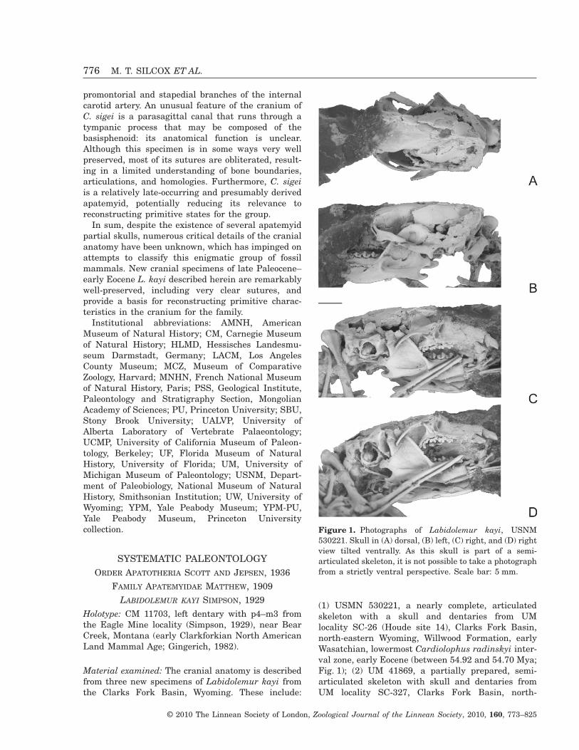
(1) USMN 530221, a nearly complete, articulatedskeleton with a skull and dentaries from UMlocality SC-26 (Houde site 14), Clarks Fork Basin,north-eastern Wyoming, Willwood Formation, earlyWasatchian, lowermost Cardiolophus radinskyi inter-val zone, early Eocene (between 54.92 and 54.70 Mya;Fig. 1); (2) UM 41869, a partially prepared, semi-articulated skeleton with skull and dentaries fromUM locality SC-327, Clarks Fork Basin, north-
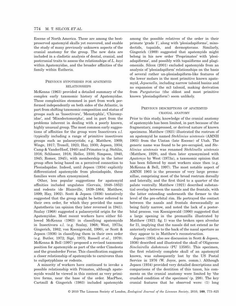
Figure 1. Photographs of Labidolemur kayi, USNM530221. Skull in (A) dorsal, (B) left, (C) right, and (D) rightview tilted ventrally. As this skull is part of a semi-articulated skeleton, it is not possible to take a photographfrom a strictly ventral perspective. Scale bar: 5 mm.
776 M. T. SILCOX ET AL.
© 2010 The Linnean Society of London, Zoological Journal of the Linnean Society, 2010, 160, 773–825
western Wyoming, lower Willwood Formation, lateClarkforkian, Phenacodus–Ectocion acme zone, latePaleocene (between 55.36 and 55.0 Mya; Fig. 2); and(3) USNM 530208, an associated rostrum and basic-ranium from UM locality SC-62 (Block Z), ClarksFork Basin, north-western Wyoming; lower WillwoodFormation, middle Clarkforkian, Uppermost Plesi-adapis cookei range zone, late Paleocene (between55.68 and 55.36 Mya; Fig. 3). See Gingerich (2003) forage model.
USNM 530221 and UM 41869 can be confidentlyattributed to Labidolemur on the basis of the pres-ence of a p3 and a short talonid on m3 (Gingerich &Rose, 1982). As for L. kayi, the molar teeth in thesespecimens are larger than in A. chardini andsmaller than in Labidolemur serus (Appendix 4),and the lower molars are higher crowned than thoseof A. chardini (Gingerich, 1982). The only apatemyidspecies currently documented from the Clarkforkianis L. kayi (Gingerich & Rose, 1982), which makesthis a likely identification for USNM 530208. Themorphology and size of the teeth in this specimenare similar to those of UM 41869 and USNM530221, and also to previously published specimensof L. kayi (e.g. UM 73616; Gingerich & Rose, 1982:text and fig. 1).
Occurrence: Late Paleocene (Clarkforkian NALMA)through early Eocene (Wasatchian NALMA).
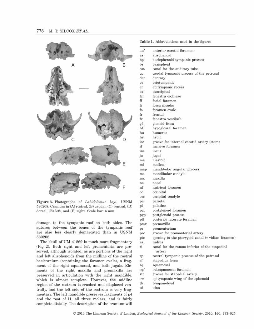
Discussion: All three new specimens of L. kayi pre-serve at least portions of the skull. USNM 530208 isthe best-preserved cranium, with an almost intactbasicranium and a well-preserved rostrum (Fig. 3).The specimen is broken into two pieces at the back ofthe orbital region, and has experienced some crushingboth near the site of the break, and rostrally andventrally on the left side. USNM 530208 preserves analmost completely intact tympanic roof on the rightside of the specimen, with well-demarcated sutures(Fig. 4, Table 1). The left basicranium of USNM530208 has some crushing to the promontorium,causing damage to the rostral pole and adjacent tym-panic roof, and a break into the promontorium later-ally, revealing part of the cochlea (Fig. 5).
USNM 530221 preserves the entire skull includingthe dentary (Fig. 1). The right side of the skull ispartially caved in, however, and the right mandiblehas become displaced so that it overlies much of themidline of the specimen. The rostral-most portions ofthis skull were embedded in epoxy during prepara-tion, and are not fully visible. In the basicranium,portions of both ectotympanics are still in place, asare fragments of the left malleus and incus (Fig. 6).However, the ultra high resolution X-ray computedtomography (uhrCT) data reveal that there is consid-erable damage to the auditory region in this speci-men, including a break running mediolaterallythrough the promontorium on the left side, and some
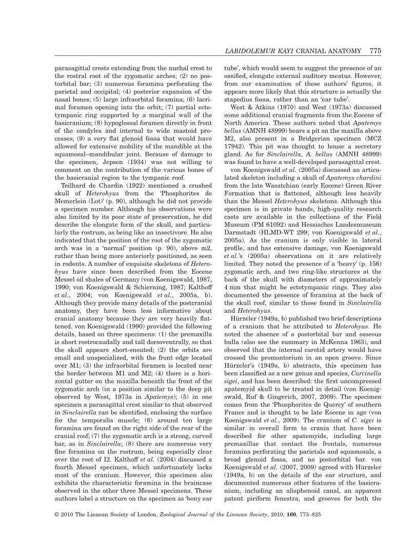
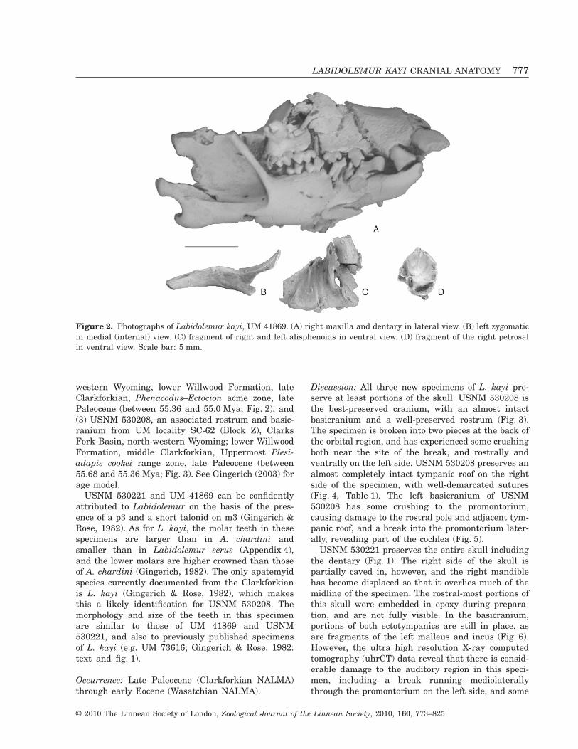
Figure 2. Photographs of Labidolemur kayi, UM 41869. (A) right maxilla and dentary in lateral view. (B) left zygomaticin medial (internal) view. (C) fragment of right and left alisphenoids in ventral view. (D) fragment of the right petrosalin ventral view. Scale bar: 5 mm.
LABIDOLEMUR KAYI CRANIAL ANATOMY 777
© 2010 The Linnean Society of London, Zoological Journal of the Linnean Society, 2010, 160, 773–825
damage to the tympanic roof on both sides. Thesutures between the bones of the tympanic roofare also less clearly demarcated than in USNM530208.
The skull of UM 41869 is much more fragmentary(Fig. 2). Both right and left promontoria are pre-served, although isolated, as are portions of the rightand left alisphenoids from the midline of the rostralbasicranium (containing the foramen ovale), a frag-ment of the right squamosal, and both jugals. Ele-ments of the right maxilla and premaxilla arepreserved in articulation with the right mandible,which is almost complete. However, the midlineregion of the rostrum is crushed and displaced ven-trally, and the left side of the rostrum is very frag-mentary. The left mandible preserves fragments of p4and the root of i1, all three molars, and is fairlycomplete distally. The description of the cranium will
Figure 3. Photographs of Labidolemur kayi, USNM530208. Cranium in (A) rostral, (B) caudal, (C) ventral, (D)dorsal, (E) left, and (F) right. Scale bar: 5 mm.
Table 1. Abbreviations used in the figures
acf anterior carotid foramenas alisphenoidbp basisphenoid tympanic processbs basisphoidcat canal for the auditory tubecp caudal tympanic process of the petrosalden dentaryec ectotympanicer epitympanic recessex exoccipitalfcf fenestra cochleaeff facial foramenfi fossa incudisfo foramen ovalefr frontalfv fenestra vestibuligf glenoid fossahf hypoglossal foramenhu humerushy hyoidicc groove for internal carotid artery (stem)if incisive forameninc incusju jugalma mastoidml malleusmap mandibular angular processmc mandibular condylemx maxillana nasalnf nutrient foramenoc occipitalocc occipital condylepa parietalpl palatinepgf postglenoid foramenpgp postglenoid processplf posterior lacerate foramenpmx premaxillapr promontoriumprc groove for promontorial arteryptc opening to the pterygoid canal (= vidian foramen)ra radiusri canal for the ramus inferior of the stapedial
arteryrp rostral tympanic process of the petrosalsf stapedius fossasq squamosalsqf subsquamosal foramenstc groove for stapedial arterysw epitympanic wing of the sphenoidth tympanohyalul ulna
778 M. T. SILCOX ET AL.
© 2010 The Linnean Society of London, Zoological Journal of the Linnean Society, 2010, 160, 773–825
be based primarily on USNM 530208, augmented byobservations on USNM 530221 and UM 41869.
Two specimens (USNM 530221 and 530208) werescanned with the OMNI-X Industrial Scanner at theCenter for Quantitative Imaging (CQI), PennsylvaniaState University, which produced uhrCT data. ForUSNM 530221, only the skull (and bones in its vicin-ity) was scanned. Only the caudal portion of USNM530208 was scanned (i.e. the portion caudal to thebreak in the specimen). These specimens were immo-bilized in floral foam (i.e. ‘oasis’) and scanned involume mode, in which 21 individual two-dimensionalslices were created for each rotation. The axial fanangle was small enough to assume parallel beamreconstructions. Each rotation consisted of 2400 viewsof the object spanning 360°. The post-acquisitionreconstruction process included all 2400 views, andeach individual slice was stored as a 1024 ¥ 1024matrix of 16-bit integers in tiff format. For USNM
530221 the reconstructed pixel size was 0.053 mmand the interslice distance was 0.058 mm; the dataset included 697 images. For USNM 530208, thereconstructed pixel size was 0.033 mm, the interslicedistance was 0.036 mm, and the data set consisted of361 images.
Images were studied using Scion Image Beta4.02 (Scion Corporation, 2002) and ImageJ 1.27w(Rasband, 2002). Parts of the data set were croppedand stacked using cropvoi and strip2raw, which areDOS programs developed by Nathan Jeffrey (Univer-sity of Liverpool). Reslicing of the data in arbitraryplanes, 2D image linking, and 3D reconstructionswere performed using Voxblast for Unix (Vaytek,Inc.) on an SGI Octane 2 workstation. Anatomicalterminology for the basicranium follows MacPhee(1981) and MacPhee, Novacek & Storch (1988). Ter-minology used to describe the rest of the skull followsNovacek (1986), unless otherwise noted.
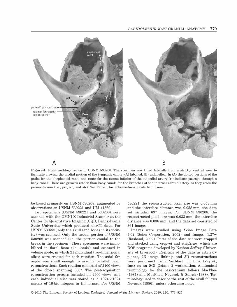
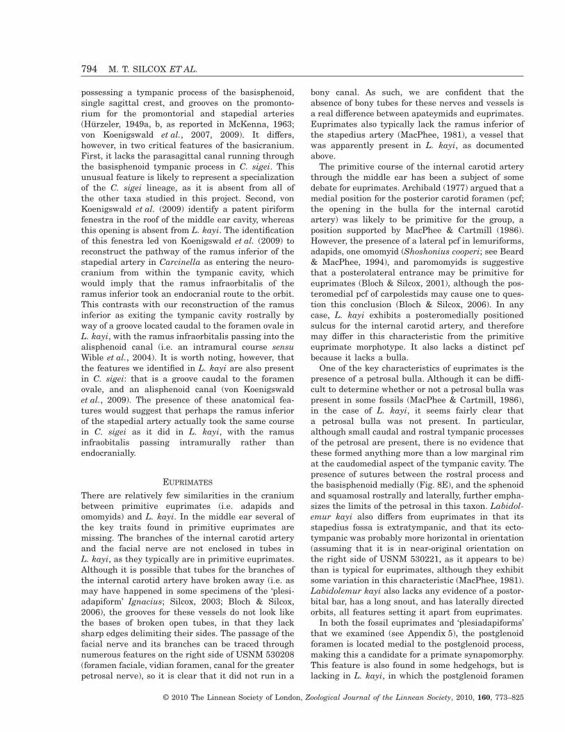
Figure 4. Right auditory region of USNM 530208. The specimen was tilted laterally from a strictly ventral view tofacilitate viewing the medial portion of the tympanic cavity: (A) labelled; (B) unlabelled. In (A) the dotted portions of thepaths for the alisphenoid canal and route for the ramus inferior of the stapedial artery (ri) indicate passage through abony canal. There are grooves rather than bony canals for the branches of the internal carotid artery as they cross thepromontorium (i.e., prc, icc, and stc). See Table 1 for abbreviations. Scale bar: 1 mm.
LABIDOLEMUR KAYI CRANIAL ANATOMY 779
© 2010 The Linnean Society of London, Zoological Journal of the Linnean Society, 2010, 160, 773–825
DESCRIPTIONTYMPANIC ROOF AND FLOOR
The tympanic roof in L. kayi is formed by threeelements: the epitympanic wing of the sphenoid(formed by the alisphenoid and basisphenoid;MacPhee, 1981), the petrosal, and the squamosal(Fig. 4). There is no evidence for a complete osseousauditory bulla in L. kayi. However, there is a tympanicprocess of the basisphenoid that extends along themedial extent of the tympanic cavity, from near thelevel of the foramen ovale rostrally, to meet the rostralprocess of the petrosal near the caudal extent of thetympanic cavity. A small alisphenoid tympanic processoverlies the ectotympanic rostrolaterally, and bears agroove for the ramus inferior of the stapedial artery(Fig. 4). A small caudal petrosal tympanic process ispresent, sitting caudal to the cochlear fenestra. Aspreserved, these various processes form more of a rimto the tympanic cavity than a floor. The ectotympanicappears to have been athictic or slightly semiphaneric,following the terminology of MacPhee (1981; Fig. 6).
SPHENOID (INCLUDING PRE-, ORBITO-, BASI-,AND ALISPHENOID, AND PTERYGOID)
The alisphenoid, basisphenoid, and pterygoid com-prise a single element without evidence of sutures,
and will therefore be discussed as a unit. A fragmentof bone that comprises a portion of the pre- andorbitosphenoid is preserved with the rostral portion ofUSNM 530208, displaced slightly off centre andpushed ventrally towards the back of the palate(Fig. 7). The optic canal is visible in its endocranialaspect on the right side of this bone, lateral to apronounced strut that would have formed the midlineof the mesocranial region.
In the basicranial region the sphenoid contacts thesquamosal, petrosal, and basioccipital. No specimen iswell enough preserved to demonstrate the rostralcontacts of the sphenoid. The uhrCT data (Fig. 8B)demonstrate that the alisphenoid extends internal tothe squamosal, such that it is dorsal to the glenoidfossa. Just lateral to the entopterygoid plate is theforamen ovale, and more rostrally, a well-preservedalisphenoid canal (Fig. 4). This canal runs rostrocau-dally, does not communicate with the braincase(Fig. 8A), and presumably carried the ramus infraor-bitalis of the ramus inferior of the stapedial artery(Wible, 1987; Wible, Novacek & Rougier, 2004). Thereis a well-demarcated groove located just caudal to theforamen ovale that is likely to have housed the ramusinferior of the stapedial artery (Fig. 4). This grooveextends into the tympanic cavity proper, through agroove in the alisphenoid tympanic process, to theforamen for the ramus superior of the stapedial
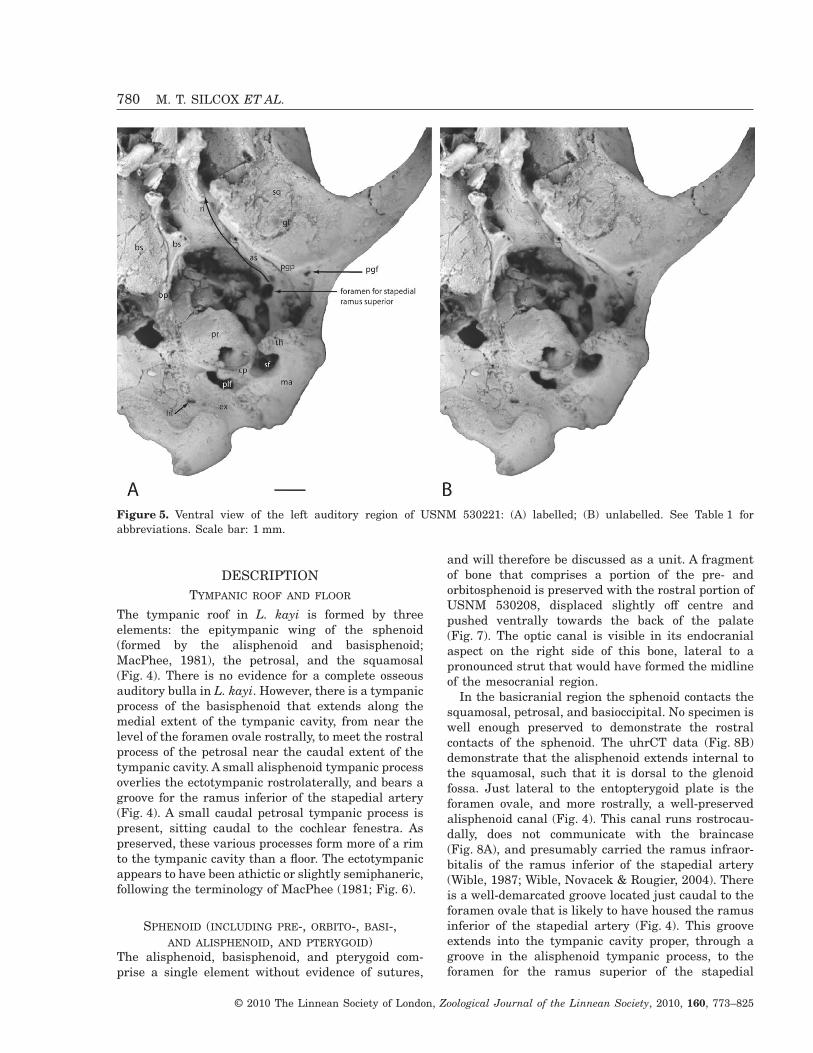
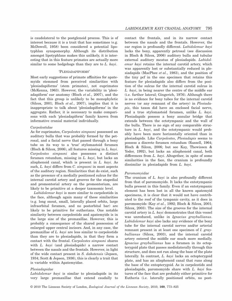
Figure 5. Ventral view of the left auditory region of USNM 530221: (A) labelled; (B) unlabelled. See Table 1 forabbreviations. Scale bar: 1 mm.
780 M. T. SILCOX ET AL.
© 2010 The Linnean Society of London, Zoological Journal of the Linnean Society, 2010, 160, 773–825
artery. A well-demarcated groove is also incised intothe sphenoid just lateral to the basisphenoid tym-panic process at the rostral extent of the tympaniccavity, which is likely to have housed the auditorytube (‘cat’ on Fig. 4). On the right side of USNM
530208, just caudal to the groove for the auditorytube, is a small foramen at the end of a short groove(‘ptc’ on Fig. 4). This is likely to be the opening to thepterygoid canal (= vidian foramen). An alternativeinterpretation is that it is the anterior carotid
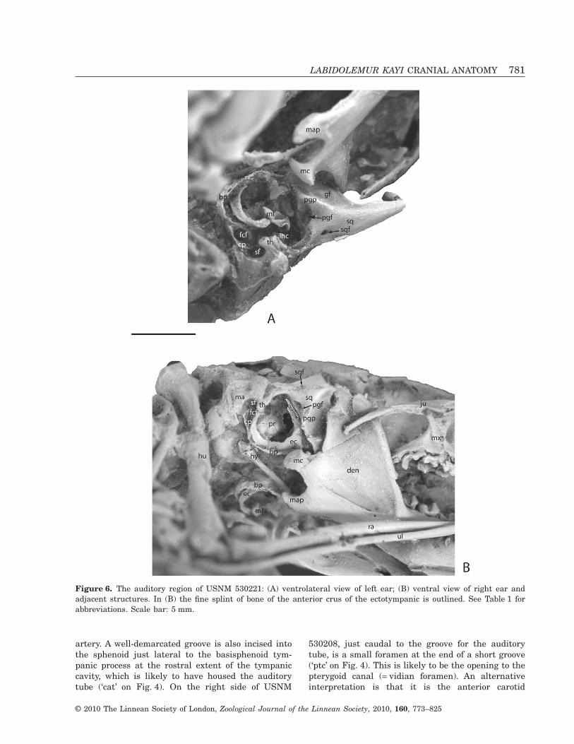
Figure 6. The auditory region of USNM 530221: (A) ventrolateral view of left ear; (B) ventral view of right ear andadjacent structures. In (B) the fine splint of bone of the anterior crus of the ectotympanic is outlined. See Table 1 forabbreviations. Scale bar: 5 mm.
LABIDOLEMUR KAYI CRANIAL ANATOMY 781
© 2010 The Linnean Society of London, Zoological Journal of the Linnean Society, 2010, 160, 773–825
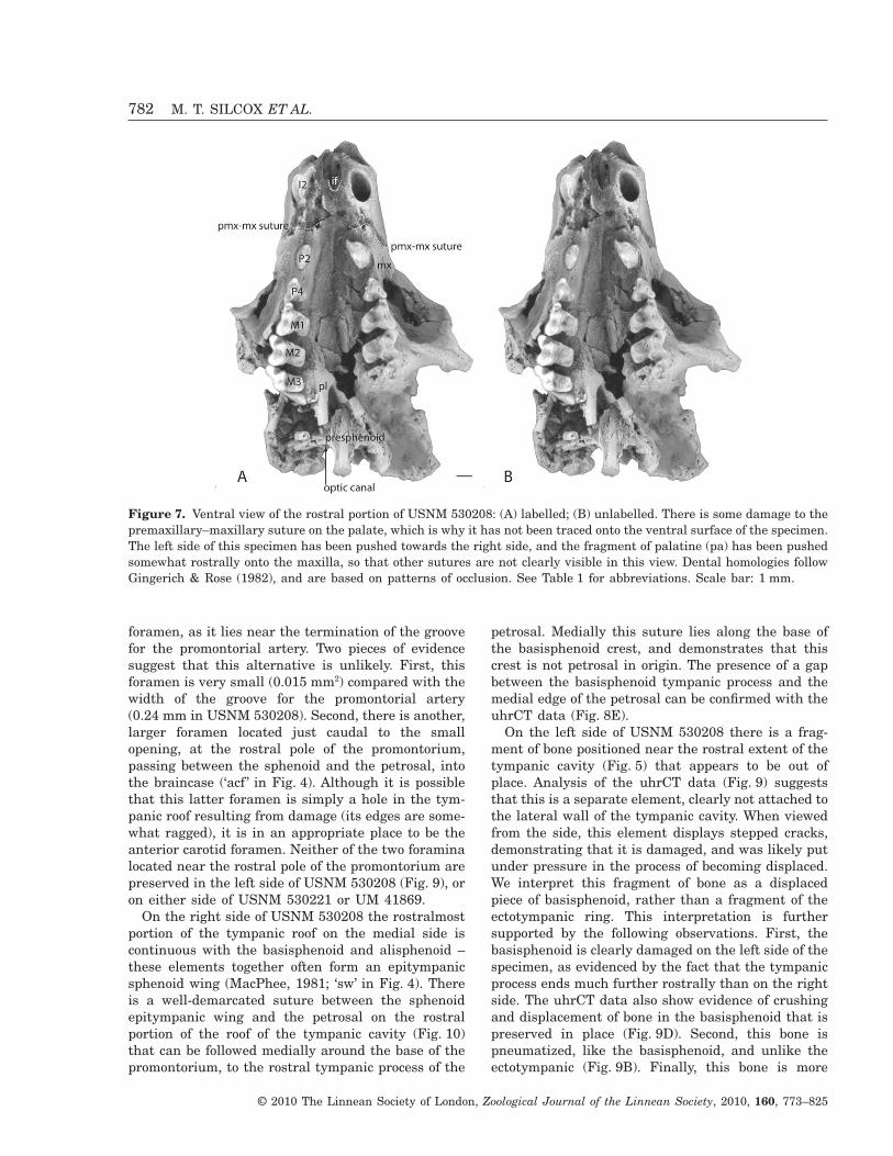
foramen, as it lies near the termination of the groovefor the promontorial artery. Two pieces of evidencesuggest that this alternative is unlikely. First, thisforamen is very small (0.015 mm2) compared with thewidth of the groove for the promontorial artery(0.24 mm in USNM 530208). Second, there is another,larger foramen located just caudal to the smallopening, at the rostral pole of the promontorium,passing between the sphenoid and the petrosal, intothe braincase (‘acf ’ in Fig. 4). Although it is possiblethat this latter foramen is simply a hole in the tym-panic roof resulting from damage (its edges are some-what ragged), it is in an appropriate place to be theanterior carotid foramen. Neither of the two foraminalocated near the rostral pole of the promontorium arepreserved in the left side of USNM 530208 (Fig. 9), oron either side of USNM 530221 or UM 41869.
On the right side of USNM 530208 the rostralmostportion of the tympanic roof on the medial side iscontinuous with the basisphenoid and alisphenoid –these elements together often form an epitympanicsphenoid wing (MacPhee, 1981; ‘sw’ in Fig. 4). Thereis a well-demarcated suture between the sphenoidepitympanic wing and the petrosal on the rostralportion of the roof of the tympanic cavity (Fig. 10)that can be followed medially around the base of thepromontorium, to the rostral tympanic process of the
petrosal. Medially this suture lies along the base ofthe basisphenoid crest, and demonstrates that thiscrest is not petrosal in origin. The presence of a gapbetween the basisphenoid tympanic process and themedial edge of the petrosal can be confirmed with theuhrCT data (Fig. 8E).
On the left side of USNM 530208 there is a frag-ment of bone positioned near the rostral extent of thetympanic cavity (Fig. 5) that appears to be out ofplace. Analysis of the uhrCT data (Fig. 9) suggeststhat this is a separate element, clearly not attached tothe lateral wall of the tympanic cavity. When viewedfrom the side, this element displays stepped cracks,demonstrating that it is damaged, and was likely putunder pressure in the process of becoming displaced.We interpret this fragment of bone as a displacedpiece of basisphenoid, rather than a fragment of theectotympanic ring. This interpretation is furthersupported by the following observations. First, thebasisphenoid is clearly damaged on the left side of thespecimen, as evidenced by the fact that the tympanicprocess ends much further rostrally than on the rightside. The uhrCT data also show evidence of crushingand displacement of bone in the basisphenoid that ispreserved in place (Fig. 9D). Second, this bone ispneumatized, like the basisphenoid, and unlike theectotympanic (Fig. 9B). Finally, this bone is more
Figure 7. Ventral view of the rostral portion of USNM 530208: (A) labelled; (B) unlabelled. There is some damage to thepremaxillary–maxillary suture on the palate, which is why it has not been traced onto the ventral surface of the specimen.The left side of this specimen has been pushed towards the right side, and the fragment of palatine (pa) has been pushedsomewhat rostrally onto the maxilla, so that other sutures are not clearly visible in this view. Dental homologies followGingerich & Rose (1982), and are based on patterns of occlusion. See Table 1 for abbreviations. Scale bar: 1 mm.
782 M. T. SILCOX ET AL.
© 2010 The Linnean Society of London, Zoological Journal of the Linnean Society, 2010, 160, 773–825
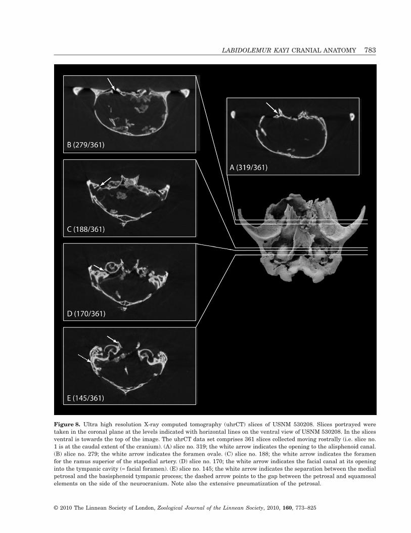
Figure 8. Ultra high resolution X-ray computed tomography (uhrCT) slices of USNM 530208. Slices portrayed weretaken in the coronal plane at the levels indicated with horizontal lines on the ventral view of USNM 530208. In the slicesventral is towards the top of the image. The uhrCT data set comprises 361 slices collected moving rostrally (i.e. slice no.1 is at the caudal extent of the cranium). (A) slice no. 319; the white arrow indicates the opening to the alisphenoid canal.(B) slice no. 279; the white arrow indicates the foramen ovale. (C) slice no. 188; the white arrow indicates the foramenfor the ramus superior of the stapedial artery. (D) slice no. 170; the white arrow indicates the facial canal at its openinginto the tympanic cavity (= facial foramen). (E) slice no. 145; the white arrow indicates the separation between the medialpetrosal and the basisphenoid tympanic process; the dashed arrow points to the gap between the petrosal and squamosalelements on the side of the neurocranium. Note also the extensive pneumatization of the petrosal.
LABIDOLEMUR KAYI CRANIAL ANATOMY 783
© 2010 The Linnean Society of London, Zoological Journal of the Linnean Society, 2010, 160, 773–825
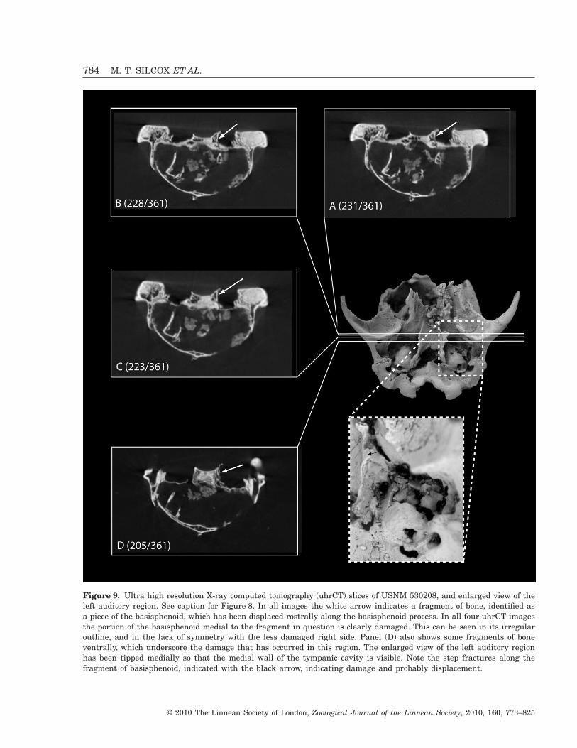
Figure 9. Ultra high resolution X-ray computed tomography (uhrCT) slices of USNM 530208, and enlarged view of theleft auditory region. See caption for Figure 8. In all images the white arrow indicates a fragment of bone, identified asa piece of the basisphenoid, which has been displaced rostrally along the basisphenoid process. In all four uhrCT imagesthe portion of the basisphenoid medial to the fragment in question is clearly damaged. This can be seen in its irregularoutline, and in the lack of symmetry with the less damaged right side. Panel (D) also shows some fragments of boneventrally, which underscore the damage that has occurred in this region. The enlarged view of the left auditory regionhas been tipped medially so that the medial wall of the tympanic cavity is visible. Note the step fractures along thefragment of basisphenoid, indicated with the black arrow, indicating damage and probably displacement.
784 M. T. SILCOX ET AL.
© 2010 The Linnean Society of London, Zoological Journal of the Linnean Society, 2010, 160, 773–825
expansive dorsoventrally than the ectotympanics pre-served in USNM 530221, extending dorsally towardsthe roof of the tympanic cavity.
Although this fragment of bone is out of place,it does suggest that the tympanic process of thebasisphenoid may have extended further over theectotympanic than it would appear to on the rightside of this specimen, contributing to a more expan-sive tympanic floor. This impression is supported bytwo observations. First, in USNM 530221, on theright side the ectotympanic is nearly in place and thebasisphenoid tympanic process appears to curl overthe ectotympanic (Fig. 6A). Second, the right side ofUSNM 530208 exhibits a broken edge along the tym-panic process from near the rostral extent of theauditory cavity, so it cannot be used as a reliableguide as to the extent of this process, implying that alarger element was originally present.
In the tympanic roof, the lateral portion of thealisphenoid extends caudally to a suture locatedrostral to the foramen for the ramus superior of thestapedial artery (Fig. 10). This portion of the boneextends as a narrow process passing between thesquamosal and petrosal. There is a small tympanicprocess that arises from this portion of the alisphenoid(Fig. 4). The edge of this bone houses one side of a canalpassing from the tympanic cavity rostrally (the brokenedge of this canal is visible on both the right and leftsides of USNM 530208). As this canal appears to leadinto the groove for the ramus inferior of the stapedialartery, it presumably carried this artery.
The precise location of the spheno-occipital syn-chondrosis is not clear, because a portion of the
central stem is caved-in distally in USNM 530208(Fig. 3), and the hyoid apparatus overlies the regionin USNM 530221 (Fig. 6A). Analysis of uhrCT datashow that the basisphenoid tympanic process origi-nates rostrally from the sphenoid, and is continuousalong its length, with no evidence of any sutures.Thus, it can be confidently identified as beingbasisphenoid, rather than basioccipital, in origin.
PETROSAL (= PETROMASTOID)The petrosal forms much of the roof of the tympaniccavity. It contacts the sphenoid rostrally, medially,and laterally, forming the caudal wall of the presum-ptive anterior carotid foramen, as discussed above(Fig. 10). The mastoid portion of the petrosal is fairlylarge, housing relatively large semicircular canals,and is extensively pneumatized (Figs 8E, 11D). Thisportion of the petrosal contacts the exoccipital medi-ally (Fig. 4), and is overlain laterally by the squamo-sal (Fig. 8E). A suture is also present laterallybetween the petrosal and the squamosal on thelateral wall of the bulla (Fig. 4), demonstrating thatthe epitympanic recess was roofed by both these ele-ments. A small indentation caudal to the epitympanicrecess may be the fossa incudis for the short processof the incus (MacPhee, 1981). The petrosal bearsfairly small caudal and rostral tympanic processes(Fig. 4). A suture can be traced separating the rostralprocess from the basisphenoid tympanic process onthe right side of USNM 530208: it extends rostrally toapproximately the same level as the rostral extent ofthe fenestra vestibuli. The caudal process is also
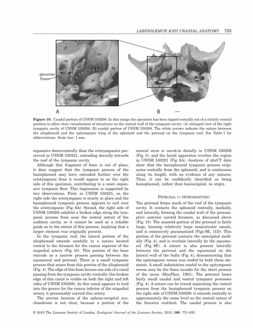
Figure 10. Caudal portion of USNM 530208. In this image the specimen has been tipped rostrally out of a strictly ventralposition to allow clear visualization of structures on the rostral wall of the tympanic cavity: (A) enlarged view of the righttympanic cavity of USNM 530208; (B) caudal portion of USNM 530208. The white arrows indicate the suture betweenthe alisphenoid and the epitympanic wing of the sphenoid and the petrosal on the tympanic roof. See Table 1 forabbreviations. Scale bar: 1 mm.
LABIDOLEMUR KAYI CRANIAL ANATOMY 785
© 2010 The Linnean Society of London, Zoological Journal of the Linnean Society, 2010, 160, 773–825
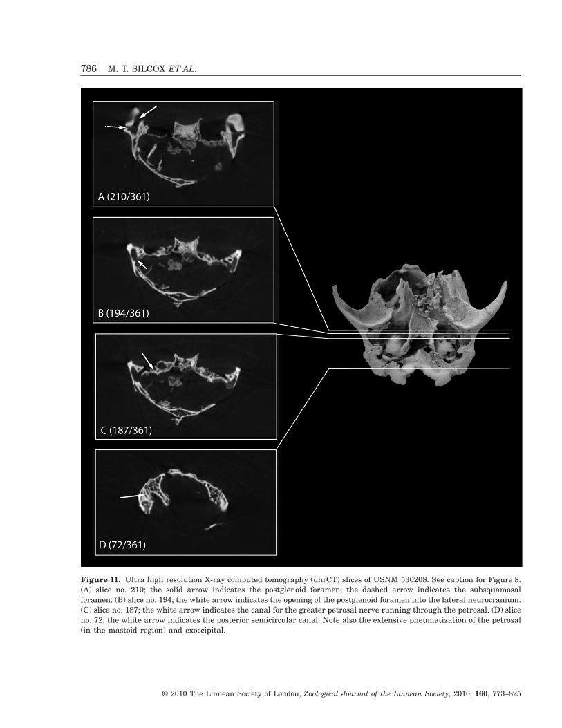
Figure 11. Ultra high resolution X-ray computed tomography (uhrCT) slices of USNM 530208. See caption for Figure 8.(A) slice no. 210; the solid arrow indicates the postglenoid foramen; the dashed arrow indicates the subsquamosalforamen. (B) slice no. 194; the white arrow indicates the opening of the postglenoid foramen into the lateral neurocranium.(C) slice no. 187; the white arrow indicates the canal for the greater petrosal nerve running through the petrosal. (D) sliceno. 72; the white arrow indicates the posterior semicircular canal. Note also the extensive pneumatization of the petrosal(in the mastoid region) and exoccipital.
786 M. T. SILCOX ET AL.
© 2010 The Linnean Society of London, Zoological Journal of the Linnean Society, 2010, 160, 773–825
preserved in this specimen, located just caudal to thefenestra cochleae, and separated by a gap from thetympanohyal (remnant of Reichert’s cartilage;MacPhee, 1981). Just caudal to this gap is the stape-dius fossa. The origin of the stapedius muscle wouldhave been extratympanic: this is clear in USNM530221, in which the stapedius fossa is preservedwith an ectotympanic that is nearly in place on theright-hand side (Fig. 6A). The posterior lacerateforamen is located just caudal to the caudal process,between the petrosal and the exoccipital (Figs 4, 5).There is only a single posterior lacerate foramen thatpresumably accommodated both the internal jugularvein and cranial nerves IX–XI. The promontorium isrelatively bulbous and inflated (e.g. in contrast to thecondition in leptictids), suggesting a long cochlea maybe present, although this awaits confirmation with anappropriate comparative data set of cochlear lengths(e.g. as alluded to by Kirk & Gosselin-Ildari,2009).
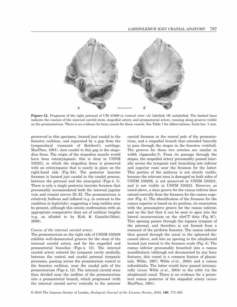
Course of the internal carotid arteryThe promontorium on the right side of USNM 530208exhibits well-demarcated grooves for the stem of theinternal carotid artery, and for the stapedial andpromontorial branches (Figs 4, 12). The internalcarotid artery entered the tympanic cavity medially,between the rostral and caudal petrosal tympanicprocesses, passing across the promotorium rostral tothe fenestra cochleae, near the caudal pole of thepromontorium (Figs 4, 12). The internal carotid stemthen divided near the midline of the promontoriuminto a promontorial branch, which progressed (withthe internal carotid nerve) rostrally to the anterior
carotid foramen at the rostral pole of the promonto-rium, and a stapedial branch that extended laterallyto pass through the stapes in the fenestra vestibuli.The grooves for these two arteries are similar inwidth (Appendix 3). From its passage through thestapes, the stapedial artery presumably passed later-ally across the tympanic roof, branching into inferiorand superior rami near the foramen for the latter.This portion of the pathway is not clearly visible,because the relevant area is damaged on both sides ofUSNM 530208, is not preserved in USNM 530221,and is not visible in USNM 530221. However, asnoted above, a clear groove for the ramus inferior doesextend rostrally from the foramen for the ramus supe-rior (Fig. 4). The identification of the foramen for theramus superior is based on its position, its associationwith the presumptive groove for the ramus inferior,and on the fact that it can be seen to open into thelateral neurocranium on the uhrCT data (Fig. 8C).This opening passes through the tegmen tympani ofthe petrosal, and therefore is not formed from aremnant of the piriform fenestra. The ramus inferiorthen passed through the canal in the sphenoid dis-cussed above, and into an opening in the alisphenoidlocated just rostral to the foramen ovale (Fig. 4). Theramus inferior presumably branched into a ramusmandibularis (although not documented by any bonyfeatures, this vessel is a common feature of placen-tals; Wible, 1987; Wible et al., 2004) and a ramusinfraobitalis. The latter would have passed intramu-rally (sensu Wible et al., 2004) to the orbit via thealisphenoid canal. There is no evidence for a persis-tent ramus posterior of the stapedial artery (sensuMacPhee, 1981).
Figure 12. Fragment of the right petrosal of UM 41869 in ventral view: (A) labelled; (B) unlabelled. The dashed linesindicate the courses of the internal carotid stem, stapedial artery, and promontorial artery, running along grooves visibleon the promontorium. There is no evidence for bony canals for these vessels. See Table 1 for abbreviations. Scale bar: 1 mm.
LABIDOLEMUR KAYI CRANIAL ANATOMY 787
© 2010 The Linnean Society of London, Zoological Journal of the Linnean Society, 2010, 160, 773–825

Course of the facial nerveThere is a small foramen (0.23 mm2) present at thebase of the promontorium, just rostral to the fenestravestibuli on the right side of USNM 530208 and theright promontorium of UM 41869 (Figs 4, 12; thisarea is damaged or not visible in the other speci-mens). This foramen can be traced on the uhrCT datafor USNM 530208 through the petrosal into theneurocranium (Fig. 8D). In light of its course, thisopening is best interpreted as the foramen for thefacial nerve. The main trunk of the facial nerve wouldpresumably have exited the tympanic cavity througha gap between the caudal petrosal process and thetympanohyal, near the fossa for the stapedius muscle.This gap then functions as the ‘foramen styloma-stoideum primitivum’ (Bromann, 1899, as cited inMacPhee, 1981), and there is no ‘definitive’ stylomas-toid foramen. There is a very small (diameter~0.17 mm) canal that runs rostrally from the facialcanal in its passage through the petrosal, visible inthe uhrCT data (Fig. 11C), that exits near the rostralpole of the promontorium on its lateral side, presum-ably for the greater petrosal nerve, making this canalthe hiatus fallopii. The exit for this canal into thetympanic cavity is not clearly visible in USNM530208 because it lies underneath a displaced frag-ment of bone. From this point, no features associatedwith the pathway of the greater petrosal nerve arevisible, until it joins the deep petrosal branch of theinternal carotid nerve to form the nerve of the ptery-goid canal, passing through the vidian foramenrostral to the rostral pole of the promontorium (seediscussion above).
SQUAMOSAL
The squamosal contacts the petrosal laterally, overly-ing this bone on the side of the cranium and meetingthe parietal to form part of the neurocranium(Fig. 8E). The squamosal contacts the sphenoid in asuture located rostrolateral to the alisphenoid tym-panic process (Fig. 10). More rostrally the squamosaloverlies the sphenoid (Fig. 8B), as discussed above,extending dorsally to contact the parietals. The squa-mosal has a fairly long zygomatic process (Fig. 13)that contacts the jugal.
The glenoid fossa is fairly wide (4.53 mm in USNM530208) and flat. The postglenoid process sits nearthe medial extent of the fossa, and is narrow rostro-caudally, descending as a lip to a point (rather than abulbous process as seen in some mammals; Figs 4, 5).Using the uhrCT data, caudal and lateral to thepostglenoid process a well-demarcated postglenoidforamen (average area = 0.51 mm2) can be traced to along canal that leads into the lateral neurocranium(Fig. 11B). This foramen would presumably have
transmitted a persistent petrosquamous sinus(MacPhee, 1981). There is also a small (average =0.12 mm2) subsquamosal foramen (sometimesreferred to as the suprameatal foramen, but see dis-cussion in Beard & MacPhee 1994) located lateral tothe postglenoid foramen, at the base of the zygomaticprocess of the squamosal. Analysis of the uhrCT datashows that these foramina are continuous in thesquamosal (Fig. 11A), so the subsquamosal foramenprobably transmitted a vein associated with thepetrosquamous sinus.
There is no entoglenoid process. A groove incisedmedial to the postglenoid process and caudal to theglenoid fossa is interpreted as the Glaserian fissure,probably marking the course of the chorda tympani(Fig. 10).
ECTOTYMPANIC
The ectotympanics are preserved nearly in place onthe right side of USNM 530221 (Fig. 6A). The ecto-tympanics appear to have been relatively free, withbony contacts (although not necessarily sutures) withthe sphenoid, squamosal, and petrosal, rostrally andlaterally, basisphenoid tympanic process and rostraltympanic process of the petrosal, medially, and caudaltympanic process of the petrosal, caudally (Fig. 6A).As noted above, the ectotympanic appears to havebeen either athictic or semiphaneric (because of aslight overlap medially by the basisphenoid; Fig. 6A).The ectotympanic is most expanded at its rostrome-dial corner, where it is fairly wide in the frontal plane(maximum width = 1.72 mm). In the frontal planethe ectotympanic narrows laterally from this point(minimum width = 0.29), ultimately forming a veryfine splint of bone (anterior crus) running along agroove located along the sutures between the sphe-noid and squamosal, and the sphenoid and petrosal(Fig. 6A). Although this splint is broken away in theleft side of USNM 530221, the position of the ecto-tympanic relative to the malleus suggests that thissplint may have contacted this ossicle, because thebroken end of the ectotympanic is curving towardsthis bone in this specimen, although the malleus istoo incompletely preserved to confirm this (Fig. 6B).Although it is unclear whether the ectotympanic ringwas complete laterally (because the lateral edgesof the bone are not perfectly preserved, suggestingsome breakage may have occurred), the generaloutline is more circular than horseshoe-shaped (inter-nal diameter = ~3.0 mm).
ENTOTYMPANIC
There is no evidence for entotympanic elements in theear of L. kayi. It is possible that the fragment of bone
788 M. T. SILCOX ET AL.
© 2010 The Linnean Society of London, Zoological Journal of the Linnean Society, 2010, 160, 773–825
preserved at the rostromedial corner of the left ear ofUSNM 530208 (Fig. 5, labelled ‘bs’), identified aboveas a probable fragment of basisphenoid, is actually aremnant of an entotympanic. However, there is noadditional evidence in support of this interpretation,
and considering the damage to the basisphenoid inthis region, it seems more likely that it is a displacedfragment of this bone, as discussed above. AsMcKenna (1963) noted, in light of the variable pres-ervation of entotympanics in leptictids (perhaps one
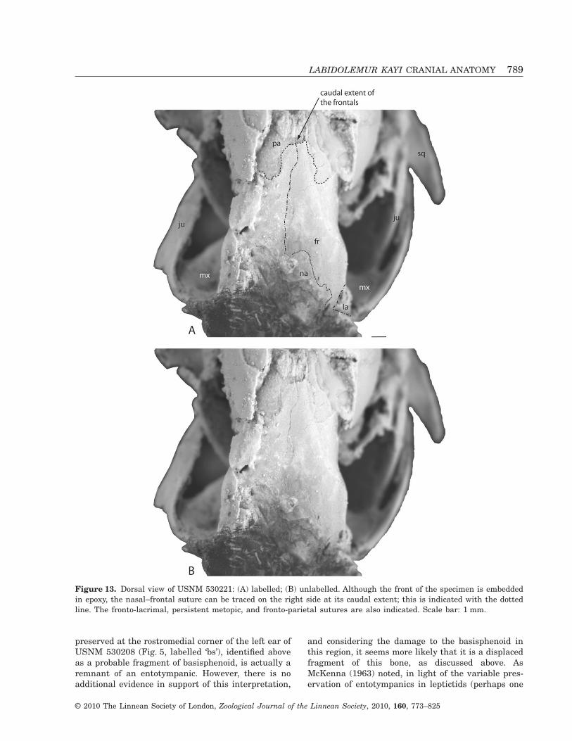
Figure 13. Dorsal view of USNM 530221: (A) labelled; (B) unlabelled. Although the front of the specimen is embeddedin epoxy, the nasal–frontal suture can be traced on the right side at its caudal extent; this is indicated with the dottedline. The fronto-lacrimal, persistent metopic, and fronto-parietal sutures are also indicated. Scale bar: 1 mm.
LABIDOLEMUR KAYI CRANIAL ANATOMY 789
© 2010 The Linnean Society of London, Zoological Journal of the Linnean Society, 2010, 160, 773–825

in every ten specimens), it is possible that an ento-tympanic element was present in apatemyids eventhough evidence of one has never been found.However, unlike in leptictids (Novacek, 1986), there isno sign of a ridge on the promontorium for contactwith the entotympanic. There are also no clear articu-lar surfaces on any of the tympanic processes thatwould have accommodated an entotympanic bulla.Therefore, although a certain degree of caution isappropriate when discussing an element as poten-tially easily dislodged as the entotympanic, it can bestated that there is no positive evidence that such abone was present in the tympanic floor of Labidol-emur. With more confidence, it can be observed thatthe entotympanic did not form part of the tympanicroof in this species, because the roof is preservednearly intact on the right side of USNM 530208, andthere is no evidence of an entotympanic contributionto it.
OCCIPITAL
There are no clear sutures between the supra-, ex-,and basioccipital elements, so these elements will bediscussed together. As noted above, the precise posi-tion of the spheno-occipital synchondrosis is unclearin USNM 530208 because of damage to the centralstem, but it probably fell fairly far caudally as thereis no evidence of a suture along the length of thebasisphenoid tympanic process. Laterally the exoc-cipital contacts the mastoid portion of the petrosal(Fig. 4). On the side of the skull the exoccipital and/or
supraoccipital form a limited contact with the squa-mosal, and a more extensive contact with the pari-etals (Fig. 14). The occipital–parietal suture sits fairlyfar rostrally, at the level of the zygomatic process ofthe squamosal, so the occipital forms a substantialportion of the caudal neurocranium. The supraoccipi-tal bears a low sagittal crest, which does not extendrostrally beyond the occipital–parietal suture. A well-developed nuchal crest is also present, forming acompound crest with the sagittal (Fig. 14).
On the ventral side of the skull, the posterior lac-erate foramen lies between the exoccipital and thepetrosal, caudal to the fenestra cochleae (Figs 4, 5).The small hypoglossal foramen (0.051 mm2) forcranial nerve XII is well preserved on the left side ofUSNM 530208, but is partially broken away on theright (Figs 4, 5). The foramen magnum faces directlyposteriorly (Fig. 3). There is a tiny nutrient foramen(0.028 mm2; ‘nf ’ in Fig. 14) on the dorsal aspect, butno evidence for any other foramina in the occipital ofLabidolemur.
JUGAL (= ZYGOMATIC)Jugals are preserved on both sides of USNM 530221and isolated in UM 41869 (Fig. 15). They are fairlylong, stretching approximately 70% of the length ofthe zygomatic arch (see Appendix 3). Rostrally, at thecontact with the zygomatic process of the maxilla, thejugal is bifurcated into dorsal and ventral processes,with the dorsal process being much longer (Fig. 15).Distally, the jugal narrows to a point, overlapping the
Figure 14. Dorsal view of the caudal portion of USNM 530208. The dashed lines indicate the sutures between theoccipital and the parietals and the squamosal and parietal. Scale bar: 5 mm.
790 M. T. SILCOX ET AL.
© 2010 The Linnean Society of London, Zoological Journal of the Linnean Society, 2010, 160, 773–825

pointed zygomatic process of the squamosal, as indi-cated by a groove on the dorsomedial surface of thecaudal end of the jugal (Fig. 15). On the medial (inter-nal) surface of the jugal a strong ridge extends cau-dally more than half the length of the bone (Fig. 15).This is presumably the origin of the masseter muscle.No foramina are preserved on the jugals, althoughthis might be a product of preservation. There is noprocess present on the jugal that would have formedthe base of a postorbital bar.
PARIETAL
The parietals contact the occipital and squamosalcaudally (Fig. 14), and presumably the frontal ros-trally, although this suture is not clearly preserved inany specimen. The poor preservation of the caudalorbital region in all specimens makes the contacts ofthe parietals in this area unclear. There are noforamina piercing the parietal in the caudolateralbraincase. As noted above, the sagittal crest from theoccipital does not extend rostrally along the sagittalsuture (Fig. 14), and parasagittal crests are absent.
The parietals are distinctly convex in the neuro-cranium, so that the braincase appears somewhatinflated.
FRONTAL
The frontals contact the nasals and premaxillae ros-trally, the parietals caudally, and the lacrimals andmaxillae rostral to the orbit. The frontals extendventrally for most of the height of the orbit, meetingthe maxilla near the base of the orbit (Fig. 16). Apartfrom its contact with the parietal caudally, the othercontacts of the frontal in the orbit are not clearbecause of damage in this region. The dorsal extent ofthe orbit is indicated by a low crest on the frontal,which ends in a blunt swelling caudally that is notextended into a distinct process ventrally (i.e. postor-bital process is absent). This crest also does notextend distally into a parasagittal crest. The frontal–parietal suture is hard to discern in USNM 530221because of damage to this area, making it difficult todifferentiate cracks from sutures. However, the likelyposition of the caudal extent of the frontal dorsally is
Figure 15. Left isolated zygomatic of UM1 in medial (internal) view. Scale bar: 5 mm.
Figure 16. Lateral view of USNM 530208. The arrow indicates the fronto-maxillary suture, near the base of the orbit.Its position indicates that the maxilla was not expanded into the orbit, unlike the condition of eulipotyphlans. Its positionalso indicates that the palatine is not expanded into the orbit. Scale bar: 5 mm.
LABIDOLEMUR KAYI CRANIAL ANATOMY 791
© 2010 The Linnean Society of London, Zoological Journal of the Linnean Society, 2010, 160, 773–825

indicated in Figure 13, based on the presence of anapparent suture at this point, and on comparisonwith Sinclairella (Scott & Jepsen, 1936: plate VI). InUM 41869 a fragment of this suture is preserved inplace, positioned rostral to the position of the back ofthe frontal in USNM 530221, which supports theinterpretation that the suture curves rostroventrally,as indicated on Figure 13. The metopic suture can beseen to be persistent in USNM 530221 (Fig. 13).
NASAL
Fragments of nasals are preserved in USNM 530208(Fig. 3) and UM 41869, and the caudal end of theleft nasal is visible in USNM 530221, at its suturewith the frontal (Fig. 13). The nasals were long thinbones that contacted the premaxillae laterally andthe frontal caudally. The very large premaxillaeexclude the nasals from contact with the maxillae, atleast laterally. The nasals widen distally, then narrowjust rostral to their contact with the frontal (Fig. 13).The rostral ends of the nasals are preserved in UM41869, although the bones are somewhat out of place.They are sharply pointed laterally, and then curvestrongly medially, so the nasal cavity was relativelyopen rostrodorsally, and the nasals did not overhangthe front of the nasal cavity.
PREMAXILLA
The premaxillae of L. kayi are relatively large,forming a substantial portion of the rostrum (Fig. 16).On the lateral sides of the skull they lie betweenthe nasals and the maxillae, extending caudally tocontact the frontals. The premaxillae form the front~25% of the palate (Fig. 7). The suture with themaxilla is somewhat damaged in USNM 530208, andis not visible in USNM 530221 or UM 41869. Theincisive foramina were contained in the premaxillae.The caudal portion of the left incisive foramen ispreserved in USNM 53208 (Fig. 7), although it is tooincomplete to be accurately measured.
PALATINE
The palatine is not well preserved in any specimen,although a fragment of the palatal process of thisbone is present at the right corner of the palate inUSNM 530208 (Fig. 7). The suture between this frag-ment and the maxilla is not clear because the frag-ment of palatine has been pushed rostrally slightlyover the caudal edge of the maxilla. The posterioremargination of the palate appears to have extendedrostrally to near the level of the front of M3. Themorphology of the distal palatine is unclear, as thisfragment of bone has also been pushed somewhat
dorsally. However, it is clear from the preservedmorphology that the back of the palate did not sporta straight, very pronounced, bar-like postpalatinetorus. The position of the frontomaxillary suture, nearthe base of the orbit, indicates that the palatine wasnot expanded into the orbit (Fig. 16).
MAXILLA
The maxilla contacts the premaxilla rostrally, andalong the side of the rostrum (Fig. 7), and the jugal atthe zygomatic process of the maxilla. It contacted thepalatine at the back of the palate, although the suturewith this bone is not clearly preserved in any speci-men. As noted above, the premaxilla excluded themaxilla from contact with the frontal on the rostrum,but the maxilla and frontal were in contact near thebase of the orbit (Fig. 16). The maxilla is pierced bya very large (1.57 mm2 in USNM 530221), roundinfraorbital foramen for the infraorbital nerve (abranch of V2), artery, and vein, located above M1, thatleads into the orbit without forming a canal (i.e. thezygomatic process of the maxilla is quite thin).
LACRIMAL
The lacrimal foramen is preserved on the left sidesof both USNM 530221 and 530208. Although thisforamen was plainly intraorbital, the caudal (orbital)contacts of the lacrimal are not visible, making itunclear whether or not the foramen was entirely inthe lacrimal. The suture between the frontal andlacrimal is clear on the left side of USNM 530221(Fig. 13), and it is obvious that the lacrimal was smallwithout a significant facial process.
DENTARY
Portions of the dentary are preserved on both sides ofUSNM 530221 and UM 41869 (Figs 2, 6). This bonehas a tall ascending ramus, stout, cylindrical condyle,and very strong angular process that extends cau-dally to the level of the neck of the condyle. Theangular process is distally inflected and faces medi-ally (Fig. 6A). The condyle would not have occupiedthe entire (rather flat) glenoid fossa of the squamosal,and the postglenoid process is quite weak, so thedentary would have been quite mobile at its joint withthe squamosal. There is a trio of mental foraminaon the right dentary of UM 41869, located beneathm1–m2 (Fig. 2). The left dentary of this specimenbears two foramina, under the rostral roots of m1 andm2, respectively. The right dentary of USNM 530221bears only a single mental foramen under m2,whereas there is at least one mental foramen on theleft side under m1 (this region is partly obscured by
792 M. T. SILCOX ET AL.
© 2010 The Linnean Society of London, Zoological Journal of the Linnean Society, 2010, 160, 773–825
the ulna and radius, which overlie the bone; Fig. 1).In other words, the number and position of themental foramina are variable in this taxon, evenwithin the same specimen, although in all cases theyare located either under m1 or further posterior. UM41869 and USNM 530221 both exhibit a deep masse-teric fossa extending below m3.
COMPARISONS
A list of the critical references and specimens referredto for the comparisons and phylogenetic analysis isprovided as Appendix 5.
OTHER APATEMYIDS
Labidolemur kayi exhibits numerous differences fromSinclairella dakotensis (PU 13585) as described byJepsen (1934). It lacks three of the most prominentfeatures of this skull: the long parasagittal crests, thelarge foramina in the parietal and occipital bones,and the wide caudal contact between the nasals andthe frontals. Nonetheless, L. kayi does share withS. dakotensis the presence of a large infraorbitalforamen, a lacrimal foramen that opens into the orbit,and a very flat glenoid fossa. Jepsen’s (1934) descrip-tion of the auditory region as bearing a ‘marginalwall’ (p. 299) supporting the ectotympanic also seemsto be similar to the series of tympanic processesforming a rim around the tympanic cavity docu-mented here. Labidolemur kayi also differs fromApatemys bellus (AMNH 48999; West, 1973a) inlacking a marked parasagittal crest, but is similar inthe intraorbital position of the lacrimal foramen, andin the possession of a deep masseteric fossa extendingbelow m3 (also found in Apatemys uintensis; West,1973a). West (1973a) also observed a deep pit aboveM2 on the front of the zygomatic process of themaxilla on a rostrum of A. bellus, which he inter-preted as having held a secretory gland. This featureis variably expressed in L. kayi: it is present justventral to the infraorbital foramen on both sides ofUSNM 530221 and on the left side of UM 41869, butis absent on the right (less damaged) side of USNM530208.
The rostrum of L. kayi is more similar to that of A.uintensis (AMNH 1903) discussed by Matthew (1921)than to that of S. dakotensis. In particular, it shareswith AMNH 1903 the relatively narrow caudalcontact between the nasals and the frontals (Fig. 13;contrasting with the wide contact in S. dakotensis),and the very large premaxillae. It is unclear fromMatthew’s (1921: fig. 2) illustration whether or notthe premaxillae contacted the frontals in AMNH1903, but it seems likely that they did, as they do inL. kayi (the extent and relative size of the premaxillae
is unclear from Jepsen’s 1934 illustrations of PU13585). Unlike the A. chardini specimen described byvon Koenigswald et al. (2005a, b), L. kayi lacksforamina in the side of the braincase. Labidolemurkayi is otherwise fairly similar to this taxon in theregions of the cranium that can be observed in A.chardini. In particular, it shares with this taxon afairly heavy zygomatic arch and relatively round ecto-tympanics. Indeed, the identification of the ringspresent at the back of the skull in the A. chardinispecimen can be confirmed based on their similarityto the ectotympanic rings of L. kayi.
Compared with the Messel specimens of Heterohyus(von Koenigswald, 1990; Kalthoff et al., 2004), Labi-dolemur kayi differs in having a less blunt snout, withpremaxillae that are relatively longer. These featuresmay be related to the level of specialization for usingthe incisors as chisels. Cartmill (1974) discussed anumber of cranial features associated with the wood-boring specializations of extant Daubentonia andDactylopsila, including: klinorhynchy (i.e. the facialskeleton is ‘bent downward with respect to the cranialbase’ Cartmill, 1974: 658); a decrease in anteroposte-rior length and an increase in dorsal–ventral height,producing a more foreshortened, globular braincase;a short, narrow, sturdily reinforced palate; rostrallypositioned zygomatic arches with a tall anterior root;increased relative interorbital breadth; and a greatlyinflated sphenoid. Cartmill (1974: 664) argued thatthese features ‘have the effect of reducing and coun-tering bending forces in the snout, thus increasing thelevel of force that these animals can exert when theytear into infested wood in search of wood-boringinsects’. Although the flattening and young age of theMessel specimens that include crania undoubtedlyinfluences their shape, there are some indicationsthat Heterohyus is more derived in these featuresthan Labidolemur. The shorter, taller premaxilla ofHeterohyus produces a shorter face, and specifically ashorter palate compared with Labidolemur. Hetero-hyus is known to have had a relatively more massiveand curved lower incisor than other apatemyids(Russell et al., 1979), also suggesting a more special-ized incisor complex in this form. In the illustrationsof the skull of Heterohyus provided by von Koenig-swald (1990: figs 3, 7), the skull looks shorter, and thebraincase looks shorter and taller, than in Labidol-emur. These tentative indications from the skullsupport indications from the hand (von Koenigswaldet al., 2005a) that Heterohyus was more specializedfor the woodpecker-like, wood-boring niche than Labi-dolemur. Alternatively, these differences may reflectthe subadult status of the available specimens ofHeterohyus.
Labidolemur kayi was similar to Carcinella sigei inlacking a postorbital bar and osseous bulla, and in
LABIDOLEMUR KAYI CRANIAL ANATOMY 793
© 2010 The Linnean Society of London, Zoological Journal of the Linnean Society, 2010, 160, 773–825
possessing a tympanic process of the basisphenoid,single sagittal crest, and grooves on the promonto-rium for the promontorial and stapedial arteries(Hürzeler, 1949a, b, as reported in McKenna, 1963;von Koenigswald et al., 2007, 2009). It differs,however, in two critical features of the basicranium.First, it lacks the parasagittal canal running throughthe basisphenoid tympanic process in C. sigei. Thisunusual feature is likely to represent a specializationof the C. sigei lineage, as it is absent from all ofthe other taxa studied in this project. Second, vonKoenigswald et al. (2009) identify a patent piriformfenestra in the roof of the middle ear cavity, whereasthis opening is absent from L. kayi. The identificationof this fenestra led von Koenigswald et al. (2009) toreconstruct the pathway of the ramus inferior of thestapedial artery in Carcinella as entering the neuro-cranium from within the tympanic cavity, whichwould imply that the ramus infraorbitalis of theramus inferior took an endocranial route to the orbit.This contrasts with our reconstruction of the ramusinferior as exiting the tympanic cavity rostrally byway of a groove located caudal to the foramen ovale inL. kayi, with the ramus infraorbitalis passing into thealisphenoid canal (i.e. an intramural course sensuWible et al., 2004). It is worth noting, however, thatthe features we identified in L. kayi are also presentin C. sigei: that is a groove caudal to the foramenovale, and an alisphenoid canal (von Koenigswaldet al., 2009). The presence of these anatomical fea-tures would suggest that perhaps the ramus inferiorof the stapedial artery actually took the same coursein C. sigei as it did in L. kayi, with the ramusinfraobitalis passing intramurally rather thanendocranially.
EUPRIMATES
There are relatively few similarities in the craniumbetween primitive euprimates (i.e. adapids andomomyids) and L. kayi. In the middle ear several ofthe key traits found in primitive euprimates aremissing. The branches of the internal carotid arteryand the facial nerve are not enclosed in tubes inL. kayi, as they typically are in primitive euprimates.Although it is possible that tubes for the branches ofthe internal carotid artery have broken away (i.e. asmay have happened in some specimens of the ‘plesi-adapiform’ Ignacius; Silcox, 2003; Bloch & Silcox,2006), the grooves for these vessels do not look likethe bases of broken open tubes, in that they lacksharp edges delimiting their sides. The passage of thefacial nerve and its branches can be traced throughnumerous features on the right side of USNM 530208(foramen faciale, vidian foramen, canal for the greaterpetrosal nerve), so it is clear that it did not run in a
bony canal. As such, we are confident that theabsence of bony tubes for these nerves and vessels isa real difference between apateymids and euprimates.Euprimates also typically lack the ramus inferior ofthe stapedius artery (MacPhee, 1981), a vessel thatwas apparently present in L. kayi, as documentedabove.
The primitive course of the internal carotid arterythrough the middle ear has been a subject of somedebate for euprimates. Archibald (1977) argued that amedial position for the posterior carotid foramen (pcf;the opening in the bulla for the internal carotidartery) was likely to be primitive for the group, aposition supported by MacPhee & Cartmill (1986).However, the presence of a lateral pcf in lemuriforms,adapids, one omomyid (Shoshonius cooperi; see Beard& MacPhee, 1994), and paromomyids is suggestivethat a posterolateral entrance may be primitive foreuprimates (Bloch & Silcox, 2001), although the pos-teromedial pcf of carpolestids may cause one to ques-tion this conclusion (Bloch & Silcox, 2006). In anycase, L. kayi exhibits a posteromedially positionedsulcus for the internal carotid artery, and thereforemay differ in this characteristic from the primitiveeuprimate morphotype. It also lacks a distinct pcfbecause it lacks a bulla.
One of the key characteristics of euprimates is thepresence of a petrosal bulla. Although it can be diffi-cult to determine whether or not a petrosal bulla waspresent in some fossils (MacPhee & Cartmill, 1986),in the case of L. kayi, it seems fairly clear thata petrosal bulla was not present. In particular,although small caudal and rostral tympanic processesof the petrosal are present, there is no evidence thatthese formed anything more than a low marginal rimat the caudomedial aspect of the tympanic cavity. Thepresence of sutures between the rostral process andthe basisphenoid medially (Fig. 8E), and the sphenoidand squamosal rostrally and laterally, further empha-sizes the limits of the petrosal in this taxon. Labidol-emur kayi also differs from euprimates in that itsstapedius fossa is extratympanic, and that its ecto-tympanic was probably more horizontal in orientation(assuming that it is in near-original orientation onthe right side of USNM 530221, as it appears to be)than is typical for euprimates, although they exhibitsome variation in this characteristic (MacPhee, 1981).Labidolemur kayi also lacks any evidence of a postor-bital bar, has a long snout, and has laterally directedorbits, all features setting it apart from euprimates.
In both the fossil euprimates and ‘plesiadapiforms’that we examined (see Appendix 5), the postglenoidforamen is located medial to the postglenoid process,making this a candidate for a primate synapomorphy.This feature is also found in some hedgehogs, but islacking in L. kayi, in which the postglenoid foramen
794 M. T. SILCOX ET AL.
© 2010 The Linnean Society of London, Zoological Journal of the Linnean Society, 2010, 160, 773–825
is caudolateral to the postglenoid process. This is ofinterest because it is a trait that has sometimes (e.g.McDowell, 1958) been considered a potential lipo-typhlan synapomorphy. Although its distributionamongst lipotyphlans makes this unlikely, it is inter-esting that in this feature primates are actually moresimilar to some hedgehogs than they are to L. kayi.
‘PLESIADAPIFORMS’Most early suggestions of primate affinities for apate-myids stemmed from perceived similarities with‘plesiadapiforms’ (stem primates), not euprimates(McKenna, 1963). However, the variability in ‘plesi-adapiform’ ear anatomy (Bloch et al., 2007), and thefact that this group is unlikely to be monophyletic(Silcox, 2001; Bloch et al., 2007), implies that it isinappropriate to talk about ‘plesiadapiforms’ in theaggregate. Rather, it is necessary to make compari-sons with each ‘plesiadapiform’ family known frominformative cranial material individually.
CarpolestidaeAs for euprimates, Carpolestes simpsoni possessed anauditory bulla that was probably formed by the pet-rosal, and a facial nerve that passed through a bonytube on its way to a ‘true’ stylomastoid foramen(Bloch & Silcox, 2006), all features missing in L. kayi.Carpolestes simpsoni also possesses a distinctforamen rotundum, missing in L. kayi, but lacks analisphenoid canal, which is present in L. kayi. Assuch, L. kayi differs from C. simpsoni in most aspectsof the auditory region. Similarities that do exist, suchas the presence of a medially positioned sulcus for theinternal carotid artery and grooves for the stapedialand promontorial artery on the promontorium, arelikely to be primitive at a deeper taxonomic level.
Labidolemur kayi is more similar to carpolestids inthe face, although again many of these similarities(e.g. long snout, small, laterally placed orbits, largeinfraorbital foramen, and no postorbital bar) arelikely to be primitive for eutherians. One notablesimilarity between carpolestids and apatemyids is inthe large size of the premaxillae. However, this isprobably a consequence of the shared possession ofenlarged upper central incisors. And, in any case, thepremaxillae of L. kayi are less similar to carpolestidsthan they are to plesiadapids, in that they form acontact with the frontal. Carpolestes simpsoni shareswith L. kayi (and plesiadapids) a narrow contactbetween the nasals and the frontals. However, in lightof the wide contact present in S. dakotensis (Jepsen,1934; Scott & Jepsen, 1936), this is clearly a trait thatis variable within Apatemyidae.
PlesiadapidaeLabidolemur kayi is similar to plesiadapids in itsvery large premaxillae that extend caudally to
contact the frontals, and in its narrow contactbetween the nasals and the frontals. However, theear region is profoundly different. Labidolemur kayilacks the bony, apparently petrosal (see discussionin Bloch & Silcox, 2006) auditory bulla and tubularexternal auditory meatus of plesiadapids. Labidol-emur kayi retains the internal carotid artery, whichwas apparently lost or substantially reduced in ple-siadapids (MacPhee et al., 1983), and the position ofthe tiny pcf in the one specimen that retains thisfeature for plesiadapids also differs from the posi-tion of the sulcus for the internal carotid sulcus inL. kayi, in being nearer the centre of the middle ear(i.e. further lateral; Gingerich, 1976). Although thereis no evidence for bony tubes for the internal carotidnerves (or any remnant of the artery) in Plesiada-pis, this taxon did have an enclosed facial nerve,and a true stylomastoid foramen, unlike L. kayi.Plesiadapids possess a bony annular bridge thatextends between the ectotympanic and the wall ofthe bulla. There is no sign of any comparable struc-ture in L. kayi, and the ectotympanic would prob-ably have been more horizontally oriented than inplesiadapids. Like Carpolestes, Plesiadapis seems topossess a discrete foramen rotundum (Russell, 1964;Bloch & Silcox, 2006; but see Kay, Thewissen &Yoder, 1992), but lacks an alisphenoid canal, bothdifferences from L. kayi. Altogether, in spite of somesimilarities in the face, the cranium is profoundlydissimilar in plesiadapids from L. kayi.
ParomomyidaeThe cranium of L. kayi is also profoundly differentfrom that of paromomyids. It lacks the entotympanicbulla present in this family. Even if an entotympanicelement has been lost in all the known apatemyidspecimens, it is clear that it would not have contrib-uted to the roof of the tympanic cavity, as it does inparomomyids (Kay et al., 1992; Bloch & Silcox, 2001;Silcox, 2003). The size of the grooves for the internalcarotid artery in L. kayi demonstrates that this vesselwas unreduced, unlike in Ignacius graybullianus.Labidolemur kayi also lacks any evidence of the bonytube for the internal carotid nerves and/or arterialremnant present in at least one specimen of I. gray-bullianus (Silcox, 2003), and the internal carotidartery entered the middle ear much more medially.Ignacius graybullianus has a foramen in its ectop-terygoid plate that passes mediolaterally through thisstructure, and does not run along the base of the platelaterally. In contrast, L. kayi lacks an ectopterygoidplate, and has an alisphenoid canal that runs alongthe base of the entopterygoid. As in carpolestids andplesiadapids, paromomyids share with L. kayi fea-tures of the face that are probably either primitive forEutheria (i.e. laterally positioned orbits, no post-
LABIDOLEMUR KAYI CRANIAL ANATOMY 795
© 2010 The Linnean Society of London, Zoological Journal of the Linnean Society, 2010, 160, 773–825
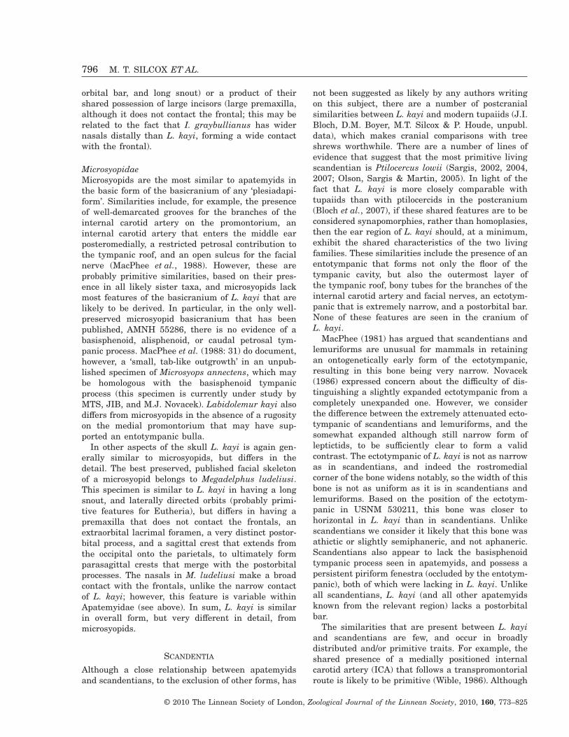
orbital bar, and long snout) or a product of theirshared possession of large incisors (large premaxilla,although it does not contact the frontal; this may berelated to the fact that I. graybullianus has widernasals distally than L. kayi, forming a wide contactwith the frontal).
MicrosyopidaeMicrosyopids are the most similar to apatemyids inthe basic form of the basicranium of any ‘plesiadapi-form’. Similarities include, for example, the presenceof well-demarcated grooves for the branches of theinternal carotid artery on the promontorium, aninternal carotid artery that enters the middle earposteromedially, a restricted petrosal contribution tothe tympanic roof, and an open sulcus for the facialnerve (MacPhee et al., 1988). However, these areprobably primitive similarities, based on their pres-ence in all likely sister taxa, and microsyopids lackmost features of the basicranium of L. kayi that arelikely to be derived. In particular, in the only well-preserved microsyopid basicranium that has beenpublished, AMNH 55286, there is no evidence of abasisphenoid, alisphenoid, or caudal petrosal tym-panic process. MacPhee et al. (1988: 31) do document,however, a ‘small, tab-like outgrowth’ in an unpub-lished specimen of Microsyops annectens, which maybe homologous with the basisphenoid tympanicprocess (this specimen is currently under study byMTS, JIB, and M.J. Novacek). Labidolemur kayi alsodiffers from microsyopids in the absence of a rugosityon the medial promontorium that may have sup-ported an entotympanic bulla.
In other aspects of the skull L. kayi is again gen-erally similar to microsyopids, but differs in thedetail. The best preserved, published facial skeletonof a microsyopid belongs to Megadelphus ludeliusi.This specimen is similar to L. kayi in having a longsnout, and laterally directed orbits (probably primi-tive features for Eutheria), but differs in having apremaxilla that does not contact the frontals, anextraorbital lacrimal foramen, a very distinct postor-bital process, and a sagittal crest that extends fromthe occipital onto the parietals, to ultimately formparasagittal crests that merge with the postorbitalprocesses. The nasals in M. ludeliusi make a broadcontact with the frontals, unlike the narrow contactof L. kayi; however, this feature is variable withinApatemyidae (see above). In sum, L. kayi is similarin overall form, but very different in detail, frommicrosyopids.
SCANDENTIA
Although a close relationship between apatemyidsand scandentians, to the exclusion of other forms, has
not been suggested as likely by any authors writingon this subject, there are a number of postcranialsimilarities between L. kayi and modern tupaiids (J.I.Bloch, D.M. Boyer, M.T. Silcox & P. Houde, unpubl.data), which makes cranial comparisons with treeshrews worthwhile. There are a number of lines ofevidence that suggest that the most primitive livingscandentian is Ptilocercus lowii (Sargis, 2002, 2004,2007; Olson, Sargis & Martin, 2005). In light of thefact that L. kayi is more closely comparable withtupaiids than with ptilocercids in the postcranium(Bloch et al., 2007), if these shared features are to beconsidered synapomorphies, rather than homoplasies,then the ear region of L. kayi should, at a minimum,exhibit the shared characteristics of the two livingfamilies. These similarities include the presence of anentotympanic that forms not only the floor of thetympanic cavity, but also the outermost layer ofthe tympanic roof, bony tubes for the branches of theinternal carotid artery and facial nerves, an ectotym-panic that is extremely narrow, and a postorbital bar.None of these features are seen in the cranium ofL. kayi.
MacPhee (1981) has argued that scandentians andlemuriforms are unusual for mammals in retainingan ontogenetically early form of the ectotympanic,resulting in this bone being very narrow. Novacek(1986) expressed concern about the difficulty of dis-tinguishing a slightly expanded ectotympanic from acompletely unexpanded one. However, we considerthe difference between the extremely attenuated ecto-tympanic of scandentians and lemuriforms, and thesomewhat expanded although still narrow form ofleptictids, to be sufficiently clear to form a validcontrast. The ectotympanic of L. kayi is not as narrowas in scandentians, and indeed the rostromedialcorner of the bone widens notably, so the width of thisbone is not as uniform as it is in scandentians andlemuriforms. Based on the position of the ectotym-panic in USNM 530211, this bone was closer tohorizontal in L. kayi than in scandentians. Unlikescandentians we consider it likely that this bone wasathictic or slightly semiphaneric, and not aphaneric.Scandentians also appear to lack the basisphenoidtympanic process seen in apatemyids, and possess apersistent piriform fenestra (occluded by the entotym-panic), both of which were lacking in L. kayi. Unlikeall scandentians, L. kayi (and all other apatemyidsknown from the relevant region) lacks a postorbitalbar.
The similarities that are present between L. kayiand scandentians are few, and occur in broadlydistributed and/or primitive traits. For example, theshared presence of a medially positioned internalcarotid artery (ICA) that follows a transpromontorialroute is likely to be primitive (Wible, 1986). Although
796 M. T. SILCOX ET AL.
© 2010 The Linnean Society of London, Zoological Journal of the Linnean Society, 2010, 160, 773–825
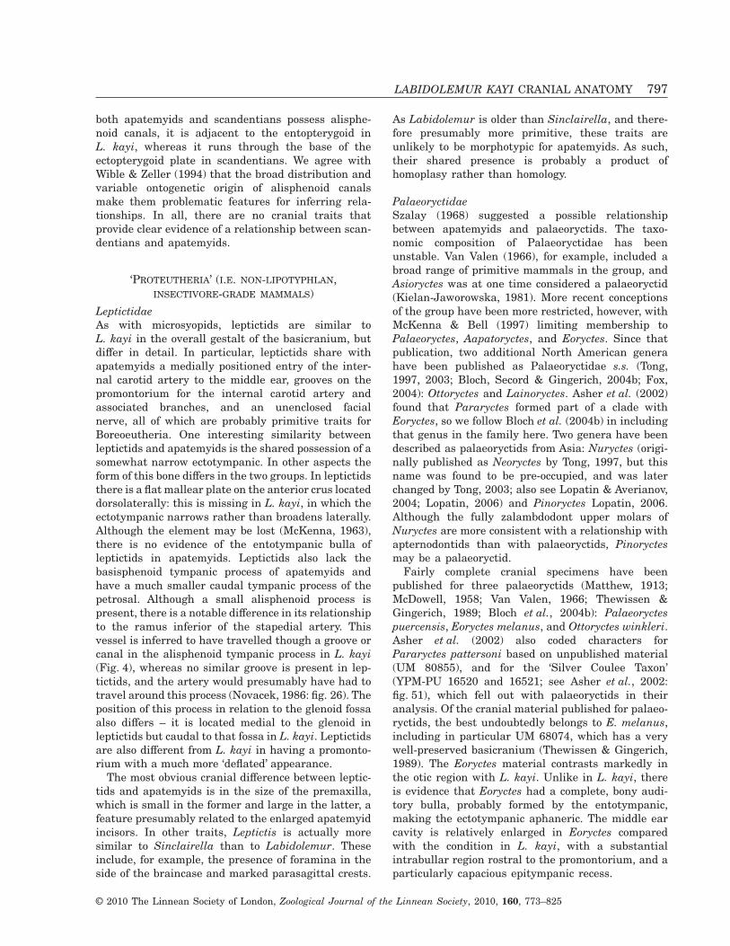
both apatemyids and scandentians possess alisphe-noid canals, it is adjacent to the entopterygoid inL. kayi, whereas it runs through the base of theectopterygoid plate in scandentians. We agree withWible & Zeller (1994) that the broad distribution andvariable ontogenetic origin of alisphenoid canalsmake them problematic features for inferring rela-tionships. In all, there are no cranial traits thatprovide clear evidence of a relationship between scan-dentians and apatemyids.
‘PROTEUTHERIA’ (I.E. NON-LIPOTYPHLAN,INSECTIVORE-GRADE MAMMALS)
LeptictidaeAs with microsyopids, leptictids are similar toL. kayi in the overall gestalt of the basicranium, butdiffer in detail. In particular, leptictids share withapatemyids a medially positioned entry of the inter-nal carotid artery to the middle ear, grooves on thepromontorium for the internal carotid artery andassociated branches, and an unenclosed facialnerve, all of which are probably primitive traits forBoreoeutheria. One interesting similarity betweenleptictids and apatemyids is the shared possession of asomewhat narrow ectotympanic. In other aspects theform of this bone differs in the two groups. In leptictidsthere is a flat mallear plate on the anterior crus locateddorsolaterally: this is missing in L. kayi, in which theectotympanic narrows rather than broadens laterally.Although the element may be lost (McKenna, 1963),there is no evidence of the entotympanic bulla ofleptictids in apatemyids. Leptictids also lack thebasisphenoid tympanic process of apatemyids andhave a much smaller caudal tympanic process of thepetrosal. Although a small alisphenoid process ispresent, there is a notable difference in its relationshipto the ramus inferior of the stapedial artery. Thisvessel is inferred to have travelled though a groove orcanal in the alisphenoid tympanic process in L. kayi(Fig. 4), whereas no similar groove is present in lep-tictids, and the artery would presumably have had totravel around this process (Novacek, 1986: fig. 26). Theposition of this process in relation to the glenoid fossaalso differs – it is located medial to the glenoid inleptictids but caudal to that fossa in L. kayi. Leptictidsare also different from L. kayi in having a promonto-rium with a much more ‘deflated’ appearance.
The most obvious cranial difference between leptic-tids and apatemyids is in the size of the premaxilla,which is small in the former and large in the latter, afeature presumably related to the enlarged apatemyidincisors. In other traits, Leptictis is actually moresimilar to Sinclairella than to Labidolemur. Theseinclude, for example, the presence of foramina in theside of the braincase and marked parasagittal crests.
As Labidolemur is older than Sinclairella, and there-fore presumably more primitive, these traits areunlikely to be morphotypic for apatemyids. As such,their shared presence is probably a product ofhomoplasy rather than homology.
PalaeoryctidaeSzalay (1968) suggested a possible relationshipbetween apatemyids and palaeoryctids. The taxo-nomic composition of Palaeoryctidae has beenunstable. Van Valen (1966), for example, included abroad range of primitive mammals in the group, andAsioryctes was at one time considered a palaeoryctid(Kielan-Jaworowska, 1981). More recent conceptionsof the group have been more restricted, however, withMcKenna & Bell (1997) limiting membership toPalaeoryctes, Aapatoryctes, and Eoryctes. Since thatpublication, two additional North American generahave been published as Palaeoryctidae s.s. (Tong,1997, 2003; Bloch, Secord & Gingerich, 2004b; Fox,2004): Ottoryctes and Lainoryctes. Asher et al. (2002)found that Pararyctes formed part of a clade withEoryctes, so we follow Bloch et al. (2004b) in includingthat genus in the family here. Two genera have beendescribed as palaeoryctids from Asia: Nuryctes (origi-nally published as Neoryctes by Tong, 1997, but thisname was found to be pre-occupied, and was laterchanged by Tong, 2003; also see Lopatin & Averianov,2004; Lopatin, 2006) and Pinoryctes Lopatin, 2006.Although the fully zalambdodont upper molars ofNuryctes are more consistent with a relationship withapternodontids than with palaeoryctids, Pinoryctesmay be a palaeoryctid.
Fairly complete cranial specimens have beenpublished for three palaeoryctids (Matthew, 1913;McDowell, 1958; Van Valen, 1966; Thewissen &Gingerich, 1989; Bloch et al., 2004b): Palaeoryctespuercensis, Eoryctes melanus, and Ottoryctes winkleri.Asher et al. (2002) also coded characters forPararyctes pattersoni based on unpublished material(UM 80855), and for the ‘Silver Coulee Taxon’(YPM-PU 16520 and 16521; see Asher et al., 2002:fig. 51), which fell out with palaeoryctids in theiranalysis. Of the cranial material published for palaeo-ryctids, the best undoubtedly belongs to E. melanus,including in particular UM 68074, which has a verywell-preserved basicranium (Thewissen & Gingerich,1989). The Eoryctes material contrasts markedly inthe otic region with L. kayi. Unlike in L. kayi, thereis evidence that Eoryctes had a complete, bony audi-tory bulla, probably formed by the entotympanic,making the ectotympanic aphaneric. The middle earcavity is relatively enlarged in Eoryctes comparedwith the condition in L. kayi, with a substantialintrabullar region rostral to the promontorium, and aparticularly capacious epitympanic recess.
LABIDOLEMUR KAYI CRANIAL ANATOMY 797
© 2010 The Linnean Society of London, Zoological Journal of the Linnean Society, 2010, 160, 773–825
There are bony tubes for the branches of the inter-nal carotid arteries in Eoryctes, rather than simplygrooves, as in L. kayi. It is unclear whether or not thefacial nerve also travelled in bony tubes in Eoryctes.Thewissen & Gingerich (1989) identified a largeindentation on the mastoid as the (definitive) stylo-mastoid foramen; however, this supposed foramendoes not appear to be perforate in uhrCT data of UM68074, and this indentation may actually be the sta-pedius fossa. Furthermore, Boyer & Georgi (2007)described the facial nerve as running in a groove witha well-developed crista parotica through the middleear cavity, based on observation of several specimensof Eoryctes and Pararyctes, including UM 68074 andUM 80855. In Palaeoryctes puercensis Van Valen(1966) identified an opening to the facial canal intothe middle ear cavity, a foramen stylomastoideumprimitivum, and a stapedius fossa near the position ofthe supposed stylomastoid foramen of Eoryctes,making it like L. kayi in the pathway of the facialnerve.
Like L. kayi, Eoryctes has an alisphenoid canal;however, in Eoryctes it is continuous with a foramenthat opens into the braincase, interpreted byThewissen & Gingerich (1989) as the sphenorbitalfissure (their ‘rotundo-orbital foramen’), whereas theCT data for L. kayi shows that this canal does notopen into the braincase (Fig. 8A). Eoryctes actuallyshares one feature with some apatemyids that ismissing in L. kayi: foramina in the parietal bones.Unlike all apatemyids, however, Eoryctes has lamb-doid plates (sensu Thewissen & Gingerich, 1989;Asher et al., 2002; large, flattened areas on the pari-etals that presumably served as muscle attach-ments) and lacks zygomatic arches. The cranialmaterial for Ottoryctes is less complete, and is verysimilar to that of Eoryctes in the areas that arepreserved, which is unsurprising in light of theirvery close (possible ancestor-descendent) relationship(Bloch et al., 2004b). The only apparent differencesbetween these taxa are the more substantial lambdoidplates (also seen in the ‘Sand Coulee Taxon’) and lessrostrally extensive tympanic cavity of Ottoryctes,although the precise dimensions of the latter aredifficult to assess because of distortion. In any case,clearly this taxon is also markedly dissimilar toLabidolemur.
Based on the descriptions of the cranium of Palaeo-ryctes puercensis (AMNH 15923l; Matthew, 1913;McDowell, 1958; Van Valen, 1966), it would appear asthough this taxon was distinctly different fromEoryctes in most features of the auditory region;indeed, the apparent contrasts between these taxa ledFox (2004) to question the monophyly of the group.McDowell (1958: 178) described the promontorium,for example, as ‘quite smooth and devoid of grooves
for the carotid arterial circulation’. He reconstructedthe blood supply to the brain as running through amedially placed carotid artery (‘entocarotid’), basedon the presence of an apparent groove and foramensitting medial to the middle ear proper (see alsoMatthew, 1913; Van Valen, 1966). At the time thatMcDowell was writing, this configuration was con-sidered characteristic of carnivorous mammals(Matthew, 1909), which led him to consider Palaeo-ryctes only distantly related to lipotyphlans and othergroups with a carotid circulation passing through themiddle ear (e.g. primates, tree shrews). However,since McDowell’s work, the existence of the entoca-rotid in any mammal has come into question as aresult of embryological work (Presley, 1979; MacPhee,1981). The groove in question in Palaeoryctes is aslikely to be a gap between the components of thetympanic roof as an arterial groove. AMNH 15923 isa severely damaged specimen, so it is unwise toover-interpret its anatomy. In light of the damage tothis specimen, the apparent absence of grooves on thepromontorium is not convincing evidence that aninternal carotid artery did not run through the middleear; indeed, Van Valen (1966) identified a number ofsubtle features he suggested were elements of thisvessel’s route. Promontorial grooves can be difficult tosee in fossils, even when well preserved, and arevariable in their depth, degree of expression, and pathamongst modern osteological specimens of the samespecies. For these reasons we consider the route of theinternal carotid artery to be impossible to resolve forPalaeoryctes based on the current evidence. There isno evidence, however, that it possessed bony tubes forthese vessels, making it more like L. kayi thanEoryctes in this feature, although based on theirabsence in primitive eutherians (e.g. Asioryctes;Kielan-Jaworowska, 1981) this similarity is presum-ably primitive.
Although McDowell’s (1958) interpretation of thecarotid circulation in Palaeoryctes is problematic, ourobservations confirm his identification of a fragmentof bone in the rostromedial corner of the left auditorycavity as an entotympanic, rather than an ectotym-panic (contra Matthew, 1913), so that this taxon wassimilar to Eoryctes, and different from L. kayi, inhaving an entotympanic auditory bulla. Our observa-tions of the original material convince us that AMNH15923 also had an enlarged auditory cavity, likeEoryctes and unlike L. kayi. Matthew (1913: 310)argued that there was no alisphenoid canal in thisspecimen, with only a ‘deep groove, incompletelybridged for a short distance, occupying its place’(Matthew, 1913: 310). Van Valen (1966) disagreed,observing a complete canal on the right, but only agroove on the left. Although he interpreted this as apossible indication of asymmetry, it is difficult to have
798 M. T. SILCOX ET AL.
© 2010 The Linnean Society of London, Zoological Journal of the Linnean Society, 2010, 160, 773–825
confidence in this finding about a specimen that is soheavily damaged. As in Eoryctes, but not Labidol-emur, Van Valen reported that the alisphenoid canalwas continuous with the sphenorbital fissure. Manyof the other characteristics of Eoryctes are impossibleto confidently assess in Palaeoryctes. AMNH 15923 ismissing the lateral portions of the cranium on boththe right and left sides, making the presence orabsence of lambdoid plates and of an enlarged epi-tympanic recess impossible to establish. In sum, thedifferences between Eoryctes and Palaeoryctes incranial morphology may not be as profound as hasbeen supposed.
One factor that may support the notion that Palaeo-ryctes originally possessed arterial grooves, but nottubes, on the promontorium is their presence inPararyctes pattersoni and in the ‘Silver Coulee Taxon’.Pararyctes pattersoni (UM 80855) demonstratesseveral other differences from Eoryctes, which mightbe primitive for the family, including the absence oflambdoid plates. Pararyctes pattersoni still exhibitsa number of differences from L. kayi, however, in-cluding the presence of an entotympanic bulla, anenlarged auditory cavity, an expansive epitympanicrecess (Boyer & Georgi, 2007), no zygomatic arches,and no alisphenoid canal. In sum, there appear tobe profound differences in the construction of thecranium between apatemyids and palaeoryctids, andfew if any derived similarities, which are suggestiveof only a distant relationship between thesegroups.
EULIPOTYPHLA
Soricomorpha s.s. (e.g. not including tenrecs, etc.)There are some basic similarities between shrewsand apatemyids in the form of the I1. Also, materialof Heterohyus was published by Filhol (1890) asNeosorex, a supposed fossil shrew, highlighting thepresence of some (limited) dental similarities betweenthese groups. Soricomorphs possess several derivedfeatures of the skull that are missing in apatemyids,however, including a persistent, large piriform fenes-tra, an incomplete zygomatic arch, and a doubledmandibular condyle. Perhaps more importantly,shrews are lacking one of the most distinctive fea-tures of the basicranium of L. kayi: the basisphenoidtympanic process. Optimization of this feature isequivocal for the group that includes shrews, hedge-hogs, and moles (Asher, 2005); it may be primitive forthis group, suggesting that its absence in modernshrews may not be a bar to its presence in an ancientfossil relative. However, there are no features of theskull that offer clear support for an apatemyid–soricomorph relationship.
ErinaceomorphaOf all of the taxa to which L. kayi was compared, thegreatest phenetic similarities are with modern erina-ceid insectivores, and in particular with Hemiechinus.Similarities between these forms include: (1) a hori-zontal ectotympanic; (2) caudal and rostral processesof the petrosal that are small but distinct; (3) a smallalisphenoid tympanic process, through (or past) whichthe ramus inferior of the stapedial artery passes;(4) an un-enclosed facial nerve; (5) no complete ossi-fied bulla; (6) a limited contribution of the petrosal tothe tympanic roof; (7) grooves on the promontoriumfor the branches of an unreduced internal carotidartery, proceeding from a posteromedial entrance tothe middle ear; (8) a substantial contribution from thesphenoid to the tympanic roof; (9) a basisphenoidtympanic process; and (10) a small or absent piriformfenestra. Some of these features (e.g. 4, 5, 6, and 7)are likely to be primitive (based on their presence inprimitive eutherians); for others (e.g. 1 and 10) thepolarity is in debate (Symonds, 2005). One of thesetraits (9) has often been highlighted as a potentialsynapomorphy of either Lipotyphla, or of a morerestricted grouping within that cluster of forms(Asher, 2005; Symonds, 2005); however, the distribu-tion of this character makes its significance uncertain(Asher, 2005).
There are additionally some fairly significant dif-ferences between erinaceomorphs and apatemyids. Apotential lipotyphlan synapomorphy is the reductionof the jugal (Symonds, 2005). Erinaeomorphs (talpidsand erinaceids) do retain a complete zygomatic arch(unlike soricomorphs); however, the jugal componentof the arch is quite small. This is not true of apate-myids, in which the jugal is a very sturdy element(e.g. see Fig. 15), and the zygomatic arch is quiterobust. However, there may be a functional explana-tion for this characteristic. As discussed above, thezygomatic arch probably had an important role toplay in resisting the bending forces in the face result-ing from using the incisors to bore into wood. As such,this difference may have more to do with functionthan phylogeny.
A feature reconstructed as primitive for Lipotyphlais an expansion of the maxilla into the orbital mosaic(Asher, 2005), with the expansion of this bone beingparticularly marked in erinaceomorphs. Labidolemurlacks this trait (Fig. 16): the maxilla does not extendto the level of the dorsal edge of the infraorbitalforamen. Another feature of some eulipotyphlans thathas been considered significant (e.g. McDowell, 1958;see discussion in Boyer & Georgi, 2007) is the absenceof a ‘true’ postglenoid process, although this trait isquite variable in insectivores, and is present in somefossil members of the group (Butler, 1988). In anycase, apatemyids do have a lip-like postglenoid
LABIDOLEMUR KAYI CRANIAL ANATOMY 799
© 2010 The Linnean Society of London, Zoological Journal of the Linnean Society, 2010, 160, 773–825
process, which is located caudolateral to the postgle-noid foramen, rather than medial to it as in someerinaceomorphs. In sum, with the exception of thebasisphenoid tympanic process, apatemyids lack mostof the features that have been considered as potentialsynapomorphies for Lipotyphla or Eulipotyphla.
There are other differences between apatemyidsand living erinaceomorphs, including the breadth ofthe ectotympanic (broader in erinaceomorphs), thedegree of development of the basisphenoid tympanicprocess (more laterally extensive in erinaceomorphs,although this may be influenced by breakage inL. kayi), and the size of the premaxilla (much smallerin erinaceomorphs). Also, L. kayi is actually moresimilar to living erinaceomorphs than it is to somefossils that have been suggested to be related to thatgroup. Brachyerix, for example, is very different fromboth modern erinaceomorphs and L. kayi in having amuch more heavily ossified auditory region, withcanals for the internal carotid artery, and an evenmore expansive basisphenoid tympanic process,forming an ossified auditory bulla (Rich & Rich,1971). Diacodon actually lacks the basisphenoidprocess (MacPhee et al., 1988), further complicatingour picture of the evolutionary history of that trait.
In sum, comparisons with a diverse array of livingand extinct eutherian groups, reveals no obvious solu-tion to the problem of apatemyid relationships. Whilesimilarities to erinaceomorphs are intriguing, theirpolarity needs to be assessed in the context of aphylogenetic analysis.
PHYLOGENETIC ANALYSIS
Performing a phylogenetic analysis to decipher thebroader relationships of L. kayi is complicated by thediverse array of possible affinities that have beensuggested for apatemyids. To test all of the hypoth-eses that have been suggested, it is necessary toinclude a very broad range of eutherians, includingother apatemyids, eulipotyphlans, ‘proteutherians’(leptictids and palaeoryctids), primates and othereuarchontans, and any other groups that might berelevant for accurately reconstructing basal states forlarger clades that include those taxa (e.g. carnivoransand gliroids). To this end we have assembled a matrixof 33 in-group taxa and one out-group (Ukhaatheriumnessovi) that were assessed for 240 morphologicalcharacters (68 postcranial, 45 cranial, and 127 dental;Appendix 1), either modified from Silcox (2001, 2008)or written for this study based on Jepsen (1934),McKenna (1963), Szalay (1968), Emry (1970), Szalay& Drawhorn (1980), Novacek (1986), Asher (1999),Gaudin & Wible (1999), Rose & Lucas (2000), Asher,Novacek & Geisler (2003), Asher et al. (2002, 2005),Bloch et al. (2004b), Flynn & Wesley-Hunt (2005),
Meng & Wyss (2005), and original observations. Adescription of these characters is given in Appendix 2and the character matrix is provided as Appendix 3.All characters were equally weighted and treated asunordered. The characters that were variable withina taxon were treated as polymorphic. A heuristicsearch with 10 000 replicates using parsimony wasperformed in PAUP 4.0b10 (Swofford, 2002). Bremersupport (decay) indices (Bremer, 1988) were calcu-lated using TreeRot 2 (Sorenson, M.D., 1999).
The cladistic analysis produced three most parsi-monious trees. The strict consensus tree is presentedin Figure 17. There were two areas of disagreementamongst the three trees. The first relates to therelationships of Leptictida + Palaeoryctidae to theother major clades in the analysis. In two trees thisgrouping is the sister taxon to Euarchontoglires +Laurasiatheria, whereas in the third it is the sistertaxon to Euarchontoglires alone. Some additionalout-groups would probably be necessary to resolvethese relationships. The second area of disagreementrelates to the branching order amongst the Palaeo-ryctidae: in two trees Palaeoryctes and Ottoryctes aresister taxa, whereas in the third Pararyctes andOttoryctes are sister taxa.
Most of the major clades that are currentlyaccepted as valid amongst molecular biologists (e.g.Springer et al., 2004) are supported by this analysis,including Laurasiatheria, Eulipotyphla, Carnivora,Euarchonta, and Euarchontoglires. An issue ofcurrent debate in mammalian systematics is thebranching order within Euarchonta (e.g. see Janeckaet al., 2007; Nie et al., 2008). This analysis providesadditional morphological support (see also Silcox,2001; Sargis, 2002, 2004, 2007; Bloch et al., 2007) forthe Sundatheria hypothesis (Olson et al., 2005)linking Dermoptera and Scandentia, to the exclusionof Primates (including ‘plesiadapiforms’).
In all three trees apatemyids are the sister taxon tothe primitive gliroid Rhombomylus at the base ofEuarchontoglires. A relationship between apatemyidsand any gliroid has not been seriously considered forsome time, and is thus a surprising outcome of ouranalysis. In conflict with this result, similaritiesbetween apatemyids and some euarchontans includepostcranial characters that probably relate to arbo-real locomotion, which are missing in the terrestrialgliroid Rhombomylus {e.g. elongation of digit III or IVof the manus [21(1)]}, and dental features sharedwith ‘plesiadapiforms’ {e.g. procumbent, enlarged i1[185(0), 187(1)], curving molar paracristids [233(1)]}.In fact, moving apatemyids into a sister-group posi-tion with Euarchonta adds only five steps to this tree,so the sister taxon relationship with Rhombomylus isquite weakly supported, and will require furthertesting with broader taxonomic sampling of Glires.
800 M. T. SILCOX ET AL.
© 2010 The Linnean Society of London, Zoological Journal of the Linnean Society, 2010, 160, 773–825
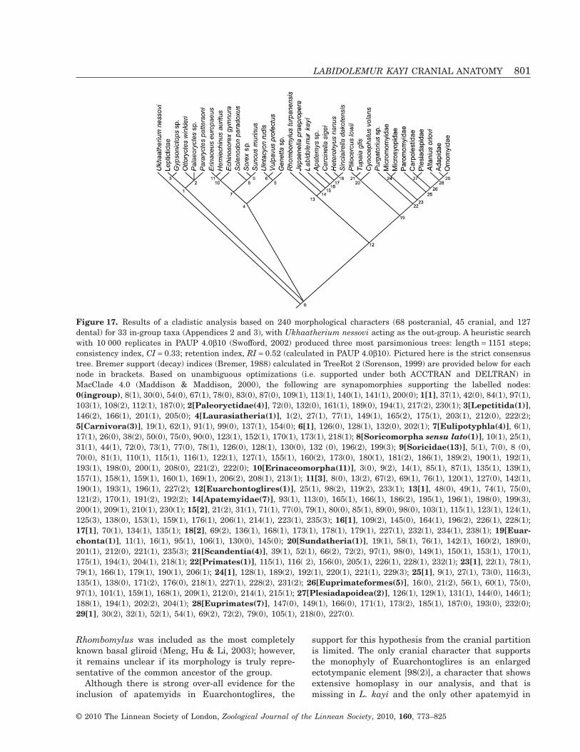
Rhombomylus was included as the most completelyknown basal gliroid (Meng, Hu & Li, 2003); however,it remains unclear if its morphology is truly repre-sentative of the common ancestor of the group.
Although there is strong over-all evidence for theinclusion of apatemyids in Euarchontoglires, the
support for this hypothesis from the cranial partitionis limited. The only cranial character that supportsthe monophyly of Euarchontoglires is an enlargedectotympanic element [98(2)], a character that showsextensive homoplasy in our analysis, and that ismissing in L. kayi and the only other apatemyid in
Figure 17. Results of a cladistic analysis based on 240 morphological characters (68 postcranial, 45 cranial, and 127dental) for 33 in-group taxa (Appendices 2 and 3), with Ukhaatherium nessovi acting as the out-group. A heuristic searchwith 10 000 replicates in PAUP 4.0b10 (Swofford, 2002) produced three most parsimonious trees: length = 1151 steps;consistency index, CI = 0.33; retention index, RI = 0.52 (calculated in PAUP 4.0b10). Pictured here is the strict consensustree. Bremer support (decay) indices (Bremer, 1988) calculated in TreeRot 2 (Sorenson, 1999) are provided below for eachnode in brackets. Based on unambiguous optimizations (i.e. supported under both ACCTRAN and DELTRAN) inMacClade 4.0 (Maddison & Maddison, 2000), the following are synapomorphies supporting the labelled nodes:0(ingroup), 8(1), 30(0), 54(0), 67(1), 78(0), 83(0), 87(0), 109(1), 113(1), 140(1), 141(1), 200(0); 1[1], 37(1), 42(0), 84(1), 97(1),103(1), 108(2), 112(1), 187(0); 2[Paleoryctidae(4)], 72(0), 132(0), 161(1), 189(0), 194(1), 217(2), 230(1); 3[Lepctitida(1)],146(2), 166(1), 201(1), 205(0); 4[Laurasiatheria(1)], 1(2), 27(1), 77(1), 149(1), 165(2), 175(1), 203(1), 212(0), 222(2);5[Carnivora(3)], 19(1), 62(1), 91(1), 99(0), 137(1), 154(0); 6[1], 126(0), 128(1), 132(0), 202(1); 7[Eulipotyphla(4)], 6(1),17(1), 26(0), 38(2), 50(0), 75(0), 90(0), 123(1), 152(1), 170(1), 173(1), 218(1); 8[Soricomorpha sensu lato(1)], 10(1), 25(1),31(1), 44(1), 72(0), 73(1), 77(0), 78(1), 126(0), 128(1), 130(0), 132 (0), 196(2), 199(3); 9[Soricidae(13)], 5(1), 7(0), 8 (0),70(0), 81(1), 110(1), 115(1), 116(1), 122(1), 127(1), 155(1), 160(2), 173(0), 180(1), 181(2), 186(1), 189(2), 190(1), 192(1),193(1), 198(0), 200(1), 208(0), 221(2), 222(0); 10[Erinaceomorpha(11)], 3(0), 9(2), 14(1), 85(1), 87(1), 135(1), 139(1),157(1), 158(1), 159(1), 160(1), 169(1), 206(2), 208(1), 213(1); 11[3], 8(0), 13(2), 67(2), 69(1), 76(1), 120(1), 127(0), 142(1),190(1), 193(1), 196(1), 227(2); 12[Euarchontoglires(1)], 25(1), 98(2), 119(2), 233(1); 13[1], 48(0), 49(1), 74(1), 75(0),121(2), 170(1), 191(2), 192(2); 14[Apatemyidae(7)], 93(1), 113(0), 165(1), 166(1), 186(2), 195(1), 196(1), 198(0), 199(3),200(1), 209(1), 210(1), 230(1); 15[2], 21(2), 31(1), 71(1), 77(0), 79(1), 80(0), 85(1), 89(0), 98(0), 103(1), 115(1), 123(1), 124(1),125(3), 138(0), 153(1), 159(1), 176(1), 206(1), 214(1), 223(1), 235(3); 16[1], 109(2), 145(0), 164(1), 196(2), 226(1), 228(1);17[1], 70(1), 134(1), 135(1); 18[2], 69(2), 136(1), 168(1), 173(1), 178(1), 179(1), 227(1), 232(1), 234(1), 238(1); 19[Euar-chonta(1)], 11(1), 16(1), 95(1), 106(1), 130(0), 145(0); 20[Sundatheria(1)], 19(1), 58(1), 76(1), 142(1), 160(2), 189(0),201(1), 212(0), 221(1), 235(3); 21[Scandentia(4)], 39(1), 52(1), 66(2), 72(2), 97(1), 98(0), 149(1), 150(1), 153(1), 170(1),175(1), 194(1), 204(1), 218(1); 22[Primates(1)], 115(1), 116( 2), 156(0), 205(1), 226(1), 228(1), 232(1); 23[1], 22(1), 78(1),79(1), 166(1), 179(1), 190(1), 206(1); 24[1], 128(1), 189(2), 192(1), 220(1), 221(1), 229(3); 25[1], 9(1), 27(1), 73(0), 116(3),135(1), 138(0), 171(2), 176(0), 218(1), 227(1), 228(2), 231(2); 26[Euprimateformes(5)], 16(0), 21(2), 56(1), 60(1), 75(0),97(1), 101(1), 159(1), 168(1), 209(1), 212(0), 214(1), 215(1); 27[Plesiadapoidea(2)], 126(1), 129(1), 131(1), 144(0), 146(1);188(1), 194(1), 202(2), 204(1); 28[Euprimates(7)], 147(0), 149(1), 166(0), 171(1), 173(2), 185(1), 187(0), 193(0), 232(0);29[1], 30(2), 32(1), 52(1), 54(1), 69(2), 72(2), 79(0), 105(1), 218(0), 227(0).
LABIDOLEMUR KAYI CRANIAL ANATOMY 801
© 2010 The Linnean Society of London, Zoological Journal of the Linnean Society, 2010, 160, 773–825
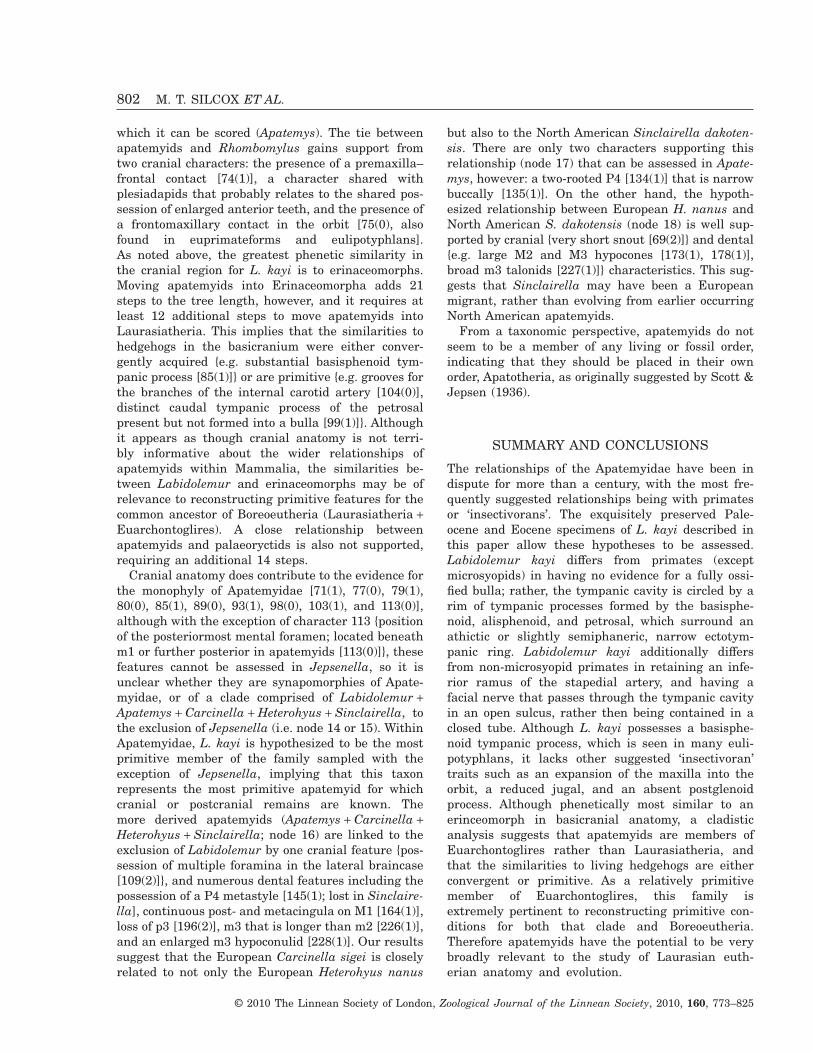
which it can be scored (Apatemys). The tie betweenapatemyids and Rhombomylus gains support fromtwo cranial characters: the presence of a premaxilla–frontal contact [74(1)], a character shared withplesiadapids that probably relates to the shared pos-session of enlarged anterior teeth, and the presence ofa frontomaxillary contact in the orbit [75(0), alsofound in euprimateforms and eulipotyphlans].As noted above, the greatest phenetic similarity inthe cranial region for L. kayi is to erinaceomorphs.Moving apatemyids into Erinaceomorpha adds 21steps to the tree length, however, and it requires atleast 12 additional steps to move apatemyids intoLaurasiatheria. This implies that the similarities tohedgehogs in the basicranium were either conver-gently acquired {e.g. substantial basisphenoid tym-panic process [85(1)]} or are primitive {e.g. grooves forthe branches of the internal carotid artery [104(0)],distinct caudal tympanic process of the petrosalpresent but not formed into a bulla [99(1)]}. Althoughit appears as though cranial anatomy is not terri-bly informative about the wider relationships ofapatemyids within Mammalia, the similarities be-tween Labidolemur and erinaceomorphs may be ofrelevance to reconstructing primitive features for thecommon ancestor of Boreoeutheria (Laurasiatheria +Euarchontoglires). A close relationship betweenapatemyids and palaeoryctids is also not supported,requiring an additional 14 steps.
Cranial anatomy does contribute to the evidence forthe monophyly of Apatemyidae [71(1), 77(0), 79(1),80(0), 85(1), 89(0), 93(1), 98(0), 103(1), and 113(0)],although with the exception of character 113 {positionof the posteriormost mental foramen; located beneathm1 or further posterior in apatemyids [113(0)]}, thesefeatures cannot be assessed in Jepsenella, so it isunclear whether they are synapomorphies of Apate-myidae, or of a clade comprised of Labidolemur +Apatemys + Carcinella + Heterohyus + Sinclairella, tothe exclusion of Jepsenella (i.e. node 14 or 15). WithinApatemyidae, L. kayi is hypothesized to be the mostprimitive member of the family sampled with theexception of Jepsenella, implying that this taxonrepresents the most primitive apatemyid for whichcranial or postcranial remains are known. Themore derived apatemyids (Apatemys + Carcinella +Heterohyus + Sinclairella; node 16) are linked to theexclusion of Labidolemur by one cranial feature {pos-session of multiple foramina in the lateral braincase[109(2)]}, and numerous dental features including thepossession of a P4 metastyle [145(1); lost in Sinclaire-lla], continuous post- and metacingula on M1 [164(1)],loss of p3 [196(2)], m3 that is longer than m2 [226(1)],and an enlarged m3 hypoconulid [228(1)]. Our resultssuggest that the European Carcinella sigei is closelyrelated to not only the European Heterohyus nanus
but also to the North American Sinclairella dakoten-sis. There are only two characters supporting thisrelationship (node 17) that can be assessed in Apate-mys, however: a two-rooted P4 [134(1)] that is narrowbuccally [135(1)]. On the other hand, the hypoth-esized relationship between European H. nanus andNorth American S. dakotensis (node 18) is well sup-ported by cranial {very short snout [69(2)]} and dental{e.g. large M2 and M3 hypocones [173(1), 178(1)],broad m3 talonids [227(1)]} characteristics. This sug-gests that Sinclairella may have been a Europeanmigrant, rather than evolving from earlier occurringNorth American apatemyids.
From a taxonomic perspective, apatemyids do notseem to be a member of any living or fossil order,indicating that they should be placed in their ownorder, Apatotheria, as originally suggested by Scott &Jepsen (1936).
SUMMARY AND CONCLUSIONS
The relationships of the Apatemyidae have been indispute for more than a century, with the most fre-quently suggested relationships being with primatesor ‘insectivorans’. The exquisitely preserved Pale-ocene and Eocene specimens of L. kayi described inthis paper allow these hypotheses to be assessed.Labidolemur kayi differs from primates (exceptmicrosyopids) in having no evidence for a fully ossi-fied bulla; rather, the tympanic cavity is circled by arim of tympanic processes formed by the basisphe-noid, alisphenoid, and petrosal, which surround anathictic or slightly semiphaneric, narrow ectotym-panic ring. Labidolemur kayi additionally differsfrom non-microsyopid primates in retaining an infe-rior ramus of the stapedial artery, and having afacial nerve that passes through the tympanic cavityin an open sulcus, rather then being contained in aclosed tube. Although L. kayi possesses a basisphe-noid tympanic process, which is seen in many euli-potyphlans, it lacks other suggested ‘insectivoran’traits such as an expansion of the maxilla into theorbit, a reduced jugal, and an absent postglenoidprocess. Although phenetically most similar to anerinceomorph in basicranial anatomy, a cladisticanalysis suggests that apatemyids are members ofEuarchontoglires rather than Laurasiatheria, andthat the similarities to living hedgehogs are eitherconvergent or primitive. As a relatively primitivemember of Euarchontoglires, this family isextremely pertinent to reconstructing primitive con-ditions for both that clade and Boreoeutheria.Therefore apatemyids have the potential to be verybroadly relevant to the study of Laurasian euth-erian anatomy and evolution.
802 M. T. SILCOX ET AL.
© 2010 The Linnean Society of London, Zoological Journal of the Linnean Society, 2010, 160, 773–825
ACKNOWLEDGEMENTS
The uhrCT scanning of the Labidolemur specimenswas performed by Özgen Karacan (Center for Quan-titative Imaging, Pennsylvania State University) andwas supported by NSF grant BCS-0003920 to A.Walker. uhrCT scanning of the Eoryctes specimen wasperformed at Stony Brook University Department ofBiomedical Engineering’s Center for Biotechnologywith permission and help from S. Judex and C.Rubin. Thanks to R. Asher for discussion on thesubject of Nuryctes and to R.C. Fox, P.D. Gingerich,W.v. Koenigswald, C. McCaffery, J. Meng, M.J.Novacek, D.L, Reed, E. J. Sargis, and N. Simmons foraccess to specimens and unpublished data. Thanks toJ.R. Wible and an anonymous reviewer for commentsthat substantially improved this paper. Additionalsupport for the work was provided by NSERCand discretionary research grants (University ofWinnipeg) to MTS, NSF EF-0629836, to JIB, MTS,and E. Sargis, NSF EAR-0308902, to D. Krause andJIB, NSF DDIG BCS-0622544, and a University ofMichigan Honors College Research Grant Award toDMB, and NSF BSR-8313209 to PH.
REFERENCES
Archibald JD. 1977. Ectotympanic bone and internal carotidcirculation of eutherians in reference to anthropoid origins.Journal of Human Evolution 6: 609–622.
Asher RJ. 1999. A morphological basis for assessing thephylogeny of the ‘Tenrecoidea’ (Mammalia, Lipotyphla). Cla-distics 15: 231–252.
Asher RJ. 2005. Insectivoran-grade placentals. In: Rose KD,Archibald JD, eds. The rise of placental mammals: originsand relationships of the major extant clades. Baltimore, MD:The Johns Hopkins University Press, 50–70.
Asher RJ, McKenna MC, Emry RJ, Tabrum AR, KronDG. 2002. Morphology and relationships of Apternodus andother extinct, zalambdodont, placental mammals. Bulletinof the American Museum of Natural History 273: 1–117.
Asher RJ, Meng J, Wible JR, McKenna MC, Rougier GW,Dashzeveg D, Novacek MJ. 2005. Stem lagomorpha andthe antiquity of glires. Science 307: 1091–1094.
Asher RJ, Novacek MJ, Geisler JH. 2003. Relationships ofendemic African mammals and their fossil relatives basedon morphological and molecular evidence. Journal of Mam-malian Evolution 10: 131–194.
Beard KC, MacPhee RDE. 1994. Cranial anatomy of Shos-honius and the antiquity of Anthropoidea. In: Fleagle JG,Kay RF, eds. Anthropoid origins. New York: Plenum Press,55–97.
de Blainville HMD. 1839–1864. Ostéographie ou descriptioniconographique comparée du squelette et du système dentairedes mammifères récents et fossiles. Paris: Baillière et Fils.
Bloch JI, Boyer DM, Silcox MT, Houde P. 2004a.New skeletons of Paleocene-Eocene Labidolemur kayi
(Mammalia, Apatemyidae): ecomorphology and relationshipof apatemyids to Primates and other mammals. Journal ofVertebrate Paleontology 24 (Suppl. 3): 40A.
Bloch JI, Secord R, Gingerich PD. 2004b. Systematics andphylogeny of late Paleocene and early Eocene Palaeorycti-nae (Mammalia, Insectivora) from the Clarks Fork andBighorn Basins, Wyoming. Contributions from the Museumof Paleontology, University of Michigan 31: 119–154.
Bloch JI, Silcox MT. 2001. New basicrania of Paleocene-Eocene Ignacius: re-evaluation of the plesiadapiform-dermopteran link. American Journal of PhysicalAnthropology 116: 184–198.
Bloch JI, Silcox MT. 2006. Cranial anatomy of Paleoceneplesiadapiform Carpolestes simpsoni (Mammalia, Primates)using ultra high-resolution X-ray computed tomography,and the relationships of plesiadapiforms to Euprimates.Journal of Human Evolution 50: 1–35.
Bloch JI, Silcox MT, Boyer DM, Sargis EJ. 2007. NewPaleocene skeletons and the relationship of plesiadapiformsto crown-clade primates. Proceedings of the NationalAcademy of Science 104: 1159–1164.
Boyer DM, Georgi JA. 2007. Cranial morphology of a pan-tolestid eutherian mammal from the Eocene Bridger forma-tion, Wyoming, USA: implications for relationships andhabitat. Journal of Mammalian Evolution 14: 239–280.
Bremer K. 1988. The limits of amino acid sequence data inangiosperm phylogenetic reconstruction. Evolution 42: 795–803.
Bromann I. 1899. Die entwickelungsgeschichte dergehörknöchelchen beim menschen. Anatomische Hefte 11:507–670.
Butler PM. 1972. The problem of insectivore classification.In: Joysey KA, Kemp TS, eds. Studies in vertebrate evolu-tion. Edinburgh: Oliver and Boyd, 253–265.
Butler PM. 1988. Phylogeny of the insectivores. In: BentonMJ, ed. The phylogeny and classification of the tetrapodsvolume 2. Oxford: Clarendon Press, 117–141.
Camp CL, VanderHoof VL. 1940. Bibliography of fossilvertebrates 1928–1933. Special Papers of the GeologicalSociety of America 27: 1–503.
Cartmill M. 1974. Daubentonia, Dactylopsila, woodpeckersand klinorhynchy. In: Martin RD, Doyle GA, Walker AC,eds. Prosimian biology. Pittsburgh, PA: University of Pitts-burgh Press, 656–669.
Dagosto M, Gebo DL, Beard KC. 1999. Revision of theWind River faunas, early Eocene of central Wyoming. Part14. Postcranium of Shoshonius cooperi (Mammalia, Pri-mates). Annals of Carnegie Museum 68: 175–211.
Emry RJ. 1970. A North American Oligocene pangolin andother additions to the Pholidota. Bulletin of the AmericanMuseum of Natural History 142: 459–510.
Filhol H. 1890. Description d’un nouveau genre d’insectivore.Bulletin de Sciences de la Société Philomatique de Paris,Ser. II 8: 174–175.
Flynn JJ, Wesley-Hunt GD. 2005. Carnivora. In: Rose KD,Archibald JD, eds. The rise of placental mammals: originsand relationships of the major extant clades. Baltimore, MD:The Johns Hopkins University Press, 145–158.
LABIDOLEMUR KAYI CRANIAL ANATOMY 803
© 2010 The Linnean Society of London, Zoological Journal of the Linnean Society, 2010, 160, 773–825
Fox RC. 2004. A new palaeoryctid (Insectivora: Mammalia)from the late Paleocene of Alberta, Canada. Journal ofPaleontology 78: 612–616.
Gaudin TJ, Wible JR. 1999. The entotympanic of pangolinsand the phylogeny of Pholidota (Mammalia). Journal ofMammalian Evolution 6: 39–65.
Gervais P. 1848–1852. Zoologie et paléontologie françaises.Paris: Arthus Bertrand.
Gingerich PD. 1976. Cranial anatomy and evolution of earlyTertiary Plesiadapidae (Mammalia, Primates). University ofMichigan Papers on Paleontology 15: 1–141.
Gingerich PD. 1982. Studies on Paleocene and early EoceneApatemyidae (Mammalia, Insectivora). II. Labidolemur andApatemys from the early Wasatchian of the Clark’s ForkBasin, Wyoming. Contributions from the Museum of Pale-ontology, University of Michigan 26: 57–69.
Gingerich PD. 1983. Systematics of Early Eocene Miacidae(Mammalia, Carnivora) in the Clark’s Fork Basin,Wyoming. Contributions from the Museum of Paleontology,University of Michigan 26: 197–225.
Gingerich PD. 1989. New earliest Wasatchian mammalianfauna from the Eocene of northwestern Wyoming: composi-tion and diversity in a rarely sampled high-floodplainassemblage. University of Michigan Papers on Paleontology28: 1–97.
Gingerich PD. 2003. Mammalian responses to climatechange at the Paleocene-Eocene boundary: polecat benchrecord in the northern Bighorn Basin, Wyoming. In: WingSL, Gingerich PD, Schmitz B, Thomas E, eds. Causes andconsequences of globally warm climates in the early Paleo-gene, Vol. 369, Special Papers. Boulder, CO: GeologicalSociety of America, 463–478.
Gingerich PD, Rose KD. 1982. Studies on Paleocene andearly Eocene Apatemyidae (Mammalia, Insectivora). I. Den-tition of Clarkforkian Labidolemur kayi. Contributions fromthe Museum of Paleontology, University of Michigan 26:49–55.
Gregory WK. 1920. On the structure and relationships ofNotharctus, an American Eocene primate. Memoirs of theAmerican Museum of Natural History 3: 49–243.
Hay OP. 1902. Bibliography and catalogue of the fossil Ver-tebrata of North America. Bulletin of the US GeologicalSurvey 179: 1–868.
Hay OP. 1930. Second bibliography and catalogue of the fossilVertebrata of North America. Washington, DC: CarnegieInstitution.
Heller F. 1930. Die Säugetierfauna der mitteleozänenBraunkohle des Geiseltales bei Halle a. S. Jahrbuch desHalleschen Verbandes 9: 13–41.
Horovitz I. 2003. Postcranial skeleton of Ukaatheriumnessovi (Eutheria, Mammalia) from the Late Cretaceous ofMongolia. Journal of Vertebrate Paleontology 23: 857–868.
Hürzeler J. 1949a. Über die europäischen Apatemyiden.Eclogae Geologicae Helvetiae 42: 485.
Hürzeler J. 1949b. Über die europaischen Apatemyiden.Verhandlungen der Schweizerischen NaturforschendenGesellschaft 129: 140–141.
Janecka JE, Miller W, Pringle TH, Wiens F, Zitzmann A,
Helgen KM, Springer MS, Murphy WJ. 2007. Molecularand genomic data identify the closest living relative ofPrimates. Science 318: 792–794.
Jepsen GL. 1934. A revision of the American Apatemyidaeand the description of a new genus, Sinclairella, from theWhite River Oligocene of South Dakota. Proceedings of theAmerican Philosophical Society 74: 287–305.
Kalthoff DC, von Koenigswald W, Kurz C. 2004. A newspecimen of Heterohyus nanus (Apatemyidae, Mammalia)from the Eocene of Messel (Germany) with unusual soft partpreservation. Courier Forschungsinstitut Senckenberg 252:1–12.
Kay RF, Thewissen JGM, Yoder AD. 1992. Cranialanatomy of Ignacius graybullianus and the affinities of thePlesiadapiformes. American Journal of Physical Anthropol-ogy 89: 477–498.
Kielan-Jaworowska Z. 1981. Evolution of the therianmammals in the Late Cretaceous of Asia. Part IV. Skullstructure in Kennalestes and Asioryctes. PalaeontologiaPolonica 42: 25–78.
Kirk EC, Gosselin-Ildari AD. 2009. Cochlear labyrinthvolume and hearing abilities in primates. AnatomicalRecord 292: 765–776.
von Koenigswald W. 1987. Apatemyiden-Skelette aus demMitteleozän von Messel und ihre paläobiologische Aussage.Carolinea 45: 31–35.
von Koenigswald W. 1990. Die Paläobiologie der Apatemy-iden (Insectivora s.l.) und die Ausdeutung der Skelettfundevon Heterohyus nanus aus dem Mittleozän von Messel beiDarmstadt. Palaeontographica A 210: 41–77.
von Koenigswald W, Rose KD, Grande L, Martin RD.2005a. First apatemyid skeleton from the Lower EoceneFossil Butte Member, Wyoming (USA), compared to theEuropean apatemyid from Messel, Germany. Palaeonto-graphica A 272: 149–169.
von Koenigswald W, Rose KD, Grande L, Martin RD.2005b. Die Lebensweise eozäner Säugetiere (Pantolestidaeund Apatemyidae) aus Messel (Europa) im Vergleich zuenuen Skelettfunden aus dem Fossil Butte Member vonWyoming (NordAmerika). Geologisches Jahrbuch Hessen272: 43–54.
von Koenigswald W, Ruf I, Gingerich PD. 2007. Wellpreserved skull of a European apatemyid (Mammalia) fromthe Phosphorites de Quercy. Journal of Vertebrate Paleon-tology 27 (Suppl. 3): 101A.
von Koenigswald W, Ruf I, Gingerich PD. 2009. Cranialmorphology of a new apatemyid, Carcinella sigei n. gen. n.sp. (Mammalia, Apatotheria) from the late Eocene of south-ern France. Paleontographica A 288: 53–91.
von Koenigswald W, Schierning H-P. 1987. The ecologicalniche of early Tertiary apatemyids – extinct group ofmammals. Nature 326: 595–596.
Lillegraven JA. 1969. Latest Cretaceous mammals of upperpart of the Edmonton formation of Alberta, Canada, andreview of marsupial-placental dichotomy in MammalianEvolution. The University of Kansas Paleontological Contri-butions 50: 1–122.
Lopatin AV. 2006. Early Paleogene insectivore mammals of
804 M. T. SILCOX ET AL.
© 2010 The Linnean Society of London, Zoological Journal of the Linnean Society, 2010, 160, 773–825
Asia and establishment of the major groups of Insectivora.Paleontological Journal 40 (Suppl. 3): S205–S405.
Lopatin AV, Averianov AO. 2004. New Palaeoryctidae(Mammalia) from the Eocene of Kyrgyzstan and Mongolia.Paleontological Journal 38: 556–562.
McDowell SB. 1958. The greater Antillean insectivores. Bul-letin of the American Museum of Natural History 115:117–214.
McKenna MC. 1963. Primitive Paleocene and Eocene Apate-myidae (Mammalia, Insectivora) and the primate-insectivoreboundary. American Museum Novitates 2160: 1–39.
McKenna MC, Bell SK. 1997. Classification of mammalsabove the species level. New York: Columbia UniversityPress.
MacPhee RDE. 1981. Auditory regions of primates and euth-erian insectivores: morphology, ontogeny, and characteranalysis. Contributions to Primatology 18: 1–282.
MacPhee RDE, Cartmill M. 1986. Basicranial structuresand primate systematics. In: Swisher DR, Erwin J, eds.Comparative primate biology, volume 1: systematics, evolu-tion, and anatomy. New York: Alan R. Liss, 219–275.
MacPhee RDE, Cartmill M, Gingerich PD. 1983. NewPaleogene primate basicrania and the definition of the orderPrimates. Nature 301: 509–511.
MacPhee RDE, Novacek MJ, Storch G. 1988. Basicranialmorphology of early Tertiary erinaceomorphs and the originof Primates. American Museum Novitates 2921: 1–42.
Maddison WP, Maddison DR. 2000. MacClade 4.0,Program and Documentation. Sunderland, MA: SinauerAssociates, Inc.
Matthew WD. 1899. A provisional classification of the fresh-water Tertiary of the West. Bulletin of the AmericanMuseum of Natural History 12: 19–75.
Matthew WD. 1909. The Carnivora and Insectivora of theBridger Basin, middle Eocene. Memoirs of the AmericanMuseum of Natural History 9: 291–567.
Matthew WD. 1913. A zalambdodont insectivore from thebasal Eocene. Bulletin of the American Museum of NaturalHistory 32: 307–314.
Matthew WD. 1921. Stehlinius, a new Eocene insectivore.American Museum Novitates 14: 1–5.
Matthew WD. 1929. Preoccupied names. Journal of Mam-malogy 10: 171.
Meng J, Hu Y, Li C-K. 2003. The osteology of Rhombomylus(Mammalia, Glires): implications for phylogeny and evolu-tion of Glires. Bulletin of the American Museum of NaturalHistory 275: 1–247.
Meng J, Wyss AR. 2005. Glires (Lagomorpha, Rodentia). In:Rose KD, Archibald JD, eds. The rise of placental mammals:origins and relationships of the major extant clades. Balti-more, MD: The Johns Hopkins University Press, 145–158.
Nie W, Fu B, O’Brien PCM, Wang J, Su W, Tanomtong A,Volobouev V, Ferguson-Smith MA, Yang F. 2008. Flyinglemurs – the ‘flying tree shrews’? Molecular cytogeneticevidence for a Scandentia-Dermoptera sister clade. BMCBiology 6: 18 (11 pages).
Novacek MJ. 1986. The skull of leptictid insectivorans andthe higher-level classification of eutherian mammals.
Bulletin of the American Museum of Natural History 183:1–111.
Novacek MJ, Rougier GW, Wible JR, McKenna MC,Dashzeveg D, Horovitz I. 1997. Epipubic bones in euth-erian mammals from the Late Cretaceous of Mongolia.Nature 389: 483–486.
Olson LE, Sargis EJ, Martin RD. 2005. Intraordinal phy-logenetics of treeshrews (Mammalia: Scandentia) based onevidence from the mitochondrial 12S rRNA gene. MolecularPhylogenetics and Evolution 35: 656–673.
Presley R. 1979. The primitive course of the internal carotidartery in mammals. Acta Anatomica 103: 238–244.
Rasband W. 2002. ImageJ version 1.27w. Available at http://rsb.info.nih.gov/ij/
Rich TH, Rich PV. 1971. Brachyerix, a Miocene hedgehogfrom western North America, with a description of thetympanic regions of Paraechinus and Podogymnura. Ameri-can Museum Novitates 2477: 1–58.
Romer AS. 1945. Vertebrate paleontology. Chicago, IL: Uni-versity of Chicago Press.
Rose KD. 1975. The Carpolestidae: early Tertiary primatesfrom North America. Bulletin of the Museum of ComparativeZoology 147: 1–74.
Rose KD. 1999. Postcranial skeleton of Eocene Leptictidae(Mammalia), and its implications for behavior andrelationships. Journal of Vertebrate Paleontology 192: 355–372.
Rose KD, Lucas SG. 2000. An early Paleocene palaeanodont(Mammalia, ? Pholidota) from New Mexico, and the origin ofPalaeanodonta. Journal of Vertebrate Paleontology 20: 139–156.
Russell DE. 1964. Les mammifères Paléocène d’Europe.Mémoires du Muséum National d’Histoire Naturelle, Nou-velle Série 13: 1–324.
Russell DE, Godinot M, Louis P, Savage DE. 1979. Apa-totheria (Mammalia) de l’Eocène inferieur de France et deBelgique. Bulletin Muséum National d’Histoire Naturelle,Paris 1: 203–243.
Sargis EJ. 2002. The postcranial morphology of Ptilocercuslowii (Scandentia, Tupaiidae): an analysis of primatomor-phan and volitantian characters. Journal of MammalianEvolution 9: 137–160.
Sargis EJ. 2004. New views on tree shrews: the role oftupaiids in primate supraordinal relationships. Evolution-ary Anthropology 13: 56–66.
Sargis EJ. 2007. The postcranial morphology of Ptilocercuslowii (Scandentia, Tupaiidae) and its implications forprimate supraordinal relationships. In: Ravosa MJ, DagostoM, eds. Primate origins: adaptations and evolution. NewYork: Plenum Press, 51–82.
Schlosser M. 1918. Klasse Mammalia. In: von Zittel, KA, ed.Grundzüge der paläontologie. Neubearbeitet von ferdinandbrolli und M.Schlosser. II abteilung: vertebrata, 3rd edn.Munich and Berlin: R. Oldenbourg, 380–659.
Scion Corporation. 2002. Scion image beta 4.02 forwindows. Available at http://www.scioncorp.com/
Scott WB, Jepsen GL. 1936. The mammalian fauna of theWhite River Oligocene. Part I. Insectivora and Carnivora.
LABIDOLEMUR KAYI CRANIAL ANATOMY 805
© 2010 The Linnean Society of London, Zoological Journal of the Linnean Society, 2010, 160, 773–825
Transactions of the American Philosophical Society NewSeries 28: 1–153.
Scott WB, Jepsen GL. 1941. The mammalian fauna of theWhite River Oligocene. Addenda et corrigenda. Transactionsof the American Philosophical Society New Series 28: xiii–xvi.
Sigé B. 1975. Insectivores primitifs de l’Éocene supérieur deOligocène inférieur d’Europe occidentale: Apatemyidés etLeptictidés. Colloque international du CNRS 218: 653–676.
Silcox MT. 2001. A phylogenetic analysis of Plesiadapiformesand their relationship to Euprimates and other archontans.Unpublished Ph.D. Dissertation, Johns Hopkins University,School of Medicine.
Silcox MT. 2003. New discoveries on the middle ear anatomyof Ignacius graybullianus (Paromomyidae, Primates) fromultra high resolution X-ray computed tomography. Journalof Human Evolution 44: 73–86.
Silcox MT. 2008. The biogeographic origins of primates andeuprimates: East, West, North, or South of Eden? In:Dagosto MJ, Sargis EJ, eds. Mammalian evolutionary mor-phology: a tribute to Frederick S. Szalay. New York:Springer-Verlag, 199–231.
Simpson GG. 1929. A collection of Paleocene mammals fromBear Creek, Montana. Annals of the Carnegie Museum 19:115–122.
Simpson GG. 1940. Studies on the earliest primates. Bulletinof the American Museum of Natural History 77: 185–212.
Simpson GG. 1945. The principles of classification and aclassification of mammals. Bulletin of the American Museumof Natural History 85: 1–350.
Sorenson MD. 1999. TreeRot, version 2. Boston, MA: BostonUniversity.
Springer MS, Stanhope MJ, Madsen O, de Jong WW.2004. Molecules consolidate the placental mammal tree.Trends in Ecology and Evolution 19: 430–437.
Stehlin HG. 1916. Die Säugetiere des SchweizerischenEocaens. Abhandlungen Schweizerischen PalaontologischenGesellschaft 41: 1299–1552.
Swofford DL. 2002. Phylogenetic analysis using parsimony(PAUP) version 4.0b10. Computer Program and documenta-tion. Sunderland, MA: Sinauer Associates.
Symonds RE. 2005. Phylogeny and life histories of the ‘Insec-tivora’: controversies and consequences. Biological Reviewsof the Cambridge Philosophical Society 80: 93–128.
Szalay FS. 1968. Origins of the Apatemyidae (Mammalia,Insectivora). American Museum Novitates 2352: 1–11.
Szalay FS, Drawhorn G. 1980. Evolution and diversificationof the Archonta in an Arboreal Milieu. In: Luckett PW, ed.Comparative biology and evolutionary relationships of treeshrews. New York: Plenum Press, 133–169.
Teilhard de Chardin P. 1922. Les mammifères de l’Éoceneinférieur français et leurs gisements. Annales de Paleontolo-gie, Paris 11: 9–116.
Thewissen JGM, Gingerich PD. 1989. Skull and endo-cranial cast of Eoryctes melanus, a new palaeoryctid (Mam-malia: Insectivora) from the early Eocene of western NorthAmerica. Journal of Vertebrate Paleontology 9: 459–470.
Tong Y-S. 1997. Middle Eocene small mammals from Liquan-qiao Basin of Henan Province and Yuanqu Basin of Shanxi
Province, Central China. Palaeontologica Sinica WholeNumber 18, New Series C 26: 1–265.
Tong Y-S. 2003. Nuryctes a new generic name to replaceNeoryctes Tong 1997 (Mammalia, ?Insectivora, Palaeoryc-tidae). Vertebrata PalAsiatica 41: 88.
Troxell EL. 1923. The Apatemyidae. American Journal ofScience 5: 503–506.
Van Valen LM. 1966. Deltatheridia, a new order ofmammals. Bulletin of the American Museum of NaturalHistory 132: 1–126.
West RM. 1973a. Review of the North American Eocene andOligocene Apatemyidae (Mammalia: Insectivora). SpecialPublications the Museum Texas Tech University 3: 1–42.
West RM. 1973b. Antemolar dentitions of the Paleoceneapatemyid insectivorans Jepsenella and Labidolemur.Journal of Mammalogy 54: 33–40.
West RM, Atkins EG. 1970. Additional middle Eocene (Brid-gerian) mammals from Tabernacle Butte, Sublette County,Wyoming. American Museum Novitates 2404: 1–26.
Wible JR. 1986. Transformations in the extracranial courseof the internal carotid artery in mammalian phylogeny.Journal of Vertebrate Paleontology 6: 313–325.
Wible JR. 1987. The eutherian stapedial artery: characteranalysis and implications for superordinal relationships.Zoological Journal of the Linnean Society 91: 107–135.
Wible JR. 2008. On the cranial osteology of the Hispaniolansolenodon, Solenodon paradoxus Brandt, 1833 (Mammalia,Lipotyphla, Solenodontidae). Annals of the CarnegieMuseum 77: 321–402.
Wible JR, Novacek MJ, Rougier GW. 2004. New data onthe skull and dentition of the Mongolian Cretaceous euth-erian mammal Zalambdalestes. Bulletin of the AmericanMuseum of Natural History 281: 1–144.
Wible JR, Zeller U. 1994. Cranial circulation of the pen-tailed tree shrew Ptilocercus lowii and relationships ofScandentia. Journal of Mammalian Evolution 2: 209–230.
Winge H. 1917. Udsigt over Insektaedernes indbyrdes Slae-gtskab. Videnskabelige Meddelelser Fra Dansk Naturhis-torisk Forening, Copenhagen 68: 83–203.
APPENDIX 1Description of characters used in phylogenetic analy-ses. Characters were treated as unweighted and unor-dered. The values in brackets are consistency indices(CIs) for each character, calculated in MacClade 4.0(Maddison & Maddison, 2000), for the tree portrayedin Figure 17. Characters are tagged so that the firstone or two letters refer to the larger partition towhich they belong (Cr, cranial; D, dental; PC, postc-ranial;), and the word(s) or abbreviation that followsindicates to which element or portion of the skeletonthe character refers (e.g. characters tagged ‘axial’ allpertain to the axial skeleton, characters tagged ‘p4’ allpertain to that tooth).
1. PC, scapula metacromion form (0.50): 0, absent;1, small but present; 2, very large – larger thanacromion.
806 M. T. SILCOX ET AL.
© 2010 The Linnean Society of London, Zoological Journal of the Linnean Society, 2010, 160, 773–825
2. PC, humerus projection of greater tuberosity(0.33): 0, small tuberosity that does not extendsuperior to head; 1, even with or slightly abovehead; 2, extends far superior to head.
3. PC, humerus projection of lesser tuberosity(0.33): 0, not projecting, small; 1, large andmedially projecting.
4. PC, humerus deltopectoral crest form (0.20): 0,sharp and elevated; 1, broad and elevated.
5. PC, humerus deltopectoral crest tip form (0.50):0, crest tapers to a point; 1, crest has broad,rounded, shelf-like distal end.
6. PC, humerus deltopectoral crest proportionallength (0.25): 0, less than 50% humerus length;1, between 50% and 67% humerus length; 2,greater than 67% humerus length.
7. PC, humerus olecranon fossa depth (0.20): 0,shallow or slit-like; 1, deep and pit-like; 2, deep,pit-like, and perforated.
8. PC, humerus medial epicondyle projection(0.25): 0, makes up less than 25% of entire distalend width; 1, makes up 25% or more of entiredistal end width.
9. PC, humerus supinator crest development(0.50): 0, present as a distinct ridge; 1, projectsprominently posterolaterally; 2, absent –rounded lateral surface of distal humeral shaft.
10. PC, humerus prominence of teres tubercle onmedial side (1.0): 0, small or indistinct; 1, promi-nent and crest-like.
11. PC, humerus capitulum shape (0.25): 0, spindle-shaped; 1, ovoid or spherical.
12. PC, humerus humeral trochlear morphology(0.25): 0, medial keel only; 1, medial and lateralkeels, trochlea and capitulum well-separated.
13. PC, radius radial head shape (0.50): 0, minimumdiameter greater than 70% maximum diameter;1, minimum diameter between 70 and 60%maximum diameter; 2, minimum diameter lessthan or equal to 60% maximum diameter.
14. PC, radius bicipital tuberosity presence (0.33): 0,present; 1, absent.
15. PC, radius distal radius–ulna contact (1.0): 0,ligamentous or synovial; 1, synostosis.
16. PC, radius ridge on dorsal surface of distal endpresence (0.33): 0, absent; 1, present.
17. PC, ulna olecranon relative length (0.50): 0, lessthan 20% total ulna length; 1, between 20 and25% of total ulna length; 2, greater than 25%total ulna length.
18. PC, ulna olecranon tip form (0.33): 0, straight,with no flare beyond more proximal part ofolecranon; 1, tip flares somewhat medially; 2, tipflares prominently medially.
19. PC, carpals scaphoid–lunate fusion (0.33): 0,unfused; 1, fused.
20. PC, metapodial MCIII dorsal surface form(0.50): 0, smooth; 1, with distinct extensortubercle.
21. PC, phalanges digit elongation index [(Interme-diate phalanx length + proximal phalanxlength)/humerus length] of digit III or IV (0.40):0, less than 35%; 1, 35–50%; 2, greater than50%.
22. PC, phalanges prehensility index of digit III ofthe manus (0.50): 0, intermediate phalanx lessthan 80% of metacarpal length; 1, greater thanor equal to 80%.
23. PC, innominate anterior inferior iliac spinedevelopment (0.20): 0, absent; 1, distinct butsmall; 2, pronounced and laterally projecting.
24. PC, innominate ilium shape (0.25): 0, rod-like; 1,blade-like.
25. PC, innominate buttressing of acetabulum(0.40): 0, no buttressing; 1, cranial buttressing;2, caudal buttressing.
26. PC, innominate ischiopubic symphysis presenceand form (0.33): 0, absent; 1, present butnarrow craniocaudally; 2, robust, longcraniocaudally.
27. PC, femur greater trochanter projection (0.20):0, below femoral head (ratio of femoral lengthincluding greater trochanter to that length notincluding trochanter, but measured to the supe-rior surface of the head, is less than 1); 1, evenwith femoral head (ratio is between 1 and 1.05);2, prominent, extending above femoral head(ratio is greater than 1.05).
28. PC, femur greater trochanter relative anteropos-terior (AP) expansion (0.20): 0, trochanter APdimension less than 120% midshaft AP dimen-sion; 1, trochanter AP dimension 120% orgreater midshaft AP dimension.
29. PC, femur lesser trochanter orientation (0.17): 0,medially projecting; 1, posteromedially or poste-riorly projecting.
30. PC, femur third trochanter position (0.50): 0, fardistal to lesser trochanter; 1, slightly distal tolesser trochanter; 2, proximal to lesser tro-chanter.
31. PC, femur third trochanter lateral projection(0.17): 0, small, not projecting; 1, prominentlyprojecting.
32. PC, femur patellar groove form (0.17): 0, proxi-modistal length less than 150% of mediolateraldimension; 1, proximodistal length greater thanor equal to 150% of mediolateral dimension.
33. PC, tibia–fibula, relative shaft length (0.20): 0,tibia no longer than femur; 1, tibia longer thanfemur.
34. PC, tibia–fibula, tibia shaft shape (0): 0,straight; 1, bowed, and thus laterally concave.
LABIDOLEMUR KAYI CRANIAL ANATOMY 807
© 2010 The Linnean Society of London, Zoological Journal of the Linnean Society, 2010, 160, 773–825
35. PC, tibia–fibula popliteal tuberosity (process onthe anteromedial surface of the proximal tibia)(0.33): 0, present; 1, absent.
36. PC, tibia–fibula distal contact form (0.50): 0,ligamentous or synovial; 1, synostosis – fused.
37. PC, tibia–fibula, tibial posterior process devel-opment (0.25): 0, small or absent; 1, prominentlydistally projecting.
38. PC, tibia–fibula medial malleolus form (1.0): 0,well-developed; 1, small; 2, absent.
39. PC, tibia–fibula medial malleolus relation tosustentaculum tali (scored as inapplicable ifmedial malleolus was scored as ‘absent’ in thepreceding character; 1.0): 0, no posterior contact;1, posterior contact.
40. PC, astragalus form of trochlea of body (0.20): 0,shallowly grooved; 1, deeply grooved.
41. PC, astragalus regions of trochlea of body (0.40):0, not clearly separated into regions or regionsequal in mediolateral width; 1, lateral regionwider than medial region; 2, medial region widerthan lateral region.
42. PC, astragalus relative height of trochlearborders (0.29): 0, medial border less than 90%height of lateral border; 1, medial border90–110% height of lateral border; 2, medialborder greater than 110% height of lateralborder.
43. PC, astragalus astragalar body medial aspect(0): 0, flat; 1, deeply concave, cotylar fossa.
44. PC, astragalus astragalar medial border of bodycrimped (medial margin is relatively deeper dor-soventrally than long proximodistally) (1.0): 0,absent; 1, present.
45. PC, astragalus sustentacular and navicularfacet contact (0.50): 0, no contact; 1, contact onlateral side; 2, contact on medial side; 3, contacton ventral side.
46. PC, astragalus fibular facet form and orienta-tion (0.29): 0, flat and faces laterodorsally; 1,faces laterally but has laterally flaring, dorsallyfacing shelf; 2, flat, and faces laterally with noshelf.
47. PC, astragalus ectal facet form (0.33): 0, evenlyconcave; 1, unevenly concave or ‘peaked’.
48. PC, astragalus head shape (0.40): 0, maximumdiameter less than 140% of minimum diameter;1, greater than or equal to 140%.
49. PC, astragalus flexor fibularis groove presence(1.0): 0, present, separate from trochlea; 1,absent.
50. PC, calcaneum fibular facet orientation (0.67): 0,large and lateral; 1, large and distal; 2, small orabsent.
51. PC, calcaneum plantar pit on cuboid facet pres-ence (0.60): 0, absent; 1, present.
52. PC, calcaneum peroneal tubercle position (0.50):0, distal; 1, proximal.
53. PC, calcaneum ectal facet proximal marginshape (0.5): 0, convex; 1, concavoconvex.
54. PC, calcaneum shaft (body and tuber) shape(0.20): 0, straight or laterally bowed; 1, mediallybowed (= laterally convex).
55. PC, entocuneiform plantodistal process presence(0.33): 0, present; 1, absent.
56. PC, entocuneiform proximal extension of medialmetatarsal I facet presence (0.33): 0, absent; 1,present.
57. PC, metapodial metatarsal I torsion (0.5): 0,absent; 1, present.
58. PC, metapodial bifurcate keel on metatarsal I(1.0): 0, absent; 1, present.
59. PC, metapodial cylindrical (instead of spherical)metapodial heads (scored from the centralmetapodials of either the manus or pes) (0.50):0, absent; 1, present.
60. PC, phalanges distal phalanx of pedal digit Ishape (1.0): 0, claw shaped; 1, flattened as anail.
61. PC, phalanges ungual pedal phalanx of digitIII–IV relative length (0.25): 0, greater than110% length of respective intermediate phalan-ges; 1, less than or equal to 110%.
62. PC, phalanges asymmetrical manual or pedalintermediate phalanx distal ends presence (1.0):0, absent; 1, present.
63. PC, phalanges flexor sheath attachments onproximal phalanges of the manus or pes (0.40):0, reduced or present as bony processes; 1,present as long ridges, but not substantiallyventrally projecting; 2, substantially ventrallyprojecting.
64. PC, axial axis spinous process orientation (0.50):0, caudal; 1, cranial.
65. PC, axial anapophysis number (0.33): 0, presenton all or all but ultimate lumbar vertebrae; 1,lacking on all or all but first lumbar vertebrae.
66. PC, axial sacral vertebra spinous process (0.67):0, all equal; 1, first reduced or absent; 2, firsttwo reduced or absent.
67. PC, axial manubrium sterni form (0.50): 0, notenlarged; 1, enlarged, with ventral keel thatextends to anterior margin; 2, dorsoventrallythickened, with ventral keel poorly developed; 3,with prominent anterior process and short pos-terior process.
68. PC, axial rib morphology (0.50): 0, narrow; 1,broad.
69. Cr, snout length (0.29): 0, long; 1, moderate; 2,very short.
70. Cr, nasal posterior extension (0.20): 0, extends toM3; 1, extends to M1; 2, extends to P4.
808 M. T. SILCOX ET AL.
© 2010 The Linnean Society of London, Zoological Journal of the Linnean Society, 2010, 160, 773–825
71. Cr, nasal frontonasal contact relative size(0.22): 0, broad, nasals reach lacrimal andmaxilla is separated from frontal; 1, semi-expanded – nasals flare posteriad but do nottouch lacrimal; 2, restricted – nasals narrowposteriad.
72. Cr, jugal zygomatic arch form (0.50): 0, incom-plete – jugal absent; 1, complete; 2, completewith postorbital bar.
73. Cr, lacrimal tubercle development (0.33): 0, dis-tinctly present; 1, poorly defined or absent.
74. Cr, premaxilla frontal contact (0.50): 0, absent;1, present.
75. Cr, maxilla orbital mosaic maxillary contacts(0.33): 0, frontal; 1, palatine excludes fromfrontal contact; 2, non-palatine bone preventsmaxillary–frontal contact.
76. Cr, maxilla infraorbital foramen size (0.17): 0,larger than 15% max breadth between cheektooth arcades; 1, 15% or less.
77. Cr, maxilla infraorbital foramen position (0.25):0, above M1; 1, above P4; 2, above P3.
78. Cr, palatine postpalatine spine presence (0.40):0, large; 1, small or absent.
79. Cr, alisphenoid tympanic process development(0.25): 0, small or absent; 1, substantial – mayform anterior margin of bulla.
80. Cr, alisphenoid ectopterygoid crest development(0.33): 0, no alisphenoid-tipped ectopterygoidcrest; 1, alisphenoid-tipped crest equal orsmaller than entopterygoid crest; 2, alisphenoid-tipped crest much larger than entopterygoidcrest.
81. Cr, alisphenoid canal for ramus infraorbitalispresence (0.33): 0, present; 1, absent.
82. Cr, alisphenoid foramen rotundum presence(0.20): 0, present; 1, absent.
83. Cr, alisphenoid borders of foramen ovale (1.0): 0,foramen ovale contained by alisphenoid; 1,between alisphenoid and squamosal and/or pet-rosal.
84. Cr, alisphenoid transverse canal (foramensubovale) presence (0.33): 0, absent; 1,present.
85. Cr, basisphenoid tympanic process development(0.50): 0, small to absent; 1, substantial – mayform much of anteromedial wall and floor ofbulla.
86. Cr, basisphenoid morphology relating to vidiannerve (0.50): 0, foramen for vidian nerve inbasisphenoid in tympanic cavity; 1, groove leadsto foramen outside of tympanic cavity; 2, nomorphological evidence of vidian nerve.
87. Cr, basisphenoid anterior carotid foramencomposition (0.67): 0, piriform fenestra; 1,basisphenoid.
88. Cr, basioccipital tympanic process development(0.13): 0, absent; 1, distinctly present.
89. Cr, basioccipital central stem breadth (0.17): 0,mediolaterally broad central stem, tympaniccavities well separated; 1, mediolaterally narrowcentral stem, tympanic cavities nearly incontact.
90. Cr, basioccipital dorsum sellae presence (1.0): 0,absent; 1, present, with prominent posteriorclinoid processes.
91. Cr, occipital tentorium cerebelli condition (0.33):0, unossified; 1, ossified.
92. Cr, occipital nuchal crest development (0.17): 0,poorly developed or absent; 1, distinct and large.
93. Cr, squamosal postglenoid process form (0.29):0, absent; 1, present rostral to postglenoidforamen; 2, present lateral or caudal to postgle-noid foramen.
94. Cr, squamosal entoglenoid process form (0.33):0, absent; 1, present but small (smaller thanpostglenoid process, if present); 2, present andlarge.
95. Cr, squamosal pathway for the ramus inferior ofthe stapedial artery location (0.33): 0, separatefrom chorda tympani nerve and Glaserianfissure; 1, within Glaserian fissure with chordatympani nerve.
96. Cr, squamosal/petrosal epitympanic recess size(0.20): 0, small; 1, large.
97. Cr, ectotympanic degree to which it is covered byother bones (0.33): 0, phaneric; 1, completelycovered by bony bulla.
98. Cr, ectotympanic shape (0.40): 0, very narrowring; 1, moderately expanded ring; 2, vastlyexpanded – may form much or all of ossifiedbulla.
99. Cr, petrosal caudal tympanic process develop-ment (0.25): 0, absent or very small; 1, presentand not very small (small to extensive sensuMacPhee et al., 1988).
100. Cr, petrosal rostral tympanic process (0.20): 0,small or absent; 1, present.
101. Cr, petrosal bulla presence (1.0) (scored as inap-plicable if rostral tympanic process is absent): 0,absent; 1, present.
102. Cr, petrosal piriform fenestra development(0.20): 0, expansive medially, laterally, and/orcaudally; 1, reduced or absent.
103. Cr, petrosal facial nerve pathway (0.25): 0, opensulcus; 1, fully closed canal.
104. Cr, petrosal expression of promontory branch ofinternal carotid artery (ICA) on promontorium(0.33): 0, groove; 1, tube; 2, no expression.
105. Cr, petrosal stapedial branch of ICA on promon-torium expression (0.25): 0, groove; 1, tube; 2, noexpression.
LABIDOLEMUR KAYI CRANIAL ANATOMY 809
© 2010 The Linnean Society of London, Zoological Journal of the Linnean Society, 2010, 160, 773–825

106. Cr, petrosal bony tube for stapedial artery con-dition (1.0) (scored as inapplicable if bony tubeis absent): 0, stops at fenestra vestibuli; 1, con-tinues through fenestra vestibuli.
107. Cr, petrosal fenestra cochleae visibility (0.25): 0,visible (when bulla, if present, is removed); 1,shielded by plate or tube of bone.
108. Cr, entotympanic presence and degree of devel-opment (0.40): 0, absent; 1, present, small; 2,present and contributes to much of an ossifiedbulla.
109. Cr, parietal/occiptal foramina in the lateralbraincase (probably for the rami temporales;Wible, 2008; = sinus canal Novacek, 1986) (0.25):0, absent; 1, one or two present; 2, manypresent, proliferated.
110. Cr, parietal temporal lines or crest form (0.25):0, single sagittal crest; 1, parallel parasagittalcrests or temporal lines.
111. Cr, parietal orbitotemporal canal (sensu Wible,2008; = sinus canal McDowell, 1958; = opthalmicsulcus Novacek, 1986) for ramus supraorbitalisof ramus superior (0.50): 0, present – groove oninternal aspect of braincase; 1, absent.
112. Cr, dentary internal ridge caudal to tooth rowdevelopment (0.50): 0, incomplete, reduced, orabsent ridge between m3 and condyle; 1, promi-nent ridge between m3 and condyle.
113. Cr, dentary posteriormost mental foramen posi-tion (0.50): 0, beneath m1 or farther distal; 1,beneath p4; 2, beneath p3 or farther mesial.
114. D, I1 size (0.50): 0, similar in size to otherincisors or premolars (if I2 and I3 are missing);1, much larger than other incisors or premolars(if I2 and I3 are missing); 2, tooth absent.
115. D, I1 tip strongly recurved (0.40): 0, absent; 1,present.
116. D, I1 accessory cuspules (0.38): 0, no accessorycuspules; 1, posterocone present, but no apicalcuspules; 2, posterocone and small cuspulesdeveloped around the tip, no strong apical divi-sion; 3, strong apical division into an anteroconeand laterocone in addition to the presence of aprotocone.
117. D, I1 restricted enamel, presence and distribu-tion (1.0): 0, absent (enamel surrounds theentire tooth); 1, restricted to an anterior band; 2,bands of enamel present on both anterior andposterior surfaces.
118. D, I2 (1.0): 0, present; 1, absent.119. D, I2 size (0.57): 0, tooth absent; 1, present but
small; 2, large.120. D, I3 (0.17): 0, present; 1, absent.121. D, C1 upper canine root number (0.29): 0, single
rooted; 1, double rooted; 2, tooth absent.122. D, P1 (0.20): 0, present; 1, absent.
123. D, P2 root number (0.20): 0, tooth absent; 1,double rooted; 2, single rooted; 3, triple rooted.
124. D, P2 parastyle (anterior basal cusp) (0.25): 0,poorly developed or absent; 1, distinct.
125. D, P3 root number (0.43): 0, triple rooted; 1,double rooted; 2, single rooted; 3, tooth absent.
126. D, P3 shape (buccal length/lingual length)(0.18): 0, less than 1.8; 1, 1.8–2.0; 2, more than2.0.
127. D, P3 size relative to P4 based on [ln(buccallength ¥ width)P3]/[ln(buccal length ¥ width)P4](0.20): 0, less than 0.6; 1, 0.6–1.2; 2, more than1.2.
128. D, P3 parastyle (anterior basal cusp) presence(0.17): 0, distinct; 1, absent.
129. D, P3 metacone (0.33): 0, absent; 1, present.130. D, P3 metastyle presence (0.25): 0, absent; 1,
present.131. D, P3 conules presence and number (1.0): 0,
absent; 1, one present; 2, two present.132. D, P3 protocone presence (0.25): 0, absent; 1,
present.133. D, P3 hypocone presence (0): 0, absent; 1,
present.134. D, P4 number of roots (0.50): 0, one; 1, two; 2,
three.135. D, P4 shape (buccal length/lingual length)
(0.22): 0, more than 1.8; 1, less than or equal to1.8.
136. D, P4 cusp acuteness (0.25): 0, acute; 1, bulbous.137. D, P4 carnassial shear with m1 (1.0): 0, absent;
1, present.138. D, P4 size relative to M1 based on [ln(buccal
length ¥ width)P4]/[ln(buccal length ¥ width)M1](0.23): 0, less than 0.9; 1, 0.90–0.98; 2, morethan 0.98.
139. D, P4 width relative to M1 (0.25): 0, P4 not aswide transversely as M1; 1, P4 as wide as orwider transversely as M1.
140. D, P4 cusp height in lateral view relative to M1(0.33): 0, P4 lower than M1; 1, P4 equal to orgreater in height than M1.
141. D, P4 stylar shelf development (0.33): 0, widelaterally and very narrow in the middle becauseof a strong ectoflexus; 1, ectoflexus weak withlittle or no stylar shelf.
142. D, P4 parastyle presence (0.50): 0, present; 1,absent.
143. D, P4 parastylar lobe morphology (0.33): 0,large, projecting; 1, smaller, not projecting.
144. D, P4 metacone presence (0.22): 0, present; 1,absent.
145. D, P4 metastyle presence (0.20): 0, absent; 1,present.
146. D, P4 conules presence and size (0.67): 0, absent;1, one large conule present located near the
810 M. T. SILCOX ET AL.
© 2010 The Linnean Society of London, Zoological Journal of the Linnean Society, 2010, 160, 773–825

midline of the tooth mesiodistally; 2, small para-conule present; 3, both conules present, strong;4, metaconule present, no paraconule.
147. D, P4 protocone lobe shape (0.17): 0, shortermesiodistally than wide; 1, equally long andwide.
148. D, P4 preprotocrista (0.33): 0, present; 1, absent.149. D, P4 protocone position (0.20): 0, not mesial to
paracone; 1, mesial to paracone.150. D, P4 postprotocrista presence (0.14): 0, present;
1, absent.151. D, P4 postprotocingulum presence (0.25): 0,
absent; 1, present.152. D, P4 hypocone presence (0.50): 0, totally
absent; 1, present, at least incipiently.153. D, M1 length relative to transverse width com-
pared with M2 or M3 (0.17): 0, M1 similarlyelongate relative to transverse width than M2 orM3; 1, M1 more elongate relative to transversewidth than M2 or M3.
154. D, M1 ectoflexus depth (0.25): 0, deep, with thestylar shelf wide at the corners and almost dis-appearing in the middle; 1, shallow.
155. D, M1 W-shaped ectoloph presence (0.33): 0,absent; 1, present.
156. D, M1 precingulum presence (0.44): 0, present,doesn’t connect to postcingulum; 1, present, con-nects to postcingulum in at least some speci-mens; 2, precingulum absent.
157. D, M1 pre- and paracingula continuity (0.50): 0,not continuous; 1, continuous; 2, no paracingu-lum.
158. D, M1 parastylar lobe morphology (0.14): 0, pro-jecting beyond the plane of the mesiolingualcorner of the tooth; 1, not projecting.
159. D, M1 preparacrista orientation (0.29): 0, angledbuccally; 1, straight; 2, crest absent.
160. D, M1 paracone and metacone relative sizes(0.33): 0, paracone larger than metacone ormetacone absent; 1, cusps are subequal; 2, meta-cone larger than paracone.
161. D, M1 paracone and metacone bases relation-ship (0.50): 0, M1 paracone and metacone clearlyseparated at their bases; 1, M1 paracone andmetacone connate (no separation at the bases ofthe cusps).
162. D, M1 metastylar region buccal projection(0.13): 0, greater than parastylar region; 1, lessthan or equal to parastylar region.
163. D, M1 metastyle presence (0.17): 0, absent; 1,present.
164. D, M1 post- and metacingula continuity (0.13):0, not continuous; 1, continuous.
165. D, M1 conules (0.25): 0, both conules present; 1,metaconule absent; 2, both weak or absent; 3,paraconule absent.
166. D, M1 conules position (0.33): 0, central or closerto protocone than to paracone and metacone; 1,appressed to paracone metacone.
167. D, M1 protocone size relative to the buccal halfof the tooth (0.25): 0, large; 1, small.
168. D, M1 protocone position (0.22): 0, skewedmesiobuccally; 1, central on the tooth.
169. D, M1 protoloph (0.50): 0, absent; 1, present.170. D, M1 hypocone (0.30): 0, absent; 1, present
(true hypocone, coming off the cingulum); 2,present (pseudohyopcone, budding off the post-protocingulum).
171. D, M1 or M2 postprotocingulum (0.75): 0,absent; 1, weak; 2, pronounced.
172. D, M2 ectoflexus depth (0.13): 0, deep, with thestylar shelf wide at the corners and almost dis-appearing in the middle; 1, shallow.
173. D, M2 hypocone size (0.22): 0, small, distinctlysmaller than the protocone; 1, large, similar insize to the protocone; 2, hypocone absent.
174. D, M3 presence (1.0): 0, present; 1, absent.175. D, M3 relative size based on [ln(buccal
L ¥ W)M3]/[ln(buccal L ¥ W)m1] (0.25): 0, morethan 0.9; 1, less than or equal to 0.9.
176. D, M3 prominent parastylar lobe presence(0.20): 0, not prominent or absent; 1, prominent.
177. D, M3 metacone presence (0.50): 0, metaconepresent as a well-developed cusp; 1, metaconeabsent.
178. D, M3 hypocone size (0.33): 0, very small orabsent; 1, large.
179. D, upper molar stylar shelf morphology (0.20): 0,broad; 1, narrow (buccal cingulum only) orabsent.
180. D, upper molar mesostyles (0.50): 0, absent; 1,one or more present.
181. D, upper molar centrocrista morphology (0.40):0, moderate; 1, strong and straight; 2, absent orvery weak; 3, strong and V-shaped.
182. D, anteriormost lower incisor continuous growth(0): 0, absent; 1, present.
183. D, anteriormost lower incisor enamel restrictedto an anterior band (0.50): 0, not restricted; 1,restricted.
184. D, anteriormost lower incisor root extent rela-tive to m3 (0.50): 0, does not extend below m3; 1,extends below m3.
185. D, i1 size (0.38): 0, much larger than otherincisors (or premolars if i2 and i3 are lost); 1,comparable with other incisors (or premolars ifi2 and i3 are lost); 2, very reduced; 3, toothabsent.
186. D, i1 form (0.63): 0, simple, not laterally com-pressed; 1, laterally compressed with no broad,flattened surface; 2 as 1, with flattened dorsalsurface; 3 as 2, but rotated medially.
LABIDOLEMUR KAYI CRANIAL ANATOMY 811
© 2010 The Linnean Society of London, Zoological Journal of the Linnean Society, 2010, 160, 773–825
187. D, i1 orientation (0.33): 0, essentially vertical(between vertical and 45°); 1, procumbent–horizontal (greater than 45°).
188. D, i1 margoconid presence (0.33): 0, absent; 1,present.
189. D, i2 presence (0.33): 0, present, large and/orlarger than i1; 1, present, small; 2, absent.
190. D, i3 presence (0.17): 0, present; 1, absent.191. D, c1 lower canine root number (0.67): 0, one; 1,
two; 2, tooth absent.192. D, c1 lower canine relative size (0.50): 0, larger
than adjacent teeth; 1, smaller than adjacentteeth; 2, tooth absent.
193. D, p1 (0.17): 0, present; 1, absent.194. D, p2 alveoli number (0.20): 0, two; 1, one; 2,
tooth absent.195. D, p2 large, procumbent, with a hatchet-like
slicing crown projecting forward (1.0): 0, absent;1, present.
196. D, p3 root number (0.29): 0, two; 1, one; 2, toothabsent.
197. D, p3 paraconid (anterior basal cusp) (0.14): 0,present; 1, absent.
198. D, p4 number of roots (0.75): 0, one; 1, two rootsfused with apical division; 2, two.
199. D, p4 mesiodistal length relative to m1 (0.33): 0,p4 somewhat shorter than m1; 1, p4 and m1subequal in length; 2, p4 much longer than m1;3, m1 much longer than p4.
200. D, p4 paraconid presence (0.56): 0, paraconiddistinct, cuspate; 1, cusp indistinct but paracris-tid present, not markedly elongate; 2, paraconidand paracristid absent or weak; 3, paracristidelongate with or without a distinct cusp.
201. D, p4 metaconid presence (0.10): 0, absent; 1,present.
202. D, p4 cristid obliqua position (0.50): 0, joinspostvallid near midline of tooth or more lin-gually; 1, joins postvallid near buccal margin oftrigonid; 2, cristid obliqua absent.
203. D, p4 hypoflexid morphology (0.67): 0, distinct,deep; 1, not distinct, shallow.
204. D, p4 talonid morphology (0.29): 0, basined; 1,not basined.
205. D, p4 talonid cusp number (0.38): 0, three welldefined; 1, two well defined; 2, one solo distinctcusp; 3, all poorly defined.
206. D, m1 crown height (m1 trigonid height overtooth length) (0.33): 0, high crowned (indexvalue more than 0.79); 1, moderate (index value0.60–0.78); 2, low crowned (index value lessthan 0.6).
207. D, m1 trigonid degree of mesiodistal compres-sion (0.50): 0, strongly compressed mesiodis-tally; 1, longer, with the paraconid positionedmore mesially relative to the metaconid.
208. D, m1 trigonid height (0.33): 0, taller than thetalonid but less than two times the height of thetalonid; 1, of a similar height to talonid; 2,trigonid two times taller than the talonid ormore.
209. D, m1 trigonid basal breadth (0.25): 0, notswollen at the base; 1, swollen basally.
210. D, m1 mesiobuccal projection (0.33): 0, absent; 1,present.
211. D, m1 paraconid distinctiveness from paracris-tid (0.40): 0, indistinct from paracristid; 1, dis-tinct from paracristid.
212. D, m1 paraconid size (0.40): 0, large; 1, small,markedly smaller than metaconid; 2, paraconidabsent.
213. D, m1 protoconid and metaconid relative height(0.43): 0, protoconid higher; 1, subequal; 2,metaconid higher.
214. D, m1 metaconid position relative to protoconid(0.33): 0, metaconid and protoconid in line ormetaconid in front of protoconid; 1, metaconidpositioned well behind the level of the proto-conid.
215. D, m1 stepped postvallid presence (1.0): 0,absent; 1, present.
216. D, m1 talonid basin form (1.0): 0, talonid withwell-defined basin surrounded by ridges con-tinuous with or comprised of the entoconid,hypoconid, and or hypoconulid; 1, talonid basinreduced or absent.
217. D, m1 talonid width near cusp apices (0.33): 0,somewhat narrower than trigonid; 1, widerthan trigonid; 2, much narrower than trigonid(the distance between the lingual marginand the point at which the cristid obliqua con-tacts the postvallid is less than half the width ofthe trigonid).
218. D, m1 talonid cusps relative height (0.29): 0,hypoconid taller than entoconid; 1, entoconidtaller than hypoconid.
219. D, m1 entoconid notch (between the ent-oconid and hypoconulid) (0.25): 0, absent; 1,present.
220. D, m1 hypoconulid notch (between the hypo-conulid and hypoconid) (0.33): 0, absent–weak;1, present.
221. D, m1 hypoconulid and entoconid relative posi-tions (see Silcox, 2001: fig. 3.18) (0.67): 0,unpaired; 1, paired (hypoconulid lingual of thecentral axis of the tooth but not directlyappressed to the entoconid); 2, twinned (hypo-conulid appressed to the entoconid in the disto-lingual corner of the tooth).
222. D, m1 hypoconulid position relative to thecentral axis of the tooth (0.40): 0, hypoconulidcentrally placed or lingual of the central axis of
812 M. T. SILCOX ET AL.
© 2010 The Linnean Society of London, Zoological Journal of the Linnean Society, 2010, 160, 773–825
the tooth; 1, hypoconulid buccal of central axis;2, cusp absent.
223. D, m2 mesiobuccal projection presence (0.50): 0,absent; 1, present.
224. D, m2 paraconid distinctiveness compared withm1 (0.50): 0, comparably distinct to the para-conid on m1; 1, less distinct than the paraconidon m1.
225. D, m3 presence (1.0): 0, present; 1, absent.226. D, m3 length relative to m2 (0.17): 0, m3 less
than or equal to m2; 1, m3 greater than m2.227. D, m3 talonid width (0.33): 0, much narrower
than trigonid; 1, similar in breadth to trigonid orwider; 2, talonid absent.
228. D, m3 hypoconulid size (0.38): 0, similar to thatcusp on m1 and m2; 1, larger than on m1 andm2, but not developed into a lobe; 2, developedinto a lobe.
229. D, lower molar relative sizes (0.31): 0, lowermolars get progressively smaller distally fromm1–m3; 1, lower molars get progressively largerfrom m1–m3; 2, all lower molars similar in size;3, m2 is the smallest lower molar; 4, m2 is thelargest lower molar.
230. D, lower molar lingual curvature (‘sweepssmoothly from the paraconid to the rear of thetalonid’; McKenna, 1963: 17) (0.33): 0, absent; 1,present.
231. D, lower molar trigonid length along the toothrow (0.33): 0, trigonids become less mesiodistally
compressed from m1 to m3: 1, no change; 2,trigonids become more mesiodistally compressedfrom m1 to m3.
232. D, lower molar trigonid mesial inflection (inmesially oriented trigonids the postvallid is at agreater than 90° angle to the floor of the talonidbasin; see Silcox, 2001: fig. 3.13) (0.43): 0,absent; 1, weak; 2, pronounced.
233. D, lower molar curving paracristids (0.33): 0,absent; 1, present.
234. D, lower molar protoconid–metaconid notchmorphology (0.50): 0, strong and sharp; 1, morerounded; 2, fold of enamel bridges notch.
235. D, lower molar buccal cingulid(s) (0.40): 0, anter-obuccal ‘precingulid’ only; 1, separate anteriorand posterior cingulids; 2, continuous buccalcingulid; 3, absent (no buccal cingulid or‘precingulid’).
236. D, lower molar hypoflexid distinctiveness (0): 0,distinct, invaginated; 1, not distinct.
237. D, molar enamel roughness (0.50): 0, smooth; 1,crenulated.
238. D, molar cusp acuteness (0.50): 0, relative acute;1, blunter (more bunodont).
239. D, diastema large upper diastema presence(1.0): 0, diastema absent or shorter than themolar tooth row; 1, diastema longer than themolar tooth row.
240. D, diastema between the lower incisors and thecheek teeth presence (1.0): 0, absent; 1, present.
LABIDOLEMUR KAYI CRANIAL ANATOMY 813
© 2010 The Linnean Society of London, Zoological Journal of the Linnean Society, 2010, 160, 773–825
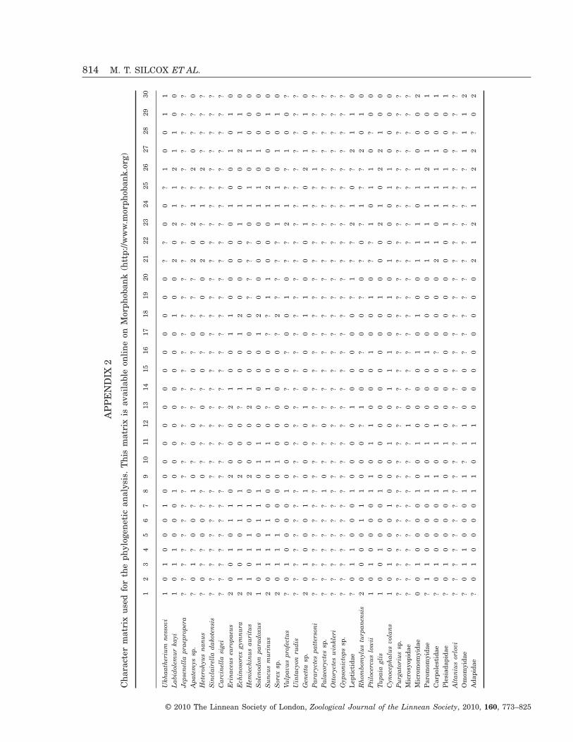
AP
PE
ND
IX2
Cha
ract
erm
atri
xus
edfo
rth
eph
ylog
enet
ican
alys
is.
Thi
sm
atri
xis
avai
labl
eon
line
onM
orph
oban
k(h
ttp:
//ww
w.m
orph
oban
k.or
g)
12
34
56
78
910
1112
1314
1516
1718
1920
2122
2324
2526
2728
2930
Ukh
aath
eriu
mne
ssov
i1
01
00
01
00
00
00
00
00
00
0?
?0
0?
10
01
1L
abid
olem
urka
yi1
01
10
00
10
00
00
00
00
10
02
02
11
21
10
0Je
psen
ella
prae
prop
era
??
??
??
??
??
??
??
??
??
??
??
??
??
??
??
Apa
tem
yssp
.?
01
?0
0?
10
?0
0?
?0
?0
??
?2
02
1?
20
??
0H
eter
ohyu
sna
nus
?0
??
00
??
0?
??
0?
0?
0?
00
20
?1
?2
??
??
Sin
clai
rell
ada
kote
nsis
??
??
??
??
??
??
??
??
??
??
??
??
??
??
??
Car
cine
lla
sige
i?
??
??
??
??
??
??
??
??
??
??
??
??
??
??
?E
rina
ceus
euro
paeu
s2
00
10
11
02
00
02
10
01
10
00
00
10
01
01
0E
chin
osor
exgy
mnu
ra2
10
10
11
12
00
0?
10
01
20
00
01
10
02
11
0H
emie
chin
usau
ritu
s2
10
11
01
02
00
02
10
00
0?
??
?0
11
01
00
0S
olen
odon
para
doxu
s1
01
10
11
10
11
00
00
01
20
00
00
11
01
00
0S
uncu
sm
urin
us2
01
11
10
00
11
0?
10
0?
?1
10
00
12
00
01
0S
orex
sp.
20
11
10
00
01
00
00
00
?2
??
??
11
10
10
10
Vulp
avus
prof
ectu
s?
01
00
00
10
00
00
?0
?0
01
0?
?2
1?
?1
00
?U
inta
cyon
rudi
s?
??
??
??
??
??
??
??
??
??
??
??
??
??
??
?G
enet
tasp
.2
01
00
01
10
00
01
00
00
11
00
01
10
21
01
0P
arar
ycte
spa
tter
soni
??
??
??
??
??
??
??
??
??
??
??
??
?1
??
??
Pal
aeor
ycte
ssp
.?
??
??
??
10
??
0?
??
??
??
??
??
??
??
??
?O
ttor
ycte
sw
inkl
eri
??
??
??
??
??
??
??
??
??
??
??
??
??
??
??
Gyp
soni
ctop
ssp
.?
??
??
??
??
??
??
??
??
??
??
??
??
??
??
?L
epti
ctid
ae?
01
10
00
10
00
01
00
00
0?
1?
?2
10
?2
11
0R
hom
bom
ylus
turp
anen
sis
20
00
01
11
00
0?
10
0?
00
?0
?0
?1
??
20
10
Pti
loce
rcus
low
ii1
01
00
00
11
01
10
00
10
01
0?
?1
01
10
?0
0Tu
paia
glis
10
01
00
01
00
00
00
00
00
10
00
21
02
21
00
Cyn
ocep
halu
svo
lans
10
10
00
10
00
10
01
11
00
10
10
00
11
00
00
Pur
gato
rius
sp.
??
??
??
??
??
??
??
??
??
??
??
??
??
??
??
Mic
rosy
opid
ae?
??
??
??
??
??
10
??
??
??
??
??
??
??
??
?M
icro
mom
yida
e0
01
00
00
10
01
00
00
10
10
01
11
01
10
00
2P
arom
omyi
dae
?1
10
00
01
10
10
00
01
00
00
11
11
12
10
01
Car
pole
stid
ae?
01
00
00
11
01
10
00
?0
00
02
10
11
11
00
1P
lesi
adap
idae
?0
10
00
01
00
10
00
00
00
00
00
11
11
00
01
Alt
aniu
sor
lovi
??
??
??
??
??
??
??
??
??
??
??
??
??
??
??
Om
omyi
dae
?0
11
00
01
1?
11
00
0?
??
??
??
??
??
11
12
Ada
pida
e?
01
00
00
11
01
10
00
00
00
02
12
11
22
?0
2
814 M. T. SILCOX ET AL.
© 2010 The Linnean Society of London, Zoological Journal of the Linnean Society, 2010, 160, 773–825
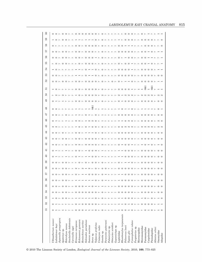
3132
3334
3536
3738
3940
4142
4344
4546
4748
4950
5152
5354
5556
5758
5960
Ukh
aath
eriu
mne
ssov
i0
01
00
00
00
00
20
02
00
00
?1
00
1?
00
01
0L
abid
olem
urka
yi1
10
00
00
00
11
00
02
10
01
20
00
00
00
?0
0Je
psen
ella
prae
prop
era
??
??
??
??
??
??
??
??
??
??
??
??
??
??
??
Apa
tem
yssp
.1
?0
00
00
00
??
?0
0?
??
?1
2?
0?
?0
00
?0
0H
eter
ohyu
sna
nus
??
00
00
00
01
10
0?
?1
0?
??
?0
??
00
0?
00
Sin
clai
rell
ada
kote
nsis
??
??
??
??
??
??
??
??
??
??
??
??
??
??
??
Car
cine
lla
sige
i?
??
??
??
??
??
??
??
??
??
??
??
??
??
??
?E
rina
ceus
euro
paeu
s0
01
00
10
2?
00
20
01
21
10
10
01
1?
00
01
0E
chin
osor
exgy
mnu
ra0
11
00
1?
?0
10
20
00
21
10
00
01
10
00
01
0H
emie
chin
usau
ritu
s0
01
00
11
2?
??
??
??
??
??
??
??
??
??
??
0S
olen
odon
para
doxu
s1
01
01
11
10
11
20
10
10
10
00
00
00
00
01
0S
uncu
sm
urin
us1
01
01
10
2?
10
20
11
21
10
00
01
10
00
01
0S
orex
sp.
10
10
01
0?
00
02
01
02
?0&
10
00
01
10
00
01
0Vu
lpav
uspr
ofec
tus
00
00
10
00
00
20
00
00
01
02
00
00
?1
?0
00
Uin
tacy
onru
dis
??
??
??
??
??
??
??
??
??
??
??
??
??
??
??
Gen
etta
sp.
01
00
10
00
01
12
00
01
11
02
00
00
00
00
00
Par
aryc
tes
patt
erso
ni?
??
??
11
0?
??
??
??
??
??
??
??
??
??
??
?P
alae
oryc
tes
sp.
??
??
??
??
??
??
??
??
??
??
??
??
??
??
??
Ott
oryc
tes
win
kler
i?
??
??
??
??
??
??
??
??
??
??
0?
??
??
??
?G
ypso
nict
ops
sp.
??
??
??
??
??
??
??
??
??
??
??
??
??
??
??
Lep
tict
idae
11
10
01
10
01
10
00
01
01
02
00
00
00
00
1?
Rho
mbo
myl
ustu
rpan
ensi
s0
11
00
01
0?
11
10
00
10
01
10
?0
0?
?0
0?
?P
tilo
cerc
uslo
wii
00
00
00
00
10
00
00
20
01
02
01
00
01
01
00
Tupa
iagl
is1
10
00
00
01
00
00
02
20
10
20
10
00
00
10
0C
ynoc
epha
lus
vola
ns0
00
00
00
00
00
10
02
00
00
21
00
11
00
10
0P
urga
tori
ussp
.?
??
??
??
??
??
??
??
??
??
??
??
??
??
??
?M
icro
syop
idae
??
??
??
??
??
??
??
??
??
??
1?
??
??
??
??
Mic
rom
omyi
dae
?0
10
00
00
00
01
00
20
01
02
10
00
00
0?
00
Par
omom
yida
e0
00
00
00
00
00
00
02
00
10
20&
10
00
00
00
0?
Car
pole
stid
ae1
0?
00
00
00
00
10
02
00
10
21
00
01
11
00
1P
lesi
adap
idae
00
00
00
00
00
00
00
20
01
02
0&1
00
00
10
00
?A
ltan
ius
orlo
vi?
??
??
??
??
??
??
??
??
??
??
??
??
??
??
?O
mom
yida
e1
11
00
00
00
00
10
02
20
00
21
10
1?
?1
??
1A
dapi
dae
01
00
00
00
00
00
00
20
01
02
11
01
11
10
01
LABIDOLEMUR KAYI CRANIAL ANATOMY 815
© 2010 The Linnean Society of London, Zoological Journal of the Linnean Society, 2010, 160, 773–825

AP
PE
ND
IX2
Con
tinu
ed
6162
6364
6566
6768
6970
7172
7374
7576
7778
7980
8182
8384
8586
8788
8990
Ukh
aath
eriu
mne
ssov
i1
00
0?
00
00
00
11
0?
02
10
0&1
11
10
0?
10
1?
Lab
idol
emur
kayi
10
00
02
20
10
11
?1
00
0?
10
0?
00
10
00
01
Jeps
enel
lapr
aepr
oper
a?
??
??
??
??
??
??
??
??
??
??
??
??
??
??
?A
pate
mys
sp.
1?
0?
??
?0
1?
?0
?1
?0
0?
??
??
??
??
??
??
Het
eroh
yus
nanu
s1
?0
?0
??
02
??
1?
1?
00
??
??
??
?1
??
??
?S
incl
aire
lla
dako
tens
is?
??
??
??
?2
10
1?
??
00
??
0?
??
??
??
?0
?C
arci
nell
asi
gei
??
??
??
??
11
1?
?1
??
00
1?
01
00
1?
01
0?
Eri
nace
useu
ropa
eus
00
00
00
20
11
21
00
01
10
02
11
00
10
11
00
Ech
inos
orex
gym
nura
00
00
00
10
01
21
00
00
10
02
01
00
10
11
00
Hem
iech
inus
auri
tus
??
?0
10
20
10
21
00
01
20
12
0&1
10
01
01
11
0S
olen
odon
para
doxu
s0
00
01
01
00
12
01
00
00
10
00
10
10
00&
10
00
Sun
cus
mur
inus
10
0?
00
10
00
20
10
00
01
00
11
00
01
00
00
Sor
exsp
.1
00
00
02
00
00
01
00
00
10
01
00
00
?0
00
0Vu
lpav
uspr
ofec
tus
11
?0
0?
?0
01
11
00
?1
10
00
??
0?
01
00
0?
Uin
tacy
onru
dis
??
??
??
??
0?
?1
??
??
??
??
??
??
??
??
??
Gen
etta
sp.
01
00
00
30
12
21
00
10
10&
10
10
00
10
0&1
01
01
Par
aryc
tes
patt
erso
ni?
??
??
??
?0
2?
00
0?
?1
?0
20
?0
?0
??
11
?P
alae
oryc
tes
sp.
??
??
??
??
0?
?0
??
??
??
0?
??
??
0?
?0
1?
Ott
oryc
tes
win
kler
i?
??
??
??
?0
21
0&1
00
??
2?
0?
01
01
00
01
1?
Gyp
soni
ctop
ssp
.?
??
??
??
??
??
??
??
??
??
??
??
??
??
??
?L
epti
ctid
ae1
00
00
?1
01
22
10
01
10
00
20
10
10
00
10
1R
hom
bom
ylus
turp
anen
sis
1?
0?
00
??
12
21
11
00
20
02
01
00
0?
?0
1?
Pti
loce
rcus
low
ii1
01
10
21
11
12
21
01
12
10
10
10
00
10
01
1Tu
paia
glis
10
00
02
?0
02
22
10
11
20
02
00
00
00
00
11
Cyn
ocep
halu
svo
lans
10
21
10
31
12
11
10
11
20
02
11
00
00
00
01
Pur
gato
rius
sp.
??
??
??
??
??
??
??
??
??
??
??
??
??
??
??
Mic
rosy
opid
ae?
??
??
??
?1
10
11
01
00
11
01
00
00
00
10
1M
icro
mom
yida
e1
02
??
?1
01
??
1?
??
02
?1
??
??
??
??
?1
?P
arom
omyi
dae
10
2?
01
?0
12
01
00
?0
21
12
11
??
0?
?1
11
Car
pole
stid
ae1
02
0?
??
01
22
10
00
02
11
21
00
?0
??
01
?P
lesi
adap
idae
00
10
00
10
12
21
01
01
21
?2
10
??
0?
?0
1?
Alt
aniu
sor
lovi
??
??
??
??
??
??
??
??
??
??
??
??
??
??
??
Om
omyi
dae
??
??
??
??
2?
22
0?
01
21
02
1?
00
0?
01
1?
Ada
pida
e1
00
00
?1
02
12
20
00
11
10
21
00
00
00
01
1
816 M. T. SILCOX ET AL.
© 2010 The Linnean Society of London, Zoological Journal of the Linnean Society, 2010, 160, 773–825

9192
9394
9596
9798
9910
010
110
210
310
410
510
610
710
810
911
011
111
211
311
411
511
611
711
811
912
0
Ukh
aath
eriu
mne
ssov
i?
02
1?
00
11
0–
00
00
?0
00
00
00
00
00
01
0L
abid
olem
urka
yi0
11
00
00
01
10
10
00
?0
?0
00
00
11
10
02
1Je
psen
ella
prae
prop
era
??
??
??
??
??
??
??
??
??
??
?0
0?
??
??
??
Apa
tem
yssp
.?
1?
??
?0
0?
??
0?
??
??
?2
??
0?
11
10
02
1H
eter
ohyu
sna
nus
?1
??
??
??
??
??
??
??
??
2?
??
01
1?
00
21
Sin
clai
rell
ada
kote
nsis
?1
10
?0
0?
??
??
??
??
??
21
?0
01
?0
20
21
Car
cine
lla
sige
i0
1?
00
00
?1
?0
00
00
?0
?2
0?
??
1?
??
02
1E
rina
ceus
euro
paeu
s0
12
10
00
11
10
10
00
?0
01
00
01
10
00
01
1E
chin
osor
exgy
mnu
ra0
11
00
00
11
0?
10
00
?0
01
00
01
10
00
01
0H
emie
chin
usau
ritu
s0
12
10
00
11
10
10
00
?0
01
00
01
10
00
01
1S
olen
odon
para
doxu
s0
10
20
?0
11
10
00
00
?0
01
00
10&
1&2
10
00
01
0S
uncu
sm
urin
us0
10
20
00
11
10
00
00
?0
01
10
0?
11
10
02
1S
orex
sp.
00
02
00
01
11
00
00
0?
00
11
00
01
11
00
10
Vulp
avus
prof
ectu
s1
11
20
00
?0
0?
00
02
?0
01
0?
0?
00
00
01
0U
inta
cyon
rudi
s?
??
??
??
??
??
??
??
??
??
0?
0?
??
??
0?
0G
enet
tasp
.1
02
00
00
10
0?
10
02
?0
20
01
01
00
00
01
0P
arar
ycte
spa
tter
soni
?0
11
?0
11
11
01
?0
0?
02
10
?1
0?
??
??
?0
Pal
aeor
ycte
ssp
.?
?1
2?
??
??
10
??
??
??
??
??
?1
??
??
??
?O
ttor
ycte
sw
inkl
eri
00
11
01
11
11
01
11
10
02
10
01
10
0?
?0
10
Gyp
soni
ctop
ssp
.?
??
??
??
??
??
??
??
??
??
??
??
??
??
??
?L
epti
ctid
ae0
01
11
01
00
10
11
00
?0
22
00
11
0?
0?
01
1R
hom
bom
ylus
turp
anen
sis
00
00
?1
02
11
00
12
2?
00
10
?0
12
0?
11
01
Pti
loce
rcus
low
ii0
01
01
01
01
10
11
11
11
21
10
01
10
00
02
1Tu
paia
glis
00
11
10
10
10
?1
11
11
02
10
00
11
00
00
21
Cyn
ocep
halu
svo
lans
00
02
?1
02
00
?1
02
2?
01
01
10
10
00
00
20
Pur
gato
rius
sp.
??
??
??
??
??
??
??
??
??
??
??
?1
12
0?
??
Mic
rosy
opid
ae1
12
0&1
11
??
01
01
00
0?
0?
10
00
11
11
00
21
Mic
rom
omyi
dae
??
21
?1
02
1?
??
10
??
1?
??
?0
11
12
00
21
Par
omom
yida
e1
12
1?
10
21
10
11
12
?1
22
00
01
11
30
01
1C
arpo
lest
idae
00
21
?0
??
11
1?
10
0?
0?
?0
?0
?1
13
00
20
Ple
siad
apid
ae0
02
10
01
?1
11
11
02
?1
?0
0?
01
11
30
01
1A
ltan
ius
orlo
vi?
??
??
??
??
??
??
??
??
??
??
??
??
??
??
?O
mom
yida
e?
02
11
01
?1
11
11
11
?1
0?
0?
0?
0&1
0&1
10
01&
21
Ada
pida
e0
02
11
01
01
11
11
11
11
01
0?
01
00
00
01&
21
LABIDOLEMUR KAYI CRANIAL ANATOMY 817
© 2010 The Linnean Society of London, Zoological Journal of the Linnean Society, 2010, 160, 773–825
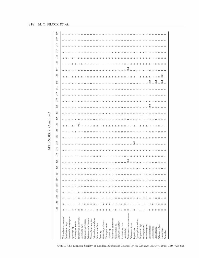
AP
PE
ND
IX2
Con
tinu
ed
121
122
123
124
125
126
127
128
129
130
131
132
133
134
135
136
137
138
139
140
141
142
143
144
145
146
147
148
149
150
Ukh
aath
eriu
mne
ssov
i0
02
00
21
00
10
10
20
00
10
00
00
11
00
00
0L
abid
olem
urka
yi2
11
13
??
??
??
??
20
00
00
11
00
11
01
00
0Je
psen
ella
prae
prop
era
??
??
??
??
??
??
??
??
0?
??
??
??
??
??
??
Apa
tem
yssp
.2
11
13
??
??
??
??
20
?0
00
01
10
00
0?
?0
?H
eter
ohyu
sna
nus
21
11
3?
??
??
??
?0
11
00
?0
10
00
0?
10
?0
Sin
clai
rell
ada
kote
nsis
21
20
3?
??
??
??
?1
0&1
10
00
01
10
11
01
00
0C
arci
nell
asi
gei
21
11
3?
??
??
??
?1
10
00
01
10
01
00
11
?1
Eri
nace
useu
ropa
eus
10
10
20
01
01
00
02
10
00
11
11
11
10
10
11
Ech
inos
orex
gym
nura
10
10
02
10
01
01
02
10
01
11
10
01
10
10
11
Hem
iech
inus
auri
tus
10
20
02
00
01
01
02
10
01
11
11
01
10
10
11
Sol
enod
onpa
rado
xus
10
0?
10
11
00
00
02
01
02
01
00
01
10
10
11
Sun
cus
mur
inus
01
10
20
01
00
00
02
00
01
01
10
01
10
10
00
Sor
exsp
.0
11
02
00
10
00
00
21
00
11
11
00
11
01
01
0Vu
lpav
uspr
ofec
tus
00
20
00
11
01
00
02
00
10
01
10
01
10
10
11
Uin
tacy
onru
dis
00
20
10
11
01
00
02
00
12
01
10
01
10
10
10
Gen
etta
sp.
00
20
02
10
01
01
02
00
12
01
10
01
10
10
10
Par
aryc
tes
patt
erso
ni1
11
01
20
00
10
00
20
00
20
11
00
11
00
00
0P
alae
oryc
tes
sp.
01
20
02
00
11
00
02
00
00
01
11
0?
10
00
10
Ott
oryc
tes
win
kler
i1
12
10
21
01
10
00
20
00
10
11
00
01
00
00
0G
ypso
nict
ops
sp.
??
??
01
10
11
01
02
00
02
01
10
00
13
10
00
Lep
tict
idae
00
20
02
10
10
21
02
00
02
01
00
00
03
00
00
Rho
mbo
myl
ustu
rpan
ensi
s2
10
?0
01
00&
11
01
02
11
01
01
10
10&
11
01
00
0P
tilo
cerc
uslo
wii
11
20
00
10
00
01
02
10
01
0?
11
01
00
10
11
Tupa
iagl
is0
12
00
21
00
00
0&1
02
00
01
01
11
01
00
00
11
Cyn
ocep
halu
svo
lans
11
0?
02
11
10
00
02
10
02
01
11
10
03
10
00
Pur
gato
rius
sp.
??
??
02
10
10
01
02
00
01
01
10
00
00
00
00
Mic
rosy
opid
ae1
12
01
00
10
00
00
10
0?
11
11
00
10
00
00
1M
icro
mom
yida
e1
12
00
21
10
00
00
20
00
10
11
00
00
01
00
0P
arom
omyi
dae
01
2?
00
1?
00
00
02
10
00&
20
11
0&1
01
00
10
01
Car
pole
stid
ae0
01
00
10
01
01
10
21
00
21
11
01
01
11
10
1P
lesi
adap
idae
01
21
01
10
10
11
01
11
00
01
10&
11
00
11
00
1A
ltan
ius
orlo
vi0
02
?0
21
00
00
10
20
00
?0
11
00
10
00
11
1O
mom
yida
e0
01
00
11
10
00
10
21
00
00
11
0&1
0&1
10
00
01
1A
dapi
dae
00
20
00
10
01
01
02
10
01
01
10
11
00
00
11
818 M. T. SILCOX ET AL.
© 2010 The Linnean Society of London, Zoological Journal of the Linnean Society, 2010, 160, 773–825
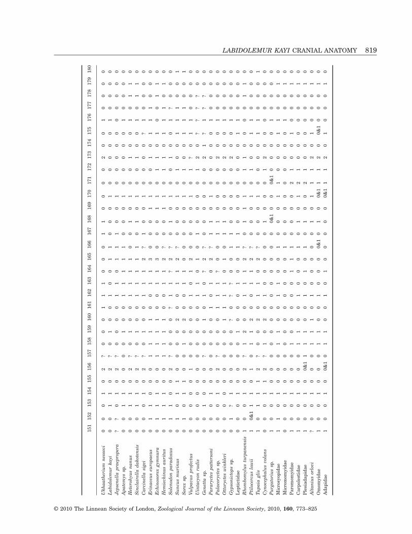
151
152
153
154
155
156
157
158
159
160
161
162
163
164
165
166
167
168
169
170
171
172
173
174
175
176
177
178
179
180
Ukh
aath
eriu
mne
ssov
i0
00
10
2?
00
01
11
00
01
10
00
02
00
10
00
0L
abid
olem
urka
yi0
01
10
2?
01
00
10
01
10
00
10
00
00
10
00
0Je
psen
ella
prae
prop
era
??
01
02
?0
00
01
10
11
10
01
00
00
00
00
00
Apa
tem
yssp
.0
01
10
00
01
00
11
11
10
00
10
00
00
10
00
0H
eter
ohyu
sna
nus
00
11
02
?0
10
01
11
10
01
01
00
10
01
01
10
Sin
clai
rell
ada
kote
nsis
00
11
02
?0
10
01
11
11
01
01
00
10
01
01
10
Car
cine
lla
sige
i0
00
10
2?
01
00
11
12
?1
00
10
00
0?
10
00
0E
rina
ceus
euro
paeu
s0
11
00
01
11
10
01
13
00
01
10
01
01
11
00
0E
chin
osor
exgy
mnu
ra0
11
10
01
11
10
01
13
00
01
10
11
01
00
11
0H
emie
chin
usau
ritu
s0
11
10
01
11
10
01
12
?0
01
10
11
01
11
01
0S
olen
odon
para
doxu
s0
11
00
2?
00
0?
11
?2
?0
10
10
01
00
1?
00
0S
uncu
sm
urin
us0
10
11
00
00
20
01
12
?0
00
10
10
01
11
00
1S
orex
sp.
01
01
10
01
02
00
11
00
00
01
01
00
11
10
01
Vulp
avus
prof
ectu
s0
00
00
10
00
00
10
12
00
00
10
1?
01
10
00
0U
inta
cyon
rudi
s0
00
00
00
00
00
11
01
01
00
00
12
0?
??
?0
0G
enet
tasp
.0
01
00
0?
00
10
10
?2
?0
00
00
02
1?
1?
?0
0P
arar
ycte
spa
tter
soni
00
01
00
00
00
10
10
2?
11
01
00
00
01
00
00
Pal
aeor
ycte
ssp
.0
00
10
2?
00
01
11
?0
01
10
00
02
00
10
00
0O
ttor
ycte
sw
inkl
eri
00
00
00
00
01
11
10
10
11
00
00
20
01
00
00
Gyp
soni
ctop
ssp
.0
1?
10
00
00
?0
??
00
11
00
00
02
00
10
00
0L
epti
ctid
ae0
00
10
00
01
00
01
00
11
00
00
00
01
10
00
0R
hom
bom
ylus
turp
anen
sis
10
01
02
?1
20
01
11
2?
01
11
01
10
00
01
10
Pti
loce
rcus
low
ii1
0&1
11
01
01
12
00
11
2?
00
01
01
00
11
00
00
Tupa
iagl
is1
01
11
2?
00
20
11
02
?0
00
10
00
01
10
00
1C
ynoc
epha
lus
vola
ns0
00
11
2?
10
20
10
00
00
10
00
12
00
00
00
0P
urga
tori
ussp
.1
00
10
00
00
00
00
00
00
0&1
00
0&1
00
00
10
00
0M
icro
syop
idae
11
01
00
01
10
01
10
01
00
01
10
00
11
00
10
Mic
rom
omyi
dae
00
01
00
00
00
00
00
01
00
00
00
00
01
00
10
Par
omom
yida
e1
00
10
00
10
00
00
11
10
00
02
10
01
00
01
0C
arpo
lest
idae
10
01
00
01
10
01
00
01
01
01
21
00
00
00
10
Ple
siad
apid
ae1
00
10
0&1
11
10
01
01
01
01
00
21
00
00
00
10
Alt
aniu
sor
lovi
?0
01
00
01
11
01
00
00
01
01
11
20
10
00
10
Om
omyi
dae
10
01
00
01
10
01
00
00&
10
10
0&1
11
20
0&1
00
01
0A
dapi
dae
10
01
00&
10
11
00
10
10
00
00
0&1
11
20
10
00
10
LABIDOLEMUR KAYI CRANIAL ANATOMY 819
© 2010 The Linnean Society of London, Zoological Journal of the Linnean Society, 2010, 160, 773–825
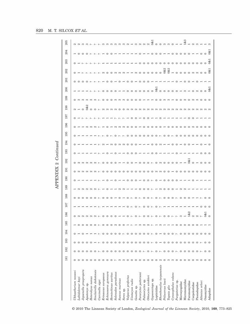
AP
PE
ND
IX2
Con
tinu
ed
181
182
183
184
185
186
187
188
189
190
191
192
193
194
195
196
197
198
199
200
201
202
203
204
205
Ukh
aath
eriu
mne
ssov
i0
00
01
01
01
00
00
00
00
20
10
00
12
Lab
idol
emur
kayi
00
01
02
10
21
22
11
11
11
31
01
10
2Je
psen
ella
prae
prop
era
00
00
02
10
21
22
11
11
?0
31
0?
?0
3A
pate
mys
sp.
00
01
02
10
21
22
11
12
?0&
23
10
??
03
Het
eroh
yus
nanu
s0
00
10
21
02
12
21
11
2?
03
??
??
??
Sin
clai
rell
ada
kote
nsis
00
11
02
10
21
22
11
12
?0
3?
??
??
?C
arci
nell
asi
gei
0?
??
??
??
??
??
??
??
??
??
??
??
?E
rina
ceus
euro
paeu
s0
00
00
01
01
10
01
2?
11
20
01
21
13
Ech
inos
orex
gym
nura
00
00
00
10
00
00
01
00
12
00
00
10
3H
emie
chin
usau
ritu
s0
00
00
01
01
10
01
2?
11
20
00
01
03
Sol
enod
onpa
rado
xus
?0
00
20
11
10
00
00
02
?2
30
12
11
3S
uncu
sm
urin
us3
00
00
11
02
10
11
2?
2?
03
10
21
13
Sor
exsp
.3
00
?0
11
12
10
11
2?
2?
03
11
21
13
Vulp
avus
prof
ectu
s0
00
01
00
01
00
00
00
01
20
00
11
12
Uin
tacy
onru
dis
00
00
??
??
10
00
00
00
02
00
01
11
1G
enet
tasp
.0
00
01
00
01
00
10
00
00
20
01
21
13
Par
aryc
tes
patt
erso
ni0
00
03
??
?0
10
01
10
11
20
00
00
02
Pal
aeor
ycte
ssp
.0
?0
02
00
00
00
01
10
00
21
00
00
12
Ott
oryc
tes
win
kler
i0
00
02
00
00
00
01
10
00
21
00
00
12
Gyp
soni
ctop
ssp
.0
??
??
??
??
?0
?0
00
01
21
01
00
00&
1L
epti
ctid
ae0
00
01
00
01
02
00
00
01
20
0&1
10
00
0R
hom
bom
ylus
turp
anen
sis
2?
11
3?
??
21
22
12
?0
02
00
10
00
0P
tilo
cerc
uslo
wii
00
00
10
10
00
00
11
01
12
30
10&
20
13
Tupa
iagl
is3
00
01
01
00
00
01
10
01
23
01
0&2
01
3C
ynoc
epha
lus
vola
ns3
00
01
?1
00
01
01
0?
01
21
01
10
00
Pur
gato
rius
sp.
0?
0?
0?
1?
10
00
00
00
12
00
00
00
1M
icro
syop
idae
00
00
03
10
21
01
11
00
12
01
10
00
1M
icro
mom
yida
e0
00
00
11
02
10
11
00
01
22
30
00
01&
3P
arom
omyi
dae
00
00
01&
21
01
10
0&1
10
00
02
01
00
00
1C
arpo
lest
idae
00
00
02
11
10
00
11
02
02
20
12
01
2P
lesi
adap
idae
20
00
02
11
11
01
11
00
12
32
02
01
3A
ltan
ius
orlo
vi0
??
01
?0
?1
10
00
00
01
20
01
00
01
Om
omyi
dae
00
00
10&
10
01
10
00
10
01
20
01
00
01
Ada
pida
e0
00
01
00
01
10
00
00
01
20
0&1
10&
10&
10&
13
820 M. T. SILCOX ET AL.
© 2010 The Linnean Society of London, Zoological Journal of the Linnean Society, 2010, 160, 773–825
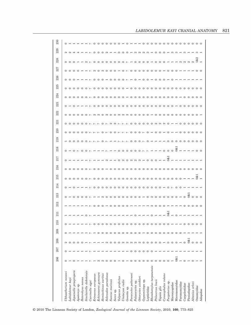
206
207
208
209
210
211
212
213
214
215
216
217
218
219
220
221
222
223
224
225
226
227
228
229
230
Ukh
aath
eriu
mne
ssov
i0
02
00
11
00
00
00
10
00
00
00
00
20
Lab
idol
emur
kayi
11
01
11
11
10
01
10
00
01
00
00
02
1Je
psen
ella
prae
prop
era
01
01
11
10
00
01
00
00
00
00
00
04
1A
pate
mys
sp.
11
01
11
10
10
01
?0
00
01
00
10
14
1H
eter
ohyu
sna
nus
11
01
11
10
10
01
00
00
01
00
11
11
1S
incl
aire
lla
dako
tens
is1
11
11
01
01
00
10
00
00
10
01
12
11
Car
cine
lla
sige
i?
??
??
??
??
??
??
??
??
??
??
??
??
Eri
nace
useu
ropa
eus
11
11
01
01
00
01
1?
??
20
00
02
00
0E
chin
osor
exgy
mnu
ra2
11
00
10
10
00
11
??
?2
00
00
00
00
Hem
iech
inus
auri
tus
21
10
01
02
00
01
10
00
00
00
02
00
0S
olen
odon
para
doxu
s0
12
00
11
00
01
2?
??
?2
00
01
00
20
Sun
cus
mur
inus
01
00
01
00
00
01
11
02
00
00
00
01
0S
orex
sp.
01
00
01
00
00
01
11
02
00
00
00
01
0Vu
lpav
uspr
ofec
tus
11
00
01
00
00
00
0?
??
20
00
01
00
0U
inta
cyon
rudi
s0
12
00
10
00
00
20
??
?2
00
00
00
01
Gen
etta
sp.
11
20
01
00
00
00
0?
??
20
11
??
?0
0P
arar
ycte
spa
tter
soni
00
20
01
10
00
02
00
00
00
00
10
11
1P
alae
oryc
tes
sp.
00
20
00
10
00
02
00
00
00
00
00
02
1O
ttor
ycte
sw
inkl
eri
00
20
01
10
00
02
00
00
00
00
10
02
1G
ypso
nict
ops
sp.
00
01
01
11
00
01
??
?0
00
00
00
02
0L
epti
ctid
ae0
00
00
11
00
00
0?
00
00
00
00
00
30
Rho
mbo
myl
ustu
rpan
ensi
s0
00
00
02
20
00
01
11
01
00
01
01
10
Pti
loce
rcus
low
ii0
10
00
10
00
00
01
10
20
00
00
00
00
Tupa
iagl
is0
10
00
10
00
00
11
10
20
00
00
00
00
Cyn
ocep
halu
svo
lans
01
10
01
00
10
01
01
12
00
00
01
00
0P
urga
tori
ussp
.0
10
00
0&1
10
00
00&
10
00
00
00
01
01
10
Mic
rosy
opid
ae1
10
01
11
0?
00
10
01
10
00
00
01
30
Mic
rom
omyi
dae
0&1
10
01
01
00
00
10&
10
11
01
00
10
13
0P
arom
omyi
dae
11
10
11
11
00
01
10
00
00
10
11
21
0C
arpo
lest
idae
01
01
01
00
11
01
10
00
00
00
11
23
0P
lesi
adap
idae
11
0&1
10
10
0&1
11
01
10
00
00
00
11
21
0A
ltan
ius
orlo
vi0
10
10
10
01
10
11
00
00
00
01
12
10
Om
omyi
dae
11
01
01
00
10&
10
10
00
00
00
01
01&
23
0A
dapi
dae
11
01
01
00
11
01
00
00
10
00
10
11
0
LABIDOLEMUR KAYI CRANIAL ANATOMY 821
© 2010 The Linnean Society of London, Zoological Journal of the Linnean Society, 2010, 160, 773–825
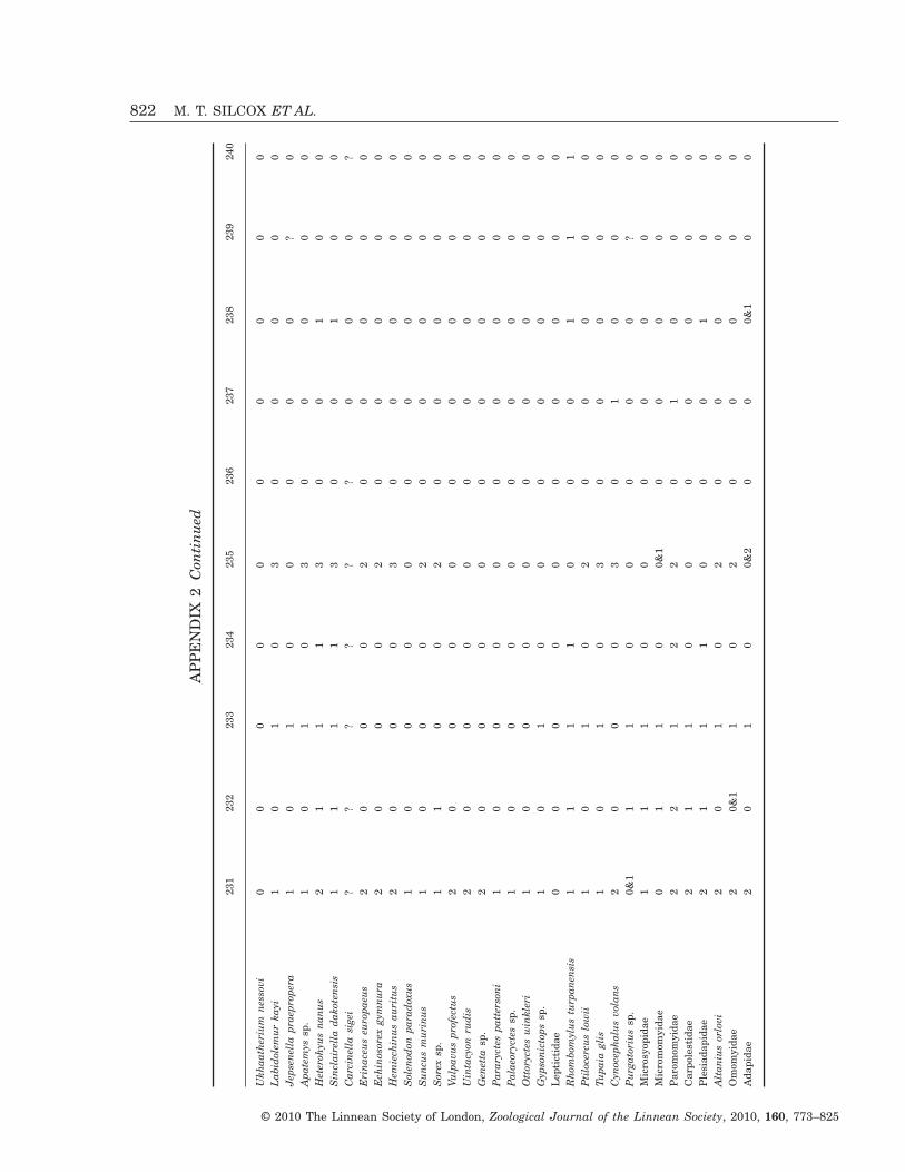
AP
PE
ND
IX2
Con
tinu
ed
231
232
233
234
235
236
237
238
239
240
Ukh
aath
eriu
mne
ssov
i0
00
00
00
00
0L
abid
olem
urka
yi1
01
03
00
00
0Je
psen
ella
prae
prop
era
10
10
00
00
?0
Apa
tem
yssp
.1
01
03
00
00
0H
eter
ohyu
sna
nus
21
11
30
01
00
Sin
clai
rell
ada
kote
nsis
11
11
30
01
00
Car
cine
lla
sige
i?
??
??
?0
00
?E
rina
ceus
euro
paeu
s2
00
02
00
00
0E
chin
osor
exgy
mnu
ra2
00
02
00
00
0H
emie
chin
usau
ritu
s2
00
03
00
00
0S
olen
odon
para
doxu
s1
00
00
00
00
0S
uncu
sm
urin
us1
00
02
00
00
0S
orex
sp.
11
00
20
00
00
Vulp
avus
prof
ectu
s2
00
00
00
00
0U
inta
cyon
rudi
s2
00
00
00
00
0G
enet
tasp
.2
00
00
00
00
0P
arar
ycte
spa
tter
soni
10
00
00
00
00
Pal
aeor
ycte
ssp
.1
00
00
00
00
0O
ttor
ycte
sw
inkl
eri
10
00
00
00
00
Gyp
soni
ctop
ssp
.1
01
00
00
00
0L
epti
ctid
ae0
00
00
00
00
0R
hom
bom
ylus
turp
anen
sis
11
11
00
01
11
Pti
loce
rcus
low
ii1
01
02
00
00
0Tu
paia
glis
10
10
30
00
00
Cyn
ocep
halu
svo
lans
20
00
30
10
00
Pur
gato
rius
sp.
0&1
11
00
00
0?
0M
icro
syop
idae
11
10
00
00
00
Mic
rom
omyi
dae
01
10
0&1
00
00
0P
arom
omyi
dae
22
12
20
10
00
Car
pole
stid
ae2
11
00
00
00
0P
lesi
adap
idae
21
11
00
01
00
Alt
aniu
sor
lovi
20
10
20
00
00
Om
omyi
dae
20&
11
02
00
00
0A
dapi
dae
20
10
0&2
00
0&1
00
822 M. T. SILCOX ET AL.
© 2010 The Linnean Society of London, Zoological Journal of the Linnean Society, 2010, 160, 773–825
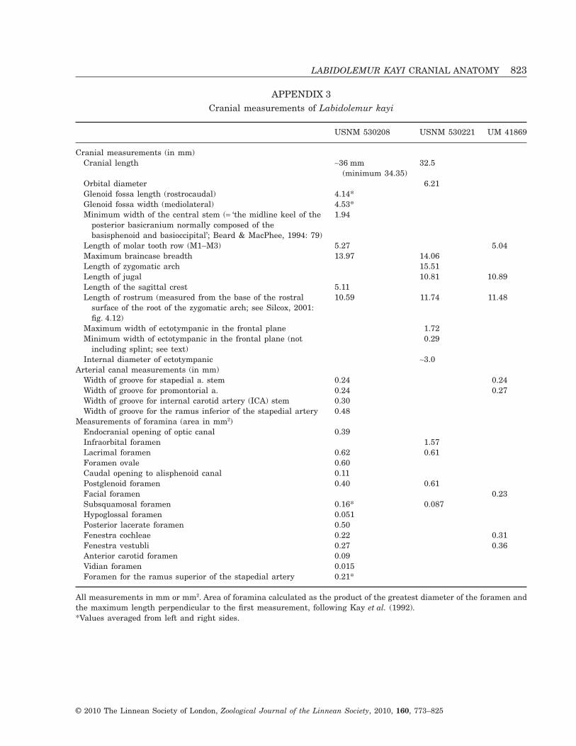
APPENDIX 3Cranial measurements of Labidolemur kayi
USNM 530208 USNM 530221 UM 41869
Cranial measurements (in mm)Cranial length ~36 mm
(minimum 34.35)32.5
Orbital diameter 6.21Glenoid fossa length (rostrocaudal) 4.14*Glenoid fossa width (mediolateral) 4.53*Minimum width of the central stem (= ‘the midline keel of the
posterior basicranium normally composed of thebasisphenoid and basioccipital’; Beard & MacPhee, 1994: 79)
1.94
Length of molar tooth row (M1–M3) 5.27 5.04Maximum braincase breadth 13.97 14.06Length of zygomatic arch 15.51Length of jugal 10.81 10.89Length of the sagittal crest 5.11Length of rostrum (measured from the base of the rostral
surface of the root of the zygomatic arch; see Silcox, 2001:fig. 4.12)
10.59 11.74 11.48
Maximum width of ectotympanic in the frontal plane 1.72Minimum width of ectotympanic in the frontal plane (not
including splint; see text)0.29
Internal diameter of ectotympanic ~3.0Arterial canal measurements (in mm)
Width of groove for stapedial a. stem 0.24 0.24Width of groove for promontorial a. 0.24 0.27Width of groove for internal carotid artery (ICA) stem 0.30Width of groove for the ramus inferior of the stapedial artery 0.48
Measurements of foramina (area in mm2)Endocranial opening of optic canal 0.39Infraorbital foramen 1.57Lacrimal foramen 0.62 0.61Foramen ovale 0.60Caudal opening to alisphenoid canal 0.11Postglenoid foramen 0.40 0.61Facial foramen 0.23Subsquamosal foramen 0.16* 0.087Hypoglossal foramen 0.051Posterior lacerate foramen 0.50Fenestra cochleae 0.22 0.31Fenestra vestubli 0.27 0.36Anterior carotid foramen 0.09Vidian foramen 0.015Foramen for the ramus superior of the stapedial artery 0.21*
All measurements in mm or mm2. Area of foramina calculated as the product of the greatest diameter of the foramen andthe maximum length perpendicular to the first measurement, following Kay et al. (1992).*Values averaged from left and right sides.
LABIDOLEMUR KAYI CRANIAL ANATOMY 823
© 2010 The Linnean Society of London, Zoological Journal of the Linnean Society, 2010, 160, 773–825
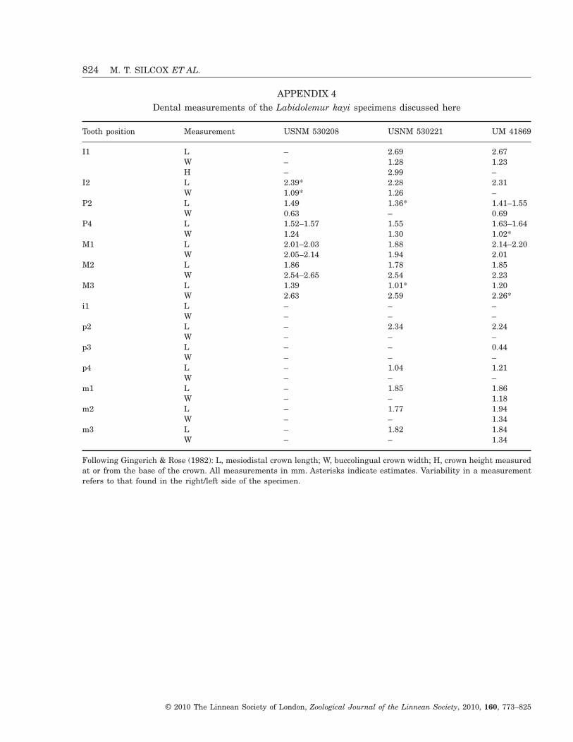
APPENDIX 4Dental measurements of the Labidolemur kayi specimens discussed here
Tooth position Measurement USNM 530208 USNM 530221 UM 41869
I1 L – 2.69 2.67W – 1.28 1.23H – 2.99 –
I2 L 2.39* 2.28 2.31W 1.09* 1.26 –
P2 L 1.49 1.36* 1.41–1.55W 0.63 – 0.69
P4 L 1.52–1.57 1.55 1.63–1.64W 1.24 1.30 1.02*
M1 L 2.01–2.03 1.88 2.14–2.20W 2.05–2.14 1.94 2.01
M2 L 1.86 1.78 1.85W 2.54–2.65 2.54 2.23
M3 L 1.39 1.01* 1.20W 2.63 2.59 2.26*
i1 L – – –W – – –
p2 L – 2.34 2.24W – – –
p3 L – – 0.44W – – –
p4 L – 1.04 1.21W – – –
m1 L – 1.85 1.86W – – 1.18
m2 L – 1.77 1.94W – – 1.34
m3 L – 1.82 1.84W – – 1.34
Following Gingerich & Rose (1982): L, mesiodistal crown length; W, buccolingual crown width; H, crown height measuredat or from the base of the crown. All measurements in mm. Asterisks indicate estimates. Variability in a measurementrefers to that found in the right/left side of the specimen.
824 M. T. SILCOX ET AL.
© 2010 The Linnean Society of London, Zoological Journal of the Linnean Society, 2010, 160, 773–825
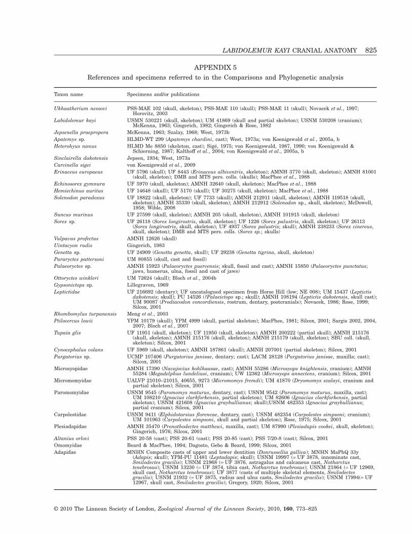
APPENDIX 5References and specimens referred to in the Comparisons and Phylogenetic analysis
Taxon name Specimens and/or publications
Ukhaatherium nessovi PSS-MAE 102 (skull, skeleton); PSS-MAE 110 (skull); PSS-MAE 11 (skull); Novacek et al., 1997;Horovitz, 2003
Labidolemur kayi USMN 530221 (skull, skeleton); UM 41869 (skull and partial skeleton); USNM 530208 (cranium);McKenna, 1963; Gingerich, 1982; Gingerich & Rose, 1982
Jepsenella praepropera McKenna, 1963; Szalay, 1968; West, 1973bApatemys sp. HLMD-WT 299 (Apatemys chardini, cast); West, 1973a; von Koenigswald et al., 2005a, bHeterohyus nanus HLMD Me 8850 (skeleton, cast); Sigé, 1975; von Koenigswald, 1987, 1990; von Koenigswald &
Schierning, 1987; Kalthoff et al., 2004; von Koenigswald et al., 2005a, bSinclairella dakotensis Jepsen, 1934; West, 1973aCarcinella sigei von Koenigswald et al., 2009Erinaceus europaeus UF 5796 (skull); UF 8443 (Erinaceus albiventris, skeleton); AMNH 3770 (skull, skeleton); AMNH 81001
(skull, skeleton); DMB and MTS pers. colls. (skulls); MacPhee et al., 1988Echinosorex gymnura UF 5970 (skull, skeleton); AMNH 32640 (skull, skeleton); MacPhee et al., 1988Hemiechinus auritus UF 14648 (skull); UF 5170 (skull); UF 30275 (skull, skeleton); MacPhee et al., 1988Solenodon paradoxus UF 18822 (skull, skeleton); UF 7733 (skull); AMNH 212911 (skull, skeleton); AMNH 119518 (skull,
skeleton); AMNH 35330 (skull, skeleton); AMNH 212912 (Solenodon sp., skull, skeleton); McDowell,1958; Wible, 2008
Suncus murinus UF 27599 (skull, skeleton); AMNH 205 (skull, skeleton), AMNH 101915 (skull, skeleton)Sorex sp. UF 26118 (Sorex longirostris, skull, skeleton); UF 1226 (Sorex palustris, skull, skeleton); UF 26113
(Sorex longirostris, skull, skeleton); UF 4937 (Sorex palustris; skull); AMNH 238233 (Sorex cinereus,skull, skeleton); DMB and MTS pers. colls. (Sorex sp.; skulls)
Vulpavus profectus AMNH 12626 (skull)Uintacyon rudis Gingerich, 1983Genetta sp. UF 24909 (Genetta genetta, skull); UF 29238 (Genetta tigrina, skull, skeleton)Pararyctes pattersoni UM 80855 (skull, cast and fossil)Palaeoryctes sp. AMNH 15923 (Palaeoryctes puercensis; skull, fossil and cast); AMNH 15850 (Palaeoryctes punctatus;
jaws, humerus, ulna, fossil and cast of jaws)Ottoryctes winkleri UM 72624 (skull); Bloch et al., 2004bGypsonictops sp. Lillegraven, 1969Leptictidae UF 216692 (dentary); UF uncatalogued specimen from Horse Hill (low; NE 008); UM 15437 (Leptictis
dakotensis; skull); PU 14526 (?Palaeictops sp.; skull); AMNH 108194 (Leptictis dakotensis, skull cast);UM 90087 (Prodiacodon concordiensis, rostrum, dentary, postcranials); Novacek, 1986; Rose, 1999;Silcox, 2001
Rhombomylus turpanensis Meng et al., 2003Ptilocercus lowii YPM 10179 (skull); YPM 4999 (skull, partial skeleton); MacPhee, 1981; Silcox, 2001; Sargis 2002, 2004,
2007; Bloch et al., 2007Tupaia glis UF 11951 (skull, skeleton); UF 11950 (skull, skeleton); AMNH 200222 (partial skull); AMNH 215176
(skull, skeleton); AMNH 215176 (skull, skeleton); AMNH 215179 (skull, skeleton); SBU coll. (skull,skeleton); Silcox, 2001
Cynocephalus volans UF 5969 (skull, skeleton); AMNH 187861 (skull); AMNH 207001 (partial skeleton); Silcox, 2001Purgatorius sp. UCMP 107406 (Purgatorius janisae, dentary; cast); LACM 28128 (Purgatorius janisae, maxilla; cast);
Silcox, 2001Microsyopidae AMNH 17390 (Navajovius kohlhaasae, cast); AMNH 55286 (Microsyops knightensis, cranium); AMNH
55284 (Megadelphus lundeliusi, cranium); UW 12362 (Microsyops annectens, cranium); Silcox, 2001Micromomyidae UALVP 21010–21015, 40655, 9273 (Micromomys fremdi); UM 41870 (Dryomomys szalayi, cranium and
partial skeleton); Silcox, 2001Paromomyidae USNM 9545 (Paromomys maturus, dentary, cast); USNM 9542 (Paromomys maturus, maxilla, cast);
UM 108210 (Ignacius clarkforkensis, partial skeleton); UM 82606 (Ignacius clarkforkensis, partialskeleton); USNM 421608 (Ignacius graybullianus; skull);USNM 482353 (Ignacius graybullianus;partial cranium); Silcox, 2001
Carpolestidae USNM 9411 (Elphidotarsius florencae, dentary, cast); USNM 482354 (Carpolestes simpsoni; cranium);UM 101963 (Carpolestes simpsoni, skull and partial skeleton); Rose, 1975; Silcox, 2001
Plesiadapidae AMNH 35470 (Pronothodectes matthewi, maxilla, cast); UM 87990 (Plesiadapis cookei, skull, skeleton);Gingerich, 1976; Silcox, 2001
Altanius orlovi PSS 20-58 (cast); PSS 20-61 (cast); PSS 20-85 (cast); PSS 7/20-8 (cast); Silcox, 2001Omomyidae Beard & MacPhee, 1994; Dagosto, Gebo & Beard, 1999; Silcox, 2001Adapidae MNHN Composite casts of upper and lower dentition (Donrussellia gallica); MNHN MaPhQ 33y
(Adapis; skull); YPM-PU 11481 (Leptadapis; skull); USNM 19997 (= UF 3878, innominate cast,Smilodectes gracilis); USNM 21968 (= UF 3876, astragalus and calcaneus cast, Notharctustenebrosus); USNM 13230 (= UF 3874, tibia cast, Notharctus tenebrosus); USNM 21864 (= UF 12969,skull cast, Notharctus tenebrosus); UF 3877 (casts of multiple skeletal elements, Smilodectesgracilis); USNM 21932 (= UF 3875, radius and ulna casts, Smilodectes gracilis); USNM 17994(= UF12967, skull cast, Smilodectes gracilis); Gregory, 1920; Silcox, 2001
LABIDOLEMUR KAYI CRANIAL ANATOMY 825
© 2010 The Linnean Society of London, Zoological Journal of the Linnean Society, 2010, 160, 773–825