Cranial anatomy of a Maastrichtian (Upper Cretaceous...
15
89 Cranial anatomy of a Maastrichtian (Upper Cretaceous) mosasaur (Squamata, Mosasauridae) from north-east Mexico Marie-Céline Buchy 1,* , Eberhard Frey 2 , Wolfgang Stinnesbeck 3 , and José Guadalupe López-Oliva 4 1 Universität Karlsruhe, Geologisches Institut, Postfach 6980, D-76128 Karlsruhe, Germany. Current address: Museo del Desierto, Apartado Postal 307, 25000 Saltillo, Coahuila, Mexico. 2 Geowissenschaftliche Abteilung, Staatliches Museum für Naturkunde, Erbprinzenstrasse 13, D-76133 Karlsruhe, Germany. 3 Universität Karlsruhe, Geologisches Institut, Postfach 6980, D-76128 Karlsruhe, Germany. 4 Universidad Autónoma de Nuevo León, Facultad de Ciencias de la Tierra, Apartado Postal 104, 67700 Linares, N.L., Mexico. * [email protected] ABSTRACT We here describe the first mosasaur from Mexico known by significant cranial remains, from the late Early Maastrichtian Méndez Formation of Nuevo León, north-east Mexico. The specimen comprises a fragmentary skull and parts of the mandibles. Some anatomical features suggest a juvenile animal. The skull possesses a rostral tuberosity on the premaxilla, as well as a combination of features known from different mosasaur genera, like its frontopremaxillary suture situated caudal to the external naris, its prefrontal and postorbitofrontal being in contact lateral to the orbit, associated with the supra- and infrastapedial processes of its quadrate which almost contact one another. Despite being clearly distinct from any hitherto described mosasaur, the affinities of this specimen with other mosasaurs remain obscure, not only because of incompleteness, but also because of the poorly understood biological significance of the characters used for the classification of Mosasauridae. Key words: Mosasauridae, Early Maastrichtian, Méndez Formation, north-east Mexico. RESUMEN Se describe el primer mosasauro conocido por restos de cráneo de México, de la Formación Méndez, del final del Maastrichtiano Temprano, de Nuevo León, noreste de México. El espécimen comprende un cráneo fragmentado y partes de las mandíbulas. Diversas estructuras anatómicas sugieren que este era un animal joven. El cráneo tiene una protuberancia rostral en la premaxila, así como una combinación de estructuras conocidas de diferentes géneros de mosasauros, entre ellas, la sutura frontopremaxilar situada caudalmente a su nariz, el prefrontal y postorbitofrontal están en contacto lateral con la órbita, asociados con los procesos supra e infrastapedial de su cuadrado, los cuales tienen un ligero contacto entre sí. A pesar de ser claramente distintos de cualquier otro mosasauro descrito hasta ahora, las afinidades de este espécimen con otros mosasauros permanecen inciertas, no solamente debido a lo incompleto, sino también porque la significancia biológica de los caracteres utilizados para la clasificación de los mosasauros (Mosasauridae) está pobremente entendida. Palabras clave: Mosasauridae, Maastrichtiano Temprano, Formación Méndez, noreste de México. Revista Mexicana de Ciencias Geológicas, v. 24, núm. 1, 2007, p. 89-103
Transcript of Cranial anatomy of a Maastrichtian (Upper Cretaceous...
Cranial anatomy of a Maastrichtian mosasaur from north-east Mexico 89
CranialanatomyofaMaastrichtian(UpperCretaceous)mosasaur(Squamata,Mosasauridae)fromnorth-eastMexico
Marie-Céline Buchy1,*, Eberhard Frey2, Wolfgang Stinnesbeck3, and José Guadalupe López-Oliva4
1 Universität Karlsruhe, Geologisches Institut, Postfach 6980, D-76128 Karlsruhe, Germany.Current address: Museo del Desierto, Apartado Postal 307, 25000 Saltillo, Coahuila, Mexico.
2 Geowissenschaftliche Abteilung, Staatliches Museum für Naturkunde, Erbprinzenstrasse 13, D-76133 Karlsruhe, Germany.
3 Universität Karlsruhe, Geologisches Institut, Postfach 6980, D-76128 Karlsruhe, Germany.4 Universidad Autónoma de Nuevo León, Facultad de Ciencias de la Tierra,
Apartado Postal 104, 67700 Linares, N.L., Mexico.* [email protected]
ABSTRACT
We here describe the first mosasaur from Mexico known by significant cranial remains, from the late Early Maastrichtian Méndez Formation of Nuevo León, north-east Mexico. The specimen comprises a fragmentary skull and parts of the mandibles. Some anatomical features suggest a juvenile animal. The skull possesses a rostral tuberosity on the premaxilla, as well as a combination of features known from different mosasaur genera, like its frontopremaxillary suture situated caudal to the external naris, its prefrontal and postorbitofrontal being in contact lateral to the orbit, associated with the supra- and infrastapedial processes of its quadrate which almost contact one another. Despite being clearly distinct from any hitherto described mosasaur, the affinities of this specimen with other mosasaurs remain obscure, not only because of incompleteness, but also because of the poorly understood biological significance of the characters used for the classification of Mosasauridae.
Key words: Mosasauridae, Early Maastrichtian, Méndez Formation, north-east Mexico.
RESUMEN
Se describe el primer mosasauro conocido por restos de cráneo de México, de la Formación Méndez, del final del Maastrichtiano Temprano, de Nuevo León, noreste de México. El espécimen comprende un cráneo fragmentado y partes de las mandíbulas. Diversas estructuras anatómicas sugieren que este era un animal joven. El cráneo tiene una protuberancia rostral en la premaxila, así como una combinación de estructuras conocidas de diferentes géneros de mosasauros, entre ellas, la sutura frontopremaxilar situada caudalmente a su nariz, el prefrontal y postorbitofrontal están en contacto lateral con la órbita, asociados con los procesos supra e infrastapedial de su cuadrado, los cuales tienen un ligero contacto entre sí. A pesar de ser claramente distintos de cualquier otro mosasauro descrito hasta ahora, las afinidades de este espécimen con otros mosasauros permanecen inciertas, no solamente debido a lo incompleto, sino también porque la significancia biológica de los caracteres utilizados para la clasificación de los mosasauros (Mosasauridae) está pobremente entendida.
Palabras clave: Mosasauridae, Maastrichtiano Temprano, Formación Méndez, noreste de México.
Revista Mexicana de Ciencias Geológicas, v. 24, núm. 1, 2007, p. 89-103

Buchy et al.90
INTRODUCTION
MosasaurswereUpperCretaceousmarinelizardswith possible varanoid affinities (De Braga and Carroll, 1993;Caldwellet al., 1995; Bell, 1997a; Zaher and Rieppel, 1999;Evanset al.,2006).Currently,allmosasaursaregrouped within Mosasauridae (Bell, 1997b). Until recently, Mosasauridae was conceived of as a morphologically ho-mogeneous group, and the classification of Williston (1895, 1897) separating three subfamilies had not been profoundly modified by subsequent analyses (Russell, 1967; De Braga and Carroll, 1993; Bell, 1997b). However, Bell and Polcyn (2005) recently suggested that fin-like limbs evolved at least twicewithinmosasauroids.Shouldtheirresultsbesupportedby further analyses, our understanding of the relationships within the group might change drastically.
Mosasaurs are known worldwide and are especially abundantintheUpperCretaceoussedimentsofNorthAmerica and Europe (Williston, 1897, 1898; Dollo, 1904, 1913; Russell, 1967; Lingham-Soliar and Nolf, 1989; Lingham-Soliar, 1992, 1994). During the Late Cretaceous, the European Archipelago and the Western Interior Seaway, as well as the North and South American Pacific coasts, wereconnectedbytheAtlanticOcean(Sohlet al.,1991).However, despite the intermediate geographical position of north-eastMexicobetweenthesecontinuousmarinerealms,only a single mosasaur has until now been described from there,Amphekepubis johnsoni(Mehl,1930).Thespeciesisbased on a pelvic girdle, hind limb bones and nine caudal vertebrae. The exact stratigraphical origin of this material is uncertain, as is the precise geographical location of the find, “about forty miles east and a little north of Monterrey, Nuevo León” (Mehl, 1930, p. 383). From the matrix, Mehl (1930) suggests that it comes from the San Felipe Formation (Coniacian-Santonian) but admits that it could also be younger. Camp (1942, p. 25) and later Lingham-Soliar (1995, p. 171-172) both consider the holotype of Amphekepubis as a member of the genus Mosasaurus.
Additionally, an undetermined fragment of a mosasaur jaw with three incomplete teeth from the Méndez Formation (Campanian-Maastrichtian) of Hualahuises, approximately 10 km north of Linares, N. L., was described by Aranda-Manteca and Stinnesbeck (1993). Some vertebrae and other undescribed postcranial elements belonging to indeterminate mosasaurs from the Méndez Formation of Nuevo León and the neighbouring State Coahuila are kept in the collections of the Universidad Autónoma de Nuevo León, Facultad de Ciencias de la Tierra, Linares (UANL-FCT) and the Museo del Desierto, Saltillo, Coahuila (MUDE), Mexico (Buchy et al., 2005). These fossils indicate a high potential to find mosasaurremainsintheuppermostCretaceoussedimentsofnorth-eastMexico.
The specimen we describe here represents the first significant cranial remains of a mosasaur from Mexico. They were collected by G. Barbosa Navéjar, S. Navéjar Torres, and A. Navéjar Ruiz in October and November of
2001 from marls of the Méndez Formation at Rancho Las Barretas, between El Canela and La Escondida, 10 km north-east of Linares, N. L. (24°57’87’’ N, 99°30’46’’ W; Figure 1). When presenting the material to one of us (J.G. L.-O.) the discoverers described an animal that was ‘flying on the ground’. Therefore it appears likely that the rest of the skeleton is still in place. Unfortunately, the discover-ers cannot remember the exact location of the find in an uniformlandscape.
We here give a description of the remains secured thus far (Figures 2-7), pending the collection of more material. The specimen is housed in the palaeontological collection of the UANL-FCT, under accession number UANL-FCT-R4.
GEOLOGY
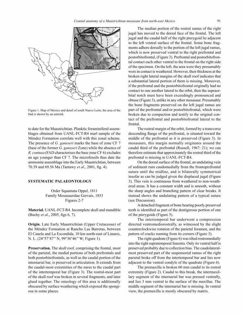
From the regional geology and marl lithologies cover-ing UANL-FCT-R4, the fossil doubtlessly comes from the Méndez Formation, a lithostratigraphic unit of Campanian-Maastrichtian age (Keller et al., 1997; Stinnesbeck et al.,2001). The Méndez Formation is widely distributed in the Gulf Coast Plain of north-east Mexico, east and south-east of the city of Monterrey (Figure 1). The unit is up to 1,000 m thick and consists of rhythmically bedded marls, shales, andminorsandstones.Theseweredepositedinanopenmarineshelfenvironmentinwaterdepthsofapproximately100 m near Los Ramones, 40 km north-east of Monterrey, and more than 400 m in the La Sierrita region, 40 km east of Montemorelos (Keller et al., 1997; Stinnesbeck et al.,2001). Planktic and benthic foraminiferal assemblages are rich and diverse throughout the Méndez Formation, whereas macrofossils, like inoceramids, ammonites, and vertebrate remains, are rare and known only from a few locations (Ifrim et al., 2004).
Foraminifers present in the marls adjacent to UANL-FCT-R4 include Heterohelix globulosa, H. striata, H. planata, H. dentata, H. pulchra, H. moremani, H. punctulata, H. glabrans; Pseudoguembelina kempensis, P. costulata, P. palpebra; Pseudotextularia elegans, P. deformis, P. nutalli; Hedbergella holmdelensis, H. monmouthensis; Rugoglobigerina pennyi, R. hexacamerata, R. macrocephala; Gansserina gansseri; Globotruncana aegyptica, G. arca, G. ventricosa, G. linneiana, G. orientalis, G. rosetta, G. esnehensis; Globotruncanita stuarti; Globotruncanella petaloidea.
Datum events and biozones presented within the zonal scheme based on planktic foraminiferal assemblages intro-duced for the Maastrichtian by Li and Keller (1998), based on Tunisian sections and DSDP sites (525), appear broadly valid throughout the central and western Tethys, including Mexico(e.g.Tantawyet al., 2001; Keller et al.,2002).ThiszonalschemesubdividestheMaastrichtianintoninezoneslabelled CF1-CF8a and b (CF for Cretaceous Foraminifera) and thus provides the highest resolution age control known
Cranial anatomy of a Maastrichtian mosasaur from north-east Mexico 91
Figure 1. Map of Mexico and detail of south Nuevo León; the area of the find is shown by an asterisk.
to date for the Maastrichtian. Planktic foraminiferal assem-blages obtained from UANL-FCT-R4 marl sample of the Méndez Formation correlate well with this zonal scheme. ThepresenceofG. gansseri marks the base of zone CF 7 (baseoftheformerG. gansseri Zone) while the absence of R. contusa (FAD characterises the base zone CF 6) excludes an age younger than CF 7. The microfossils thus date the ammonite assemblage into the Early Maastrichtian, between 70.39 and 69.56 Ma (Tantawy et al., 2001, fig. 4).
SYSTEMATIC PALAEONTOLOGY
OrderSquamataOppel,1811Family Mosasauridae Gervais, 1853
Figures 2-7
Material.UANL-FCT-R4. Incomplete skull and mandible (Buchy et al., 2005, figs 6, 7).
Origin. Late Early Maastrichtian (Upper Cretaceous) of the Méndez Formation at Rancho Las Barretas, between El Canela and La Escondida, 10 km north-east of Linares, N. L. (24°57’87’’ N, 99°30’46’’ W; Figure 1).
Preservation. The skull roof, comprising the frontal, most oftheparietal,themedialportionsofbothprefrontalsandbothpostorbitofrontals,aswellasthecaudalportionoftheinternarial bar, is preserved in articulation. It extends from thecaudal-mostextremitiesofthenarestothecaudalpartof the intertemporal bar (Figure 3). The rostral-most part of the skull roof was broken in several fragments, and later glued together. The osteology of this area is additionally obscured by surface weathering which exposed the spongi-osainsomeplaces.
The median portion of the rostral ramus of the right jugal has moved to the dorsal face of the frontal. The left jugal and the caudal half of the right pterygoid lie adjacent to the left ventral surface of the frontal. Some bone frag-ments adhere dorsally to the portion of the left jugal ramus, which is now preserved ventral to the right prefrontal and postorbitofrontal, (Figure 3). Prefrontal and postorbitofron-tal contact each other ventral to the frontal on the right side ofthespecimen.Ontheleft,theareaweretheypresumablywere in contact is weathered. However, their thickness at the broken right lateral margins of the skull roof indicates that a substantial lateral portion of them is missing. Moreover, if the prefrontal and the postorbitofrontal originally had no contacttooneanotherlateraltotheorbit,thenthesupraor-bital notch must have been exceedingly pronounced and obtuse (Figure 3), unlike in any other mosasaur. Presumably the bone fragments preserved on the left jugal ramus are partoftheprefrontaland/orpostorbitofrontal,whichwerebroken due to compaction and testify to the original con-tactoftheprefrontalandpostorbitofrontallateraltothefrontal.
The rostral margin of the orbit, formed by a transverse descending flange of the prefrontal, is situated toward the middle of the prefrontal as it is preserved (Figure 3). In mosasaurs, this margin normally originates around the caudal third of the prefrontal (Russell, 1967: 21); we can thereforeestimatethatapproximatelytherostralthirdoftheprefrontal is missing in UANL-FCT-R4.
On the dorsal surface of the frontal, an undulating vein ofsedimentrunscaudomediallyfromthefrontoprefrontalsutureuntil themidline,andisbilaterallysymmetricalinsofar as can be judged given the displaced jugal (Figure 3).Thisveiniscontinuousfromweatheredtonon-weath-ered areas. It has a constant width and is smooth, without the sharp angles and branching pattern of clear breaks. It instead shows the undulating pattern of a typical suture (see Discussion).
A detached fragment of bone bearing poorly preserved teeth is identified as part of the dentigerous portion of one of the pterygoids (Figure 5).
Theintertemporalbarunderwentacompressiondirected ventromediorostrally, as witnessed by the slight counterclockwise rotation of the parietal foramen, and the pattern of cracks running from its corners (Figure 3).
The right quadrate (Figure 6) was tilted rostromedially into the right supratemporal fenestra. Only its ventral half is preservedprobablyduetocollectionbias.Thecaudolateral-most preserved part of the suspensorial ramus of the right parietal broke off from the intertemporal bar and lies now adjacent to the ventral condyle of the quadrate (Figure 6).
The premaxilla is broken 60 mm caudal to its rostral extremity (Figure 2). Caudal to this break, the intermaxil-lary segment of the internarial bar was pressed ventrally, andlies3mmventraltothesurfaceofthemaxillae.Themiddle segment of the internarial bar is missing. In ventral view,thepremaxillaismostlyobscuredbymatrix.
Buchy et al.92
The dentigerous portions of both maxillae are almost completelypreservedandarticulatewiththerostralextrem-ity of the premaxilla (Figure 2). The dorsal margin of each maxilla was pressed medially and is broken. Some frag-ments of the dorsal maxillary margins are now preserved tilted ventromedially against the ventral dentigerous part ofthemaxillae.Theentiremuzzleunit(asdefinedbyRussell, 1967: 14) was subject to erosion after breakage. All maxillary and premaxillary tooth crowns are broken, and the enamel of the detached preserved fragments of crownsiseroded.
Fragments of both vomers are preserved between the maxillae, separated by a 45 mm long rod of bone identi-fied as the ventral internarial process of the premaxilla (Figure 2). The right vomer is broken caudally level with the seventh right maxillary tooth; the left vomer is broken 10 mm further rostrally. In ventral view, both are obscured rostrallybymatrixandindorsalviewbythemaxillaeandthepremaxilla.
Themiddleportionofeachanteriorlowerjaw(asdefined by Russell, 1967: 49-51) is preserved (Figure 4). Asdeterminedbycomparisonwiththeupperjaw,thelength of the missing rostral portion is at least 50 mm. The intramandibular articulation is also missing. The lateral and medialsurfacesofbothanteriorlowerjawsareweathered.Theventralhalfofthelateralsurfaceofthebetterpreservedright one is damaged, as witnessed by the splenial which islaterallyexposedoverthreequartersofthespecimen(inmosasaursthesplenialdisappearsrostraltothecaudalthirdof the dentary in lateral view [Russell, 1967: 50]). The lateral surface of the rostroventral-most portion of the right splenial of UANL-FCT-R4 is depressed (Figure 4). This depression is most likely the imprint of the dentary and therefore marks its original extension lateral and ventral to the splenial.
Most of the preserved functional dentary teeth lack enamel.
Description
Skull Premaxilla (Figures 2, 3). Therostralextremityofthepremaxilla bears a rostrally directed 5 mm thick circular tuberositythecentreofwhichexhibitsaforamen2mmindiameter.Thistuberosityissituated10mmdorsaltotheventral margin of the bone, on the midline. Faint regular wrinkles radiate from the tuberosity. They disappear ap-proximately10mmawayfromthetuberosity.Ventraltothetuberosity,therostralsurfaceofthepremaxillaisverti-cal and slightly concave. The lateral face of the premaxilla gently curves caudolaterally until the maxillopremaxillary suture.Thedorsalsurfaceoftheboneisweathered;itapparently also gently curves caudodorsally to form the internarialbar.Theforaminaoftheophthalmicramusofthefifth cranial nerve (Russell, 1967) are randomly distributed aroundtherostraltuberosity.
Themaxillopremaxillarysuturecommences35mmcaudal to the rostral extremity of the premaxilla. It has a sinusoidoutlineinlateralview.Theinternarialportionofthepremaxilla emerges from a triangular intermaxillary base.
Theinternarialprocessofthepremaxillaisdrop-shaped in transverse cross-section, with an overall height of 8 to 9 mm and a maximal width of 3 mm. The dorsal margin of this process forms a thin, damaged blade.
Thepremaxillaeextend55mmfurthercaudallythanthe caudal extremity of the external nares and are wedged betweenthepairedrostralprocessesofthefrontal.Onthiscaudalportion,theinterpremaxillarysutureisdistinct.Atthe rostral break of the preserved portion of skull roof, the frontopremaxillarysuturecommences20mmlateraltothemidline.Onthedorsalsurfaceofthespecimen,thefronto-premaxillary suture is visible running straight caudally for approximately 45 mm. It then undulates medially and further joins the midline in a gentle caudomedial curve. In ventral view,thecaudal-mostportionofthefrontopremaxillarysuture is situated level with its dorsal counterpart. Its rostral continuation on the left side is obscured by matrix and break-age. On the right side it partly appears as a straight, rostrally directedsuture18to19mmlateraltothemidline.
Thecaudal-mostportionofthepremaxillaeiselevatedandformsamediancrest,whichcontinuesfor65mmontothe frontal. The premaxillae have a median thickness of 7 mm at the rostral break of the specimen. Laterally, the ven-tralsurfaceofthepremaxillaeraisesdorsolaterally,andthebones are less than 2 mm in height at their lateral margin.
Maxilla (Figure 2). The ventral margin of the dentigerous portion of the maxilla is straight, horizontal. The maxil-larycanalisvisibleontheerodeddorsalsurfaceoftheleftmaxilla,withadiameterofapproximately5mm.
Because of distortion and erosion of the dorsal-most portion of the maxillae in UANL-FCT-R4, the outline of the lateral margin of the naris (the dorsal margin of the maxilla) can only be reconstructed by reversing the medial compression that the dorsal portion of the right maxilla underwent.Thenariscommenceslevelwiththerostralmargin or the middle of the fourth maxillary tooth. The height of the maxilla at the level of the caudal termination of the maxillopremaxillary suture could have reached 75 mm. The dorsal margin of the maxilla can be traced until the seventh maxillary tooth. It was gently concave, and the height of the maxilla was approximately 65 mm at the level oftheseventhmaxillarytooth.
On the right maxilla, the articular facet for the jugal is visible in lateral view. It is semicircular in outline, com-mencing level with the middle of the 11thmaxillarytooth.Thefacetcanbetraceduntilapointapproximately20mmfurthercaudally.
Frontal (Figure 3). In dorsal aspect, the rostral processes of the frontal wedging the premaxillae are approximately 20mmwideandcontacttheprefrontallaterally.Thefron-
Cranial anatomy of a Maastrichtian mosasaur from north-east Mexico 93
a)
b)
c) d)
a’)
b’)
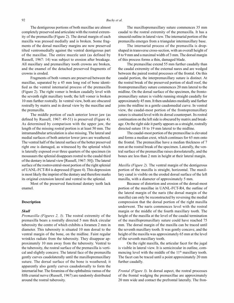
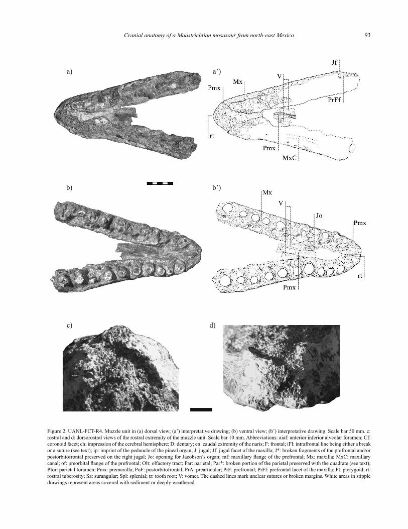
Figure 2. UANL-FCT-R4. Muzzle unit in (a) dorsal view; (a’) interpretative drawing; (b) ventral view; (b’) interpretative drawing. Scale bar 50 mm. c: rostral and d: dorsorostral views of the rostral extremity of the muzzle unit. Scale bar 10 mm. Abbreviations: aiaf: anterior inferior alveolar foramen; Cf: coronoid facet; ch: impression of the cerebral hemisphere; D: dentary; en: caudal extremity of the naris; F: frontal; iFl: intrafrontal line being either a break or a suture (see text); ip: imprint of the peduncle of the pineal organ; J: jugal; Jf: jugal facet of the maxilla; J*: broken fragments of the prefrontal and/or postorbitofrontal preserved on the right jugal; Jo: opening for Jacobson’s organ; mf: maxillary flange of the prefrontal; Mx: maxilla; MxC: maxillary canal; of: preorbital flange of the prefrontal; Olt: olfactory tract; Par: parietal; Par*: broken portion of the parietal preserved with the quadrate (see text); Pfor: parietal foramen; Pmx: premaxilla; PoF: postorbitofrontal; PrA: prearticular; PrF: prefrontal; PrFf: prefrontal facet of the maxilla; Pt: pterygoid; rt: rostral tuberosity; Sa: surangular; Spl: splenial; tr: tooth root; V: vomer. The dashed lines mark unclear sutures or broken margins. White areas in stipple drawings represent areas covered with sediment or deeply weathered.
Buchy et al.94
a)
b)
a’)
b’)
toprefrontalsuturecommencesatthecaudal-mostpointofthe naris and runs straight caudally. In ventral aspect, these processes are flat and horizontal for most of their width. Laterally they abruptly curve ventrally and form a thin, longitudinally oriented flange. This flange is bordered later-ally by a similar flange formed by the medial-most margin oftheprefrontal.
In ventral aspect, the frontoprefrontal suture contin-ues caudally straight until the caudomedial corner of the prefrontal (which was originally ventrally covered by the postorbitofrontal: see Preservation and description of the postorbitofrontal). In dorsal aspect, the frontoprefrontal suturecurveslaterally,andthefrontaloverlapstheprefron-tal. The frontoprefrontal suture then gently curves caudally, andalmostparallelsthemidlineforapproximately60mm,before it curves caudolaterally until the caudolateral break oftheprefrontal.Therethefrontalformsaroundedcaudola-teral wing overlapping the postorbitofrontal and reaches its maximumwidthof190mm.
Thefrontoparietalsuturecommencesatthecaudal-most extension of the caudolateral wing of the frontal. It describesanarcrostraltothesupratemporalfenestrain
ventral and dorsal aspects. However, as is visible along the left lateral break of the specimen, the rostral margin of the supratemporal fenestra is formed by a 17 mm long caudal process of the frontal wedged between a 17 mm long dorsal process and a 11 mm long ventral process of the parietal. Therefore the parietal and frontal interdigitate strongly at thatlevel.
Medialtothesupratemporalfenestra,apairedcaudalprocess of the frontal overlaps the parietal. The original di-mensionofthisprocesscannotbedeterminedduetosurfaceweathering. Medial to this process, the frontoparietal suture runsmediallyuntilitreachesthemidline.
In ventral view the medial portion of the frontoparietal suture is visible at the same level as it is in dorsal view, 140 mmcaudaltothefrontopremaxillarysutureasmeasuredalong the midline. The frontoparietal suture undulates later-allyfor5mmoneithersideofthemidline.
Thepartialimpressionofthecerebralhemispheresareprobablyvisibleasapairofshallowsulcusatthecaudalend of the olfactory tract (see Camp, 1942: 40-42; Russell, 1967: 21). The olfactory bulbs are enclosed laterally by a paired descending flange commencing approximately 50
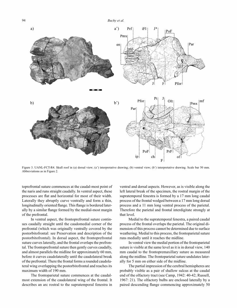
Figure 3. UANL-FCT-R4. Skull roof in (a) dorsal view; (a’) interpretative drawing; (b) ventral view; (b’) interpretative drawing. Scale bar 50 mm. Abbreviations as in Figure 2.
Cranial anatomy of a Maastrichtian mosasaur from north-east Mexico 95
flange of the prefrontal originally contacting the maxilla ispartlypreserved,approximately60mmlateraltothemidline. This maxillary flange curves medially, and forms the ventrorostrally sloping rostral margin of the orbit, at thelevelofthemiddleoftheprefrontalaspreserved(seePreservation). It then curves rostrally, and contacts the lat-eral longitudinal flange of the frontal caudal to the naris.
Rostral to the longitudinal preorbital flange, the ventral surface of the prefrontal is flat, horizontal, mostly obscuredbymatrixontheleftside,andweatheredontheright one.
The prefrontojugal suture is most likely visible dorsal to the articular facet for the jugal on the right maxilla. It runs dorsorostrally, dorsally convex, for 15 mm, before fading intotheerodeddorsalareaofthemaxilla.
Parietal (Figures 3, 6),. In dorsal aspect, the parietal forms most of the rostral margin of the supratemporal fossa, the postorbitofrontal forming only its lateral corner. The rostral margin of the supratemporal fossa is almost straight and caudomediallyorienteduntilitreachestheintertemporalbar. The lateral margins of the intertemporal bar form a subcircularhorizontaltabledorsaltothejunctionwiththerostral margin of the supratemporal fossa, 20 mm caudal to the caudal margin of the parietal foramen. The suspensorial ramus of the parietal emerges from the medial margin of the supratemporalfenestrajustcaudaltothatlevel.
In dorsal view, the parietal foramen is subrectangular, transversely oriented. The middle of its rostral margin is situated15mmcaudaltothefrontoparietalsuture.
The ventral aperture of the parietal foramen is longi-tudinally oval. Its rostral margin slopes ventrorostrally; its caudal margin is subvertical. Its lateral margins are formed by a pair of descending ridges of the parietal. These ridges extend rostrally onto the frontal. Caudal to the caudal margin of the parietal foramen, the flanges abut against the ventrally curving medial portion of the parietal. The lateral faces of the parietal caudal to the parietal foramen strongly curve ventrally and form the descending medial margins of the supratemporalfenestrae.
A triangular area of smooth bone caudal to the parietal foramenisprobablytheimprintofthepeduncleofthepinealorgan (see Camp, 1942: 40-42, fig. 24). This smooth area extends 20 mm caudal to the caudal margin of the parietal foramen. It is caudally prolonged by a low, blunt, median ridge for 20 mm. This ridge abruptly terminates as a cau-dally pointed triangle. Caudal to it, the median surface of theparietalispoorlypreserved,butitapparentlycurvesdorsally. A 3 mm long paired ridge bifurcates laterally on eitherside,from5mmcaudaltotheextremityoftheme-dian ridge, and runs caudally parallel to the midline over approximately 5 mm. Both ridges tend to converge toward thecaudalextremityofthespecimen.
Quadrate (Figure 6). Theventralcondyleofthequadrateisdrop-shaped in ventral view, tapering medially.
mm rostral to the frontoparietal suture. This flange runs rostrally, slightly medially, and is vaulted ventromedially, for 30 mm. Its ventral margin is convex. The flange is 3 mm in maximal dorsoventral height when compared to the ventral surface of the frontal lateral to it. Between the flanges, the oflactory tract is 4 mm wide and approximately 5mmdeep.
Postorbitofrontal (Figure 3). In dorsal view, the postor-bitofrontal as preserved is visible only on the right side of thespecimen,rostralandcaudolateraltothecaudolateralwing of the frontal. It forms the rostrolateral corner of the supratemporal fenestra. It was also exposed lateral to this wing, as is suggested by the lateral break of the specimen (see Preservation). Along this longitudinal break, the pa-rietopostorbitofrontal suture is exposed. It runs vertically in a sinusoidal manner, with a rostrocaudal amplitude of 4 mm. In dorsal aspect, the suture emerges from the caudal-most point of the caudolateral wing of the frontal, and runs caudolaterally until the caudal margin of the supratemporal fenestra.
As is visible along the left lateral break of the speci-men, a 15 to 20 mm long, 2 mm thick caudal process of thepostorbitofrontalventrallycoversthefrontoparietalcontact.
In ventral aspect, the medial portion of the postor-bitofrontal is subrectangular, poorly preserved on the left side, and covered by the jugal and pterygoid on the right. The caudal margin of the postorbitofrontal is formed by the suture with the parietal. This suture runs medially straight, then curves rostrally, forming the caudal 10 mm of the medial margin of the postorbitofrontal. Further rostrally, the medial margin of the postorbitofrontal as preserved contactsthefrontal.Thefrontopostorbitofrontalsuturerunsslightly rostrolaterally, then rostrally, and finally curves laterally, forming the rostromedial corner of the postor-bitofrontal. The suture runs laterally until the lateral break ofthespecimen.
As is suggested by the weathering pattern of the speci-men (see Preservation), the postorbitofrontal was most likely originally in contact with the prefrontal dorsal to the orbit. In ventral aspect, the rostral margin of the right postorbitof-rontal is intact, though partly covered by the jugal and the pterygoid. The right postorbitofrontal extends until 80 mm rostraltothefrontoparietalsuture,andcoverstheprefrontalventrally. At the dorsal margin of the orbit, therefore, the prefrontal was wedged between the frontal dorsally and the postorbitofrontalventrally.
Prefrontal (Figures 2, 3). The original outline of the supraor-bital wing of both prefrontals is only partly preserved, com-mencing in dorsal aspect at the lateral-most extension of the frontal as preserved, and gently curving rostromedially.
Theventralsurfaceofthecaudalhalfoftheprefrontalis subhorizontal and flat, as is its lateral-most portion, which forms the supraorbital wing. The longitudinal ventrolateral
Buchy et al.96
a)
b)
a’)
b’)
In rostral view, the lateral margin of the quadrate is concave, curving slightly rostrolaterally toward its preserved dorsal margin, and forms the tympanic wing. As is visible along the dorsal break of the quadrate, the tympanic wing at its ventral base was about 1 mm thick.
The rostromedial margin of the quadrate is dorsola-terally oriented. In medial view, this rostromedial margin consists of a 2 mm thick ridge. A sulcus separates it from a caudomedialprocessofthequadrateshaft,whichformsthecaudomedial margin of the quadrate. Seen from caudally, this process merges caudolaterally with the infrastapedial process.
The dorsolateral margin of the infrastapedial process rises mediodorsally and is straight until the level of the middle of the ventral condyle. From there, the dorsal mar-gin of the infrastapedial process forms a transversely flat surface, inclined caudally at an angle of about 40°. Medial to this surface, the infrastapedial process merges with the caudomedialprocessofthequadrateshaft.Theventral-mostextremityofthesuprastapedialprocessispreservedincloseproximity,butisseparatedby1to2mmofsedimentfromtheinfrastapedialprocessandthecaudomedialprocessofthequadrateshaft.
Jugal (Figures 2, 3). The median portion of the left jugal ispreservedinmedialview.Thepreservedportionofthehorizontal rostral ramus is 100 mm long, the preserved portion of the ascending dorsal ramus, 40 mm long. They form a 90° angle to one another. The rostral ramus is 20 mm high, 10 mm wide. It is drop-shaped in cross-section, its dorsal margin converging into a ridge, possibly due to compaction and weathering. In cross-section the dorsal ramus is subtriangular, with a maximal rostral width of 8 mm, and a length of 20 mm. Its medial margin abruptly curveslaterallyatthejunctionwiththehorizontalrostralramus and forms a 7 to 9 mm wide tuberosity. Further de-tailsarenotpreserved,oraredistortedinsuchawaythataninterpretationwouldbeunreliable.
Pterygoid (Figures 3, 5). Thecaudal,quadrateprocessofthepterygoid is oval in cross-section, and dorsoventrally com-pressed. Neither its height nor its width can be determined with certainty. The medial process, caudally terminating the pterygoid tooth row, is 10 mm wide, emerging at a right angle from the quadrate process. The isolated portion of pterygoid is L-shaped. As it is preserved, it bears five toothpositions.
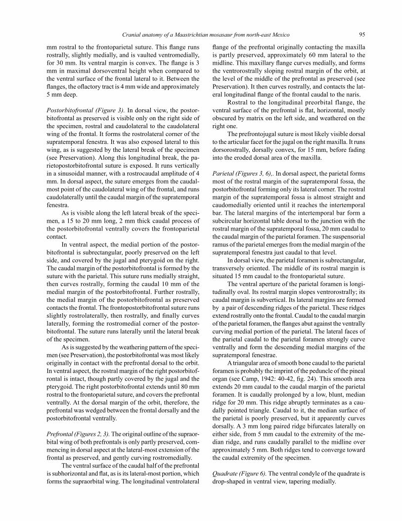
Figure 4. UANL-FCT-R4. Right anterior mandibular unit in (a) medial view; (a’) interpretative drawing; (b) lateral view; (b’) interpretative drawing. Scale bar 50 mm. Abbreviations as in Figure 2. Arrows and numbers in b’) refer to the sections illustrated in Figure 7.
Cranial anatomy of a Maastrichtian mosasaur from north-east Mexico 97
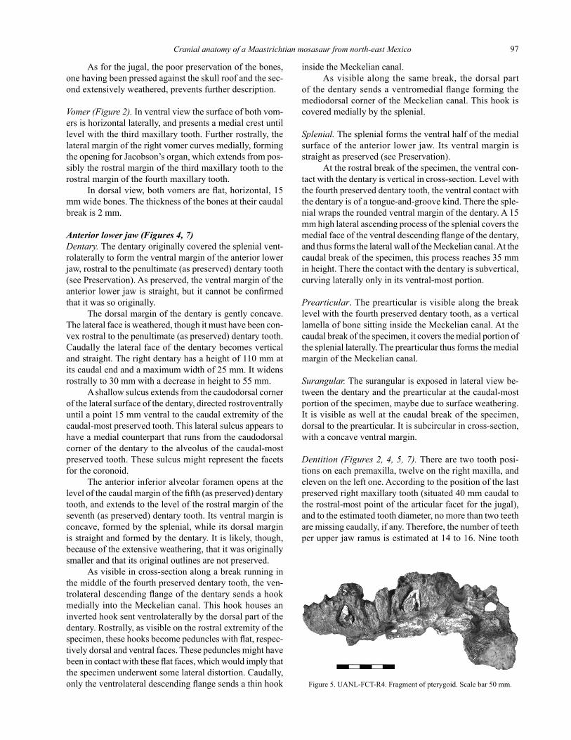
As for the jugal, the poor preservation of the bones, one having been pressed against the skull roof and the sec-ondextensivelyweathered,preventsfurtherdescription.
Vomer (Figure 2). In ventral view the surface of both vom-ersishorizontallaterally,andpresentsamedialcrestuntillevel with the third maxillary tooth. Further rostrally, the lateral margin of the right vomer curves medially, forming the opening for Jacobson’s organ, which extends from pos-sibly the rostral margin of the third maxillary tooth to the rostral margin of the fourth maxillary tooth.
In dorsal view, both vomers are flat, horizontal, 15 mm wide bones. The thickness of the bones at their caudal break is 2 mm.
Anterior lower jaw (Figures 4, 7)Dentary. The dentary originally covered the splenial vent-rolaterally to form the ventral margin of the anterior lower jaw,rostraltothepenultimate(aspreserved)dentarytooth(see Preservation). As preserved, the ventral margin of the anterior lower jaw is straight, but it cannot be confirmed that it was so originally.
The dorsal margin of the dentary is gently concave. The lateral face is weathered, though it must have been con-vexrostraltothepenultimate(aspreserved)dentarytooth.Caudallythelateralfaceofthedentarybecomesverticaland straight. The right dentary has a height of 110 mm at its caudal end and a maximum width of 25 mm. It widens rostrally to 30 mm with a decrease in height to 55 mm.
Ashallowsulcusextendsfromthecaudodorsalcornerofthelateralsurfaceofthedentary,directedrostroventrallyuntilapoint15mmventraltothecaudalextremityofthecaudal-mostpreservedtooth.Thislateralsulcusappearstohaveamedialcounterpartthatrunsfromthecaudodorsalcornerofthedentarytothealveolusofthecaudal-mostpreserved tooth. These sulcus might represent the facets forthecoronoid.
Theanteriorinferioralveolarforamenopensatthelevel of the caudal margin of the fifth (as preserved) dentary tooth, and extends to the level of the rostral margin of the seventh (as preserved) dentary tooth. Its ventral margin is concave, formed by the splenial, while its dorsal margin is straight and formed by the dentary. It is likely, though, because of the extensive weathering, that it was originally smaller and that its original outlines are not preserved.
As visible in cross-section along a break running in themiddleofthefourthpreserveddentarytooth,theven-trolateral descending flange of the dentary sends a hook medially into the Meckelian canal. This hook houses an inverted hook sent ventrolaterally by the dorsal part of the dentary. Rostrally, as visible on the rostral extremity of the specimen, these hooks become peduncles with flat, respec-tively dorsal and ventral faces. These peduncles might have been in contact with these flat faces, which would imply that thespecimenunderwentsomelateraldistortion.Caudally,only the ventrolateral descending flange sends a thin hook
inside the Meckelian canal. As visible along the same break, the dorsal part
of the dentary sends a ventromedial flange forming the mediodorsal corner of the Meckelian canal. This hook is coveredmediallybythesplenial.
Splenial. Thesplenialformstheventralhalfofthemedialsurface of the anterior lower jaw. Its ventral margin is straight as preserved (see Preservation).
At the rostral break of the specimen, the ventral con-tact with the dentary is vertical in cross-section. Level with thefourthpreserveddentarytooth,theventralcontactwiththe dentary is of a tongue-and-groove kind. There the sple-nial wraps the rounded ventral margin of the dentary. A 15 mm high lateral ascending process of the splenial covers the medial face of the ventral descending flange of the dentary, and thus forms the lateral wall of the Meckelian canal. At the caudal break of the specimen, this process reaches 35 mm in height. There the contact with the dentary is subvertical, curving laterally only in its ventral-most portion.
Prearticular. The prearticular is visible along the break levelwiththefourthpreserveddentarytooth,asaverticallamella of bone sitting inside the Meckelian canal. At the caudal break of the specimen, it covers the medial portion of thespleniallaterally.Theprearticularthusformsthemedialmargin of the Meckelian canal.
Surangular. The surangular is exposed in lateral view be-tweenthedentaryandtheprearticularatthecaudal-mostportion of the specimen, maybe due to surface weathering. It is visible as well at the caudal break of the specimen, dorsal to the prearticular. It is subcircular in cross-section, with a concave ventral margin.
Dentition (Figures 2, 4, 5, 7).Therearetwotoothposi-tions on each premaxilla, twelve on the right maxilla, and eleven on the left one. According to the position of the last preserved right maxillary tooth (situated 40 mm caudal to the rostral-most point of the articular facet for the jugal), andtotheestimatedtoothdiameter,nomorethantwoteethare missing caudally, if any. Therefore, the number of teeth per upper jaw ramus is estimated at 14 to 16. Nine tooth
Figure 5. UANL-FCT-R4. Fragment of pterygoid. Scale bar 50 mm.
Buchy et al.98
a) b) c)
b’)
d)
a’) b’) c’) d’)
positionsaredocumentedoneachdentary.Thenumberofmissing dentary teeth might be two to five, according to the estimated length of the tooth row of the upper jaw.
Because of the broken tooth crowns, the ontogenetic conditionoftheteethandthereforetheactualdiameterofthe functional upper teeth are difficult to assess. The upper dentition shows a slight anisodonty, as the two premaxillary andtherostral-mosttwomaxillarycrownbaseshavearos-trocaudal length of 10 to 12 mm, compared to the estimated basal length of 15 to 17 mm of the more caudally situated maxillarytoothcrowns.
Alldentarytoothcrownsaspreserved(i.e.,withoutenamel coverage, and with the apices of functional teeth broken or abraded) have a basal mesiodistal length of 16 to 18 mm, and a height of 22 to 25 mm.
The preserved tooth crowns, most of them being of non-erupted replacement teeth, are slightly and gently curvedcaudallyandmedially,withasubcircularbasalcross-section. Two mesially and distally aligned carinae dividethecrownuntiltheapexintotwoequalhalves,andthe cross-section becomes slightly oval apically. The crown gently tapers until 3 to 4 mm below its apex. Then the sur-facetapersmoreabruptlyandformsabluntconicalapex.Where preserved, the enamel surface is smooth.
The better preserved pterygoid tooth documents a tooth that was at least 20 mm high, slightly more massive thananyofthemandibularteeth,andwithasharperapex.
Discussion
Rostral extension of the frontal, individual age of the specimen. Thetransverselinevisibleonthedorsalsurfaceof the frontal (see Preservation; Figure 3) could be a break, although due to its symmetry it much resembles a suture.
If this line is actually a suture, assuming that it marks therostralextremityofthefrontalwouldmeanthatan-other bone, forming partly the medial margin of the naris, surroundsthecaudal-mostportionofthepremaxillaandoverlapstheprefrontallaterally.Thisbonecouldbepartofthemaxilla,onlyifitisassumedthatthemaxillaformsthe internarial bar together with the premaxilla. Such an anatomy has never been observed in any mosasaur (Romer, 1956; Russell, 1967).
Especiallybecausethisboneislocatedmedialtothenaris, it could also represent the nasal. However, nasals are rarely preserved in mosasaurs (Russell, 1967: 18; Bell, 1997b: 302). If they are, they are reduced in size and situated lateraltotheinternarialbar,theircaudalextremityarticulat-ing with the rostral extremity of the frontal (Camp, 1942: 27-28, fig. 14). Regardless of whether nasals in mosasaurs arefusedwiththefrontalorthepremaxillainsometaxa,ormissing due to a loose connection with the internarial bar in others, as discussed by Camp (1942: 28), it appears highly unlikely that they can form such a well-expressed caudal process as is seen in UANL-FCT-R4.
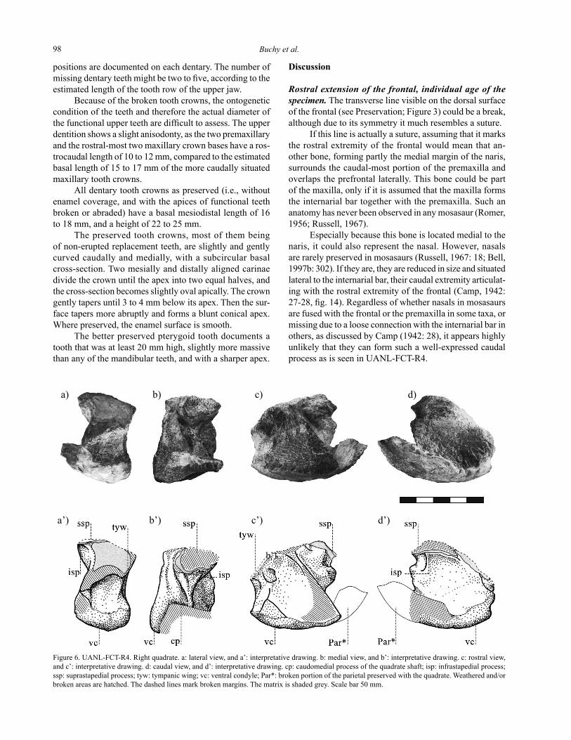
Figure 6. UANL-FCT-R4. Right quadrate. a: lateral view, and a’: interpretative drawing. b: medial view, and b’: interpretative drawing. c: rostral view, and c’: interpretative drawing. d: caudal view, and d’: interpretative drawing. cp: caudomedial process of the quadrate shaft; isp: infrastapedial process; ssp: suprastapedial process; tyw: tympanic wing; vc: ventral condyle; Par*: broken portion of the parietal preserved with the quadrate. Weathered and/or broken areas are hatched. The dashed lines mark broken margins. The matrix is shaded grey. Scale bar 50 mm.
Cranial anatomy of a Maastrichtian mosasaur from north-east Mexico 99
1)
2)
3)
1’)
2’)
3’)
Anotherhypothesisisthatthisstructurerepresentsa supernumerary bone (or wormian bone, Gray, 1901: 49-50), which appears as unlikely as the preceding hypoth-eses, because the bone in UANL-FCT-R4 is overlapping the prefrontal, instead of growing between two bones as wouldbethecaseforasupernumerarybone(Gray,1901;Piveteau, 1954).
Aseptomaxillahasneverbeenreportedformosas-aurs,andnootherdermalboneexistsintheirrostrofrontalarea.Therefore,ifthislineisasuture,ithastobeasuturewithinthefrontalitself.Suchanintrafrontalsuturecouldbe either pathological, due to a break early in ontogeny. It could also mark the unossified contact of two ossification centres (Gray, 1901; Piveteau, 1954; Romer, 1956). In that latter case, the specimen would be a juvenile, as is also sug-gested by the presence of an interpremaxillary suture, for in mosasaurs the premaxillae are normally ‘‘co-ossified with no indication of a suture’’ (Russell, 1967: 14). Even if the interpremaxillary suture of UANL-FCT-R4 is exposed in dorsal view only due to surface weathering, it indicates that thefusionofthepremaxillaeproceedsinacaudaldirectionduring ontogeny, as no interpremaxillary suture is visible ontheerodeddorsomedialareaoftherostral-mostpartofthepremaxilla.Underthehypothesisofajuvenileanimal,the unfused but almost contacting infra- and suprastapedial processes of the quadrate could also be related to young individual age.
Comparative palaeontology. Mosasaurinae, Plioplatecar-pinae and Tylosaurinae were characterised by Russell (1967) byfeaturesoftheopisthoticandbasioccipital,andofthepostcranial skeleton. These elements are missing in UANL-FCT-R4. Subsequent studies (Wright and Shannon, 1988; Lingham-Soliar, 1992; De Braga and Carroll, 1993) could not define other diagnostic characters of the subfamilies that would allow the identification of UANL-FCT-R4 to subfamiliallevel.There-assessmentoftherelationshipsamong Mosasauridae proposed by Bell (1997b) is of little help, due to the inadequate preservation of UANL-FCT-R4. The few characters defined by Bell (1997b), whose states can be determined in UANL-FCT-R4, yield contradic-tory results. For example, the large supraorbital process oftheprefrontalandthecoincidencebetweenthecaudalterminationofthenarisandtherostralterminationofthefrontoprefrontal suture would place UANL-FCT-R4 within Mosasaurinae. However, the triangular ventrally inflated ‘boss’ (Bell, 1997b: 305) on the frontal caudal to the ol-factory tract, as is seen on UANL-FCT-R4, is one of the unambiguous derived characters of the “Russellosaurinae”, new subfamily informally introduced by Bell (1997b; see Bell and Polcyn, 2005) in order to accommodate Tylosaurusand the members of the tribe Plioplatecarpini. However, the genus Hainosaurus,theothermemberofthesubfamilyTylosaurinaesensu Russell, 1967, was not included in Bell’s analysis (1997b). Therefore the status of the subfamily remains uncertain under this phylogenetical hypothesis.
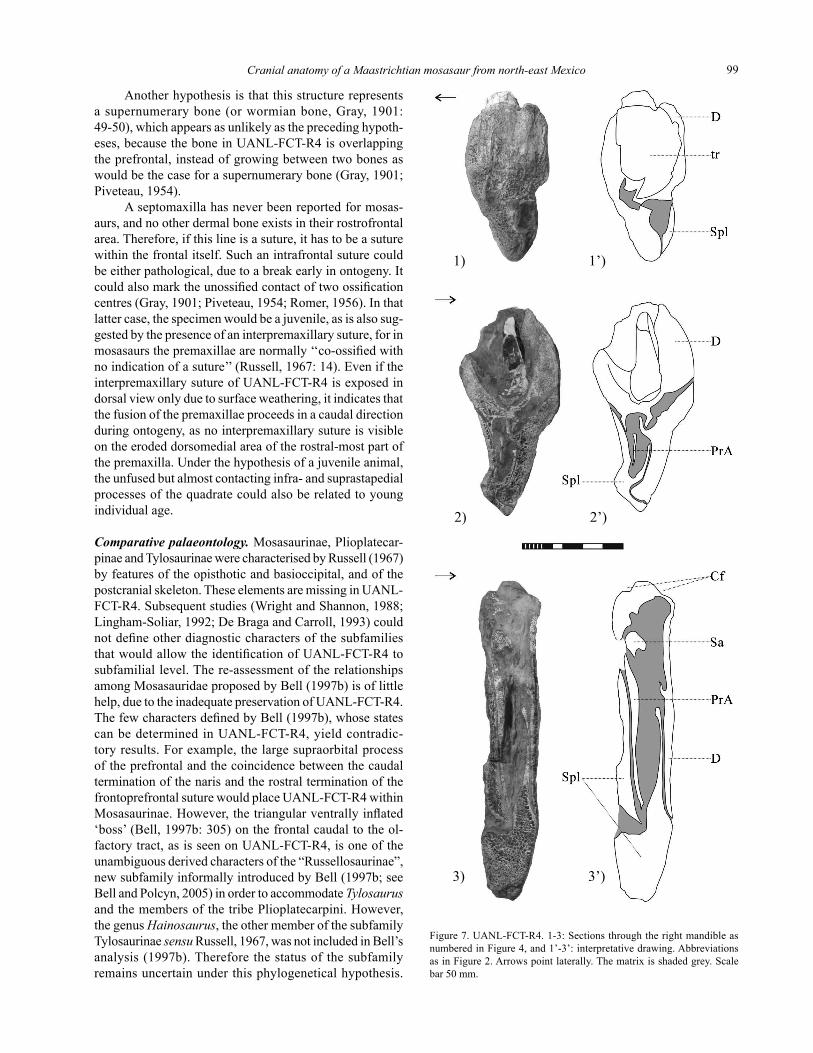
Figure 7. UANL-FCT-R4. 1-3: Sections through the right mandible as numbered in Figure 4, and 1’-3’: interpretative drawing. Abbreviations as in Figure 2. Arrows point laterally. The matrix is shaded grey. Scale bar50mm.
Buchy et al.100
11).Globidens has a very massive skull and characteristic low bulbous teeth (Russell, 1975), which are clearly dis-tinct from what is known of UANL-FCT-R4. However, inGlobidenstoo,theprefrontalandpostorbitofrontalex-clude the frontal from the dorsolateral margin of the orbit. Prognathodon,traditionallyconsideredaplioplatecarpinemosasaur (Russell, 1967), was reassigned by Bell (1997b; see also Bell and Polcyn, 2005) to the tribe Globidensini of the subfamilly Mosasaurinae, together with GlobidensandPlesiotylosaurus.Thisrelationshipissupportedbythefu-sionoftheinfra-andsuprastapedialprocessesinthesethreegenera, which is considered convergent in Ectenosaurus.However, UANL-FCT-R4 differs from known members of the genus Prognathodon as diagnosed by Lingham-Soliar andNolf(1989)intheexposedcontactoftheprefrontalandpostorbitofrontallateraltothefrontal,thesubcircularinter-temporal table, and the thin tympanic wing of the quadrate. Thesinusoidalmaxillopremaxillarysuture,andtheroundedcaudolateral wings of the frontal are also unknown in the genus Prognathodon (Russell, 1967; Welles and Gregg, 1971; Lingham-Soliar and Nolf, 1989).
UANL-FCT-R4 therefore cannot be confidently assigned to any of the previously described taxa sharing withittheclosecontact(orpossiblyfusioninadults)oftheinfra-andsuprastapedialprocessesoftheirquadrates;the remainder of its skull anatomy yields no further hint at its affinities.
CONCLUSIONS
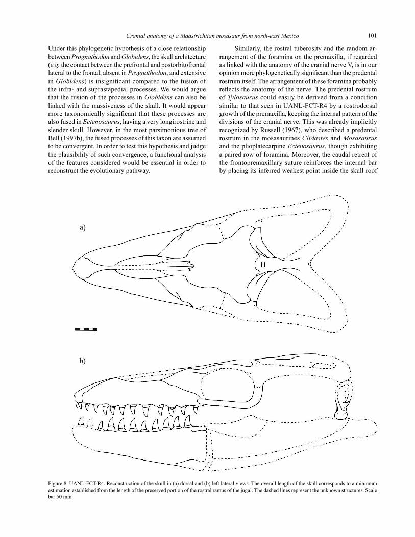
UANL-FCT-R4 is the first substantially documented mosasaurfromnorth-eastMexicoandcombinesfeaturesknown from genera assigned to different subfamilies. Although it comprises an almost complete skull roof, muzzle and anterior mandibular units, and partial quadrate and jugal, allowing us to draw a reconstruction of its skull (Figure 8), its affinities with other mosasaurs cannot be determined with confidence. This is partly linked with the inadequacy ofitspreservationcomparedtoclaimedtaxonomicallysignificant features in mosasaurs, but also we shall argue, with the poor understanding of the biological meaning of thesefeatures,nottomentionthedebatable,current,almostexclusive cladistic approach to phylogeny (see e.g.Vermeij,1999; Pavlinov, 2003; Hawks, 2004).
As an example, the skull architecture of UANL-FCT-R4issimilartothatofGlobidens (Russell, 1975), if not taking into account the massiveness of the skull in the latter genus, and differs from that of Prognathodon (Lingham-Soliar and Nolf, 1989). The skull architecture of Globidensisconsideredasrelatedtoitsmassivenessandlowbulbousteeth, allowing the animal to feed on hard-shelled prey (Russell, 1967, 1975; Schulp, 2005). Interpreting the anatomyofGlobidens as a consequence of the feeding strategy having no taxonomical meaning above the genus level, allowed Bell (1997b) to relate it to Prognathodon.
Due to its preservation status, UANL-FCT-R4 cannot be referredper setoanyofthesubfamiliesortribesofthefamilyMosasauridae.
UANL-FCT-R4 is excluded a priori from theTylosaurinaesensu Russell (1967) because it lacks a predental premaxillary rostrum. However, it is similar toTylosaurusintheextensionofthepremaxillaecaudaltothenarisandthecontactbetweentheprefrontalandthe postorbitofrontal lateral to the orbit (Russell, 1967; Everhart,2005).Therostraltuberosityofthepremaxillain UANL-FCT-R4 and the arrangement of foramina on the rostrodorsalsurfaceofthisbonearoundthetuberosity,areverysimilartowhatisdescribedforTylosaurus by Russell (1967: 16), but the tuberosity of UANL-FCT-R4issituatedmoredorsally.
The morphology of the quadrate clearly excludes UANL-FCT-R4frombothTylosaurus andHainosaurus.Theinfra-andsuprastapedialprocessesofthequadrateofUANL-FCT-R4arealmostincontact,whileinTylosaurusandHainosaurusthesuprastapedialprocessisnotexpandedventrally,andnevercontactsthereducedinfrastapedialprocess (Russell, 1967; Nicholls, 1988; Lingham-Soliar, 1992;Everhart,2005).
Among mosasaurs evincing a contact between in-fra-andsuprastapedialprocessesorprocessesalmostincontactistheplioplatecarpineSelmasaurus (Wright and Shannon, 1988). However, Selmasaurus lacks an inter-temporal table, has a longitudinally oval parietal foramen in dorsal aspect, and the caudolateral wings of its frontal are triangular (Wright and Shannon, 1988, fig. 1). Only in Clidastes propythondoesthesuprastapedialprocesscontactthe infrastapedial process and a crest emerging from the quadrate shaft (Russell, 1967; Wright and Shannon, 1988), asinUANL-FCT-R4. Still, UANL-FCT-R4 is excluded from the genus Clidastes in having a more massive and probably more brevirostrine skull, as far as can be judged from what is preserved, and 12 to 14 maxillary and 11 to 14 dentary teeth while Clidasteshas16to18maxillaryanddentary teeth (Russell, 1967, 1975). UANL-FCT-R4alsodiffers from members of the genus Clidastes in having a maxillopremaxillarysuturesinusoidalinlateralview,therostralextremityofthenarissituatedbetweenthethirdandfourthmaxillaryteeth,asubcircularintertemporaltable,andadorsallyexposedcontactbetweentheprefrontalandthe postorbitofrontal lateral to the frontal (Merriam, 1894; Russell, 1967).
Theinfra-andsuprastapedialprocessesofthequadratearefusedinGlobidens,Prognathodon,PlesiotylosaurusandEctenosaurus (Russell, 1967, 1975; Wright and Shannon, 1988). The latter genus is a very longirostrine plioplate-carpine, sharing with UANL-FCT-R4onlythethintympanicwing of their quadrates, and the position of the rostral terminationof thenaris levelwiththethirdmaxillarytooth (Russell, 1967: 158). No similarity can be observed betweenthequadrateofUANL-FCT-R4andthesameboneinPlesiotylosaurus as illustrated by Camp (1942, fig.
Cranial anatomy of a Maastrichtian mosasaur from north-east Mexico 101
Under this phylogenetic hypothesis of a close relationship betweenPrognathodonandGlobidens, the skull architecture (e.g.thecontactbetweentheprefrontalandpostorbitofrontallateraltothefrontal,absentinPrognathodon,andextensiveinGlobidens) is insignificant compared to the fusion of the infra- and suprastapedial processes. We would argue thatthefusionoftheprocessesinGlobidenscanalsobelinked with the massiveness of the skull. It would appear more taxonomically significant that these processes are alsofusedinEctenosaurus, having a very longirostrine and slender skull. However, in the most parsimonious tree of Bell (1997b), the fused processes of this taxon are assumed to be convergent. In order to test this hypothesis and judge the plausibility of such convergence, a functional analysis ofthefeaturesconsideredwouldbeessentialinordertoreconstructtheevolutionarypathway.
Similarly,therostraltuberosityandtherandomar-rangement of the foramina on the premaxilla, if regarded as linked with the anatomy of the cranial nerve V, is in our opinion more phylogenetically significant than the predental rostrum itself. The arrangement of these foramina probably reflects the anatomy of the nerve. The predental rostrum ofTylosauruscouldeasilybederivedfromaconditionsimilartothatseenin UANL-FCT-R4byarostrodorsalgrowth of the premaxilla, keeping the internal pattern of the divisionsofthecranialnerve.Thiswasalreadyimplicitlyrecognized by Russell (1967), who described a predental rostruminthemosasaurinesClidastesandMosasaurusandtheplioplatecarpineEctenosaurus, though exhibiting apairedrowofforamina.Moreover,thecaudalretreatofthefrontopremaxillarysuturereinforcestheinternalbarby placing its inferred weakest point inside the skull roof
Figure 8. UANL-FCT-R4. Reconstruction of the skull in (a) dorsal and (b) left lateral views. The overall length of the skull corresponds to a minimum estimation established from the length of the preserved portion of the rostral ramus of the jugal. The dashed lines represent the unknown structures. Scale bar50mm.
a)
b)
Buchy et al.102
table (Lingham-Soliar, 1992). UANL-FCT-R4sharesbothfeatureswithTylosaurus, despite its lacking the classical, tylosaurine type of predental rostrum. The suggested func-tion of this rostrum is linked with the predatory strategy of the animal (or breeding behaviour), as was discussed by Russell (1967) and Lingham-Soliar (1992). The impor-tance given to this predental rostrum in the diagnosis of the subfamily by Russell (1967: 170; this diagnosis is the latest available for the subfamily, as Bell, 1997b, did not consider all members of it, see above and Bell and Polcyn, 2005), compared to its suggested function, appears dispro-portionate, especially if the modifications linked with the samefunctioninGlobidens, but affecting the entire skull, only diagnose the genus.
We cannot place UANL-FCT-R4 within the phyloge-netical frame currently hypothesised for mosasaurs. Within this frame, its anatomy indicates conflicting affinities, and we do not feel that we possess convincing, biologically supported arguments to define, which one is the better sup-ported. Following Herkner (1999) and Salisbury (2001), we suggest that an analysis focusing on the constructional aspects of the structures resulting in the reconstruction of an evolutionary pathway should greatly help clarifying mosasaurs’ systematics, and in peculiar, the affinities of UANL-FCT-R4.
ACKNOWLEDGEMENTS
The authors warmly thank Sr. Gustavo Barbosa Navéjar, Ing. Sigifredo Navéjar Torres, and Sr. Alberto Navéjar Ruiz, who discovered and collected the speci-men, and transferred it to the UANL-FCT. Special thanks go to M.P. Pedro Rodríguez Saavedra (UANL-FCT), Stefan Unrein (University of Karlsruhe) and René Kastner (Staatliches Museum für Naturkunde Karlsruhe) for prepara-tion. This manuscript was kindly improved by the excellent J. Liston (Glasgow), and it or extracts benefited from com-ments by M. Caldwell (Edmonton), R. Holmes (Ottawa), E.W.A. Mulder (Maastricht) and other participants of the 1st Mosameeting (Maastricht, 2004); it owes a great deal to K. T. Smith (Yale). I. Ferrusquía-Villafranca and V. H. Reynoso (Mexico City) were thorough reviewers. J.G.L.O. thanks the Instituto Nacional de Antropología y Historia, Monterrey, for support. This research was financially supported by the Deutsche Forschungsgemeinschaft (grants number FR 1314/4-1, FR 1314/6-1, FR 1314/9-1 and 9-2).
REFERENCES
Aranda-Manteca, F., Stinnesbeck, W., 1993, Primer registro de mosasauridos en Mexico: Universidad Autónoma de Nuevo León, Actas de la Facultad de Ciencias de la Tierra, 8, 1-8.
Bell, G.L., 1997a, Introduction to Part IV, Mosasauridae, inCallaway,J.M., Nicholls, E.L. (eds.), Ancient Marine Reptiles: San Diego, USA, Academic Press, 281-292.
Bell, G.L., 1997b, A phylogenetic revision of North American and Adriatic Mosasauroidea, in Callaway, J.M., Nicholls, E.L. (eds.), Ancient Marine Reptiles: San Diego, USA, Academic Press, 293-332.
Bell, G.L., Polcyn, M.J., 2005, Dallasaurus turneri,anewprimitivemo-sasauroidfromtheMiddleTuronianofTexasandcommentsonthe phylogeny of Mosasauridae (Squamata): Netherlands Journal of Geosciences, 84, 177-194.
Buchy, M.-C., Smith, K.T., Frey, E., Stinnesbeck, W., González González, A.H., Ifrim, C., López Oliva,J.G., Poras-Muzquiz, H., 2005, Annotated catalogue of marine squamates (Reptilia) from the Upper Cretaceous of northeastern Mexico: Netherlands Journal of Geosciences, 84, 195-205.
Caldwell, M.W., Carroll, R.L., Kaiser, H., 1995, The pectoral girdle andforelimbofCarsosaurus marchesetti (Aigialosauridae), with a preliminary phylogenetic analysis of Mosasauroids and Varanoids: Journal of Vertebrate Paleontology, 15(3), 516-531.
Camp, C.L., 1942, California mosasaurs: Memoirs of the University of California,13,1-68.
De Braga, M., Carroll, R.L., 1993, The origin of mosasaurs as a model of macroevolutionary patterns and processes: Evolutionary Biology, 27, 245-322.
Dollo, L., 1904, Les Mosasauriens de la Belgique: Bulletin de la Société belge de Géologie, Paléontologie et Hydrologie, Mémoires, 18, 207-216.
Dollo, L., 1913, Globidens fraasi,mosasaurienmylodontenouveauduMaastrichtien (Crétacé supérieur) du Limbourg, et l’éthologie de la nutrition chez les mosasaures: Archives de Biologie, 28, 609-626.
Evans, S. E., Manabe, M., Noro, M., Isaji, S., Yamaguchi, M., 2006, A long-bodied lizard from the Lower Cretaceous of Japan: Palaeontology, 49,1143-1165.
Everhart, M.J., 2005, Tylosaurus kansasensis,anewspeciesoftylo-saurine (Squamata, Mosasauridae) from the Niobrara Chalk of western Kansas, USA: Netherlands Journal of Geosciences, 84, 231-240.
Gervais, P., 1853, Observations relatives aux reptiles fossiles de France: Comptes rendus de l’Académie des Sciences de Paris, 36, 374-377, 470-474.
Gray, H., 1901, Anatomy descriptive and surgical: New York, Barnes and Noble,15thedition,1096p.
Hawks, J., 2004, How much can cladistics tell us about early hominid relationships?: American journal of physical anthropology, 125, 207-219.
Herkner, B., 1999, Über die evolutionäre Entstehung des Tetrapodenlokomotionsapparates der Landwirbeltiere. Ein konstruktionsmor-phologisches Transformationsmodell auf evolutionstheoretischer Grundlage: Carolinea Beihefte, 13, 1-353.
Ifrim, C., Stinnesbeck, W., López-Oliva, J.G., 2004, Maastrichtian cephalopods from the Méndez Formation at Cerralvo, Nuevo León, northeastern Mexico: Palaeontology, 47: 1575-1627.
Keller, G., López-Oliva, J.G., Stinnesbeck, W., Adatte, T., 1997, Age, stratigraphy and deposition of near K/T siliciclastic deposits in Mexico: Relation to bolide impact?: Geological Society of America Bulletin, 109, 410-428.
Keller, G.,Adatte,T.,Stinnesbeck, W., Affolter,M.,Schilli, L., López-Oliva,J.-G.,2002,MultiplespherulelayersinthelateMaastrichtianofnortheastern Mexico: Geological Society of America Special Paper, 356, 145-161.
Li, L., Keller, G., 1998, Maastrichtian climate, productivity and faunal turnovers in planctic foraminifera on South Atlantic DSDP sites 525A and 21: Marine Micropaleontology, 33, 55-86.
Lingham-Soliar, T., 1992, The Tylosaurine mosasaurs (Reptilia, Mosasauridae) from the Upper Cretaceous of Europe and Africa: Bulletin de l‘Institut Royal des Sciences naturelles de Belgique, Sciences de la Terre, 62, 171-194.
Lingham-Soliar, T., 1994, The mosasaur Plioplatecarpus (Reptilia, Mosasauridae) from the Upper Cretaceous of Europe: Bulletin de l’Institut Royal des Sciences naturelles de Belgique, Sciences de la Terre, 64, 177-211.
Lingham-Soliar, T., 1995, Anatomy and functional morphology of the larg-
Cranial anatomy of a Maastrichtian mosasaur from north-east Mexico 103
est marine reptile known, Mosasaurus hoffmanni(Mosasauridae,Reptilia) from the Upper Cretaceous, Upper Maastrichtian of the Netherlands: Philosophical Transactions of the Royal Society of London, B, 347, 155-180.
Lingham-Soliar, T., Nolf, D., 1989, The mosasaur Prognathodon (Reptilia, Mosasauridae) from the Upper Cretaceous of Belgium: Bulletin de l’Institut Royal des Sciences naturelles de Belgique, Sciences de la Terre, 59, 137-190.
Mehl, M.G., 1930, A new genus of mosasaurs from Mexico, and notes on the pelvic girdle of Platecarpus: Denison University, Journal of the Scientific Laboratories, 24, 383-400.
Merriam, J.C., 1894, Über die Pythonomorphen der Kansas-Kreide: Palaeontographica, 41, 1-39.
Nicholls, E.L., 1988, The first record of the mosasaur Hainosaurus(Reptilia: Lacertilia) from North America: Canadian Journal of Earth Sciences, 29, 1564-1570.
Oppel, M., 1811, Die Ordnungen, Familien, und Gattungen der Reptilien als Prodrom einer Naturgeschichte derselben: Munchen, Lindauer, xii+86p.
Pavlinov, I.Y., 2003, The new phylogenetics: an essay: Wulfenia, 10, 1-14.
Piveteau, J., 1954, Le problème du crâne, in Grassé, P.P. (ed.), Traité de Zoologie, tome XII: Paris, Masson, 1145 p.
Romer A.S., 1956, Osteology of the reptiles: Chicago, University of Chicago Press, 772 p.
Russell, D.A., 1967, Systematics and morphology of American Mosasaurs (Reptilia, Sauria): Peabody Museum of Natural History, Yale University, Bulletin, 23, 1-241.
Russell, D.A., 1975, A new species of Globidens from South Dakota, and a review of Globidentine mosasaurs: Fieldiana Geology, 33 (13),235-256.
Salisbury, S.W., 2001, A biomechanical transformation model for the evolution of the eusuchian-type bracing system: Sydney, Australia, University of New South Wales, unpublished PhD thesis, 554pp.
Schulp, A.S., 2005, Feeding the mechanical mosasaur: what did Carinodenseat?: Netherlands Journal of Geosciences, 84, 345-358.
Sohl, N.F, Martínez, R.E., Salmerón-Ureña, P., Soto-Jaramillo, F., 1991, UpperCretaceous,inSalvador,A.(ed.),TheGulfofMexicoBasin: Boulder, Colorado, Geological Society of America, The Geology of North America, Volume J, 205-215.
Stinnesbeck, W., Schulte, P., Lindenmaier, F., Adatte, T., Affolter, M., Schilli, L., Stüben, D., Berner, Z., Kramar, U., López-Oliva, J.G., 2001, Late Maastrichtian age of spherule deposits in northeastern Mexico: Implication for Chicxulub scenario: Canadian Journal ofEarthSciences,38,229-238.
Tantawy, A. A., Keller, G., Adatte, T., Stinnesbeck, W., Kassab, A., Schulte, P., 2001, Maastrichtian to Paleocene depositional environment of the Dakhla Formation, Western Desert, Egypt: sedimentology, mineralogy, and integrated micro -and macrofossil biostratigra-phies: Cretaceous Research, 22, 795-827.
Vermeij, G.J., 1999, A serious matter with character-taxon matrices: Paleobiology, 25, 431-433.
Welles, S.P., Gregg, D.R., 1971, Late Cretaceous marine reptiles of New Zealand: Records of the Canterbury Museum, 9(1), 1-111.
Williston, S.W., 1895, New or little-known extinct vertebrates: Kansas University Quarterly, 3, 165-176.
Williston, S.W., 1897, Range and distribution of the mosasaurs: Kansas University Quarterly, 6, 177-189.
Williston, S.W., 1898, Mosasaurs: University Geological Survey of Kansas, 4, 83-221.
Wright, K.R., Shannon, S.W., 1988, Selmasarus russelli, a newplioplatecarpinemosasaur(Squamata,Mosasauridae)fromAlabama: Journal of Vertebrate Paleontology, 8 (1), 102-107.
Zaher, H., Rieppel, O., 1999, Tooth implantation and replacement in squamates, with special reference to mosasaur lizards and snakes: American Museum of Natural History Novitates, 3271, 1-19.
Manuscript received: January 12, 2005Corrected manuscript received: December 8, 2006Manuscript accepted: February 13, 2007