CPG Shoulder Instability 2019 Handouts...shoulder pain related to shoulder instability and movement...
23
CSM 2019 Shoulder Instability Clinical Practice Guidelines 1/24/19 Property of presenting authors, not to be copied without written permission 1 Shoulder Stability and Movement Coordination Impairments: Shoulder Instability Clinical Practice Guideline Amee L Seitz, PT, PhD, DPT, OCS & Timothy Uhl, PT, ATC PhD Lori Michener, PT, ATC, PhD, SCS* Michael Shaffer, PT, MS, ATC Martin Kelley, PT, DPT Angela Tate, PT, PhD Margie Olds, PT MHSC, BPHty Aaron Sciascia, PhD, ATC, PES Kyle Matsel, PT, DPT, SCS, CSCS Eric Hegedus, PT, PhD, DPT, OCS, CSCS Carolyn Hettrich, MD, MPH Nitin Jain, MD Shoulder Instability Guideline Leaders Amee Seitz and Tim Uhl Co-Leaders Clinical Course/ Prognosis Section Leaders 5000 titles/abstracts Tim Uhl PhD, PT, ATC Kyle Matsel DPT, SCS, CSCS Carolyn Hettrich MD, MPH Marty Kelley PT, DPT, OCS DPT Students/ Research Assistants Non-authors; acknowledged contributors) Diagnosis Section Leaders 2600 articles, 260 full-text reviews Eric Hegedus PT, DPT, PhD, OCS, CSCS Aaron Scisascia PhD, ATC, PES Nitin Jain, MD, MPH Examination Section Leaders 12,300 titles /abstracts Lori Michener PhD, PT, ATC, SCS, FAPTA Angela Tate PhD, PT, Cert MDT Orthopaedic DPT Residents (Non-authors; acknowledged contributors) Intervention Section Leaders 6500 titles and abstracts, 120 full text reviews Amee Seitz PhD PT, DPT, OCS Mike Shaffer PT, MSPT, OCS, ATC Margie Olds MHSC, BPHty DPT Students at Northwestern (Non-auhtors; acknowledged contributors) Support by Orthopedic Section Office (Brenda Johnson, Christine McDonough) and APTA and Linda O'Dwyer Certified Science Librarian Disclosures • All Authors have no conflicts related to development of the CPG • Funded by the Academy of Orthopaedic Physical Therapy & Academy of Sports Physical Therapy and by the APTA. Objectives of Session 1. Explain the shoulder pain classification and methods used to categorize patients into shoulder CPG categories, specifically shoulder instability 2. Understand the evidence with regard to establishing a prognosis for patients with shoulder instability including pathoanatomic features as well as the limitation of evidence for the risk factors for operative versus non-operative treatment 3. Recognize current best practice and recent evidence supporting the physical therapy examination, treatment and outcome assessment in patients with shoulder pain related to shoulder instability and movement coordination deficits 4. Recognize the strengths and limitations in CPGs to define best practices that meet the needs of patients under most circumstances but do not replace the need for sound clinical decision making for individual patients Outline • Staged Algorithm for Rehabilitation of Shoulder Pain – (STAR) Shoulder Movement Diagnosis and Rehabilitation Classification Overview (Tim Uhl) • Clinical Course: Typical outcomes of patients with instability including pathoanatomic diagnoses and other potential clinical factors that may impact prognosis of rehabilitation (Kyle Matsel) • Diagnosis: Best evidence and clinical recommendations for examination procedures to identify patients with shoulder instability (Eric Hegedus) • Intervention: Best evidence and clinical recommendations for physical therapy interventions including immobilization, exercise, neuromuscular retraining, and bracing (Amee Seitz) • Outcome Assessment: What self-reported and performance based measures best capture patient rehabilitation treatment outcomes in patients with shoulder instability (Lori Michener) • Questions 2013 First Shoulder Clinical Practice Guideline
Transcript of CPG Shoulder Instability 2019 Handouts...shoulder pain related to shoulder instability and movement...

CSM 2019 Shoulder Instability Clinical Practice Guidelines
1/24/19
Property of presenting authors, not to be copied without written permission 1
Shoulder Stability and Movement Coordination Impairments: Shoulder Instability Clinical Practice Guideline
Amee L Seitz, PT, PhD, DPT, OCS & Timothy Uhl, PT, ATC PhDLori Michener, PT, ATC, PhD, SCS*
Michael Shaffer, PT, MS, ATC Martin Kelley, PT, DPTAngela Tate, PT, PhD
Margie Olds, PT MHSC, BPHtyAaron Sciascia, PhD, ATC, PES
Kyle Matsel, PT, DPT, SCS, CSCS Eric Hegedus, PT, PhD, DPT, OCS, CSCS
Carolyn Hettrich, MD, MPHNitin Jain, MD
Shoulder Instability Guideline Leaders Amee Seitz and Tim Uhl Co-Leaders
C lin ica l C o u rse / P ro gn o sis
Se ctio n Le a d e rs5000 titles/abstracts
Tim Uhl PhD, PT, ATC
Kyle M atsel DPT, SCS, CSCSCarolyn Hettrich M D, M PH
M arty Kelley PT, DPT, O CS
DPT Students/ Research Assistants Non-authors;
acknow ledged contributors)
DiagnosisSection Leaders
2600 articles, 260 full-text reviews
Eric Hegedus PT, DPT, PhD, O CS, CSCS
Aaron Scisascia PhD, ATC, PES
Nitin Jain, M D, M PH
ExaminationSection Leaders
12,300 titles /abstracts
Lori M ichener PhD, PT, ATC, SCS, FAPTA
Angela Tate PhD, PT, Cert M DT
O rthopaedic DPT Residents
(Non-authors; acknow ledged contributors)
InterventionSection Leaders
6500 titles and abstracts, 120 full text reviews
Am ee Seitz PhD PT, DPT, O CS
M ike Shaffer PT, M SPT, O CS, ATC
M argie O lds M HSC, BPHty
DPT Students at Northw estern
(Non-auhtors; acknow ledged contributors)
Support by O rthopedic Section O ffice (Brenda Johnson, Christine M cDonough) and APTA and
Linda O 'Dw yer Certified Science Librarian
Disclosures• All Authors have no conflicts related to
development of the CPG• Funded by the Academy of Orthopaedic Physical
Therapy & Academy of Sports Physical Therapy and by the APTA.
Objectives of Session
1. Explain the shoulder pain classification and methods used to categorize patients into shoulder CPG categories, specifically shoulder instability
2. Understand the evidence with regard to establishing a prognosis for patients with shoulder instability including pathoanatomic features as well as the limitation of evidence for the risk factors for operative versus non-operative treatment
3. Recognize current best practice and recent evidence supporting the physical therapy examination, treatment and outcome assessment in patients with shoulder pain related to shoulder instability and movement coordination deficits
4. Recognize the strengths and limitations in CPGs to define best practices that meet the needs of patients under most circumstances but do not replace the need for sound clinical decision making for individual patients
Outline
• Staged Algorithm for Rehabilitation of Shoulder Pain – (STAR) Shoulder Movement Diagnosis and Rehabilitation Classification Overview (Tim Uhl)• Clinical Course: Typical outcomes of patients with instability including
pathoanatomic diagnoses and other potential clinical factors that may impact prognosis of rehabilitation (Kyle Matsel)• Diagnosis: Best evidence and clinical recommendations for examination
procedures to identify patients with shoulder instability (Eric Hegedus)• Intervention: Best evidence and clinical recommendations for physical therapy
interventions including immobilization, exercise, neuromuscular retraining, and bracing (Amee Seitz)• Outcome Assessment: What self-reported and performance based measures
best capture patient rehabilitation treatment outcomes in patients with shoulder instability (Lori Michener)• Questions
2013 First Shoulder Clinical Practice Guideline

CSM 2019 Shoulder Instability Clinical Practice Guidelines
1/24/19
Property of presenting authors, not to be copied without written permission 2
4 Component Model with Tissue Irritability
SCREEN
DIFFFERENTIALEVALUATION
TISSUE IRRITABILITY
TREAT
-McClure & Michener Phy Ther 2015
OSPRO ROSOSPRO YF
Pathoanatomic Diagnosis vs. Rehab Classification
• Level 2 Pathoanatomic Dx• Primary Tissue Pathology• Stable over an episode of
care • Guides general Rx strategy • Informs prognosis• Surgical Decisions
• Level 3 Rehabilitation Classification• Irritability / Impairment • Often changes over episode of care• Guides specific rehab treatment
• Physical stress dosage• Specific Impairments
• May inform prognosis
Key positive findings•impingement signs•Painful arc•Pain w/ isom resist•Weakness•Atrophy (tear)
Key negative findings• Sig loss of motion• Instability signs
Key positive findings•Spontaneous progressive pain•Loss of motion in multiple planes•Pain at end-range
Key negative findings• Normal motion• Age < 40
Key positive findings•Age usu < 40•Hx disloc / sublux•Apprehension•Generalized laxity
Key negative findings• No hx disloc• No apprehension
•GH Arthritis•Fractures•AC jt•Neural Entrap•Myofascial•Fibromyalgia•Post-Op
Subacromial PainSyndrome
Rotator Cuff
AdhesiveCapsulitis
GlenohumeralInstability Other
Pathoanatomic diagnosis based on specific physical examination (+/- imaging). Most diagnostic accuracy studies address this level. As examples, findings are listed for the three most common diagnoses only.
“Rule in”
“Rule Out”
Pathoanatomic DiagnosesLevel 2 Rehabilitation ClassificationLevel 3
• Tissue Irritability ( guides intensity of physical stress )• Impairments ( guides specific intervention tactics)
Tissue Irritability: Pain , Motion, DisabilityHigh Moderate Low
History and Exam
• High Pain (> 7/10)• night or rest pain
• consistent• Pain before end ROM• AROM < PROM• High Disability
•(DASH, ASES)
• Mod Pain (4-6/10)• night or rest pain
• intermittent• Pain at end ROM• AROM ~ PROM • Mod Disability
•(DASH, ASES)
•Low Pain (< 3/10)• night or rest pain
• none• Min pain w/overpressure• AROM = PROM• Low Disability
•(DASH, ASES)
Intervention Focus
Minimize Physical Stress
• Activity modification
• Monitor impairments
Mild - Moderate Physical Stress
• Address impairments • Basic level functional activity restoration
Mod – High Physical Stress
• Address impairments • High demand functional activity restoration

CSM 2019 Shoulder Instability Clinical Practice Guidelines
1/24/19
Property of presenting authors, not to be copied without written permission 3
Rehab Classification • Tissue Irritability ( guides intensity of physical stress )
• Impairments ( guides specific intervention tactics)
ImpairmentHigh Irritability Moderate Irritability Low Irritability
Pain: Assoc Local Tissue
Injury
M odalities
Activity m odification
Lim ited m odality use
Activity m odification
N o m odalities
Pain: Assoc with Central
Sensitization
Progressive exposure to activity
M edical M gm t
Limited Passive Mobility:
joint / muscle / neural
RO M , stretching, m anual therapy: Pain-free
only, typically non-end range
RO M , stretching, m anual therapy:
Com fortable end-range stretch,
typically interm ittent
RO M , stretching, m anual therapy:
Tolerable stretch sensation at end range.
Typically longer duration and frequency
Excessive Passive Mobility Protect joint or tissue from end-range Develop active control in m id-
range w hile avoiding end-range in
basic activity
Address hypom obility of adjacent joints or tissues
Develop active control during full-range
during high level functional activity
Address hypom obility of adjacent joints
or tissues
Neuromuscular Weakness:
A sso c w ith atro p h y, d isu se ,
d e co n d itio n in g
ARO M w ithin pain-free ranges Light à m od resistance to fatigue
M id-ranges
M od à high resistance to fatigue
Include End-ranges
Neuromuscular Weakness : A sso c w ith p o o r m o to r co n tro l
o r n e u ra l activatio n
ARO M w ithin pain-free ranges
Consider use of biofeedback, neurom uscular
electric stim ulation or other activation
strategies
Basic m ovem ent training w ith
em phasis on quality/precision
rather than resistance according to
m otor learning principles
H igh dem and m ovem ent training w ith
em phasis on quality rather than
resistance according to m otor learning
principles
Functional Activity
intolerance
Protect joint or tissue from end-range,
encourage use of unaffected regions
Progressively engage in basic
functional activity
Progressively engage in high dem and
functional activity
Poor patient understanding
leading to inappropriate
activity (or avoidance of activity)
Appropriate patient education Appropriate patient education Appropriate patient education
CPG for Shoulder Instability• Systematic Review of Evidence• Summarize highest level for
Diagnosis/Classification, Examination, Intervention
• Make a recommendation
Thank You
• Next• Discuss the evidence with regard to establishing a prognosis for
patients with shoulder instability • Classification• Incidence• Pathoanatomical features• Clinical Course• Risk Factors
Clinical Practice Guidelines for Shoulder Instability
Prognosis
Kyle Matsel DPT, SCS, CSCS
Defining Shoulder Instability
• Numerous shoulder classifications exist but most are based on expert opinion and lack consistency and widespread acceptance
• Kuhn JSES 2011
• Without established, validated, and well defined diagnostic criteria for classifying shoulder instability, comparing studying and compiling data in a systematic manner is difficult
• Kuhn JSES 2011
• Shoulder instability = discomfort and a feeling of looseness, slipping, or the shoulder “going out”
• Kuhn JSES 2010
FEDS Classification System• Frequency – The patient is asked, “how many episodes have you had in the last year?”
• Solitary – “1 episode”• Occasional – “2 to 5 episodes”• Frequent – “> 5 episodes”
• Etiology – The patient is asked, “did you have an injury to cause this?”• Traumatic – “Yes”• Atraumatic – “No”
• Direction – The patient is asked, “what direction does the shoulder go out most of the time?”• Anterior – “Out the front”• Posterior – “Out the back”• Inferior – “Out the bottom”
• Severity – The patient is asked, “have you ever needed help getting the shoulder back in the joint?”• Dislocation – “Yes”• Subluxation – “No”

CSM 2019 Shoulder Instability Clinical Practice Guidelines
1/24/19
Property of presenting authors, not to be copied without written permission 4
FEDS Classification System
• The categorical definitions prevent ambiguity in classification
• 36 possible combinations
• 6 categories are most meaningful - Hettrich JSES 2019
• Solitary traumatic anterior dislocation (STAD) – 24.8%
• Occasional traumatic anterior dislocation (OTAD) – 16.4%
• Solitary traumatic anterior subluxation (STAS) – 8.4%
• Frequent traumatic anterior subluxation (FTAS) – 7.6%
• Frequent traumatic anterior dislocation (FTAD) – 8.1%
• Occasional traumatic anterior subluxation (OTAS) – 6.8%
• This classification system relies on history and the patient’s perception, however, a physical exam can be utilized to determine the direction of instability• Interobserver reliability: k= 0.69 – 0.87• Interobserver reliability: k=0.44 - 0.7
• Kuhn JSES 2011
5869 references imported for screening
5868 screened
1034 full-text assessed for eligibility
185 studies included
1 duplicate removed
4834 studies irrelevant
849 studies excluded• 722 interventions or examinations outside of
scope of PT practice • 51 Duplicate• 31 Outcome data not presented• 19 Wrong population of subjects no in
conscious state• 15 Abstract only• 5 Equipment or device not readily available in
everyday practice • 4 Not in English• 1 Intervention out of date• 1 Unable to locate
Incidence
• Shoulder instability has been classified by several different systems over the years incorporating mechanism, severity (subluxation vs dislocation), frequency, and direction of instability.
• The lack of consistent classification system creates a challenge to identify incidence rates for each category of instability.
Incidence – Primary Traumatic Anterior Dislocations
• Overall US incidence for traumatic shoulder instability = 0.24 per 1000 exposures (CI95 0.21 – 0.27)
• Zacchilli J Bone Joint Surg. 2010, Nordqvist JSES 1995
• The incidence of instability is greater in males over females and tends to be higher in individuals under 30 in high demand activities such as sport or military
• Zacchilli J Bone Joint Surg. 2010, Kardouni Med Sci Sports Exerc. 2016
• Collegiate athletes = 0.12 (CI95 0.12-0.13 per 1000 exposures)• Owens J Bone Joint Surg. 2009
• Military = 1.69 to 3.13 per 1000 exposures• Owens J Bone Joint Surg. 2009, Kardouni Med Sci Sports Exerc. 2016
Incidence – Primary Traumatic/Recurrent Posterior and Inferior Dislocations
• Rarely studies identify distinct direction of instability or frequency of occurrence • United States Military Academy – Prospective cohort• 117/4141 total traumatic shoulder dislocations (2.8%)
• 5/117 first time posterior subluxations (4.2%)• 6/117 recurrent posterior subluxations (5.1%)
• 11/117 inferior instabilities (10%)• Owens Am J Sports Med 2007 (LOE 4)
• This area has limited research and is a prime area for PT, ATC to perform epidemiological studies on this population
Pathoanatomical
What are the associated lesions with shoulder instabilities

CSM 2019 Shoulder Instability Clinical Practice Guidelines
1/24/19
Property of presenting authors, not to be copied without written permission 5
Primary Traumatic Anterior Dislocation
Primary Traumatic Anterior Dislocation• 60% presence of associated lesions following PRIMARY anterior shoulder
dislocation in people over the age of 43• Atef Int Orthop. 2016 (LOE 4)
• Labral Bankart Lesions• In younger individuals < 40 years of age the probability of sustaining an associated
labral Bankart lesion following traumatic anterior dislocation ranges from 72 – 97% • O’Brien Eur J Radiol. 2012 (LOE 3)• Taylor AJSM 1997 (LOE 2)
• Bony Bankart Lesions• In younger individuals < 40 years of ago the probability of sustaining a bony
Bankart lesion is variable across the literature ranging from 22 – 73% following a traumatic anterior dislocation
• Widjaja ANZ J Surg. 2006 (LOE 4)• Taylor AJSM 1997 (LOE 2)
• Hill Sachs Lesions• Hill Sachs lesions are common following traumatic anterior shoulder
dislocation ranging from 13 – 90% • O’Brien Eur J Radiol. 2012 (LOE 3)• Simank Arch Orthop Trauma Surg. 2006 (LOE 2)• Spatschil Arch Orthop Trauma Surg. 2006 (LOE 3)• Taylor AJSM 1997 (LOE 2)• Widjaja ANZ J Surg. 2006 (LOE 4)
• 76% (48/63) had an associated Hill Sachs lesion• Perron J Emerg Med. 2003 (LOE 4)
Primary Traumatic Anterior Dislocation Primary Traumatic Anterior Dislocation• Rotator Cuff Lesions• The probability of an associated rotator cuff tear following traumatic anterior
dislocation appears to increase with age• 54% of the patients over the age of 40 have an associated cuff tear; the
frequency increases with advancing age to 100% in patients over the age of 70. • Simank Arch Orthop Trauma Surg. 2006 (LOE 2)
• The presence of rotator cuff lesions with PRIMARY traumatic anterior dislocation ranges from 4 – 38%• Atef Int Orthop. 2016 (LOE 4)• Davy Injury 2002 (LOE 3)• Spatschil Arch Orthop Trauma Surg. 2006 (LOE 3)• Tollanen Acta Orthop Scand 1993 (LOE 4)
• Rotator cuff lesions can occur in addition to other pathologies • Rotator cuff tear with axillary nerve injury – 6% • Rotator cuff tear with Bankart lesion – 7.5%
• Atef Int Orthop. 2016 (LOE 4)
• Nerve Lesions• Axillary nerve lesions are most common following traumatic anterior
dislocation• Axillary – 6 - 73%
• Davy Injury 2002 (LOE 3)
• Yeap Med J Malaysia. 2004 (LOE 3)
• Ulnar – 10%• Radial – 1.4%• Musculocutaneous – <1%• Median – 3.8%
• Robinson JBJS 2012 (LOE 2)
Primary Traumatic Anterior Dislocation Primary Traumatic Anterior Dislocation
• Greater Tuberosity Fracture• The presence of greater tuberosity fractures following traumatic anterior
shoulder dislocation is 15-16% • Simank Arch Orthop Trauma Surg. 2006 (LOE 2)• Perron J Emerg Med 2003 (LOE 4)• Robinson JBJS 2012 (LOE 2)

CSM 2019 Shoulder Instability Clinical Practice Guidelines
1/24/19
Property of presenting authors, not to be copied without written permission 6
Associated Lesions
• Pediatric patients (age 11-18) with anterior shoulder dislocations • Plain radiography identified a lower incidence of fractures than those
reported from adult studies. • 3% associated fractures• 4% associated Hill Sach lesions
• Reid Pediatr Emerg Care 2013 (LOE 4)
Primary Traumatic Anterior Subluxation
Primary Traumatic Anterior Subluxation
• In younger individuals age 18-24 who had a first time, traumatic subluxation event results in a high rate of labral and Hill Sachs lesions.• Labral Bankart • 74% (20/27) had an associated labral Bankart
• Owens JBJS 2010 (LOE 2)
• Bony Bankart• 22% (6/27) had an associated labral Bankart
• Owens JBJS 2010 (LOE 2)• Hill Sachs Lesion• 93% (25/27) had an associated Hill Sachs lesion
• Owens JBJS 2010 (LOE 2)
Recurrent Traumatic Anterior Subluxation
Recurrent Traumatic Anterior Subluxation
• Labral Bankart lesion• 39% (11/28) had an associated labral Bankart lesion
• Isolated rotator cuff tear• 3.5% (1/28) had an associated rotator cuff lesion
• Bony Bankart lesion • 2% (6/28) had an associated bony Bankart lesion
• Glenoid chondral injury • 7% (2/28) had erosion of the glenoid
• Hill Sachs • 2% (6/28) had an associated Hill Sachs defect
• Younger individuals (18-35) who present with recurrent traumatic anterior subluxations appear to be at greater risk for labral Bankart lesions compared to other bony pathologies
• Shin Arthroscopy 2016 (LOE 4) Recurrent Traumatic Anterior Dislocation

CSM 2019 Shoulder Instability Clinical Practice Guidelines
1/24/19
Property of presenting authors, not to be copied without written permission 7
Recurrent Traumatic Anterior Dislocation• Labral Bankart
• Labral Bankart lesions are common following RECURENT traumatic anterior dislocation ranging from 45% - 97% • 97% (101/104) associated labral Bankart or Alspsa lesions
• Yiannakopoulos Arthroscopy 2007 (LOE 4)• 45% (38/84) had an associated labral Bankart lesion
• Shin Arthroscopy 2016 (LOE 4)
• SLAP Lesions – 20% (21/104)• Yiannakopoulos Arthroscopy. 2007 (LOE 4)
• Hill Sach lesion• The presence of a Hill Sachs lesion following a RECURRENT traumatic Anterior
dislocation is high ranging from 80% - 93%. • 93% (97/104) had and associated Hill Sach lesion
• Yiannakopoulos Arthroscopy. 2007 (LOE 4)• 80% (67/84) had an associated Hill Sach lesion
• Shin Arthroscopy 2016 (LOE 4)
Recurrent Traumatic Anterior Dislocation• The correlation between labral Bankart and Hill Sachs showed that
if one of the lesions was identified, the chance of the other being present was more than 2.5 times as likely (OR = 2.67 (0.83-8.61). P=0.10)• 79% of those with a labral Bankart lesion also had a Hill Sachs lesion• 81% of those with a Hill Sachs lesion also had a labral Bankart lesion
• Widjaja ANZ J Surg. 2006 (LOE 4)
Recurrent Traumatic Anterior Dislocation
• Glenoid bone loss seen in 48% (55/114)• 13% (15/114) had critical glenoid bone loss. • Average age of patients with no glenoid bone loss was 14.7 years (range 6.5-
18.1) • Average age of 15.6 years (11.4-18) male can expect more glenoid bone loss
than females - Male to female ratio 6:1• Ellis J Pediatr Orthop. 2017 (LOE 3)
Recurrent Traumatic Anterior Dislocation
• Bony Bankart Lesion• The presence of a bony Bankart lesion following a RECURRENT traumatic
anterior dislocation ranges from 10.5% - 72%• 10.5% (11/104) had an associated bony Bankart lesion
• Yiannakopoulos Arthroscopy. 2007 (LOE 4)
• 29% (24/84) had an associated bony Bankart lesion• Shin Arthroscopy 2016 (LOE 4)
• 72% (33/46) had an associated bony Bankart lesion• Widjaja ANZ J Surg. 2006
Recurrent Traumatic Anterior Dislocation
• Rotator cuff lesion• Associated rotator cuff lesions ranges from 4.7% - 11.5%
• 11.5% (12/104) hand an associated rotator cuff lesion• Yiannakopoulos Arthroscopy. 2007 (LOE 4)
• 4.7% (4/84) had an associated rotator cuff lesion• Shin Arthroscopy 2016 (LOE 4)
• Nerve Lesions (age 16-86)• 1.3% (1/75) associated neuropraxia of the axillary and radial nerve• 4% (3/75) associated neuropraxia of only the axillary nerve
• Gumina Chir Organi Mov. 2005 (LOE 2)
Primary and Recurrent Inferior Dislocations
• Rotator cuff lesions (ages 14-78)• 6.3% (19/303) had an associated rotator cuff lesion
• PRIMARY inferior dislocations 20% (12/61) were more common to have associated rotator cuff lesion than RECURRENT interior dislocations 11% (27/242)• Spatschil Arch Orthop Trauma Surg. 2006 (LOE 2)
• Hill Sachs Lesion (ages 14-78)• 80.5% (244/303) had an associated Hill Sachs lesion
• PRIMARY inferior dislocations made up 67% (41/61) of the Hill Sachs lesions where as RECURRENT inferior dislocations made up 84%
• RECURRENT inferior dislocations = more associated Hill Sachs lesions compared to PRIMARY events
• Spatschil Arch Orthop Trauma Surg. 2006 (LOE 2)
• MGHL, IGHL, and Hill Sachs lesions were all more common in RECURRENT inferior dislocations
• Spatschil Arch Orthop Trauma Surg. 2006 (LOE 2)
CSM 2019 Shoulder Instability Clinical Practice Guidelines
1/24/19
Property of presenting authors, not to be copied without written permission 8
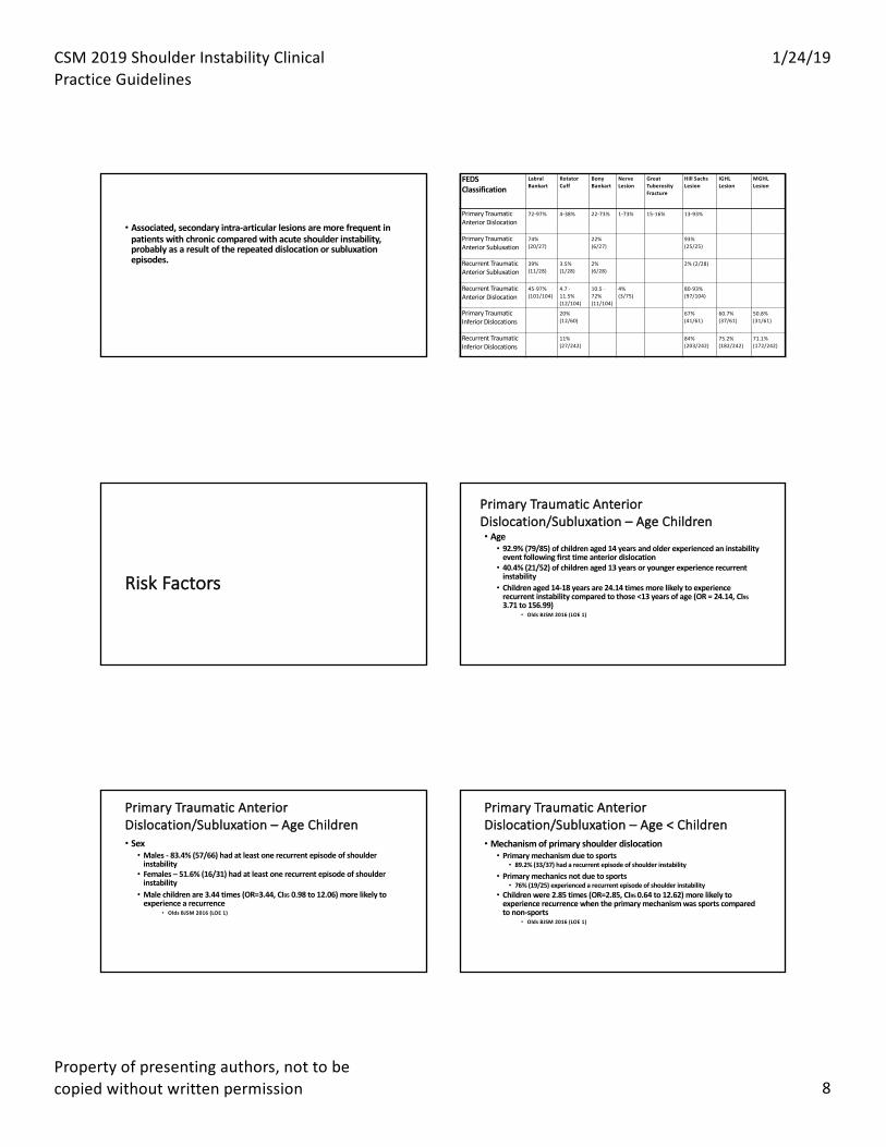
• Associated, secondary intra-articular lesions are more frequent in patients with chronic compared with acute shoulder instability, probably as a result of the repeated dislocation or subluxation episodes.
FEDS Classification
Labral Bankart
Rotator Cuff
Bony Bankart
Nerve Lesion
Great Tuberosity Fracture
Hill Sachs Lesion
IGHL Lesion
MGHL Lesion
Primary Traumatic Anterior Dislocation
72-97% 4-38% 22-73% 1-73% 15-16% 13-93%
Primary Traumatic Anterior Subluxation
74% (20/27)
22% (6/27)
93% (25/25)
Recurrent Traumatic Anterior Subluxation
39% (11/28)
3.5% (1/28)
2% (6/28)
2% (2/28)
Recurrent Traumatic Anterior Dislocation
45-97% (101/104)
4.7 -11.5% (12/104)
10.5 -72% (11/104)
4% (3/75)
80-93% (97/104)
Primary Traumatic Inferior Dislocations
20% (12/60)
67% (41/61)
60.7% (37/61)
50.8% (31/61)
Recurrent Traumatic Inferior Dislocations
11% (27/242)
84% (203/242)
75.2% (182/242)
71.1% (172/242)
Risk Factors
Primary Traumatic Anterior Dislocation/Subluxation – Age Children• Age• 92.9% (79/85) of children aged 14 years and older experienced an instability
event following first time anterior dislocation
• 40.4% (21/52) of children aged 13 years or younger experience recurrent instability
• Children aged 14-18 years are 24.14 times more likely to experience recurrent instability compared to those <13 years of age (OR = 24.14, CI95
3.71 to 156.99)• Olds BJSM 2016 (LOE 1)
• Sex• Males - 83.4% (57/66) had at least one recurrent episode of shoulder
instability• Females – 51.6% (16/31) had at least one recurrent episode of shoulder
instability• Male children are 3.44 times (OR=3.44, CI95 0.98 to 12.06) more likely to
experience a recurrence• Olds BJSM 2016 (LOE 1)
Primary Traumatic Anterior Dislocation/Subluxation – Age Children
Primary Traumatic Anterior Dislocation/Subluxation – Age < Children• Mechanism of primary shoulder dislocation• Primary mechanism due to sports
• 89.2% (33/37) had a recurrent episode of shoulder instability
• Primary mechanics not due to sports• 76% (19/25) experienced a recurrent episode of shoulder instability
• Children were 2.85 times (OR=2.85, CI95 0.64 to 12.62) more likely to experience recurrence when the primary mechanism was sports compared to non-sports
• Olds BJSM 2016 (LOE 1)

CSM 2019 Shoulder Instability Clinical Practice Guidelines
1/24/19
Property of presenting authors, not to be copied without written permission 9
• Open/closed proximal humeral physis
• Open physis – 61.1% (39/59) had at least one recurrent episode of shoulder
instability
• Closed physis – 94.1% (16/17) had at least one recurrent episode of
shoulder instability
• Children with a closed physis are 14 times (OR=14.0, CI95 1.46 to 134.25)
more likely to experience recurrent instability compared to those with open
physis
• Olds BJSM 2016 (LOE 1)
Primary Traumatic Anterior Dislocation/Subluxation – Age < Children
• Hill Sachs Lesion• 100% (13/13) of subjects with a Hill Sachs lesion had at least one recurrent
episode of shoulder instability• 72% (13/18) of subjects without a Hill Sachs lesion had at least one
recurrent episode of shoulder instability• Individuals under the age of 18 years with a Hill Sachs lesion were 17.18
times (OR=17.18, CI95 0.76 to 390.92) more likely to experience recurrence• Olds BJSM 2016 (LOE 1)
Primary Traumatic Anterior Dislocation/Subluxation – Age < Children
Primary Traumatic Anterior Dislocation/Subluxation – Adults • Age
• < 40 years of age had a 44% increased risk for an recurrence of instability
compared to those > 40 years (11%)
• Individuals who are < 40 years of age are 13.46 times (OR=13.46, CI95 (5.25
to 34.49) more likely to have a recurrent instability compared to those > 40
years• Olds BJSM 2015 (LOE 1)
Primary Traumatic Anterior Dislocation/Subluxation – Adults • Sex• Men are 3.18 times (OR=3.18, CI95 (1.28 to 7.89) more likely to have a
recurrent instability compared women• Olds BJSM 2015 (LOE 1)
Primary Traumatic Anterior Dislocation/Subluxation – Adults • Greater Tuberosity Fractures• Individuals with a greater tuberosity fracture were over 7 times less likely to
have a recurrence (OR=0.13 CI95 0.06 to 0.30)• Olds BJSM 2015 (LOE 1)
• Hyperlaxity• Individuals with hyperlaxity are 2.68 times (OR=2.68, CI95 (1.33 to 5.39)
more likely to have a recurrent instability compared to those who don’t.• Olds BJSM 2015 (LOE 1)
Primary Traumatic Posterior Dislocation
• Glenoid retroversion• Increased glenoid retroversion was associated with increased risk for
posterior instability• HR= 1.17 CI95 1.03 to 1.34 for every 1 degree of increased retroversion there
was a 17% increased risk of posterior shoulder instability. • Owens AJSM 2013 (LOE 2)
• Strength• Increased external rotation strength in adduction (HR =1.06, CI95 1.01 to
1.12) and at 45 degrees of abduction (HR=1.07, CI95 1.01 to 1.13) was associated with those who had a posterior dislocation• Increased internal rotation strength in adduction (HR= 1.05 CI95 1.00 to
1.11) was associated with those who had a posterior dislocation• Owens AJSM 2013 (LOE 2)
CSM 2019 Shoulder Instability Clinical Practice Guidelines
1/24/19
Property of presenting authors, not to be copied without written permission 10
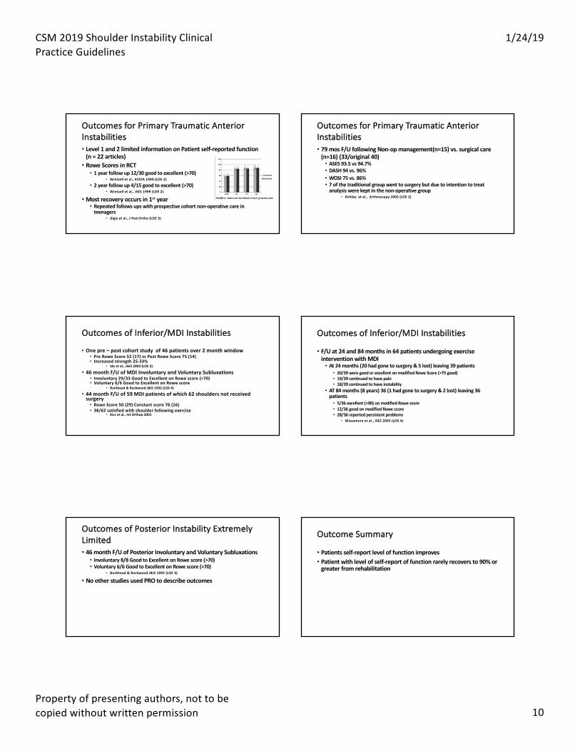
Outcomes for Primary Traumatic Anterior Instabilities• Level 1 and 2 limited information on Patient self-reported function
(n = 22 articles)• Rowe Scores in RCT• 1 year follow up 12/30 good to excellent (>70)
• Wintzell et al., KSSTA 1999 (LOE 2)
• 2 year follow up 4/15 good to excellent (>70)• Wintzell et al., JSES 1999 (LOE 2)
• Most recovery occurs in 1st year• Repeated follows ups with prospective cohort non-operative care in
teenagers• Gigis et al., J Ped Ortho (LOE 3)
Outcomes for Primary Traumatic Anterior Instabilities• 79 mos F/U following Non-op management(n=15) vs. surgical care
(n=16) (33/original 40)• ASES 93.5 vs 94.7%• DASH 94 vs. 96%• WOSI 75 vs. 86% • 7 of the traditional group went to surgery but due to intention to treat
analysis were kept in the non-operative group• Kirkley et al., Arthroscopy 2005 (LOE 2)
Outcomes of Inferior/MDI Instabilities
• One pre – post cohort study of 46 patients over 2 month window
• Pre Rowe Score 52 (17) vs Post Rowe Score 75 (14)
• Increased strength 25-33%• Ide et al., JSES 2003 (LOE 2)
• 46 month F/U of MDI Involuntary and Voluntary Subluxations
• Involuntary 29/33 Good to Excellent on Rowe score (>70)• Voluntary 6/6 Good to Excellent on Rowe score
• Burkhead & Rockwood JBJS 1992 (LOE 4)
• 44 month F/U of 59 MDI patients of which 62 shoulders not received surgery
• Rowe Score 50 (29) Constant score 76 (16)
• 38/62 satisfied with shoulder following exercise• Kiss et al., Int Orthop 2001
Outcomes of Inferior/MDI Instabilities
• F/U at 24 and 84 months in 64 patients undergoing exercise intervention with MDI• At 24 months (20 had gone to surgery & 5 lost) leaving 39 patients
• 20/39 were good or excellent on modified Rowe Score (>75 good)• 19/39 continued to have pain• 18/39 continued to have instability
• AT 84 months (8 years) 36 (1 had gone to surgery & 2 lost) leaving 36 patients• 5/36 excellent (>90) on modified Rowe score• 12/36 good on modified Rowe score• 28/36 reported persistent problems
• Missamore et al., JSES 2005 (LOE 4)
Outcomes of Posterior Instability Extremely Limited• 46 month F/U of Posterior Involuntary and Voluntary Subluxations• Involuntary 8/8 Good to Excellent on Rowe score (>70)• Voluntary 6/6 Good to Excellent on Rowe score (>70)
• Burkhead & Rockwood JBJS 1992 (LOE 4)
• No other studies used PRO to describe outcomes
Outcome Summary
• Patients self-report level of function improves • Patient with level of self-report of function rarely recovers to 90% or
greater from rehabilitation

CSM 2019 Shoulder Instability Clinical Practice Guidelines
1/24/19
Property of presenting authors, not to be copied without written permission 11
Thank You
• Up next…. Diagnosis of shoulder instability DiagnosisClinical examination to identify patients with shoulder instability
ERIC HEGEDUS, PT, PhD, DPT, OCS, CSCS
History
Instability
Congenital
Trauma
Overuse
V- Expert Opinion Tests & Measures- Motion Testing
• Motion testing• AROM may be painful• PROM may be excessive with reports of apprehension at end range• Accessory motions likely show greater excursion and maybe subluxation
V- Expert Opinion
Tests & Measures- Muscle Testing
• Muscle testing• Often no issue as MMT• Performance tests may show decreased function, pain, apprehension
V- Expert Opinion
Tests & Measures- Palpation
• Palpation is often unremarkable• Unique tests as follows
V- Expert Opinion

CSM 2019 Shoulder Instability Clinical Practice Guidelines
1/24/19
Property of presenting authors, not to be copied without written permission 12
Unique TestsAnterior Instability
Level II- Lesser Quality Diagnostic StudiesGrade- A
TESTNAME(S)
Pathology LeadAuthor
LR+ LR- RiskofBiasfromQUADAS2
Apprehension AnteriorInstability Jia 20 0.29 UnclearVanKampen 3.5 0.02 LowFarber 20 0.29 LowLo 48 0.48 HighHegedus 17 0.39 SystematicReview
Relocation AnteriorInstability Farber 10 0.20 LowLo 1 1 HighSpeer 67 0.33 HighVanKampen 4 0.04 LowHegedus 5.5 0.55 SystematicReview
Surprise AnteriorInstability Lo 59 0.37 HighGross 8 0.09 HighVanKampen 6 0.10 LowHegedus 5 0.45 SystematicReview
AnteriorDrawer
AnteriorInstability Farber 4 0.56 LowVanKampen 8 0.45 Low
Apprehension LR+ = 17
GRADE A
Relocation LR+ = 5.5
GRADE A
Surprise LR+ = 5.0
GRADE B
Anterior Drawer LR+ = 4-8
GRADE A
CSM 2019 Shoulder Instability Clinical Practice Guidelines
1/24/19
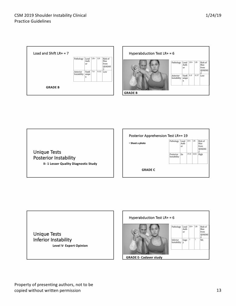
Property of presenting authors, not to be copied without written permission 13
Load and Shift LR+ = 7
GRADE B
Pathology LeadAuthor
LR+ LR- RiskofBiasfromQUADAS2
AnteriorInstability
VanKampen
7.0 0.32 Low
Hyperabduction Test LR+ = 6
Pathology LeadAuthor
LR+ LR- RiskofBiasfromQUADAS2
AnteriorInstability
VanKampen
6.0 0.37 Low
GRADE B
Unique TestsPosterior Instability
II- 1 Lesser Quality Diagnostic Study
Posterior Apprehension Test LR+= 19
• Shoot a photoPathology Lead
Author
LR+ LR- RiskofBiasfromQUADAS2
PosteriorInstability
Jia 19.0 0.82 High
GRADE C
Unique TestsInferior Instability
Level V- Expert Opinion
Hyperabduction Test LR+ = 6
Pathology LeadAuthor
LR+ LR- RiskofBiasfromQUADAS2
InferiorInstability
Gagey
? ? NA
GRADE E- Cadaver study

CSM 2019 Shoulder Instability Clinical Practice Guidelines
1/24/19
Property of presenting authors, not to be copied without written permission 14
Unique TestsMultidirectional Instability
V- Expert Opinion
In My Opinion- Grade F
• Beighton index 5/9 or greater• Almost always congenital• Comparisons to opposite shoulder largely meaningless• Best tests for anterior, inferior, and posterior instability to rule in
Diagnosis and Classification- Summary
• Diagnosis of specific shoulder pathology is not easy• In other areas of the body where diagnosis is also challenging,
classification systems are developed• Many classification systems have been developed for the shoulder
and are based often on etiology (ex: trauma) and direction (ex: anterior) • Recent classification systems have added frequency and severity
• No classification system has the requisite proven psychometric properties (ex: validity)
Diagnosis and Classification- Summary
• Traumatic instability is often suspected from patient history and confirmed by imaging• Non-traumatic instability is more difficult but there are physical
examination tests that can help• Research on physical examination tests is primarily focused on
anterior instability and secondarily on posterior instability while inferior and multidirectional instability are largely ignored
Other ConsiderationsTraumatic Dislocation & Hypermobility
Trauma
• X-ray for bony lesions• MRI for soft tissue lesions• MR arthrography for labral tear
CSM 2019 Shoulder Instability Clinical Practice Guidelines
1/24/19
Property of presenting authors, not to be copied without written permission 15
Hypermobility Syndrome– Beighton Score
The ability to: Right Yes/No LeftPassively extend the 5th MCP to > 90 degrees
1 1Passively oppose the thumb to the ipsilateral forearm
1 1Elbow hyperextension of > 10 degrees
1 1Knee hyperextension of > 10 degrees
1 1Hands flat on floor without bending knees
1
Total Possible Score = 9 Note: 5/9 + HMS
Marfan Syndrome- 2010 Nosology
• *Points for systemic score• Wrist AND thumb sign = 3 (wrist OR thumb sign = 1)• Pectus carinatum deformity = 2 (pectus excavatum or chest asymmetry = 1)• Hindfoot deformity = 2 (plain pes planus = 1)• Dural ectasia = 2• Protrusio acetabula = 2• Reduced upper segment/lower segment ratio AND increased arm/height AND no severe scoliosis = 1• Scoliosis or thoracolumbar kyphosis = 1• Reduced elbow extension = 1• Facial features (3/5) = 1 (dolichocephaply, enophthalmos, downslanting palpebral fissures, malar
hypoplasia, retrognathia)• Skin striae = 1• Myopia > 3 diopters = 1• Mitral valve prolapse = 1
Ehlers Danlos Syndrome
• Requires genetic testing
Thank You
• Up next…..Intervention: • physical therapy interventions including • immobilization • exercise • motor control/neuromuscular retraining• bracing
InterventionEvidence Based Recommendations for Treatment of Patients with
Shoulder Instability
Amee L. Seitz, PT, PhD, DPT, OCS
Interventions
1. Immobilization (following Dislocation)• Duration • Position
2. Exercise • Strengthening• Motor Control/ Neuromuscular Retraining
3. Bracing for return to high demand activity/sport
Terminology for population not consistent
• Atraumatic/Multidirectional Instability • Anterior Dislocation (traumatic / atraumatic)

CSM 2019 Shoulder Instability Clinical Practice Guidelines
1/24/19
Property of presenting authors, not to be copied without written permission 16
• 1 week vs 3-4 weeks duration • ER vs IR position
Interventions Immobilization Duration & Position
Population• 1st Time Anterior Dislocation
Immobilization Duration (1 week vs. 3 or 4 weeks)Paterson et alLevel 3 Studies
Level of Evidence... Careful review
• No level I/II studies on duration of immobilization
• Excluded Hovelius 1983: allocation to group at 6/27 centers was based on date of shoulder dislocation. At 21/27 centers treatment was given according to customary practice= Not randomized/quasi (prospective observational study)• Excluded Kiviluto 1980: of 99 patients, 53 immobilized for 1 week and 46 for 3
weeks. No indication of method of allocation. No response from study authors.• Robinson 2006 was not included. it is a prospective cohort examining factors
associated with recurrent instability. No formal statistics were conducted to compare recurrence as it related to duration of immobilization. Level I prognosis but not level I intervention study
Hanchard 2014
Results: Recurrence Pooled meta-analysis
Patients younger <30 yo rate of recurrence:
41% (40/97) in patients immobilized for one week or less
37% (34/93) in patients immobilized for three weeks or longer
(p = 0.52). bottom Line………
Paterson et al
•No randomized clinical trials (Level I /II evidence) for duration of immobilization•High risk of bias or confounding in currently published
observational study results
Guideline Recommendation: Immobilization Duration
There is no harm in immobilizing a patient for 1 week instead of 3 weeks following a first time
anterior dislocation
Immobilization: Position
What is best position toimmobilize shoulder s/ptraumatic dislocation?
• Itoi et al 2001
• MRI study 19 patients• IR vs ER position • Separation and displacement of the labrum were both
significantly less
• Miller et al 2004 Hart 2005• Cadeveric and arthroscopic observations supports ER
optimal healing position that approximates labrum to bone

CSM 2019 Shoulder Instability Clinical Practice Guidelines
1/24/19
Property of presenting authors, not to be copied without written permission 17
Randomized Trials Immobilization Position
Itoi 2007• N=198 participants• 3 weeks immobilization • Randomized IR vs ER (10°)
Liavaag 2011 • N=51 participants• 3 weeks immobilization• Randomized IR vs ER (15°)
Taskoparan 2010 (level 2)• N=188 participants• 3 week immobilization • Randomized IR vs ER (10°)
Finestone 2009 • N=33 participants• 4 weeks immobilization• Randomized IR vs ER (15°-20°)
N= 470 total; 371 (79%) males
Quality Assessment: Bias Risk High & Overall “low” grading
Hanchard 2014
N= 6 Randomized Trials Immobilization Position (Level 1-2 evidence)
Whelan 2014• N=60 participants• 4 weeks immobilization • Randomized IR vs ER (10°)
Heidari 2014• N= 102 participants• 3 week immobilization • Randomized IR vs ER (10°)
Pooled DATAN= 632 total patients; 517 (82%) males
Mean age= 30 years
Whelan 2016
Summary Evidence Immobilization Position
Recurrence (2yr f/u) 6 studies
• No significant difference between positions in low and high risk groups
Patient Reported Outcomes WOSI lower is better (3 studies)
• Mean WOSI 83 (ER) vs. 89 (IR) • No significant difference
between groups
Return Pre-Injury Activities (Itoi2007 & Liavaag 2011)• No significant difference
(p>0.05)
Adherence
5 studies• No significant difference in self-
reported adherence (p>0.05)
Whelan 2016 AJSM
Recommendation Immobilization: Position
• Evidence for superiority of immobilization in ER over traditional sling in IR is lacking
There is no justification for change in current clinical practice
Interventions
2. Exercise • Strengthening• Motor Control/ Neuromuscular Retraining
Terminology for population not consistent
• Anterior Dislocation (traumatic / atraumatic)

CSM 2019 Shoulder Instability Clinical Practice Guidelines
1/24/19
Property of presenting authors, not to be copied without written permission 18
Bottoni et al. 2002• N=24 male active military• Mean age 22yrs• 4 weeks immobilization +
rehabilitation vs Bankart repair
Wintzell et al. 1999• N=30 (46 males)• Mean age 24 years• 1 week immobilization + normal
use vs Arthroscopic lavage
Kirkley et al. 1999 N=40 participants (35 males)Mean age 22yrs• 3 week immobilization +
rehabilitation vs Bankart repair
Sandow et al. (abstract only)• N=39 <26 year old• 4 weeks immobilization +
rehabilitation vs Bankart repair
N= 143 total; >80% males
4 Randomized Trials Surgery vs Non-surgical 1st anterior dislocation
Handoll 2004
Rehabilitation Protocol Bottoni et al. 1. 4 weeks sling immobilization, limited active ROM and “some exercises” under
physiotherapist supervision;2. 4 weeks of progressive passive motion exercises followed by active-assisted ROM exercises
without resistance3. 4 weeks of progressively greater resistance exercises4. Return to full active duty, contact sports and activities requiring over-head or heavy lifting
restricted until 4 months
Kirkley et al. 1. 3 Weeks immobilization, then both groups had the same staged (4 to 6 weeks; 7 to 8 weeks;
9 to 12 weeks) rehabilitation protocol of progressive exercises, including easing of the restrictions in ER ROM
2. 3 month for return to non-contact or non-overhead sports; 3. 4 months for contact sports
Wintzel et al.• 1 week immobilization + normal use
Which is the most effective treatment for instability, surgery vs rehabilitation?
Randomized trials compare surgical intervention to non-surgical management:
• Bottoni et al. 2002* 24 males in military• Kirkley et al. 1999 * 40 patients• Wintzell et al. 1999*à lavage vs no rehab• Sandow et al 1996* à abstract only
“limited evidence supporting primary surgery for young adults, usually male, engaged in high demand physical activities following their 1st acute traumatic shoulder dislocation”
“There is no evidence for other patient groups” Handoll, Cochrane Review 2004
Results: Recurrence Dislocation 2yr f/u
43%13%
54%21%
1 week immobilization + normal use
64%21%
3-4 weeks immobilization + rehabilitation
Summary Evidence: Surgery vs. PT for 1st Dislocation
• First time only dislocation: Rehab vs Bankart • 2 published randomized trials - 1999• 59/64 total patients males, 24 military population• Mean age 22 years• Greater likelihood of recurrence with rehab (64% recurrence vs 33%)
• Both are successful at improving patient rated outcomes and return to activity • The rehabilitation program in these studies (strengthening initiated at 8
weeks) is not current evidence-based standard of care • Immobilization time (3-4 weeks versus shorter duration) is not standard
of care for non-operative treatment of acute shoulder dislocation
Surgery first in high demand patient?
• A key area of controversy• Limited evidence with 2 randomized control trials recruited the
population at highest risk of recurrence Level 2• Shoulder instability also occurred in the surgical treatment
group- pooled data 6/28 =21% (versus rehabilitation 43%)•Only 50% of patients with recurrence in the conservative
treatment group chose subsequent surgery

CSM 2019 Shoulder Instability Clinical Practice Guidelines
1/24/19
Property of presenting authors, not to be copied without written permission 19
• GAP: More aggressive non-operative treatment rehabilitation program àneuromuscular control / strengthening started < one week with is warranted (Prior recommendation Grade C)• GAP: proprioception and motor control exercises
Guideline Recommendation: Progressive Exercise
Following first time dislocation PT consisting of graded exercise improves pain, function, and allows for return
to activity
Interventions
2. Exercise • Strengthening• Motor Control/ Neuromuscular Retraining
• Atraumatic/Multidirectional Instability
Low quality evidence surgery better than exercise • Shoulder kinematics • Likelihood return to sport
4 studies: No RCTs, 2 retrospective cohorts, 2 pre/post cohorts
Both treatments improve pain, function, and activity participation
Low quality evidence exercise better than surgery • Satisfaction• Self-report outcome
measures
Rockwood Program, 1992
Warby S, et al
“Motor Control + Strengthening” programScapular motor control + GH joint strengthening- 4 phases
Watson et al. Shoulder & Elbow 2017
• Greater improvements in motor control /strengthening program (Watson)• WOSI total score 12 weeks and 24 (>MCID) weeks (difference 11-12%)• Melbourne Instability Shoulder Score at 24 weeks (15% difference)
• Not included in rehabilitation program• Proprioceptive training- joint repositioning activities• Closed chain exercises • Kinetic Chain/ trunk/ core/ LE strengthening

CSM 2019 Shoulder Instability Clinical Practice Guidelines
1/24/19
Property of presenting authors, not to be copied without written permission 20
• GAP: More aggressive non-operative treatment rehabilitation program àneuromuscular control / strengthening started < one week with is warranted (Prior recommendation Grade C)
Guideline Recommendation: Motor Control + Strengthening
In patients with multi-directional instability, clinicians should consider progressive motor control and strengthening exercises
to improve pain, function, and ABDuction ROM
Interventions
3. Bracing for return to high demand activity/sport
• Anterior Dislocation
Bracing for return to sport/high demand activity following dislocation/subluxation• 5 studies including patients ages 17-31 years
• 2 prospective cohort studies• 1 prospective case series • 1 retrospective study• 1 cross-sectional study (joint reposition sense with/without brace)
• All subjects were managed nonoperatively and treated with a brace during return to activity or sport.
• Bracing allowed for early RTS, 10 to 40 days, but recurrence rate is higher with an earlier return: • 73% with 10 days RTS (n=45); • 53% with 11.7 days RTS (n=19); • 10% at 40 days RTS (n=20).
• 90% of athletes (n=27/30) RTS without pain within 40 days- 10 had recurrence (37%)• Joint reposition sense improves with use of brace near end range (10 degrees from
full) shoulder external rotation.
Risk of Bias
• GAP: no comparative studies have been conducted examining the use of a brace for return to sport activities
Guideline Recommendation: Rehabilitation + Bracing for Return to Activity/Sport
In patients who experienced a dislocation /subluxation, clinicians may consider recommending a brace for return to sport or high demand activity although the risk of recurrence increases with earlier return
Thank You
• Up next…..Examination:
• Key impairments • Self-reported outcome tools
CSM 2019 Shoulder Instability Clinical Practice Guidelines
1/24/19
Property of presenting authors, not to be copied without written permission 21
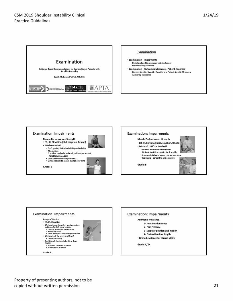
ExaminationEvidence Based Recommendations for Examination of Patients with
Shoulder Instability
Lori A Michener, PT, PhD, ATC, SCS
Examination
• Examination - Impairments• Deficits related to prognosis and risk factors• Functional requirements
• Examination – Outcomes Measures - Patient-Reported• Disease-Specific, Shoulder-Specific, and Patient-Specific Measures• Anchoring the scores
Examination: ImpairmentsMuscle Performance - Strength• ER, IR, Elevation (abd, scaption, flexion)• Methods: MMT• 0 – 5 grades; limited reliability and validity• Alternative:
3 grades: markedly reduced, reduced, or normalReliable (Wainner, 2003)
• Used to determine impairments• Limited ability to assess change over time
Grade: B
Examination: ImpairmentsMuscle Performance - Strength• ER, IR, Elevation (abd, scaption, flexion)• Methods: HHD or Isokinetic• Used to determine impairments• Reliable in athletes, patients, & healthy• Improved ability to assess change over time• Isokinetic – concentric and eccentric
Grade: B
Examination: ImpairmentsRange of Motion• ER, IR, Elevation• Methods: goniometer, inclinometer -
bubble, digital, smartphone• Used to determine impairments• Reliability for all• Good ability to assess change over time
• Methods: IR by vertebral level• Limited reliability
• Additional- horizontal add or low flexion• Posterior shoulder tightness• Inclinometer as above
Grade: B
Examination: ImpairmentsAdditional Measures
1- Joint Position Sense2- Pain Pressure3- Scapular position and motion4- Pectoralis minor length
• Limited evidence for clinical utility
Grade: C/ D

CSM 2019 Shoulder Instability Clinical Practice Guidelines
1/24/19
Property of presenting authors, not to be copied without written permission 22
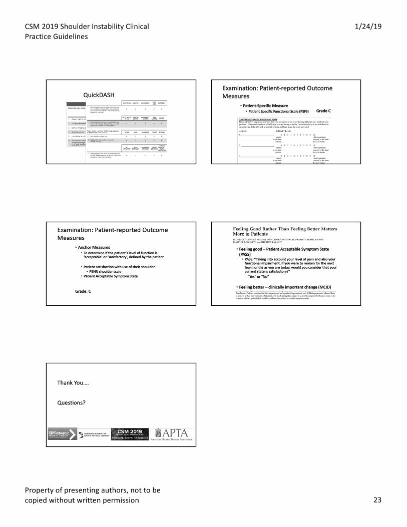
Examination: Patient-reported Outcome Measures
Disease Specific Measures• Western Ontario Shoulder Instability Index (WOSI)• Oxford Shoulder Instability (OSSI)
• Shoulder Specific Measures• ASES• PENN• DASH
Grade: B
WOSI
Examination: Patient-reported Outcome Measures
Disease Specific Measures• Western Ontario Shoulder Instability Index (WOSI)• Oxford Shoulder Instability (OSSI)
• Shoulder Specific Measures• ASES• PENN• DASH / QuickDASH
Grade: B
PennShoulder Score• Pain
(0–30 pts)• Rest• Normal ADL• Strenuous
• Satisfaction –shoulder use (0-10 pts)
CSM 2019 Shoulder Instability Clinical Practice Guidelines
1/24/19
Property of presenting authors, not to be copied without written permission 23
QuickDASHExamination: Patient-reported Outcome Measures
• Patient-Specific Measure• Patient Specific Functional Scale (PSFS) Grade C
Examination: Patient-reported Outcome Measures
• Anchor Measures• To determine if the patient’s level of function is
‘acceptable’ or ‘satisfactory’, defined by the patient
• Patient satisfaction with use of their shoulder• PENN shoulder scale
• Patient Acceptable Symptom State
Grade: C
• Feeling good – Patient Acceptable Symptom State (PASS)• PASS: “Taking into account your level of pain and also your
functional impairment, if you were to remain for the next few months as you are today, would you consider that your current state is satisfactory?”
“Yes” or “No”
• Feeling better – clinically important change (MCID)
Thank You….
Questions?