Correlations of structure and function and mechanisms of recovery in acute tubular necrosis
23
Review Correlations of Structure and Function and Mechanisms of Recovery in Acute Tubular Necrosis* JEAN OLIVER, M.D. Brooklyn, New York T HE problems that I wish to discuss are concerned with questions of epistemologic nosology; what constitutes a legitimate entity in the field of renal disease; how can such entities best be established; what are the causal relationships that bind them together; in general, what processes of reasoning, observa- tion and experiment can we most usefully bring to bear on the involved entanglements of pathogenesis in matters renal? I shall not attempt to discuss such recondite considerations but I do intend to illustrate their importance by means of specific example. another attempt. That our labor is not wasted and that our method is sound we can believe from what Gerhard wrought with the use of those ancient tools, the correlation of structural and functional change which had been handed down to him through Morgagni from still earlier times. As he then proved, typhoid is not typhus, even if the current distinction is not much concerned with the structural landmarks of intestinal ulcer and swollen mesenteric gland. Similar questions confronted William Ger- hard in the early 1800’s when he faced and solved such problems as distinguishing typhus from typhoid or of establishing the relation of tuberculous meningitis to pulmonary phthisis. It is true that in the field of infectious disease such difficulties have today been resolved or, as the philosophically minded might somewhat reluctantly think, brushed aside by the intro- duction of a strict etiologic basis for the entity; “tuberculosis” is now whatever happens when Mycobacterium tuberculosis is at work and we worry no more about the structural aspects of tubercle and caseation, of phthisis and scirrhus. But as Gerhard had to do things the hard and slow way, just so are we pathologists and clinicians still doomed to Sisyphean labors in our classifications of renal disease; no sooner is the final stone of our theoretic construction in place but it rolls back upon us. The collapse is, however, seldom complete; some part of the imposing edifice we have momentarily admired remains to encourage To get to our immediate point: it was about thirty years after its first recognition that Dr. Baldwin Luckt’ pointed out common structural and functional characteristics in a seemingly heterogenous group of clinical conditions in which sudden failure of renal activity was an obvious common denominator. Being a pa- thologist, he quite naturally approached the problem from its structural aspect and estab- lished the entity on the basis of a structural change. That further investigations brought out certain additional minutiae and led to what are sometimes tolerantly called “semantic difficul- ties” is of no great importance. However, very real difficulties did appear when the functional- ists in their survey of the problem came to their conclusions, for as Dr. Homer Smith2 summed matters up, it was the functional phenomenon of “acute renal failure” with its myriad con- trasting dynamic mechanisms that was the unifying key to the solution of the nosologic puzzle. The man in the middle, in particular the clinician, was therefore left with an “entity” that in its one aspect was a complex of diverse functional disturbances and in the other a restricted structural lesion. The very existence ” From the Department of Pathology, State University of New York, Medical Center at New York City. This work was supported by the Life Insurance Medical Research Fund and the New York Heart Association. Address before the Philadelphia Pathological Society, November 13, 1952 on the award of the William Wood Gerhard Medal. OCTOBER, 1953 535
-
Upload
jean-oliver -
Category
Documents
-
view
213 -
download
0
Transcript of Correlations of structure and function and mechanisms of recovery in acute tubular necrosis
Review
Correlations of Structure and Function
and Mechanisms of Recovery in
Acute Tubular Necrosis*
JEAN OLIVER, M.D.
Brooklyn, New York
T HE problems that I wish to discuss are concerned with questions of epistemologic nosology; what constitutes a legitimate
entity in the field of renal disease; how can such entities best be established; what are the causal relationships that bind them together; in general, what processes of reasoning, observa- tion and experiment can we most usefully bring to bear on the involved entanglements of pathogenesis in matters renal? I shall not attempt to discuss such recondite considerations but I do intend to illustrate their importance by means of specific example.
another attempt. That our labor is not wasted and that our method is sound we can believe from what Gerhard wrought with the use of those ancient tools, the correlation of structural and functional change which had been handed down to him through Morgagni from still earlier times. As he then proved, typhoid is not typhus, even if the current distinction is not much concerned with the structural landmarks of intestinal ulcer and swollen mesenteric gland.
Similar questions confronted William Ger- hard in the early 1800’s when he faced and solved such problems as distinguishing typhus from typhoid or of establishing the relation of tuberculous meningitis to pulmonary phthisis. It is true that in the field of infectious disease such difficulties have today been resolved or, as the philosophically minded might somewhat reluctantly think, brushed aside by the intro- duction of a strict etiologic basis for the entity; “tuberculosis” is now whatever happens when Mycobacterium tuberculosis is at work and we worry no more about the structural aspects of tubercle and caseation, of phthisis and scirrhus. But as Gerhard had to do things the hard and slow way, just so are we pathologists and clinicians still doomed to Sisyphean labors in our classifications of renal disease; no sooner is the final stone of our theoretic construction in place but it rolls back upon us.
The collapse is, however, seldom complete; some part of the imposing edifice we have momentarily admired remains to encourage
To get to our immediate point: it was about thirty years after its first recognition that Dr. Baldwin Luckt’ pointed out common structural and functional characteristics in a seemingly heterogenous group of clinical conditions in which sudden failure of renal activity was an obvious common denominator. Being a pa- thologist, he quite naturally approached the problem from its structural aspect and estab- lished the entity on the basis of a structural change. That further investigations brought out certain additional minutiae and led to what are sometimes tolerantly called “semantic difficul- ties” is of no great importance. However, very real difficulties did appear when the functional- ists in their survey of the problem came to their conclusions, for as Dr. Homer Smith2 summed matters up, it was the functional phenomenon of “acute renal failure” with its myriad con- trasting dynamic mechanisms that was the unifying key to the solution of the nosologic puzzle. The man in the middle, in particular the clinician, was therefore left with an “entity” that in its one aspect was a complex of diverse functional disturbances and in the other a restricted structural lesion. The very existence
” From the Department of Pathology, State University of New York, Medical Center at New York City. This work was supported by the Life Insurance Medical Research Fund and the New York Heart Association. Address before the Philadelphia Pathological Society, November 13, 1952 on the award of the William Wood Gerhard Medal.
OCTOBER, 1953 535
Structure and Function in Acute Tubular Necrosis-Oliver
of such an “entity,” much less an understanding of its causal relationships, seemed incredible.
Now all of us, as pathologists, must have felt certain that this pathogenetic dilemma could in the end only be resolved by the same means that had proved so effective in the hands of
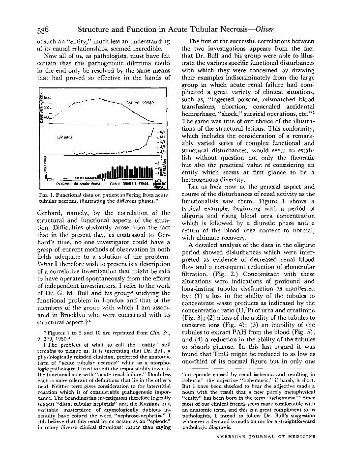
FIG. 1. Functional data on patient suffering from acute tubular necrosis, illustrating the different phases. *
Gerhard, namely, by the correlation of the structural and functional aspects of the situa- tion. Difficulties obviously arose from the fact that in the present day, as contrasted to $Ser- hard’s time, no one investigator could have a grasp of current methods of observation in both fields adequate to a solution of the problem. What I therefore wish to present is a description of a correlative investigation that might be said to have operated spontaneously from the efforts of independent investigators. I refer to the work of Dr. G. M. Bull and his group3 studying the functional problem in London and that of the members of the group with which I am associ- ated in Brooklyn who were concerned with its structural aspect.i_*
* Figures 1 to 5 and 10 are reprinted from Clin. SC., 9: 379, 1950.3
t The problem of what to call the “entity” still remains to plague us. It is interesting that Dr. Bull, a physiologically minded clinician, preferred the anatomic term of “acute tubular necrosis” while as a morpho- logic pathologist I tried to shift the responsibility towards the functional side with “acute renal failure.” Doubtless each is more tolerant of definitions that lie in the other’s field. Neither term gives consideration to the interstitial reaction which is of considerable pathogenetic impor- tance. The Scandinavian investigators therefore logically suggest “distal tubular nephritis” and the Russians in a veritable masterpiece of etymologically dubious in- genuity have coined the word “nephroso-nephritis.” I still believe that this renal lesion occurs as an “episode” in many diverse clinical situations: rather than saying
The first of the successful correlations between the two investigations appears from the fact that Dr. Bull and his group were able to illus- trate the various specific functional disturbances with which they were concerned by drawing their examples indiscriminately from the large group in which acute renal failure had com- plicated a great variety of clinical situations, such as, “ingested poisons, mismatched blood transfusions, abortion, concealed accidental hemorrhage, “shock,” surgical operations, etc.“3 The same was true of our choice of the illustra- tions of the structural lesions. This conformity, which includes the consideration of a remark- ably varied series of complex functional and structural disturbances, would seem to estab- lish without question not only the theoretic but also the practical value of considering an entity which seems at first glance to be a heterogenous diversity.
Let us look now at the general aspect and course of the disturbances of renal activity as the functionalists saw them. Figure 1 shows a typical example, beginning with a period of oliguria and rising blood urea concentration which is followed by a diuretic phase and a return of the blood urea content to normal, with ultimate recovery.
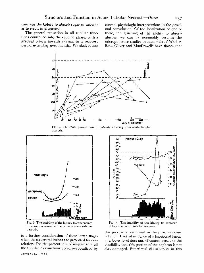
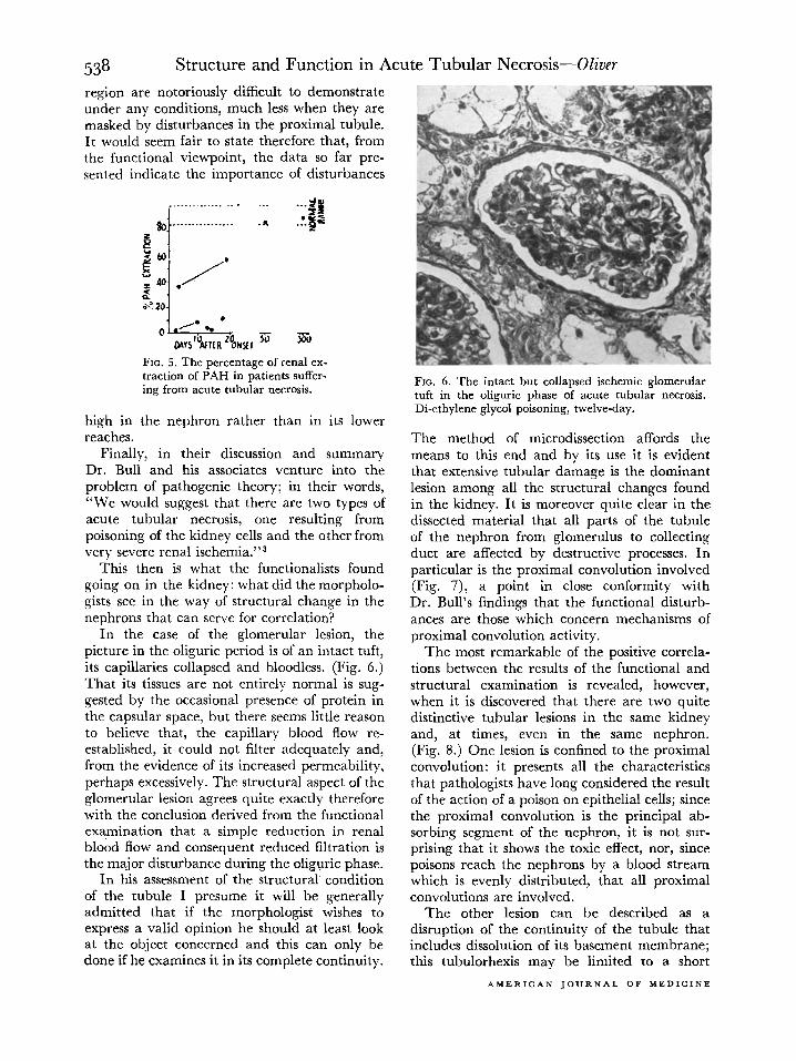
A detailed analysis of the data in the oliguric period showed disturbances which were inter- preted as evidence of decreased renal blood flow and a consequent reduction of glomerular filtration. (Fig. 2.) Concomitant with these alterations were indications of profound and long-lasting tubular dysfunction as manifested by: (1) a loss in the ability of the tubules to concentrate waste products as indicated by the concentration ratio (U/P) of urea and creatinine (Fig. 3); (2) a loss of the ability of the tubules to conserve ions (Fig. 4) ; (3) an inability of the tubules to extract PAH from the blood (Fig. 5); and (4) a reduction in the ability of the tubules to absorb glucose. In this last regard it was found that TmG might be reduced to as low as one-third of its normal figure but in only one
“an episode caused by renal ischemia and resulting in ischuria” the adjective “ischemuric,” if harsh, is short. But I have been shocked to hear the adjective made a noun with the result that a new purely metaphysical “entity” has been born in the term “ischemuria”! Since most of our clinical friends seem more comfortable with an anatomic term, and this is a great compliment to us pathologists, I intend to follow Dr. Bull’s suggestion whenever a demand is made on me for a straightforward pathologic diagnosis.
AMERICAN JOURNAL OF MEDICINE
Structure and Function in Acute Tubular Necrosis-Oliver 537 case was the failure to absorb sugar so extreme current physiologic interpretations in the proxi- as to result in glycosuria. ma1 convolution. Of the localization of one of
The general reduction in all tubular func- these, the lessening of the ability to absorb tions continued into the diuretic phase, with a glucose, we can be reasonably certain; the gradual return towards normal in a recovery micropuncture studies in mammals of Walker, period extending over months. We shall return Bott, Oliver and MacDowel15 have shown that
DAYS AFlLR ONSCr FIG. 2. The renal plasma flow in patients suffering from acute tubular necrosis.
FIG. 3. The inability of the kidney to concentrate urea and creatinine in the urine in acute tubular necrosis.
to a further consideration of these latter stages when the structural lesions are presented for cor- relation. For the present it is of interest that all the tubular dysfunctions noted are localized b)
OCTOBER, 1953
19/l - PAlIf NT sC549 ra/l - n I
FIG. 4. The inability of the kidney to conserve chloride in acute tubular necrosis.
this process is completed in the proximal con- volution. Lack of evidence of a functional lesion
at a lower level does not, of course, preclude the possibility that this portion of the nephron is not also damaged. Functional disturbances in this
538 Structure and Function in Acute Tubular Necrosis-Oliver
region are notoriously difficult to demonstrate under any conditions, much less when they are masked by disturbances in the proximal tubule. It would seem fair to state therefore that, from the functional viewpoint, the data so far pre- sented indicate the importance of disturbances
FIG. 5. The percentage of renal ex- traction of PAH in patients suffer- ing from acute tubular necrosis.
high in the nephron rather than in its lower reaches.
Finally, in their discussion and summary Dr. Bull and his associates venture into the problem of pathogenic theory; in their words, “We would suggest that there are two types of acute tubular necrosis, one resulting from poisoning of the kidney cells and the other from very severe renal ischemia.“3
This then is what the functionalists found going on in the kidney: what did the morpholo- gists see in the way of structural change in the nephrons that can serve for correlation?
In the case of the glomerular lesion, the picture in the oliguric period is of an intact tuft, its capillaries collapsed and bloodless. (Fig. 6.) That its tissues are not entirely normal is sug- gested by the occasional presence of protein in the capsular space, but there seems little reason to believe that, the capillary blood flow re- established, it could not filter adequately and, from the evidence of its increased permeability, perhaps excessively. The structural aspect of the glomerular lesion agrees quite exactly therefore with the conclusion derived from the functional examination that a simple reduction in renal blood flow and consequent reduced filtration is the major disturbance during the oliguric phase.
In his assessment of the structural condition of the tubule I presume it will be generally admitted that if the morphologist wishes to express a valid opinion he should at least look at the object concerned and this can only be done if he examines it in its complete continuity.
FIG. 6. The intact but collapsed ischemic glomerular tuft in the oliguric phase of acute tubular necrosis. Di-ethylene glycol poisoning, twelve-day.
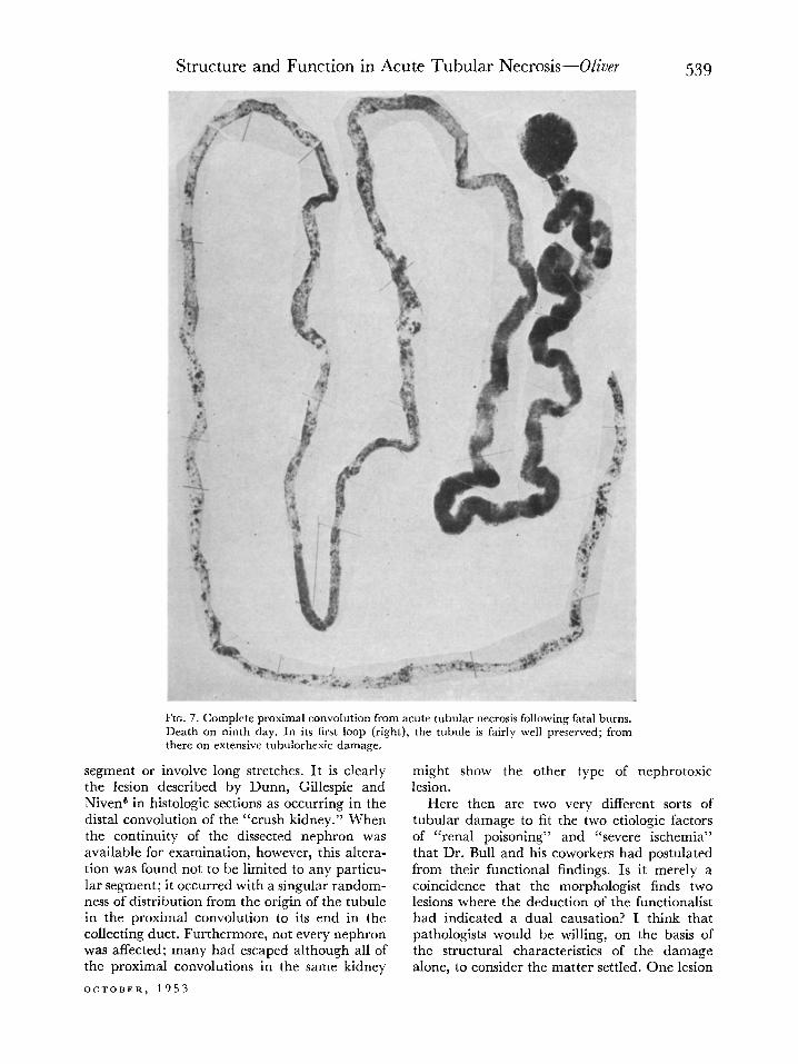
The method of microdissection affords the means to this end and by its use it is evident that extensive tubular damage is the dominant lesion among all the structural changes found in the kidney. It is moreover quite clear in the dissected material that all parts of the tubule of the nephron from glomerulus to collecting duct are affected by destructive processes. In particular is the proximal convolution involved (Fig. 7), a point in close conformity with Dr. Bull’s findings that the functional disturb- ances are those which concern mechanisms of proximal convolution activity.
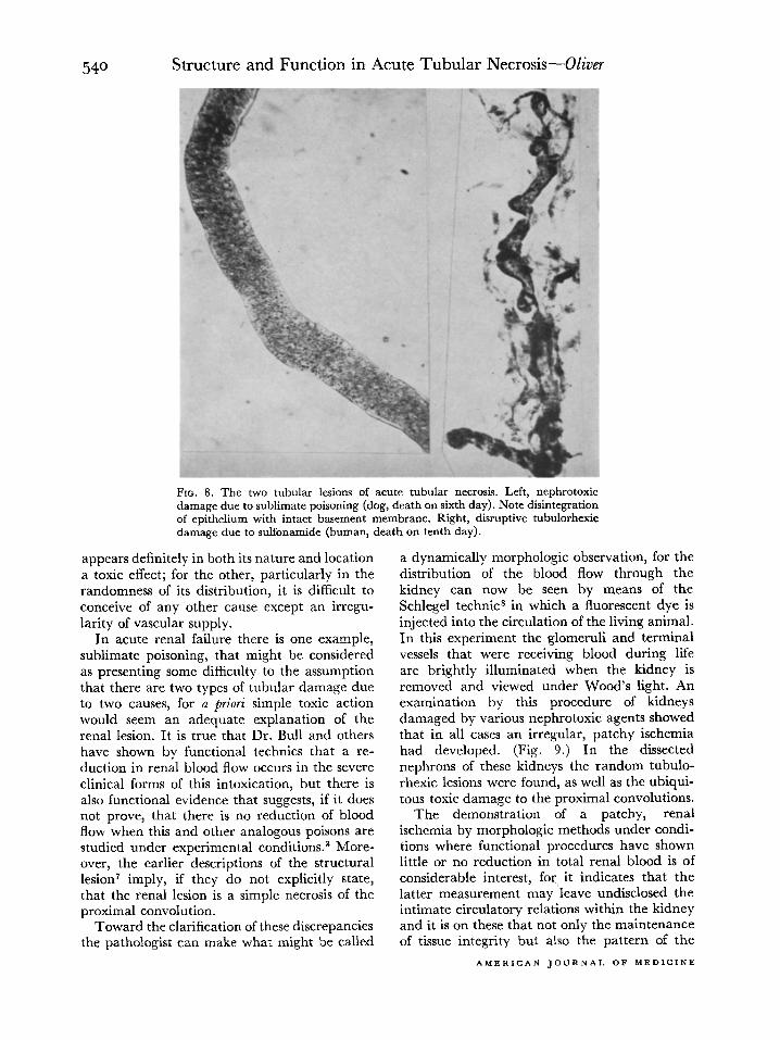
The most remarkable of the positive correla- tions between the results of the functional and structural examination is revealed, however, when it is discovered that there are two quite distinctive tubular lesions in the same kidney and, at times, even in the same nephron. (Fig. 8.) One lesion is confined to the proximal convolution: it presents all the characteristics that pathologists have long considered the result of the action of a poison on epithelial cells; since the proximal convolution is the principal ab- sorbing segment of the nephron, it is not sur- prising that it shows the toxic effect, nor, since poisons reach the nephrons by a blood stream which is evenly distributed, that all proximal convolutions are involved.
The other lesion can be described as a disruption of the continuity of the tubule that includes dissolution of its basement membrane; this tubulorhexis may be limited to a short
AMERICAN JOURNAL OF MEDICINE
Structure and Function in Acute Tubular Necrosis--Oliver
FIG. 7. Complete proximal convolution from acute tubular necrosis following fatal burns. Death on ninth day. In its first loop (right), the tubule is fairly well preserved; from there on extensive tubulorhexic damage.
segment or involve long stretches. It is clearly the lesion described by Dunn, Gillespie and Niven6 in histologic sections as occurring in the distal convolution of the “crush kidney.” When the continuity of the dissected nephron was available for examination, however, this altera- tion was found not to be limited to any particu- lar segment; it occurred with a singular random- ness of distribution from the origin of the tubule in the proximal convolution to its end in the collecting duct. Furthermore, not every nephron was affected; many had escaped although all of the proximal convolutions in the same kidney
OCTOBER, 1953
might show the other type of nephrotoxic lesion.
Here then are two very different sorts of tubular damage to fit the two etiologic factors of “renal poisoning” and “severe ischemia” that Dr. Bull and his coworkers had postulated from their functional findings. Is it merely a coincidence that the morphologist finds two lesions where the deduction of the functionalist had indicated a dual causation? I think that pathologists would be willing, on the basis of the structural characteristics of the damage alone, to consider the matter settled. One lesion
Structure and Function in Acute Tubular Necrosis-Oliver
FIG. 8. The two tubular lesions of acute tubular necrosis. Left, nephrotoxic damage due to sublimate poisoning (dog, death on sixth day). Note disintegration of epithelium with intact basement membrane. Right, disruptive tubulorhexic damage due to sulfonamide (human, death on tenth day).
appears definitely in both its nature and location a toxic effect; for the other, particularly in the randomness of its distribution, it is difficult to conceive of any other cause except an irregu- larity of vascular supply.
In acute renal failure there is one example, sublimate poisoning, that might be considered as presenting some difficulty to the assumption that there are two types of tubular damage due to two causes, for a priori simple toxic action would seem an adequate explanation of the renal lesion. It is true that Dr. Bull and others have shown by functional technics that a re- duction in renal blood flow occurs in the severe clinical forms of this intoxication, but there is also functional evidence that suggests, if it does not prove, that there is no reduction of blood flow when this and other analogous poisons are studied under experimental conditions.3 More- over, the earlier descriptions of the structural lesion’ imply, if they do not explicitly state, that the renal lesion is a simple necrosis of the proximal convolution.
Toward the clarification of these discrepancies the pathologist can make what might be called
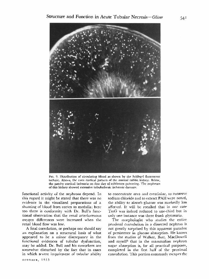
a dynamically morphologic observation, for the distribution of the blood flow through the kidney can now be seen by means of the Schlegel technics in which a fluorescent dye is injected into the circulation of the living animal. In this experiment the glomeruli and terminal vessels that were receiving blood during life are brightly illuminated when the kidney is removed and viewed under Wood’s light. An examination by this procedure of kidneys damaged by various nephrotoxic agents showed that in all cases an irregular, patchy ischemia had developed. (Fig. 9.) In the dissected nephrons of these kidneys the random tubulo- rhexic lesions were found, as well as the ubiqui- tous toxic damage to the proximal convolutions.
The demonstration of a patchy, renal ischemia by morphologic methods under condi- tions where functional procedures have shown little or no reduction in total renal blood is of considerable interest, for, it indicates that the latter measurement may leave undisclosed the intimate circulatory relations within the kidney and it is on these that not only the maintenance of tissue integrity but also the pattern of the
AMERICAN JOURNAL OF MEDICINE
Structure and Function in Acute Tubular Necrosis--Oliver
FIG. 9. Distribution of circulating blood as shown by the Schlegel fluoresce-nt technic. Above, the even cortical pattern of the normal rabbit kidney. Below, the patchy cortical ischemia on first day of sublimate poisoning. The nephrons of this kidney showed extensive tubulorhexic ischemic damage.
functional activity of the nephrons depend. In this regard it might be stated that there was no evidence in the visualized preparations of a shunting of blood from cortex to medulla; here too there is conformity with Dr. Bull’s func- tional observation that the renal arteriovenous oxygen differences were increased when the renal blood flow was low.
A final correlation, or perhaps one should say an explanation on a structural basis of what appeared to be a minor discrepancy in the functional evidences of tubular dysfunction, may be added. Dr. Bull and his coworkers are somewhat disturbed by the fact that in cases in which severe impairment of tubular ability
OCTOBER, 1953
to concentrate urea and creatinine, to conserve sodium chloride and to extract PAH were noted, the ability to absorb glucose was markedly less affected. It will be recalled that in one case TmG was indeed reduced to one-third but in only one instance was there frank glycosuria.
The morphologist who studies the entire proximal convolution in a dissected nephron is not greatly surprised by this apparent paradox of persistence in glucose absorption. He knows from the studies of Walker, Bott, MacDowell and myself5 that in the mammalian nephron sugar absorption is, for all practical purposes, completed in the first half of the proximal convolution. This portion commonly escapes the
542 Structure and Function in Acute Tubular Necrosis-Oliver
maximum damage that is produced by most toxic substances, so that even in the severely affected proximal convolution there is, at least from the morphologist’s viewpoint, a consider- able Tm remaining. This is also true when a considerable part of the proximal convolution
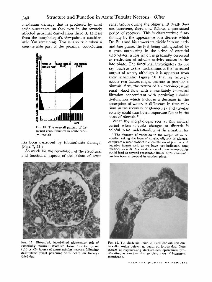
FIG. 10. The over-all pattern of dis- turbed renal function in acute tubu- lar necrosis.
has been destroyed by tubulorhexic damage. (Figs. 7, 21.)
So much for the correlation of the structural and functional aspects of the lesions of acute
renal failure during the oliguria. If death does not intervene, there now follows a protracted period of recovery. This is characterized func- tionally by the appearance of a diuresis which Dr. Bull and his coworkers divide into an early and late phase, the first being distinguished by a gross outpouring in the urine of essential electrolytes, a loss which is gradually corrected as restitution of tubular activity occurs in the late phase. The functional investigators do not say much as to the mechanisms of the increased output of water, although it is apparent from their schematic Figure 10 that as recovery occurs two factors might operate to produce a diuresis; first, the return of an ever-increasing renal blood flow with immediately increased filtration concomitant with persisting tubular dysfunction which includes a decrease in the absorption of water. A difference in time rela- tions in the recovery of glomerular and tubular activity could thus be an important factor in the onset of diuresis.*
What the morphologist sees at this critical period when oliguria changes to diuresis is helpful to an understanding of the situation for
*The “cause” of variation in the output of water, whether taking the form of anuria, oliguria or diuresis, comprises a most elaborate constellation of positive and negative factors and, as we have just indicated, time relations as well. A consideration of these complexities would lead us beyond reasonable limits in this discussion but has been attempted in another place.4
FIG. 11. Distended, blood-filled glomerular tuft of essentially normal structure from diuretic phase (115 cc./24 hours) of acute tubular necrosis following di-ethylene glycol poisoning with death on twenty- third day.
FIG. 12. Tubulorhexic lesion in distal convolution due to sulfonamide poisoning; death on fourth day. Note masses of regenerating dark-stained epithelium pro- liferating at random due to disruption of basement membrane.
AMERICAN JOURNAL OF MEDICINE
Structure and Function in Acute Tubular Necrosis-Oliver 543
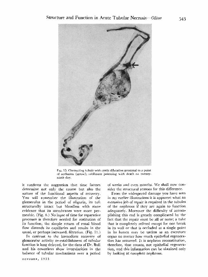
Fro. 13. Connecting tubule with cystic dilatation proximal to a point of occlusion (arrow); sublimate poisoning with death on twenty- ninth day.
it confirms the suggestion that time factors determine not only the course but also the nature of the functional aspects of recovery. You will remember the illustration of the glomerulus in the period of oliguria, its tuft structurally intact but bloodless with some evidence that its membranes were more per- meable. (Fig. 6.) No lapse of time for reparative processes is therefore needed for restitution of its function; the simple return of renal bIood flow distends its capillaries and results in the usual, or perhaps increased, filtration. (Fig. 11.)
In contrast to the immediate recovery of glomerular activity re-establishment of tubular function is long delayed, for the data of Dr. Bull and his coworkers show irregularities in the balance of tubular mechanisms over a period
OCTOBER, 1953
of weeks and even months. We shall now con- sider the structural reasons for this difference.
From the widespread damage you have seen in my earlier illustrations it is apparent what an extensive job of repair is required in the tubules of the nephrons if they are again to function adequately. Moreover the difficulty of accom- plishing this end is greatly complicated by the fact that the repair must be all or none; a tube that is completely relined except for one break in its wall or that is occluded at a single point in its lumen may be useless as an excretory organ no matter how much epithelial regenera- tion has occurred. It is nephron reconstitution, therefore, that counts, not epithelial regenera- tion, and this information can be obtained only by looking at complete nephrons.
544 Structure and Function in Acute Tubular Necrosis-Oliver
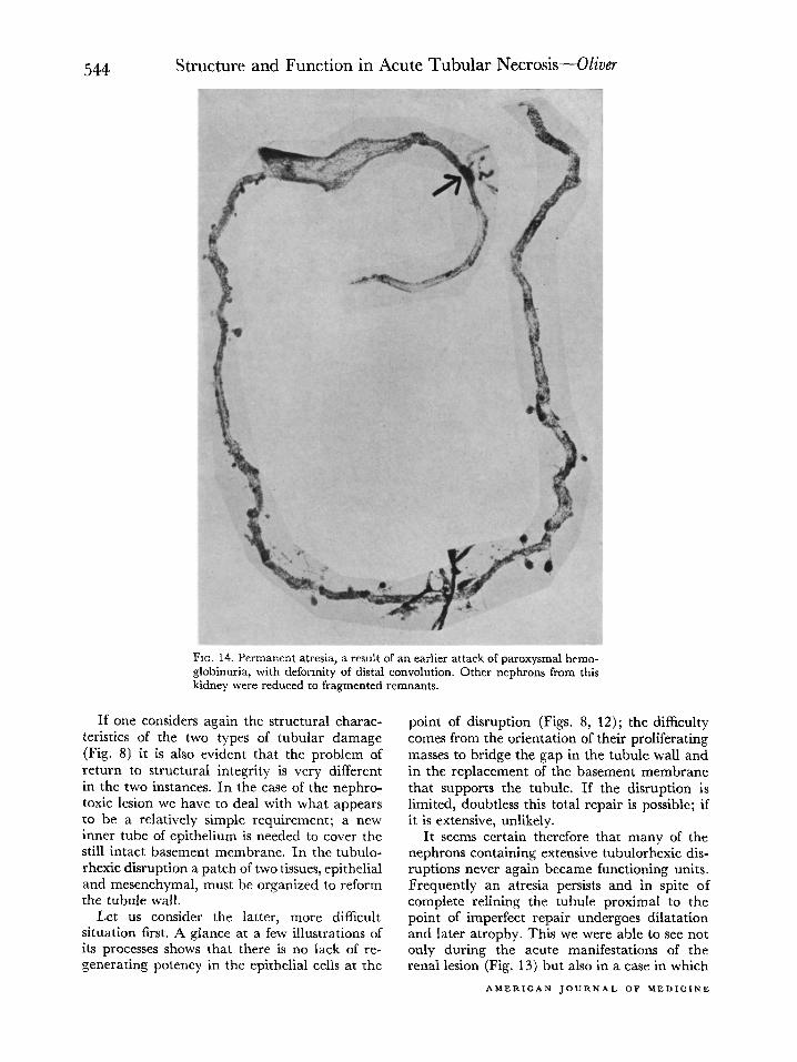
FIG. 14. Permanent atresia, a result of an earlier attack of paroxysmal hemo- globinuria, with deformity of distal convolution. Other nephrons from this kidney were reduced to fragmented remnants.
If one considers again the structural charac- teristics of the two types of tubular damage (Fig. 8) it is also evident that the problem of return to structural integrity is very different in the two instances. In the case of the nephro- toxic lesion we have to deal with what appears to be a relatively simple requirement; a new inner tube of epithelium is needed to cover the still intact basement membrane. In the tubulo- rhexic disruption a patch of two tissues, epithelial and mesenchymal, must be organized to reform the tubule wall.
Let us consider the latter, more difficult situation first. A glance at a few illustrations of its processes shows that there is no lack of re- generating potency in the epithelial cells at the
point of disruption (Figs. 8, 12) ; the difficulty comes from the orientation of their proliferating masses to bridge the gap in the tubule wall and in the replacement of the basement membrane that supports the tubule. If the disruption is limited, doubtless this total repair is possible; if it is extensive, unlikely.
It seems certain therefore that many of the nephrons containing extensive tubulorhexic dis- ruptions never again became functioning units. Frequently an atresia persists and in spite of complete relining the tubule proximal to the point of imperfect repair undergoes dilatation and later atrophy. This we were able to see not only during the acute manifestations of the renal lesion (Fig. 13) but also in a case in which
AMERICAN JOURNAL OF MEDICINE
Structure and Function in Acute Tubular Necrosis--Oliver 545
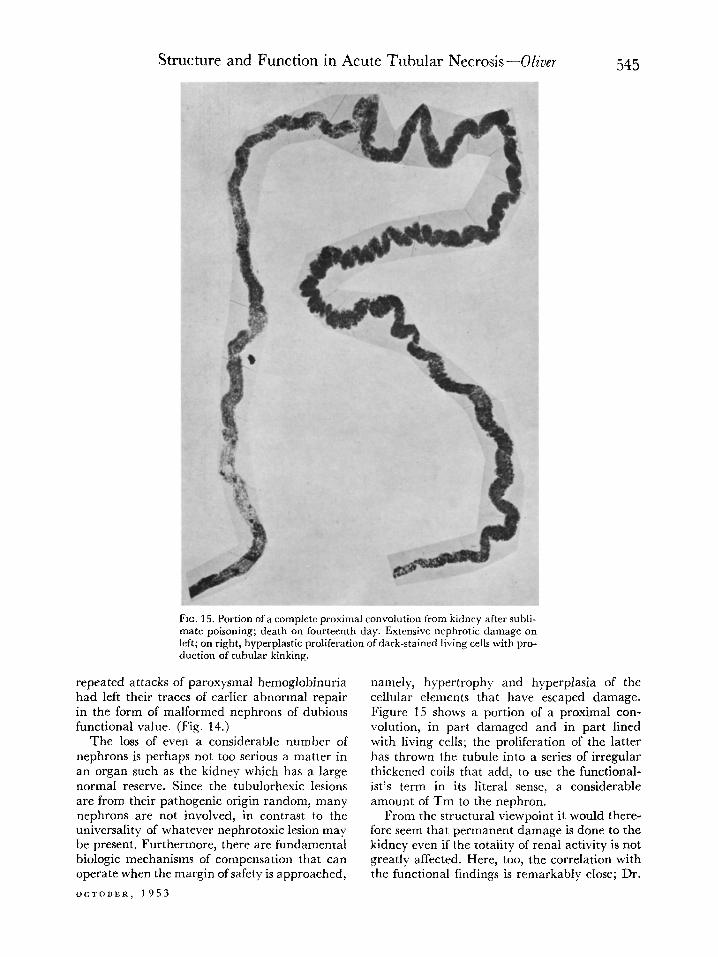
FIG. 15. Portion of a complete proximal convolution from kidney after subli- mate poisoning; death on fourteenth day. Extensive nephrotic damage on left; on right. hyperplastic proliferation of dark-stained living cells with pro- duction o? &b&r l&king.
repeated attacks of paroxysmal hemoglobinuria had left their traces of earlier abnormal repair in the form of malformed nephrons of dubious functional value. (Fig. 14.)
The loss of even a considerable number of nephrons is perhaps not too serious a matter in an organ such as the kidney which has a large normal reserve. Since the tubulorhexic lesions are from their pathogenic origin random, many nephrons are not involved, in contrast to the universality of whatever nephrotoxic lesion may be present. Furthermore, there are fundamental biologic mechanisms of compensation that can operate when the margin of safety is approached,
OCTOBER, 1953
namely, hypertr-ophy and hyperplasia of the cellular elements that have escaped damage. Figure 15 shows a portion of a proximal con- volution, in part damaged and in part lined with living cells; the proliferation of the latter has thrown the tubule into a series of irregular thickened coils that add, to use the functional- ist’s term in its literal sense, a considerable amount of Tm to the nephron.
From the structural viewpoint it would there- fore seem that permanent damage is done to the kidney even if the totality of renal activity is not greatly affected. Here, too, the correlation with the functional findings is remarkably close; Dr.
546 Structure and Function in Acute Tubular Necrosis-Oliver
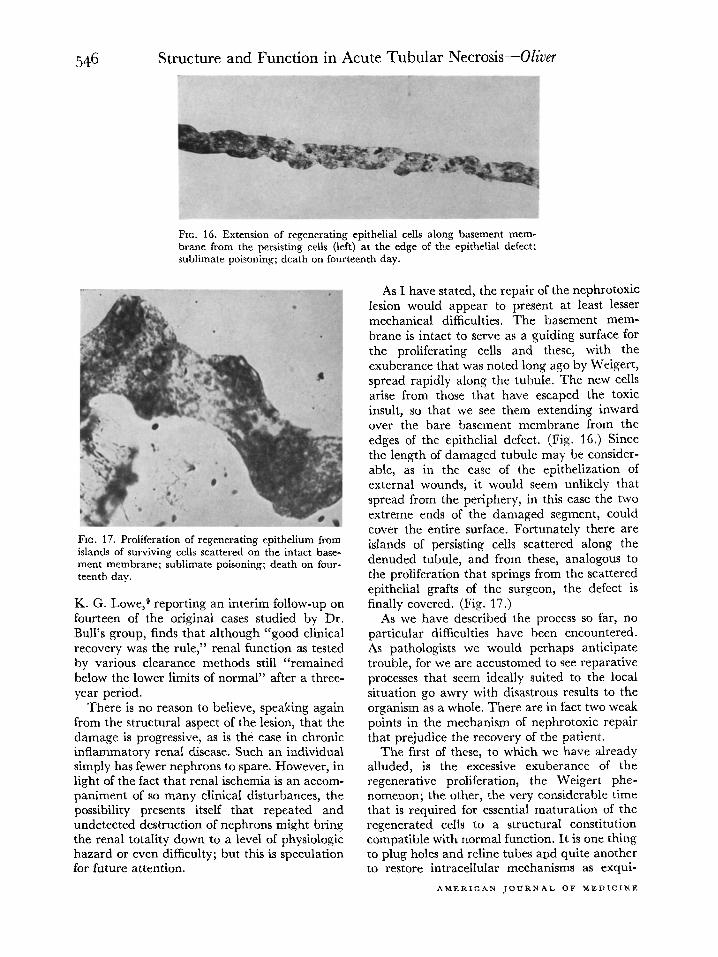
FIG. 16. Extension of regenerating epithelial cells along basement mem- brane from the persisting cells (left) at the edge of the epithelial defect; sublimate poisoning; death on fourteenth day.
FIG. 17. Proliferation of regenerating epithelium from islands of surviving cells scattered on the intact base-
ment membrane; sublimate poisoning; death on four- teenth day.
K. G. Lowe,s reporting an interim follow-up on fourteen of the original cases studied by Dr. Bull’s group, finds that although “good clinical recovery was the rule,” renal function as tested by various clearance methods still “remained below the lower limits of normal” after a three- year period.
There is no reason to believe, speaking again from the structural aspect of the lesion, that the damage is progressive, as is the case in chronic inflammatory renal disease. Such an individual simply has fewer nephrons to spare. However, in light of the fact that renal ischemia is an accom- paniment of so many clinical disturbances, the possibility presents itself that repeated and undetected destruction of nephrons might bring the renal totality down to a level of physiologic hazard or even difficulty; but this is speculation for future attention.
As I have stated, the repair of the nephrotoxic lesion would appear to present at least lesser mechanical difficulties. The basement mem- brane is intact to serve as a guiding surface for the proliferating cells and these, with the exuberance that was noted long ago by Weigert, spread rapidly along the tubule. The new cells arise from those that have escaped the toxic insult, so that we see them extending inward over the bare basement membrane from the edges of the epithelial defect. (Fig. 16.) Since the length of damaged tubule may be consider- able, as in the case of the epithelization of external wounds, it would seem unlikely that spread from the periphery, in this case the two extreme ends of the damaged segment, could cover the entire surface. Fortunately there are islands of persisting cells scattered along the denuded tubule, and from these, analogous to the proliferation that springs from the scattered epithelial grafts of the surgeon, the defect is finally covered. (Fig. 17.)
As we have described the process so far, no particular difficulties have been encountered. As pathologists we would perhaps anticipate trouble, for we are accustomed to see reparative processes that seem ideally suited to the local situation go awry with disastrous results to the organism as a whole. There are in fact two weak points in the mechanism of nephrotoxic repair that prejudice the recovery of the patient.
The first of these, to which we have already alluded, is the excessive exuberance of the regenerative proliferation, the Weigert phe- nomenon; the other, the very considerable time that is required for essential maturation of the regenerated cells to a structural constitution compatible with normal function. It is one thing to plug holes and reline tubes apd quite another to restore intracellular mechanisms as exqui-
AMERICAN JOURNAL OF MEDICINE
Structure and Function in Acute Tubular Necrosis-Oliver 547 sitely delicate as those that operate in the is well to remember what is commonly over- epithelium of the proximal convolution, and it looked, namely, that in the last analysis we shall is in this vitally functioning portion of the be arguing from analogy not identity. nephron that the nephrotoxic lesions occur. A series of rats was given the same dose of
For examination of these intimate cellular corrosive sublimate in such amount that the details it becomes necessary to turn to the majority of them survived after a period of
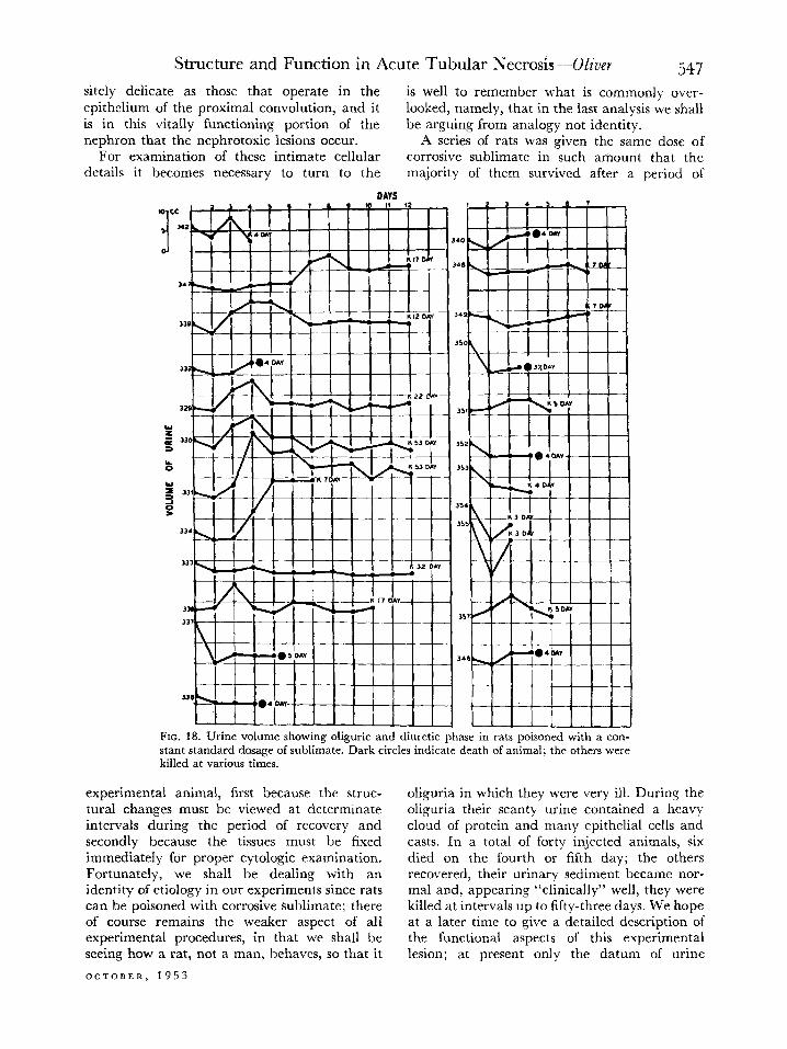
FIG. 18. Urine volume showing oliguric and diuretic phase in rats poisoned with a con- stant standard dosage of sublimate. Dark circles indicate death of animal; the others were killed at various tin&
experimental animal, first because the struc- tural changes must be viewed at determinate intervals during the period of recovery and secondly because the tissues must be fixed immediately for proper cytologic examination. Fortunately, we shall be dealing with an identity of etiology in our experiments since rats can be poisoned with corrosive sublimate; there of course remains the weaker aspect of all experimental procedures, in that we shall be seeing how a rat, not a man, behaves, so that it
OCTOBER, 1953
oliguria in which they were very ill. During the oliguria their scanty urine contained a heavy cloud of protein and many epithelial cells and casts. In a total of forty injected animals, six died on the fourth or fifth day; the others recovered, their urinary sediment became nor- mal and, appearing “clinically” well, they were killed at intervals up to fifty-three days. We hope at a later time to give a detailed description of the functional aspects of this experimental lesion; at present only the datum of urine
Structure and Function in Acute Tubular Necrosis-Oliver
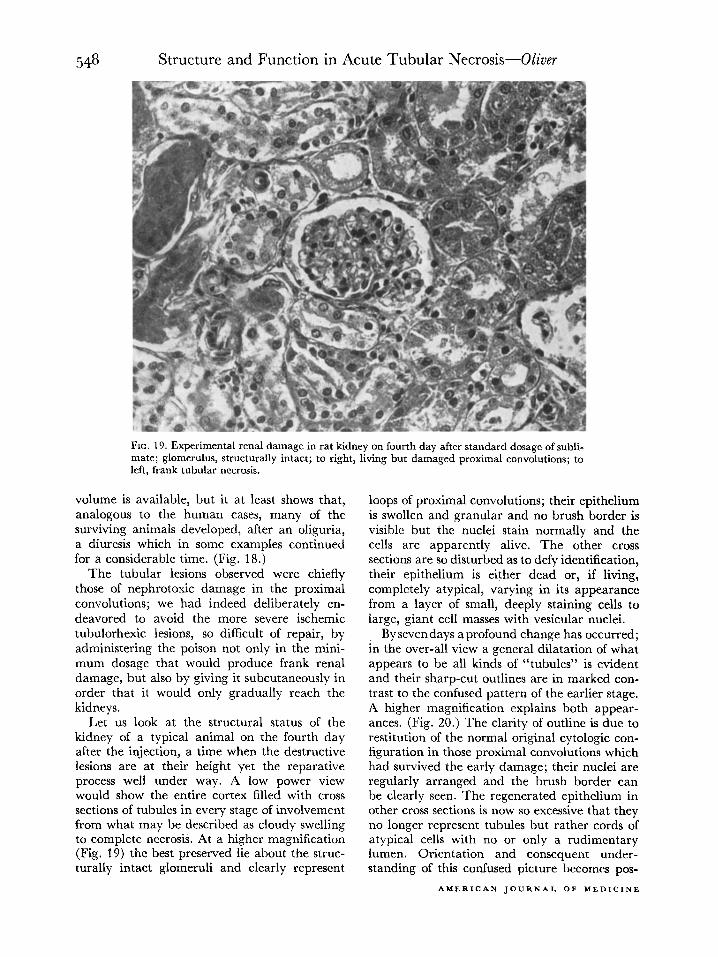
FIG. 19. Experimental renal damage in rat kidney on fourth day after standard dosage of subli- mate; glomerulus, structurally intact; to right, living but damaged proximal convolutions; to left, frank tubular necrosis.
volume is available, but it at least shows that, analogous to the human cases, many of the surviving animals developed, after an oliguria, a diuresis which in some examples continued for a considerable time. (Fig. 18.)
The tubular lesions observed were chiefly those of nephrotoxic damage in the proximal convolutions; we had indeed deliberately en- deavored to avoid the more severe ischemic tubulorhexic lesions, so difficult of repair, by administering the poison not only in the mini- mum dosage that would produce frank renal damage, but also by giving it subcutaneously in order that it would only gradually reach the kidneys.
Let us look at the structural status of the kidney of a typical animal on the fourth day after the injection, a time when the destructive lesions are at their height yet the reparative process well under way. A low power view would show the entire cortex filled with cross sections of tubules in every stage of involvement from what may be described as cloudy swelling to complete necrosis. At a higher magnification (Fig. 19) the best preserved lie about the struc- turally intact glomeruli and clearly represent
loops of proximal convolutions; their epithelium is swollen and granular and no brush border is visible but the nuclei stain normally and the cells are apparently alive. The other cross sections are so disturbed as to defy identification, their epithelium is either dead or, if living, completely atypical, varying in its appearance from a layer of small, deeply staining cells to large, giant cell masses with vesicular nuclei.
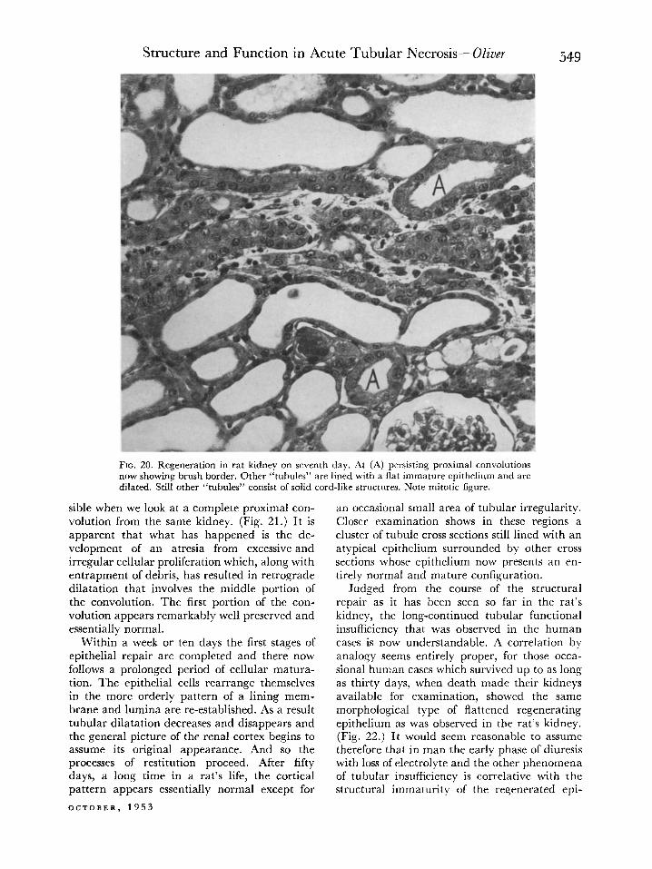
Bysevendays a profound change has occurred; in the over-all view a general dilatation of what appears to be all kinds of “tubules” is evident and their sharp-cut outlines are in marked con- trast to the confused pattern of the earlier stage. A higher magnification explains both appear- ances. (Fig. 20.) The clarity of outline is due to restitution of the normal original cytologic con- figuration in those proximal convolutions which had survived the early damage; their nuclei are regularly arranged and the brush border can be clearly seen. The regenerated epithelium in other cross sections is now so excessive that they no longer represent tubules but rather cords of atypical cells with no or only a rudimentary lumen. Orientation and consequent under- standing of this confused picture becomes pos-
AMERICAN JOURNAL OF MEDICINE
Structure and Function in Acute Tubular Necrosis-Oliver 549
FIG. 20. Regeneration in rat kidney on seventh day. At (A) persisting proximal convolutions now showing brush border. Other “tubules” are lined with a flat immature epithelium and are dilated. Still other “tubules” consist of solid cord-like structures. Note mitotic figure.
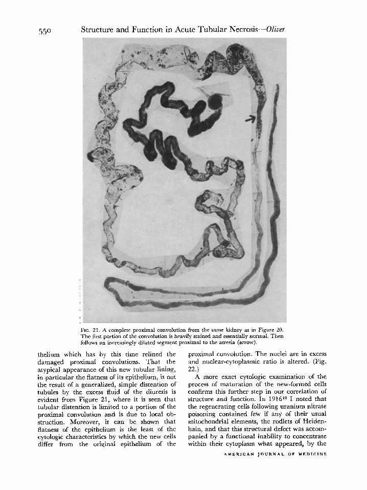
sible when we look at a complete proximal con- volution from the same kidney. (Fig. 21.) It is apparent that what has happened is the de- velopment of an atresia from excessive and irregular cellular proliferation which, along with entrapment of debris, has resulted in retrograde dilatation that involves the middle portion of the convolution. The first portion of the con- volution appears remarkably well preserved and essentially normal.
Within a week or ten days the first stages of epithelial repair are completed and there now follows a prolonged period of cellular matura- tion. The epithelial cells rearrange themselves in the more orderly pattern of a lining mem- brane and lumina are re-established. As a result tubular dilatation decreases and disappears and the general picture of the renal cortex begins to assume its original appearance. And so the processes of restitution proceed. After fifty days, a long time in a rat’s life, the cortical pattern appears essentially normal except for
OCTOBER. 1953
an occasional small area of tubular irregularity. Closer examination shows in these regions a cluster of tubule cross sections still lined with an atypical epithelium surrounded by other cross sections whose epithelium now presents an en- tirely normal and mature configuration.
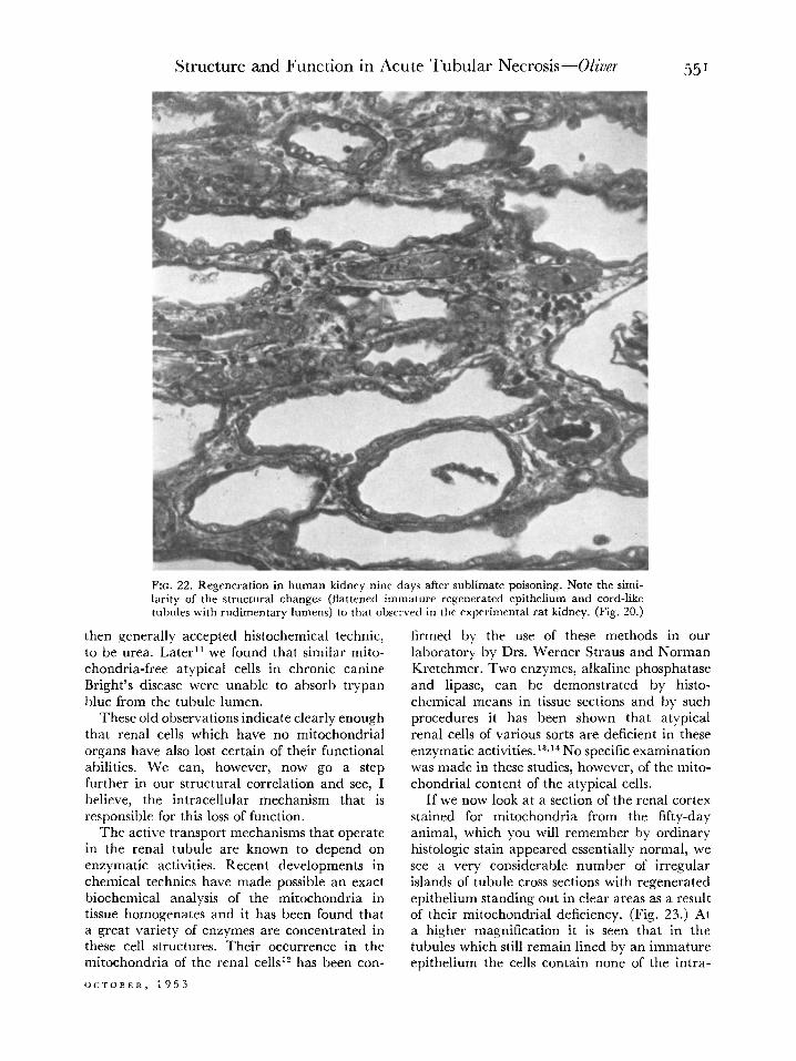
Judged from the course of the structural repair as it has been seen so far in the rat’s kidney, the long-continued tubular functional insufficiency that was observed in the human cases is now understandable. A correlation by analogy seems entirely proper, for those occa- sional human cases which survived up to as long as thirty days, when death made their kidneys available for examination, showed the same morphological type of flattened regenerating epithelium as was observed in the rat’s kidney. (Fig. 22.) It would seem reasonable to assume therefore that in man the early phase of diuresis with loss of electrolyte and the other phenomena of tubular insufficiency is correlative with the structural immaturity of the regenerated epi-
5.50 Structure and Function in Acute Tubular Necrosis-Oliver
FIG. 21. A complete proximal convolution from the same kidney as in Figure 20. The first portion of the convolution is heavilv stained and essentially normal. Then follows an increasingly diIated segment proximal to the atresia (arrow).
thelium which has by this time relined the damaged proximal convolutions. That the atypical appearance of this new tubular lining, in particular the flatness of its epithelium, is not the result of a generalized, simple distention of tubules by the excess fluid of the diuresis is evident from Figure 21, where it is seen that tubular distention is limited to a portion of the proximal convolution and is due to local ob- struction. Moreover, it can be shown that flatness of the epithelium is the least of the cytologic characteristics by which the new cells differ from the original epithelium of the
proximal convolution. The nuclei are in excess and nuclear-cytoplasmic ratio is altered. (Fig. 22.)
A more exact cytologic examination of the process of maturation of the new-formed cells confirms this further step in our correlation of structure and function. In 1 916r” I noted that the regenerating cells following uranium nitrate poisoning contained few if any of their usual mitochondrial elements, the rodlets of Heiden- hain, and that this structural defect was accom- panied by a functional inability to concentrate within their cytoplasm what appeared, by the
AMERICAN JOURNAL OF MEDICINE
Structure qd Function in Acute Tubular Necrosis--Oliver 55’
FIG. 22. Regeneration in human kidney nine days after sublimate poisoning. Note the simi- larity of the structural changes (flattened immature regenerated epithelium and cord-like tubules with rudimentary lumens) to that observed in the experimental rat kidney. (Fig. 20.)
then generally accepted histochemical technic, to be urea. Later” we found that similar mito- chondria-free atypical cells in chronic canine Bright’s disease were unable to absorb trypan blue from the tubule lumen.
These old observations indicate clearly enough that renal cells which have no mitochondrial organs have also lost certain of their functional abilities. We can, however, now go a step further in our structural correlation and see, I believe, the intracellular mechanism that is responsible for this loss of function.
The active transport mechanisms that operate in the renal tubule are known to depend on enzymatic activities. Recent developments in chemical technics have made possible an exact biochemical analysis of the mitochondria in tissue homogenates and it has been found that a great variety of enzymes are concentrated in these cell structures. Their occurrence in the mitochondria of the renal cellsi has been con-
OCTOBER, 1953
firmed by the use of these methods in our laboratory by Drs. Werner Straus and Norman Kretchmer. Two enzymes, alkaline phosphatase and lipase, can be demonstrated by histo- chemical means in tissue sections and by such procedures it has been shown that atypical renal cells of various sorts are deficient in these enzymatic activities. 13vi4 No specific examination was made in these studies, however, of the mito- chondrial content of the atypical cells.
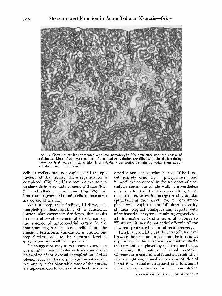
If we now look at a section of the renal cortex stained for mitochondria from the fifty-day animal, which you will remember by ordinary histologic stain appeared essentially normal, we see a very considerable number of irregular islands of tubule cross sections with regenerated epithelium standing out in clear areas as a result of their mitochondrial deficiency. (Fig. 23.) At a higher magnification it is seen that in the tubules which still remain lined by an immature epithelium the cells contain none of the intra-
Structure and Function in Acute Tubular Necrosis-Oliver
FIG. 23. Cortex of rat kidney stained with iron hematoxylin fifty days after standard dosage of sublimate. Most of the cross sections of proximal convolution are filled with the dark-staining mitochondrial rodlets. Lighter islands of tubular cross section remain in which these intra- cellular structures are absent.
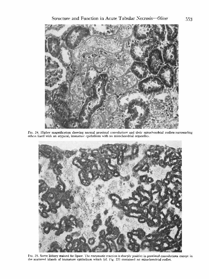
cellular rodlets that so completely fill the epi- thelium of the tubules where regeneration is completed. (Fig. 24.) If the sections are stained to show their enzymatic content of lipase (Fig. 25) and alkaline phosphatase (Fig. 26), the immature regenerated tubule cells in these areas are devoid of enzyme.
We can accept these findings, I believe, as a morphologic demonstration of a functional intracellular enzymatic deficiency that results from an observable structural defect, namely, the absence of mitochondrial organs in the immature regenerated renal cells. Thus the functional-structural correlation is pushed one step further back to the elemental level of enzyme and intracellular organella.
This suggestion may seem to some so much an oversimplification as to derive from a somewhat naive view of the dynamic complexities of vital phenomena, but the morphologist by nature and training is, in the charitable sense of the phrase, a simple-minded fellow and it is his business to
describe and believe what he sees. If he is not yet entirely clear how “phosphatase” and “lipase” are concerned in the transport of elec- trolytes across the tubule wall, it nevertheless may be admitted that the ever-shifting struc- tural patterns he sees in the regenerating tubular epithelium as they slowly evolve from amor- phous cell complex to the full-blown maturity of their original configuration, replete with mitochondrial, enzymes-containing organelles- all this makes at least a series of pictures to “illustrate” if they do not entirely “explain” the slow and protracted course of renal recovery.
This final correlation at the intracellular level between the structural aspect and the functional expression of tubular activity emphasizes again the essential part played by relative time factors in shaping the pattern of renal recovery. Glomerular structural and functional restitution is, one might say, immediate to the restitution of blood flow; tubular structural and functional recovery require weeks for their completion
AMERICAN JOURNAL OF MEDICINE
FId othl
FIG
the
Structure and Function in Acute Tubular Necrosis-Oliver
24. Higher magnification showing normal proximal convolutions and their mitochondrial rodlets surror srs lined with an atypical, immature epithelium with no mitochondrial organelles.
ending
25. Same kidney stained for lipase. The enzymatic reaction is sharply positive in proximal convolutions exe scattered islands of immature epithelium which (cf. Fig. 23) contained no mitochondrial rodlet.
:ept in
554 Structure and Function in Acute Tubular Necrosis-Oliver
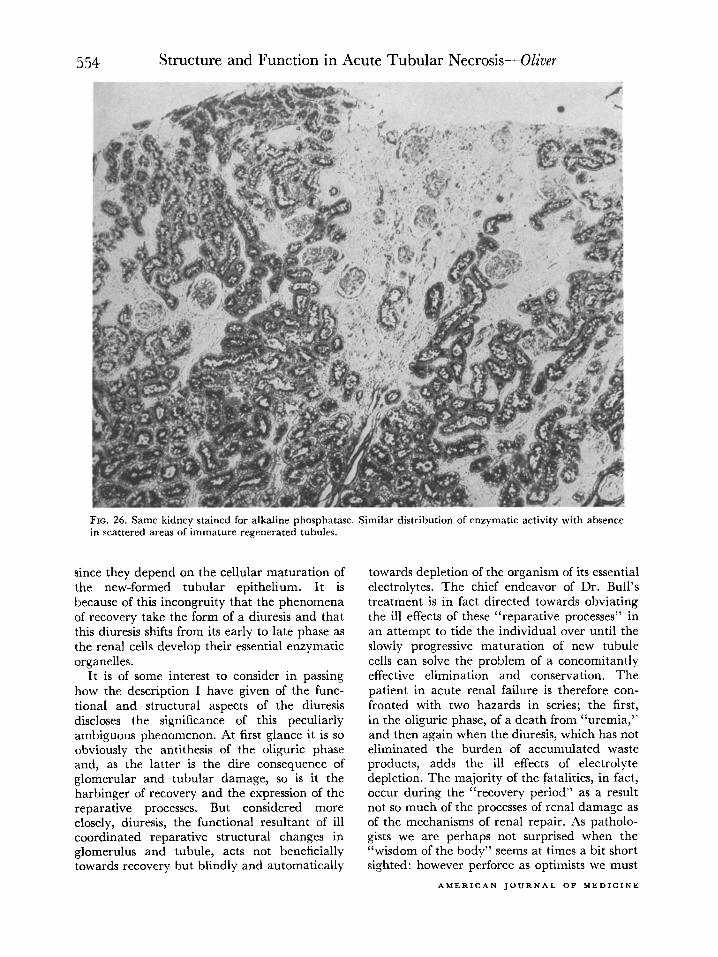
FIG. 26. Same kidney stained for alkaline phosphatase. Similar distribution of enzymatic activity with absence in scattered areas of immature regenerated tubules.
since they depend on the cellular maturation of the new-formed tubular epithelium. It is because of this incongruity that the phenomena of recovery take the form of a diuresis and that this diuresis shifts from its early to late phase as the renal cells develop their essential enzymatic organelles.
It is of some interest to consider in passing how the description I have given of the func- tional and structural aspects of the diuresis discloses the significance of this peculiarly ambiguous phenomenon. At first glance it is so obviously the antithesis of the oliguric phase and, as the latter is the dire consequence of glomerular and tubular damage, so is it the harbinger of recovery and the expression of the reparative processes. But considered more closely, diuresis, the functional resultant of ill coordinated reparative structural changes in glomerulus and tubule, acts not beneficially towards recovery but blindly and automatically
towards depletion of the organism of its essential electrolytes. The chief endeavor of Dr. Bull’s treatment is in fact directed towards obviating the ill effects of these “reparative processes” in an attempt to tide the individual over until the slowly progressive maturation of new tubule cells can solve the problem of a concomitantly effective elimination and conservation. The patient in acute renal failure is therefore con- fronted with two hazards in series; the first, in the oliguric phase, of a death from “uremia,” and then again when the diuresis, which has not eliminated the burden of accumulated waste products, adds the ill effects of electrolyte depletion. The majority of the fatalities, in fact, occur during the “recovery period” as a result not so much of the processes of renal damage as of the mechanisms of renal repair. As patholo- gists we are perhaps not surprised when the “wisdom of the body” seems at times a bit short sighted: however perforce as optimists we must
AMERICAN JOURNAL OF MEDICINE
Structure and Function in Acute Tubular Necrosis-Oliver 555
believe that in the evolutionary long run doubt- ruption of a progressive continuum into neatly less Nature knows best or we should not be disarticulated parts and the stowing of them, discussing the matter now. each into its proper compartment, but also
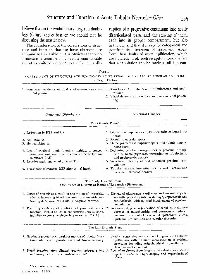
The consideration of the correlations of struc- in the demand that it makes for categorical and ture and function that we have observed are oversimplified terseness of statement. Apart summarized in Table I. It is obvious that such from these faults of oversimplification, which Procrustean treatment involved a considerable are inherent in all such recapitulations, the fact use of expository violence, not only in its dis- that a tabulation can be made at all is a con-
TABLE 1
CORRELATIONS OFSTRUCTURE AND FUNCTION IN ACUTE RENAL FAILURE (ACUTE TUBULAR NECROSIS)
Etiologic Factors
1. Functional evidence of dual etiology-ischemia and / 1. Two types of tubular lesion-tubulorhexic and neph- renal poison rotoxic
2. Visual demonstration of focal ischemia in renal poison- ing
5.
6.
Functional Disturbances
The Oliguric Phase *
Structural Changes
Reduction in RBF and GF
Albuminuria Hemoglobinuria
1. Glomerular capillaries empty with tufts collapsed but intact
1 2. Protein in capsular space i 3. Heme pigments in capsular space and tubule lumens,
heme casts Loss of proximal tubule function; inability to concen- ~ 4. Beginning tubular damage-lack of proximal absorp-
trate urea and creatinine, to conserve electrolyte and tion of heme pigments, increasing to tubulorhexic to extract PAH and nephrotoxic necrosis
Relative maintenance of glucose Tm 5. Structural integrity of first one-third proximal con- volution
Persistence of reduced RBF after initial insult ~ 6. Tubular leakage, interstitial edema and reaction and increased intrarenal tension
The Early Diuretic Phase Occurrence of Diuresis as Result of Reparative Phenomena
I Onset of diuresis as a result of absorption of interstitial 1. Distended glomerular capillaries and normal appear-
edema, increasing blood flow and filtration with con- I ing tufts; persisting tubular damage, nephrotoxic and tinuing depression of tubular absorption of water tubulorhexic, with especial involvement of proximal
convolution Persisting evidence of abolition of proximal tubule 2. Extensive atypical regeneration of renal epithelium-
function (lack of ability to concentrate urea in urine, inability to conserve electrolyte or extract PAH.) /
absence of mitochondria and consequent reduced enzymatic content of new renal epithelium; excess epithrlial proliferation and tubular dilatation
~__~. _~ ~~_~ ~~. The Late Diuretic Phase
_ ._~ .~ _.. ~_~ ~
Gradual recovery over weeks or months of tubular func- , 1. Slowly progressive maturation of regenerated tubular tional ability with possible eventual clinical recovery epithelium with ultimate reconstitution of original
structures including mitochondrial organelles with their enzymatic content
Renal function after clinical recovery adequate but 2. Loss of nephrons from irreparable tubulorhexic dam- remaining below lower limits of normal9 age and associated hypertrophy and hyperplasia of
others
* See footnote on page 542
OCTOBER, 1953
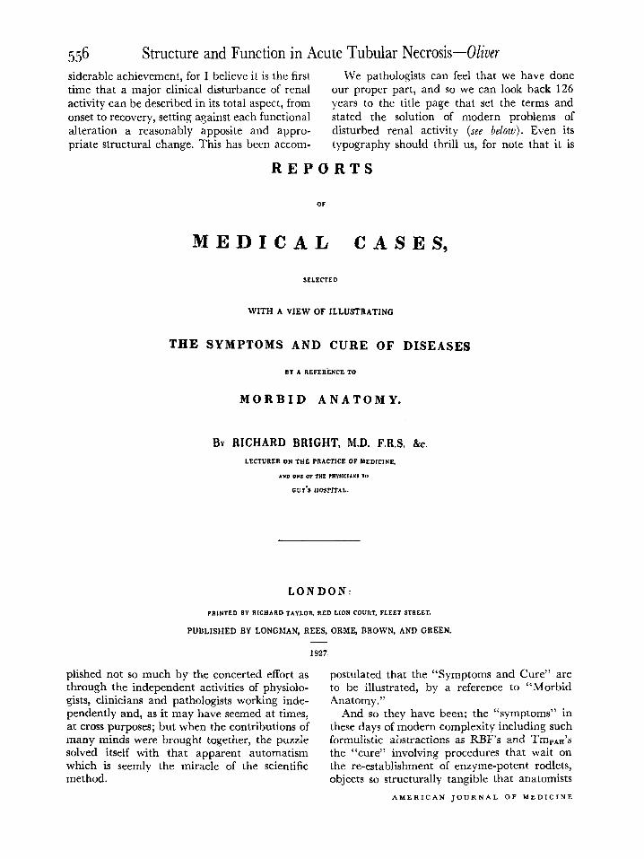
556 Structure and Function in Acute Tubular Necrosis-Oliver siderable achievement, for I believe it is the first We pathologists can feel that we have done time that a major clinical disturbance of renal our proper part, and so we can look back 126 activity can be described in its total aspect, from years to the title page that set the terms and onset to recovery, setting against each functional stated the solution of modern problems of alteration a reasonably apposite and appro- disturbed renal activity (see below). Even its priate structural change. This has been accom- typography should thrill us, for note that it is
REPORTS
OF
MEDICAL CASES,
SELECTED
WITH A VIEW OF ILLUSTRATING
THE SYMPTOMS AND CURE OF DISEASES
BY A REFEEbNCE TO
MORBID ANATOMY.
BY RICHARD BRIGHT, M.D. F.R.S. &c.
LECTURER ON THE PRACTICE OF MEDICINE.
A”D DIE 01 m ,RYSm*P, m
GUY’S IIDSPITAL.
LONDON:
PRlNTED BY RICBARD TAYLOR. RED ‘ION CODnT. FLEET STREET.
PUBLISHED BY LONGMAN, REES, ORME, BROWN, AND GREEN. -
1927.
plished not so much by the concerted effort as through the independent activities of physiolo- gists, clinicians and pathologists working inde- pendently and, as it may have seemed at times, at cross purposes; but when the contributions of many minds were brought together, the puzzle solved itself with that apparent automatism which is seemly the miracle of the scientific method.
postulated that the “Symptoms and Cure” are to be illustrated, by a reference to “Morbid Anatomy.”
And so they have been; the “symptoms” in these days of modern complexity including such formuhstic abstractions as RBF’s and TmFAH’s the “cure” involving procedures that wait on the re-establishment of enzyme-potent rodlets, objects so structurally tangible that anatomists
AMERICAN JOURNAL OF MEDICINE
Structure and Function in Acute Tubular Necrosis-Oliver 557
have collected and biochemists weighed and analysed them.
Apparently, therefore, we have come safely out of that phantasmagorical Looking Glass world through which the functional transcendentalists led us, showing us strange visions of things working by means other than through the operation of their physical constitution. * As pathologists we were perhaps never greatly disturbed by such fantasies, but we did have to listen to considerable hortatory expostulation that we “come out of the dead-house” or that we be “more dynamically functional”! If our contribution to the general effort seemed meagre at time, it was not more functional activity on our part that was needed but, and I use the old word gladly, more morbid anatomy. Today, with the new tools that the physicists and biochemists have prepared for us, we can deliver this ever essential commodity in a new and dynamic aspect.
So we may feel confirmed in our belief that the method we are using is adequate and that the way we are following leads straight. From time immemorial that method has been observa- tion and experiment; that way, a correlative fusion of what, appearing as a duality, is in fact unity, the disease process in its structural and functional aspects. These were the “grand old traditions of the classical curriculum” in the pathology of Gerhard’s time; and what is pathology today, if it he not informed with their spirit and attributes?
* SCHLAYER, K. R. Sic! “Der eine Fundamentalsatz nach dieser Richtung lautet: die Nierenfunktion an sich ist in ihrer Vergnderung unabhlngig von der anatomischen Art der Erkrankung.” Beitr. zur med. Klin., 8: 211, 1912.
REFERENCES
1. LUCK& B. Lower nephron nephrosis (renal lesions of the crush syndrome, of burns, transfusions and
other conditions affecting lower segment of nephrons). Mil. Surgeon, 99: 371, 1946.
2. SMITH, H. The Kidney; Structure and Function in Health and Disease. New York, 1951. Oxford Univ. Press.
3. BULL, G. M., JOEKES, A. M. and LOWE, K. G. Renal function studies in acute tubular necrosis. Clin. SC., 9: 379, 1950.
4. OLIVER, J., MACDOWELL, M. and TRACY, A. The pathogenesis of acute renal failure associated with traumatic and toxic injury. Renal ischemia, nephrotoxic damage and the ischemuric episode. J. Clin. Investigation, 30: 1305, 1951.
5. WALKER, A. M., BOTT, P. A., OLIVER, J. and MACDOWELL, M. The collection and analysis of fluid from single nephrons of the mammalian kidney. Am. J. Physiol., 134: 562, 1941.
6. DUNN, J. S., GILLESPIE, M. and NIVEN, J. Renal lesions in two cases of crush syndrome. Lancet, 2: 549, 1941.
7. SUSUKI, T. Zur Morphologie der Nierensekretion unter physiologischen und pathologischen Bedin- gungen. Jena, 1912. Fisher.
8. SCHLEGEL, J. U. Demonstration of blood vessels and lymphatics with a fluorescent dye in ultraviolet light. Anat. Rec., 105: 433, 1949.
9. LOWE, K. G. The late prognosis in acute tubular necrosis: an interim follow-up report on 14 pa- tients. Lancet, 1: 1086, 1952.
10. OLIVER, J. A further study of the regenerated epi- thelium in chronic uranium nephritis. An anatomical investigation of its function. J. Exper. Med., 23: 301, 1916.
11. OLIVER, J., BLOOM, F. and MACDOWELL, M. Structural and functional transformations in the tubular epithelium of the dogs kidney in chronic Bright’s disease and their relation to mechanisms of renal compensation and failure. J. &per. Med., 73: 141, 1941.
12. SCHNEIDER, W. C. and HOGEBOOM, G. H. Cyto- chemical studies of mammalian tissues: the isola- tion of cell components by differential centrifuga- tion: a review. Cancer Research, 11: 1, 1951.
13. KABAT, E. A. and FURTH, J. A histochemical study of the distribution of alkaline phosphatase in various normal and neoplastic tissues. Am. J. Path., 17: 393, 1941.
14. WACHSTEIN, M. Influence of experimental kidney damage on histochemically demonstrable lipase activity in the rat. Comparison with alkaline phosphatase activity. J. &per. Med., 84: 25, 1946.
OCTOBER, 1953