Leptomeningeal involvement in prostate cancer: a case report
Upload
dominic-greerCategory
view
218download
3
Correlation of Leptomeningeal Disease on MRI Between the Brain and Spine in Patients Presenting to a Tertiary Referral Center
Poster #: EP-47 Control #: 1681

1Syed S. Hashmi, 2J. Matthew Debnam, 3Dima Suki, 4Rory R. Mayer, 2T Linda Chi,
2Leena Ketonen, 3Jeffrey S. Weinberg, 2Nandita Guha-Thakurta
1Department of Diagnostic Radiology, The University of Texas, Houston, TX; 2Department of Diagnostic Radiology, Section of
Neuroradiology, 3Department of Neurosurgery, The University of Texas MD Anderson Cancer Center, Houston, TX; 4Department
of Neurosurgery, Baylor College of Medicine, Houston, TX

• The Institutional Review Board approved this study and waived the requirement for informed consent.
• Data acquisition was performed in compliance with all applicable Health Insurance Portability and Accountability Act regulations.
• No Disclosures

Leptomeningeal disease (LMD)Background
• Spread of cancer cells within the leptomeningeal space in brain and/or spine
• Associated with poor prognosis, increased mortality rates
• Incidence of LMD has been increasing– now occurring in 3-8% of patients with cancer

Leptomeningeal disease (LMD)Background
• Cytological evaluation remains the gold standard for diagnosis, however,...
• MRI is often used in the assessment of patients with suspected LMD involving the brain and spine

Purpose of Study
• To determine if there is a correlation between findings in the brain and spinal canal on MRI in patients with LMD

Materials & Methods
• Inclusion criteria • MRI brain with contrast• MRI spine with contrast• CSF cytology documenting the
presence of LMD

Materials & Methods
• Total cases reviewed – 295• 157 patients met the inclusion criteria • MRI brain review
– consensus by 4 neuroradiologists• MRI of the spine review
– consensus by 2 neuroradiologists
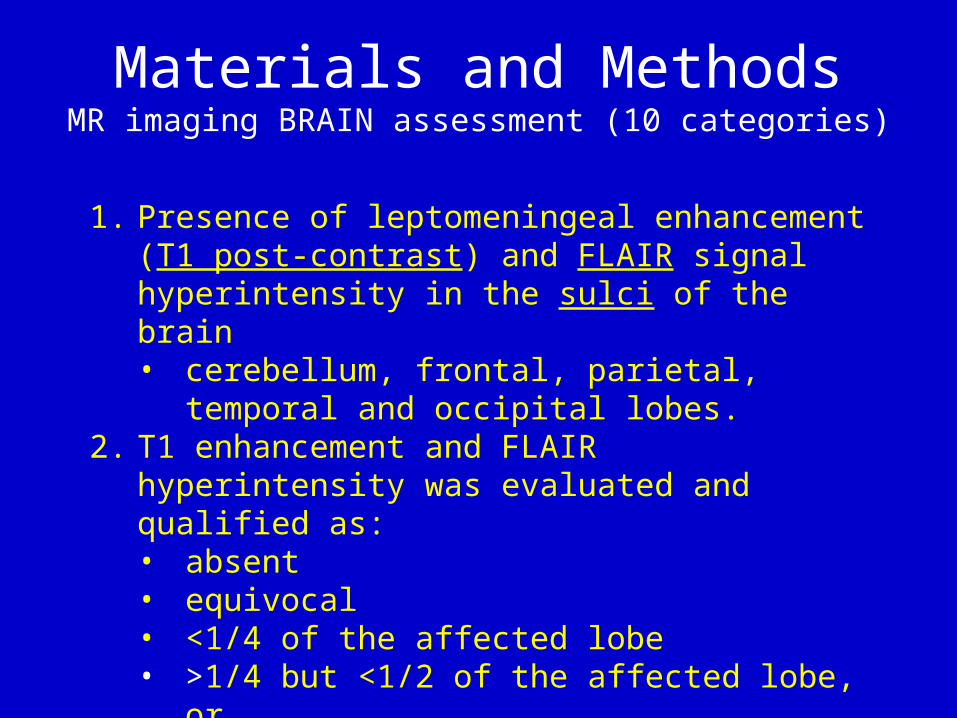
Materials and MethodsMR imaging BRAIN assessment (10 categories)
1. Presence of leptomeningeal enhancement (T1 post-contrast) and FLAIR signal hyperintensity in the sulci of the brain • cerebellum, frontal, parietal, temporal and
occipital lobes.2. T1 enhancement and FLAIR hyperintensity was
evaluated and qualified as:• absent • equivocal • <1/4 of the affected lobe• >1/4 but <1/2 of the affected lobe, or • >1/2 of the affected lobe

Materials and MethodsBRAIN MR imaging assessment (10 categories)
3. Ependymal enhancement in the ventricular system • linear • nodular, or • mixed
4. Number of parenchymal lesions• solitary • 2-5 lesions, or • >5 lesions
5. Size of the largest parenchymal metastasis 6. ± vasogenic edema7. Related mass effect

Materials and MethodsBRAIN MR imaging assessment (10 categories)
8. Cranial nerve enhancement• CN III• CN V• Meckel’s cave• CN VII/VIII
9. Basal cisterns enhancement10. Pituitary stalk enhancement

Materials and MethodsSPINE MR imaging assessment
• T1 enhancement was qualified as one or more of the following:1. thin and linear2. thick and linear3. nodular 4. diffuse filling the spinal canal

Statistical Analysis
• Frequencies and descriptive statistics on the various entities under study were obtained.• Chi square test or the Fisher exact test• univariate and multivariate logistic
regression analysis• Odds ratios and their 95% confidence
intervals were obtained• A multivariate model was utilized if a critical
p value was 0.05

ResultsPatient demographics
• 157 patients positive for LMD on cytology• 88 women: 69 men• age 9-82 (mean age 51 ± 13.2 years)
• For patients < 36.5 years:• 11 (41%) patients were positive for spinal LMD• 16 (59%) patients were positive for spinal LMD
• For patients ≥ 36.5 years• 36 (28%) patients were positive for spinal LMD• 94 (72%) patients were positive for spinal LMD
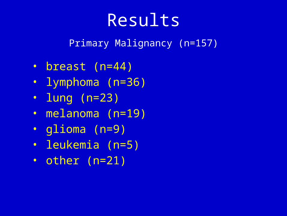
ResultsPrimary Malignancy (n=157)
• breast (n=44) • lymphoma (n=36)• lung (n=23)• melanoma (n=19)• glioma (n=9)• leukemia (n=5)• other (n=21)

ResultsBrain –T1 post contrast enhancement in each lobe
None Equivocal Less than one quarter
one quarter to one half
greater than one half
Combined 68 16 43 14 16
Right cerebellum 95 18 26 8 10
Left cerebellum 93 18 27 10 9
Right occipital 101 22 24 2 8
Left occipital 110 17 20 2 8
Right parietal 121 7 20 4 5
Left parietal 128 7 14 4 4
Right temporal 128 5 17 4 3
Left temporal 127 9 15 2 4
Right frontal 126 11 14 3 3
Left frontal 130 9 12 3 4
Axial T1 postcontrast:A, LMD in the sulci of the frontal and parietal lobes
B, LMD in the cerebellar folia
A
B

ResultsBrain – T2/FLAIR hyperintensity in each lobe
None Equivocal Less than one quarter
one quarter to one half
greater than one half
Combined 61 22 35 16 23
Right cerebellum 92 12 29 11 13
Left cerebellum 92 12 30 11 13
Right occipital 91 19 25 9 13
Left occipital 89 22 24 8 14
Right parietal 107 11 23 4 12
Left parietal 115 9 15 7 11
Right temporal 122 5 18 6 6
Left temporal 124 7 13 7 6
Right frontal 123 9 17 1 7
Left frontal 128 7 13 2 7
Axial T2/FLAIRA, LMD in the sulci of the frontal and parietal lobes
B, LMD in the cerebellar folia
A
B

ResultsBrain LMD-Cranial nerve involvement
• right CN III (n=6), left CN III (n=5) • right CNV (n=16), left CN V (n=14) • right Meckel’s cave (n=15) • left Meckel’s cave (n=12)
A. CN III B. CN V C. Meckel’s cave
A B C

ResultsBrain LMD-Cranial nerve involvement
• right CN VII/VIII (n=34), left CN VII/VII (n=28)• basal cisterns (n=28)• pituitary stalk (n=7) • some patients exhibited more than one site
involvement
A. CN VII/VIII B, basal cisterns C. pituitary stalk
CBA

ResultsBrain LMD-Other sites of disease involvement
• Ependymal enhancement: 28 patients (17.8%) • linear (n=16), nodular (n=4), or mixed (n=8).
• Parenchymal metastasis: 61 patients (38.8%) • Number: solitary (n=21), between 2-5 (n=25), or >5 (n=15) • Size: <10 mm (n= 26), between 10 and <20 mm (n=18), greater or equal to
20 mm (n=17)• Vasogenic edema present: 56 patients (35.7%) • Mass effect present: 40 patients (25.5%)
A. ependymal B. parenchymal met C. Vasogenic edema/ mass effect
CBA

ResultsSpinal canal -Leptomeningeal enhancement
• 110 of 157 patients (70.1%) • thin/linear (n=55)• thick (n=36)• nodular (n=15)• diffuse (n=5)
• Patients may have exhibited more than one type of enhancement
A. thin linear B, thick linear C. nodular D. diffuse
A B C D

Results-Statistics
• gender (n=0.9) • tumor type (0.85)• ependymal enhancement (p=0.78)• parenchymal mass
• presence of a mass (p=0.2)• size (p=0.22)• vasogenic edema (p=0.24) • associated mass effect (p=0.68)
No significant association was noted between spinal leptomeningeal enhancement and….

Results-StatisticsNo significant association was noted between brain parenhcymal enhancement and spinal leptomeningeal enhancement
T1 post contrast T2/FLAIRp value p value
Right cerebellum 0.57 0.27Left cerebellum 0.38 0.18
Right occipital 0.24 0.06Left occipital 0.11 0.06
Right parietal 0.90 0.48Left parietal 0.76 0.02
Right temporal 0.26 0.46Left temporal 0.44 0.53Right frontal 0.35 0.34
Left frontal 0.49 0.41
T2/FLAIR hyperintensity in the occipital lobe closely approached and in the left parietal lobe was significantly associated with spinal canal leptomeningeal enhancement

Results-Statistics
• All cranial nerves combined: approached significance (p=0.09)
• right CN III (p=0.18)• left CN III (p=0.32)• right Meckel’s cave (p=0.56)• left Meckel’s cave (p=0.51
• pituitary stalk (p=0.43)
Relation between cranial nerve involvement with LMD and spinal LMD

Results-Statistics
• All cranial nerves combined approached significance (p=0.09)
• Significant association• right CNV (p=0.003) • left CNV (p=0.01)• right CNVII/VIII (p=0.01) • left CN VII/VIII (p=0.01)
• Basal cistern (0.17)
Relation between cranial nerve involvement with LMD and spinal LMD
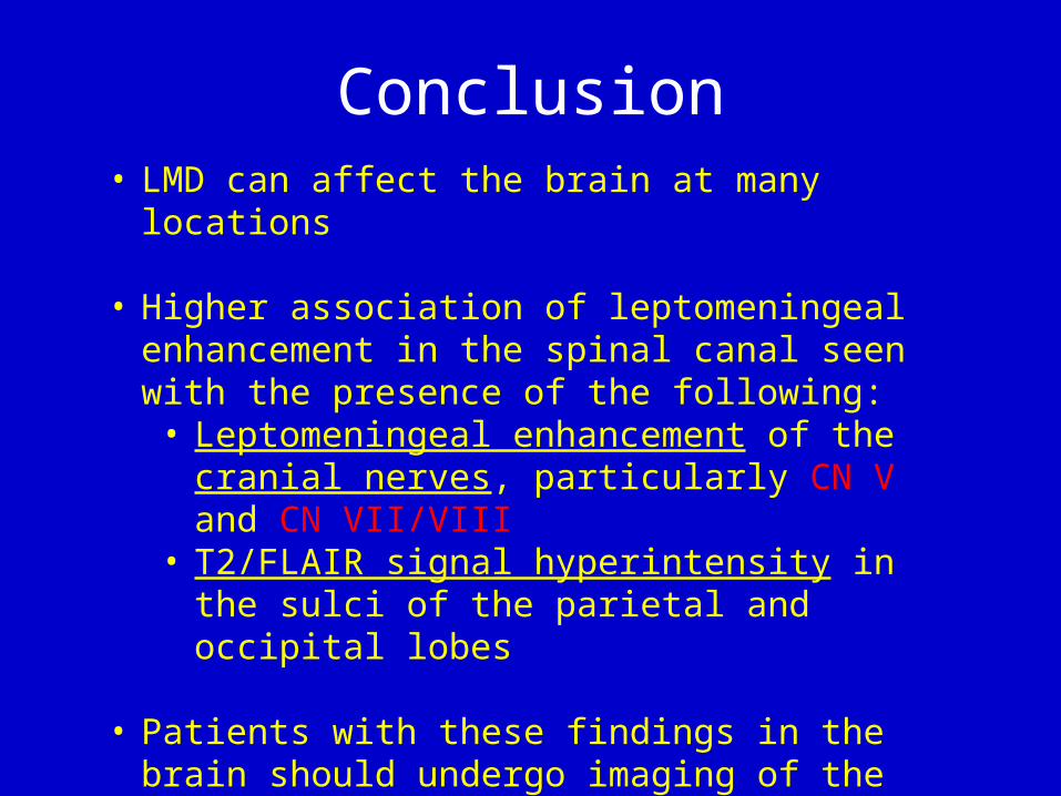
Conclusion• LMD can affect the brain at many locations
• Higher association of leptomeningeal enhancement in the spinal canal seen with the presence of the following:
• Leptomeningeal enhancement of the cranial nerves, particularly CN V and CN VII/VIII
• T2/FLAIR signal hyperintensity in the sulci of the parietal and occipital lobes
• Patients with these findings in the brain should undergo imaging of the spine to assess or spinal LMD