Corporate Structureimagingsystemsspecialists.com/uploads/products/ziehm/Corp...1 Headline Ziehm...
56
Corporate Structure 0
-
Upload
nguyenliem -
Category
Documents
-
view
222 -
download
2
Transcript of Corporate Structureimagingsystemsspecialists.com/uploads/products/ziehm/Corp...1 Headline Ziehm...

Corporate Structure
0
1
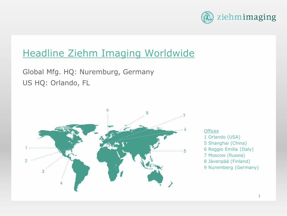
Headline Ziehm Imaging Worldwide
Global Mfg. HQ: Nuremburg, Germany
US HQ: Orlando, FL
Offices
1 Orlando (USA)
5 Shanghai (China)
6 Reggio Emilia (Italy)
7 Moscow (Russia)
8 Jävenpää (Finland)
9 Nuremberg (Germany)
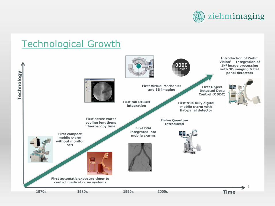
Technological Growth
2
First DSA integrated into mobile c-arms
First compact mobile c-arm
without monitor cart
First active water cooling lengthens fluoroscopy time
First full DICOM integration
First true fully digital mobile c-arm with flat-panel detector
First Virtual Mechanics and 3D imaging
First Object Detected Dose Control (ODDC)
Introduction of Ziehm Vision2 – Integration of 1k2 image processing with 3D imaging & flat
panel detectors
2000s 1990s 1980s 1970s
First automatic exposure timer to control medical x-ray systems
Ziehm Quantum Introduced
Tech
no
log
y
Time
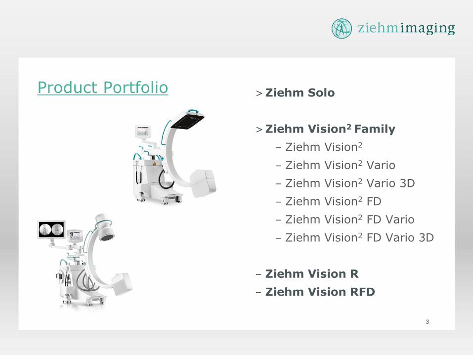
Product Portfolio >Ziehm Solo
>Ziehm Vision2 Family
– Ziehm Vision2
– Ziehm Vision2 Vario
– Ziehm Vision2 Vario 3D
– Ziehm Vision2 FD
– Ziehm Vision2 FD Vario
– Ziehm Vision2 FD Vario 3D
– Ziehm Vision R
– Ziehm Vision RFD
3
Product Overview
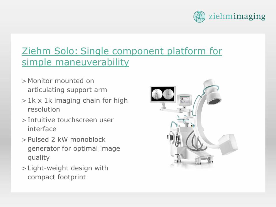
Single component platform for simple maneuverability Ziehm Solo:
>Monitor mounted on
articulating support arm
>1k x 1k imaging chain for high
resolution
> Intuitive touchscreen user
interface
>Pulsed 2 kW monoblock
generator for optimal image
quality
>Light-weight design with
compact footprint
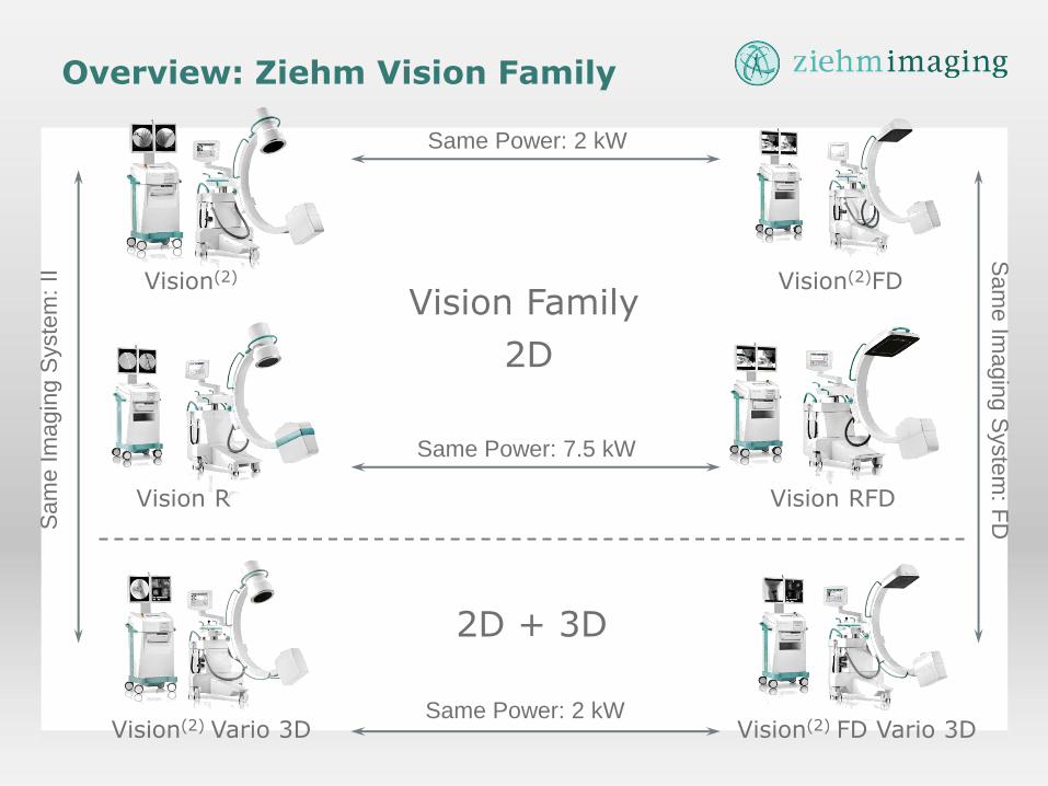
Vision Family Vision(2) Vision(2)FD
Vision RFD Vision R
Vision(2) FD Vario 3D Vision(2) Vario 3D
Overview: Ziehm Vision Family
Same Power: 2 kW
Same Power: 7.5 kW
Sam
e Im
ag
ing S
yste
m:
II
Same Power: 2 kW
Sam
e Im
ag
ing
Syste
m: F
D
2D
2D + 3D
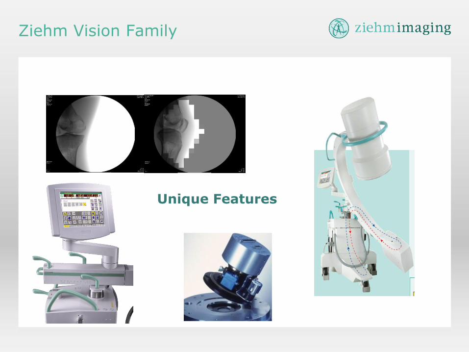
Unique Features
Ziehm Vision Family

Always Pulsing
> 1-25 pps. > (1-30 pps. For I.I. based systems)
> Conventional C-arm generators only pulse up to 8 pps unless using high dose level modes
> Delivers higher mA than real-time fluoro while limiting patient dose
> Improved anatomic visualization
> Reduced noise
> Short pulse widths can reduce blurring and motion lag when needed
> 4-30 ms., determined by unit selected and anatomic program
- For high IQ at a lower dose
Vision PulseTM Generator
Confidential - For Ziehm internal use only
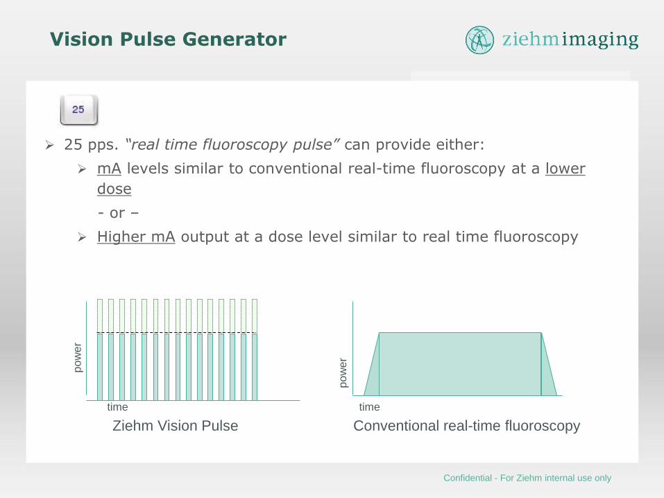
25 pps. “real time fluoroscopy pulse” can provide either:
mA levels similar to conventional real-time fluoroscopy at a lower
dose
- or –
Higher mA output at a dose level similar to real time fluoroscopy
time
Vision Pulse Generator
po
we
r
time
po
we
r
Conventional real-time fluoroscopy Ziehm Vision Pulse
Confidential - For Ziehm internal use only
10
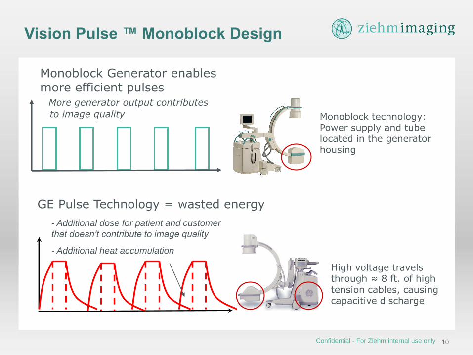
Monoblock Generator enables more efficient pulses More generator output contributes
to image quality
Vision Pulse ™ Monoblock Design
GE Pulse Technology = wasted energy
- Additional dose for patient and customer
that doesn’t contribute to image quality
- Additional heat accumulation
Monoblock technology: Power supply and tube located in the generator housing
High voltage travels through ≈ 8 ft. of high tension cables, causing capacitive discharge
Confidential - For Ziehm internal use only

11
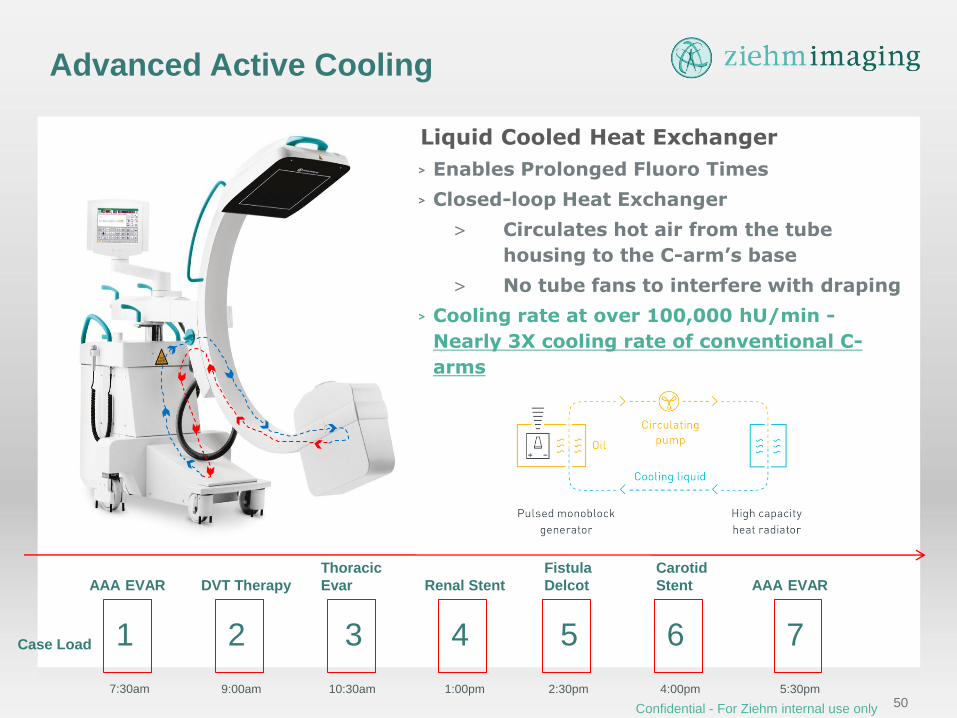
Liquid Cooled Heat Exchanger
> Enables Prolonged Fluoro Times
> Closed-loop Heat Exchanger
> Circulates hot air from the tube
housing to the C-arm’s base
> No tube fans to interfere with draping
> Cooling rate at over 100,000 hU/min -
Nearly 3X cooling rate of conventional C-
arms
Advanced Active Cooling
7:30am 9:00am 10:30am 1:00pm 2:30pm 4:00pm 5:30pm
Case Load 1 2 3 4 5 6 7
AAA EVAR DVT Therapy
Thoracic
Evar Renal Stent
Fistula
Delcot
Carotid
Stent AAA EVAR
Confidential - For Ziehm internal use only
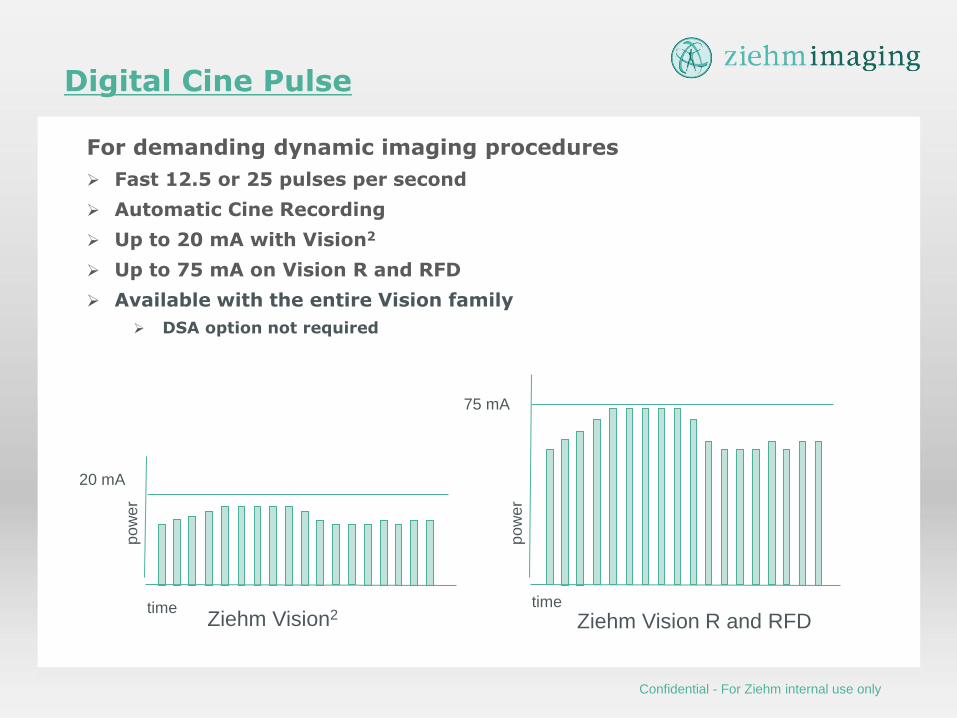
Digital Cine Pulse
For demanding dynamic imaging procedures
Fast 12.5 or 25 pulses per second
Automatic Cine Recording
Up to 20 mA with Vision2
Up to 75 mA on Vision R and RFD
Available with the entire Vision family
DSA option not required
Ziehm Vision2 time
po
we
r
75 mA
po
we
r
20 mA
Ziehm Vision R and RFD time
Confidential - For Ziehm internal use only
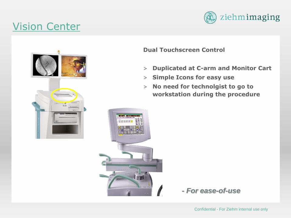
Vision Center
Dual Touchscreen Control
> Duplicated at C-arm and Monitor Cart
> Simple Icons for easy use
> No need for technolgist to go to
workstation during the procedure
- For ease-of-use
Confidential - For Ziehm internal use only
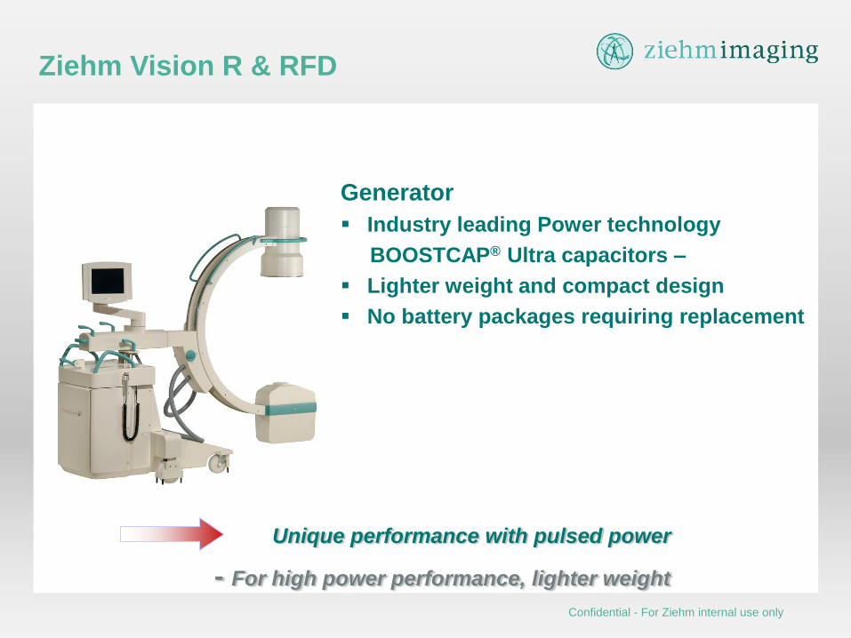
Generator
Industry leading Power technology
BOOSTCAP® Ultra capacitors –
Lighter weight and compact design
No battery packages requiring replacement
Ziehm Vision R & RFD
Unique performance with pulsed power
- For high power performance, lighter weight
Confidential - For Ziehm internal use only
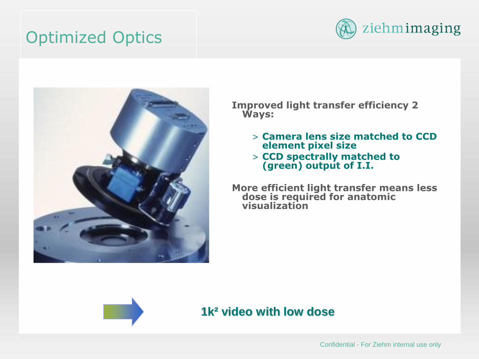
Improved light transfer efficiency 2 Ways:
> Camera lens size matched to CCD
element pixel size > CCD spectrally matched to
(green) output of I.I. More efficient light transfer means less
dose is required for anatomic visualization
Optimized Optics
1k² video with low dose
Confidential - For Ziehm internal use only
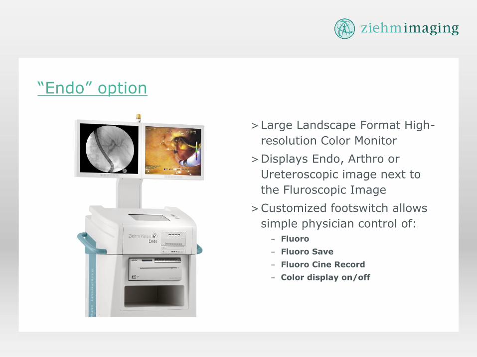
“Endo” option
>Large Landscape Format High-
resolution Color Monitor
>Displays Endo, Arthro or
Ureteroscopic image next to
the Fluroscopic Image
>Customized footswitch allows
simple physician control of:
– Fluoro
– Fluoro Save
– Fluoro Cine Record
– Color display on/off

ADCS – active S-distortion correction
package
Real-time magnetic field correction coils for
eliminating S-distortion
For 31 cm Image intensifier
Standard on Ziehm Vision & Vision R
31 cm/12“ I.I.
Without correction
Distortion correction
With correction
Confidential - For Ziehm internal use only

Ziehm Vision
ODDC Object Detected Dose Control
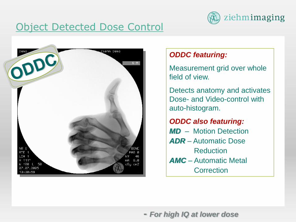
ODDC featuring:
Measurement grid over whole
field of view.
Detects anatomy and activates
Dose- and Video-control with
auto-histogram.
ODDC also featuring:
MD – Motion Detection
ADR – Automatic Dose
Reduction
AMC – Automatic Metal
Correction
Object Detected Dose Control
- For high IQ at lower dose
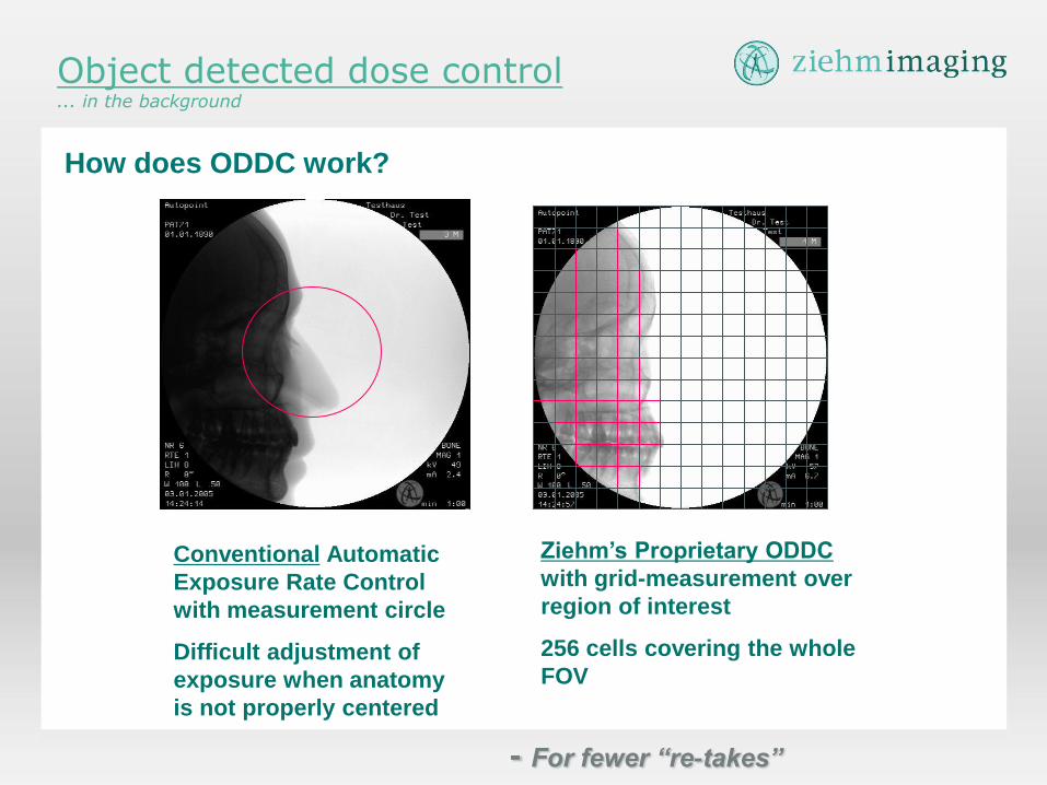
Object detected dose control ... in the background
How does ODDC work?
Ziehm’s Proprietary ODDC
with grid-measurement over
region of interest
256 cells covering the whole
FOV
Conventional Automatic
Exposure Rate Control
with measurement circle
Difficult adjustment of
exposure when anatomy
is not properly centered
- For fewer “re-takes”
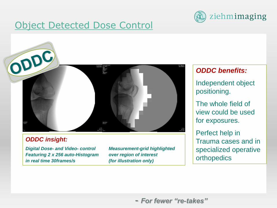
ODDC benefits:
Independent object
positioning.
The whole field of
view could be used
for exposures.
Perfect help in
Trauma cases and in
specialized operative
orthopedics
ODDC insight:
Digital Dose- and Video- control Measurement-grid highlighted
Featuring 2 x 256 auto-Histogram over region of interest
in real time 30frames/s (for illustration only)
Object Detected Dose Control
- For fewer “re-takes”
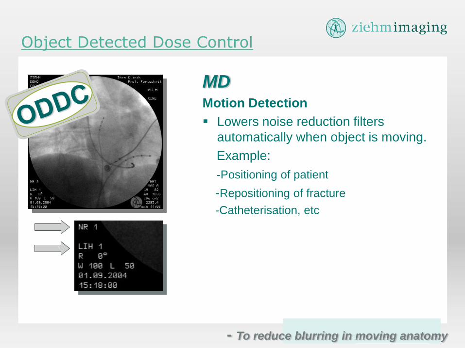
MD Motion Detection
Lowers noise reduction filters
automatically when object is moving.
Example:
-Positioning of patient
-Repositioning of fracture
-Catheterisation, etc
Object Detected Dose Control
- To reduce blurring in moving anatomy
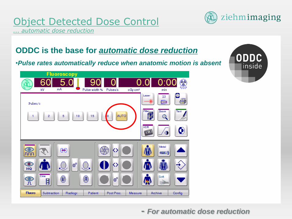
Object Detected Dose Control ... automatic dose reduction
ODDC is the base for automatic dose reduction
•Pulse rates automatically reduce when anatomic motion is absent
- For automatic dose reduction

AMC Automatic Metal Correction
Introducing real-time
interactive Metal correction for
elimination of Metal artifacts
such as blooming effect.
- For automatic adjustment to metal
Intraoperative 3D Imaging

Vision Family Vision(2) Vision(2)FD
Vision RFD Vision R
Vision(2) FD Vario 3D Vision(2) Vario 3D
Overview: Ziehm Vision Family
Same Power: 2.2 kW
Same Power: 7.5 kW
Sam
e Im
ag
ing S
yste
m:
II
Same Power: 2.2 kW
Sam
e Im
ag
ing
Syste
m: F
D
2D
2D + 3D
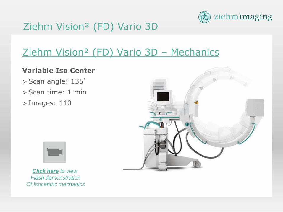
Ziehm Vision² (FD) Vario 3D – Mechanics
Variable Iso Center
>Scan angle: 135°
>Scan time: 1 min
> Images: 110
Ziehm Vision² (FD) Vario 3D
Click here to view
Flash demonstration
Of Isocentric mechanics

28
Ziehm Vision² (FD) Vario 3D
Mechanics
„Truly Isocentric C“ Ziehm Vision² (FD) Vario 3D – Variable Isocenter
29
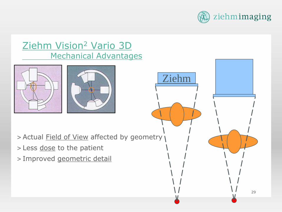
Ziehm Vision2 Vario 3D
Mechanical Advantages
>Actual Field of View affected by geometry
>Less dose to the patient
> Improved geometric detail
Ziehm
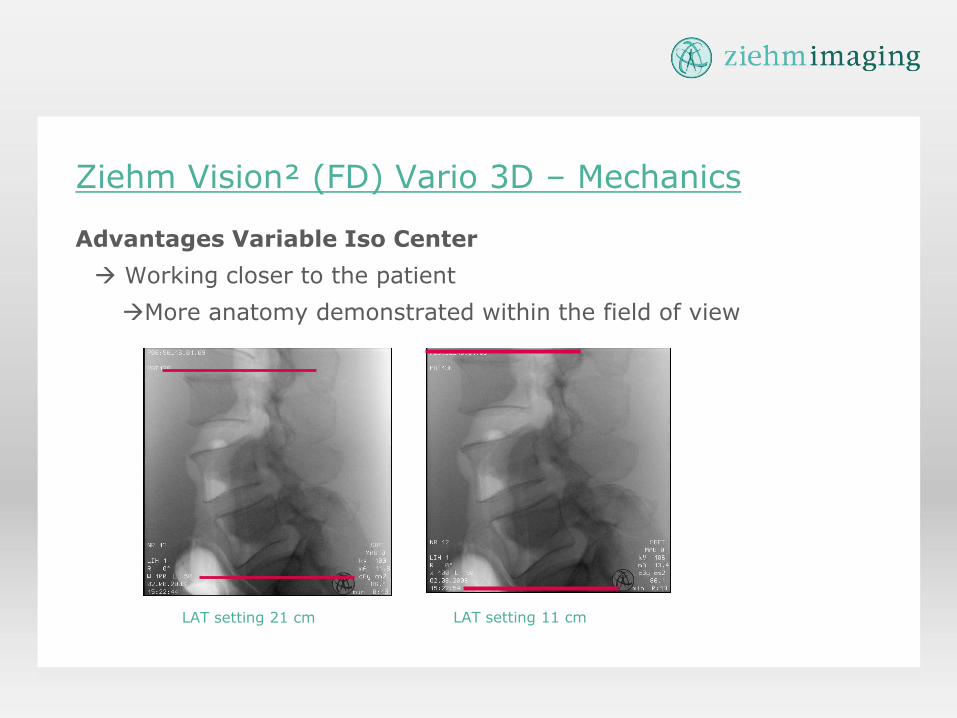
Ziehm Vision² (FD) Vario 3D – Mechanics
Advantages Variable Iso Center
Working closer to the patient
More anatomy demonstrated within the field of view
LAT setting 21 cm LAT setting 11 cm
31
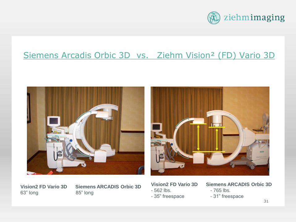
Siemens Arcadis Orbic 3D vs. Ziehm Vision² (FD) Vario 3D
Vision2 FD Vario 3D Siemens ARCADIS Orbic 3D
- 562 lbs. - 765 lbs.
- 35” freespace - 31” freespace
Vision2 FD Vario 3D Siemens ARCADIS Orbic 3D
63” long 85” long
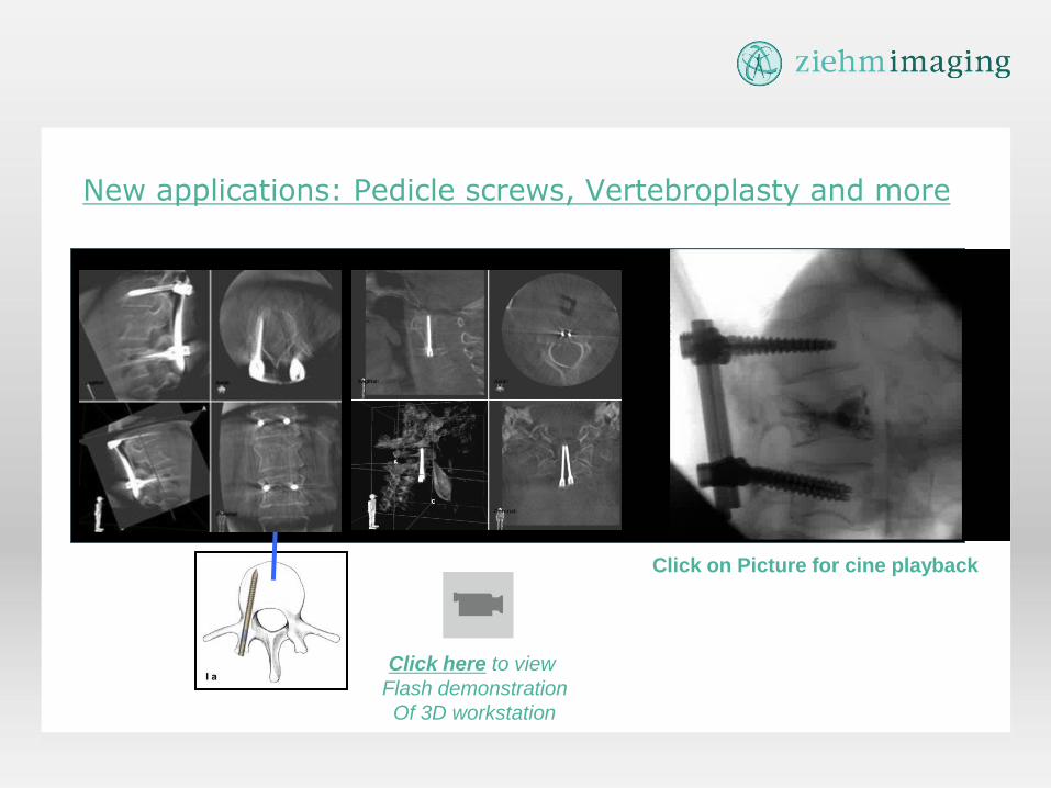
New applications: Pedicle screws, Vertebroplasty and more
Click on Picture for cine playback
Click here to view
Flash demonstration
Of 3D workstation
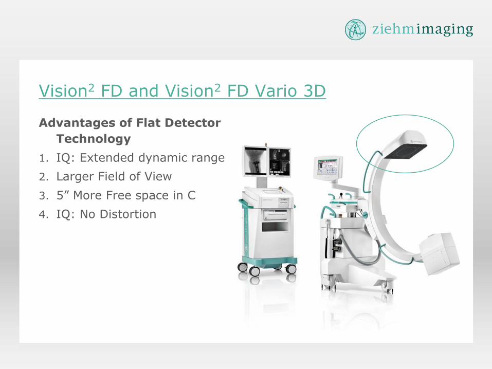
Vision2 FD and Vision2 FD Vario 3D
Advantages of Flat Detector
Technology
1. IQ: Extended dynamic range
2. Larger Field of View
3. 5” More Free space in C
4. IQ: No Distortion
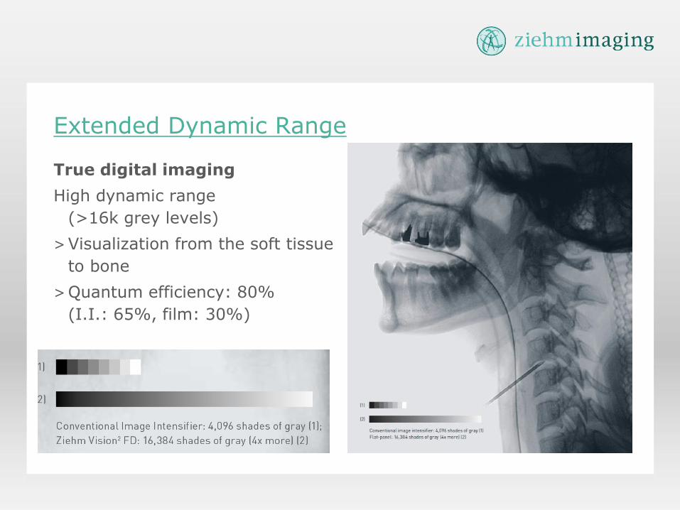
Extended Dynamic Range
True digital imaging
High dynamic range
(>16k grey levels)
>Visualization from the soft tissue
to bone
>Quantum efficiency: 80%
(I.I.: 65%, film: 30%)
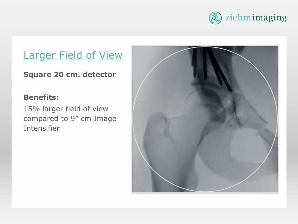
Larger Field of View
Square 20 cm. detector
Benefits:
15% larger field of view
compared to 9” cm Image
Intensifier
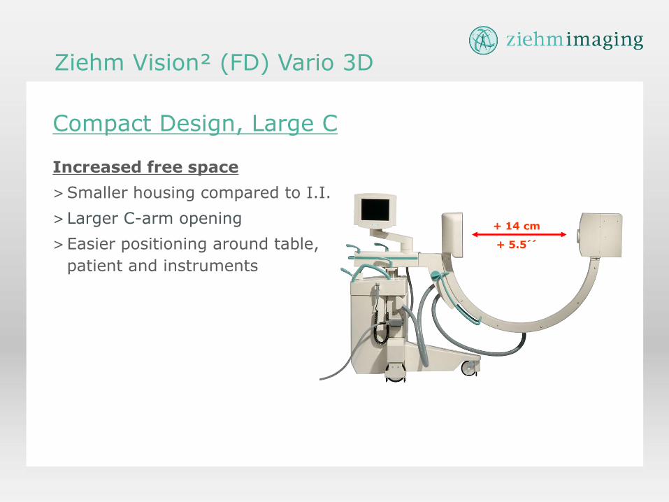
Compact Design, Large C
Increased free space
>Smaller housing compared to I.I.
>Larger C-arm opening
>Easier positioning around table,
patient and instruments
+ 14 cm
+ 5.5´´
Ziehm Vision² (FD) Vario 3D
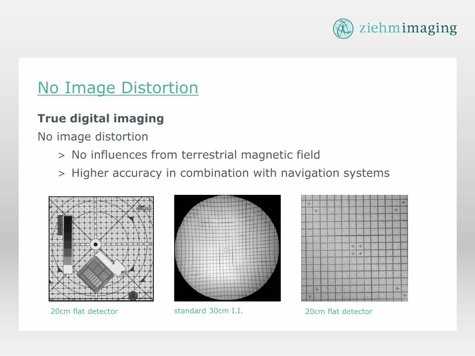
No Image Distortion
standard 30cm I.I.
True digital imaging
No image distortion
> No influences from terrestrial magnetic field
> Higher accuracy in combination with navigation systems
20cm flat detector 20cm flat detector
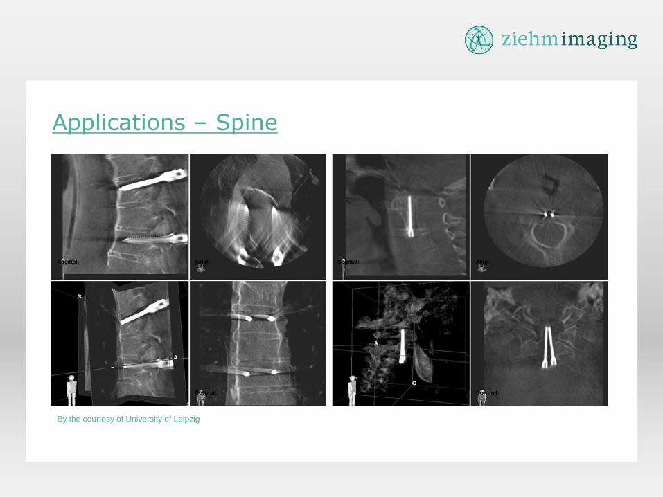
Applications – Spine
By the courtesy of University of Leipzig
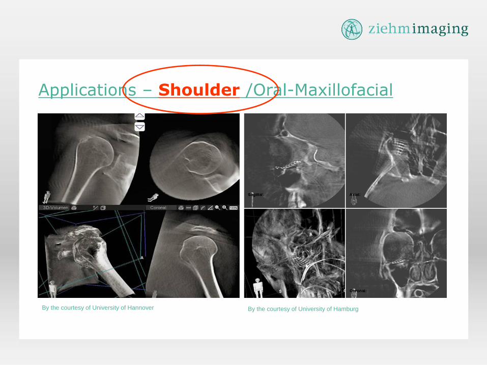
Applications – Shoulder /Oral-Maxillofacial
By the courtesy of University of Hannover By the courtesy of University of Hamburg
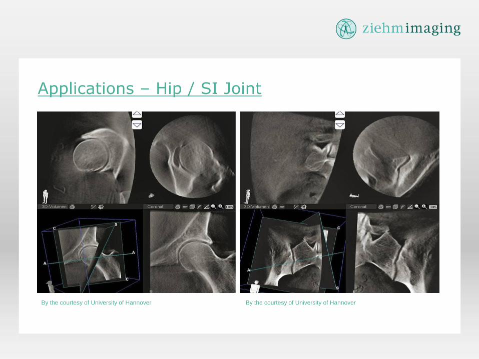
Applications – Hip / SI Joint
By the courtesy of University of Hannover By the courtesy of University of Hannover

Applications – Brachytherapy – Prostate NEW!
By the courtesy of University of Erlangen
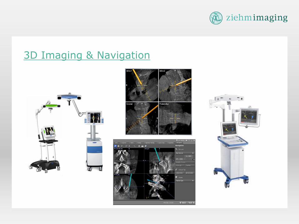
3D Imaging & Navigation
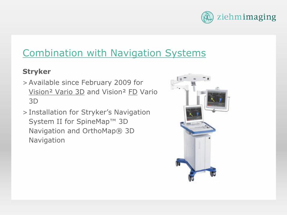
Stryker
>Available since February 2009 for
Vision² Vario 3D and Vision² FD Vario
3D
> Installation for Stryker’s Navigation
System II for SpineMap™ 3D
Navigation and OrthoMap® 3D
Navigation
Combination with Navigation Systems
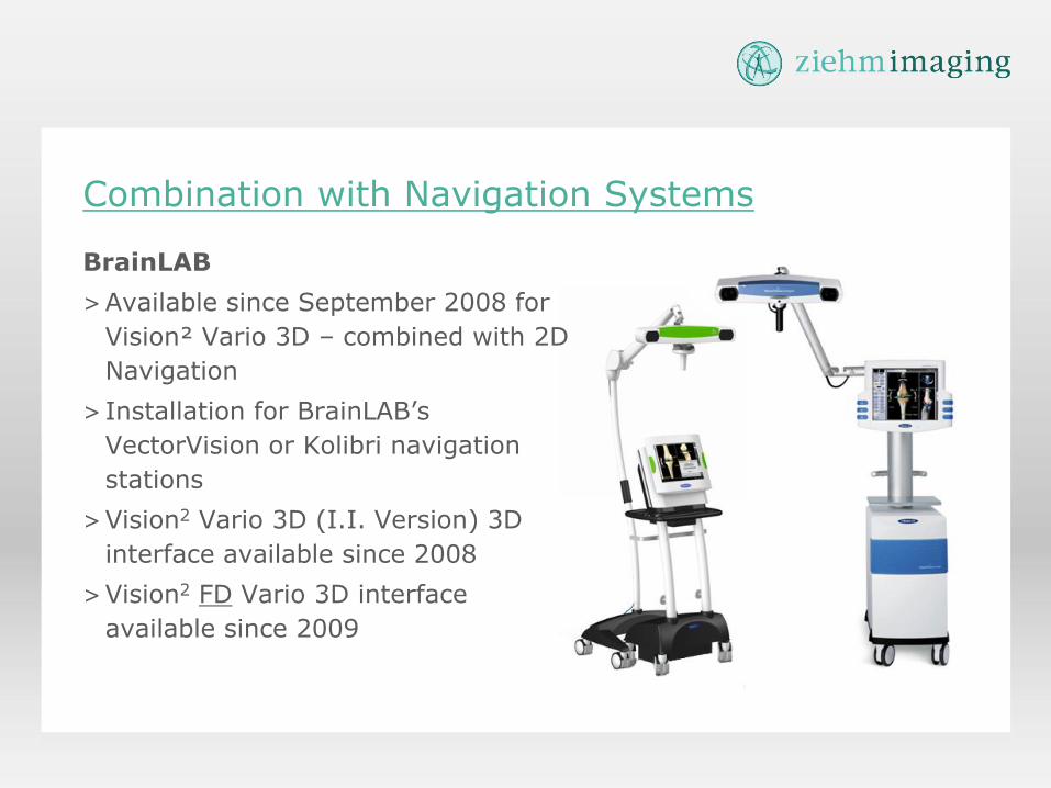
Combination with Navigation Systems
BrainLAB
>Available since September 2008 for
Vision² Vario 3D – combined with 2D
Navigation
> Installation for BrainLAB’s
VectorVision or Kolibri navigation
stations
>Vision2 Vario 3D (I.I. Version) 3D
interface available since 2008
>Vision2 FD Vario 3D interface
available since 2009
Medtronic
>Work in progress for 2D and 3D
navigation interface
Combination with Navigation Systems
Vision RFD
47
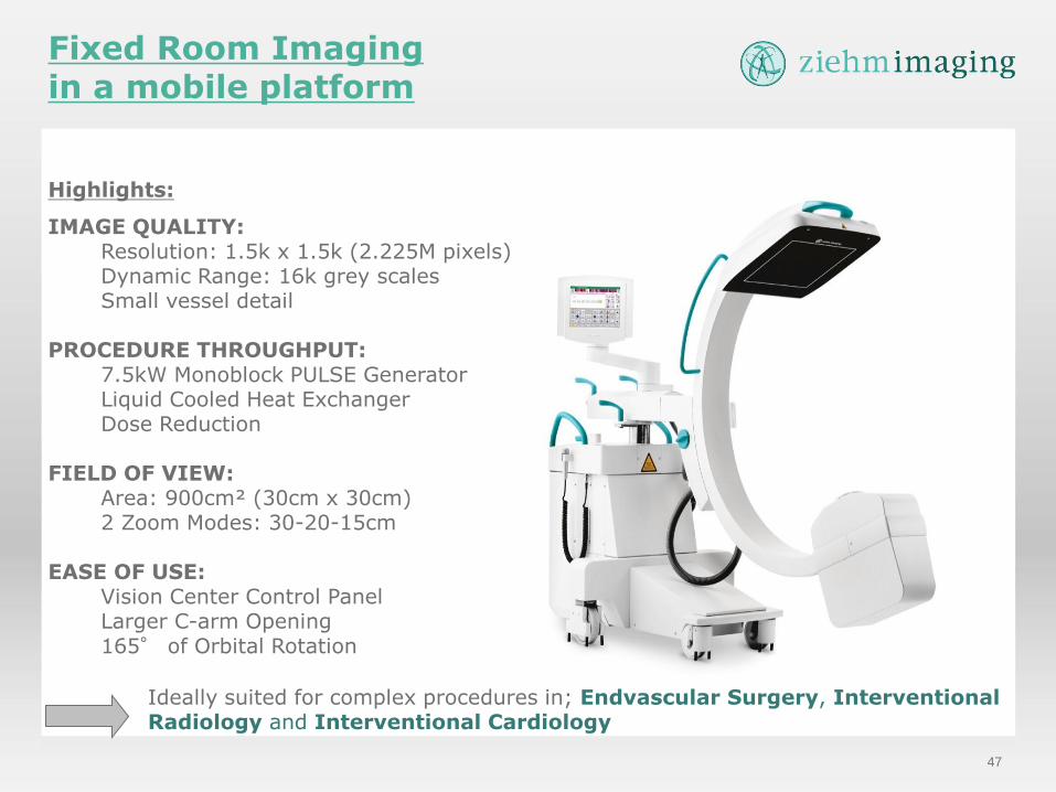
Highlights:
IMAGE QUALITY: Resolution: 1.5k x 1.5k (2.225M pixels) Dynamic Range: 16k grey scales Small vessel detail
PROCEDURE THROUGHPUT: 7.5kW Monoblock PULSE Generator Liquid Cooled Heat Exchanger Dose Reduction
FIELD OF VIEW: Area: 900cm² (30cm x 30cm) 2 Zoom Modes: 30-20-15cm
EASE OF USE:
Vision Center Control Panel Larger C-arm Opening 165° of Orbital Rotation
Ideally suited for complex procedures in; Endvascular Surgery, Interventional Radiology and Interventional Cardiology
Fixed Room Imaging in a mobile platform
48
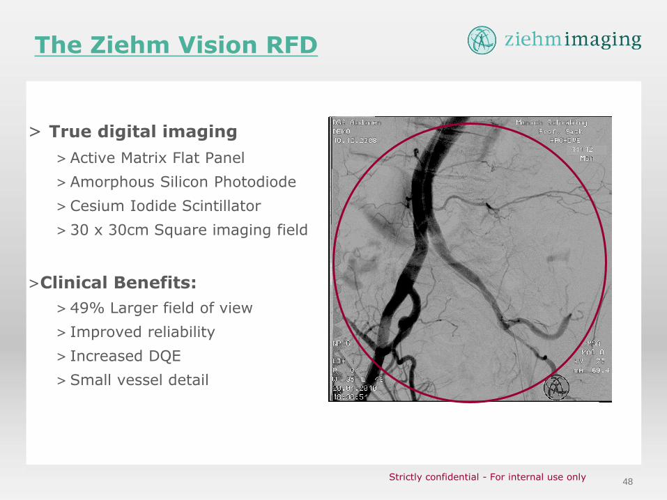
The Ziehm Vision RFD
> True digital imaging
> Active Matrix Flat Panel
> Amorphous Silicon Photodiode
> Cesium Iodide Scintillator
> 30 x 30cm Square imaging field
>Clinical Benefits:
> 49% Larger field of view
> Improved reliability
> Increased DQE
> Small vessel detail
Strictly confidential - For internal use only

49
Vision RFD – Flat Panel Image Quality
True Digital Imaging
>Crisp Clear Vascular Imaging
>Dose Quantum efficiency: 80%
(I.I.: 65%, foil: 30%)
>High dynamic range –
visualization from the soft tissue
to bone
50
Liquid Cooled Heat Exchanger
> Enables Prolonged Fluoro Times
> Closed-loop Heat Exchanger
> Circulates hot air from the tube
housing to the C-arm’s base
> No tube fans to interfere with draping
> Cooling rate at over 100,000 hU/min -
Nearly 3X cooling rate of conventional C-
arms
Advanced Active Cooling
7:30am 9:00am 10:30am 1:00pm 2:30pm 4:00pm 5:30pm
Case Load 1 2 3 4 5 6 7
AAA EVAR DVT Therapy
Thoracic
Evar Renal Stent
Fistula
Delcot
Carotid
Stent AAA EVAR
Confidential - For Ziehm internal use only
51
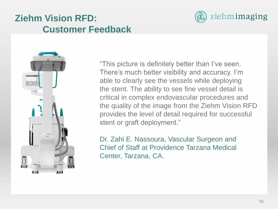
Ziehm Vision RFD:
Customer Feedback
“This picture is definitely better than I’ve seen.
There’s much better visibility and accuracy. I’m
able to clearly see the vessels while deploying
the stent. The ability to see fine vessel detail is
critical in complex endovascular procedures and
the quality of the image from the Ziehm Vision RFD
provides the level of detail required for successful
stent or graft deployment.”
Dr. Zahi E. Nassoura, Vascular Surgeon and
Chief of Staff at Providence Tarzana Medical
Center, Tarzana, CA.
52
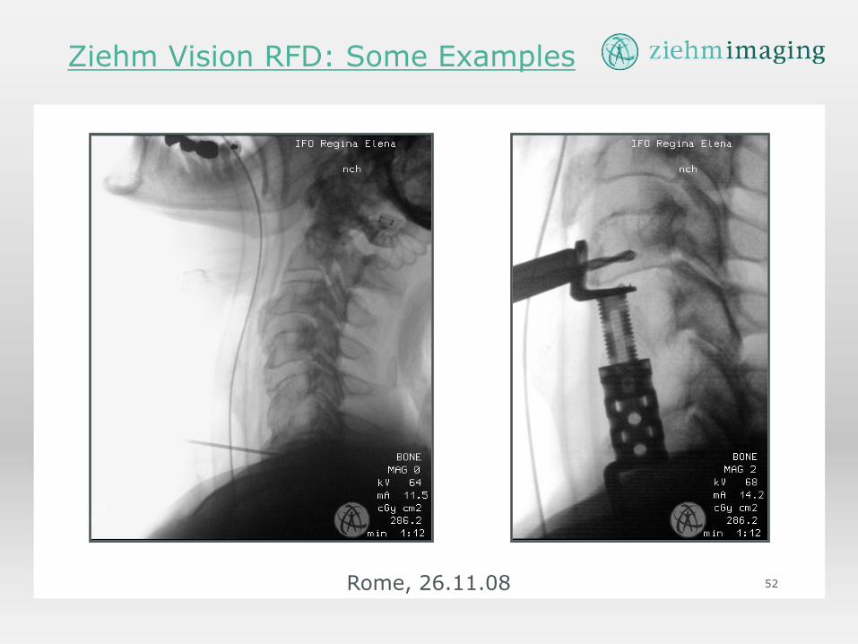
Ziehm Vision RFD: Some Examples
Rome, 26.11.08
53
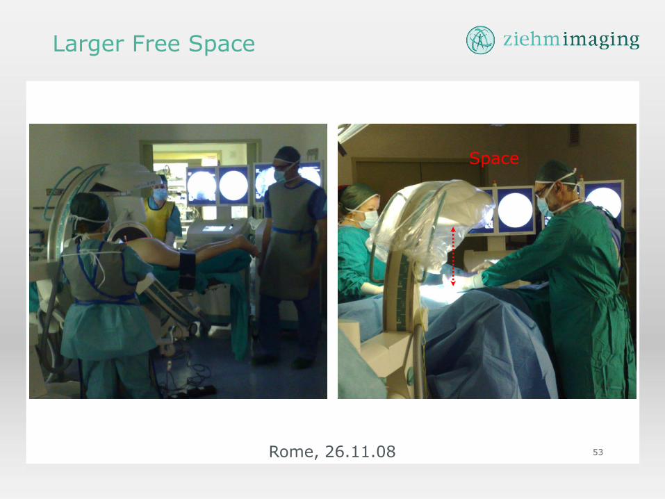
Larger Free Space
Rome, 26.11.08
Space

54 Rome, 26.11.08
Thank you!