Cornea Grand Rounds - nceyes.org grand rounds.pdf · Most common post. dyst ... LP OS. Diagnosis...
44
Cornea Grand Rounds Charlie Ficco, O.D. Milan Eye Center Cumming, GA [email protected] 81 y. o. w. f. c/o chronic fbs ou. “Equate” AT ou qid + w/ minimal relief. Coumadin, Premarin, Antivert, Toprol XL, Diovan, Lanoxin, Tricor, Xanax. 20/25 20/25 -2
-
Upload
hoangnguyet -
Category
Documents
-
view
217 -
download
0
Transcript of Cornea Grand Rounds - nceyes.org grand rounds.pdf · Most common post. dyst ... LP OS. Diagnosis...

Cornea Grand Rounds
Charlie Ficco, O.D.Milan Eye Center
Cumming, GA
81 y. o. w. f. c/o chronic fbs ou. “Equate” AT ou qid + w/ minimal relief. Coumadin, Premarin, Antivert, Toprol XL, Diovan, Lanoxin, Tricor, Xanax.
20/25 20/25-2
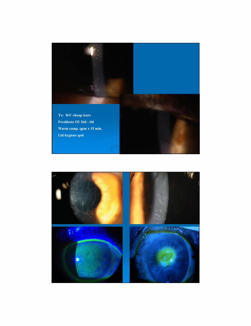
Tx: D/C cheap tears
Freshkote OU bid - tid
Warm comp. qpm x 15 min.
Lid hygiene qod

Pathophysiology
Morphologic changes in BM of epithelium
Inadequate hemidesmosome formation
• “fingerprint” lines
Fibro-granular deposits
• “map” and “dot” deposits
Pham, LTL, et. al. Treatment of Epithelial Basement Membrane Dystrophy With Manual Superficial Keratectomy. EyeRounds.org. Feb 22, 2010; Available from: http://www.eyerounds.org/cases/78-EBMD-treatment.htm.

Pham, LTL, et. al. Treatment of Epithelial Basement Membrane Dystrophy With Manual Superficial Keratectomy. EyeRounds.org. Feb 22, 2010; Available from: http://www.eyerounds.org/cases/78-EBMD-treatment.htm.
Pham, LTL, et. al. Treatment of Epithelial Basement Membrane Dystrophy With Manual Superficial Keratectomy. EyeRounds.org. Feb 22, 2010; Available from: http://www.eyerounds.org/cases/78-EBMD-treatment.htm

Pham, LTL, et. al. Treatment of Epithelial Basement Membrane Dystrophy With Manual Superficial Keratectomy. EyeRounds.org. Feb 22, 2010; Available from: http://www.eyerounds.org/cases/78-EBMD-treatment.htm
Basement Membrane Dystrophy
Most common anterior corneal dystrophy!
AD
2% of population

EBMD Tx
Early• Lubrication• Hypertonic gtts• DTS control
Later• Recurrent erosions
Debridement or PTK BCL Anti-inflamatory Tx
• Topical and Oral Antibiotic Tx
Dursun D, Kim MC, Solomon A, Pflugfelder SC. Treatment of recalcitrant recurrent corneal erosions with inhibitors of matrix metalloproteinase-9, doxycycline and corticosteroids. Am J Ophthalmol. 2001;132:8-13.
Dry Sponge v. Wet Sponge

RCE Treatment
Abrasions Antibiosis
BCL
Anti-inflammatory• Topical and / or Oral Tetracyclines
Chronic Treatments Copious Lubrication
Topical Sodium Chloride
Topical Anti-inflammatory
Oral Tetracyclines
RCE Treatment
Chronic Treatments Surgical
• PTK
• PRK
• Stromal Puncture
• Epithelial Debridement

Anterior K Dystrophies
Reis Buckler’s Dystrophy Scarring Bowman’s membrane, 1st
decade
Anterior K Dystrophies
Meesman’s Dystrophy AKA juvenile epithelial dystrophy
Extremely rare, multiple sm. clr. cysts

Anterior K Dystrophies Lisch dystrophy
? EBMD
Thiel-Behnke dystrophy ? Reis Buckler
35 year-old male
Myopia
-5.75 OD; -6.50 OS
Pre-operative ocular exam was normal Scant small white subepithelial flecks in cornea
OU
Ladarwave CustomCornea LASIK in 2005 Amadeus Microkeratome used
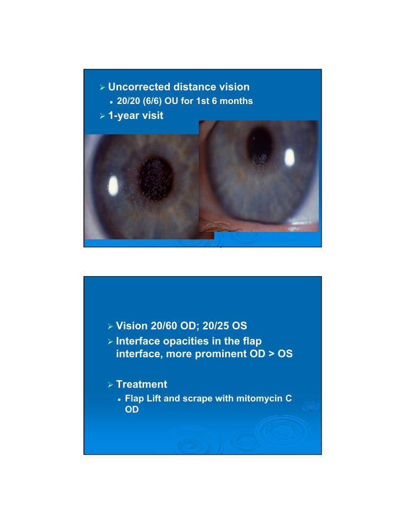
Uncorrected distance vision 20/20 (6/6) OU for 1st 6 months
1-year visit
Vision 20/60 OD; 20/25 OS
Interface opacities in the flap interface, more prominent OD > OS
Treatment Flap Lift and scrape with mitomycin C
OD

Vision was 20/80 OD and 20/25 OS
Penetrating keratoplasty OD

Granular corneal dystrophy Type II (Avellino Corneal Dystrophy - ACD) (Granular-lattice corneal dystrophy)
Excimer laser corneal ablation Contraindicated1-4
Includes PTK with or without mitomycin1
PRK, LASEK (Advanced surface ablation)2
LASIK3,4
1) Dogru M et al. Ophthalmology 2001;108:810-7.2) Banning CS et al. Cornea 2006;25:482-4.3) Lee WB et al. JRCS 2007;33:133-8 4) Lee WB et al. AAO Abstract 2007;253.
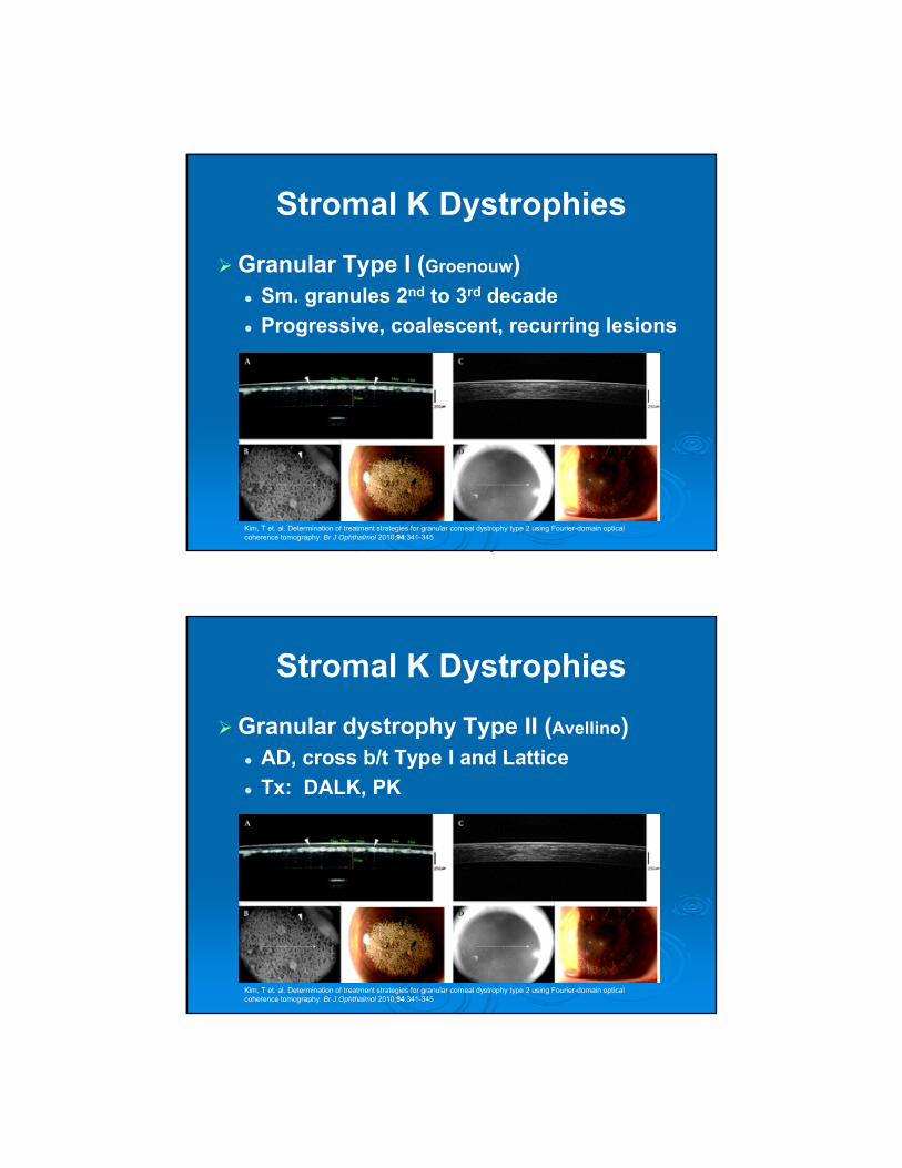
Stromal K Dystrophies
Granular Type I (Groenouw) Sm. granules 2nd to 3rd decade
Progressive, coalescent, recurring lesions
Kim, T et. al. Determination of treatment strategies for granular corneal dystrophy type 2 using Fourier-domain optical coherence tomography. Br J Ophthalmol 2010;94:341-345
Stromal K Dystrophies
Granular dystrophy Type II (Avellino) AD, cross b/t Type I and Lattice
Tx: DALK, PK
Kim, T et. al. Determination of treatment strategies for granular corneal dystrophy type 2 using Fourier-domain optical coherence tomography. Br J Ophthalmol 2010;94:341-345

Stromal K Dystrophies
Lattice dystrophy Type I Most common stromal dystrophy
Lattice or cracked glass appearance, 1st decade
PTK, DALK, PK
H&E stain of cornea with lattice. Note pink amorphous deposits in stroma

Congo red stain, highlighting amyloid Apple-green birefringence of amyloid with cross-polarization.
Stromal K Dystrophies
Macular dystrophy AR, gray dep. w/ stromal haze, limbus to
limbus.
1st decade, PK rather than PTK

Mucopolysaccharide deposits
Staining w/ Alcian Blue
Table 1: Corneal Stromal Dystrophies Table 2: Mnemonic for remembering corneal stromal dystrophies
*Lattice*Granular*Avellino*Macular*Gelatinous Droplike dystrophy*Schnyder Crystalline Corneal Dystrophy
(SCCD)*Francois-Neetens Fleck Dystrophy*Congenital Hereditary Stromal Dystrophy
*Marilyn – Macular Dystrophy*Monroe – Mucopolysaccharide*Always – Alcian Blue stain*Gets – Granular Dystrophy*Her – Hyaline*Man in – Masson Trichrome stain*Los - Lattice Dystrophy*Angeles – Amyloid*California – Congo Red
Birkholz M, Sayed N, Wagoner M. Corneal Stromal Dystrophies: A Clinicopathologic Review. eyerounds.org. Aug 17, 2009.

Posterior K Dystrophies
Fuch’s Dystrophy
Most common post. dyst
AD, 4th - 6th decade, var. exp., polymegathism
Dec. Va, k edema late

Posterior K Dystrophies
Palliative Treatment:
Topical osmotic agents
Definitive Treatment
DSAEK
DMEK
Posterior K Dystrophies
Congenital Hereditary Endothelial dystrophy AR (Most Common)
• Edema at birth, nystagmus
AD• Opacities dev. Later, no nystagmus

DSAEK
Make it stop!!!
34 y. o. w. m. c/o 1 d Hx of severe FBS, photophobia, OD, after waking this morning.
Fourth occurrence
Initial occurrence fingernail injury


RCE Healing
S/P Debridement BCL or AMT
Antibiotic
Steroid
Oral Tetracycline or Azythromycin
RCE Treatment
Epithelial debridement Weck cell sponge
Beaver Blade
Foreign body spud / debridement hoe
Bandage CL / Amniotic Memrane Topical antibiotic
Topical anti-inflammatory
Oral Tetracycline antibiotic therapy

RCE
Treatment Surgical
• PTK / PRK
• Debridement – Amniotic Membrane CL

C. B.
40 y. o. w.m. LEE 2 years prior. Si HyDW CL wearer. Obvious non-compliant SCL wear.
Noticed dec. Va, FBS, injection OS 1 d prior. No discharge or pain. Removed CL OS immediately.
BVa: 20/20 OD, 20/100 OS
P, EOM, MB, CF = Normal
TA: 16 mmHg, OU

C. B.
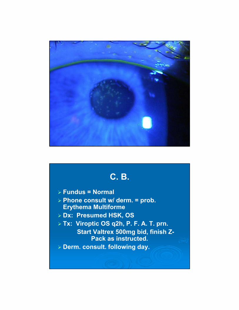
Fundus = Normal Phone consult w/ derm. = prob.
Erythema MultiformeDx: Presumed HSK, OS Tx: Viroptic OS q2h, P. F. A. T. prn.
Start Valtrex 500mg bid, finish Z-Pack as instructed.
Derm. consult. following day.
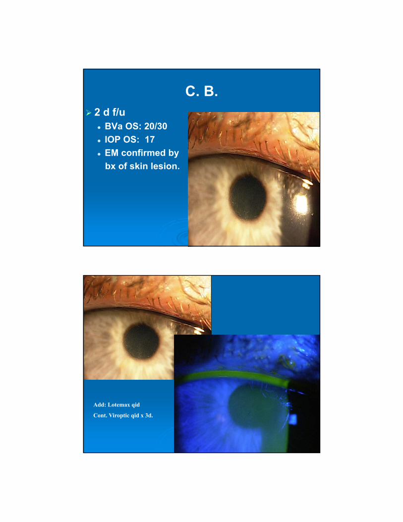
C. B. 2 d f/u
BVa OS: 20/30
IOP OS: 17
EM confirmed by
bx of skin lesion.
Add: Lotemax qid
Cont. Viroptic qid x 3d.

C. B.
8 d f/u
BVa: 20/20
IOP: 16 mmHg
Tx: Lotemax qid x 1 more wk. then stop.
Herpes Simplex Keratitis
Epithelial Disease Vesicles, Dendrites, or
Geographic ulcers.
Check K sensitivity• Viroptic q2h or 8x/d
• Zirgan gel
• Debridement?
• Orals? HEDS II
**Absolutely NO STEROIDS w epi. defects.
• Most common cause of corneal blindness in US, 50,000 new or recurrent cases/yr.1
1. Herpetic Eye Disease Study Group. Acyclovir for the Prevention of Recurrent Herpes Simplex Eye Disease. New Eng J Med July 30, 1998.

HS Stromal Keratitis
Recurrent disease ISK
Retained Viral antigen in stroma.
Nec. Strom. Keratitis Dense infil., ulceration,
and necrosis
Tx: Top. Steroids, Top. Antiviral, Oral Antivirals.
HEDS I HEDS II
Sig. benefit tx w/ orals.
1. Herpetic Eye Disease Study Group. Oral Acyclovir for Herpes Simplex Virus Eye Disease: Effect on Prevention of Epithelial Keratitis and Stromal Keratitis. Arch Ophtalmol Aug 2000; 118(8):1030-1036.

H S Endothelialitis
KPs, Cells and Flare
Stromal / Epi. edema
No neovasc. or infiltr.
Disciform, Linear, Diffuse
? CMV
Tx: Steroids, Top. Antivirals, and Oral Antivirals (1-2 gm/d)*
Koizumi N, et. al. Cytomegalovirus as an etiologic factor in corneal endothelialitis. Ophthalmology 2008; 115(2):292-297.
HZO72 y. o. 1 wk hx “shingles” c/o dec. Va OS. Acyclovir 800 mg 5x/d.
Va: 20/30 OD, 20/100 OS

20/100
(-) K staining20/50
Cont. Valtrex 800mg 5x/d
Tobradex oph ung bid / Lotemax qid
3wk: Cont. Lotemax bid
“Pseudo-dendrites” v. “Dendrites”
Pseudodendrites: Tree branches w/o terminal end bulbs.
Dendrites: Tree branches with terminal end bulbs.

“I Can See Clearly Now”
40 year old female
30 years of blindness in left eye following children’s Motrin at 10 yo
VA 20/30 OD; LP OS

Diagnosis
Toxic Epidermal Necrolysis (Lyell sydrome)
Total Stem cell deficiency and keratinization of the cornea
Diffuse Symblephara and/or ankyloblephara
Treatment Options
Total Keratolimbal allograft and penetrating keratoplasty
Artificial cornea transplant

PRE-OPLight Perception
1 MOS POST-OP ; 20/40
(6 Months Postop)Kpro and Amniotic Membrane
Reconstruction
20/30 Distance Acuity

Boston KPro Indications
Two prior failed grafts
Poor prognosis for cadaveric grafts
Vision less than 20/400
Suboptimal vision in other eye
No end-stage glaucoma or retinal detachment
KPro Components
Type 2 Device
Type 1 Device (PMMA)
Inert Titanium Fixation Screw
Collar Button Back Plate

KPro Assembly
KPro Assembly
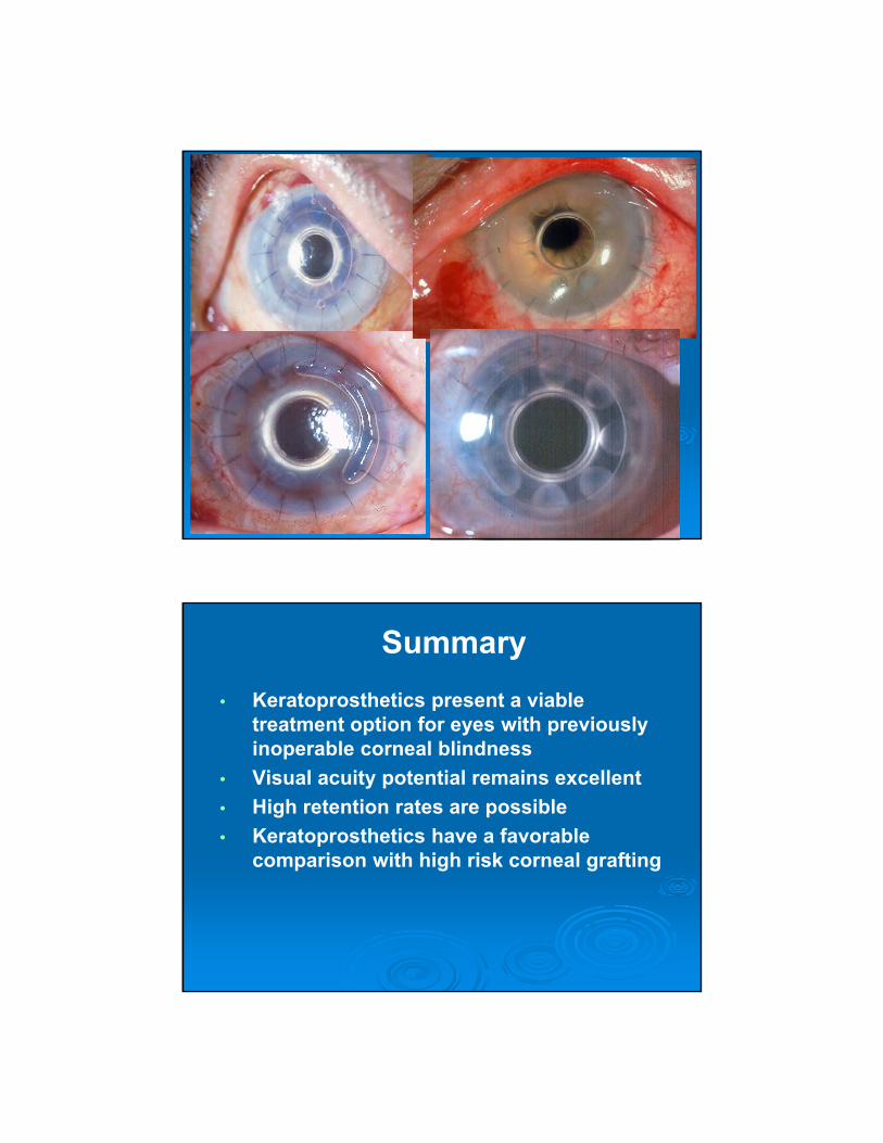
Summary
• Keratoprosthetics present a viable treatment option for eyes with previously inoperable corneal blindness
• Visual acuity potential remains excellent
• High retention rates are possible
• Keratoprosthetics have a favorable comparison with high risk corneal grafting

Anterior Segment Predictors of Death
Xerophthalmia
Toxic Epidermal Necrolysis (Stevens-Johnson Syndrome)
Mooren’s Ulcer
Fabry’s Disease
Mooren’s Ulcer
Pain
Unilateral > Bilateral
Males > Females
Limbus to central cornea
Autoimmune
Prognosis Depends on underlying autoimmune
disorder• Can lead to death if not treated properly

Fabry Disease
AKA: Alpha Galactosidase-A Deficiency
X-Linked
Acroparesthesias, angiokeratomas, hypohidrosis, tinnitus
Ocular signs conj. bv tortuosity, radiating psc, K
verticillata
Kidney damage, cardiac damage and stroke potential = life threatening cond.
FABRY DISEASE MOOREN’S ULCER
Klaus D. Teichmann, MD, FRCS, FRACO; Michael D. Wagoner, MD. Mooren Ulcer Following Epikeratoplasty for Keratoconus. Arch Ophthalmol. 1998;116(10):1381-1382
National Fabry Disease Foundation. 2009

The Masquerade: A Case for the FDA
17 year old female
Contact lens wearer
Redness, pain, and decreased vision for 4 days
Medical History benign except for “fever blisters”
Initial Examination
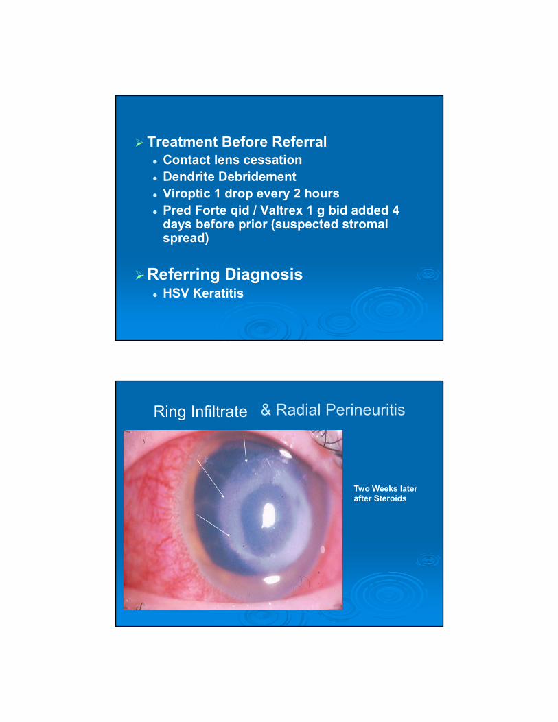
Treatment Before Referral Contact lens cessation Dendrite Debridement Viroptic 1 drop every 2 hours Pred Forte qid / Valtrex 1 g bid added 4
days before prior (suspected stromal spread)
Referring Diagnosis HSV Keratitis
Ring Infiltrate & Radial Perineuritis
Two Weeks later after Steroids

Differential for Dendritic Lesions
Herpes simplex /zoster Epstein Barr Tyrosinemia Healing epithelial defect Rosacea Lee, Mannis, Schwab. Cornea 2005
Superficial hypertrophic dendritic epitheliopathy (SHDE)
Lee et Mannis, Cornea 2006
Acanthamoeba
Diagnostics
Cornea Culture Chocolate and blood agar
• No growth Sabouraud agar
• No growth Nonnutrient agar
(E. Coli overlay)
• Positive
Cornea Scraping• Positive for ameobic cysts

Confocal Microscopy(Confoscan 4)
40 X view
40 X View
Diagnosis & Treatment
Acanthamoeba Keratitis 1) Stop Pred Forte & Antivirals
2) Start antiamoebic agents• Polyhexamethalene biguanide (PHMB) .02%
• Chlorhexidine .02%
• Brolene (Propanidine)
• Desmadine (Hemxamidine)
3) Cycloplegia