Copyright © 2010 Pearson Education, Inc. DO NOW WORK Explain in terms of homeostasis why exercise...
24
Copyright © 2010 Pearson Education, Inc. DO NOW WORK Explain in terms of homeostasis why exercise results in increased respiration and heart rates. Are these examples of positive and negative feedback mechanisms? How do you know?
-
Upload
edgar-elliott -
Category
Documents
-
view
213 -
download
0
Transcript of Copyright © 2010 Pearson Education, Inc. DO NOW WORK Explain in terms of homeostasis why exercise...

Copyright © 2010 Pearson Education, Inc.
DO NOW WORKExplain in terms of homeostasis why exercise results in increased respiration and heart rates.
Are these examples of positive and negative feedback mechanisms?
How do you know?

Copyright © 2010 Pearson Education, Inc.
Thought of the Day:
Q: Why don’t skeletons get in fights?

Copyright © 2010 Pearson Education, Inc.
Thought of the Day:
Q: Why don’t skeletons get in fights?
A: Because they don’t have the guts!

Copyright © 2010 Pearson Education, Inc.
Figure 1.7 Regional terms used to designate specific body areas.
CervicalCervical
Back (dorsal)
(a) Anterior/Ventral (b) Posterior/Dorsal
Pubic (genital)
Cephalic Frontal Orbital Nasal Oral Mental
Thoracic Sternal Axillary Mammary
Scapular
Vertebral
Lumbar
Sacral
Gluteal
Perineal (between anus and external genitalia)
Abdominal Umbilical
Pelvic Inguinal
(groin)
Upper limb Acromial
Brachial (arm) Antecubital Olecranal Antebrachial
(forearm) Carpal (wrist)
Manus (hand) Pollex Metacarpal Palmar Digital
Lower limb Coxal (hip) Femoral (thigh) Patellar Popliteal Crural (leg) Sural (calf) Fibular or peroneal
Pedal (foot) Tarsal (ankle) Calcaneal Metatarsal Digital Plantar Hallux
Cephalic Otic Occipital (back
of head)
ThoraxAbdomenBack (Dorsum)

Copyright © 2010 Pearson Education, Inc.
Figure 1.7a Regional terms used to designate specific body areas.
Cervical
(a) Anterior/Ventral
Pubic(genital)
CephalicFrontalOrbitalNasalOralMental
ThoracicAxillaryMammarySternal
AbdominalUmbilical
PelvicInguinal(groin)
Upper limbAcromialBrachial (arm)AntecubitalAntebrachial (forearm)Carpal (wrist)
Manus (hand)PalmarPollexDigital
Lower limbCoxal (hip)Femoral (thigh)
PatellarCrural (leg)Fibular or peroneal
Pedal (foot)Tarsal (ankle)
MetatarsalDigitalHallux
ThoraxAbdomenBack (Dorsum)

Copyright © 2010 Pearson Education, Inc.
Figure 1.7b Regional terms used to designate specific body areas.
Cervical Back (dorsal)
(b) Posterior/Dorsal
Scapular Vertebral Lumbar Sacral Gluteal Perineal (between anus and external genitalia)
Upper limb AcromialBrachial (arm)
Olecranal Antebrachial (forearm)Manus (hand) Metacarpal DigitalLower limb Femoral (thigh) Popliteal Sural (calf) Fibular or peroneal
Pedal (foot) Calcaneal Plantar
Cephalic Otic Occipital (back of head)
ThoraxAbdomenBack (Dorsum)

Copyright © 2010 Pearson Education, Inc.
Figure 1.8 Planes of the body with corresponding magnetic resonance imaging (MRI) scans.
Transverse plane
Median (midsagittal) plane
Frontal plane
Liver
Spleen
Pancreas
Aorta
Vertebralcolumn
Spinal cord
Subcutaneous fat layerBody wall
Rectum IntestinesLeft andright lungs
Liver HeartStomach
SpleenArm
(a) Frontal section (through torso)
(b) Transverse section (through torso, inferior view)
(c) Median section (midsagittal)

Copyright © 2010 Pearson Education, Inc.
Figure 1.8a Planes of the body with corresponding magnetic resonance imaging (MRI) scans.
Frontalplane
Left and right lungs
Liver HeartStomach
SpleenArm
(a) Frontal section (through torso)

Copyright © 2010 Pearson Education, Inc.
Figure 1.8b Planes of the body with corresponding magnetic resonance imaging (MRI) scans.
Transverseplane
Liver
Spleen
PancreasAorta
Spinal cordSubcutaneous fat layer Body wall
(b) Transverse section (through torso, inferior view)

Copyright © 2010 Pearson Education, Inc.
Figure 1.8c Planes of the body with corresponding magnetic resonance imaging (MRI) scans.
Median(midsagittal)plane
Vertebral columnRectum Intestines
(c) Median section (midsagittal)
Copyright © 2010 Pearson Education, Inc.
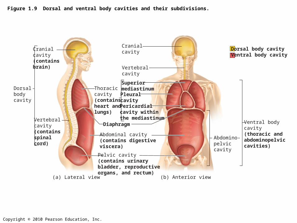
Figure 1.9 Dorsal and ventral body cavities and their subdivisions.
Cranialcavity(contains brain)
Dorsalbodycavity
Vertebralcavity(contains spinal cord)
Cranialcavity
Superiormediastinum
Pericardialcavity withinthe mediastinum
Pleuralcavity
Vertebralcavity
Abdomino-pelviccavity
Ventral bodycavity(thoracic andabdominopelviccavities)
Abdominal cavity(contains digestiveviscera)
Diaphragm
Pelvic cavity(contains urinary bladder, reproductive organs, and rectum)
Thoraciccavity(containsheart andlungs)
(a) Lateral view (b) Anterior view
Dorsal body cavityVentral body cavity

Copyright © 2010 Pearson Education, Inc.
Figure 1.9a Dorsal and ventral body cavities and their subdivisions.
Cranial cavity(contains brain)
Dorsalbodycavity
Vertebral cavity(contains spinalcord) Abdominal cavity
(contains digestiveviscera)
Diaphragm
Pelvic cavity(contains urinary bladder, reproductive organs, and rectum)
Thoracic cavity(contains heart and lungs)
(a) Lateral view
Dorsal body cavityVentral body cavity

Copyright © 2010 Pearson Education, Inc.
Figure 1.9b Dorsal and ventral body cavities and their subdivisions.
Cranialcavity
Superiormediastinum
Pericardialcavity within the mediastinum
Pleuralcavity
Vertebralcavity
Abdomino-pelviccavity
Ventral bodycavity(thoracic andabdominopelviccavities)
Abdominal cavity(contains digestiveviscera)
Diaphragm
Pelvic cavity(contains urinary bladder, reproductive organs, and rectum)
Thoraciccavity(containsheart andlungs)
(b) Anterior view
Dorsal body cavityVentral body cavity

Copyright © 2010 Pearson Education, Inc.
Figure 1.10 Serous membrane relationships.
Outer balloon wall(comparable to parietal serosa)
Air (comparable to serous cavity)
Inner balloon wall(comparable to visceral serosa)
Heart
Parietalpericardium
Pericardialspace withserous fluid
Visceralpericardium
(a) A fist thrust into a flaccid balloon demonstratesthe relationship between the parietal and visceralserous membrane layers.
(b) The serosae associated with the heart.

Copyright © 2010 Pearson Education, Inc.
Figure 1.11 The four abdominopelvic quadrants.
Right upperquadrant(RUQ)
Right lowerquadrant(RLQ)
Left upperquadrant(LUQ)
Left lowerquadrant(LLQ)

Copyright © 2010 Pearson Education, Inc.
Figure 1.12 The nine abdominopelvic regions.
Epigastricregion
Umbilicalregion
Rightlumbarregion
Leftlumbarregion
Righthypochondriac
region
Lefthypochondriac
region
Hypogastric(pubic)region
Right iliac(inguinal)
region
Left iliac(inguinal)
region
Liver
Gallbladder
Ascending colon oflarge intestine
Small intestine
Appendix
Cecum
Diaphragm
Stomach
Descending colonof large intestine
Transverse colonof large intestine
Initial part ofsigmoid colon
Urinary bladder
(a) Nine regions delineated by four planes (b) Anterior view of the nine regions showing the superficial organs

Copyright © 2010 Pearson Education, Inc.
Figure 1.12a The nine abdominopelvic regions.
Epigastricregion
Umbilicalregion
Rightlumbarregion
Leftlumbarregion
Righthypochondriac
region
Lefthypochondriac
region
Hypogastric(pubic)region
Right iliac(inguinal)
region
Left iliac(inguinal)
region
(a) Nine regions delineated by four planes

Copyright © 2010 Pearson Education, Inc.
Figure 1.12b The nine abdominopelvic regions.
LiverGallbladderAscending colonof large intestine Small intestine
AppendixCecum
Diaphragm Stomach
Descending colonof large intestine
Transverse colonof large intestine
Initial part ofsigmoid colonUrinary bladder
(b) Anterior view of the nine regions showing the superficial organs

Copyright © 2010 Pearson Education, Inc.
Table 1.1 Orientation and Directional Terms (1 of 3)

Copyright © 2010 Pearson Education, Inc.
Table 1.1 Orientation and Directional Terms (2 of 3)

Copyright © 2010 Pearson Education, Inc.
Table 1.1 Orientation and Directional Terms (3 of 3)

Copyright © 2010 Pearson Education, Inc.
A Closer Look 1.1a Medical Imaging: Illuminating the Body.
Left
Liver Vertebra(a) A CT scan through the superior abdomen.
Left kidney SpleenPancreas
Right

Copyright © 2010 Pearson Education, Inc.
A Closer Look 1.1b Medical Imaging: Illuminating the Body.
(b) A DSA image of the arteries that supply the heart.
Arterysupplyingheart
Narrowingof the artery

Copyright © 2010 Pearson Education, Inc.
A Closer Look 1.1c Medical Imaging Illuminating the Body