Coordinate control of terminal dendrite patterning...
12
RESEARCH ARTICLE Coordinate control of terminal dendrite patterning and dynamics by the membrane protein Raw Jiae Lee, Yun Peng, Wen-Yang Lin and Jay Z. Parrish* ABSTRACT The directional flow of information in neurons depends on compartmentalization: dendrites receive inputs whereas axons transmit them. Axons and dendrites likewise contain structurally and functionally distinct subcompartments. Axon/dendrite compartmentalization can be attributed to neuronal polarization, but the developmental origin of subcompartments in axons and dendrites is less well understood. To identify the developmental bases for compartment-specific patterning in dendrites, we screened for mutations that affect discrete dendritic domains in Drosophila sensory neurons. From this screen, we identified mutations that affected distinct aspects of terminal dendrite development with little or no effect on major dendrite patterning. Mutation of one gene, raw, affected multiple aspects of terminal dendrite patterning, suggesting that Raw might coordinate multiple signaling pathways to shape terminal dendrite growth. Consistent with this notion, Raw localizes to branch-points and promotes dendrite stabilization together with the Tricornered (Trc) kinase via effects on cell adhesion. Raw independently influences terminal dendrite elongation through a mechanism that involves modulation of the cytoskeleton, and this pathway is likely to involve the RNA-binding protein Argonaute 1 (AGO1), as raw and AGO1 genetically interact to promote terminal dendrite growth but not adhesion. Thus, Raw defines a potential point of convergence in distinct pathways shaping terminal dendrite patterning. KEY WORDS: Dendrite, Compartmentalization, Terminal dendrite dynamics INTRODUCTION The directional flow of information in the nervous system relies on the compartmentalization of neurons. On a basic level, a neuron has two compartments apart from the soma: dendrites, which receive inputs, and axons, which transmit signals. To meet their respective functions, axons and dendrites have distinct morphological and molecular properties. For example, the microtubule cytoskeleton is organized differently in axons and dendrites, allowing distinct modes of trafficking (Baas et al., 1988; Horton and Ehlers, 2003). Beyond this basic level of compartmentalization, neurons display extreme diversity in axon and dendrite morphology, and both axons and dendrites contain structurally and functionally distinct subdomains (Katsuki et al., 2011; Masland, 2004). Several lines of evidence support the existence of compartments within dendrites. First, some neurons have morphologically distinct dendrites. For example, mammalian olfactory bulb mitral cells extend a tufted primary dendrite radially and structurally and functionally distinct lateral dendrites horizontally (Imamura and Greer, 2009). Likewise, apical/basal dendrites of hippocampal neurons and ipsilateral/contralateral dendrites of motoneurons are morphologically and biophysically distinct. Second, specialized structures are asymmetrically distributed in many dendrites. Notable among these is the dendritic spine, an isolated compartment that is electrically and biochemically distinct from the rest of the dendrite arbor, and dendritic spines likewise have distinctive microdomains (Chen and Sabatini, 2012; Yuste, 2013). Many organelles are selectively deployed in dendrites, including a satellite secretory pathway containing endoplasmic reticulum and Golgi outposts; the number and location of these organelles locally influences dendrite growth and dynamics (Aridor et al., 2004; Gardiol et al., 1999; Horton and Ehlers, 2003; Ye et al., 2007). In highly branched dendrite arbors, for example cerebellar Purkinje neurons and insect sensory neurons, major dendrites and terminal dendrites have distinct cytoskeletal compositions and growth properties (Fujishima et al., 2012; Jinushi-Nakao et al., 2007). Finally, dendrite arbors often contain functionally distinct domains as well. For example, the proximal-distal compartmentalization of chloride co-transporters underlies directional selectivity in starburst amacrine cells (Gavrikov et al., 2006). Whereas axon/dendrite compartmentalization can be attributed to neuronal polarization, the developmental origin of dendrite subcompartments is less well understood. Drosophila peripheral nervous system (PNS) class IV dendrite arborization (C4da) neurons have highly branched dendrite arbors, consisting of major dendrites emanating radially from the soma and terminal dendrites that fill in the space in the receptive field (Grueber et al., 2002). Main branches and terminal arbors have distinct growth properties in these neurons, with terminal dendrites exhibiting dynamic growth and containing a cytoskeleton largely devoid of microtubules (Grueber et al., 2002; Jinushi-Nakao et al., 2007), suggesting that different cellular programs pattern main dendrites and terminal arbors. To identify the developmental bases for compartment-specific patterning in these dendrites, we used a genetic screen to identify mutations that affect distinct dendritic compartments. From this screen, we identified mutants that selectively affected terminal dendrites, including their placement along the proximal-distal axis and their patterning, suggesting that distance from the soma and branch type (major or terminal dendrite) are two key pieces of positional information in the patterning of dendrite arbors. Mutations in raw were unique in that they simultaneously affected multiple aspects of terminal dendrite patterning, suggesting that raw coordinately controls multiple aspects of terminal dendrite growth. Indeed, we found that Raw regulates terminal dendrite adhesion and elongation via distinct pathways, the former involving the Trc kinase and the latter involving cytoskeletal remodeling and the RNA-binding protein AGO1. Thus, Raw appears to be a crucial component of a spatially localized program controlling terminal dendrite patterning. Received 28 May 2014; Accepted 31 October 2014 Department of Biology, University of Washington, Seattle, WA 98195, USA. *Author for correspondence ( [email protected]) 1 © 2015. Published by The Company of Biologists Ltd | Development (2015) 142, 1-12 doi:10.1242/dev.113423 DEVELOPMENT Development ePress. Posted online 5 December 2014 http://dev.biologists.org/lookup/doi/10.1242/dev.113423 Access the most recent version at First posted online on 5 December 2014 as 10.1242/dev.113423
Transcript of Coordinate control of terminal dendrite patterning...

RESEARCH ARTICLE
Coordinate control of terminal dendrite patterning and dynamicsby the membrane protein RawJiae Lee, Yun Peng, Wen-Yang Lin and Jay Z. Parrish*
ABSTRACTThe directional flow of information in neurons depends oncompartmentalization: dendrites receive inputs whereas axonstransmit them. Axons and dendrites likewise contain structurallyand functionally distinct subcompartments. Axon/dendritecompartmentalization can be attributed to neuronal polarization, butthe developmental origin of subcompartments in axons and dendritesis less well understood. To identify the developmental bases forcompartment-specific patterning in dendrites, we screened formutations that affect discrete dendritic domains in Drosophilasensory neurons. From this screen, we identified mutations thataffected distinct aspects of terminal dendrite development with little orno effect on major dendrite patterning. Mutation of one gene, raw,affected multiple aspects of terminal dendrite patterning, suggestingthat Raw might coordinate multiple signaling pathways to shapeterminal dendrite growth. Consistent with this notion, Raw localizes tobranch-points and promotes dendrite stabilization together with theTricornered (Trc) kinase via effects on cell adhesion. Rawindependently influences terminal dendrite elongation through amechanism that involves modulation of the cytoskeleton, and thispathway is likely to involve the RNA-binding protein Argonaute 1(AGO1), as raw and AGO1 genetically interact to promote terminaldendrite growth but not adhesion. Thus, Raw defines a potential pointof convergence in distinct pathways shaping terminal dendritepatterning.
KEY WORDS: Dendrite, Compartmentalization, Terminal dendritedynamics
INTRODUCTIONThe directional flow of information in the nervous system relies onthe compartmentalization of neurons. On a basic level, a neuron hastwo compartments apart from the soma: dendrites, which receiveinputs, and axons, which transmit signals. To meet their respectivefunctions, axons and dendrites have distinct morphological andmolecular properties. For example, the microtubule cytoskeleton isorganized differently in axons and dendrites, allowing distinctmodes of trafficking (Baas et al., 1988; Horton and Ehlers, 2003).Beyond this basic level of compartmentalization, neurons displayextreme diversity in axon and dendrite morphology, and both axonsand dendrites contain structurally and functionally distinctsubdomains (Katsuki et al., 2011; Masland, 2004).Several lines of evidence support the existence of compartments
within dendrites. First, some neurons have morphologically distinctdendrites. For example, mammalian olfactory bulb mitral cells
extend a tufted primary dendrite radially and structurally andfunctionally distinct lateral dendrites horizontally (Imamura andGreer, 2009). Likewise, apical/basal dendrites of hippocampalneurons and ipsilateral/contralateral dendrites of motoneurons aremorphologically and biophysically distinct. Second, specializedstructures are asymmetrically distributed in many dendrites. Notableamong these is the dendritic spine, an isolated compartment that iselectrically and biochemically distinct from the rest of the dendritearbor, and dendritic spines likewise have distinctive microdomains(Chen and Sabatini, 2012; Yuste, 2013). Many organelles areselectively deployed in dendrites, including a satellite secretorypathway containing endoplasmic reticulum and Golgi outposts; thenumber and location of these organelles locally influences dendritegrowth and dynamics (Aridor et al., 2004; Gardiol et al., 1999;Horton and Ehlers, 2003; Ye et al., 2007). In highly brancheddendrite arbors, for example cerebellar Purkinje neurons and insectsensory neurons, major dendrites and terminal dendrites havedistinct cytoskeletal compositions and growth properties (Fujishimaet al., 2012; Jinushi-Nakao et al., 2007). Finally, dendrite arborsoften contain functionally distinct domains as well. For example, theproximal-distal compartmentalization of chloride co-transportersunderlies directional selectivity in starburst amacrine cells (Gavrikovet al., 2006). Whereas axon/dendrite compartmentalization can beattributed to neuronal polarization, the developmental origin ofdendrite subcompartments is less well understood.
Drosophila peripheral nervous system (PNS) class IV dendritearborization (C4da) neurons have highly branched dendrite arbors,consisting of major dendrites emanating radially from the soma andterminal dendrites that fill in the space in the receptive field(Grueber et al., 2002). Main branches and terminal arbors havedistinct growth properties in these neurons, with terminal dendritesexhibiting dynamic growth and containing a cytoskeleton largelydevoid of microtubules (Grueber et al., 2002; Jinushi-Nakao et al.,2007), suggesting that different cellular programs pattern maindendrites and terminal arbors. To identify the developmental basesfor compartment-specific patterning in these dendrites, we used agenetic screen to identify mutations that affect distinct dendriticcompartments. From this screen, we identified mutants thatselectively affected terminal dendrites, including their placementalong the proximal-distal axis and their patterning, suggesting thatdistance from the soma and branch type (major or terminal dendrite)are two key pieces of positional information in the patterning ofdendrite arbors. Mutations in raw were unique in that theysimultaneously affected multiple aspects of terminal dendritepatterning, suggesting that raw coordinately controls multipleaspects of terminal dendrite growth. Indeed, we found that Rawregulates terminal dendrite adhesion and elongation via distinctpathways, the former involving the Trc kinase and the latterinvolving cytoskeletal remodeling and the RNA-binding proteinAGO1. Thus, Raw appears to be a crucial component of a spatiallylocalized program controlling terminal dendrite patterning.Received 28 May 2014; Accepted 31 October 2014
Department of Biology, University of Washington, Seattle, WA 98195, USA.
*Author for correspondence ( [email protected])
1
© 2015. Published by The Company of Biologists Ltd | Development (2015) 142, 1-12 doi:10.1242/dev.113423
DEVELO
PM
ENT
Development ePress. Posted online 5 December 2014http://dev.biologists.org/lookup/doi/10.1242/dev.113423Access the most recent version at First posted online on 5 December 2014 as 10.1242/dev.113423
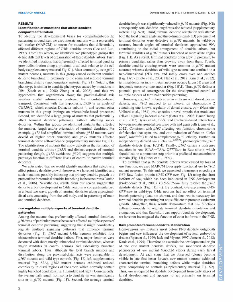
RESULTSIdentification of mutations that affect dendritecompartmentalizationTo identify the developmental bases for compartment-specificpatterning in dendrites, we used mosaic analysis with a repressiblecell marker (MARCM) to screen for mutations that differentiallyaffected different regions of C4da dendrite arbors (Lee and Luo,1999). From this screen, we identified two phenotypic groups thatdefine different levels of organization of these dendrite arbors. First,we identified mutations that differentially affected terminal dendritegrowth/distribution along a proximal-distal axis relative to the cellbody (supplementary material Fig. S1). Most commonly, as in jj329mutant neurons, mutants in this group caused exuberant terminaldendrite branching in proximity to the soma and reduced terminalbranching distally (supplementary material Fig. S1). Notably, thisphenotype is similar to dendrite phenotypes caused by mutations inDlic (Satoh et al., 2008; Zheng et al., 2008), and thus wehypothesize that organization along the proximal-distal axisinvolves spatial information conferred by microtubule-basedtransport. Consistent with this hypothesis, jj329 is an allele ofCG12042, which encodes Dynactin subunit 4, and several othermutants in this group impinge on microtubule-based processes.Second, we identified a large group of mutants that preferentiallyaffect terminal dendrite patterning without affecting majordendrites. Within this group, we identified mutants that affectedthe number, length and/or orientation of terminal dendrites. Forexample, jj472 had simplified terminal arbors; jj835 mutants weredevoid of higher order branches; and jj599 mutants affectedterminal dendrite self-avoidance (supplementary material Fig. S1).The identification of mutants that show deficits in the formation ofterminal dendrite arbors ( jj835) and distinct aspects of terminalpatterning (length, jj472; avoidance, jj599) suggests that multiplepathways function at different levels of control to pattern terminaldendrites.We anticipated that we would identify mutations that selectively
affect primary dendrite growth; however, we have not identified anysuch mutations, possibly indicating that primary dendrite growth is aprerequisite for terminal dendrite branching in these neurons (Parrishet al., 2006). Nevertheless, results from our screen suggest thatdendrite arbor development in C4da neurons is compartmentalizedin at least two ways: growth of terminal dendrites along a proximal-distal axis emanating from the cell body, and in patterning of mainand terminal dendrites.
raw regulates multiple aspects of terminal dendritepatterningAmong the mutants that preferentially affected terminal dendrites,jj102was of particular interest because it affected multiple aspects ofterminal dendrite patterning, suggesting that it might coordinatelyregulate multiple signaling pathways that influence terminaldendrites (Fig. 1). jj102 mutant C4da neurons exhibited fourcharacteristic terminal dendrite defects. First, major dendrites weredecorated with short, mostly unbranched terminal dendrites, whereasmajor dendrites in control neurons had extensively branchedterminal arbors. Thus, although the total branch number anddistribution along the proximal-distal axis were comparable injj102 mutants and wild-type controls (Fig. 1E, left; supplementarymaterial Fig. S2A), jj102 mutant neurons exhibited reducedcomplexity in distal regions of the arbor, which normally containhighly branched dendrites (Fig. 1E,middle and right). Consequently,the average path length from soma to dendrite tip was significantlyshorter in jj102 mutants (Fig. 1F). Second, the average terminal
dendrite length was significantly reduced in jj102mutants (Fig. 1G);consequently, total dendrite length was also reduced (supplementarymaterial Fig. S2B). Third, terminal dendrite orientation was altered:both the local branch angle and three-dimensional (3D) placement ofterminal dendrites were defective in jj102 mutants. In wild-typeneurons, branch angles of terminal dendrites approached 90°,contributing to the radial arrangement of dendrite arbors, butterminal dendrites of jj102 mutants branched at more acute angles(Fig. 1H). As a result, terminal dendrites often grew in proximity toprimary dendrites, rather than growing away from them. Fourth,dendrite-dendrite crossing events were common in jj102 mutantneurons; whereas dendrites of wild-type neurons are confined to atwo-dimensional (2D) area and rarely cross over one another(Fig. 1A′) (Emoto et al., 2004; Han et al., 2012; Kim et al., 2012),terminal dendrites in rawmutant neurons occupy larger 3D areas andfrequently cross over one another (Fig. 1B′,I). Thus, jj102 defines apotential point of convergence for the developmental control ofmultiple aspects of terminal dendrite patterning.
Homozygous jj102mutants arrest as embryos with dorsal closuredefects, and jj102 mapped to an interval on chromosome 2containing one known regulator of dorsal closure, raw (Nusslein-Volhard et al., 1984). raw encodes a novel protein that modulatescell-cell signaling in dorsal closure (Bates et al., 2008; Bauer Huanget al., 2007; Byars et al., 1999) and Cadherin-based interactionsbetween somatic gonadal precursor cells and germ cells (Jemc et al.,2012). Consistent with jj102 affecting raw function, chromosomedeficiencies that span raw and raw reduction-of-function alleles(raw1, rawk01021) failed to complement jj102 lethality. Further, anindependently derived raw allele (rawk01021) phenocopied the jj102dendrite defects (Fig. 1C,F-I). Finally, jj102 carries a nonsensemutation in raw (CAA→TAA, Q777Stop in Raw-short), whichshould lead to a premature stop prior to a predicted transmembranedomain (Fig. 1J) (Jones et al., 1994).
To establish that jj102 dendrite defects were caused by loss ofraw function, we used MARCM to resupply functional raw to jj102mutant neurons. To this end, we generated a transgene encoding aGFP-Raw fusion protein (UAS-GFP-raw, Fig. 1J) using the shortisoform of raw, which has been implicated in PNS development(Prokopenko et al., 2000). UAS-GFP-raw fully rescued the jj102dendrite defects (Fig. 1D,F-I). By contrast, overexpressing UAS-GFP-raw in wild-type C4da neurons had no effect on terminalbranch patterning (data not shown), and thus raw is necessary forterminal dendrite patterning but not sufficient to promote exuberantgrowth. Altogether, these results demonstrate that raw functionscell-autonomously to regulate terminal dendrite orientation andelongation, and that Raw-short can support dendrite development;we have not investigated the function of other isoforms in the PNS.
Raw promotes terminal dendrite stabilizationHomozygous raw mutants arrest before PNS dendrite outgrowthbegins and raw influences the development of several embryonictissues (Byars et al., 1999; Jack andMyette, 1997; Jemc et al., 2012;Kania et al., 1995). Therefore, to ascertain the developmental originof the raw mutant dendrite defects, we monitored dendritephenotypes of raw mutant MARCM clones during early larvaldevelopment. At each stage that we observed (clones becomevisible in late first instar larvae), raw mutant neurons exhibitedcharacteristic terminal branching defects while major dendritesgrew comparably to controls (supplementary material Fig. S2).Thus, raw is required for dendrite development from early stages oflarval development and appears to act primarily on terminaldendrites.
2
RESEARCH ARTICLE Development (2015) 142, 1-12 doi:10.1242/dev.113423
DEVELO
PM
ENT

The rawmutant dendrite branching defects could reflect a failurein terminal branch elongation, increased branch retraction, or somecombination of both. To distinguish between these possibilities, wemonitored terminal dendrite dynamics in wild-type or raw mutantC4da MARCM clones using time-lapse microscopy. Initially, wechose a 4-h window beginning at 96 h after egg laying (AEL), whenlarval growth is nearly complete and C4da dendrite arbors are no
longer expanding (Parrish et al., 2009). During this time-lapse mostterminal dendrites exhibit dynamics with equivalent rates of branchextension and retraction (Fig. 2A,C,D). By contrast, a significantlylarger proportion of terminals exhibited dynamics in raw mutantneurons, with dynamic branches retracting more frequently thanthey grew (Fig. 2B-D). Additionally, the extent of dynamics wassignificantly increased in raw mutants (3.8 µm on average,
Fig. 1. Rawaffects terminal dendritepatterning.RepresentativeMARCMclones forwild-type (wt) control (A), rawjj102 (B), rawk01021 (C) and rawjj102+UAS-GFP-raw(D). Arrowheadsmark dendrite-dendrite crossings. The boxed regions of the control and rawjj102 aremagnified in A′ and B′, respectively, to show the axial position ofterminal dendrites. (E-I) Morphometric analysis of dendrites from C4da MARCM clones of the indicated genotypes. (E) Sholl analysis depicting the distributionof branch-points, intersections and dendrite length. (F) Scatter plot showing path lengths from the cell body to branch endings. Data points reflectmeasurements froma representative neuron; error bars indicate aggregate mean and 95% confidence interval (n=8 for wt, 6 for jj102). (G) Average terminal branch length; error barsindicate s.d. (H) Terminal branch angles; box plots depict mean values and first/third quartile, whiskers mark minimum/maximum values. (I) Dendrite-dendritecrossing, normalized to total dendrite length. Error bars indicate s.d.; ns, not significant; **P<0.01, ***P<0.001, relative to control; one-way ANOVA with post-hocDunnett’s test. The number of neurons analyzed for each genotype is indicated in (I), and branch angles for 100 terminal dendrites weremeasured for each neuron in(H). Scale bars: 50 µm, except 10 µm for A′,B′. (J) Schematic of the raw locus, predicted polypeptides, andGFP-raw constructs. k01021 carries a P{lacW} insertion,and jj102 carries a missense mutation (red asterisk) in the last coding exon. Raw has two ∼100 amino acid repeats of unknown function and a putative C-terminaltransmembrane domain (TM). Raw-L, Raw-long; Raw-S, Raw-short; ECD, extracellular domain; ICD, intracellular domain.
3
RESEARCH ARTICLE Development (2015) 142, 1-12 doi:10.1242/dev.113423
DEVELO
PM
ENT

compared with 1.8 µm for control neurons) (Fig. 2E). Thus, rawappears to regulate terminal dendrite stability.Our developmental analysis of rawmutant neurons suggested that it
is required for the stabilization of terminal branches throughoutdevelopment. We therefore monitored dendrite dynamics over anearlier time-lapse when arbors are growing rapidly and adding newterminal dendrites. Between 72 and 76 h AEL, control neuronsexhibited an increased frequency and a different distribution ofdynamics: terminal growth occurred almost twice as frequently asretraction during the early interval, whereas growth and retractionoccurred at equivalent rates during the late interval (Fig. 2C,D). As inthe late time-lapse, rawmutants exhibited significantly more dynamicbehavior than wild-type controls between 72 and 76 h AEL (Fig. 2C).Notably, whereas the dynamic behavior of control neurons changedduring development, the rate of dynamics and the frequency of growth/retraction in rawmutant clones remained similar throughout both time-lapses, suggesting that terminal dendrite growth is constantly offset byretraction in raw mutants (Fig. 2D). Thus, raw affects terminal branchstability; at each time point examined, terminal dendrites of rawmutants were more dynamic than those of controls, with growthmatched by retraction. Hence, terminal dendrites of rawmutants rarelybranch or elongate, preventing the elaboration of terminal arbors.
Raw may function locally in dendrites to regulate terminaldendrite patterningTo gain insight into Raw control of terminal dendrite dynamics, wemonitored the intracellular distribution of GFP-Raw in raw mutantC4da neuron clones. GFP-Raw rescues the rawjj102 dendrite defects,
and thus we reasoned that GFP-Raw localization in these clones islikely to reflect the site of action of Raw. In dendrites, GFP-Raw isconcentrated in puncta, most of which localize to branch-pointsalong major dendrites (Fig. 3A, green arrowheads), consistent withRaw locally regulating terminal dendrite patterning. However, GFP-Raw was rarely detected in terminal dendrites, suggesting that Rawmight function at branch-points to stabilize nascent dendrites orrecruit other factors that function in the terminals.
Terminal dendrites rapidly turn over in raw mutants; therefore,we monitored whether GFP-Raw localization correlated withterminal branch stabilization. Using time-lapse microscopy, wemonitored dendrite dynamics during an 18-h interval in GFP-Raw-expressing rawjj102 mutant C4da MARCM clones and examinedwhether the presence of GFP-Raw puncta at branch-points wasassociated with branch stabilization (Fig. 3B). We sampled over 500dynamic terminal dendrites from six neurons and found that thegrowth behavior of terminal dendrites originating in branch-pointscontaining GFP-Raw puncta [χ2=23.80, degrees of freedom (d.f.)=2,P<0.01] or lacking GFP-Raw puncta (χ2=14.90, d.f.=2,P<0.01) wassignificantly different from that of the population of terminaldendrites as a whole. Specifically, branches associated with GFP-Raw puncta were more likely to grow or remain the same length andless likely to retract, whereas branches emanating from branch-pointslacking GFP-Raw were more likely to retract, consistent with Rawpromoting terminal dendrite stabilization/elongation or markingstabilized terminals (Fig. 3C).
raw encodes a protein with a putative C-terminal transmembranedomain (Jones et al., 1994), and thus rawmight encode a membrane
Fig. 2. Raw regulates terminalbranch dynamics. (A-B″) Time-lapse analysis of terminal dendritedynamics in wild-type (A-A″) andrawjj102 (B-B″) ddaC MARCM clonesimagedat 96 h (A,B) and 100 h (A′,B′)AEL. (A″,B″) Growth (cyan) andretraction (red) are pseudocolored in acomposite of the two time points.(C-E) Quantification of terminaldendrite dynamics (72-76 h or 96-100 h AEL) in wild-type and rawjj102
MARCM clones. n=5 neurons foreach genotype. Dynamics weremeasured for at least 100 terminaldendrites in each neuron. Theproportion of (C) stable and(D) dynamic terminals is shown. Errorbars indicate s.d. (E) Change interminal dendrite length for dynamicterminals. Box plots depict meanvalues and first/third quartile;whiskers mark minimum/maximumvalues. *P<0.05, **P<0.01,***P<0.001; ns, not significant; one-way ANOVA with post-hoc Dunnett’stest in C and E, binomial test inD. Scale bars: 100 μm.
4
RESEARCH ARTICLE Development (2015) 142, 1-12 doi:10.1242/dev.113423
DEVELO
PM
ENT

protein that coordinately regulates multiple aspects of terminaldendrite development in response to extracellular cues.We thereforeinvestigated whether Raw is membrane associated. We fractionatedDrosophila S2 cells expressing GFP-Raw and probed the cellularfractions with antibodies to GFP, α-Tubulin (cytoplasmic marker)and Robo (membrane marker). Indeed, GFP-Raw was present in thecytoplasmic and membrane fractions, demonstrating that some ofthe GFP-Raw was membrane associated (Fig. 4A). Similarly, theC. elegans Raw ortholog (OLRN-1) is membrane associated, butOLRN-1 has multiple putative transmembrane domains that are notpresent in Raw (Bauer Huang et al., 2007).Our membrane topology prediction suggested that the N-terminal
∼700 amino acids of Raw constitute an extracellular domain (ECD),whereas the short C-terminus is located intracellularly (Jones et al.,1994). We therefore examined Raw distribution and topology usingsurface staining of ‘rescued’ rawmutantMARCM clones expressingGFP-Raw, reasoning that GFP should be surface exposed if Raw istargeted to the plasma membrane. As a negative control, we firstassayed for surface-exposed GFP in MARCM clones expressingEB1-GFP, a microtubule-associated protein that should not besurface exposed. Although EB1-GFP was dispersed throughout thedendrite arbor, no GFP surface staining was evident, demonstratingthat the plasma membrane was not permeabilized by our stainingprotocol (Fig. 4B). By contrast, C4da neurons expressing GFP-Rawexhibited punctate GFP surface staining (Fig. 4C), and these punctafrequently occurred at dendrite branch-points (green arrowheads).Similarly, GFPwas surface exposed in S2 cells expressingGFP-Raw(data not shown). We conclude that Raw can associate withmembranes and that the N-terminal domain can be exposed to theextracellular environment, with the potential to locally influenceterminal dendrite development in response to extracellular cues.
raw interacts with the trc pathway to regulate terminaldendrite adhesionWe next set out to characterize downstream pathways by which Rawregulates terminal dendrite patterning. In dorsal closure and gonadal
ensheathment, raw negatively regulates Jnk signaling to modulate cell-cell interactions (Byars et al., 1999; Jemc et al., 2012).Wehypothesizedthat raw similarly regulates Jnk signaling in C4da neurons to patternterminal dendrites. We therefore monitored effects of raw on phospho-Jnk accumulation, on expression of the Jnk pathway target puckered,and on AP-1 reporter expression in C4da neurons (Chatterjee andBohmann, 2012), and in each case we observed no effect(supplementary material Fig. S3). Likewise, modulating Jnk activityhad no apparent effect on C4da dendrite patterning. We thereforeconclude that raw is likely to signal through distinct pathways in theepidermis and C4da neurons, with raw function in dendrite patterningbeing largely independent of Jnk signaling.
To identify genes that function with raw to regulate terminaldendrite patterning, we assayed for genetic interactions between rawand known regulators of terminal dendrite development, includinggenes in the Trc signaling pathway, that regulate dendrite-dendriterepulsion and terminal dendrite adhesion: turtle (tutl), whichregulates dendrite-dendrite repulsion; Dscam, which regulatesdendrite self-avoidance; and myospheroid (mys), which is requiredfor dendrite-extracellular matrix (ECM) interactions (Emoto et al.,2004; Han et al., 2012; Kim et al., 2012; Koike-Kumagai et al.,2009; Long et al., 2009; Matthews et al., 2007; Soba et al., 2007).On its own, heterozygosity for mutation in raw or any of the othergenes had no significant effect on the number of terminal dendritecrossing events, but larvae doubly heterozygous for mutations inraw and Trc signaling pathway genes exhibited significant increasesin dendrite-dendrite crossing (Fig. 5), suggesting that Raw functionstogether with the Trc pathway to regulate terminal dendritepatterning. Similar to raw, trc regulates multiple aspects ofterminal dendrite patterning, namely terminal dendrite numberand adhesion, and these activities are genetically separable (Emotoet al., 2004). However, we observed significant changes in dendrite-dendrite crossing but not dendrite number in raw trc doubleheterozygotes, suggesting that the different functions of raw interminal branching are likewise genetically separable, with terminaldendrite adhesion involving trc.
Fig. 3. Raw accumulates at branch-points ofpersistent dendrites. (A) Time-lapse imagingof rawjj102 C4da neuron MARCM cloneexpressing UAS-GFP-raw (structure illustratedat top), which rescues rawjj102 dendrite defects.The same neuron is imaged at 72 h and 90 hAEL. (Top row) Merge of GFP-Raw (yellow) andmembrane-targeted mRFP (mCD8-mRFP; red)signals. (Bottom row) GFP-Raw distribution.Arrowheads mark GFP-Raw puncta: green,branch-point-localized puncta; white, puncta ininterstitial regions. (B) Trace of region of interest(box in A) depicting dynamic terminal dendrites(red, retracting; cyan, growing) and GFP-Rawpuncta localized at branch-points (green). 3/18retracting terminals emanate from GFP-Raw-containing branch-points. (C) Quantification ofdynamics for terminal dendrites emanating frombranch-points containing GFP-Raw puncta(green), branch-points lackingGFP-Raw puncta(white), or all branch-points (black). n=6 clones;∼100 terminals were scored in each clone. Errorbars indicate s.d.
5
RESEARCH ARTICLE Development (2015) 142, 1-12 doi:10.1242/dev.113423
DEVELO
PM
ENT

Next, we assayed for physical association between Raw and Trc. Weco-expressedGFP-Rawand Trc-mCherry-HA in S2 cells and examinedwhether the epitope-tagged proteins co-immunoprecipitated. Indeed,Raw co-immunoprecipitated with Trc and vice versa (Fig. 6A), and atruncated Raw protein containing the transmembrane domain andintracellular domain (TM-ICD) interacted with Trc, whereas the RawECD did not (Fig. 6B). Trc activation is potentiated by membrane
association (Hergovich et al., 2005; Koike-Kumagai et al., 2009); thus,our findings that Raw is a membrane-associated protein, that Rawphysically associates with Trc, and that the Raw TM-ICD mediatesthis interaction, suggested that Raw might play some role in Trcphosphorylation/activation.
To assess the functional relevance of the Raw-Trc interaction, wedeveloped antibodies that allowedus tomonitorTrc phosphorylation onthreonine 449 (T449), the residue associated with maximal kinaseactivation (Fig. 6C; supplementarymaterial Fig. S4) (Tamaskovic et al.,2003). First, we examined the relationship between Raw and Trcactivation in S2 cells. In control or GFP-raw-transfected S2 cells, TrcP-T449 was present at low levels (Fig. 6D; for overexposed blot seesupplementary material Fig. S4), suggesting that Raw is not sufficientto promote Trc phosphorylation. To examine whether Raw couldfacilitate Trc phosphorylation, we treated S2 cells with okadaic acid(OA) andmonitored the effect of Raw onTrc P-T449 accumulation. Asexpected, OA treatment induced Trc phosphorylation (Koike-Kumagaiet al., 2009; Millward et al., 1999), and Raw overexpression led to a62% increase inOA-inducedTrc phosphorylation (Fig. 6D). Thus,Rawcan potentiate Trc activation in S2 cells. Further, the RawTM-ICDwassufficient to potentiate Trc phosphorylationwhereas the ECD exhibitedno activity in this assay (Fig. 6E), suggesting that Raw enhances Trcphosphorylation by promoting Trc membrane association/proximity.
To examine whether Raw influences Trc activity in vivo, weassayed effects of raw mutation on Trc phosphorylation in C4daneurons. In the larval PNS, Trc P-T449 immunoreactivity waspresent at high levels in C4da dendrites, where it appeared toaccumulate in puncta (Fig. 6F, rawjj102/+ heterozygote), consistentwith prior reports that Trc is required for dendritic tiling in theseneurons (Emoto et al., 2004). Trc P-T449 was also detectable inClass III da neurons, albeit at much lower levels, and trc is requiredfor dendrite morphogenesis in these neurons as well (Fig. 6F, doublearrowheads). Finally, Trc P-T449 was present at high levels in theaxons of da neurons (Fig. 6F, bracket). By contrast, in raw mutantC4da MARCM clones, Trc P-T449 levels were substantiallyreduced to levels comparable to those in Class III da neurons(Fig. 6G). Thus, raw appears to potentiate Trc phosphorylation inC4da neurons as well as in S2 cells.
If raw functions through Trc to regulate dendrite-dendrite crossing,we reasoned that ectopic Trc activation in rawmutant neurons shouldmitigate the raw mutant dendrite crossing phenotype. This is indeedwhat we found.Although overexpressingwild-type Trc in rawmutantneurons had a dominant-negative effect (Fig. 6H,J), overexpression ofmyristoylated Trc, which is membrane targeted and henceconstitutively active (Koike-Kumagai et al., 2009), significantlyreduced dendrite-dendrite crossing in rawmutant neurons (Fig. 6I,J).Likewise, overexpression of a Trc phosphomimetic (UAS-trc T449E)partially suppressed the rawmutant dendrite-dendrite crossing defect(Fig. 6J). Thus, the raw mutant dendrite-dendrite crossing defect islikely to be caused, in part, by deficits in Trc activation, which Rawmay potentiate by promoting Trc membrane association.
Trc can modulate dendrite adhesion to the ECM (Han et al.,2012); therefore, we investigated whether Raw can likewisemodulate adhesion. We compared the adhesion of control orGFP-raw-expressing S2 cells to different ECM components(Fig. 7A). GFP-raw expression significantly enhanced S2 celladhesion to collagen and, to a lesser degree, fibronectin (Fig. 7B).Since Raw can modulate Trc activity, and one output of Trc is celladhesion (Han et al., 2012), we tested whether Raw promotesadhesion in a Trc-dependent manner. Indeed, a dominant-negativeversion of Trc (Trc K112A; Emoto et al., 2004) abrogated the abilityof Raw to enhance adhesion to collagen (Fig. 7C).
Fig. 4. Raw localizes to the plasma membrane. (A) Raw associates withmembranes in Drosophila S2 cells. (Left) Cell fractionation workflow. (Right)Western blot of cytoplasm and membrane fractions probed with GFP (GFP-Raw), Tubulin (cytoplasm) and Robo (membrane) antibodies. (B-C′) GFP-Rawlocalizes to the dendritic plasma membrane. Total GFP fluorescence afterfixation (B) and surface-exposed GFP revealed by immunostaining under non-permeabilizing conditions (B′) for a wild-type C4da MARCM clone expressingUAS-EB1-GFP. Total GFP fluorescence after fixation (C) and surface-exposedGFP-Raw (C′) for a rawjj102 C4da neuron MARCM clone expressing UAS-GFP-raw. Boxed regions are magnified beneath. Arrowheads indicate GFP-Raw puncta: green arrowheads, puncta at branch-points; white arrowheads,additional GFP-Raw puncta. Additional puncta are likely to localize to branch-points, but we cannot unambiguously identify all branch-points with this fixationprotocol. Scale bars: 50 µm.
6
RESEARCH ARTICLE Development (2015) 142, 1-12 doi:10.1242/dev.113423
DEVELO
PM
ENT

Since Raw promotes S2 cell adhesion to collagen together withTrc, and Trc promotes Integrin-based attachment of C4da dendritesto a collagen-rich ECM (Han et al., 2012), we examined whetherdefects in Integrin-based adhesion contribute to the raw mutantdendrite phenotype. If raw modulates dendrite adhesion, wereasoned that increasing dendrite-ECM attachment shouldsuppress the raw mutant dendrite-dendrite crossing defects, asIntegrin overexpression suppresses dendrite-dendrite crossingdefects of Trc pathway mutants (Han et al., 2012). To test thisprediction, we assayed the effects of neuronal overexpression ofIntegrins (UAS-mys+UAS-mew) on terminal dendrite patterningin raw mutant C4da MARCM clones. Integrin overexpressionsuppressed dendrite-dendrite crossings in raw mutant neurons(Fig. 7F), suggesting that dendrite-ECM attachment iscompromised in raw mutants, and significantly increased terminalbranch number (Fig. 7G), which is likely to be the result ofstabilizing the exuberant, short-lived branches found in raw mutantC4da neurons. Indeed, Integrin overexpression altered raw mutantterminal dendrite dynamics, increasing the fraction of stableterminals (Fig. 7H). However, Integrin overexpression did notrescue the raw mutant terminal dendrite elongation defect (Fig. 7I),suggesting that raw regulates branch stabilization and elongation viadistinct mechanisms, with the former involving Trc.
Coordinate regulation of terminal dendrite adhesion andelongation by rawRaw modulates F-actin assembly in cuticular hairs (Blake et al.,1999), and we noted that raw cell-autonomously regulated theformation of Actin-rich protrusions in oenocytes (supplementarymaterial Fig. S5), suggesting that changes in Actin assembly mightcontribute to the dendrite extension defects of raw mutants. Toinvestigate this possibility, we monitored F-actin distribution usingGMA-GFP in wild-type and raw mutant C4da MARCM clones(supplementary material Fig. S6) (Bloor and Kiehart, 2001). Inwild-type C4da dendrites, GMA-GFP was evenly distributedthroughout the arbor with occasional concentrations at branch-points and in terminal dendrites. By contrast, terminal dendrites inraw mutant C4da neurons exhibited substantially higher levels ofGMA-GFP than major dendrites. Thus, raw mutant terminal
dendrites appear to be enriched in F-actin, which is likely tocontribute to the dynamic behavior of these terminals. We examinedwhether Raw physically interacts with Actin or Tubulin, but wewere unable to detect either Actin or Tubulin in Rawimmunoprecipitates (data not shown). We also found that raw hadlittle effect on the dendritic microtubule cytoskeleton visualizedwith Tubulin-GFP (supplementary material Fig. S6). Thus, weconclude that Raw indirectly regulates cytoskeletal composition toinfluence terminal dendrite elongation. Consistent with Rawregulating branch elongation independently of the trc/fry pathway,we did not observe GMA-GFP accumulation in terminal dendritesof fry mutants (supplementary material Fig. S7).
If indeed raw regulates terminal dendrite adhesion and elongationvia distinct pathways, we reasoned that mutations in the signalingpathway(s) involved in branch elongation would selectivelyenhance the dendrite elongation phenotype of raw mutantneurons. To test this hypothesis, we screened our collection ofterminal branching mutants for genetic interactions with raw. Weidentified one mutant ( jj472) that genetically interacts with raw toaffect terminal dendrite length and higher order branching withoutaffecting the 3D orientation of terminal dendrites (Fig. 8A-C),further demonstrating that raw functions in terminal dendriteadhesion and elongation are genetically separable. Similar to rawmutant neurons, raw jj472 double heterozygotes had shorterterminal dendrites and less complex terminal arbors; thus, themean path length from the soma to branch endings was reduced inraw jj472 double heterozygotes, as in rawmutant neurons (data notshown). On its own, homozygosity for jj472 caused a significantdecrease in terminal branch number and length, but only a modestincrease in dendrite-dendrite crossing (Fig. 8D,E). jj472 is loss-of-function allele of AGO1, which encodes an RNA-binding proteininvolved in miRNA-mediated translational repression (Förstemannet al., 2007; Kataoka et al., 2001), as jj472 fails to complementAGO1k00208 and AGO1k08121. jj472 carries a premature stop codon(Q319Stop) that truncates AGO1 before the catalytic domain (Hockand Meister, 2008), and AGO1 RNAi and AGO1k08121 phenocopythe dendrite defects of jj472 (Fig. 8F,G; data not shown).
Altogether, our results support a model in which raw regulatesterminal dendrite adhesion/stability and elongation by distinct
Fig. 5. Raw and Trc geneticallyinteract. (A-C) C4da dendritesvisualized using pickpocket::mCD8-GFP in rawjj102/+ heterozygous (A)rawjj102/fry1 double-heterozygous (B)and rawjj102/trc1 double-heterozygous(C) larvae. (D) Quantification ofdendrite crossing phenotypes anddendrite branch number. Error barsindicate s.d. **P<0.01, ***P<0.001;one-way ANOVA with post-hocDunnett’s test; number of neuronsanalyzed is indicated for eachgenotype.
7
RESEARCH ARTICLE Development (2015) 142, 1-12 doi:10.1242/dev.113423
DEVELO
PM
ENT

pathways (Fig. 8J). Knockdown of one of these pathways causesterminal dendrite elongation defects (AGO1-RNAi, Fig. 8G) ordendrite-dendrite crossing defects (trc-RNAi, Fig. 8H), whereasknockdown of both pathways has an additive effect, resulting indendrite defects similar to those of raw mutants (Fig. 8I). We havenot yet characterized the raw-AGO1 interaction in detail, as AGO1overexpression causes severe patterning defects and occasionalneuron death, and we have been unable to generate double-mutant
MARCM clones, so their respective functions in dendriteelongation remain to be determined. One possibility is that Rawinteracts with AGO1 to influence the local translation of keyeffectors of terminal dendrite growth, as miRNAs are knownto regulate local translation in dendrites in some neurons (Voet al., 2010) and several factors with roles in local translationaffect terminal dendrite growth in C4da neurons (Olesnicky et al.,2014).
Fig. 6. Raw promotes Trc activation. (A,B) Raw-Trc association. Lysates of S2 cells transfected with Trc-mCherry-HA and/or (A) GFP-raw and (B) GFP-raw:TM-ICD or GFP-raw:ECD were immunoprecipitated (IP) and immunoblotted (IB) with the indicated antibodies. Loading controls were provided by β-Actin (A) and Histone H3 (B). (C-E) Raw potentiates Trc phosphorylation. (C) Trc schematic depicting phosphorylation sites (left), and the phosphoepitopeused to generate Trc P-T449 antibodies (right). (D) S2 cells were transfected with GFP or GFP-raw and Trc phosphorylation on T449 was assayed in theabsence or presence of okadaic acid (OA) treatment using phospho-specific antibodies (see also supplementary material Fig. S2). Bar chart shows themean levels (four independent experiments) of total Trc and phospho-Trc in cell lysates. (E) The Raw TM-ICD fragment but not the ECD fragmentpotentiates Trc phosphorylation. (F-G′) Raw promotes Trc phosphorylation in C4da neurons. Immunostaining in rawjj102/+ larvae carrying C4da rawjj102/jj102
MARCM clones. Phospho-Trc (P-T449) immunoreactivity (magenta), anti-HRP to label sensory neurons (blue) and anti-GFP to label MARCM clones(inset, G) are shown. White dotted lines outline C4da cell bodies; brackets mark axons. Arrows indicate C4da dendrites; double-chevrons mark C3dadendrites. (H-J) Raw promotes Trc activation to regulate dendrite-dendrite crossing. UAS-trc (H), UAS-trc-myr (I) or UAS-trc T449E (J) was expressed inrawjj102 C4da MARCM clones and effects on dendrite-dendrite crossing were analyzed (J). Red arrowheads indicate dendrite-dendrite crossings.**P<0.01, ***P<0.001; one-way ANOVA with post-hoc Dunnett’s test. Error bars indicate s.d.
8
RESEARCH ARTICLE Development (2015) 142, 1-12 doi:10.1242/dev.113423
DEVELO
PM
ENT

DISCUSSIONAlthough the concept of positional information was first applied toembryonic development (Wolpert, 1969), intracellular positionalinformation governs morphogenesis of individual cells as well. Forexample, positioning the nucleus at the cell center and growth zonesat the cell periphery depends on positional information from themicrotubule cytoskeleton in Schizosaccharomyces pombe (Bählerand Pringle, 1998; Castagnetti et al., 2007; Hagan and Yanagida,1997). Several lines of evidence support the existence of distinctsubcompartments in axons and dendrites, but the forms ofintracellular positional information and the coordinate systems thatguide the development of these subcompartments have not beenextensively characterized. Results from our screen and other studiessuggest that at least two types of positional information govern C4dadendrite patterning. First, terminal branch distribution along theproximal-distal axis depends on microtubule-based processes;perturbing microtubule-based transport leads to a distal-proximalshift in the distribution of terminal dendrites in C4da arbors (Satohet al., 2008; Zheng et al., 2008). Interestingly, modulating theactivity of the F-actin nucleator Spire also affects terminal dendritepositioning along the proximal-distal axis (Ferreira et al., 2014),suggesting that multiple pathways contribute to the fidelity of branchplacement. Second, terminal dendrites rely on dedicated programsthat may act locally to regulate terminal dendrite patterning. Ourobservation that different pathways regulate different aspects ofterminal dendrite development suggests that multiple signalingsystems exist for the local control of dendrite growth.
We identified raw as a key regulator of terminal dendritepatterning. raw encodes a membrane protein that accumulates atbranch-points and coordinately regulates terminal dendrite adhesion/stability via a pathway that involves Trc and terminal dendriteelongation via a pathway that is likely to involve cytoskeletalremodeling and AGO1. Raw therefore provides a potential pointof integration for external signals that regulate these downstreamgrowth programs. These pathways could be responsive to the samesignal – for example, Raw association with an extracellular ligand ora co-receptor – or could be spatially/sequentially segregated.Identification of additional raw-interacting genes should helpclarify the architecture of these signaling pathways.
Raw regulates cell-cell signaling (Bates et al., 2008; Bauer Huanget al., 2007; Byars et al., 1999; Jemc et al., 2012), and in gonadmorphogenesis Raw modulates Cadherin-based interactionsbetween somatic gonadal precursor cells and germ cells, in partby localizing Armadillo to the cell surface (Jemc et al., 2012).Likewise, our data support a role for Raw in promoting Trcactivation by localizing Trc to the plasma membrane. Thus, oneplausible model for Raw function in dendrite development is that itinteracts with an extracellular signal, which might be a componentof the ECM or a cell surface protein on epithelial cells, and signalstogether with a co-receptor to stimulate downstream pathways foradhesion and cytoskeletal remodeling. Several analogous signalingsystems involving interactions with the epidermis that influenceterminal dendrite or sensory axon patterning have been described(Chiang et al., 2011; Dong et al., 2013; Han et al., 2012; Kim et al.,
Fig. 7. Raw modulates cell adhesion. (A) S2cell adhesion assay. (B) Cell adhesion todifferent substrates. S2 cells were transfectedwith Actin-Gal4+UAS-GFP or UAS-GFP-raw,and adhesion was assayed 2 days post-transfection. (C) Cell adhesion to collagen wasmeasured as in B, but cells were additionallyco-transfected with a dominant-negativeversion of Trc (Trc-DN; UAS-trc K112A). Boxplots depict mean values and first/third quartilefrom five experiments (>100 cells scored foreach experiment); whiskers denote maximum/minimum values. ns, not significant; *P<0.05,***P<0.001, compared with Actin-Gal4+UAS-GFP-transfected S2 cells; Student’s t-test in B,one-way ANOVA with a post-hoc Dunnett’stest in C. (D-G) Integrin-based adhesionpromotes terminal dendrite stability.(D,E) Overexpression of Integrins(UAS-mys+UAS-mew) in a control (D) orrawjj102 (E) C4da neuron. Arrowheads indicatedendrite-dendrite crossings. (F-I)Quantification of terminal dendrite crossings(F), number (G), dynamics (H) and length (I) inthe indicated genotypes. The number ofneurons analyzed for each genotype isindicated within each bar. ns, not significant;*P<0.05, ***P<0.001, compared withUAS-Integrins; one-way ANOVA with post-hocDunnett’s test. Error bars indicate s.d.
9
RESEARCH ARTICLE Development (2015) 142, 1-12 doi:10.1242/dev.113423
DEVELO
PM
ENT

2012; Salzberg et al., 2013), but how many of these signalingsystems are at work in a given neuron, and how Raw interfaces withother signaling pathways, remain to be determined.Although Raw has no obvious vertebrate counterpart, stretches of
theECDbear similarity tomucins and leucine-rich repeat proteins, oneofwhichmight serve an analogous function.Moreover, components ofboth downstream signaling pathways that we identified are conservedin vertebrates and play known roles in dendrite patterning, includingroles in the local control of dendrite growth: theTrc orthologsNDR1/2(STK38/STK38L) regulate aspects of dendrite branch and spinemorphogenesis (Ultanir et al., 2012), and Argonaute proteins mediatemiRNA-mediated control of dendrite patterning, in part through localeffects on translation (Vo et al., 2010). Additionally, dendrites containstructures related to P-granules, andArgonaute proteins may influencelocal translation in P-granules as well (Cougot et al., 2008). Thus,versions of the Raw-regulated signaling pathways might controlterminal dendrite patterning in vertebrates.
MATERIALS AND METHODSFly stocksA list of alleles used in this study and details of the genetic screen areprovided in supplementary material methods and Table S1.
Live imagingImaging was performed as described (Jiang et al., 2014). For time-lapseanalysis, larvae were imaged at the indicated time, recovered to yeasted agarplates with vented lids, aged at 25°C, and imaged again.
ImmunohistochemistryLarval fillets were dissected and processed as described (Grueber et al.,2002) and stained with HRP conjugated with Cy5 (1:250; 123-175-021,Jackson ImmunoResearch), anti-mCD8 (1:100; MCD0800, LifeTechnologies), anti-GFP (1:500; A11122, Life Technologies), anti-β-gal(1:500; A11132, Life Technologies), anti-Trc-P-T449 (1:500; this study),and secondary antibodies from Jackson ImmunoResearch (112-225-003,111-295-144; 1:250).
Fig. 8. Coordinate control of terminal dendrite adhesion and elongation by raw. (A-C) AGO1 and raw genetically interact to regulate terminal dendritelength. Representative images of C4da neurons (ppk-mCD8-GFP) are shown for AGO1jj472/+ (A) and AGO1jj472/rawjj102 (B) heterozygotes. (C) Mean terminaldendrite length (left) and average number of dendrite crossing points (right) in larvae of the indicated genotypes. The number of C4da neurons analyzedfor each genotype is indicated within each bar. (D-F) AGO1 regulates terminal dendrite growth. Representative C4da MARCM clones shown for wild-typecontrol (D) and AGO1jj472 (E). (F) Mean terminal dendrite length (left) and crossing points (right) for the indicated genotypes. **P<0.01, ***P<0.001; one-wayANOVA with post-hoc Dunnett’s test. (G-I) Additive effects of trc and AGO1 knockdown on dendrite patterning. (G) UAS-AGO1-RNAi, (H) UAS-trc-RNAi or(I) UAS-AGO1-RNAi+UAS-trc-RNAi. Arrowheads indicate dendrite-dendrite crossing points. (J) Genetic pathway for raw control of terminal dendritepatterning. Raw independently regulates terminal dendrite adhesion/stability and branching/branch elongation. raw functions upstream of trc to promoteterminal dendrite adhesion and stability, perhaps via integrins. raw regulates terminal branching and branch elongation via a pathway that involves AGO1 andF-actin assembly, but the position and relative contribution of each component in this pathway is unknown, hence the dashed lines. Error bars indicate s.d.Scale bars: 50 µm.
10
RESEARCH ARTICLE Development (2015) 142, 1-12 doi:10.1242/dev.113423
DEVELO
PM
ENT

Phospho-Trc antibodyAntibodies from rabbits immunized with a KLH-conjugated phosphorylatedpeptide (CKDWVFINY-pT-YKRFE) were affinity purified with bead-conjugated phospho-peptide. Non-phospho-specific antibodies wereremoved by absorption against a non-phosphorylated version of the antigen(Yenzym). In Trc phosphorylation assays, total Trc levels were assessed withanti-Trc antibodies [1:1000; courtesy of K. Emoto; Koike-Kumagai et al.(2009)] and protein input was assessed with anti-Actin (1:5000; ab8224,Abcam) or anti-Histone H3 (1:2000; H0164, Sigma) antibodies.
Surface stainingFormaldehyde (4%) fixed fillets were washed/stained without detergentto prevent permeabilization. Rhodamine-conjugated secondary antibodies(1:250; 123-175-021; Jackson Immunoresearch) were used for surfacestaining to ensure that surface-exposed GFP could be differentiated fromtotal GFP.
Cell cultureS2 cells were grown as described (Rogers and Rogers, 2008), transfectedusing Effectene (Qiagen), and treated with 100 nM okadaic acid (Sigma) for30 min before harvesting, where indicated.
ImmunoprecipitationsTwo days post-transfection, cells were lysed in NP-40 buffer. One milligramof extract was incubated with primary antibodies to GFP (2 μg; A11120,Life Technologies) or HA (2 μg; 11867431001, Roche) for 2 h, followed byProtein-G Agarose (Roche) for 1 h. Beads were washed five times in lysisbuffer and bound proteins were analyzed by SDS-PAGE and westernblotting with antibodies to GFP (1:500; A11122, Life Technologies) or HA(1:2000; 11867431001, Roche).
Cell adhesion assayTransfected S2 cells were plated for 1 h in a 6-well cell culture plate coatedwith collagen I, fibronectin, or laminin (Life Technologies). Cells werewashed twice with PBS and counted in four fields of view, then washedthree time with PBS and counted as before, from which we calculated anadhesion index as the ratio of GFP-positive cells before/after washing.
Cell fractionationTransfected S2 cells were lysed in fractionation buffer [250 mM sucrose,20 mM HEPES pH 7.4, 10 mM KCl, 1.5 mM MgCl2, 1 mM EDTA, 1 mMEGTA, 1 mM DTT, Complete protease inhibitor (Life Technologies)] withDounce homogenization. Lysates were fractionated by centrifugation: 720 gfor 5minutes (nuclear), 10,000 g 5 for minutes (mitochondria) and 100,000 gfor 1 h for membrane (pellet) and cytosolic (soluble) fractions. Fractionswere analyzed by SDS-PAGE and western blotting with antibodies againstGFP (1:500; A11122, Life Technologies), Robo and Tubulin (13C9 at1:200, 12G10 at 1:2000, respectively; both from Developmental StudiesHybridoma Bank).
Molecular biologyRaw-short was PCR amplified from cDNA clone GH23250 (DrosophilaGenomics ResourceCenter, Bloomington, IN,USA) and cloned into pUAST(DrosophilaGenomicsResourceCenter)withN-terminal EGFPderived frompEGFP-N1 (Clontech). Transgenics services were provided by BestGene.
Dendrite measurementsDendrite length, crossing and dynamicswere analyzed as described previously(Jiang et al., 2014); for details, see supplementary material methods.
Statistical analysisDifferences between group means were analyzed by ANOVA with a post-hoc Dunnett’s test; pairwise comparisons of group means were performedwith Student’s t-test. Binomial tests were used to evaluate whether terminaldynamics differed between control and raw mutant neurons (Fig. 2D).Chi-squared tests were used to evaluate whether the presence/absence ofGFP-Raw at branch-points influenced terminal dynamics (Fig. 3C).
AcknowledgementsWe thank the Bloomington Stock Center, Giovanni Bosco, Simon Collier, KazuoEmoto, Eric Lai, Anthea Letsou, Peter Soba and Tadashi Uemura for fly stocks;Kazuo Emoto and the Developmental Studies Hybridoma Bank for antibodies; theDrosophila Genomics Resource Center for cDNA clones; Ashley Lau and HannahLampert for screening and stereology assistance; David Parichy, Michael Kim andPeter Soba for critical reading of the manuscript.
Competing interestsThe authors declare no competing financial interests.
Author contributionsJ.L. and J.Z.P. designed the experiments. Y.P. constructed truncated UAS-rawtransgenes and performed cell adhesion assays. W.Y.L. constructed UAS-rawtransgenes. J.L. and J.Z.P. conducted all other experiments. J.L. and J.Z.P.analyzed the data and wrote the manuscript.
FundingThis work was supported by the National Institutes of Health [NIMHR00-MH084277,NINDS R01-NS076614], a March of Dimes Basil O’Connor Award, a KlingensteinFellowship in Neuroscience (J.Z.P.) and a Benjamin Hall Fellowship (J.L.).Deposited in PMC for release after 12 months.
Supplementary materialSupplementary material available online athttp://dev.biologists.org/lookup/suppl/doi:10.1242/dev.113423/-/DC1
ReferencesAridor, M., Guzik, A. K., Bielli, A. and Fish, K. N. (2004). Endoplasmic reticulum
export site formation and function in dendrites. J. Neurosci. 24, 3770-3776.Baas, P. W., Deitch, J. S., Black, M. M. and Banker, G. A. (1988). Polarity
orientation of microtubules in hippocampal neurons: uniformity in the axon andnonuniformity in the dendrite. Proc. Natl. Acad. Sci. USA 85, 8335-8339.
Bahler, J. and Pringle, J. R. (1998). Pom1p, a fission yeast protein kinase thatprovides positional information for both polarized growth and cytokinesis. GenesDev. 12, 1356-1370.
Bates, K. L., Higley, M. and Letsou, A. (2008). Raw mediates antagonism of AP-1activity in Drosophila. Genetics 178, 1989-2002.
Bauer Huang, S. L., Saheki, Y., VanHoven, M. K., Torayama, I., Ishihara, T.,Katsura, I., van der Linden, A., Sengupta, P. and Bargmann, C. I. (2007). Left-right olfactory asymmetry results from antagonistic functions of voltage-activatedcalcium channels and the Raw repeat protein OLRN-1 in C. elegans. Neural Dev.2, 24.
Blake, K. J., Myette, G. and Jack, J. (1999). ribbon, raw, and zipper have distinctfunctions in reshaping the Drosophila cytoskeleton. Dev. Genes Evol. 209,555-559.
Bloor, J. W. and Kiehart, D. P. (2001). zipper Nonmuscle Myosin-II functionsdownstream of PS2 Integrin in Drosophila Myogenesis and is necessary forMyofibril formation. Dev. Biol. 239, 215-228.
Byars, C. L., Bates, K. L. and Letsou, A. (1999). The dorsal-open group gene rawis required for restricted DJNK signaling during closure. Development 126,4913-4923.
Castagnetti, S., Novak, B. and Nurse, P. (2007). Microtubules offset growth sitefrom the cell centre in fission yeast. J. Cell Sci. 120, 2205-2213.
Chatterjee, N. and Bohmann, D. (2012). A versatile ΦC31 based reporter systemfor measuring AP-1 and Nrf2 signaling in Drosophila and in tissue culture. PLoSONE 7, e34063.
Chen, Y. and Sabatini, B. L. (2012). Signaling in dendritic spines and spinemicrodomains. Curr. Opin. Neurobiol. 22, 389-396.
Chiang, L.-Y., Poole, K., Oliveira, B. E., Duarte, N., Sierra, Y. A. B., Bruckner-Tuderman, L., Koch, M., Hu, J. and Lewin, G. R. (2011). Laminin-332coordinates mechanotransduction and growth cone bifurcation in sensoryneurons. Nat. Neurosci. 14, 993-1000.
Cougot, N., Bhattacharyya, S. N., Tapia-Arancibia, L., Bordonne, R., Filipowicz,W., Bertrand, E. and Rage, F. (2008). Dendrites of mammalian neurons containspecialized P-body-like structures that respond to neuronal activation.J. Neurosci. 28, 13793-13804.
Dong, X., Liu, O. W., Howell, A. S. and Shen, K. (2013). An extracellular adhesionmolecule complex patterns dendritic branching and morphogenesis. Cell 155,296-307.
Emoto, K., He, Y., Ye, B., Grueber, W. B., Adler, P. N., Jan, L. Y. and Jan, Y.-N.(2004). Control of dendritic branching and tiling by the Tricornered-kinase/Furrysignaling pathway in Drosophila sensory neurons. Cell 119, 245-256.
Ferreira, T., Ou, Y., Li, S., Giniger, E. and van Meyel, D. J. (2014). Dendritearchitecture organized by transcriptional control of the F-actin nucleator Spire.Development 141, 650-660.
11
RESEARCH ARTICLE Development (2015) 142, 1-12 doi:10.1242/dev.113423
DEVELO
PM
ENT

Forstemann, K., Horwich, M. D., Wee, L., Tomari, Y. and Zamore, P. D. (2007).Drosophila microRNAs are sorted into functionally distinct argonaute complexesafter production by dicer-1. Cell 130, 287-297.
Fujishima, K., Horie, R., Mochizuki, A. and Kengaku, M. (2012). Principles ofbranch dynamics governing shape characteristics of cerebellar Purkinje celldendrites. Development 139, 3442-3455.
Gardiol, A., Racca, C. and Triller, A. (1999). Dendritic and postsynaptic proteinsynthetic machinery. J. Neurosci. 19, 168-179.
Gavrikov, K. E., Nilson, J. E., Dmitriev, A. V., Zucker, C. L. and Mangel, S. C.(2006). Dendritic compartmentalization of chloride cotransporters underliesdirectional responses of starburst amacrine cells in retina. Proc. Natl. Acad. Sci.USA 103, 18793-18798.
Grueber, W. B., Jan, L. Y. and Jan, Y. N. (2002). Tiling of the Drosophila epidermisby multidendritic sensory neurons. Development 129, 2867-2878.
Hagan, I. and Yanagida, M. (1997). Evidence for cell cycle-specific, spindle polebody-mediated, nuclear positioning in the fission yeast Schizosaccharomycespombe. J. Cell Sci. 110, 1851-1866.
Han, C., Wang, D., Soba, P., Zhu, S., Lin, X., Jan, L. Y. and Jan, Y.-N. (2012).Integrins regulate repulsion-mediated dendritic patterning of drosophila sensoryneurons by restricting dendrites in a 2D space. Neuron 73, 64-78.
Hergovich, A., Bichsel, S. J. and Hemmings, B. A. (2005). Human NDR kinasesare rapidly activated by MOB proteins through recruitment to the plasmamembrane and phosphorylation. Mol. Cell. Biol. 25, 8259-8272.
Hock, J. and Meister, G. (2008). The Argonaute protein family. Genome Biol. 9,210.
Horton, A. C. and Ehlers, M. D. (2003). Dual modes of endoplasmic reticulum-to-Golgi transport in dendrites revealed by live-cell imaging. J. Neurosci. 23,6188-6199.
Imamura, F. and Greer, C. A. (2009). Dendritic branching of olfactory bulb mitraland tufted cells: regulation by TrkB. PLoS ONE 4, e6729.
Jack, J. and Myette, G. (1997). The genes raw and ribbon are required for propershape of tubular epithelial tissues in Drosophila. Genetics 147, 243-253.
Jemc, J. C., Milutinovich, A. B., Weyers, J. J., Takeda, Y. and Van Doren, M.(2012). raw Functions through JNK signaling and cadherin-based adhesion toregulate Drosophila gonad morphogenesis. Dev. Biol. 367, 114-125.
Jiang, N., Soba, P., Parker, E., Kim, C. C. and Parrish, J. Z. (2014). ThemicroRNAbantam regulates a developmental transition in epithelial cells that restrictssensory dendrite growth. Development 141, 2657-2668.
Jinushi-Nakao, S., Arvind, R., Amikura, R., Kinameri, E., Liu, A. W. and Moore,A. W. (2007). Knot/Collier and cut control different aspects of dendritecytoskeleton and synergize to define final arbor shape. Neuron 56, 963-978.
Jones, D. T., Taylor, W. R. and Thornton, J. M. (1994). A model recognitionapproach to the prediction of all-helical membrane protein structure and topology.Biochemistry 33, 3038-3049.
Kania, A., Salzberg, A., Bhat, M., D’Evelyn, D., He, Y., Kiss, I. and Bellen, H. J.(1995). P-element mutations affecting embryonic peripheral nervous systemdevelopment in Drosophila melanogaster. Genetics 139, 1663-1678.
Kataoka, Y., Takeichi, M. and Uemura, T. (2001). Developmental roles andmolecular characterization of a Drosophila homologue of ArabidopsisArgonaute1, the founder of a novel gene superfamily. Genes Cells 6, 313-325.
Katsuki, T., Joshi, R., Ailani, D. and Hiromi, Y. (2011). Compartmentalizationwithin neurites: its mechanisms and implications. Dev. Neurobiol. 71, 458-473.
Kim,M. E., Shrestha, B. R., Blazeski, R., Mason, C. A. andGrueber,W. B. (2012).Integrins establish dendrite-substrate relationships that promote dendritic self-avoidance and patterning in drosophila sensory neurons. Neuron 73, 79-91.
Koike-Kumagai, M., Yasunaga, K.-i., Morikawa, R., Kanamori, T. and Emoto, K.(2009). The target of rapamycin complex 2 controls dendritic tiling of Drosophilasensory neurons through the Tricornered kinase signalling pathway. EMBO J. 28,3879-3892.
Lee, T. and Luo, L. (1999). Mosaic analysis with a repressible cell marker for studiesof gene function in neuronal morphogenesis. Neuron 22, 451-461.
Long, H., Ou, Y., Rao, Y. and van Meyel, D. J. (2009). Dendrite branching and self-avoidance are controlled by Turtle, a conserved IgSF protein in Drosophila.Development 136, 3475-3484.
Masland, R. H. (2004). Neuronal cell types. Curr. Biol. 14, R497-R500.Matthews, B. J., Kim,M. E., Flanagan, J. J., Hattori, D., Clemens, J. C., Zipursky,
S. L. and Grueber, W. B. (2007). Dendrite self-avoidance is controlled by Dscam.Cell 129, 593-604.
Millward, T. A., Hess, D. and Hemmings, B. A. (1999). Ndr protein kinase isregulated by phosphorylation on two conserved sequence motifs. J. Biol. Chem.274, 33847-33850.
Nusslein-Volhard, C., Wieschaus, E. and Kluding, H. (1984). Mutations affectingthe pattern of the larval cuticle in Drosophila melanogaster.Rouxs Arch. Dev. Biol.193, 267-282.
Olesnicky, E. C., Killian, D. J., Garcia, E., Morton, M. C., Rathjen, A. R., Sola, I. E.and Gavis, E. R. (2014). Extensive use of RNA-binding proteins in Drosophilasensory neuron dendrite morphogenesis. G3 (Bethesda) 4, 297-306.
Parrish, J. Z., Kim, M. D., Jan, L. Y. and Jan, Y. N. (2006). Genome-wide analysesidentify transcription factors required for proper morphogenesis of Drosophilasensory neuron dendrites. Genes Dev. 20, 820-835.
Parrish, J. Z., Xu, P., Kim, C. C., Jan, L. Y. and Jan, Y. N. (2009). The microRNAbantam functions in epithelial cells to regulate scaling growth of dendrite arbors indrosophila sensory neurons. Neuron 63, 788-802.
Prokopenko, S. N., He, Y., Lu, Y. and Bellen, H. J. (2000). Mutations affecting thedevelopment of the peripheral nervous system in Drosophila: a molecular screenfor novel proteins. Genetics 156, 1691-1715.
Rogers, S. L. andRogers, G. C. (2008). Culture of Drosophila S2 cells and their usefor RNAi-mediated loss-of-function studies and immunofluorescence microscopy.Nat. Protoc. 3, 606-611.
Salzberg, Y., Dıaz-Balzac, C. A., Ramirez-Suarez, N. J., Attreed, M., Tecle, E.,Desbois, M., Kaprielian, Z. and Bulow, H. E. (2013). Skin-derived cues controlarborization of sensory dendrites in Caenorhabditis elegans. Cell 155, 308-320.
Satoh, D., Sato, D., Tsuyama, T., Saito, M., Ohkura, H., Rolls, M. M., Ishikawa, F.and Uemura, T. (2008). Spatial control of branching within dendritic arbors bydynein-dependent transport of Rab5-endosomes. Nat. Cell Biol. 10, 1164-1171.
Soba, P., Zhu, S., Emoto, K., Younger, S., Yang, S.-J., Yu, H.-H., Lee, T., Jan,L. Y. and Jan, Y.-N. (2007). Drosophila sensory neurons require Dscam fordendritic self-avoidance and proper dendritic field organization. Neuron 54,403-416.
Tamaskovic, R., Bichsel, S. J., Rogniaux, H., Stegert, M. R. and Hemmings,B. A. (2003). Mechanism of Ca2+-mediated regulation of NDR protein kinasethrough autophosphorylation and phosphorylation by an upstream kinase. J. Biol.Chem. 278, 6710-6718.
Ultanir, S. K., Hertz, N. T., Li, G., Ge, W.-P., Burlingame, A. L., Pleasure, S. J.,Shokat, K. M., Jan, L. Y. and Jan, Y.-N. (2012). Chemical genetic identification ofNDR1/2 kinase substrates AAK1 and Rabin8 Uncovers their roles in dendritearborization and spine development. Neuron 73, 1127-1142.
Vo, N. K., Cambronne, X. A. and Goodman, R. H. (2010). MicroRNA pathways inneural development and plasticity. Curr. Opin. Neurobiol. 20, 457-465.
Wolpert, L. (1969). Positional information and the spatial pattern of cellulardifferentiation. J. Theor. Biol. 25, 1-47.
Ye, B., Zhang, Y., Song, W., Younger, S. H., Jan, L. Y. and Jan, Y. N. (2007).Growing dendrites and axons differ in their reliance on the secretory pathway.Cell130, 717-729.
Yuste, R. (2013). Electrical compartmentalization in dendritic spines. Annu. Rev.Neurosci. 36, 429-449.
Zheng,Y.,Wildonger,J.,Ye,B., Zhang,Y.,Kita,A., Younger,S.H., Zimmerman,S.,Jan, L. Y. and Jan, Y. N. (2008). Dynein is required for polarized dendritic transportand uniform microtubule orientation in axons. Nat. Cell Biol. 10, 1172-1180.
12
RESEARCH ARTICLE Development (2015) 142, 1-12 doi:10.1242/dev.113423
DEVELO
PM
ENT