CooperatIve extenSIon ServICe UnIverSIty of KentUCKy ... · scale, plant diseases cause an...
24
Introduction Anyone who has ever planted a garden knows not only the rewards of beauful flowers, fruit, and/ or vegetables, but also the disappointment when plants become diseased or damaged. Many factors cause plants to exhibit poor vigor, changes in appearance, or even death. Both abioc (non-living) and bioc (living) factors can negavely impact plant heath. Disorders that result from non-living factors (such as nutrient deficiencies, over/under watering, temperature stress, and chemical damage) are discussed in subsequent chapters. This chapter focuses on those living organisms that cause disease: fungi, water molds, bacteria, viruses, nematodes, phytoplasmas, and parasic plants. History & Significance Plant loss to homeowners may result in frustraon and minor monetary cost. However, on a global scale, plant diseases cause an esmated $38 billion 1 in annual losses. History also provides some perspecve on the impacts of plant disease. One of the most notable historical impacts of plant disease was caused by late blight of potato. This disease was a major contribung factor in the Irish potato famine of 1845. During this me, approximately one million people perished from starvaon; a million and a half more are believed to have leſt Ireland and immigrated to the United States. The late blight pathogen is sll present in producon systems today, but it is managed by resistant variees, sanitaon, and fungicides. Near complete loss of the American chestnut was caused by a fungal disease that nearly wiped out forests in the eastern United States. The trees once grew to majesc heights within their nave forest habitat, as well as in urban planngs. They provided high quality hardwood for building construcon and nuts as a food source for people and wildlife. However, in the late 1800s, chestnut blight was accidentally introduced to the United States through imported Chinese chestnut trees. While Chinese chestnuts are tolerant to the blight, American chestnut trees are not, and in less than 40 years, approximately 30 million acres of chestnut trees died. Chestnut blight remains a problem, and researchers are sll seeking opons for management. Disease management changed drascally when the French wine industry gave way to the first fungicide, Bordeaux mixture. France’s grape producon had CooperatIve extenSIon ServICe UnIverSIty of KentUCKy College of agrICUltUre, food and envIronment, lexIngton, Ky, 40546 PPA-46 Plant Diseases Kentucky Master Gardener Manual Chapter 6 Kimberly Leonberger, Plant Pathology Extension Associate Kelly Jackson and Robbie Smith, Extension Horculture Agents Nicole Ward Gauthier, Extension Plant Pathologist University of Kentucky In this chapter: History & Significance.............................. Pathogens................................................ Condions for Disease Development.... Fungi...................................................... Oomycetes (Water Molds)..................... Bacteria.................................................. Viruses................................................... Plant Parasic Nematodes..................... Phytoplasmas......................................... Parasic Seed Plants.............................. Table of Common Symptoms................... Plant Disease Diagnoscs......................... Plant Disease Management..................... Resources................................................. 1 1 2 2 3 8 8 13 13 18 18 19 21 22 23
Transcript of CooperatIve extenSIon ServICe UnIverSIty of KentUCKy ... · scale, plant diseases cause an...

IntroductionAnyone who has ever planted a garden knows not only the rewards of beautiful flowers, fruit, and/or vegetables, but also the disappointment when plants become diseased or damaged. Many factors cause plants to exhibit poor vigor, changes in appearance, or even death. Both abiotic (non-living) and biotic (living) factors can negatively impact plant heath. Disorders that result from non-living factors (such as nutrient deficiencies, over/under watering, temperature stress, and chemical damage) are discussed in subsequent chapters. This chapter focuses on those living organisms that cause disease: fungi, water molds, bacteria, viruses, nematodes, phytoplasmas, and parasitic plants.
History & SignificancePlant loss to homeowners may result in frustration and minor monetary cost. However, on a global scale, plant diseases cause an estimated $38 billion1 in annual losses. History also provides some perspective on the impacts of plant disease.
One of the most notable historical impacts of plant disease was caused by late blight of potato. This disease was a major contributing factor in the Irish potato famine of 1845. During this time, approximately one million people perished from starvation; a million and a half more are believed to have left Ireland and immigrated to the United States. The late blight pathogen is still present in production systems today, but it is managed by resistant varieties, sanitation, and fungicides.
Near complete loss of the American chestnut was caused by a fungal disease that nearly wiped out forests in the eastern United States. The trees once grew to majestic heights within their native forest habitat, as well as in urban plantings. They provided high quality hardwood for building construction and nuts as a food source for people and wildlife. However, in the late 1800s, chestnut blight was accidentally introduced to the United States through imported Chinese chestnut trees. While Chinese chestnuts are tolerant to the blight, American chestnut trees are not, and in less than 40 years, approximately 30 million acres of chestnut trees died. Chestnut blight remains a problem, and researchers are still seeking options for management.
Disease management changed drastically when the French wine industry gave way to the first fungicide, Bordeaux mixture. France’s grape production had
CooperatIve extenSIon ServICeUnIverSIty of KentUCKy College of agrICUltUre, food and envIronment, lexIngton, Ky, 40546
PPA-46
Plant DiseasesKentucky Master Gardener Manual Chapter 6
Kimberly Leonberger, Plant Pathology Extension AssociateKelly Jackson and Robbie Smith, Extension Horticulture Agents
Nicole Ward Gauthier, Extension Plant PathologistUniversity of Kentucky
In this chapter:History & Significance..............................Pathogens................................................
Conditions for Disease Development....Fungi......................................................Oomycetes (Water Molds).....................Bacteria..................................................Viruses...................................................Plant Parasitic Nematodes.....................Phytoplasmas.........................................Parasitic Seed Plants..............................
Table of Common Symptoms...................
Plant Disease Diagnostics.........................
Plant Disease Management.....................
Resources.................................................
1
1 2 2 3 8 813131818
19
21
22
23

long been devastated by powdery mildew and downy mildew. Then in 1882, the discovery of a copper sulfate and lime mixture helped manage these vineyard diseases. Bordeaux mixture is still used in a modified form to manage powdery mildew, downy mildew, and other fungal diseases on numerous types of plants.
These examples represent situations in which plant diseases have reached historical proportions. However, the amount of damage that plant diseases cause varies depending upon factors such as environment, host health and susceptibility, and pathogen biology. There are many options for managing disease development and spread. The effectiveness of management techniques begins with proper identification of the disease and/or causal organism.
pathogensA plant disease is any physiological or structural abnormality that is caused by a living organism. Organisms that cause disease are referred to as ‘pathogens,’ and affected plants are referred to as ‘hosts.’ Many organisms rely on other species for sources of nutrients or as a means of survival, but are not always harmful to the host. For example, saprophytic organisms obtain nutrients from dead organic material and are a vital part of many ecosystems. Plant pathogens, on the other hand, utilize hosts for nutrients and/or reproduction at the hosts’ expense. Disease causing organisms include fungi, oomycetes (fungus-like organisms called water molds), bacteria, viruses, nematodes, phytoplasmas, and parasitic seed plants.
Once a pathogen infects a host, symptoms often develop. Symptoms are the outward changes in the physical appearance of plants. Symptoms take time to develop, and thus, disease development may be delayed for several days, weeks, months, or even years after initial infection occurs. Examples of symptoms include wilt, leaf spots, cankers, rots, and decline.
Physical evidence of pathogens (called ‘signs’) may also be observed on diseased tissue. Examples of signs include fungal fruiting bodies, bacterial ooze, nematode cysts, and fungal mycelia. Both symptoms and signs are utilized in making disease diagnoses.
Conditions for disease developmentDisease development is dependent upon three conditions: a susceptible host plant, a favorable environment, and a viable pathogen. All three of these factors must be present for disease to occur. Figure 1 presents this concept as a ‘disease triangle.’ Each side of the triangle represents one of these factors: host plant, environment, or pathogen. When all three sides of the triangle are complete, disease occurs. If one of the conditions is not present (one side of the triangle is missing), then disease does not occur. By altering the susceptibility of host plants, the surrounding environment, and/or the viability of pathogens, the disease triangle can be broken and disease development prevented.
Host plant genetic makeup determines its susceptibility to disease. This susceptibility depends upon various physical and biochemical factors within the plant. A plant’s stature, growth habit, cuticle thickness (a protective outer layer on plant tissues), and shape of stomata (small openings that allow water, oxygen, and carbon dioxide in and out of plant tissues) are a few physical factors that influence disease development. Plants may also produce biochemical compounds that limit or prevent colonization or infection. Growth stage and ability to deter pathogens can also impact plant susceptibility to disease. For example, young leaves are often more susceptible to infection than mature leaves.
Figure 1: Disease Triangle—PlanT Disease resulTs when There is a suscePTible hosT, viable PaThogen, anD favorable environmenT.
disease
2
Chapter 6 — plant diseases

Environment plays an important role in disease development. Pathogens generally require specific environmental conditions for infection and spread. Most plant pathogens require high humidity and moderate temperatures. Other pathogens, such as bacteria and water molds, require surface water for spread. In some disease cycles, environmental conditions influence the development of symptoms. For example, extreme temperatures or drought can cause plant stress; this loss of vigor can increase host susceptibility to both infection and disease development. Other environmental factors affecting disease can include those resulting from planting and maintenance practices. For example, high density plantings can a have higher relative humidity, while overhead watering increases leaf surface moisture needed by pathogens to infect plant leaves. pathogens must be present and viable in order to infect plants and cause disease. Removal of infected plant parts and other remnants of pathogens makes them unavailable for infection. Fungicides also reduce amounts of inoculum (infective propagules) available for infection. Many pathogens, however, have developed specialized structures that ensure survival during adverse conditions. For example, several water molds and fungi are capable of surviving in soil for many years until conditions are favorable for infection. Pathogens may also survive winter temperatures and other harsh conditions in infected plant tissue. If a susceptible host and favorable environment are not available, some pathogens can assume a dormant state for many years.
fungiFungi are the most abundant group of plant pathogens. There are thousands of fungi capable of causing plant diseases. These multicellular organisms are typically microscopic. The ‘body’ of a fungus is composed of filament-like threads called ‘hyphae.’ Masses of hyphae are called ‘mycelia.’ When large enough, these masses can be seen without the aid of a microscope. Powdery mildew is one example of a disease in which fungal mycelia is visible.
Fungi reproduce via spores, which can be produced sexually or asexually. Spores vary in color, shape, size, and function, and this variation can often be used by diagnosticians as a means to identify pathogens. Some fungi produce spores within sexual fruiting structures (ascocarps, pustules, mushrooms) or asexual fruiting structures (pycnidia, acervuli). While many of these fruiting structures are small and may require the aid of a microscope, some (such as mushrooms or rust pustules) can be seen with the naked eye. Other types of fungi produce exposed or unprotected spores that are not enclosed in structures. These spore types are more sensitive to environmental conditions than enclosed spores.
Once a fungal spore makes contact with a plant surface, it germinates, much like a seed, as long as conditions are favorable for the pathogen. Hyphae emerge from spores and are capable of infecting plants via natural plant openings (such as stomata), through wounds, or by direct penetration of the plant epidermis. Upon infection, fungi utilize nutrients from their hosts. Many fungi produce additional spores as they grow, which aids in the spread of the pathogen.
Spores or mycelial fragments (these infective units are referred to as ‘propagules’) can be spread via wind, water, soil, insects, animals, and humans. Fungi spread not only from plant to plant, but also within a single plant. Fungi may spread through a single plant as a systemic infection by utilizing the vascular system or by splashing spores that result in multiple infection sites on multiple plant tissues.
Common symptoms caused by fungi include leaf spots, wilts, blights, cankers, fruit rots, and dieback. For additional information on symptoms that can result from fungal infections, refer to Table 1. Fungi cause a wide range of diseases including Septoria leaf spot, powdery mildew, cedar-apple rust, and Armillaria root rot. Life cycles of these diseases are presented in Figures 2, 3, 4, and 5 respectively.
3
Chapter 6 — plant diseases

Figure 2: sePToria leaf sPoT of TomaTo is a Disease familiar To many garDeners. sPores survive winTer on infecTeD PlanT Debris (A) anD weeD hosTs. in sPring, when TemPeraTures are favorable, rain sPlashes sPores (B) onTo new PlanT growTh where infecTion occurs. small circular lesions DeveloP on leaves (C), PeTioles, anD sTems. This PaThogen is noT known To infecT fruiT. symPToms firsT DeveloP on lower PlanT ParTs where humiDiTy is high anD The firsT sPlasheD sPores lanD. as The season Progresses The fungus ProDuces aDDiTional sPores, which are sPlasheD by waTer furTher uP PlanTs. severely infecTeD PlanTs may exhibiT necrosis anD DefoliaTion (D).
4
Chapter 6 — plant diseases
A
B C
d

Figure 3: PowDery milDew is a common Disease ThaT is known To infecT numerous lanDscaPe anD garDen PlanTs. sPores of This fungal PaThogen survive winTer on infecTeD PlanT Debris or wooDy Tissue (A). in sPring, when TemPeraTures are favorable, rain sPlashes sPores (B) from leaf liTTer onTo new PlanT growTh where infecTion occurs. ThroughouT The season The fungus ProDuces aDDiTional sPores (C) causing subsequenT infecTions. The resulT is a whiTe PowDery PlanT (D) ThaT may exhibiT early DefoliaTion.
5
Chapter 6 — plant diseases
A B
C
d

Figure 4: ceDar-aPPle rusT is causeD by a unique grouP of fungi known as rusTs. many rusT PaThogens require mulTiPle hosTs To comPleTe Their life cycles. in The case of ceDar-aPPle rusT, The PaThogen overwinTers as galls on juniPer (A). once sPring rains begin, galls ProDuce slimy ‘horns’ comPriseD of sPores (B). These sPores infecT leaves anD fruiT of aPPle (anD relaTeD hosTs) (C). in laTe summer, aPPle lesions ProDuce anoTher sPore TyPe ThaT infecTs juniPer (D).
6
Chapter 6 — plant diseases
A B
C d
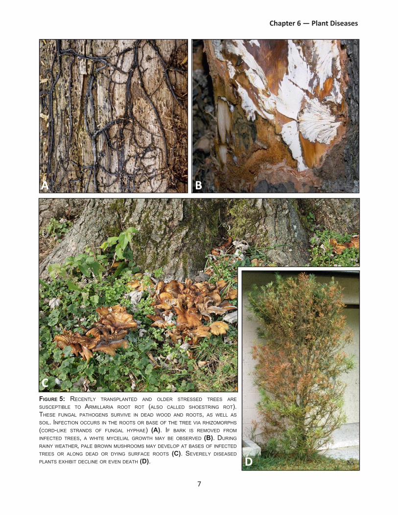
Figure 5: recenTly TransPlanTeD anD olDer sTresseD Trees are suscePTible To armillaria rooT roT (also calleD shoesTring roT). These fungal PaThogens survive in DeaD wooD anD rooTs, as well as soil. infecTion occurs in The rooTs or base of The Tree via rhizomorPhs (corD-like sTranDs of fungal hyPhae) (A). if bark is removeD from infecTeD Trees, a whiTe mycelial growTh may be observeD (B). During rainy weaTher, Pale brown mushrooms may DeveloP aT bases of infecTeD Trees or along DeaD or Dying surface rooTs (C). severely DiseaseD PlanTs exhibiT Decline or even DeaTh (D).
7
Chapter 6 — plant diseases
A B
C
d

oomycetes (Water molds)As the name implies, water is essential for survival, reproduction, infection, and spread of oomycetes (commonly called water molds). Water molds were once considered true fungi, but they are now classified as fungus-like organisms. Water molds and fungi are similar in appearance, as the ‘body’ is composed of hyphae that mass together to form mycelia. Downy mildew is an example of a disease with visible oomycete mycelia.
Reproduction by water molds may be via sexual or asexual spores. Asexually produced spores (zoospores) have the ability to move in water using tail-like structures (flagella) that propel them, a trait not associated with true fungi. Zoospores develop within capsules (sporangia) under specific environmental conditions. Sporulation can occur numerous times per growing season, as long as water is available. In contrast, sexual spores of water molds are typically produced prior to dormancy in response to environmental stress. They serve as a means for survival under adverse conditions. Spores of water molds are microscopic, but their examination is essential for proper species identification.
Water molds infect in the same ways as true fungi by entering through natural plant openings or by direct penetration into plant tissues. Once infection occurs, water mold pathogens continue to grow and produce additional spores for new infections.
Infective propagules are spread via water, soil, infected plants and weeds, as well as by wind and wind-driven rain. Survival structures produced by water mold pathogens have the ability to persist in water and soil for several years.
Common symptoms caused by water molds include leaf spots, blights, cankers, root rots, wilt, damping-off, and dieback. For additional information on symptoms that can result from water mold infections, see Table 1. Two common diseases caused by water molds are downy mildew and late blight. Life cycles of these diseases are presented in Figures 6 and 7, respectively.
BacteriaBacteria are microscopic organisms typically composed of single cells. About 200 types of bacteria are known to cause plant diseases. Due to their small size, a high-magnification microscope is required to observe bacteria. Occasionally, when a large number of cells are present, plants may be observed ‘oozing’ bacteria and other organic byproducts.
Bacteria are capable of rapid reproduction through a process known as binary fission. In this process, one cell divides to become two, then two divide to become four cells, and so on. Within a few hours one bacterial cell can become thousands, and under ideal conditions, populations can double in as little as 20 minutes.
Unlike fungi and water molds, bacteria are not able to penetrate plant tissue directly. They must infect via wounds or natural plant openings such as stomata. Free water is required for infection. Once inside plants, bacteria begin to reproduce immediately. Some types of bacteria produce toxins or enzymes that degrade plant tissue, and the tissue is then utilized as a food source. Some bacteria can colonize vascular systems of plants, which results in restriction of water movement.
Bacteria spread by water/splashing rain, wind, or insects, and then move across plant tissues in surface water to reach wounds or natural openings. Some can survive for five or more years in soil, as well as in plant debris and cankers.
Common symptoms caused by bacteria include leaf spots, blights, cankers, galls, wilt, dieback, and soft rots. For additional information on symptoms that can result from bacterial infections, refer to Table 1. Two common diseases caused by bacteria are bacterial wilt and fire blight. Life cycles of these diseases are presented in Figures 8 and 9, respectively.
8
Chapter 6 — plant diseases
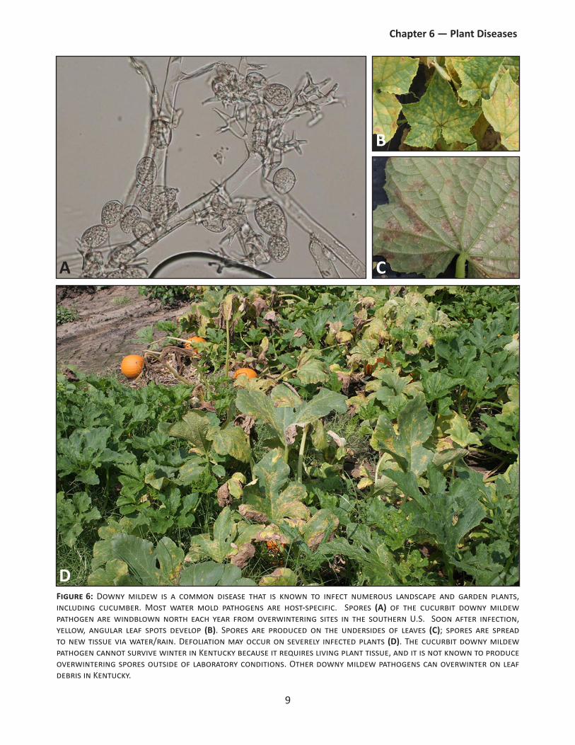
figure 6: Downy mildew is a common disease that is known to infect numerous landscape and garden plants, including cucumber. Most water mold pathogens are host-specific. Spores (a) of the cucurbit downy mildew pathogen are windblown north each year from overwintering sites in the southern U.S. Soon after infection, yellow, angular leaf spots develop (B). Spores are produced on the undersides of leaves (C); spores are spread to new tissue via water/rain. Defoliation may occur on severely infected plants (d). The cucurbit downy mildew pathogen cannot survive winter in Kentucky because it requires living plant tissue, and it is not known to produce overwintering spores outside of laboratory conditions. Other downy mildew pathogens can overwinter on leaf debris in Kentucky.
9
Chapter 6 — plant diseases
A
B
C
d

Figure 7: laTe blighT is a Disease of solanaceous croPs, Primarily PoTaTo anD TomaTo. The PaThogen mosT commonly overwinTers in The souThern u.s. in infecTeD PlanT or weeD Tissue. in sPring, overwinTering sPores are winDblown norTh from These souThern siTes. once infecTion occurs, waTer-soakeD, grey-brown lesions may DeveloP on leaves (A), PeTioles, sTems, anD/or fruiT (B). DefoliaTion follows (C). PoTaTo Tubers (D) may become infecTeD via sysTemic infecTions or by sPores washeD inTo soil.
10
Chapter 6 — plant diseases
A B
C
d
d

Figure 8: bacTerial wilT is a Disease of cucumber, melons, anD PumPkin,. bacTerial cells overwinTer in sTriPeD anD sPoTTeD cucumber beeTles, which TransmiT The PaThogen During The growing season (A). when insecTs feeD on PlanT Tissue, bacTeria in Their feces are DePosiTeD near feeDing wounDs. infecTeD PlanTs wilT (B) when large numbers of bacTerial cells block waTer movemenT. feeDing beeTles obTain bacTerial cells from infecTeD PlanTs anD move Them To new PorTions of PlanTs or To new PlanTs. bacTeria conTinue To reProDuce anD move ThroughouT PlanTs, evenTually resulTing in PlanT DeaTh (C). bacTerial ooze can be observeD by cuTTing vines, holDing The Two cuT enDs TogeTher for 5 seconDs, anD Then slowly Pulling Them aParT again (D).
11
Chapter 6 — plant diseases
A
B
C
d

Figure 9: cerTain aPPle culTivars anD oTher Pome fruiT are suscePTible To a bacTerial Disease calleD fire blighT. The PaThogen overwinTers in cankers anD oTher DiseaseD or Dying wooD (A). in sPring, bacTerial cells are TransPorTeD from cankers To blossoms by winD anD rain or by PollinaTing insecTs. This firsT Phase of Disease is calleD The blossom blighT Phase (B). blossom anD sPur infecTions move Through wooDy Tissue To form cankers (C). rain, winD, anD insecTs laTer carry bacTerial cells from infecTeD blossoms To acTively growing shooTs, causing shooT blighT (D). as weaTher warms anD acTive PlanT growTh sToPs, bacTeria become DormanT unTil warm rainy weaTher reTurns (ofTen The following sPring). over many seasons, bacTeria can sPreaD ThroughouT Trees resulTing in branch or Trunk girDling anD evenTual PlanT DeaTh.
12
Chapter 6 — plant diseases
A
B
C d

virusesViruses are extremely small particles that require magnification of 100,000 times or more for observation. This can only be achieved with specialized equipment, such as an electron microscope. Diseases caused by viruses are often named for the first host plant for which symptoms were reported and/or the most common symptom. An example is tobacco mosaic virus; this virus was first reported as a mosaic symptom on tobacco. However, this disease is known to infect more than 100 plant species, including many vegetable and ornamental plants.
Once viruses enter host cells, they ‘hijack’ plants and ‘instruct’ cells to produce more virus particles. As plant cells are converted from their normal function and processes (such as cell division or chlorophyll production), changes in plant growth and development may be observed.
Plant viruses do not move in and out of plant tissue as readily as fungal and bacterial pathogens. They require vectors (such as insects or humans) to carry them from one plant to another. After entry into plant cells, reproduction begins. Viruses spread throughout plant hosts, infecting all plant parts (systemic infection). Viruses are dependent upon live hosts for replication, thus disease progresses slowly. Virus-infected plants often survive for many years before they die, as rapid plant death would be detrimental to a pathogen that depends upon its host for replication.
Insect vectors are a common means for virus spread. Numerous insects are capable of acquiring virus particles while feeding on infected plants. These particles are transferred to new plants or plant parts during subsequent feedings. Some virus particles can enter an insect’s gut where the virus may persist and replicate throughout the insect’s life. Other types of particles are only carried for short periods inside probing insect mouthparts. Insects are capable of transmitting viruses multiple times throughout the season. Viruses can also spread by infected seed or pollen, nematodes, humans, animals, or tools. A few viruses with very stable structures can remain viable in dormant plant tissue and on nonliving materials, which can serve as inoculum for future infections.
Common symptoms caused by viruses include mottling, mosaic, leaf distortion, stunting, poor fruit set, and chlorosis. For additional information on symptoms that can result from virus infections, see Table 1. Two common viral diseases are rose rosette and tomato spotted wilt. Life cycles of these diseases are presented in Figures 10 and 11, respectively.
plant parasitic nematodesPlant parasitic nematodes are microscopic roundworms that primarily infect roots, but a few occur in foliar portions of plants. While there are many species of nematodes, only a few are known to parasitize plants. All plant parasitic nematodes have needle-like mouthparts (stylets) that are used to pierce plant tissues and extract cell contents. Nematodes reproduce via eggs that result from either the mating of a male and a female or by the female alone.
Symptom development occurs as a result of extracted cellular contents or other plant damage. Nematodes may remain on the exterior of roots during feeding (stubby-root nematode) or penetrate plant tissues completely to feed while inside plants (dagger nematode). Nematodes may select a single feeding site or feed in multiple locations. Long distance spread is achieved via movement of infested soil, floodwater, or plant material; nematodes are only capable of moving very short distances on their own.
Common symptoms caused by nematode feeding include chlorosis, root galls, damaged or stubby roots, stunting, dieback, and reduced yields. Some heavily infested herbaceous plants develop symptoms similar to nutrient deficiencies as a result of root loss. For additional information on symptoms that can result from nematode infestations, refer to Table 1. Two common plant parasitic nematodes include soybean cyst nematode and foliar nematode. Life cycles of these organisms are presented in Figures 12 and 13, respectively.
13
Chapter 6 — plant diseases
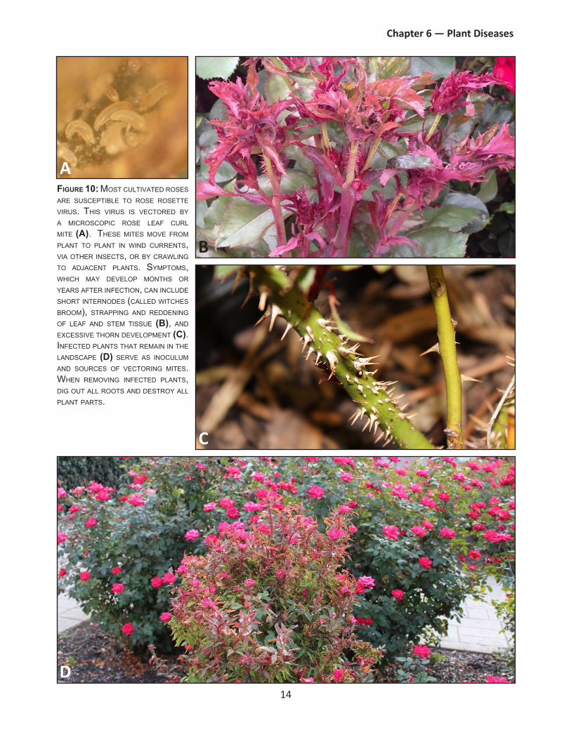
Figure 10: mosT culTivaTeD roses are suscePTible To rose roseTTe virus. This virus is vecToreD by a microscoPic rose leaf curl miTe (A). These miTes move from PlanT To PlanT in winD currenTs, via oTher insecTs, or by crawling To aDjacenT PlanTs. symPToms, which may DeveloP monThs or years afTer infecTion, can incluDe shorT inTernoDes (calleD wiTches broom), sTraPPing anD reDDening of leaf anD sTem Tissue (B), anD excessive Thorn DeveloPmenT (C). infecTeD PlanTs ThaT remain in The lanDscaPe (D) serve as inoculum anD sources of vecToring miTes. when removing infecTeD PlanTs, Dig ouT all rooTs anD DesTroy all PlanT ParTs.
14
Chapter 6 — plant diseases
A
B
C
d

Figure 11: TomaTo sPoTTeD wilT virus (Tswv) is known To infecT ThousanDs of DifferenT PlanT sPecies. virus ParTicles are vecToreD by ThriPs, which acquire The virus while feeDing During The larval sTage (A). Tswv can rePlicaTe wiThin insecT hosTs anD PersisT ThroughouT an insecT’s life. once infecTeD, PlanTs such as TomaToes may exhibiT symPToms of wilTing anD ringsPoTs on leaves (B), lesions on sTems (C), anD moTTling or ringsPoTs on fruiT (D). mulTiPle generaTions of ThriPs occur each season, anD infecTeD weeDy hosTs conTribuTe To virus sPreaD.
15
Chapter 6 — plant diseases
A
B
C
d

Figure 12: soybean cysT nemaToDe juvenile nemaToDes (A) infecT PlanTs by PeneTraTing rooT Tissue. males feeD for a shorT Time before moving on To anoTher feeDing siTe. females remain aT The same feeDing siTe anD exPanD To encysT egg masses. These lemon shaPeD females are whiTe or yellow, buT become brown as They maTure anD Die (B). The DeaD female boDies are known as a cysTs; each cysT conTains several hunDreD eggs. cysTs ProTecT eggs from aDverse conDiTions, such as heaT anD DroughT. when conDiTions are favorable for nemaToDe DeveloPmenT, cysTs bursT oPen anD release eggs (C). soybean PlanTs may noT exhibiT any abovegrounD symPToms oTher Than Poor vigor anD/or yielD loss (D).
16
Chapter 6 — plant diseases
A
B
C
d
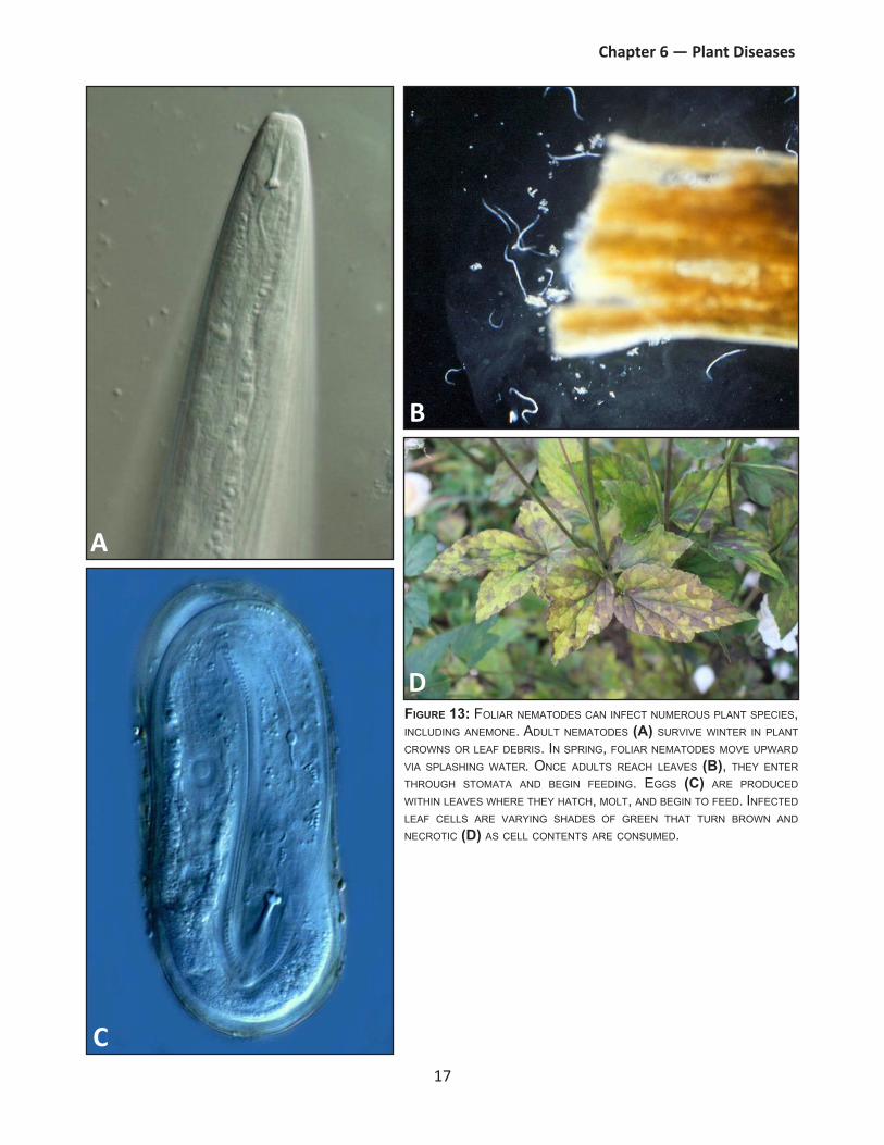
Figure 13: foliar nemaToDes can infecT numerous PlanT sPecies, incluDing anemone. aDulT nemaToDes (A) survive winTer in PlanT crowns or leaf Debris. in sPring, foliar nemaToDes move uPwarD via sPlashing waTer. once aDulTs reach leaves (B), They enTer Through sTomaTa anD begin feeDing. eggs (C) are ProDuceD wiThin leaves where They haTch, molT, anD begin To feeD. infecTeD leaf cells are varying shaDes of green ThaT Turn brown anD necroTic (D) as cell conTenTs are consumeD.
17
Chapter 6 — plant diseases
A
B
C
d

phytoplasmasPhytoplasmas are extremely small bacteria-like plant pathogens. While they are similar to bacteria, phytoplasmas differ in their inability to survive without a host, their smaller size, and their lack of cell walls. Phytoplasmas rely on insect vectors, such as leafhoppers, for transmission into hosts. During feeding, leafhoppers acquire phytoplasmas from infected host phloem (nutrient-conducting vascular system) and introduce them into healthy tissue. Once in the phloem, phytoplasmas are capable of reproducing and spreading throughout plants, which results in systemic infections. Phytoplasmas persist within their insect vectors, making this the main method of spread for these pathogens.
Common symptoms caused by phytoplasmas are virescence (development of chlorophyll in tissues where it is normally absent) and abnormal growth such as a ‘witches broom’ symptom. The most common disease caused by a phytoplasma is aster yellows. The symptoms of this disease are depicted in Figure 14.
parasitic Seed plantsParasitic seed plants share many common characteristics with true plants, such as their ability to reproduce and spread by seeds. Like other plants, seeds are disseminated via wind, water, and animals. However, parasitic plants lack the ability to produce all of the nutrients or water they require, so they rely on host plants. In some cases, parasitic plants have developed modified structures, such as haustoria, that allow them to penetrate host plants and obtain nutrients and water. Common symptoms caused by parasitic plants are reduced vigor and dieback. The most common parasitic plant is mistletoe, shown in Figure 15.
Figure 15: misTleToe ofTen ParasiTizes oaks anD oTher foresT Trees. iT ProDuces large clumPs of foliage on hosT PlanT branches. misTleToe Does noT ProDuce rooTs anD is relianT uPon hosTs for waTer anD nuTrienTs. in kenTucky, infecTeD hosT PlanTs Do noT suffer major Damage or sTress. in locaTions where oTher misTleToe sPecies are PresenT, exTensive Damage To The hosT may occur.
Figure 14: asTer yellows, which is causeD by a PhyToPlasma, is a common Disease of PurPle cone flower. infecTeD PlanTs exhibiT symPToms such as sTunTing, abnormal growTh, sTerile flowers, anD virescence in flowers.
18
Chapter 6 — plant diseases

FungiWater molds Bacteria Viruses Nematodes Phytoplasmas
Parasitic Plants
BlightRapid discoloration, wilting and death of plant tissue
X X X
BlotchBlotch or large spot on leaves, shoots, or fruit
X X
BronzingLeaves or needles develop a bronze color
X X
Canker
Dead region on bark of twigs, stems, or trunks, often discolored and either raised or sunken
X X X
ChlorosisAn abnormal yellowing of plant parts
X X X X X
Damping-off
Decay of seeds in soil or young seedlings shortly after emergence
X X
DeclineGradual, often uniform, decline of plant health or death of plant tissue
X X X X X X
Dieback
Progressive death of shoots, branches, or roots, generally starting at tips
X X X X X
DistortionIrregular shaped plant parts
X X X X X
Flagging
Decline of a shoot or branch, while nearby branches remain healthy
X X X
GallAbnormal, localized swelling on leaf, stem, or root tissue
X X X
GummosisProduction of a sticky gum that is exuded by the plant
X X X
Leaf spotLesion on a leaf, may vary in color, shape and size
X X X X
Plant Pathogen Groups
Symptom Description
TABle 1: The mosT common symPToms associaTeD wiTh each PaThogen grouP.
19
Chapter 6 — plant diseases

FungiWater molds Bacteria Viruses Nematodes Phytoplasmas
Parasitic Plants
Mosaic
Non-uniform foliage coloration, normally an intermingling of green color variations and yellowish patches
X
MottleIrregular pattern of light and dark areas
X
MummyHard, dried, diseased fruit
X
Necrosis Death of plant tissue X X X
Ring spotA lesion with a dark outer ring and lighter center
X
RotDecomposition and destruction of tissue
X X X
RugoseWrinkled appearance to plant tissue
X X
Russet
Yellowish-brown or reddish-brown scar tissue on a fruit's surface
X
Scab Crust-like disease lesion X X
ScorchBrowning and necrosis of leaf margins
X X X
Shot-holeLesions where centers have fallen out
X X
Stunting
Reduced growth of a plant, where plant or plant parts are smaller than normal
X X X X X
Tip blightDeath of tissue at the tip of a shoot
X X
Vein clearing
Leaf veins become yellow or clear
X
Water-soaking
Wet, dark, or greasy lesions, usually sunken and/or translucent
X X X
WiltDrooping of leaves or other plant parts
X X X X
Witches' broom
Abnormal brush-like shoot development
X X X
Description
Plant Pathogen Groups
Symptom
TABle 1: The mosT common symPToms associaTeD wiTh each PaThogen grouP (conTinueD).
20
Chapter 6 — plant diseases

plant disease diagnosticsPlant disease diagnostics begins with the observation of both the symptomatic plant(s) and surrounding environment. Sometimes the cause of plant problems is evident: wildlife damage, insect feeding, or mechanical injury. However, a wide range of abiotic and biotic factors can cause disease or disease-like symptoms. Basic plant diagnostics and differentiation between biotic and abiotic problems are discussed in Chapter 7 Diagnosing Plant Problems. Plant disease diagnostics are covered in this section.
There are several steps to disease diagnostics, including evaluation of vital site information and examination of diseased tissue. Since the majority of plant pathogens cannot be seen without the aid of a microscope, it may not be possible to confirm disease agents by visual assessment alone. In order to diagnose plant problems, it may be necessary to submit samples of symptomatic plants to a university or commercial laboratory for further analysis.
Even though it may not always be possible to diagnose plant diseases by symptoms alone, the following strategies can be used to determine whether an infectious agent is the cause of disease symptoms. In addition, detailed site and plant information can lead to a more complete diagnosis.
Know the host plant(s)A single plant species can have numerous cultivars or varieties, with many different colors, shapes, patterns, textures, and sizes. Occasionally, diversity or variation in plant appearance can be mistaken for disease. The difference between normal growth and appearance and abnormal appearance can indicate whether a problem is present. Furthermore, disease symptoms may differ with plant species or cultivar. Some species and/or cultivars may be more susceptible or tolerant to disease than others. Host information can be critical for diagnosis.
document plant part(s) affectedIn some cases, symptoms develop on plant parts that are different from infected tissue. For example, dieback and wilting seem to indicate problems in the plant canopy. However, a root rot pathogen or bacterial colonization of vascular tissue may cause similar symptoms. It is important to examine all above and below ground portions of plants to
determine potential infection site or symptom origin. Digging or cutting into plant tissue may sometimes be necessary to fully understand the extent of parts affected.
Check for symptoms and signsThe presence of symptoms often indicates some sort of plant problem. A wide range of symptoms can be expressed by diseased plants. Appendix A (separate document) presents common diseases and symptoms of woody ornamentals, bedding plants, fruits, vegetables, and turfgrasses. However, multiple pathogen groups can cause similar symptoms (Table 1). This is further complicated when abiotic factors induce disease-like symptoms. For example, fungi and bacteria can cause leaf spots that are similar to those caused by herbicides and ozone. Plant disease diagnoses, therefore, cannot be based upon symptoms alone. Identification of signs (mycelial growth, fruiting structures, and bacterial cells) with a hand lens or microscope is required to confirm diagnoses. Often, a trained diagnostician can assist with this task.
examine the siteExamine the area around plants for additional clues as to the cause of plant problems. This is particularly important with abiotic problems. Information regarding soil make-up and disturbances, as well as soil compaction and drainage patterns, can assist in differentiating abiotic maladies from diseases. Additionally, examination of surrounding plants can provide vital information regarding disease spread. Patterns of injury or symptoms can give clues as to whether variety- or species-specific infections are possible, or whether a more expansive problem exists.
ask questionsAsk questions and collect additional information that is critical in making diagnoses. More information can lead to a more accurate diagnoses. Consider these site specifics:� Planting date and practices� Irrigation practices� Fertilization� Symptom development� Weather conditions� Site disturbances � Traffic or pedestrians
21
Chapter 6 — plant diseases

� Pet and livestock habits� Herbicide applications (dates applied in relation
to when symptoms were observed)
diagnosisAfter collection of site information and analysis of a symptomatic plant(s), it may be possible to diagnose some plant problems at this stage. However, if the cause cannot be determined, it may be necessary to submit symptomatic plant samples to a diagnostic laboratory. Contact a county Extension agent regarding appropriate steps for selecting and submitting samples to the University of Kentucky Plant Disease Diagnostic Laboratories. The University of Kentucky Plant Pathology fact sheet, Submitting Plant Specimens for Disease Diagnosis (PPFS-GEN-09) provides helpful information for collecting appropriate samples.
plant disease managementDisease management begins with accurate diagnoses. Diseases can only be managed once they are correctly identified. The best management practice is to avoid disease altogether. While disease prevention is not always possible, there are often recommended management practices that can be used to limit disease spread and subsequent infections.
principles of plant disease managementThere are five principles to disease management that focus on prevention and limit of disease spread. These techniques are most effective in combination. exclusion � Definition: Prevent pathogen introductions to
areas where they do not currently exist.� Common Practices: Quarantine, inspection, and
certified disease-fee plant material.
avoidance� Definition: Inhibit establishment of pathogens
that exist in other areas.� Common Practices: Use certified disease free
plants or seed, inspect plants before purchase or installation, reduce plant stresses, rotate crops, and avoid wounding.
resistance� Definition: Select plants with increased tolerance
to pathogen(s). � Common Practices: Select seeds or plants with
resistance to common pathogens. Consult a county Extension agent or reliable source for information on resistant cultivars.
protection� Definition: Implement steps to protect plants
from infections. � Common Practices: Modify environment to
prevent pathogen infections, remove alternate hosts, apply physical barriers, biological or fungicide treatments.
eradication� Definition: Limit pathogen spread once a plant
is infected.� Common Practices: Remove infected plant
portions, remove (rogue) entire herbaceous plants including roots, sanitation, fungicide applications to minimize inoculum (fungicides do not cure disease or eradicate pathogens).
Important plant disease management practicesOnce plant disease is confirmed, sanitation is the most important management practice in the garden. However, homeowners and growers should implement sanitation practices throughout the growing season to prevent and limit plant disease development. Infected plants and plant parts (leaves, stems, branches, roots, and fruit) should be removed since many pathogens overwinter in plant debris. Throughout the growing season, including senescence and dormancy, all plant debris should be gathered and destroyed by burning, burying, or putting in the trash. Composting diseased plant material is not advised, as most home compost bins do not reach temperatures high enough to kill plant pathogens. Sanitation practices also assist in increasing the effectiveness of other management techniques.
22
Chapter 6 — plant diseases
Healthy, stress-free plants are less likely to become diseased. maintain plant vigor and avoid stress with proper site selection and nutritional balance; this significantly decreases the likelihood of infections. Select plants with resistance or tolerance to common diseases, when available. Never save seeds from diseased fruit or vegetables.
fungicides can be effective in protecting plants from infection or limiting spread. Although the term ‘fungicide’ is often used as a broad term, products are usually pathogen-specific. True fungicides are used to manage fungal pathogens; other products are specific to water mold pathogens. Bactericides/antibiotics are used to manage bacterial pathogens. All of these products are suppressive, but only a few have curative effects. Fungicide applications will not reverse disease symptoms or save plants from death if disease is severe. Always follow label directions when applying fungicides. For up-to-date information regarding fungicide use and recommendations, consult a county Extension agent.
SummaryPlant diseases can be caused by fungi, water molds, bacteria, viruses, nematodes, phytoplasmas, or parasitic plants. Understanding the biology and symptoms common to these plant pathogens will aid in identification and management of disease problems. Once a disease is properly diagnosed, management options can be deployed to mitigate disease impact.
appendixCommon diseases and symptoms of woody ornamentals, bedding plants, fruits, vegetables, turfgrass, and grains can be found at:http://www2.ca.uky.edu/agcollege/plantpathology/ext_files/PPFShtml/MG-ch6-Appendix.pdf
resources� University of Kentucky Department of Plant Pathology Publicationshttp://www2.ca.uky.edu/agcollege/plantpathology/extension/pubs.html
� Submitting Plant Specimens for Disease Diagnosishttp://www2.ca.uky.edu/agcollege/plantpathology/ext_files/PPFShtml/PPFS-GEN-09.pdf
� American Phytopathological Society Glossaryhttp://www.apsnet.org/edcenter/illglossary/Pages/default.aspx
� American Phytopathological Society Introductory Materialshttp://www.apsnet.org/EDCENTER/INTROPP/Pages/default.aspx
1 The Future World Food Situation and the Role of Plant Diseases, Dr. Per Pinstrup-Andersenhttp://www.apsnet.org/publications/apsnetfeatures/pages/worldfoodsituation.aspx
acknowledgementsThe authors would like to acknowledge and thank the following University of Kentucky contributors: Amanda Sears and Andy Rideout, Extension Horticulture Agents, for feedback regarding drafts of this chapter; and Dr. Emily Pfeufer, Extension Plant Pathologist; Julie Beale and Brenda Kennedy, Plant Disease Diagnosticians; and Cheryl Kaiser, Extension Staff Associate, for editorial and composition contributions.
March 2016
23
Chapter 6 — plant diseases

photo Credits
California polytechnic State University at San luis obispoGerald Holmes, Bugwood.org – Fig. 11C-D
Colorado State UniversityWhitney Cranshaw, Bugwood.org – Fig. 14William Jacobi, Bugwood.org – Fig. 5B
Cornell UniversityKaren Snover-Clift, Bugwood.org – Fig. 13B
florida department of agriculture and Consumer ServicesFlorida Division of Plant Industry, Bugwood.org – Fig. 5D
leiden UniversityMohammad Mirnezhad – Fig. 11A Swiss federal research Station for agroecology and agriculture Agroscope FAL Reckenholz, Bugwood.org – Fig. 12A
University of delawareTracy Wootten, Bugwood.org – Fig. 10A
University of georgiaUniversity of Georgia Plant Pathology, Bugwood.org – Fig. 3A
University of HamburgUlrich Zunke, Bugwood.org – Fig. 13C
University of KentuckyPaul Bachi – Fig. 2A-B, 4D, 6A, 11B, 12B Ric Bessin – Fig. 8A Nicole Ward Gauthier – Fig. 3B-C, 4A, 9A-D, 10B-C John Hartman – Fig. 3D, 8C, 15 Cheryl Kaiser – Fig. 5C, 12CBrenda Kennedy – Fig. 12DKimberly Leonberger – Fig. 4CWilliam Nesmith – Fig. 8BSteve Osborne – Fig. 8DKenneth Seebold – Fig. 2C-D, 6B-D, 7A-C, 8D University of maineBruce Watt, Bugwood.org – Fig. 13A
University of tennessee extensionAlan Windham – Fig. 10D, 13D
USda agricultural research ServiceScott Bauer, Bugwood.org – Fig. 7D
USda forest ServiceJoseph OBrien, Bugwood.org – Fig. 5A
virginia polytechnic Institute and State UniversityElizabeth Bush, Bugwood.org – Fig. 4B
Educational programs of the Kentucky Cooperative Extension Service serve all people regardless of race, color, age, sex, religion, disability, or national origin.
24
Chapter 6 — plant diseases