Cooperation between SMYD3 and PC4 drives a distinct ...
16
8868–8883 Nucleic Acids Research, 2015, Vol. 43, No. 18 Published online 8 September 2015 doi: 10.1093/nar/gkv874 Cooperation between SMYD3 and PC4 drives a distinct transcriptional program in cancer cells Jin-Man Kim 1 , Kyunghwan Kim 1,2 , Thomas Schmidt 3 , Vasu Punj 4 , Haley Tucker 5 , Judd C. Rice 1 , Tobias S. Ulmer 3 and Woojin An 1,* 1 Department of Biochemistry and Molecular Biology, University of Southern California, Norris Comprehensive Cancer Center, Los Angeles, CA 90033, USA, 2 Department of Biology, College of Natural Sciences, Chungbuk National University, Cheongju, Chungbuk 361-763, Republic of Korea, 3 Department of Biochemistry and Molecular Biology, Zilkha Neurogenetic Institute, Keck School of Medicine, University of Southern California, 1501 San Pablo Street, Los Angeles, CA 90033, USA, 4 Department of Medicine, Norris Comprehensive Cancer Center, 1450 Biggy Street, Los Angeles, CA 90033, USA and 5 University of Texas at Austin, Institute forCellular and Molecular Biology, Austin, TX 78712, USA Received April 09, 2015; Revised July 30, 2015; Accepted August 19, 2015 ABSTRACT SET and MYND domain containing protein 3 (SMYD3) is a histone methyltransferase, which has been im- plicated in cell growth and cancer pathogenesis. In- creasing evidence suggests that SMYD3 can influ- ence distinct oncogenic processes by acting as a gene-specific transcriptional regulator. However, the mechanistic aspects of SMYD3 transactivation and whether SMYD3 acts in concert with other transcrip- tion modulators remain unclear. Here, we show that SMYD3 interacts with the human positive coactiva- tor 4 (PC4) and that such interaction potentiates a group of genes whose expression is linked to cell proliferation and invasion. SMYD3 cooperates func- tionally with PC4, because PC4 depletion results in the loss of SMYD3-mediated H3K4me3 and target gene expression. Individual depletion of SMYD3 and PC4 diminishes the recruitment of both SMYD3 and PC4, indicating that SMYD3 and PC4 localize at tar- get genes in a mutually dependent manner. Artificial tethering of a SMYD3 mutant incapable of binding to its cognate elements and interacting with PC4 to target genes is sufficient for achieving an ac- tive transcriptional state in SMYD3-deficient cells. These observations suggest that PC4 contributes to SMYD3-mediated transactivation primarily by stabi- lizing SMYD3 occupancy at target genes. Together, these studies define expanded roles for SMYD3 and PC4 in gene regulation and provide an unprece- dented documentation of their cooperative functions in stimulating oncogenic transcription. INTRODUCTION SET and MYND domain-containing proteins (SMYD) are a special class of protein lysine methyltransferases involved in methylation of histones and non-histone proteins (1– 3). In human cells there are five members of the SMYD protein family, SMYD1–5, which share a distinctive ar- chitecture of their SET domain, split into two parts by a Myeloid-Nervy-DEAF1 (MYND) domain (4–6). SMYD3, which is the major focus of the current study, mediates tri- methylation of histone H3 at lysine 4 (H3K4me3) and acti- vates transcription of a set of downstream genes containing a specific DNA motif, 5 - CCCTCC - 3 (2). The MYND domain of SMYD3 is highly positively charged and likely contributes to the binding of SMYD3 to target sites (5,7). SMYD3 may regulate chromatin remodeling and gene tran- scription by interacting with other transcription factors. In support of this idea, it has been shown that SMYD3 in- teracts with RNA polymerase II and RNA helicase HELZ and promotes gene expression by facilitating transcrip- tional elongation (2,8). Our previous work also showed that SMYD3 directly interacts with estrogen receptor (ER) and up-regulates ER target genes via H3K4me3 (9). Besides its H3K4me3-dependent function, SMYD3 also modifies non- histone proteins to regulate specific cellular reactions as ex- emplified by MAP3K2 methylation (1). SMYD3 has been regarded as an important factor in cancer, based on the fact that high level expression of SMYD3 has cell proliferative effects and up-regulates a number of genes involved in cell growth and proliferation (1,2,10–12). Further support for such an oncogenic function of SMYD3 is provided by stud- ies demonstrating that suppression of SMYD3 expression by RNAi or other inhibitory reagents induces apoptosis and inhibits cell proliferation (2,13,14). * To whom correspondence should be addressed. Tel: + 1 323 442 4398; Fax: + 1 323 442 4433; Email: [email protected] C The Author(s) 2015. Published by Oxford University Press on behalf of Nucleic Acids Research. This is an Open Access article distributed under the terms of the Creative Commons Attribution License (http://creativecommons.org/licenses/by/4.0/), which permits unrestricted reuse, distribution, and reproduction in any medium, provided the original work is properly cited. Downloaded from https://academic.oup.com/nar/article-abstract/43/18/8868/2414494 by guest on 12 April 2018
Transcript of Cooperation between SMYD3 and PC4 drives a distinct ...
8868–8883 Nucleic Acids Research, 2015, Vol. 43, No. 18 Published online 8 September 2015doi: 10.1093/nar/gkv874
Cooperation between SMYD3 and PC4 drives adistinct transcriptional program in cancer cellsJin-Man Kim1, Kyunghwan Kim1,2, Thomas Schmidt3, Vasu Punj4, Haley Tucker5, JuddC. Rice1, Tobias S. Ulmer3 and Woojin An1,*
1Department of Biochemistry and Molecular Biology, University of Southern California, Norris Comprehensive CancerCenter, Los Angeles, CA 90033, USA, 2Department of Biology, College of Natural Sciences, Chungbuk NationalUniversity, Cheongju, Chungbuk 361-763, Republic of Korea, 3Department of Biochemistry and Molecular Biology,Zilkha Neurogenetic Institute, Keck School of Medicine, University of Southern California, 1501 San Pablo Street, LosAngeles, CA 90033, USA, 4Department of Medicine, Norris Comprehensive Cancer Center, 1450 Biggy Street, LosAngeles, CA 90033, USA and 5University of Texas at Austin, Institute for Cellular and Molecular Biology, Austin, TX78712, USA
Received April 09, 2015; Revised July 30, 2015; Accepted August 19, 2015
ABSTRACT
SET and MYND domain containing protein 3 (SMYD3)is a histone methyltransferase, which has been im-plicated in cell growth and cancer pathogenesis. In-creasing evidence suggests that SMYD3 can influ-ence distinct oncogenic processes by acting as agene-specific transcriptional regulator. However, themechanistic aspects of SMYD3 transactivation andwhether SMYD3 acts in concert with other transcrip-tion modulators remain unclear. Here, we show thatSMYD3 interacts with the human positive coactiva-tor 4 (PC4) and that such interaction potentiates agroup of genes whose expression is linked to cellproliferation and invasion. SMYD3 cooperates func-tionally with PC4, because PC4 depletion results inthe loss of SMYD3-mediated H3K4me3 and targetgene expression. Individual depletion of SMYD3 andPC4 diminishes the recruitment of both SMYD3 andPC4, indicating that SMYD3 and PC4 localize at tar-get genes in a mutually dependent manner. Artificialtethering of a SMYD3 mutant incapable of bindingto its cognate elements and interacting with PC4to target genes is sufficient for achieving an ac-tive transcriptional state in SMYD3-deficient cells.These observations suggest that PC4 contributes toSMYD3-mediated transactivation primarily by stabi-lizing SMYD3 occupancy at target genes. Together,these studies define expanded roles for SMYD3 andPC4 in gene regulation and provide an unprece-dented documentation of their cooperative functionsin stimulating oncogenic transcription.
INTRODUCTION
SET and MYND domain-containing proteins (SMYD) area special class of protein lysine methyltransferases involvedin methylation of histones and non-histone proteins (1–3). In human cells there are five members of the SMYDprotein family, SMYD1–5, which share a distinctive ar-chitecture of their SET domain, split into two parts by aMyeloid-Nervy-DEAF1 (MYND) domain (4–6). SMYD3,which is the major focus of the current study, mediates tri-methylation of histone H3 at lysine 4 (H3K4me3) and acti-vates transcription of a set of downstream genes containinga specific DNA motif, 5′ - CCCTCC - 3′ (2). The MYNDdomain of SMYD3 is highly positively charged and likelycontributes to the binding of SMYD3 to target sites (5,7).SMYD3 may regulate chromatin remodeling and gene tran-scription by interacting with other transcription factors. Insupport of this idea, it has been shown that SMYD3 in-teracts with RNA polymerase II and RNA helicase HELZand promotes gene expression by facilitating transcrip-tional elongation (2,8). Our previous work also showed thatSMYD3 directly interacts with estrogen receptor (ER) andup-regulates ER target genes via H3K4me3 (9). Besides itsH3K4me3-dependent function, SMYD3 also modifies non-histone proteins to regulate specific cellular reactions as ex-emplified by MAP3K2 methylation (1). SMYD3 has beenregarded as an important factor in cancer, based on the factthat high level expression of SMYD3 has cell proliferativeeffects and up-regulates a number of genes involved in cellgrowth and proliferation (1,2,10–12). Further support forsuch an oncogenic function of SMYD3 is provided by stud-ies demonstrating that suppression of SMYD3 expressionby RNAi or other inhibitory reagents induces apoptosis andinhibits cell proliferation (2,13,14).
*To whom correspondence should be addressed. Tel: + 1 323 442 4398; Fax: + 1 323 442 4433; Email: [email protected]
C© The Author(s) 2015. Published by Oxford University Press on behalf of Nucleic Acids Research.This is an Open Access article distributed under the terms of the Creative Commons Attribution License (http://creativecommons.org/licenses/by/4.0/), whichpermits unrestricted reuse, distribution, and reproduction in any medium, provided the original work is properly cited.
Downloaded from https://academic.oup.com/nar/article-abstract/43/18/8868/2414494by gueston 12 April 2018

Nucleic Acids Research, 2015, Vol. 43, No. 18 8869
The human positive coactivator 4 (PC4) is a multifunc-tional protein which plays a regulatory role in diverse cel-lular processes, including RNA polymerase II transcrip-tion, replication, heterochromatinization and DNA repair(15–18). In transcription reactions, PC4 interacts with un-winding DNA, the basal transcription machinery and gene-specific transcription factors (15,19,20). PC4 positively reg-ulates transcription by enhancing the effects of gene-specificactivators at the initiation levels and stimulating subsequentpromoter escape (21,22). PC4 does not have any chromatinremodeling and histone modification activities, but is in-volved in the regulation of chromatin dynamics (16–18,23).Besides serving as a transcription regulator, PC4 has beenreported to have additional functions in regulating DNAend joining and repair through its single stranded DNAbinding activity (16,18). By interacting with DNA repairprotein XPG, PC4 could also prevent spontaneous and in-duced oxidative mutagenesis (16). However, in contrast tothe proposed tumor suppressive function, PC4 has recentlybeen linked to cancer, as its aberrant expression has beenreported in several human cancers and cancer cell lines(24,25). A role for PC4 in cancer development is further sup-ported by the demonstration that PC4 expression in normaldermal fibroblasts drives the tumorigenic transformation ofthe cells (25). However, the molecular mechanisms underly-ing these phenomena are poorly understood.
In this study, we employed a combination of gene ex-pression profiling, transcription assays, ChIP-qPCR andCRISPR/dCas9 system to investigate a possible functionalinteraction between SMYD3 and PC4. Our data showthat SMYD3 colocalizes with PC4 at genes regulating cellproliferation and invasion and establishes transcriptionalcompetence in bladder and colon cancer cells. SMYD3and PC4 execute transcriptional programs in a cooper-ative manner, as shRNA-mediated knockdown of eitherSMYD3 or PC4 inhibits target gene transcription andcell proliferation/invasion. Furthermore, CRISPR/dCas9-based tethering of SMYD3 mutant deficient in PC4 inter-action and DNA binding to target genes is sufficient to me-diate transcriptional activation, strongly suggesting that thefunctional contribution of PC4 to SMYD3 transactivationis mainly through the enhancement of SMYD3 occupancyat target genes.
MATERIALS AND METHODS
Cell lines, constructs and antibodies
HT29, T24 and RT4 cells were maintained in McCoy’s5A medium containing 10% fetal bovine serum (FBS).HCT116, CaCO2, J82, MCF7, MDA-MB-231, DU145and UROtsa cells were maintained in Dulbecco’s mod-ified Eagle’s medium (DMEM) supplemented with 10%FBS (HCT116, CaCO2, J82, MCF7, MDA-MB-231 andDU145 cells) or 5% FBS (UROtsa cells). LNCaP andMLC cells were maintained in RPMI culture media andT medium with 10% FBS, respectively. MCF-10–2A cellswere grown in a 1:1 mixture of DMEM and Ham’s F12supplemented with 20 ng/ml epidermal growth factor, 100ng/ml cholera toxin, 0.01 mg/ml insulin, 500 ng/ml hy-drocortisone and 5% horse serum. CCD-18Co cells werecultured in Eagle’s Minimum Essential Medium with 10%
FBS. For mammalian expression of SMYD3 and PC4, theircDNAs were amplified by PCR and ligated into the correctreading frames of lentiviral expression vector pLenti-Hygro(Addgene) containing FLAG coding sequences. To gener-ate mutant SMYD3/PC4 expression vectors, SMYD3 andPC4 cDNAs were mutated by the QuikChange R© II site-directed mutagenesis kit (Agilent Technologies) before theconstruction. For bacterial expression of wild-type and mu-tant versions of SMYD3 and PC4, the corresponding cD-NAs were amplified by PCR and inserted into pGEX-4T1and pET15b vectors. For mammalian expression of dCas9-FLAG/dCas9-FLAG-SMYD3 wild-type and mutant, thenuclease-deficient dCas9 (D10A and H840A) cDNA (NotI-dCas9-EcoRI) was PCR-amplified, and ligated into pIRESvector digested by NotI and EcoRI. Wild-type or mutantSMYD3 coding sequences were excised from a vector andcloned into the EcoRI-BamH1-digested pIRES-dCas9 vec-tor. For sgRNA expression vectors, oligonucleotides listedin Supplementary Table S2 were annealed and ligated intoBsmBI-digested pMLM3636 vector (Addgene). Targetingspecificity and the extent of potential off-target activity ofdCas9-FLAG-SMYD3 were analyzed by using TagScan(http://www.isrec.isb-sib.ch/tagger), which is a web portalto provide tools for finding short exact matches in large se-quence databases (26). Further details of plasmid construc-tions are available upon request. Antibodies used in thisstudy are as follows: SMYD3, HSP90A, H3K4me1, H2Band H3 antibodies from Abcam, H3K4me2 antibody fromMillipore, Actin and FLAG antibodies from Sigma, SPT16and Nucleolin antibodies from Santa Cruze, PARP1 andH3K4me3 antibodies from Active Motif and PC4 antibodyfrom Bethyl Laboratories.
RNA interference
For the depletion of SMYD3 and PC4, DNA oligonu-cleotides encoding shRNAs specific for SMYD3 3′UTR region (5′-GCTGTGTGAACCTCTCTTATT-3′) and PC4 mRNA coding region (5′-GAACAGATTTCTGACATTGAT-3′) were annealedand ligated into the lentiviral expression vector pLKO.1(Addgene). Lentivirus particles were generated in 293Tcells by co-transfecting plasmids encoding VSV-G, NL-BHand the shRNAs. HCT116, CaCO2, J82 and T24 cells wereinfected by these viruses, and were selected with 2 �g/mlpuromycin (Sigma) for two weeks.
Gene expression microarray, qRT-PCR and ChIP-qPCR
Total RNA was isolated from two biological replicates ofmock- or SMYD3-depleted HCT116 cells using the TRI-zol reagent according to the manufacturer’s instructions(Invitrogen). Gene expression microarray experiments wereconducted using a whole-genome expression array (Hu-man HT-12 v4 Expression BeadChip, Illumina). Differen-tial gene expression was detected by using the ArrayPipesoftware (www.pathogenomics.ca/arraypipe). Genes show-ing detection P < 0.05 with fold change >2 were func-tionally analyzed in the context of gene ontology by us-ing Ingenuity pathway software (IPA; www.ingenuity.com).For qRT-PCR, total RNA was isolated as for microar-ray and subjected to reverse transcription using the iScript
Downloaded from https://academic.oup.com/nar/article-abstract/43/18/8868/2414494by gueston 12 April 2018

8870 Nucleic Acids Research, 2015, Vol. 43, No. 18
cDNA Synthesis Kit (Bio-Rad) and the PerfeCta SYBRGreen FastMix (Quanta biosciences) as recently described(27). Gene set enrichment analysis (GSEA) was performedusing GSEA (http://www.broad.mit.edu/gsea; Broad Insti-tute, Cambridge, MA) as detailed previously (28). Theprimers used for qRT-PCR are listed in Supplementary Ta-ble S3. ChIP assays with HCT116 cells were performedusing the ChIP assay kit (Millipore) as recently described(29). Antibodies specific to SMYD3, PC4, H3K4me1,H3K4me2, H3K4me3 and H3 were used for immunoprecip-itation. Immunoprecipitated DNA was analyzed with theprimers that amplify the different regions of the FNBP1,MFGE8, PDLIM7, WNT3A, SMUG1 and KRT81 loci.The primers used for qPCR are summarized in Supplemen-tary Table S4.
Cell proliferation and invasion assays
HCT116 cells were incubated with 0.5 mg/ml MTT(2-[4,5-dimethylthiazol-2-yl]-2,5-di-phenyltetrazolium bro-mide) (Sigma) for 2 h at 37◦C. The MTT formazan was dis-solved in 200 �l of DMSO. The absorbance of the solutionwas quantified at 570 nm against 650 nm by a microplatereader (Bio-Rad). For cell invasion assays, cells were har-vested and suspended in culture medium containing 5%FBS and then seeded to the upper chamber coated with Ma-trigel (BD Biosciences). Cells were allowed to invade toward10% FBS in the lower chamber for 48 h. The invaded cellson the underside of the transwell filters were fixed with 10%formaldehyde for 15 min and stained with 1% crystal violetfor 1 h. Cells were photographed and counted.
Purification and identification of SMYD3-interacting part-ners
HCT116 cells continuously expressing FLAG-HA-SMYD3were generated by stable transfection of pIRES-FLAG-HA-SMYD3. Nuclear extracts were prepared as described(30), and ectopic SMYD3 and its interacting proteinswere purified by sequential immunoprecipitation using anti-FLAG M2 and anti-HA antibodies (Sigma) in the precip-itation buffer (20 mM Tris-HCl, pH 7.3, 300 mM KCl, 0.2mM EDTA, 20% glycerol and 0.1% Nonidet P-40). Thepurified proteins were resolved by 4–20% gradient SDS-PAGE, and stained with Coomassie blue. Major proteinbands were excised and analyzed by liquid chromatography-tandem mass spectrometry (LC-MS/MS).
Histone methyltransferase assays
293 cells were transfected with dCas9-FLAG plasmid orplasmids expressing dCas9-FLAG-SMYD3 wild-type ormutant for 48 h, and the proteins were purified with anti-FLAG antibody. Recombinant histone octamers (1 �g)were incubated with immunoprecipitated dCas9-FLAG ordCas9-FLAG-SMYD3 for 1 h at 30◦C in HMT reactionbuffer (100 mM HEPES, pH 7.8, 300 mM KCl, 2.5 mMEDTA, 25 mM dithiothreitol and 50 mM sodium butyrate)in the presence of 50 �M S-adenosyl-methionine. Histoneproteins were resolved by 15% SDS-PAGE, and analyzedby immunoblotting with anti-H3K4me3 antibody.
Protein-protein interactions
For in vitro interaction studies with SMYD3, His-taggedSMYD3 wild-type and mutants were synthesized by us-ing TNT-Quick-coupled transcription/translation system(Promega), and incubated overnight with GST-PC4 immo-bilized on glutathione-Sepharose beads (Amersham Bio-sciences) at 4◦C in binding buffer (20 mM Tris-HCl,pH 7.3, 0.2 M KCl, 0.2 mM EDTA, 20% glycerol and0.01% Nonidet P-40). After washing the beads three timeswith the binding buffer, the beads were subjected toSDS-PAGE and immunoblotting with anti-His antibody.For binding assays with PC4, GST-PC4 wild-type andmutants were prepared from E.coli and incubated withHis-tagged SMYD3. For co-immunoprecipitation assays,FLAG-tagged SMYD3 proteins were expressed in HCT116cells, and whole cell lysates were prepared from the cellswith lysis buffer (50 mM Tris-HCl, pH 8.0, 150 mM NaCl,1 mM EDTA, 0.5% sodium deoxycholate, 0.1% SDS and1% Nonidet P-40). The cell lysates were mixed with anti-FLAG antibody-conjugated Sepharose beads (Sigma) andincubated overnight with gentile rotation at 4◦C. Afterremoving the supernatant, the sample pellets were an-alyzed by immunoblot analysis using anti-PARP1, anti-SPT16, anti-Nucleolin, anti-H2B and anti-PC4 antibodies.To co-immunoprecipitate endogenous SMYD3 and PC4,HCT116 cell lysates were incubated with anti-SMYD3 an-tibody or normal rabbit IgG for overnight. After centrifu-gation, immunocomplexes in the supernatants were precipi-tated with Protein A/G-Sepharose (Santa Cruz Biotechnol-ogy) and separated on SDS-PAGE. Immunoblot analyseswere performed with anti-PC4 and anti-SMYD3 antibod-ies.
DNA binding assays
For in vitro binding assays, streptavidin-coated 96wellplates were incubated with biotin-conjugated oligonu-cleotides containing three copies of SMYD3 bindingsites for 2 h at room temperature. The sequencesof oligonucleotides were: 5′- CGTATTCCCTCCAT-ACTCGCGTATTCCCTCCATACTCGCGTATTCC-CTCCATACTCG -3′-biotin (sense) and 5′- CGAGTATG-GAGGGAATACGCGAGTATGGAGGGAATACGC-GAGTATGGAGGGAATACG -3′-biotin (antisense).The plates were washed three times with wash buffer(20 mM Tris, pH 7.2, 150 mM NaCl, 0.1% BSA and0.05% Tween-20), and incubated with SMYD3 wild-typeor mutant in binding buffer (20 mM Tris, pH 7.2, 150mM NaCl, 0.4 mM EDTA, 0.5 mM DTT, 12% glyceroland 1 �g/�l poly dI/dC) for 1 h at room temperaturewith gentle rotation, added PC4 wild-type or mutant,and then further incubated for 1 h at room temperature.After washing with wash buffer, the plates were incubatedwith FLAG/His antibodies for 1 h and then horseradishperoxidase-conjugated secondary antibody for 0.5 h. TMBsubstrate (Pierce) was used for color development at 450nm, and absorbance was read by a Plate Chameleon Vplate reader.
Downloaded from https://academic.oup.com/nar/article-abstract/43/18/8868/2414494by gueston 12 April 2018

Nucleic Acids Research, 2015, Vol. 43, No. 18 8871
RESULTS
SMYD3 activates genes governing cell proliferation and in-vasion in cancer cells
As SMYD3 is often overexpressed in cancer cells, we firstexamined SMYD3 levels in bladder, breast, colon andprostate cell lines by immunoblotting. Three bladder (J82,T24 and RT4) and three colon (HCT116, CaCO2 andHT29) cancer cell lines expressed significantly higher lev-els of SMYD3 compared to their normal counterparts(UROtsa and CCD-18Co) (Supplementary Figure S1A). Incontrast, higher expression of SMYD3 was not evident inbreast (MCF7 and MDA-MB-231) and prostate (LNCaPand DU145) cancer cell lines compared to normal cell lines(MCF-10–2A and MLC). The observed overexpression ofSMYD3 in bladder and colon cancer cell lines encour-ages the possibility that SMYD3 may facilitate key tumori-genic processes such as cell proliferation and invasion. Totest this possibility, we depleted SMYD3 in two bladder(J82 and T24) and two colon (HCT116 and CaCO2) can-cer cells expressing high levels of SMYD3. In this study,it was important that SMYD3 is depleted for prolongedperiods, as this allows the study of progressive alterationsof cell proliferation and invasion under identical condi-tions. This was achieved by using a lentiviral shRNA in-fection system. Immunoblot analysis confirmed that sta-ble transfection of SMYD3 shRNA efficiently silenced theexpression of SMYD3 in the cancer cells (SupplementaryFigure S1B). MTT assays over a period of 4 days repro-ducibly showed that the bladder and colon cancer cells pro-liferate much more slowly following the depletion of en-dogenous SMYD3 (Supplementary Figure S1C). SMYD3knockdown also led to a significant decrease in cell invasioncompared with mock-depleted control cells (Supplemen-tary Figure S1D). These observations are consistent withthe hypothesis that SMYD3 is one of the key players stim-ulating proliferation and invasiveness of bladder and coloncancer cells.
To assess the functional contributions made by SMYD3in the above results, we performed comprehensive mi-croarray analysis using total RNA isolated from mock-or SMYD3-depleted HCT116 colon cancer cells. With afold-change cutoff of 2.0 and P-value cutoff of 0.005, thegene expression profiling identified 264 genes whose ex-pression was decreased in response to SMYD3 knock-down (Figure 1A; Supplementary Table S1). GSEA ofthe genes ranked by the ratios of transcripts from con-trol and SMYD3-depleted cells identified significant enrich-ments of nine gene sets with P-value of <0.01 and FDR<0.05 (Supplementary Table S5). Representative GSEA-scoring plots and their corresponding heat maps indicatingthe enrichments of proliferation/invasion-related genes areshown (Figure 1B). IPA functional analysis of the repressedgenes revealed that many of the genes down-regulatedupon SMYD3 knockdown encode cell death/survival andcell proliferation/growth regulators (Supplementary Ta-ble S6), including those known to be key components ofcancer initiation and progression. Our transcript profilingalso showed that 84 genes were activated when SMYD3was depleted. However, growth-stimulatory and prolifer-
ative genes appeared preferentially down-regulated un-der SMYD3 knockdown conditions (Supplementary Ta-bles S6 and S7), suggesting that SMYD3-mediated acti-vation of target genes is more functional in cancer cells.These genes also tend to exhibit higher ratios of expres-sion changes, which is in agreement with the primarily ac-tivating function expected for SMYD3 during cancer de-velopment (2,10,12,13). As an experiment to confirm themicroarray results, qRT-PCR analysis of 17 putative targetgenes in HCT116 and three other cancer cell lines (CaCO2,J82 and T24) showed that SMYD3 knockdown caused 40–70% decreases in their mRNA levels (Supplementary Fig-ures S2 and S4). To further validate our microarray results,we checked the rescue potential of ectopic SMYD3 (Sup-plementary Figure S3A). The expression of SMYD3 wild-type in SMYD3-depleted cells fully restored the transcrip-tion of target genes, whereas SMYD3 enzymatic dead mu-tant (F183A) was much less efficient in restoring their tran-scription rates (Figure 1C).
Since SMYD3 functions as a transcription regulator bybinding to a specific DNA sequence (5′-CCCTCC-3′) in itstarget genes, we searched for this SMYD3 binding elementaround the transcription start sites (TSSs) of up- and down-regulated genes. We found that 176 (67%) out of 264 down-regulated genes and 49 (58%) out of 84 up-regulated genesin SMYD3-depleted cells carry the potential SMYD3 bind-ing site (Supplementary Table S8). Thus, it is likely that thesite specific binding of SMYD3 to its consensus site un-derlie the ability of SMYD3 to regulate these target genes.To explore this possibility, we tested whether SMYD3 wasbound to the four selected target genes (FNBP1, MFGE8,PDLIM7 and WNT3A) which were repressed after SMYD3knockdown and contain putative SMYD3 binding elementswithin 300 bp upstream of the TSSs (Supplementary Fig-ures S5A and S5B). The selected target genes are known tofacilitate cell proliferation via integrin/ERK1/2 signaling(31) or ERK-mediated Op18/stathmin signaling (32) andcell invasion via invadopodia formation (33) or epithelial-mesenchymal transition (34). As expected, ChIP and quan-titative PCR (ChIP-qPCR) assays showed high levels ofSMYD3 occupancy at the SMYD3 binding elements inmock-depleted bladder (J82 and T24)/colon (HCT116 andCaCO2) cancer cells, but SMYD3 levels were markedly de-creased after SMYD3 knockdown (Supplementary FigureS5C). On the contrary, ChIP analysis showed no enrich-ments of SMYD3 at the KRT81 gene whose expressionwas not affected by SMYD3 knockdown in our microar-ray analysis. The presence of a putative SMYD3 cognateelement in this gene indicates that SMYD3 is incapable ofbinding to all genes containing its cognate element. To in-vestigate the mechanisms by which SMYD3 depletion leadsto inactivation of target genes, we also checked H3K4me3on target genes in mock-depleted and SMYD3-depletedcells. High levels of H3K4me3 were detected around theSMYD3 binding elements of target genes, but such en-richment was significantly reduced upon SMYD3 deple-tion (Supplementary Figure S5D). These results imply thatSMYD3 is important for establishing the active H3K4me3mark on proliferation/invasion-stimulatory genes in cancercells.
Downloaded from https://academic.oup.com/nar/article-abstract/43/18/8868/2414494by gueston 12 April 2018

8872 Nucleic Acids Research, 2015, Vol. 43, No. 18
Figure 1. SMYD3 knockdown suppresses cell proliferation/invasion-related genes. (A) Control or SMYD3-depleted HCT116 cells were subjected tomicroarray analysis. Dots in the two-dimensional scatter-plot represent expression values for the genes with a folder change >2 in either of two independentanalyses (Batch #1 and Batch #2). These analyses identified 264 genes that were down-regulated (indicated by green dots) and 84 genes that were up-regulated (indicated by red dots) in the SMYD3-depleted cells, compared to the control cells. (B) Summary of GSEA of microarray data. The Y-axis plotsthe enrichment score and X-axis is the rank of genes differentially expressed in SMYD3-depleted cells. Bar codes below enrichment plots show the rankposition of individual genes differentially expressed in SMYD3-depleted cells. (C) SMYD3 wt and SMYD3 F183A were expressed in SMYD3-depletedHCT116 cells, and relative mRNA levels of the four down-regulated (FNBP1, MFGE8, PDLIM7 and WNT3A) and two unaffected (KRT81 and GAPDH)genes in SMYD3-depleted HCT116 cells were quantified by qRT-PCR. (D) ChIP-qPCR assays were performed in control or SMYD3-depleted HCT116cells complemented with SMYD3 wt or SMYD3 F183A by using SMYD3, H3K4me1, H3K4me2, H3K4me3 and H3 antibodies. Precipitation efficienciesrelative to non-enriched input samples were determined for the six locations across the FNBP1 locus by qPCR with primers depicted on the left andlisted in Supplementary Table S4. Percent input is determined as the amount of immunoprecipitated DNA relative to input DNA. The enrichment ofH3K4me1/me2/me3 was calculated as the ratio of anti-H3K4me1/me2/me3 ChIP to anti-H3 which is an indication of local nucleosome density. Errorbars represent the SD obtained from three independent experiments. The vertical red bar represents the putative SMYD3 binding site. (E) SMYD3-depletedHCT116 cells were infected with SMYD3 wt or SMYD3 F183A as in Supplementary Figure S3A, and changes in cell proliferation rates were measuredby the MTT colorimetric assay every day for 4 days post-infection. (F) SMYD3-depleted HCT116 cells were complemented with SMYD3 wt or SMYD3F183A, and cell invasion activity was determined by the Matrigel invasion assay.
Downloaded from https://academic.oup.com/nar/article-abstract/43/18/8868/2414494by gueston 12 April 2018
Nucleic Acids Research, 2015, Vol. 43, No. 18 8873
In order to gain further insight into how SMYD3 reg-ulates its target genes, SMYD3 occupancy was probed forthe six different regions of the FNBP1 and MFGE8 genes byChIP-qPCR, as depicted in Figure 1D and SupplementaryFigure S3B. In mock-depleted control cells, SMYD3 sig-nals were high at the proximal promoter region containinga putative SMYD3 binding element (region C) and prox-imal coding region (region D), but low SMYD3 signalswere detected at four other regions (regions A, B, E andF) (Figure 1D and Supplementary Figure S3B). It has beendemonstrated that SMYD3 participates in the early stepsof transcriptional elongation and facilitates the transitionfrom transcription initiation to early elongation (8). Thisnotion is supported by the fact that the levels of SMYD3occupancy are higher at the proximal coding region (po-sition D) than at promoter region (position C). Moreover,SMYD3 showed a pattern of accumulation indistinguish-able from that of H3K4me3, strongly suggesting a majorrole for SMYD3 in mediating higher levels of H3K4me3in these target genes (Figure 1D and Supplementary Fig-ure S3B). Indeed we found that SMYD3 knockdown re-duced levels of SMYD3 in the FNBP1 and MFGE8 genes,and such changes diminished H3K4me3 levels (Figure 1Dand Supplementary Figure S3B). These results were furthervalidated by rescue experiments demonstrating that ectopicexpression of SMYD3 wild-type (wt), but not SMYD3 en-zymatic dead mutant (F183A), largely overrides H3K4me3defects caused by SMYD3 knockdown. MTT and cell inva-sion assays also showed that SMYD3 depletion decreasedthe proliferation and invasion of HCT116 cancer cells andthat the expression of SMYD3 wild-type, but not SMYD3F183A mutant, restored cell proliferation and invasion rates(Figure 1E and F).
SMYD3 interacts directly with PC4
Overall, our microarray and ChIP data establish the rolefor SMYD3 in regulating particular sets of genes in bladderand colon cancer cells. However, it is not clear whether theobserved effects of SMYD3 reflect its physical interactionwith other factors. To check this possibility, we generatedHCT116 colon cancer cell lines stably expressing SMYD3fused to FLAG and HA epitope tags. After preparing nu-clear extracts from cultured cells, ectopic SMYD3 andits associated partners were purified from nuclear extractsthrough sequential immunoprecipitations with FLAG andHA antibodies under stringent conditions (300 mM KCland 0.1% NP40). The proteins co-purified with SMYD3were resolved by SDS-PAGE, and major protein bands wereexcised and subjected to tandem mass spectrometric anal-ysis in conjunction with a sequence database search. Assummarized in Supplementary Table S9, we detected thestable association of 15 proteins with ectopic SMYD3 inthis analysis. As predicted from recent studies, we identi-fied HELZ and HSP90A that have been shown to inter-act with SMYD3 (2,35). Interestingly, our analysis also re-vealed 13 proteins that have never been described in as-sociation with SMYD3 (Figure 2A; Supplementary TableS9). The mass spectrometry data were validated by im-munoblot analysis using available antibodies (Figure 2B).A noteworthy observation emerged from our purification
was a substantial interaction of SMYD3 with PC4, whichis a transcription factor possessing the ability to co-activateRNA polymerase II-mediated transcription. PC4 has beenreported to be overexpressed in several types of cancer andcontribute to tumorigenesis via transcriptional activation ofother genes involved in cancer pathogenesis (24,25,36). PC4has also been shown to play a role in the regulation of chro-matin transcription through its influence on histone mod-ifications (23,37). Thus the fact that PC4 stably associateswith SMYD3 in our purification prompted us to study thepossible influences of PC4 on SMYD3-mediated transacti-vation and cancer cell proliferation/invasion.
To gain support for the binding results described above,we first immunoprecipitated HCT116 cell lysates with anti-SMYD3 antibody and checked the coimmunoprecipitationof endogenous PC4. In addition to SMYD3, we could con-firm the presence of PC4 in our immunoprecipitates (Fig-ure 2C). We then conducted in vitro pull-down assays usingbacterially produced His-tagged SMYD3 and GST-fusedPC4. As shown in Figure 2D, GST-PC4 efficiently inter-acted with His-SMYD3, whereas GST alone did not. Insimilar binding experiments with truncated versions of PC4,SMYD3 interacted with two PC4 deletion mutants lack-ing the first 21 or last 40 residues (PC4 22–127 and PC41–87), but not with another PC4 mutant lacking the last66 residues (PC4 1–61), indicating that amino acid residues62–87 play a major role in PC4 binding to SMYD3. In map-ping PC4-interacting region of SMYD3, the binding of theN-terminal domains (residues 1–100 and 1–250) was readilydetectable, but the two C-terminal domains (residues 101–428 and 251–428) showed no interaction with PC4 (Fig-ure 2E). In additional binding assays, SMYD3 deletion mu-tants lacking residues 1–44 and 1–74 retained their affinityfor PC4, while no apparent interaction was observed withtwo SMYD3 mutants lacking residues 75–87 and 75–100(Supplementary Figure S6). These results reinforce the con-clusion that the primary PC4-binding capacity of SMYD3resides between residues 75 and 87.
We next attempted to identify amino acid residues crit-ical for the SMYD3–PC4 interaction. Circular dichroismspectroscopy of SMYD3, PC4 and the SMYD3–PC4 com-plex showed that all proteins were folded with the com-plex representing the superposition of the individual struc-tures (Supplementary Figure S7). To facilitate the search forresidues in the above-outlined interaction interface, we cre-ated a structural model of the SMYD3–PC4 complex us-ing the crystal structures of SMYD3 (4) and PC4 (19,20).Our model highlighted the N-terminal helix of SMYD3 andthe central �-sheet of PC4 to be important in their interac-tion (Figure 2F). Specifically, K78 and D82 of SMYD3 ap-peared positioned to make contacts with Q65 and R75 ofPC4, respectively (Figure 2F). Individual mutation of Q65and R75 to alanine had some effect on the PC4 interactionwith SMYD3 (Figure 2G, low panel). However, an essen-tially complete abrogation of the PC4–SMYD3 interactionrequired the alanine substitution of both Q65 and R75. Insimilar binding assays using SMYD3 mutants, the muta-tion of K78 and D82 individually did not affect SMYD3interaction with PC4, and mutation of both residues onlyminimally decreased PC4 binding activity of SMYD3 (Fig-ure 2H, right panel). This result indicated that additional
Downloaded from https://academic.oup.com/nar/article-abstract/43/18/8868/2414494by gueston 12 April 2018

8874 Nucleic Acids Research, 2015, Vol. 43, No. 18
Figure 2. PC4 is an interaction partner of SMYD3. (A) FLAG-HA-tagged SMYD3 was expressed in SMYD3-depleted HCT116 colon cancer cells andsequentially immunoprecipitated using anti-FLAG and anti-HA antibodies. The co-purified proteins were separated by 4–20% gradient SDS-PAGE andidentified by liquid chromatography-tandem mass spectrometry (LC-MS/MS). The positions of the molecular mass markers are indicated on the left. (B)The purified SMYD3 sample shown in (A) was resolved in 4–20% gradient SDS-PAGE and analyzed by immunoblotting with the indicated antibodies. (C)Whole cell lysates were prepared from HCT116 cells, immunoprecipitated with anti-SMYD3 antibody or control IgG, and analyzed by immunoblottingwith anti-PC4, anti-HSP90A and anti-SMYD3 antibodies. Input represents 20% of the cell extracts used in immunoprecipitation. Asterisk indicates non-specific bands. (D) GST alone or GST-PC4, immobilized on glutathione-Sepharose beads, was incubated with full length His-SMYD3 or its deletionmutants. After extensive washing with washing buffer, bound SMYD3 proteins were fractionated by 10% SDS-PAGE, and immunoblotted with anti-Hisantibody. Five percent of the input proteins were examined by immunoblotting. (E) His-tagged SMYD3 was incubated with immobilized GST or theindicated GST-H1.2 fusions, and bound SMYD3 proteins were visualized by immunoblotting with anti-His antibody. Input represents 5% of SMYD3protein used in the binding reactions. Asterisks indicate non-specific bands. (F) The model of the SMYD3–PC4 complex was built on the basis of crystalstructures of SMYD3 (4) and PC4 (19,20) using the molecular modeling program Insight II (Molecular Simulations Inc.). Residues located at the interfaceare indicated. SMYD3 is in blue and PC4 is in green. (G) Pull-down experiments were performed as described in (E), but using GST-PC4 1–127 carryingthe indicated point mutations. The three short parallel �-sheets are indicated above the sequence. (H) GST pull-down assays were conducted as in (E), butusing His-SMYD3 1–428 harboring the indicated point mutants. Alignment of the amino-acid stretch encompassing the PC4 binding region of SMYD3relative to SMYD1 and SMYD2 is shown on the left for comparison. The �-helical structural elements are indicated above the sequence.
Downloaded from https://academic.oup.com/nar/article-abstract/43/18/8868/2414494by gueston 12 April 2018

Nucleic Acids Research, 2015, Vol. 43, No. 18 8875
SMYD3 residues were important for the interaction withPC4. Among the SMYD family, R85 is, aside K78 andD82, unique to the PC4 binding domain of SMYD3 (Fig-ure 2H). We found that simultaneous alanine substitutionsof K78, D82 and R85 significantly incapacitated SMYD3from interacting with PC4, whereas R85-mutated SMYD3had a binding affinity comparable to wild-type SMYD3(Figure 2H). These results clearly establish SMYD3–PC4interaction centered on the SMYD3 N-terminal �-helix andPC4 central �-sheet, suggesting that PC4 can directly mod-ulate SMYD3 transactivation.
SMYD3 and PC4 cooperate to drive gene expression pro-gram in cancer cells
The observed interaction between SMYD3 and PC4 raisedthe possibility that SMYD3 functionally cooperates withPC4 to establish the active state of target genes in bladderand colon cancer cells. Congruent with these ideas, whenthe genes that were down-regulated in SMYD3-depletedcells were analyzed by qRT-PCR, their transcription lev-els were completely recovered after the ectopic expres-sion of SMYD3 (Figure 3A and Supplementary FigureS8A). However, the expression of SMYD3 K78/D82/R85mutant, which is unable to interact with PC4, failed toshow any recovery effect on target gene transcription (Fig-ure 3A). Having demonstrated a functional interaction be-tween SMYD3 and PC4, we then asked whether the ob-served function of PC4 in SMYD3 transactivation reflectsits effects on SMYD3 binding to target DNA sequences.To this end, we immobilized biotinylated SMYD3 cog-nate DNA elements on streptavidin-coated microtiter platesand carried out SMYD3 binding assays in the presence ofPC4. In our color reaction-based measurements of SMYD3binding affinity, more efficient association of SMYD3 withits binding elements was seen if SMYD3 was added with in-creasing concentration of PC4, while only minor effects ofPC4 were observed when SMYD3 mutant was used (Figure3B).
To elucidate the significance of PC4 with respect toSMYD3 transactivation, we next investigated PC4 localiza-tion at the FNBP1 and MFGE8 genes by ChIP assays em-ploying cross-linked chromatin isolated from control andSMYD3-depeleted cancer cells. Immunoblotting confirmedthat SMYD3 knockdown had no effect on the expres-sion levels of PC4 in HCT116 cells (Supplementary FigureS8A). The distribution patterns of PC4 across the FNBP1and MFGE8 genes were similar to those of SMYD3 andH3K4me3, with a peak around the proximal promoter andcoding regions (regions C and D) and a gradual decreaseover the distal promoter and coding regions (regions A,B, E and F) (Figure 3C and Supplementary Figure S8B).By comparison, SMYD3 depletion reduced the levels ofPC4 at target genes, strongly indicating that SMYD3 isindispensable for the initial recruitment of PC4 to targetgenes. Although beyond the scope of the present study,higher PC4 occupancy at the proximal coding region (po-sition D) is consistent with accumulating evidence suggest-ing that PC4 stimulates transcription beyond the initiationstage (17,19,22,38). The requirement of SMYD3 in PC4 re-cruitment was further supported by the fact that expressing
SMYD3 wild-type, but not SMYD3 K78/D82/R85 mutantwhich is deficient in PC4 binding, in SMYD3-depleted cellsresulted in a substantial increase in the levels of PC4 at tar-get genes. Given that SMYD3 promotes cell proliferationand invasion, we also examined whether SMYD3 capacityto interact with PC4 is necessary for the observed effects.Unlike SMYD3 wild-type, PC4 binding-deficient SMYD3mutant failed to recover the proliferation and invasivenessof SMYD3-depleted cancer cells (Figure 3D and E), thusdemonstrating that SMYD3-mediated recruitment of PC4is critical for SMYD3 function at genes involved in cell pro-liferation and invasion.
To further investigate the impact of PC4 onSMYD3function, we depleted PC4 and evaluated itseffects on target gene expression. As confirmed by im-munoblot, the transfection of PC4-targeted shRNAresulted in an almost complete disappearance of PC4without affecting SMYD3 expression in HCT116 cells(Supplementary Figure S8C). PC4 knockdown generated adistinct repression of SMYD3 target genes, and PC4 wild-type, but not SMYD3-bindining deficient PC4 mutant,could rescue the expression of target genes (Figure 4A andSupplementary Figure S8C). In our mechanistic studiesaddressing how PC4 contributes to transactive function ofSMYD3 at target genes, free SMYD3 exhibited circulardichroism spectra almost identical to those of PC4-boundSMYD3 (Supplementary Figure S7), thus ruling outthe possibility that PC4 alters SMYD3 structure morefavorable for its DNA binding. Another possibility is thatPC4 interacts with SMYD3 MYND domain bound toits target sites, and stabilizes the SMYD3–DNA interfacewithout altering overall structures. Consistent with thisidea, our binding assays employing SMYD3 cognate DNAelements immobilized on streptavidin-coated microtiterplates demonstrated that SMYD3 binds to its targetsequence more strongly in the presence of PC4 wild-typecompared to PC4 mutant (Figure 4B). ChIP analyses toelucidate the nature of PC4 recruitment patterns furtherrevealed a perfect correlation of PC4 localization withSMYD3 occupancy and H3K4me3 enrichment on theFNBP1 and MFGE8 genes (Figure 4C and SupplementaryFigure S8D). PC4 occupancy of the target genes wasreduced after knockdown or Q65/R75 mutation of PC4,but such changes also diminished the levels of SMYD3and H3K4me3. The rescue of PC4 knockdown resulted ina return of SMYD3-mediated H3K4me3, demonstratingthe reversibility of this modification process. As a control,SMYD3 knockdown and rescue had no effect on PC4localization and transcription activity at the SMUG1 genewhich is a known PC4 target gene and is not affected bySMYD3 knockdown in our microarray studies (Supple-mentary Figure S9). The importance of PC4 with respectto SMYD3 function was further confirmed by the findingthat wild-type PC4 fully restored the proliferation ratesof the cancer cells depleted of PC4, whereas SMYD3binding-deficient PC4 was much less efficient in restoringthe proliferation rates (Figure 4D). Essentially identicalresults were obtained from invasion assays (Figure 4E).Thus, the expression of wild-type PC4, but not SMYD3binding-deficient PC4, rescued cell invasion rates.
Downloaded from https://academic.oup.com/nar/article-abstract/43/18/8868/2414494by gueston 12 April 2018

8876 Nucleic Acids Research, 2015, Vol. 43, No. 18
Figure 3. SMYD3 is required for PC4 localization and function at target genes. (A) SMYD3-depleted HCT116 cells were infected with SMYD3 wild-type(wt) or K78/D82/R85 mutant (mt) as in Supplementary Figure S7A, and the levels of target gene expression were measured by qRT-PCR. (B) FLAG-tagged versions of SMYD3 wt and SMYD3 mt were stably bound to biotin-conjugated SMYD3 binding sites attached to avidin-coated 96 well plates.After incubating wells with increasing concentrations of His-tagged PC4 wt, changes in SMYD3 affinity to its binding sites were quantitatively analyzedby sequential incubations with anti-FLAG antibody, a horseradish peroxidase conjugated secondary antibody, and a colorimetric peroxidase substrate.Absorbance was read at 450 nm on a microplate reader. (C) ChIP-qPCR assays of the FNBP1 gene were carried out using indicated antibodies in control orSMYD3-depleted HCT116 cells complemented with SMYD3 wt or SMYD3 mt. ChIP-enriched DNA was quantified by qPCR using the primers indicatedin the scheme at the top. (D) SMYD3-depleted HCT116 cells were complemented with SMYD3 wt or SMYD3 mt, and MTT proliferation assays werecarried out over a period of 4 days. (E) Matrigel cell invasion assays were performed with SMYD3-depleted HCT116 cells expressing SMYD3 wt orSMYD3 mt.
Downloaded from https://academic.oup.com/nar/article-abstract/43/18/8868/2414494by gueston 12 April 2018

Nucleic Acids Research, 2015, Vol. 43, No. 18 8877
Figure 4. PC4 regulates SMYD3 occupancy and function at target genes. (A) Total RNAs from control or PC4-depleted HCT116 cells complemented withPC4 wt or PC4 mt were subjected to qRT-PCR analysis to assess target gene expression levels. (B) Quantitative binding assays were performed as describedin Figure 3B, but using His-SMYD3 wt together with FLAG-tagged PC4 wt or PC4 mt. (C) ChIP-qPCR assays were performed in control or PC4-depletedHCT116 cells complemented with PC4 wt or PC4 mt. The relative position of the qPCR primers is indicated at the top. (D) MTT proliferation assays wereperformed with PC4-depleted HCT116 cells complemented with PC4 wt or PC4 mt over a period of 4 days. (E) Invasive potential of PC4-depleted HCT116cells complemented with PC4 wt or PC4 mt was monitored by Matrigel invasion assay.
Downloaded from https://academic.oup.com/nar/article-abstract/43/18/8868/2414494by gueston 12 April 2018

8878 Nucleic Acids Research, 2015, Vol. 43, No. 18
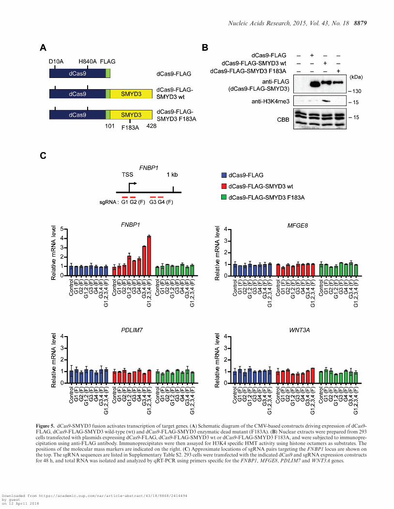
Artificial tethering of PC4-binding deficient SMYD3 to tar-get genes can generate
H3K4me3 and transactivation. The results of the aboveChIP and knockdown experiments argue persuasively thatPC4 is indispensable for the stable recruitment of SMYD3to target genes. However, these studies do not discount thepossibility that PC4 could contribute to SMYD3 transac-tivation via some other mechanisms. If PC4 exerts its co-operative effects mainly by enhancing SMYD3 binding toits cognate elements, then we can predict that artificiallydirecting a SMYD3 mutant incapable of interacting withPC4 and binding to target genes is sufficient to establishan active state of transcription. To examine this possibility,we chose to adapt CRISPR system for targeting SMYD3-mediated H3K4me3 to specific genes. We created genesencoding catalytically dead Cas9-FLAG (dCas9-FLAG)and dCas9-FLAG fused to N-terminal truncated SMYD3(residues 101–428) wild-type (wt) or enzymatic dead mu-tant (F183A), and constructed their expression plasmidsusing pIRES mammalian expression vector containing theCMV promoter (Figure 5A). When dCas9 fusion proteinswere immunoprecipitated from transfected cells and an-alyzed by HMT assays, dCAS-FLAG-SMYD3 wild-type,but not its mutant counterpart (F183A), methylated H3 inhistone octamers (Figure 5B). These results indicate that theN-terminal domain (amino acids 1–100) is not necessary forSMYD3 enzymatic activity toward histone H3 in the Cas9system. We also generated plasmids expressing four differ-ent small guide RNAs (sgRNAs) targeted to the proximalpromoter (G1 and G2) and coding (G3 and G4) regionsof the FNBP1 and MFGE8 genes under the control of U6promoter (Figure 5C and Supplementary Figure S10, up-per left panel). We then transfected dCas9 fusion proteinsinto 293 cells that express SMYD3 at low to undetectablelevels (2) together with one to four separate sgRNA ex-pression constructs targeting either the FNBP1 or MFGE8gene. It is noteworthy that in the absence of N-terminal do-main (residues 1–100), SMYD3 can neither interact withPC4 (Figure 2E) nor bind to its cognate DNA sequence intarget genes (4). Thus, although FNBP1 and MFGE8 genescontain SMYD3 cognate elements, dCas9-SMYD3 has nospecific DNA binding ability, and can activate gene expres-sion, only when guided to target genes by sgRNAs. Also,the use of the N-terminally truncated SMYD3 in dCAS9-SMYD3 fusion constructs excludes the possible effects ofendogenous PC4 on our CRISPR experiments.
As shown in Figure 5C and Supplementary Figure S10(middle left panels), individual transfection of G3 andG4 sgRNA constructs into cells expressing dCas9-FLAG-SMYD3 resulted in a detectable enhancement of FNBP1and MFGE8 mRNA levels (G3 and G4), whereas G1 orG2 sgRNA transfection displayed no obvious changes intranscription (G1 and G2). Also, targeting dCas9-FLAG-SMYD3 to the proximal promoter region by G1 and G2sgRNA pair or to proximal coding region by G3 and G4sgRNA pair activated the target genes by ∼2–3 fold (G1,2and G3,4). To further support these results, expression ofall four sgRNAs together with dCas9-FLAG-SMYD3 gen-erated even higher elevation in FNBP1 and MFGE8 expres-sion (G1,2,3,4). In parallel assays, dCas9-FLAG-SMYD3
enzymatic dead mutant (F183A) failed to show any changein transcription (dCas9-FLAG-SMYD3 F183A), clearly in-dicating the specificity and essential role of SMYD3 HMTactivity in SMYD3 function on target genes. The targetingspecificity of sgRNA-guided dCas9-FLAG-SMYD3 wasfurther confirmed by the observation that sgRNAs target-ing the FNBP1 and MFGE8 genes caused no silencing ef-fects on MFGE8 and FNBP1 gene activation, respectively(Figure 5C and Supplementary Figure S10, middle rightpanel). Also of note, transfecting dCas9-FLAG-SMYD3into cells expressing sgRNAs targeting FNBP1/MFGE8genes had no effect on two other SMYD3 target genesPDLIM7 and WNT3A (Figure 5C and Supplementary Fig-ure S10, bottom panel).
Based on the transcription results described above, wethen tested the ability of dCas9-FLAG-SMYD3 to bindto target sites and generate H3K4me3 by ChIP analysis.Two sets of primers were employed to detect dCas9-FLAG-SMYD3 and H3K4me3 in the proximal promoter and cod-ing regions by qPCR (Figure 6 and Supplementary Fig-ure S11, first/top panel). Cotransfection of dCas9-FLAG-SMYD3 wild type or enzymatic dead mutant (F183A) withG1 and G2 sgRNAs resulted in a specific accumulation ofthe dCas9 fusion proteins in the proximal promoter regionsof the FNBP1 and MFGE8 genes, whereas the dCas9 fu-sion proteins were mainly localized in the proximal codingregions when transfected with G3 and G4 sgRNAs (Fig-ure 6 and Supplementary Figure S11, second and thirdpanels, FLAG). In exploring the alteration of H3K4me3,we found that H3K4me3 levels were increased in the prox-imal promoter regions, when dCas9-FLAG-SMYD3 wascotransfected with G1 and G2 sgRNAs, but not with G3and G4 sgRNAs (second and third panels, H3K4me3/H3).Expectedly, parallel ChIP assays on the proximal cod-ing regions repeatedly demonstrated efficient accumula-tion of H3K4me3 in cells transfected with dCas9-FLAG-SMYD3 and G3 and G4 sgRNAs (second and third pan-els, H3K4me3/H3). By simultaneously transfecting all foursgRNAs, we also found that dCas9-FLAG-SMYD3 is ableto mediate H3K4me3 at both proximal promoter and cod-ing regions (fourth/bottom panels). To further confirm theabove results, the ChIP assays were repeated using cellstransiently transfected with dCas9-FLAG-SMYD3 F183A;this catalytically inactive SMYD3 mutant failed to supportH3K4me3 in all cases (Figure 6 and Supplementary Fig-ure S11, second, third and fourth panels). These data arguestrongly against the possibility that H3K4me3 is mediatedby some other cellular proteins, and indicate that SMYD3can accurately establish H3K4me3 and modulate transcrip-tion if stably recruited to the proximal promoter and cod-ing regions of target genes. Also, these results overall sug-gest that the effect of PC4 on SMYD3 transactivation ofgrowth-stimulatory genes is mainly related to its ability toenhance SMYD3 binding to its target DNA regions.
DISCUSSION
It has been known for years that SMYD3 overexpres-sion is related to transcriptional changes in cancer cells(2,11,12,39), but it is only recently that molecular studieshave merged to elucidate the oncogenic actions of this chro-
Downloaded from https://academic.oup.com/nar/article-abstract/43/18/8868/2414494by gueston 12 April 2018
Nucleic Acids Research, 2015, Vol. 43, No. 18 8879
Figure 5. dCas9-SMYD3 fusion activates transcription of target genes. (A) Schematic diagram of the CMV-based constructs driving expression of dCas9-FLAG, dCas9-FLAG-SMYD3 wild-type (wt) and dCas9-FLAG-SMYD3 enzymatic dead mutant (F183A). (B) Nuclear extracts were prepared from 293cells transfected with plasmids expressing dCas9-FLAG, dCas9-FLAG-SMYD3 wt or dCas9-FLAG-SMYD3 F183A, and were subjected to immunopre-cipitation using anti-FLAG antibody. Immunoprecipitates were then assayed for H3K4 specific HMT activity using histone octamers as substrates. Thepositions of the molecular mass markers are indicated on the right. (C) Approximate locations of sgRNA pairs targeting the FNBP1 locus are shown onthe top. The sgRNA sequences are listed in Supplementary Table S2. 293 cells were transfected with the indicated dCas9 and sgRNA expression constructsfor 48 h, and total RNA was isolated and analyzed by qRT-PCR using primers specific for the FNBP1, MFGE8, PDLIM7 and WNT3A genes.
Downloaded from https://academic.oup.com/nar/article-abstract/43/18/8868/2414494by gueston 12 April 2018

8880 Nucleic Acids Research, 2015, Vol. 43, No. 18
Figure 6. CRISPR/dCas9 system guides SMYD3 to target genes and generates H3K4me3. 293 cells were transfected with dCas9 and sgRNA expressionconstructs as in Figure 5, and the levels of dCas9 fusions and H3K4me3 at the proximal promoter and coding regions (regions C and D) of the FNBP1gene were assessed by ChIP. Precipitation efficiencies were determined for the two regions by qPCR with primers listed in Supplementary Table S4.
matin regulator. Its canonical mechanism of action involvesbinding to cognate DNA sequence and recruiting othertranscription factors which contribute to specific aspectsof SMYD3-driven transcription. To search for SMYD3-regulated genes in cancer cells, the genome-wide regulatorypotential of SMYD3 was analyzed by gene expression pro-filing of colon cancer cells. In agreement with recent reports(11,13,39), our microarray results indicated that the genesencoding cell proliferation and invasion regulators weredown-regulated in response to SMYD3 knockdown, func-tionally linking SMYD3 to oncogenic gene expression. Ear-lier studies established the enzymatic activity of SMYD3 onH3K4 methylation is critical for SMYD3 transactivationfunction (2,10), but the observed function could occur in-dependently of the ability of SMYD3 to mediate H3K4me3(1,8). However, our examination of H3K4me3 at SMYD3
target genes revealed that SMYD3 knockdown has a ma-jor impact on H3K4me3 levels in the cancer cell lines em-ployed in this study. Although these effects could be indi-rect, the striking correlation between the levels of SMYD3and H3K4me3 at target genes suggests that SMYD3 is aprominent regulator of H3K4me3 in the cancer cell lines.Related, a recent report indicated that SMYD3 also methy-lates K5 on H4 tails in breast and liver cancer cell lines (40),suggesting a possible role of H4K5me3 in SMYD3-drivenoncogenic processes. This observation might seem contra-dictory to our results; however, we provided compelling ev-idence that SMYD3 has a bona fide stimulation effect onH3K4me3 and that this activity is critical for target gene ex-pression in our assay system. Thus, once H4K5me3-specificantibodies are available, it will be important to determinewhether H4K5me3 may impact on chromatin function at
Downloaded from https://academic.oup.com/nar/article-abstract/43/18/8868/2414494by gueston 12 April 2018
Nucleic Acids Research, 2015, Vol. 43, No. 18 8881
the global level whereas H3K4me3 effects are rather focusedon specific genes, as recently proposed (41).
The previous demonstration that SMYD3 participates inthe recruitment of regulatory factors (e.g., BRD4, HELZ,HSP90A and p-TEFb) through protein-protein interactionmakes it likely that other factors might be required forSMYD3-induced transactivation and tumorigenesis (2,8).Using affinity purification and mass spectrometric ap-proaches, we purified proteins that stably associate withSMYD3 from colon cancer cells and identified PC4 as oneof the associated factors. This suggests a possible cooper-ative function of PC4 in SMYD3 transcription network incancer cells. Supporting this idea, our knockdown and res-cue experiments provided a clear and convincing demon-stration that SMYD3 transactivation of growth/invasion-stimulatory genes in cancer cells is dependent of PC4. To-ward an understanding of the underlying mechanism, wedemonstrated that there is a direct interaction betweenSMYD3 and PC4 which offers a mean for PC4 recruit-ment and stabilization at SMYD3 target genes. The molecu-lar modeling and amino acid sequence alignment suggestedthat residues K78, D82 and R85 of SMYD3 and residuesQ65 and R75 of PC4 are critical for SMYD3–PC4 inter-action. In fact, the importance of these amino acids wasconfirmed by the finding that their mutations caused an im-paired interaction of SMYD3 with PC4 and a correspond-ing loss of target gene transactivation. Thus, despite the factthat SMYD3 bears amino acid sequence similarity to otherSMYD family members, several specific amino acids un-derlie the distinct structural and functional properties ofSMYD3 in attracting PC4 to target genes and establishingactive transcriptional states.
Another intriguing finding of our study is that the stablelocalization of SMYD3 at target genes is dependent uponPC4, which underscores a complex relationship betweenSMYD3 and PC4 in gene transcription. Further indicativeof a PC4 role in enhancing SMYD3 occupancy at targetgenes is much lower levels of H3K4me3 in PC4-depletedcells, compared to control cells. The mechanistic basis forthis reciprocal effect of PC4 on SMYD3 occupancy andfunction at target genes is not fully elucidated in this study.However, considering that SMYD3 binds to its cognate sitethrough the MYND domain containing alpha helices andthat PC4 beta sheet region interacts with SMYD3 MYNDalpha helical motif, PC4-induced stabilization of SMYD3alpha helical contact with target DNA sequence is likelyto be part of the mechanism by which PC4 contributes toSMYD3-mediated transactivation. This finding also sug-gests that the presence of PC4 is critical for the stable accu-mulation of H3K4me3 at SMYD3 target genes, function-ally linking PC4 to an active histone mark. Structural in-vestigations of PC4-bound SMYD3 will provide informa-tion on the nature of SMYD3–PC4 interaction and howPC4 triggers a stable retention of SMYD3 at target genes.Also indicative of an apparent role of SMYD3 in early tran-scriptional elongation, our ChIP analysis detected higheramounts of SMYD3-mediated H3K4me3 at the proximalcoding region than promoter region of target genes. Our re-sults agree with those of previous work (8), which found thatSMYD3 directly interacts with RNA polymerase II and fa-cilitates the recruitment of transcription elongation factors.
Thus it is tempting to speculate that SMYD3 moves alongwith RNA polymerase II during the transition step frominitiation to elongation and mediates H3K4me3. These re-sults also suggest that PC4 plays a role in the transitionfrom transcription initiation to elongation in collaborationwith SMYD3. Accordingly, we observed that PC4 is con-stitutively associated within the target genes in a manneroverlapping with the distribution patterns of SMYD3 andH3K4me3. In fact, this is consistent with recent reportsshowing that PC4 actively orchestrates with other transcrip-tion regulators and chromatin remodelers during the earlyelongation steps (15,21). Understanding of the precise con-tributions of SMYD3 and PC4 to the organization of tran-scription programs downstream of TSSs awaits further ex-perimentation.
Notably, and consistent with the primary mechanism ofaction of PC4 in SMYD3 transactivation, our CRIPSRexperiments demonstrated that simultaneous transfectionof dCas9-FLAG-SMYD3 (residues 101–428) and sgRNAsets targeting the proximal promoter and coding regionswas sufficient to boost the transcription of two SMYD3target genes FNBP1 and MFGE8 in 293 cells. IndividualsgRNA could not effectively activate the correspondingtarget genes, whereas a mixture of two or four sgRNAsefficiently generated transcriptional activation. Currently,we do not have mechanistic explanation about this phe-nomenon, but we think that multiple sgRNAs increase thelikelihood of dCas9-FLAG-SMYD3 localization at a suf-ficiently high level to promote transcription. In additionalsupport of the physiological relevance of the transcriptionresults, our ChIP analyses showed that transcriptional ac-tivation of endogenous target genes by dCas9-SMYD3 co-incides with H3K4me3. Perhaps more important are obser-vations that there is a strong correlation between the levelof dCas9-SMYD3-mediated H3K4me3 and the level ofdCas9-SMYD3-induced transcriptional activation and thatdCas9-SMYD3 enzymatic dead mutant is unable to gener-ate productive transcription. Furthermore, our observationthat combined application of sgRNAs targeting the prox-imal promoter and coding regions generates a synergisticeffect is supportive of the idea that SMYD3 acts to regulateboth initiation and elongation events. In relation to the useof N-terminal truncated version of SMYD3 in our CRIPSRexperiments, a previous study suggested that the N-terminaldomain of SMYD3 is important for SMYD3 HMT ac-tivity and histone substrate specificity (42). However, ouranalysis of dCas9-SMYD3 revealed that N-terminal deletedSMYD3 is able to mediate H3K4me3 in our CRISPR ex-periments. This result is consistent with the idea, supportedby studies of a naturally occurring N-terminal deletion mu-tant of SMYD3 (43), that the N-terminal domain is dis-pensable for SMYD3 enzymatic activity toward histone H3.It is our view that the N-terminal domain plays a role inmodulating the DNA binding affinity and specificity ofSMYD3.
In summary, we have demonstrated that SMYD3promotes cell proliferation and invasion by mediatingH3K4me3 and PC4 recruitment at the proximal promoterand coding regions of target genes, thereby positively in-fluencing their expression in bladder and colon cancercells. Dynamic regulation of SMYD3-mediated H3K4me3
Downloaded from https://academic.oup.com/nar/article-abstract/43/18/8868/2414494by gueston 12 April 2018
8882 Nucleic Acids Research, 2015, Vol. 43, No. 18
by PC4 also contributes to productive transcription ofSMYD3 responsive genes in cancer cells. Thus, targetingtherapeutic intervention to cooperative activity of SMYD3and PC4 could provide effective strategy for cancer treat-ment.
SUPPLEMENTARY DATA
Supplementary Data are available at NAR Online.
ACKNOWLEDGEMENTS
We thank Dr R. G. Roeder for the gift of pGST-PC4 plas-mid.
FUNDING
National Institutes of Health (NIH) [GM84209 to W.A.].Funding for open access charge: NIH [GM84209 to W.A.].Conflict of interest statement. None declared.
REFERENCES1. Mazur,P.K., Reynoird,N., Khatri,P., Jansen,P.W., Wilkinson,A.W.,
Liu,S., Barbash,O., Van Aller,G.S., Huddleston,M., Dhanak,D. et al.(2014) SMYD3 links lysine methylation of MAP3K2 to Ras-drivencancer. Nature, 510, 283–287.
2. Hamamoto,R., Furukawa,Y., Morita,M., Iimura,Y., Silva,F.P.,Li,M., Yagyu,R. and Nakamura,Y. (2004) SMYD3 encodes ahistone methyltransferase involved in the proliferation of cancercells. Nat. Cell Biol., 6, 731–740.
3. Huang,J., Perez-Burgos,L., Placek,B.J., Sengupta,R., Richter,M.,Dorsey,J.A., Kubicek,S., Opravil,S., Jenuwein,T. and Berger,S.L.(2006) Repression of p53 activity by Smyd2-mediated methylation.Nature, 444, 629–632.
4. Xu,S., Wu,J., Sun,B., Zhong,C. and Ding,J. (2011) Structural andbiochemical studies of human lysine methyltransferase Smyd3 revealthe important functional roles of its post-SET and TPR domains andthe regulation of its activity by DNA binding. Nucleic Acids Res., 39,4438–4449.
5. Sirinupong,N., Brunzelle,J., Ye,J., Pirzada,A., Nico,L. and Yang,Z.(2010) Crystal structure of cardiac-specific histone methyltransferaseSmyD1 reveals unusual active site architecture. J. Biol. Chem., 285,40635–40644.
6. Du,S.J., Tan,X. and Zhang,J. (2014) SMYD proteins: key regulatorsin skeletal and cardiac muscle development and function. Anat. Rec.(Hoboken), 297, 1650–1662.
7. Sirinupong,N., Brunzelle,J., Doko,E. and Yang,Z. (2011) Structuralinsights into the autoinhibition and posttranslational activation ofhistone methyltransferase SmyD3. J. Mol. Biol., 406, 149–159.
8. Proserpio,V., Fittipaldi,R., Ryall,J.G., Sartorelli,V. and Caretti,G.(2013) The methyltransferase SMYD3 mediates the recruitment oftranscriptional cofactors at the myostatin and c-Met genes andregulates skeletal muscle atrophy. Genes Dev., 27, 1299–1312.
9. Kim,H., Heo,K., Kim,J.H., Kim,K., Choi,J. and An,W. (2009)Requirement of histone methyltransferase SMYD3 for estrogenreceptor-mediated transcription. J. Biol. Chem., 284, 19867–19877.
10. Hamamoto,R., Silva,F.P., Tsuge,M., Nishidate,T., Katagiri,T.,Nakamura,Y. and Furukawa,Y. (2006) Enhanced SMYD3expression is essential for the growth of breast cancer cells. CancerSci., 97, 113–118.
11. Sponziello,M., Durante,C., Boichard,A., Dima,M., Puppin,C.,Verrienti,A., Tamburrano,G., Di Rocco,G., Redler,A., Lacroix,L.et al. (2014) Epigenetic-related gene expression profile in medullarythyroid cancer revealed the overexpression of the histonemethyltransferases EZH2 and SMYD3 in aggressive tumours. Mol.Cell. Endocrinol., 392, 8–13.
12. Liu,C., Wang,C., Wang,K., Liu,L., Shen,Q., Yan,K., Sun,X.,Chen,J., Liu,J., Ren,H. et al. (2013) SMYD3 as an oncogenic driverin prostate cancer by stimulation of androgen receptor transcription.J. Natl. Cancer Inst., 105, 1719–1728.
13. Dong,S.W., Zhang,H., Wang,B.L., Sun,P., Wang,Y.G. and Zhang,P.(2014) Effect of the downregulation of SMYD3 expression by RNAion RIZ1 expression and proliferation of esophageal squamous cellcarcinoma. Oncol. Rep., 32, 1064–1070.
14. Luo,X.G., Zou,J.N., Wang,S.Z., Zhang,T.C. and Xi,T. (2010)Novobiocin decreases SMYD3 expression and inhibits the migrationof MDA-MB-231 human breast cancer cells. IUBMB Life, 62,194–199.
15. Conesa,C. and Acker,J. (2010) Sub1/PC4 a chromatin associatedprotein with multiple functions in transcription. RNA Biol., 7,287–290.
16. Wang,J.Y., Sarker,A.H., Cooper,P.K. and Volkert,M.R. (2004) Thesingle-strand DNA binding activity of human PC4 preventsmutagenesis and killing by oxidative DNA damage. Mol. Cell. Biol.,24, 6084–6093.
17. Calvo,O. and Manley,J.L. (2005) The transcriptional coactivatorPC4/Sub1 has multiple functions in RNA polymerase IItranscription. EMBO J., 24, 1009–1020.
18. Mortusewicz,O., Roth,W., Li,N., Cardoso,M.C., Meisterernst,M.and Leonhardt,H. (2008) Recruitment of RNA polymerase IIcofactor PC4 to DNA damage sites. J. Cell Biol., 183, 769–776.
19. Werten,S. and Moras,D. (2006) A global transcription cofactorbound to juxtaposed strands of unwound DNA. Nat. Struct. Mol.Biol., 13, 181–182.
20. Brandsen,J., Werten,S., van der Vliet,P.C., Meisterernst,M., Kroon,J.and Gros,P. (1997) C-terminal domain of transcription cofactor PC4reveals dimeric ssDNA binding site. Nat. Struct. Biol., 4, 900–903.
21. Malik,S., Guermah,M. and Roeder,R.G. (1998) A dynamic modelfor PC4 coactivator function in RNA polymerase II transcription.Proc. Natl. Acad. Sci. U.S.A., 95, 2192–2197.
22. Fukuda,A., Nakadai,T., Shimada,M., Tsukui,T., Matsumoto,M.,Nogi,Y., Meisterernst,M. and Hisatake,K. (2004) Transcriptionalcoactivator PC4 stimulates promoter escape and facilitatestranscriptional synergy by GAL4-VP16. Mol. Cell. Biol., 24,6525–6535.
23. Das,C., Hizume,K., Batta,K., Kumar,B.R., Gadad,S.S., Ganguly,S.,Lorain,S., Verreault,A., Sadhale,P.P., Takeyasu,K. et al. (2006)Transcriptional coactivator PC4, a chromatin-associated protein,induces chromatin condensation. Mol. Cell. Biol., 26, 8303–8315.
24. Qian,D., Zhang,B., Zeng,X.L., Le Blanc,J.M., Guo,Y.H., Xue,C.,Jiang,C., Wang,H.H., Zhao,T.S., Meng,M.B. et al. (2014) Inhibitionof human positive cofactor 4 radiosensitizes human esophagealsqumaous cell carcinoma cells by suppressing XLF-mediatednonhomologous end joining. Cell Death Dis., 5, e1461.
25. Peng,Y., Yang,J., Zhang,E., Sun,H., Wang,Q., Wang,T., Su,Y. andShi,C. (2012) Human positive coactivator 4 is a potential noveltherapeutic target in non-small cell lung cancer. Cancer Gene Ther.,19, 690–696.
26. Iseli,C., Ambrosini,G., Bucher,P. and Jongeneel,C.V. (2007) Indexingstrategies for rapid searches of short words in genome sequences.PLoS One, 2, e579.
27. Kim,J.M., Heo,K., Choi,J., Kim,K. and An,W. (2013) The histonevariant MacroH2A regulates Ca(2+) influx through TRPC3 andTRPC6 channels. Oncogenesis, 2, e77.
28. Punj,V., Matta,H. and Chaudhary,P.M. (2012) A computationalprofiling of changes in gene expression and transcription factorsinduced by vFLIP K13 in primary effusion lymphoma. PLoS One, 7,e37498.
29. Kim,K., Lee,B., Kim,J., Choi,J., Kim,J.M., Xiong,Y., Roeder,R.G.and An,W. (2013) Linker Histone H1.2 cooperates with Cul4A andPAF1 to drive H4K31 ubiquitylation-mediated transactivation. CellRep., 5, 1690–1703.
30. Heo,K., Kim,H., Choi,S.H., Choi,J., Kim,K., Gu,J., Lieber,M.R.,Yang,A.S. and An,W. (2008) FACT-mediated exchange of histonevariant H2AX regulated by phosphorylation of H2AX andADP-ribosylation of Spt16. Mol. Cell, 30, 86–97.
31. Wang,M., Fu,Z., Wu,J., Zhang,J., Jiang,L., Khazan,B.,Telljohann,R., Zhao,M., Krug,A.W., Pikilidou,M. et al. (2012)MFG-E8 activates proliferation of vascular smooth muscle cells viaintegrin signaling. Aging Cell, 11, 500–508.
32. Lin,X., Tang,M., Tao,Y., Li,L., Liu,S., Guo,L., Li,Z., Ma,X., Xu,J.and Cao,Y. (2012) Epstein-Barr virus-encoded LMP1 triggersregulation of the ERK-mediated Op18/stathmin signaling pathwayin association with cell cycle. Cancer Sci., 103, 993–999.
Downloaded from https://academic.oup.com/nar/article-abstract/43/18/8868/2414494by gueston 12 April 2018

Nucleic Acids Research, 2015, Vol. 43, No. 18 8883
33. Yamamoto,H., Sutoh,M., Hatakeyama,S., Hashimoto,Y.,Yoneyama,T., Koie,T., Saitoh,H., Yamaya,K., Funyu,T.,Nakamura,T. et al. (2011) Requirement for FBP17 in invadopodiaformation by invasive bladder tumor cells. J. Urol., 185, 1930–1938.
34. Qi,L., Sun,B., Liu,Z., Cheng,R., Li,Y. and Zhao,X. (2014) Wnt3aexpression is associated with epithelial-mesenchymal transition andpromotes colon cancer progression. J. Exp. Clin. Cancer Res., 33,107.
35. Brown,M.A., Foreman,K., Harriss,J., Das,C., Zhu,L., Edwards,M.,Shaaban,S. and Tucker,H. (2015) C-terminal domain of SMYD3serves as a unique HSP90-regulated motif in oncogenesis.Oncotarget, 6, 4005–4019.
36. Yokoi,S., Yasui,K., Saito-Ohara,F., Koshikawa,K., Iizasa,T.,Fujisawa,T., Terasaki,T., Horii,A., Takahashi,T., Hirohashi,S. et al.(2002) A novel target gene, SKP2, within the 5p13 amplicon that isfrequently detected in small cell lung cancers. Am. J. Pathol., 161,207–216.
37. Das,C., Gadad,S.S. and Kundu,T.K. (2010) Human positivecoactivator 4 controls heterochromatinization and silencing of neuralgene expression by interacting with REST/NRSF and CoREST. J.Mol. Biol., 397, 1–12.
38. Akimoto,Y., Yamamoto,S., Iida,S., Hirose,Y., Tanaka,A.,Hanaoka,F. and Ohkuma,Y. (2014) Transcription cofactor PC4 plays
essential roles in collaboration with the small subunit of generaltranscription factor TFIIE. Genes Cells, 19, 879–890.
39. Cock-Rada,A.M., Medjkane,S., Janski,N., Yousfi,N., Perichon,M.,Chaussepied,M., Chluba,J., Langsley,G. and Weitzman,J.B. (2012)SMYD3 promotes cancer invasion by epigenetic upregulation of themetalloproteinase MMP-9. Cancer Res., 72, 810–820.
40. Van Aller,G.S., Reynoird,N., Barbash,O., Huddleston,M., Liu,S.,Zmoos,A.F., McDevitt,P., Sinnamon,R., Le,B., Mas,G. et al. (2012)Smyd3 regulates cancer cell phenotypes and catalyzes histone H4lysine 5 methylation. Epigenetics, 7, 340–343.
41. Medjkane,S., Cock-Rada,A. and Weitzman,J.B. (2012) Role of theSMYD3 histone methyltransferase in tumorigenesis: local or globaleffects? Cell Cycle, 11, 1865.
42. Foreman,K.W., Brown,M., Park,F., Emtage,S., Harriss,J., Das,C.,Zhu,L., Crew,A., Arnold,L., Shaaban,S. et al. (2011) Structural andfunctional profiling of the human histone methyltransferaseSMYD3. PLoS One, 6, e22290.
43. Silva,F.P., Hamamoto,R., Kunizaki,M., Tsuge,M., Nakamura,Y. andFurukawa,Y. (2008) Enhanced methyltransferase activity of SMYD3by the cleavage of its N-terminal region in human cancer cells.Oncogene, 27, 2686–2692.
Downloaded from https://academic.oup.com/nar/article-abstract/43/18/8868/2414494by gueston 12 April 2018