Controversies and Problematic Issues in Core Needle … · Fibroepithelial Lesions of the Breast...
89
Controversies and Problematic Issues in Core Needle Biopsies (To excise or not to excise) Laura C. Collins, M.D. Beth Israel Deaconess Medical Center and Harvard Medical School Boston, MA
Transcript of Controversies and Problematic Issues in Core Needle … · Fibroepithelial Lesions of the Breast...

Controversies and Problematic Issues in Core
Needle Biopsies(To excise or not to excise)
Laura C. Collins, M.D.Beth Israel Deaconess Medical Center
and Harvard Medical SchoolBoston, MA
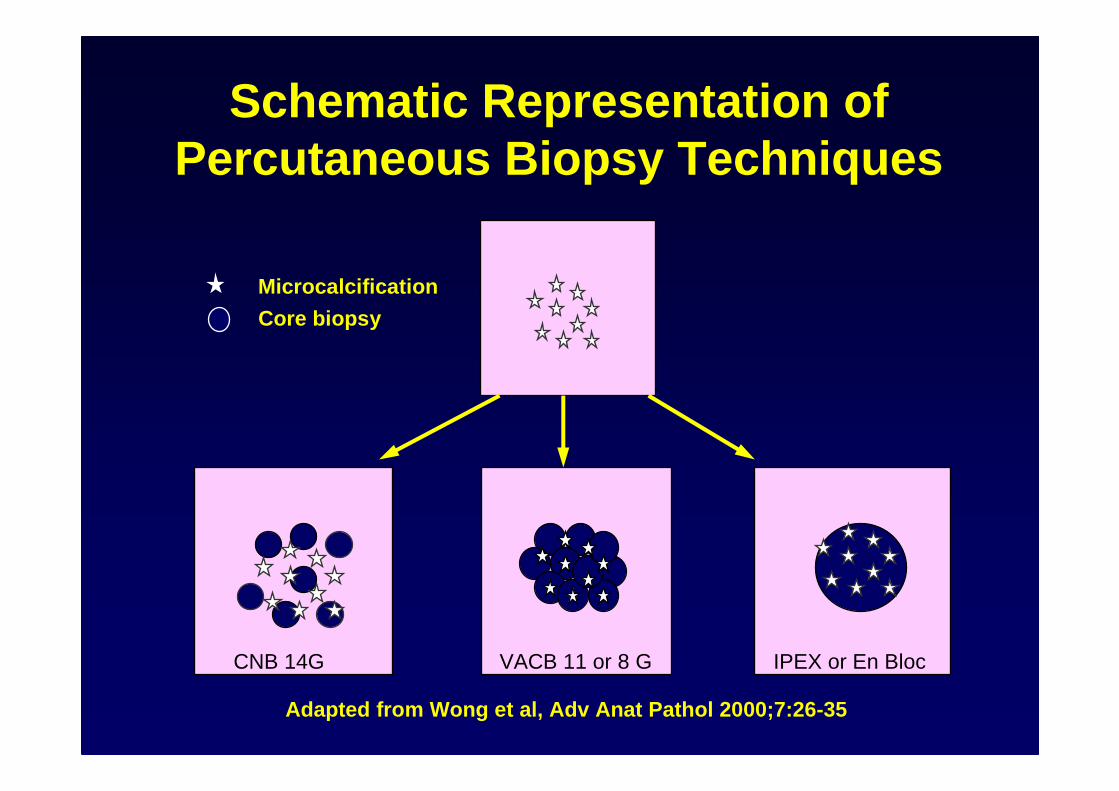
Schematic Representation of Percutaneous Biopsy Techniques
Adapted from Wong et al, Adv Anat Pathol 2000;7:26-35
Microcalcification
Core biopsy
IPEX or En BlocVACB 11 or 8 GCNB 14G

Comparison of Specimen Size
8G 14G

Upgrade Rates are Dependent on Needle Size
Bx Device Underestimation• 14g Gun 45%• 14g DVA 25%• 11g DVA 18%• 8g DVA <10%

Diagnoses On Core Needle Biopsies
• Specific diagnoses– Invasive cancer– DCIS– LCIS– Atypical hyperplasias– Fibroadenoma
• Non-specific diagnoses– Cysts– Fibrosis– “Fibrocystic changes”– Normal breast tissue

CNB Diagnoses may be Specific but not Definitive
• Atypical ductal hyperplasia-14G carcinoma in 50-60% (2/3-3/4 DCIS;
remainder invasive)
-11G (and 9G) DVA carcinoma in ~20%• Ductal carcinoma in situ
-14 G invasive carcinoma in ~20%-11G DVA invasive carcinoma in ~ 10%

Likelihood of Specific Diagnosis Related to Presence of Calcifications on
Specimen Radiograph(Liberman, 1994)
Specific Diagnosis
Ca++ present 118/146 (81%)
Ca++ absent 81/215 (38%)

Likelihood of Malignant Diagnosis Related to Presence of Calcifications on
Specimen Radiograph(Margolin, 2004)
Malignant DiagnosisCa++ present 98/116 (84%)
Ca++ absent 82/116 (71%)
p=0.02

Likelihood of Missed Malignant Diagnosis Related to Absence of
Calcifications on Specimen Radiograph(Margolin, 2004)
Missed Malignant Diagnosis
Ca++ present 1/116 (1%)
Ca++ absent 13/116 (11%)P<0.001

Pathologist Agreement: Local vs. Central DxCollins, Am J Surg Pathol, 2004
CNB(n=2002)
Overall 96%Benign 99%Invasive 97%DCIS 84%ADH 64%ALH/LCIS 56%
Open p(n=596)
93% 0.00896% ns98% ns92% ns58% ns67% ns

Mammographic-Pathologic Correlation
• The pathologic diagnosis on a core biopsy must be concordant with the impression from imaging studies
• Discordant diagnoses must be reconciled; may require repeat core biopsies or surgical excision

Examples of Discordance
Imaging Pathology
Spiculated mass any benign dx (?except RS/CSL)
Circumscribed mass benign, non-specific dx
“Malignant” Ca ++ any benign dx, even if Ca ++ present

Diagnostic Problems
• Similar to those encountered in open surgical biopsies:– ADH vs. DCIS– Identifying foci of invasion in association
with DCIS– DCIS vs. LCIS– Tubular carcinoma vs. benign sclerosing
lesions– Papillary lesions– Mucocele-like lesion vs. mucinous
carcinoma– Fibroepithelial lesions

Diagnostic Problems
• Err on the conservative side• Avoid overdiagnosis when
findings are equivocal

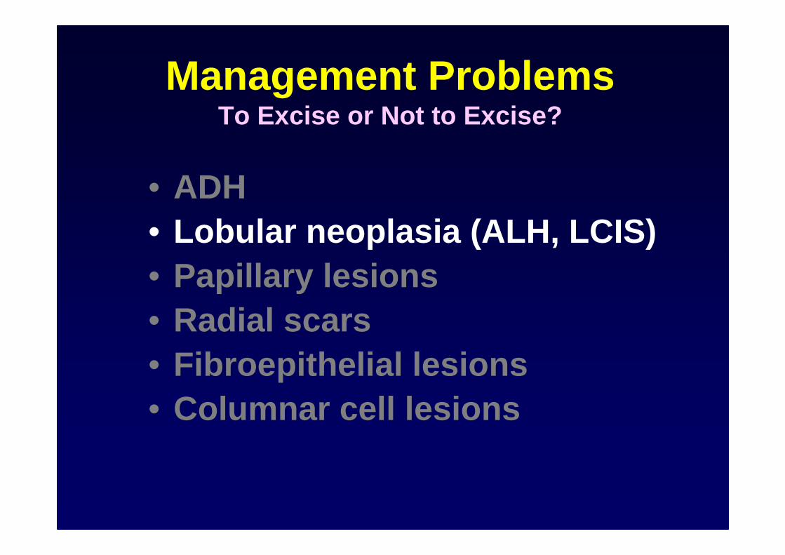
Management ProblemsTo Excise or Not to Excise?
• ADH• Lobular neoplasia (ALH, LCIS)• Papillary lesions• Radial scars• Fibroepithelial lesions• Columnar cell lesions

Management ProblemsTo Excise or Not to Excise?
• ADH• Lobular neoplasia (ALH, LCIS)• Papillary lesions• Radial scars• Fibroepithelial lesions• Columnar cell lesions

ADH on CNB
Conventional Wisdom:ADH on CNB requires surgical excision to exclude carcinoma (DCIS + invasion)

ADH on CNBLikelihood of Carcinoma on Excision
Related to:
• Technical aspects:– Gauge of needle– Lesion targeted (calcs vs. mass)– Completeness of removal
• Pathologic aspects:– Extent of ADH on core– Histologic features of ADH

Attempts at Pathologic Stratification
• Extent of ADH on CNB– # of foci of ADH
• Features of ADH on CNB– Micropapillary pattern of ADH– Marked ADH– Cytologic features bordering on DCIS
• Features of Microcalcifications– Linear, branching vs. fine, rounded
calcificationsEly, 2001; Sneige, 2003; Dalton, 2003; Eby, 2008; H oang, 2008; Wagoner, 2009; VandenBussche, 2013

Attempts at StratificationForgeard, AJS 2008

ADH Diagnosed at 11G VA Breast Biopsy
Villa, AJR 2011

AJSP, 2013

AJSP, 2013
MADH involving a large duct significantly more likely to show DCIS on excision (p<0.01)

AJSP, 2013
MADH arising in association with CCL significantly more likely to show ADH or benign findings on excision (p<0.05)

Attempts at Stratification
• We may be getting closer to identifying a subset of patients with ADH on CNB who can safely be spared excision – Larger gauge needles– Multiple cores– No residual calcifications– Limited ADH on histology

Distinction between ADH and DCIS (on CNB)
• ADH is composed of the same population of atypical epithelial cells as LG DCIS
• Incompletely filling the space• Some features of UDH• Comprises 2 spaces or less or 2 mm or
less• May be difficult to be definitive on CNB


Management of ADH vs. DCIS on CNB
• On CNB, determination of the extent is not possible, it may be more prudent to classify a lesion as “ADH bordering on low grade DCIS” or “severely atypical intraductal proliferation bordering on low grade DCIS”
• Both ADH and DCIS are managed with excisional biopsy
• As such it may be more appropriate to classify a lesion as ADH rather than labeling a patient with DCIS on a limited amount of tissue

Our current practice is to perform excision for patients with ADH diagnosed on CNB
Management ProblemsTo Excise or Not to Excise?
• ADH• Lobular neoplasia (ALH, LCIS)• Papillary lesions• Radial scars• Fibroepithelial lesions• Columnar cell lesions

Management ProblemsTo Excise or Not to Excise?
• ADH• Lobular neoplasia (ALH, LCIS)• Papillary lesions• Radial scars• Fibroepithelial lesions• Columnar cell lesions

Intraductal Papilloma on CNBIssues of Concern
• Papilloma vs. papillary DCIS may be difficult, especially with limited material
• ? Representative of lesion as whole -otherwise benign papillomas may harbor foci of ADH or DCIS
• Limited data available

Benign Papilloma on CNB with Excision Follow-up
Author # with excision f/u CA on
Philpots 6 1 (17%)Liberman 4 0Ivan 6 0Renshaw 18 0Mercado 36 2 (6%) Kil 76 6 (8%)Bernik 47 14 (36%)Tseng 24 7 (29%)Rizzo (2012) 234 21 (9%)Linda (2012) 64 4 (6%)Lu (2012) 66 4 (6%)Fu (2012) 203 34 (6%)Li (2012) 370 7 (2%)Swapp (2013) 77 0

Intraductal Papilloma on CNBRizzo, J Am Coll Surg, 2012
• 234 with IDP only• 21/234 (9%) upgraded to DCIS or IDC• Among many clinical and radiologic
variables analyzed, only older age was predictive
• Recommend excision due to lack of predictors for upgrade

Solitary Intraductal Papilloma on CNB
Swapp, Ann Surg Oncol, 2013
• 77 with IDP only and excision• 100 with no excision and stable f/u• No upgrades to atypia or malignancy• Recommend imaging f/u rather than
excision for solitary intraductal papillomas with no atypia and radiologic concordance

• 34 studies• 2,236 non malignant papillary lesions• 346 upgraded to malignant• Pooled underestimation rate of 15.7%• Rate for benign papillomas =7.0% (5.6-8.3%)• Rate for atypical papillomas =36.9% (29.5-44.3%)
Ann Surg Oncol 2013

Micropapillomas
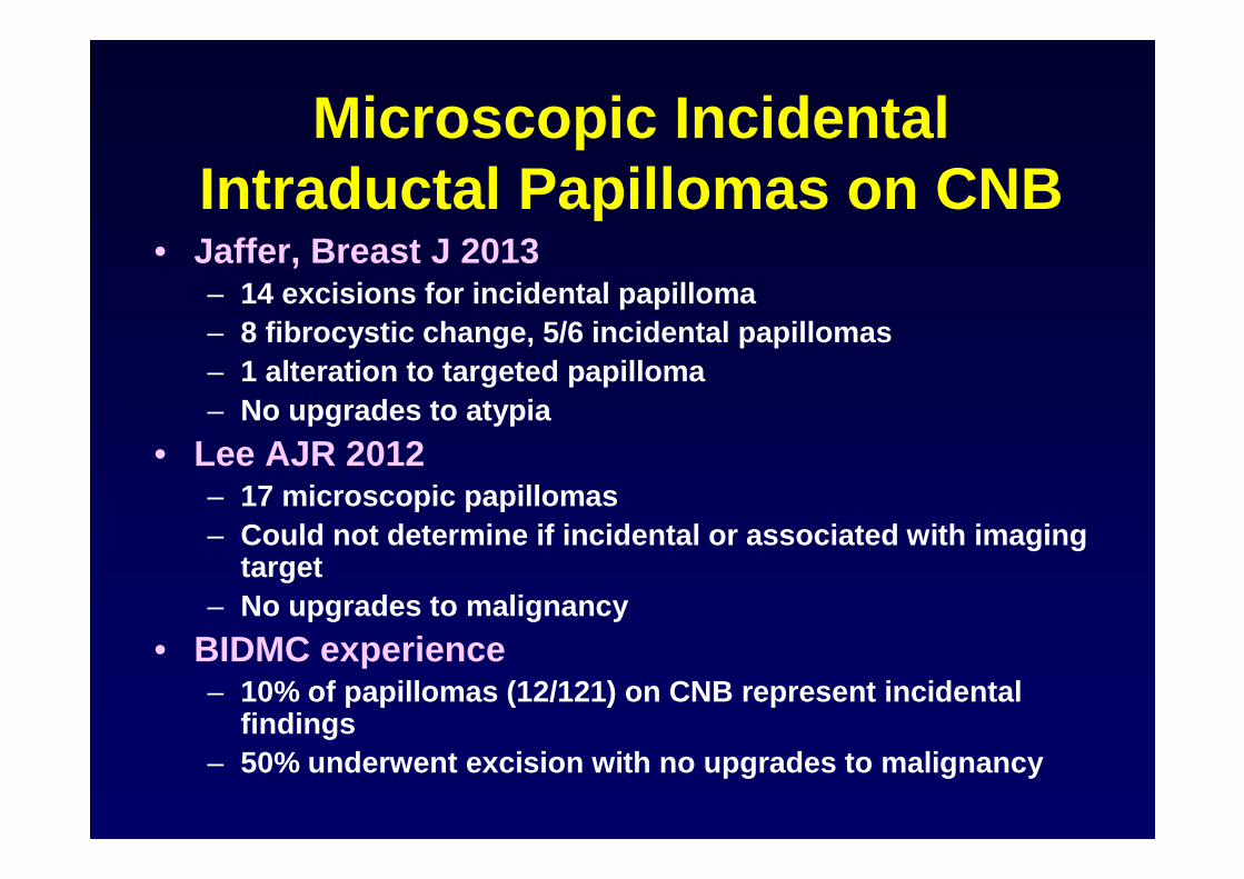
Microscopic Incidental Intraductal Papillomas on CNB
• Jaffer, Breast J 2013– 14 excisions for incidental papilloma– 8 fibrocystic change, 5/6 incidental papillomas– 1 alteration to targeted papilloma– No upgrades to atypia
• Lee AJR 2012– 17 microscopic papillomas– Could not determine if incidental or associated with imaging
target– No upgrades to malignancy
• BIDMC experience– 10% of papillomas (12/121) on CNB represent incident al
findings– 50% underwent excision with no upgrades to malignan cy

Targeted benign papilloma on CNB requires excision

Management ProblemsTo Excise or Not to Excise?
• ADH• Lobular neoplasia (ALH, LCIS)• Papillary lesions• Radial scars• Fibroepithelial lesions• Columnar cell lesions

Radial Scar on CNB• 7 studies (1996-2008)
– 7/113 pts (6.1%) with RS on CNB had carcinoma on excision
• Largest study to date (Brenner, 2002)
– 157 cases with RS on CNB with either surgical excision (n=102) or 24 month follow-up
– Malignancy in 13 cases (8.3%)– No malignancy if
• No associated AH• CNB >12 specimens• Mammotome used

Radial Scar on CNBLinda, AJR, 2012
• 54 women with radial scar diagnosed on US or MRI guided core needle biopsy underwent excision
• 2 upgrades (3.7%)– 1 ILC– 1 “incidental” low grade DCIS
• Suggest that with negative MRI, may be able to “observe” patients with radial scar

Performance of MRILinda, ACR, 2012

All Patients with Radial Scar on CNB Undergo Excision

Management ProblemsTo Excise or Not to Excise?
• ADH• Lobular neoplasia (ALH, LCIS)• Papillary lesions• Radial scars• Fibroepithelial lesions• Columnar cell lesions

Fibroepithelial Lesions on CNB
• Dx of fibroadenoma usually readily made on CNB; excision not required….
Fibroepithelial Lesions of the BreastIssues for Core Needle Biopsy
Predictors of phyllodes tumors on core needle biopsy

Fibroepithelial Lesions of the BreastIssues for Core Needle Biopsy
• Several studies have attempted to stratify cellular fibroepithelial lesions
• Jacobs et al. (Am J Clin Pathol, 2005)
– Evaluated 29 FELCS on CNB (16 FA and 12 PT)
• Lee et al. (Histopathol, 2007)
– Evaluated 36 PT and 38 FA with prior CNB• Jara-Lazaro et al. (Histopathol, 2010)
– Evaluated 261 CNB of FEL

Fibroepithelial Lesions of the BreastIssues for Core Needle Biopsy
• Jacobs et al. (Am J Clin Pathol, 2005)
– Evaluated 29 FELCS on CNB (16 FA and 12 PT)– Features assessed included:
• Stromal cellularity (mild, moderate, marked)• Stromal cell nuclear atypia• Stromal mitotic count• Stromal to epithelial ratio (%)• Stromal overgrowth (4x field)• Infiltrative edge• Epithelial hyperplasia• Stromal condensation• Growth pattern (peri-, intracanalicular, mixed)• Leaf-like pattern• Multinucleated stromal giant cells

FELCS on CNBJacobs et al. (Am J Clin Pathol, 2005)
0
10
20
30
40
50
60
70
80
% of cases
Stromal CellularityMildly Increased
ModeratelyIncreased
Markedly Increased

Fibroepithelial Lesions of the BreastIssues for Core Needle Biopsy
• Lee et al. (Histopathol, 2007)– Evaluated 36 PT and 38 FA with prior CNB– Features assessed included:
• Stromal cellularity• Stromal condensation• Stromal cell pleomorphism• Stromal overgrowth (10x and 20x)• Percentage of lesion composed of stroma• Lesion edge (circumscribed, infiltrative, NA)• Stromal mitoses• Specimen fragmentation• Intracanalicular pattern (present vs. absent)• Clefting• Stromal adipose tissue or heterologous elements

Fibroepithelial Lesions of the BreastIssues for Core Needle Biopsy
• Jara-Lazaro et al. (Histopathol, 2010)
– Evaluated 261 CNB of FEL (21 FA and 36 PT on excision included in the histologic assessment)
– Features assessed included:• Lesional edge• Stromal cellularity• Stromal overgrowth (4x)• Stromal distribution (condensation)• Stromal nuclear atypia• Stromal mitoses• Ratio of epithelial to stromal elements• Leaf-like pattern• Epithelial hyperplasia• Presence of PASH

Fibroepithelial Lesions of the BreastIssues for Core Needle Biopsy
Predictors of phyllodes tumors on core needle biopsy
Immunohistochemistry

Fibroepithelial Lesions of the BreastImmunohistochemistry
• Jacobs et al. (Am J Clin Pathol, 2005)
– Evaluated Ki-67, topoisomerase II and p53

Fibroepithelial Lesions of the BreastImmunohistochemistry
• Jara-Lazaro et al. (Histopathol, 2010)
– Evaluated Bcl-2, CD117, Ki-67, topoisomerase II and CD34

Fibroepithelial Lesions of the BreastImmunohistochemistry
• Jara-Lazaro et al. (Histopathol, 2010)

Fibroepithelial Lesions of the BreastIssues for Core Needle Biopsy
• Management recommendations include:– Excision with margin of normal tissue if two or
more features present– Less extensive excision if only one feature is
present– Possibly observation
– BUT…..given sampling issues excision recommended for all cellular fibroepithelial lesion s
– Core needle biopsy diagnosis of fibroadenoma does not completely exclude phyllodes tumor
Jacobs, 2005Lee, 2007Jara-Lazaro, 2010Resetkova, 2010

Management ProblemsTo Excise or Not to Excise?
• ADH• Lobular neoplasia (ALH, LCIS)• Papillary lesions• Radial scars• Fibroepithelial lesions• Columnar cell lesions

Columnar Cell Change/Hyperplasia
Found on excision:• No further
treatment
Found on CNB:• No excision
necessary
No additional levels obtained

Flat Epithelial Atypia
FEA on core biopsy– Excision required– “Upgraded” in
0-30% of cases
Bianchi, Virchow Arch, 2012Peres, BCRT, 2012De Mascarel, Mod Pathol, 2011Lee, Breast Journal, 2010Ingegnoli, Breast Journal, 2010Tomasino, J Cell Physiol, 2009Chivukula, Am J Clin Pathol, 2009Senetta, Mod Pathol, 2009Piubello, Am J Surg Pathol, 2009Kunju, Hum Pathol, 2007

Flat Epithelial AtypiaFEA on core biopsy
– “Upgraded” in 0-30% of cases
– But need for excision remains uncertain
– Rad-path correlation required
WHO, 2012

Management ProblemsTo Excise or Not to Excise?
• A major role of CNB is to spare patients with probably benign lesions open surgical excision where possible
• Threshold for recommending open biopsy should be low
• If there is doubt, take it out

Consequences, Complications and Artifacts Related to Core
Needle Biopsies
• Hemorrhage, granulation tissue, scarring and bx site
• Infarction• Epidermoid cysts• The missing cancer• Epithelial displacement

Consequences, Complications and
Artifacts Related to Core Needle Biopsies
• Hemorrhage, granulation tissue, scarring and bx site
• Infarction• Epidermoid cysts• The missing cancer• Epithelial displacement

Consequences, Complications and
Artifacts Related to Core Needle Biopsies
• Hemorrhage, granulation tissue, scarring and bx site
• Infarction• Epidermoid cysts• The missing cancer• Epithelial displacement

Biopsy Site Marking DevicesGuarda et al, AJSP 2005
Two major types• Pellets of resorbable copolymer of
polylactic acid/polyglycolic acid– Cell poor fibrotic reaction around empty
spaces followed by FBGCR• Plug of bovine collagen
– Eosinophilic hyalinized acellular material with lymphocytic infiltrate
– Degradation of plug associated with deposition of native collagen
– Absence of significant FBGCR


Consequences, Complications and
Artifacts Related to Core Needle Biopsies
• Hemorrhage, granulation tissue, scarring and bx site
• Infarction• Epidermoid cysts• The missing cancer• Epithelial displacement


Consequences, Complications and
Artifacts Related to Core Needle Biopsies
• Hemorrhage, granulation tissue, scarring and bx site
• Infarction• Epidermoid cysts• The missing cancer• Epithelial displacement


Consequences, Complications and
Artifacts Related to Core Needle Biopsies
• Hemorrhage, granulation tissue, scarring and bx site
• Infarction• Epidermoid cysts• The missing cancer• Epithelial displacement


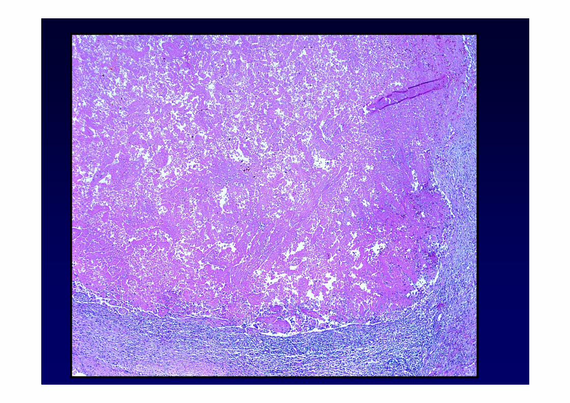
The Missing Cancer
• Cancer in core; no cancer in surgical specimen
• Uncommon• Likely to be seen
increasingly with larger gauge needles and use of mammotome

Schematic Representation of Percutaneous Biopsy Techniques
Adapted from Wong et al, Adv Anat Pathol 2000;7:26-35
Microcalcification
Core biopsy
ABBI or En BlocVACB 11 or 8 GCNB 14G

The Missing Cancer
• Patient misidentification• False positive CNB• Biopsy site not excised• Inadequate sampling• Lesion entirely removed by CNB• Obliteration of residual cancer
by healing process

Contents of the Surgical Pathology Report
• Sufficient information to permit radiologic-pathologic correlation and to get patient into appropriate therapeutic algorithm
• Optimal amount of information to include for cases of invasive cancer controversial– Inclusion of too much information can
be problematic

Which features should be noted in pathology reports
of CNB specimens containing an invasive
cancer?

Assessment of Standard Prognostic Factors on CNB
Sharifi, 1999
Factor Agreement Between CNB and Excision
Size 21%Histologic type 81% (72-82%)*Histologic grade 75% (62-83%)*
*range reported in literature
Rakha, J Clin Pathol, 2007Park, Am J Surg, 2009

Assessment of Standard Prognostic Factors on CNB
• If size, special histologic type, and/or grade are reported, should have a caveat– “in this limited sample”– for size: “this represents a
minimum size”

Assessment of Prognostic and Predictive Markers
Jacobs, 1998
Factor Agreement Between CNB and Excision
ER 100%HER2 100%p53 100%bcl2 100%
Rakha, J Clin Pathol, 2007Park, Am J Surg, 2009Sutela, Acta Oncol, 2008

Assessment of Prognostic and Predictive Markers
• False negative ER on CNB (Lee, J Clin Pathol, 2008)
• Intratumor heterogeneity for HER2 (Striebel, Am J Clin Pathol, 2008; Chivukula, Mod P athol, 2008)
• Our practice is to perform markers on all CNBs and not on excisions
• Repeat on excision for aberrant results, negative results or cases of neoadjuvant therapy
Conclusions
• Core needle biopsies represent an important advance in the evaluation of non-palpable breast lesions
• Pathologists, radiologists, and clinicians need to understand limitations
• Pathologists need to be aware of the artifacts associated with CNB
• Radiologic-pathologic correlation is essential in every case