Seed-mediated growth of MOF-encapsulated Pd@Ag core–shell ...

Controllable Preparation of Core−Shell Au−Ag Nanoshuttles withImproved Refractive Index Sensitivity and SERS ActivityTingting Bai,† Jianfei Sun,† Renchao Che,§ Lina Xu,† Chenyue Yin,† Zhirui Guo,*,‡ and Ning Gu*,†
†State Key Laboratory of Bioelectronics, Jiangsu Key Laboratory for Biomaterials and Devices, School of Biological Science andMedical Engineering, Southeast University, Nanjing 210096, P. R. China‡The Second Affiliated Hospital of Nanjing Medical University, Nanjing 210011, P. R. China§Department of Materials Science and Laboratory of Advanced Materials, Fudan University, Shanghai 200433, P. R. China
*S Supporting Information
ABSTRACT: Recent studies have conclusively shown that theplasmonic performance of Au nanostructures can be enhancedby incorporating Ag. Here, we developed a simple and robustapproach for preparing core−shell Au−Ag nanoshuttles (NSs)using single-crystal Au nanorods (NRs) as cores. Upontailoring the temperature of the reaction system containingalkaline glycine buffer (pH 8.5), exceptionally monodisperseAu−Ag NSs with sharp tips and tunable shell thickness couldbe routinely achieved in high yield through an epitaxial growthprocess. In particular, high-resolution transmission electronmicroscopy and nitric acid corrosive experiments revealed thatthe shells of these NSs consisted of a homogeneous Au−Agalloy, rather than pure Ag or Au as previously reported. It wasfound that glycine played an important role in determining the final metal contents of the shell by regulating the reductionkinetics. In addition, the obatined Au−Ag NSs with sharp tips were shown to have significantly improved refractive indexsensitivity and surface-enhanced Raman scattering activity relative to the original Au NRs, making these materials promising forbiomedical applications, such as biosensing and biolabeling.
KEYWORDS: Ag, Au, nanorods, shape control, core−shell structure, refractive index sensitivity, surface-enhanced Raman scattering
1. INTRODUCTION
Noble metal nanoparticles (NPs), especially Au and Ag NPs,have received extensive attention, mainly owing to their uniquelocalized surface plasmon resonance (SPR) in the visible andnear infrared (NIR) regions and wide applications in sensing,catalysis, biomedicine, and so on.1,2 Numerous investigationshave revealed that plasmonic properties of Au and Ag NPs arehighly sensitive to their shapes.3−5 It is also known that Au orAg NPs with anisotropic shapes exhibit enhanced plasmonicproperties.6 Attempts to control the shape of Au NPs has metwith great successes. However, the aqueous-preparation ofanisotropic Ag NPs with a uniform shape remains verychallenging. Compared with Au NPs, Ag NPs have muchstronger and more sensitive SPR, which makes them morecompetitive in plasmonic sensing and surface-enhanced Ramanscattering (SERS) applications.2 Hence, it is of greatimportance to develop reliable approaches to prepareanisotropic Ag NPs with good uniformity. To meet this end,an effective solution is to fabricate Au−Ag core−shellnanostructures. Because both Au and Ag are face-centeredcubic metals and have close lattice parameters (4.0786 Å for Auand 4.0862 Å for Ag), Ag can grow epitaxially onto a Au NPcore under the appropriate conditions.7 Here, we choose single-
crystal Au nanorods (NRs) as cores owing to their uniquesurface structures, rich plasmonic properties, and ease ofpreparation.8 Au NRs have two plasmon bands as aconsequence of their one-dimensional anisotropy. One weakband around 510 nm is assigned to the transverse SPR (TSPR),and the other strong band is contributed by a longitudinal SPR(LSPR). Importantly, the LSPR of Au NRs is quite sensitive tothe aspect ratio (AR, is the length divided by the diameter). Byincreasing the AR, the LSPR can be easily tailored from thevisible to the NIR region (700−11000 nm), which makes themwell-suited for biomedical applications.9,10 Moreover, the LSPRband of Au NRs shows excellent sensitivity to changes ofrefractive index, enabling versatile sensing applications.11,12 Inaddition, Au NRs were also widely used in SERS owing to theirenhanced electric fields at the tips compared with spheres.13,14
To date, a variety of Au−Ag nanostructures, such ascylinders,15 bars,16 boats,17 octahedrons,18 dumbbells,19 andshuttles17,20 have been obtained using Au NRs as cores for theepitaxial growth of Ag. The results show that the refractive
Received: November 25, 2013Accepted: February 17, 2014
Research Article
www.acsami.org
© XXXX American Chemical Society A dx.doi.org/10.1021/am405357v | ACS Appl. Mater. Interfaces XXXX, XXX, XXX−XXX

index sensitivity of these core−shell NRs increases with the Agshell thickness or the aspect ratio.21,22 On the basis of theimproved index sensitivity, a simple and sensitive biosensor canbe fabricated to detect molecular binding events in medical andlife sciences with a basic spectrophotometer.23 Furthermore,these core−shell nanostructures can also serve as excellentSERS substrates owing to their significantly higher enhance-ment factors.24 Among these nanostructures, Au−Ag nano-shuttles (NSs) with two arrow-headed tips at the ends show themost promise for practical applications. Theoretical calculationspredict that, from the combination of tip effects and the Agcomponent, Au−Ag NSs have a larger extinction cross-sectionof the LSPR and exhibit much stronger local field enhance-ments than Au NRs.20 The theoretical findings from the abovestudies indicate that Au−Ag NSs can be promising candidatesin plasmonic applications, such as optical sensing and SERSdetection, and thus systematic experimental verification is quitenecessary.In this work, we developed a convenient and reliable
approach for the synthesis of Au−Ag NSs based onmodifications of previously reported methods. In our presentwork, monodisperse Au−Ag NSs with a tunable shell thicknesswere achieved by simply adjusting the reaction temperature inthe glycine buffer. In particular, subsequent structure analysisby high-resolution transmission electron microscopy(HRTEM) and nitric acid corrosive experiments support thatthese NSs have a homogeneous Au−Ag alloy shell around theAu NR core, rather than the pure Ag or Au shell described inprevious documents.25,26 Moreover, our experimental resultsrevealed that, in addition to being a pH stabilizer, glycine alsoplayed an important role in regulating the reduction kinetics.Subsequently, the plasmonic properties of Au−Ag NSs weresystematically examined in serial experiments. It was found thatthese Au−Ag NSs possessed higher refractive index sensitivityas well as excellent SERS activity compared with the original AuNRs.
2. MATERIALS AND METHODS2.1. Materials. Hexadecyltrimethylammonium bromide (CTAB,
99%) and poly(styrenesulfonate) (PSS, Mw 70,000) were purchasedfrom Sigma. Sodium oleate (NaOL, >97.0%) was purchased from TCIAmerica. Glycine (C2H5NO2, ≥ 99.0%) and Tris-base (C4H11NO3, ≥99.5%) was purchased from Biosharp Company (China). 4-Amino-thiophenol (4-ATP) was purchased from Aldrich Chemical Company.Hydrogen tetrachloroauric acid (HAuCl4·4H2O, 99%), hydrochloricacid (HCl, 37 %), sliver nitrate (AgNO3), sodium borohydride(NaBH4, 96%), and L-ascorbic acid were all purchased from ShanghaiChemical Reagent Co., Ltd (China). All chemicals were used asreceived. Deionized water with resistance of 18MΩ was usedthroughout the experiments. All glassware was cleaned by aqua regia(HCl/HNO3 in 3:1 ratio by volume).2.2. Synthesis of Monodisperse Au NRs. Original Au NRs with
excellent uniformity were prepared by a binary surfactant assistant,seed-mediated method developed by Ye et al.27 The gold seeds wereprepared by the addition of a freshly prepared, ice-cold NaBH4solution (0.6 mL, 10 mM) into a solution made by combiningHAuCl4 (0.1 mL, 25 mM) and CTAB (9.9 mL, 0.1 M), followed bystirring for 2 min. The seed solution was aged at 30 °C for 2 h allowingfor the hydrolysis of unreacted NaBH4. The Au NR growth solutionwas made by first mixing 100 mL binary surfactant mixture (CTAB0.037 M, NaOL 0.078 M) with 1.44 mL AgNO3 (10 mM) and 2 mLHAuCl4 (25 mM). The solution was kept at 30 °C for 90 min understirring (700 rpm). And then, 0.42 mL HCl (37 %, 12.1 M) wasintroduced to adjust the pH. After the solution was gently mixed, afreshly prepared aqueous ascorbic acid solution (0.16 mL, 0.1 M) was
added, and the solution was stirred for 30 s. Finally, 0.08 mL seedsolution was injected into the growth solution and kept undisturbed at30 °C overnight.
2.3. Preparation of bimetallic Core−Shell Au−Ag nano-shuttles. The as-prepared Au NRs were purified by centrifugation at10000 rcf for 15 min twice. For each kind of Au−Ag NSs, 5 mL ofpurified Au NRs in 0.1 M CTAB was mixed with 5 mL of 0.2 Mglycine acid and 30 μL of 2 M NaOH solutions to obtain a stable pHvalue of 8.5. Additional HAuCl4 (40 ul, 25 mM) and AgNO3 (0.2 mL,10 mM) were introduced under vigorously stirring and incubated atdifferent temperatures (23, 25, 27, and 29 °C, respectively). Finally, aportion of ascorbic acid solution (0.2 mL, 0.1 M) was added to thegrowth solution and stirred for 30 s, and then the whole solution wasaged for 1 h. The resulting nanostructures were purified bycentrifugation at 7000 rcf for 10 min and denoted as Au−Ag NSs a,Au−Ag NSs b, Au−Ag NSs c, and Au−Ag NSs d in the followingparagraphs according to 23, 25, 27, and 29 °C, respectively.
2.4. Nitric Acid Corrosive Experiment. In this experiment, Au@Ag nanobars were used as a control model, which were synthesized bya seed-mediated growth procedure from Okuno et al.16 200 μL of 4 Mnitric acid was added to 1 mL Au−Ag NSs d and 1 mL Au@Agnanobars respectively. After 30 min of corrosion, both samples werecentrifuged at 7000 rcf for 5 min, redispersed into the water, and thencharacterized by scanning electron microscopy.
2.5. Refractive Index Sensitivity Measurement. Water−glycerol mixtures of various volume ratios were used to adjust therefractive index of the surrounding medium of nanostructures. Therefractive indices of the pure water and glycerol are 1.3334 and 1.4746,respectively. The volume percentage of glycerol in the liquid mixturewas varied from 0% to 90% at a step of 10%. The as-prepared Au NRsand Au−Ag NSs were first centrifuged at 10000 rcf for 15 min and7000 rcf for 10 min, respectively, and then redispersed into the water−glycerol mixture. Extinction spectra of the resulting dispersionsolutions were measured. The plasmon shifts relative to the refractiveindex were plotted, and the refractive index sensitivity was determinedby linear fitting. A figure of merit (FOM) is also used to characterizethe capability of noble metal nanostructures in plasmonic sensing,which is defined as the ratio of the refractive index sensitivity to the fullwidth at half maximum (FWHM) of the plasmon resonance profile.
2.6. SERS Activity of Au−Ag NSs. The fabrication of Au−Ag NSsmonolayer as SERS substrate was conducted through three steps andall the five kinds of NPs (Au NRs, Au−Ag NSs a, Au−Ag NSs b, Au−Ag NSs c, Au−Ag NSs d) were treated with the same procedure. Thefirst step is surface modification of NPs. Twenty milliliters ofnanoparticles and Raman reporter molecules (4-ATP) were mixedtogether in a proportion of 1:10 000 (the molar ratio of nanoparticlesto dye molecules) and incubated for 1 h to form the conjugation. Themixture was then centrifuged twice, and the precipitate wasredispersed in 20 mL of water. Subsequently, 2 mL PSS (10 mg/L)in 5 mM NaCl was added dropwise, and the mixture solution was keptundisturbed for 1 h. Then, the solution was centrifuged twice toremove excess polyelectrolyte and redispersed in water. All the fiveNPs were diluted to the same concentration. The second step isfunctionalization of silicon wafers. The silicon wafers were washedwith water, acetone, and ethanol in an ultrasonic bath for 5 minrespectively. To increase the coverage of surface hydroxyl groups, thesilicon wafers were treated in Piranha solution (3:7 (v/v) H2O2/H2SO4) for 20 min. Subsequently, these wafers were immersed into a0.5 wt % PDDA solution for 1 h to make the substrate positivelycharged. Next, they were rinsed several times with deionized water anddried gently with nitrogen gas. The last step is immobilization. Thefunctionalized wafers were immersed in the five NPs solutions for 36h, resulting in a monolayer of NPs films on the silicon wafers.
2.7. Instruments. The UV−vis−NIR extinction spectra wererecorded by a Shimadzu UV-3600 spectrophotometer in a range of300−1200 nm. Transmission electron microscopy (TEM) was takenwith a JEM-2100EX (JEOL) transmission electron microscopeoperated at 200 kV. Scanning electron microscopy (SEM) imagesand the EDS spectra were obtained by a Carl Zeiss ULTRA Plus FieldEmission Scanning Electron Microscope. A Renishaw Invia Reflex
ACS Applied Materials & Interfaces Research Article
dx.doi.org/10.1021/am405357v | ACS Appl. Mater. Interfaces XXXX, XXX, XXX−XXXB

Figure 1. TEM images of the original Au NRs (A) and newly formed Au−Ag NSs grown at 23 (B), 25 (C), 27 (D), and 29 °C (E), respectively. Allscale bars are 200 nm. (F) Non-normalized UV−vis−NIR extinction spectra for the original Au NRs and Au−Ag NSs. The inset shows thecorresponding photographs of the NPs solutions. All the samples were diluted threefold by deionized water before optical measurements.
Table 1. Physical Dimensions and SPR Bands of Au NRs and Au−Ag NSsa
L (nm) D1 (nm) D2 (nm) aspect ratio 1 (L/D1) aspect ratio 2 (L/D2) λT (nm) λL (nm)
Au NRs 83 ± 5 19 ± 1 19 ± 1 4.3 4.3 509 861Au−Ag NSs a (23 °C) 110 ± 6 38 ± 2 24 ± 2 2.9 4.6 529 899Au−Ag NSs b (25 °C) 109 ± 6 38 ± 2 25 ± 1 2.9 4.4 506 865Au−Ag NSs c (27 °C) 108 ± 6 38 ± 2 27 ± 1 2.8 4.0 492 794Au−Ag NSs d (29 °C) 109 ± 5 39 ± 2 29 ± 2 2.8 3.8 472 748
aThe descriptors L, D1, and D2 are defined in Scheme 1. The descriptors λT and λL represent the maximum wavelengths of the transverse andlongitudinal SPR extinctions, respectively.
Scheme 1. Schematics Illustrating the Formation Process of the Au−Ag NSs: (A) Side View, (B) Top View, (C) Shell ThicknessIncreasing with the Rise of Temperature, and (D) the Details of Alloy Shell Formation
ACS Applied Materials & Interfaces Research Article
dx.doi.org/10.1021/am405357v | ACS Appl. Mater. Interfaces XXXX, XXX, XXX−XXXC

system (equipped with a Peltier-cooled charge-coupled device (CCD)detector and Leica confocal microscope) was used to carry Ramanexperiments. Samples were excited with 785 nm diode laser underlinefocus mode and a grating of 1200 mm−1 was used. The laser wasfocused on the sample surface by using a 50× long working distanceobjective. The exposure time was 10 s, and the laser power wasadjusted to 1%, which was about 1.2 mW. Each SERS spectrum wasaveraged from 3 measurements. Results were given as means ± thestandard deviation (SD). All Raman experiments were carried out atroom temperature (∼25 °C).
3. RESULTS AND DISCUSSION3.1. Characterization and UV−vis−NIR Analysis of the
Au−Ag NSs. Figure 1A−E show the TEM images of the AuNR templates and resulting Au−Ag NSs. The images clearlyindicate that monodisperse Au NRs with negligible shapeimpurities can be obtained (Figure 1A and SupportingInformation Figure S1), making the growth of NSs withexcellent size and shape uniformity. Therefore, the NSs can beproduced in high-yield with noteworthy size uniformity andshape purity. Figure 1B−E show the TEM images of the Au−Ag NSs prepared at 23, 25, 27, and 29 °C, respectively. All ofthem have sharp tips at both ends, like a double-headed arrow.The electron density contrast implies that these NSs have acore−shell structure. The thickness of the shell can beincreased by raising the temperature of the solution. Thelength and diameter are measured from the TEM images of 100particles (Table 1). The descriptors L, D1, and D2 are defined inScheme 1.Corresponding UV−vis−NIR spectra of these NPs are
shown in Figure 1F and the SPR bands are summarized inTable 1. Similar to Au NRs, Au−Ag NSs also exhibit bothTSPR and LSPR bands. As shown in Figure 1B−E, metal ionsare preferentially deposited at the two ends of the Au NRs andgradually extend to the side when the temperature increasesfrom 23 to 29 °C. Their shapes were effectively controlled byadjusting the reaction temperature. Compared with the changein the length, which is nearly the same after the shapeconversion, the increase in diameter (D2) is more significantand steady (from 24 ±2 nm to 29 ±2 nm). Table 1 summarizesthe blue fhift of the LSPR band from 899 to 748 nm, whichcorresponds to the change in aspect ratio 2 (L/D2) from 4.6 to3.8. According to the SPR, the final colors of the four colloidsolutions are distinctly different from that of the Au NRs(Figure 1F). For the TSPR band, the position blue-shifts whenthe diameter of the NSs (D2) increases. Note that theconcentration of the Au NRs and Au−Ag NSs for the UVdetection were nearly the same as a result of using an equivalentamount of Au NR seeds. These nanostructures can be almostcompletely conserved after careful centrifugation, whichbenefits from their high monodispersity. Thus, we point outthat the significant increase in intensity of the SPR bands isascribed to the formation of the Ag-containing shell on Au NRcores.HRTEM imaging and the corresponding fast Fourier
transformation (FFT) pattern of the selected area wereperformed to ascertain the crystalline structure of the Au−AgNSs. Figure 2A shows the HRTEM image of Au−Ag NSs d.The high contrast between the center and periphery clearlyreveals the core−shell structure of this NS. The Au NRs werecompletely embedded into a homogeneous shell, and no pureAu or Ag NPs were observed. This result indicates that the shellcannot be composed of pure Au, in which case the contrast ofthe core and shell would not exist.26 The corresponding Fourier
spectrum obtained from area 1 in the HRTEM image is alsoshown as an inset. The FFT exhibits a hexagonal symmetrypattern, which is the characteristic of reflections in a face-centered cubic metal (fcc). It strongly suggests that this NS hasa single crystalline shell and that the flat surface at the end is a{111} plane. Considering the symmetry of Au NRs, theHRTEM and the FFT pattern show that the two ends of the AuNRs are enclosed by {111} facets. The local enlarged HRTEMimage of area 2 at the vertex of the arrow is shown in Figure 2B.In accordance with the FFT results, two types of lattice fringeswith interplanar spacings of 0.200 and 0.235 nm are clearlyobserved, which are attributed to the {002} and {111} planes ofAu or Ag. In the bright-field imaging mode of TEM, theelectron density contrast is formed by the occlusion andabsorption of the electrons in the sample. Thus, the bright fieldcontrast is related to the crystalline structure, thickness, andatomic number of the specimen.28 Under the same conditions,imaging parameters, and neighboring position at the atomicscale, the brightness of the atomic spots would be determinedsolely by the atomic number. As marked in area 3, the six spotsobviously have a different brightness. Considering the fact thatthere are only two kinds of metal ions involved in thepreparation, it can be concluded that the NSs have an alloyshell, which is a homogeneous solid solution of Au and Ag.Energy dispersive spectrometer (EDS) analysis and nitric
acid corrosive experiments were performed to furtherinvestigate of the shell composition. If the shell around theAu NR was composed of pure Au or Ag, the content of thecorresponding metal should increase with the growth of theshell. However, EDS analysis on randomly selected areas showsthat the average Au/Ag atomic ratio remains almost unchanged(Supporting Information Table S1), which verifies that the shellis not pure Au or Ag. Besides, it is well-known that pure AgNPs can be dissolved by a strong oxidizing acid like nitricacid.29 These Au−Ag NSs were then tested using nitric acidtogether with Au@Ag nanobars (Au NRs coated with a pure Agshell) as a control. After 30 min of nitric acid corrosion, theshells of the Au@Ag nanobars were nearly corroded, while theprofile of Au−Ag NSs d remained almost unchanged (Figure3). These results agree well with the HRTEM finding that theas-synthesized NSs have an Au−Ag alloy shell. This is contraryto previous reports, in which the shell is composed of pure Agor Au.25,26 By combination of these two metals into one NP,the localized SPR can be further enhanced and variedcontinuously beyond the limits of monometallic Au and AgNPs. In addition, the Au−Ag alloy is thermodynamically more
Figure 2. (A) HRTEM image of Au−Ag NSs d. The inset shows thecorresponding FFT spot pattern of area 1 demonstrating that thearrow tip is enclosed by {111} facets. (B) Magnification of area 2 inpanel A. Scale bars for panels A and B are 10 and 2 nm, respectively.
ACS Applied Materials & Interfaces Research Article
dx.doi.org/10.1021/am405357v | ACS Appl. Mater. Interfaces XXXX, XXX, XXX−XXXD

stable than pure Au or Ag.7 All these advantages make the Au−Ag NSs more suitable for plasmonic applications.3.2. Mediation of the Reduction Kinetics by Glycine.
Since it has been documented that amino acids could have theability to direct the growth of gold nanostructures.30,31 A seriesof experiments was carried out to investigate the role of glycinein the reduction kinetics besides the pH stabilization.The NS growth was performed at varying glycine
concentrations (0−0.2 M, 25 °C, pH 8.5). The results showthat the Au NRs can grow into NSs at various glycineconcentrations or even without glycine (Figure 4A−C). Themorphology of the NSs can be seen clearly from the SEMimages (Figure 4B). There are four sides around the NS andfour symmetrical irregular quadrilaterals at the both tipsforming an approximately 45° torsional angle with the sides.Figure 4E shows the extinction spectra of the products grown atvarying glycine concentrations up to 0.2 M. It is observed thatthe SPR intensities of the products obtained using lower glycineconcentrations (0−0.02 M) are far lower than those of theproducts obtained using 0.05−0.2 M glycine. This suggests thatthe amount of metal ions deposited on the surface of the AuNRs at lower glycine concentrations is smaller than that athigher glycine concentrations. The above results indicate thatglycine has little effect on the final morphology of the Au−AgNSs, but it may have some influence on the deposition of themetal precursors.Furthermore, Tris, a common pH buffer with an effective pH
range between 7 and 9, was also used as a control for preparingAu−Ag NSs. Similar to glycine, NSs could be obtained atvarying Tris concentrations (0−0.2 M, 25°C, pH 8.5) withoutany obvious variation in the morphological architecture (Figure4D). The average Ag/Au atomic ratio from EDS analysis alsoproves that Tris can mediate the reduction kinetics during theovergrowth of the Au−Ag NSs (Figure 5). Therefore, ourinvestigation indicates that both of the two buffers have similareffects on the dispersion of metal precursors but to differingdegrees, as there is a difference in the Ag/Au atomic ratiobetween glycine and Tris at low concentration (0.02 M).
3.3. Formation Mechanism of Au−Ag NSs with AlloyShell. In this study, the synthesis of the original Au NRs isbased on the seed-mediated method under the assistance ofsmall amount Ag+ ions. It has been known that such Au NRshave an single crystal structure and an octagonal cross-section:32,33 the side facets of the rod are dominated by four{110} and four {100} facets, whereas the ends of the rod areenclosed by {111} and {110} planes (as shown in Scheme 1Aand B). In the Ag+-assisted growth of Au NRs, the preferentialunderpotential deposition (UPD) of Ag atoms on the Au high-energetic {110} facets prevents Au deposition on them. ThusAu atoms have to deposit onto the {111} facets, resulting inone-dimensional growth in the [100] direction.34 Furthermore,Xiang et al. in 2008 were the first to demonstrate that thegrowth mode of the Au NRs could be changed from {110}prevention to preference by adjusting the reaction parameters,including Ag+/Au3+ and AA/Au3+ ratios.25 As a result, morethermodynamically favored shape, such as NSs, can be formed.In this study, by tuning the molar ratio of Au3+/Ag+/ascorbic
acid to be 1:2:20, we switched the growth mode of metal ionson the surface of Au NRs into {110} preference. Since the onlydriving force of the formation of arrow-like shape is tominimize the surface energy. On the basis of Xiang et al.’sinvestigation25 and the observations we found here, theformation process of the Au−Ag NSs with alloy shell isillustrated as following: under higher Ag+/Au3+ and AA/Au3+
ratios, the amount of reduced Ag atoms is higher enough tofully cover all exposed facets of Au NR and thus the preferentialUPD deposition of Ag on Au {110} facets would not work.Therefore, the minimization of the surface energy becomes themain driving force to direct the subsequent overgrowth of AuNRs. At the end of Au NR, the four high-energetic {110} facetsgrow more quickly and disappear finally, while four {111} facetsextend and form an arrow-head top. Meanwhile, at the side ofthe Au NR, metal atoms also deposit more quickly on the{110} facets. As a result, four side {110} facets disappear,leaving the four enlarged {100} facets to form a rod shape witha square cross-section (Scheme 1A and B). During the
Figure 3. (A) SEM image of Au−Ag NSs d with an alloy shell. (B) SEM image of Au@Ag nanobars (Au NRs coated with a pure Ag shell). The insetshows the corresponding TEM image (scale bar is 40 nm). (C) Au−Ag NSs d after treatment with nitric acid. (D) Au@Ag nanobars after treatmentwith nitric acid. All scale bars are 200 nm.
ACS Applied Materials & Interfaces Research Article
dx.doi.org/10.1021/am405357v | ACS Appl. Mater. Interfaces XXXX, XXX, XXX−XXXE

morphology transformation, new triangular {110} facets appearat the junction between the four side {100} facets and the four{111} facets at the end. Morover, the stronger reducing abilityof ascorbic acid at a higher temperature accelerates the shellgrowth process. Much more metal ions deposit on theemerging triangular {110} facets, leading to increasing ofshell thickness (Scheme 1C).Following reactions are involved in the epitaxial growth
process of the alloy shell. Firstly, the Ag+ is reduced to Ag0 onthe Au facets through UPD deposition. Then Au3+ reacts withAg0 on the surface of Au NRs. Meanwhile, the free ions can alsobe reduced by the large amount ascorbic acid. The Au−Ag alloyshell is thus formed as the reduction and in situ galvanicreplacement reaction continues. During this process, glycine isfound to mediate the reduction kinetics of metal ions andregulate the molar ratio of Au and Ag in the final products,apart from stabilizing the pH value. On the basis of the above
Figure 4. (A) SEM image of Au−Ag NSs b obtained without any buffer solution. (B) Magnification of the selected area from (A) and a sketch of twoparticles from this image. (C) SEM images of Au−Ag NSs b obtained in glycine solution with different concentrations: (I) 0.02 M, (II) 0.05 M, (III)0.1 M, and (IV) 0.2 M. (D) SEM images of Au−Ag NSs b obtained in Tris solution with different concentrations: (I) 0.02 M, (II) 0.05 M, (III) 0.1M, and (IV) 0.2 M. All scale bars are 200 nm. Non-normalized UV−vis−NIR extinction spectra for the original Au NRs and Au−Ag NSs b preparedin glycine (E) and Tris (F) solution with different concentrations.
Figure 5. Average Au/Ag atomic ratio of Au−Ag NSs b prepared inglycine and Tris solution with different concentrations.
ACS Applied Materials & Interfaces Research Article
dx.doi.org/10.1021/am405357v | ACS Appl. Mater. Interfaces XXXX, XXX, XXX−XXXF
discussion, it can be summarized that the Au−Ag NSs withalloy shell are obtained by coupling co-reduction and galvanicreplacement (Scheme 1D).3.4. Refractive Index Sensitivities of the Au−Ag NSs.
To investigate the refractive index sensitivities of the plasmonresonance peaks of Au−Ag NSs, they were dispersed in binarymixtures of water and glycerol with varying volume percentagesof glycerol. The original Au NRs were used as a control tocompare the different refractive index sensitivities after thegrowth of the Au−Ag alloy shell. In all experiments, the highestvolume percentage of glycerol was 90%, because pure glycerolis highly viscous. As the refractive index of the solvent mixtureincreased, all the plasmon peaks shifted to longer wavelengths(Figure 6A−D and Supporting Information Figure S2). Thepeak wavelengths in different solvents were measured, and the
plasmon shifts were determined by taking the difference ofplasmon peak wavelengths when the NPs were dispersed in thesolvent mixture and in pure water. The refractive indices of thesolvent mixtures were calculated according to the Lorenzequation.12 When the plasmon shifts are plotted as a functionof the refractive index of the solvent mixture, we observe anapproximately linear dependence for both the TSPR and LSPR.All the plots can be fitted well with a linear approximation, andthe slopes of the lines are the refractive index sensitivities(Figure 6F and G). Their values are summarized in Table 2,and the FWHM of the longitudinal peaks and the FOMs arealso included.Table 2 shows that Au−Ag NSs with different aspect ratios
exhibit different refractive index sensitivities, which are largerthan that of the original Au NRs. In a previous study, the noble
Figure 6. Extinction spectra of Au NRs (A, TSPR; B, LSPR) and Au−Ag NSs b (C, TSPR; D, LSPR) in water−glycerol mixtures of varyingcompositions. Each extinction spectrum is normalized to its maximum intensity. The arrows indicate the direction of increasing volume percentageof glycerol. Dependence of the TSPR intensity (E), TSPR shift (F), and LSPR shift (G) as a function of the refractive index of the liquid mixture forAu NRs and Au−Ag NSs prepared at the different temperatures.
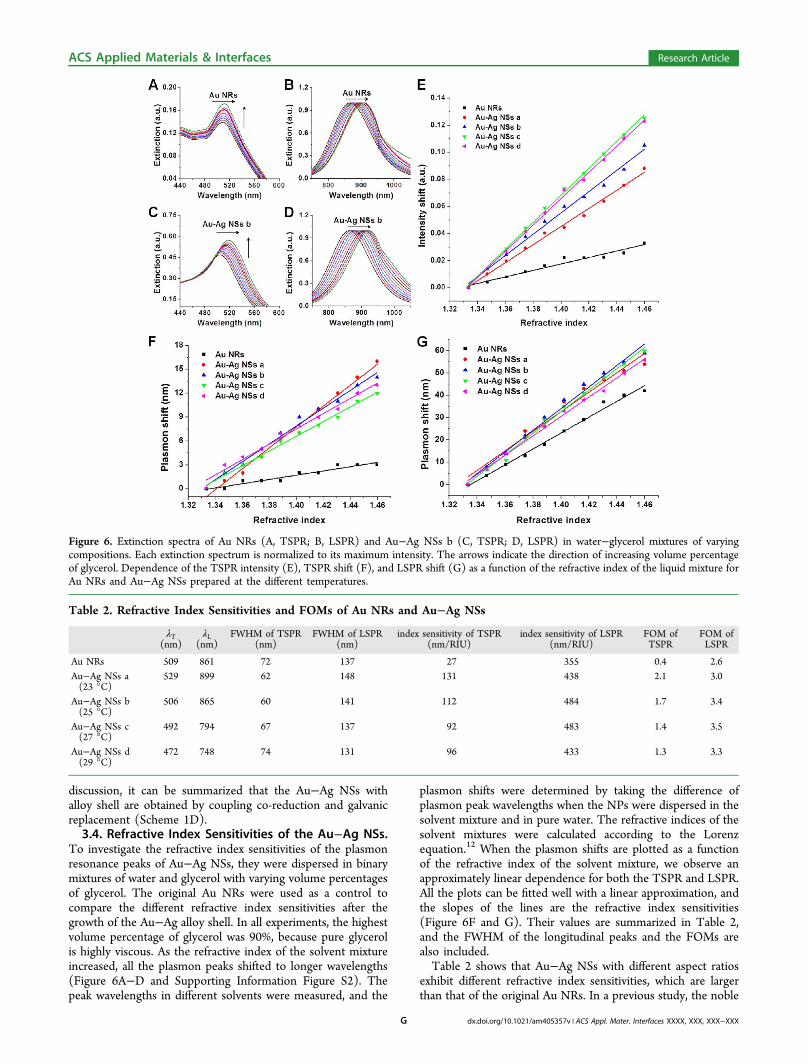
Table 2. Refractive Index Sensitivities and FOMs of Au NRs and Au−Ag NSs
λT(nm)
λL(nm)
FWHM of TSPR(nm)
FWHM of LSPR(nm)
index sensitivity of TSPR(nm/RIU)
index sensitivity of LSPR(nm/RIU)
FOM ofTSPR
FOM ofLSPR
Au NRs 509 861 72 137 27 355 0.4 2.6Au−Ag NSs a(23 °C)
529 899 62 148 131 438 2.1 3.0
Au−Ag NSs b(25 °C)
506 865 60 141 112 484 1.7 3.4
Au−Ag NSs c(27 °C)
492 794 67 137 92 483 1.4 3.5
Au−Ag NSs d(29 °C)
472 748 74 131 96 433 1.3 3.3
ACS Applied Materials & Interfaces Research Article
dx.doi.org/10.1021/am405357v | ACS Appl. Mater. Interfaces XXXX, XXX, XXX−XXXG

metal NPs were shown to have a larger refractive indexsensitivity when they had a larger aspect ratio or a shape withsharper corners.12 In addition, the refractive index sensitivityalso depends on the type of metal, as Ag nanocubes are twice assensitive to the index change in the surrounding medium as Aunanocubes of nearly the same size.35 Here, after the overgrowthof the Au−Ag alloy shell on the surface of the Au NRs and theformation of the sharp tips at the ends, both the refractive indexsensitivities from the TSPR and LSPR shifts are greater thanthat of the Au NRs (Figure 6F and G). Especially when therefractive index sensitivity from the TSPR is concerned, a largerdifference was found between the Au−Ag NSs and Au NRscompared with that in the LSPR (Figure 6F). However, amongthe four kinds of NSs, a nonlinear relationship between therefractive index sensitivities and shell thickness was found. Takethe index sensitivities from the LSPR as an example (Figure6G). With the same shape, although the Au−Ag NSs a have thelarger aspect ratio and a smaller shell thickness, the indexsensitivity is still smaller than that of Au−Ag NSs b, which havea smaller aspect ratio but a thicker shell. This phenomenon canbe attributed to the much larger amount of Ag in the alloy shell,since the index sensitivity should decrease with the reducedaspect ratio. This is the result of a combination of two reasons:metal type and aspect ratio. As for Au−Ag NSs b and Au NRs,they have very similar plasmon resonance wavelengths (865and 861 nm) and aspect ratios (4.4 and 4.3), but they aredifferent in geometry and composition. The measured indexsensitivity of Au−Ag NSs b is ∼1.4-fold larger than that of AuNRs. Combined with the previous experimental study (sharperapexes increase the index sensitivity)12 and finite-differencetime-domain (FDTD) calculations,20 we can infer that theincreasing of Au−Ag NSs b index sensitivity by a large margin iscaused by the combination of the sharp-tip geometry and thehigh quantity Ag in the shell. Similarly, the aspect ratios of Au−Ag NSs c and Au−Ag NSs d are greatly decreased, while theirindex sensitivities decrease only slightly. As shown by previousreports and our results, the metal type, aspect ratio and shapeare all influencing factors. However, it is hard to ascertain whichfactor is dominant in this study, because they are codependentand change together when the Au NRs grow into NSs. Andmore studies are still needed yet to solve this issue.The FOMs of the different SPRs of Au−Ag NSs listed in
Table 2 were also determined from the measured indexsensitivities and the resonance peak widths in the extinctionspectra. The peak widths for the determination of the FOMswere obtained by fitting the extinction spectra with multipleGaussian functions.35 Although the TSPRs of these NPs havenarrower peak widths than the of LSPRs, their FOMs are verysmall, owing to their relatively lower index sensitivities. Incontrast, the LSPRs of these NPs possess larger FOMs becauseof their higher index sensitivities. It is worth mentioning thatthe FOMs of the NSs are all above 3.0, which is greater thanthat of the Au NRs. The possession of larger index sensitivitiesand FOMs makes the Au−Ag NSs appealing for use in highlysensitive plasmonic sensors. Besides the index sensitivity andFOM, it is very interesting that the intensities of all the TSPRsfrom the normalized UV−vis spectra gradually increase as therefractive index of the solvent increases (Figure 6A and C andSupporting Information Figure S2A, C, and E). When theintensity shifts of the extinction spectra were plotted as afunction of the refractive index, an approximately lineardependence was also observed (Figure 6E). Figure 6E clearlyshows a gradual increase and variation in the slope of the TSPR
intensity shifts with an increase in the alloy shell thickness.Therefore, the slope of these curves can also be used todetermine the refractive index sensitivity of the NPs, which isproportional to the alloy shell thickness.
3.5. SERS Enhancement of the Au−Ag NSs. To studythe SERS enhancement of the Au−Ag NSs compared with AuNRs, a monolayer film of these metallic NPs was prepared by achemical assembly method using electrostatic interactions. Thecationic polyelectrolyte PDDA was used as an adhesive layer forthe NPs on the surface of a silicon substrate. A negativelycharged anionic polyelectrolyte, PSS, is needed to modify theNPs as they are capped by a positively charged bilayer ofCTAB. Supporting Information Figure S3 shows the extinctionspectra of the Au−Ag NSs (a, b, c, d)/4-ATP/PSS and AuNRs/4-ATP/PSS. A slight red-shift is observed in the spectra ofall the NSs except the NRs, which exhibit nearly no shift. Aftercoating, the NPs can still disperse uniformly in water withoutaggregation. Supporting Information Figure S4 shows the SEMimages of the surface morphologies of the monolayers ofmetallic NPs. It can be seen clearly that both the NSs withdifferent shell thicknesses and the NRs can assemble uniformlyon the substrate. In particular, most of the NPs exist separatelywith a large enough interparticle distance, to allow aninvestigation on the SERS effects of the individual NSs.The Au−Ag NSs have two sharp tips at both ends and a large
amount of Ag in the alloy shell. According to theoretical andexperimental studies, Ag NPs are more SERS-active than AuNPs, and the NSs were expected to have larger localelectromagnetic field enhancements. The Au−Ag NSs shouldtherefore be more advantageous than Au NRs when applied inRaman sensing because of their larger SERS enhancement. TheSERS spectra of the 4-ATP molecules adsorbed on themonolayers of metallic NPs are shown in Figure 7A. A SERSenhancement with the a1 vibrational modes of 4-ATP on the AuNRs monolayer, at 1591 (C−C) and 1083 cm−1 (C−S), isobserved. The band at 394 cm−1 represents the C−S bendingvibration. The observed peak positions of 4-ATP agree with thevalues reported in the literature.36 Relative to the spectrumobtained from the Au NRs, there are slight frequency shifts andchanges in the relative intensity of some bands. For example,the two obviously enhanced peaks obtained from Au−Ag NSsd, at 1598 cm−1 (C−C) and at 1083 cm−1 (C−S), were slightlyshifted. An apparent stronger intensity of the SERS spectrum of4-ATP on the surface of the NSs was obtained, which indicatesthat the Au−Ag NSs can be used as a good SERS substrate. Asa characteristic of 4-ATP, the peak at 1083 cm−1 exhibits thehighest intensity and was used to compare the enhancement ofthe different NP monolayers. As shown in Figure 7B, theincrease of the alloy shell thickness leads to a strongenhancement of the SERS intensity. Our findings show thatNSs with a Au−Ag alloy shell tend to be more SERS active thanpure Au NRs and more stable than Ag NPs, and thus theyprovide an advantage when measuring low concentrations ofanalyte.
4. CONCLUSIONSIn summary, we have developed a facile approach for high-quality Au−Ag NSs with two sharp tips at both ends usinghighly monodisperse Au NRs as cores. By simply adjusting thereaction temperature in a glycine buffer, we could finelymodulate the shell thickness of the Au−Ag NSs to achievetunable plasmonic properties over a wide range. Subsequentinvestigations indicated that these Au−Ag NSs have more
ACS Applied Materials & Interfaces Research Article
dx.doi.org/10.1021/am405357v | ACS Appl. Mater. Interfaces XXXX, XXX, XXX−XXXH

stable Au−Ag alloy shells, the formation of which can beascribed to a seed-mediated co-reduction mechanism driven bythermodynamics and the mediation of the reduction kenitics byglycine. Because of their highly anisotropic shape and bimetalliccomposition, these Au−Ag NSs exhibited higher refractiveindex sensitivities and excellent SERS enhancements comparedto the original Au NRs. Therefore, the Au−Ag NSs areexpected to have wide applications in biosensing andbiolabeling. In addition, these Au−Ag NSs also have potentialfor use in imaging, hyperthermia therapy, catalysis andchemical-specific optical sensing.
■ ASSOCIATED CONTENT
*S Supporting InformationAdditional UV−vis−NIR spectra and SEM images. Thismaterial is available free of charge via the Internet at http://pubs.acs.org.
■ AUTHOR INFORMATION
Corresponding Authors*Fax: +86 25 83738572. E-mail: [email protected].*Fax: +86 25 83272460. E-mail: [email protected].
NotesThe authors declare no competing financial interest.
■ ACKNOWLEDGMENTS
We acknowledge the support from the National Key BasicResearch Program of China (2011CB933503), the NationalNatural Science Foundation of China (61127002, 21273002),the Open Project Program (KF-GN-201107), Jiangsu provincescience and technology support project (BE2013725), and theBasic Research Program of Jiangsu Province (BK2011036,BK2011590). A project funded by the Priority AcademicProgram Development of Jiangsu Higher Education Institu-tions.
■ REFERENCES(1) Daniel, M.-C.; Astruc, D. Chem. Rev. 2004, 104, 293−346.(2) Rycenga, M.; Cobley, C. M.; Zeng, J.; Li, W.; Moran, C. H.;Zhang, Q.; Qin, D.; Xia, Y. Chem. Rev. 2011, 111, 3669−3712.(3) Burda, C.; Chen, X.; Narayanan, R.; El-Sayed, M. A. Chem. Rev.2005, 105, 1025−1102.(4) Xia, Y.; Halas, N. J. MRS Bull. 2005, 30, 338−348.(5) Liz-Marzan, L. M. Langmuir 2006, 22, 32−41.(6) Sau, T. K.; Rogach, A. L.; Jackel, F.; Klar, T. A.; Feldmann, J. Adv.Mater. 2010, 22, 1805−1825.(7) Sun, Y.; Xia, Y. Nano Lett. 2003, 3, 1569−1572.(8) Lohse, S. E.; Murphy, C. J. Chem. Mater. 2013, 25, 1250−1261.(9) Huang, X.; El-Sayed, I. H.; Qian, W.; El-Sayed, M. A. J. Am.Chem. Soc. 2006, 128, 2115−2120.(10) Ding, H.; Yong, K.-T.; Roy, I.; Pudavar, H. E.; Law, W. C.;Bergey, E. J.; Prasad, P. N. J. Phys. Chem. C 2007, 111, 12552−12557.(11) Yu, C.; Irudayaraj, J. Anal. Chem. 2007, 79, 572−579.(12) Chen, H.; Kou, X.; Yang, Z.; Ni, W.; Wang, J. Langmuir 2008,24, 5233−5237.(13) Wei, W.; Chen, K.; Ge, G. Adv. Mater. 2013, 25, 3863−3868.(14) Wang, C.; Chen, Y.; Wang, T.; Ma, Z.; Su, Z. Adv. Funct. Mater.2008, 18, 355−361.(15) Becker, J.; Zins, I.; Jakab, A.; Khalavka, Y.; Schubert, O.;Sonnichsen, C. Nano Lett. 2008, 8, 1719−1723.(16) Okuno, Y.; Nishioka, K.; Kiya, A.; Nakashima, N.; Ishibashi, A.;Niidome, Y. Nanoscale 2010, 2, 1489−1493.(17) Park, K.; Drummy, L. F.; Vaia, R. A. J. Mater. Chem. 2011, 21,15608−15618.(18) Sanchez-Iglesias, A.; Carbo-Argibay, E.; Glaria, A.; Rodríguez-Gonzalez, B.; Perez-Juste, J.; Pastoriza-Santos, I.; Liz-Marzan, L. M.Chem.Eur. J. 2010, 16, 5558−5563.(19) Huang, C.-C.; Yang, Z.; Chang, H.-T. Langmuir 2004, 20,6089−6092.(20) Li, M.; Zhang, Z.; Zhang, X.; Li, K.; Yu, X. Opt. Express 2008,16, 14288−14293.(21) Fu, Q.; Zhang, D. G.; Yi, M. F.; Wang, X. X.; Chen, Y. K. J. Opt.2012, 14, 085001−086005.(22) Lee, K.-S.; El-Sayed, M. A. J. Phys. Chem. B 2006, 110, 19220−19225.(23) Huang, H.; Liu, X.; Liao, B.; Chu, P. K. Plasmonics 2011, 6, 1−9.(24) Fernanda Cardinal, M.; Rodriguez-Gonzalez, B.; Alvarez-Puebla,R. A.; Perez-Juste, J.; Liz-Marzan, L. M. J. Phys. Chem. C 2010, 114,10417−10423.(25) Xiang, Y.; Wu, X.; Liu, D.; Feng, L.; Zhang, K.; Chu, W.; Zhou,W.; Xie, S. J. Phys. Chem. C 2008, 112, 3203−3208.(26) Park, K.; Vaia, R. A. Adv. Mater. 2008, 20, 3882−3886.(27) Ye, X.; Zheng, C.; Chen, J.; Gao, Y.; Murray, C. B. Nano Lett.2013, 13, 765−771.(28) Midgley, P.; Weyland, M. Ultramicroscopy 2003, 96, 413−431.(29) Ozmetin, C.; Copur, M.; Yartasi, A.; Kocakerim, M. Chem. Eng.Technol. 2000, 23, 707−711.(30) Shao, Y.; Jin, Y.; Dong, S. Chem. Commun. 2004, 9, 1104−1105.(31) Kou, X.; Zhang, S.; Yang, Z.; Tsung, C.-K.; Stucky, G. D.; Sun,L.; Wang, J.; Yan, C. J. Am. Chem. Soc. 2007, 129, 6402−6404.(32) Wang, Z. L.; Mohamed, M. B.; Link, S.; El-Sayed, M. A. Surf. Sci.1999, 440, L809−L814.
Figure 7. (A) SERS spectra of 4-ATP molecules on the self-assembledmonolayers of Au NRs and various Au−Ag NSs, which are markedbeside each curve. (B) Measured SERS intensity of the 4-ATPcharacteristic band at 1083 cm−1.
ACS Applied Materials & Interfaces Research Article
dx.doi.org/10.1021/am405357v | ACS Appl. Mater. Interfaces XXXX, XXX, XXX−XXXI

(33) Smith, D. K.; Miller, N. R.; Korgel, B. A. Langmuir 2009, 25,9518−9524.(34) Liu; Guyot-Sionnest, P. J. Phys. Chem. B 2005, 109, 22192−22200.(35) Lee, Y. H.; Chen, H.; Xu, Q.-H.; Wang, J. J. Phys. Chem. C 2011,115, 7997−8004.(36) Hu, X.; Cheng, W.; Wang, T.; Wang, Y.; Wang, E.; Dong, S. J.Phys. Chem. B 2005, 109, 19385−19389.
ACS Applied Materials & Interfaces Research Article
dx.doi.org/10.1021/am405357v | ACS Appl. Mater. Interfaces XXXX, XXX, XXX−XXXJ