Control of Diphteria, Pertussis, Tetanus, Influenza type B, Hepatitis Field Guide
107
-
Upload
joke-channel -
Category
Health & Medicine
-
view
6.154 -
download
2
Transcript of Control of Diphteria, Pertussis, Tetanus, Influenza type B, Hepatitis Field Guide
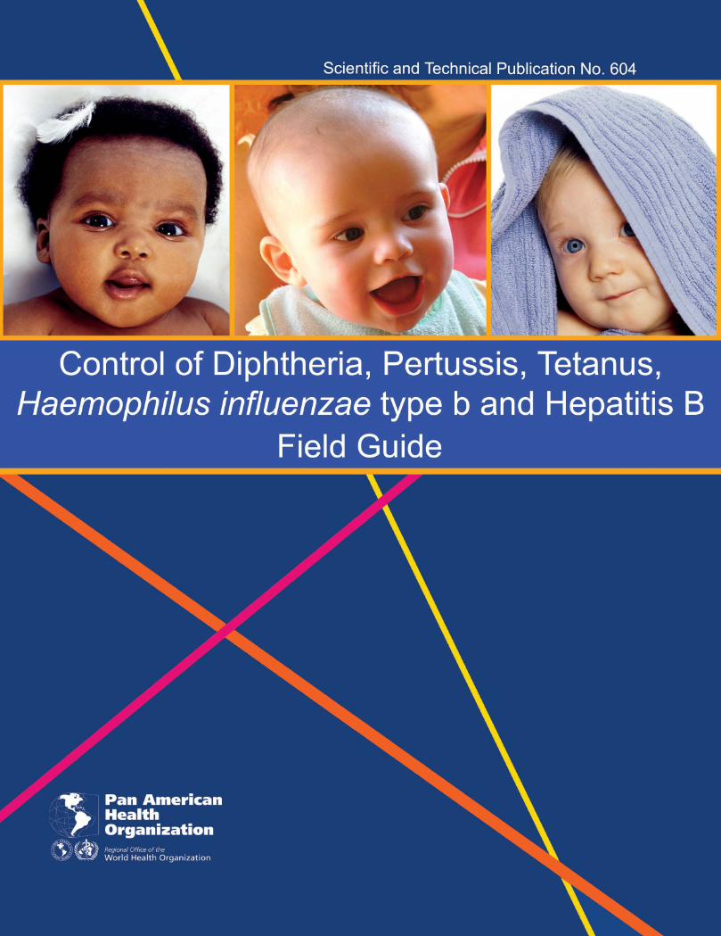

Control of Diphtheria,Pertussis, Tetanus,
Haemophilus influenzaetype b, andHepatitis B
FIELD GUIDE
Scientific and Technical Publication No. 604
PAN AMERICAN HEALTH ORGANIZATIONPan American Sanitary Bureau, Regional Office of the
WORLD HEALTH ORGANIZATION525 Twenty-third Street, N.W.
Washington, D.C. 20037www.paho.org
2005

Also published in Spanish (2005) with the title:Control de la difteria, tos ferina, tétanos, Haemophilus influenzae tipo b y hepatitis B.
Guía práctica. (ISBN 92 75 31604 X)
PAHO HQ Library Cataloguing-in-Publication Data
Pan American Health OrganizationControl of diphtheria, pertussis, tetanus, Haemophilus influenzae type b, and hepatitis B: field guide.Washington, D.C.: PAHO, © 2005.(Scientific and Technical Publication No. 604)
ISBN 92 75 11604 0
I. Title II. Series
1. DIPHTHERIA - prevention & control2. WHOOPING COUGH - prevention & control3. TETANUS - prevention & control4. HAEMOPHILUS INFLUENZAE TYPE B5. HEPATITIS B - prevention & control6. GUIDEBOOKS [PUBLICATION TYPE]
NLM WC100
This guide was prepared by the Immunization Unit of the Pan American Health Organization.
Cover photos: left, Stockbyte.com, © Stockbyte; center, courtesy of Ignacio Astorga; right,Stockbyte.com, © Stockbyte.
The Pan American Health Organization welcomes requests for permission to reproduce ortranslate its publications, in part or in full. Applications and inquiries should be addressed to thePublications Program, Pan American Health Organization, Washington, D.C., U.S.A., which willbe glad to provide the latest information on any changes made to the text, plans for new editions,and reprints and translations already available.
© Pan American Health Organization (2005)
Publications of the Pan American Health Organization enjoy copyright protection in accor-dance with the provisions of Protocol 2 of the Universal Copyright Convention. All rights arereserved.
The designations employed and the presentation of the material in this publication do notimply the expression of any opinion whatsoever on the part of the Secretariat of the Pan Ameri-can Health Organization concerning the status of any country, territory, city, or area or of itsauthorities, or concerning the delimitation of its frontiers or boundaries.
The mention of specific companies or of certain manufacturers’ products does not imply thatthey are endorsed or recommended by the Pan American Health Organization in preference toothers of a similar nature that are not mentioned. Errors and omissions excepted, the names ofproprietary products are distinguished by initial capital letters.

CONTENTS
About the immunization field guides . . . . . . . . . . . . . . . . . . . . . . . . . . . . . . xi
Preface . . . . . . . . . . . . . . . . . . . . . . . . . . . . . . . . . . . . . . . . . . . . . . . . . . . . . xiii
Glossary . . . . . . . . . . . . . . . . . . . . . . . . . . . . . . . . . . . . . . . . . . . . . . . . . . . . . xv
Acronyms . . . . . . . . . . . . . . . . . . . . . . . . . . . . . . . . . . . . . . . . . . . . . . . . . . . . xix
1 DIPHTHERIA . . . . . . . . . . . . . . . . . . . . . . . . . . . . . . . . . . . . . . . . . . . . . . . . . 1
1.1 Introduction . . . . . . . . . . . . . . . . . . . . . . . . . . . . . . . . . . . . . . . . . . . . . . . . 1
1.2 Epidemiology . . . . . . . . . . . . . . . . . . . . . . . . . . . . . . . . . . . . . . . . . . . . . . . . 1
1.2.1 Infectious Agent . . . . . . . . . . . . . . . . . . . . . . . . . . . . . . . . . . . . . . . . . . . 1
1.2.2 Occurrence . . . . . . . . . . . . . . . . . . . . . . . . . . . . . . . . . . . . . . . . . . . . . . . 2
1.2.3 Transmission . . . . . . . . . . . . . . . . . . . . . . . . . . . . . . . . . . . . . . . . . . . . . 2
1.2.4 Reservoir . . . . . . . . . . . . . . . . . . . . . . . . . . . . . . . . . . . . . . . . . . . . . . . . 2
1.2.5 Incubation . . . . . . . . . . . . . . . . . . . . . . . . . . . . . . . . . . . . . . . . . . . . . . . 2
1.2.6 Communicability . . . . . . . . . . . . . . . . . . . . . . . . . . . . . . . . . . . . . . . . . . 2
1.2.7 Immunity . . . . . . . . . . . . . . . . . . . . . . . . . . . . . . . . . . . . . . . . . . . . . . . . 3
1.2.8 Changing Epidemiology . . . . . . . . . . . . . . . . . . . . . . . . . . . . . . . . . . . . . 3
1.3 Clinical aspects . . . . . . . . . . . . . . . . . . . . . . . . . . . . . . . . . . . . . . . . . . . . . . 4
1.3.1 Pathogenesis . . . . . . . . . . . . . . . . . . . . . . . . . . . . . . . . . . . . . . . . . . . . . 4
1.3.2 Clinical Features . . . . . . . . . . . . . . . . . . . . . . . . . . . . . . . . . . . . . . . . . . . 5
1.3.3 Laboratory Diagnosis . . . . . . . . . . . . . . . . . . . . . . . . . . . . . . . . . . . . . . 6
1.3.4 Differential Diagnosis . . . . . . . . . . . . . . . . . . . . . . . . . . . . . . . . . . . . . . 6
1.3.5 Complications . . . . . . . . . . . . . . . . . . . . . . . . . . . . . . . . . . . . . . . . . . . . 7
1.3.6 Treatment . . . . . . . . . . . . . . . . . . . . . . . . . . . . . . . . . . . . . . . . . . . . . . . . 7
1.3.7 Management of Contacts . . . . . . . . . . . . . . . . . . . . . . . . . . . . . . . . . . . 9
1.4 Vaccination activities . . . . . . . . . . . . . . . . . . . . . . . . . . . . . . . . . . . . . . . . . 9
1.4.1 Routine Immunization . . . . . . . . . . . . . . . . . . . . . . . . . . . . . . . . . . . . . . 9
iiiiii
iivv CONTROL OF DIPHTHERIA, PERTUSSIS, TETANUS, HAEMOPHILUS INFLUENZAE TYPE B, AND HEPATITIS B: FIELD GUIDE
1.4.2 Other Vaccination Activities . . . . . . . . . . . . . . . . . . . . . . . . . . . . . . . . . 10
1.4.3 Outbreak Control . . . . . . . . . . . . . . . . . . . . . . . . . . . . . . . . . . . . . . . . 10
2 PERTUSSIS (WHOOPING COUGH) . . . . . . . . . . . . . . . . . . . . . . . . . . . . . . 11
2.1 Introduction . . . . . . . . . . . . . . . . . . . . . . . . . . . . . . . . . . . . . . . . . . . . . . . 11
2.2 Epidemiology . . . . . . . . . . . . . . . . . . . . . . . . . . . . . . . . . . . . . . . . . . . . . . . 11
2.2.1 Infectious Agent . . . . . . . . . . . . . . . . . . . . . . . . . . . . . . . . . . . . . . . . . . . . . 11
2.2.2 Occurrence . . . . . . . . . . . . . . . . . . . . . . . . . . . . . . . . . . . . . . . . . . . . . . . . . 11
2.2.3 Transmission . . . . . . . . . . . . . . . . . . . . . . . . . . . . . . . . . . . . . . . . . . . . . . . . 11
2.2.4 Reservoir . . . . . . . . . . . . . . . . . . . . . . . . . . . . . . . . . . . . . . . . . . . . . . . . . . 12
2.2.5 Incubation . . . . . . . . . . . . . . . . . . . . . . . . . . . . . . . . . . . . . . . . . . . . . . . . . 12
2.2.6 Communicability . . . . . . . . . . . . . . . . . . . . . . . . . . . . . . . . . . . . . . . . . . . . 12
2.2.7 Immunity . . . . . . . . . . . . . . . . . . . . . . . . . . . . . . . . . . . . . . . . . . . . . . . . . . 12
2.2.8 Changing Epidemiology . . . . . . . . . . . . . . . . . . . . . . . . . . . . . . . . . . . . . . 12
2.3 Clinical aspects . . . . . . . . . . . . . . . . . . . . . . . . . . . . . . . . . . . . . . . . . . . . . 13
2.3.1 Pathogenesis . . . . . . . . . . . . . . . . . . . . . . . . . . . . . . . . . . . . . . . . . . . . . . . 13
2.3.2 Clinical Features . . . . . . . . . . . . . . . . . . . . . . . . . . . . . . . . . . . . . . . . . . . . . 13
2.3.3 Laboratory Diagnosis . . . . . . . . . . . . . . . . . . . . . . . . . . . . . . . . . . . . . . . . 14
2.3.4 Differential Diagnosis . . . . . . . . . . . . . . . . . . . . . . . . . . . . . . . . . . . . . . . . 16
2.3.5 Complications . . . . . . . . . . . . . . . . . . . . . . . . . . . . . . . . . . . . . . . . . . . . . . 16
2.3.6 Treatment . . . . . . . . . . . . . . . . . . . . . . . . . . . . . . . . . . . . . . . . . . . . . . . . . . 17
2.3.7 Management of Contacts . . . . . . . . . . . . . . . . . . . . . . . . . . . . . . . . . . . . . 17
2.4 Vaccination activities . . . . . . . . . . . . . . . . . . . . . . . . . . . . . . . . . . . . . . . . 18
2.4.1 Routine Immunization . . . . . . . . . . . . . . . . . . . . . . . . . . . . . . . . . . . . . . . . 18
2.4.2 Outbreak Control . . . . . . . . . . . . . . . . . . . . . . . . . . . . . . . . . . . . . . . . . . . 18
3 TETANUS . . . . . . . . . . . . . . . . . . . . . . . . . . . . . . . . . . . . . . . . . . . . . . . . . . 19
3.1 Introduction . . . . . . . . . . . . . . . . . . . . . . . . . . . . . . . . . . . . . . . . . . . . . . . 19
3.2 Epidemiology . . . . . . . . . . . . . . . . . . . . . . . . . . . . . . . . . . . . . . . . . . . . . . . 19
3.2.1 Infectious Agent . . . . . . . . . . . . . . . . . . . . . . . . . . . . . . . . . . . . . . . . . . . . . 19
3.2.2 Occurrence . . . . . . . . . . . . . . . . . . . . . . . . . . . . . . . . . . . . . . . . . . . . . . . . . 19
3.2.3 Transmission . . . . . . . . . . . . . . . . . . . . . . . . . . . . . . . . . . . . . . . . . . . . . . . 20

CONTENTS vv
3.2.4 Reservoir . . . . . . . . . . . . . . . . . . . . . . . . . . . . . . . . . . . . . . . . . . . . . . . . . . 20
3.2.5 Incubation . . . . . . . . . . . . . . . . . . . . . . . . . . . . . . . . . . . . . . . . . . . . . . . . . 20
3.2.6 Communicability . . . . . . . . . . . . . . . . . . . . . . . . . . . . . . . . . . . . . . . . . . . . 20
3.2.7 Immunity . . . . . . . . . . . . . . . . . . . . . . . . . . . . . . . . . . . . . . . . . . . . . . . . . . 20
3.2.8 Changing Epidemiology . . . . . . . . . . . . . . . . . . . . . . . . . . . . . . . . . . . . . . 20
3.3 Clinical aspects . . . . . . . . . . . . . . . . . . . . . . . . . . . . . . . . . . . . . . . . . . . . . 21
3.3.1 Pathogenesis . . . . . . . . . . . . . . . . . . . . . . . . . . . . . . . . . . . . . . . . . . . . . . . 21
3.3.2 Clinical Features . . . . . . . . . . . . . . . . . . . . . . . . . . . . . . . . . . . . . . . . . . . . . 21
3.3.3 Laboratory Diagnosis . . . . . . . . . . . . . . . . . . . . . . . . . . . . . . . . . . . . . . . . 22
3.3.4 Differential Diagnosis . . . . . . . . . . . . . . . . . . . . . . . . . . . . . . . . . . . . . . . . 22
3.3.5 Complications . . . . . . . . . . . . . . . . . . . . . . . . . . . . . . . . . . . . . . . . . . . . . . 22
3.3.6 Treatment . . . . . . . . . . . . . . . . . . . . . . . . . . . . . . . . . . . . . . . . . . . . . . . . . 23
3.4 Vaccination activities . . . . . . . . . . . . . . . . . . . . . . . . . . . . . . . . . . . . . . . . 23
3.4.1 Routine Immunization . . . . . . . . . . . . . . . . . . . . . . . . . . . . . . . . . . . . . . . . 23
3.4.2 Other Vaccination Activities . . . . . . . . . . . . . . . . . . . . . . . . . . . . . . . . . . . 23
3.4.3 Outbreak Response . . . . . . . . . . . . . . . . . . . . . . . . . . . . . . . . . . . . . . . . . 24
4 HAEMOPHILUS INFLUENZAE TYPE B . . . . . . . . . . . . . . . . . . . . . . . . . . . 25
4.1 Introduction . . . . . . . . . . . . . . . . . . . . . . . . . . . . . . . . . . . . . . . . . . . . . . 25
4.2 Epidemiology . . . . . . . . . . . . . . . . . . . . . . . . . . . . . . . . . . . . . . . . . . . . . . . 25
4.2.1 Infectious Agent . . . . . . . . . . . . . . . . . . . . . . . . . . . . . . . . . . . . . . . . . . . . . 25
4.2.2 Occurrence . . . . . . . . . . . . . . . . . . . . . . . . . . . . . . . . . . . . . . . . . . . . . . . . 25
4.2.3 Transmission . . . . . . . . . . . . . . . . . . . . . . . . . . . . . . . . . . . . . . . . . . . . . . . 25
4.2.4 Reservoir . . . . . . . . . . . . . . . . . . . . . . . . . . . . . . . . . . . . . . . . . . . . . . . . . . 25
4.2.5 Incubation . . . . . . . . . . . . . . . . . . . . . . . . . . . . . . . . . . . . . . . . . . . . . . . . . 25
4.2.6 Communicability . . . . . . . . . . . . . . . . . . . . . . . . . . . . . . . . . . . . . . . . . . . . 26
4.2.7 Immunity . . . . . . . . . . . . . . . . . . . . . . . . . . . . . . . . . . . . . . . . . . . . . . . . . . 26
4.2.8 Changing Epidemiology . . . . . . . . . . . . . . . . . . . . . . . . . . . . . . . . . . . . . . 26
4.3 Clinical aspects . . . . . . . . . . . . . . . . . . . . . . . . . . . . . . . . . . . . . . . . . . . . 27
4.3.1 Pathogenesis . . . . . . . . . . . . . . . . . . . . . . . . . . . . . . . . . . . . . . . . . . . . . . . 27
4.3.2 Clinical Features . . . . . . . . . . . . . . . . . . . . . . . . . . . . . . . . . . . . . . . . . . . . 28

vvii CONTROL OF DIPHTHERIA, PERTUSSIS, TETANUS, HAEMOPHILUS INFLUENZAE TYPE B, AND HEPATITIS B: FIELD GUIDE
4.3.3 Laboratory Diagnosis . . . . . . . . . . . . . . . . . . . . . . . . . . . . . . . . . . . . . . . . 28
4.3.4 Differential Diagnosis . . . . . . . . . . . . . . . . . . . . . . . . . . . . . . . . . . . . . . . . 28
4.3.5 Complications . . . . . . . . . . . . . . . . . . . . . . . . . . . . . . . . . . . . . . . . . . . . . . 29
4.3.6 Treatment . . . . . . . . . . . . . . . . . . . . . . . . . . . . . . . . . . . . . . . . . . . . . . . . . 29
4.3.7 Management of Contacts . . . . . . . . . . . . . . . . . . . . . . . . . . . . . . . . . . . . . 29
4.4 Vaccination activities . . . . . . . . . . . . . . . . . . . . . . . . . . . . . . . . . . . . . . . . 30
4.4.1 Routine Immunization . . . . . . . . . . . . . . . . . . . . . . . . . . . . . . . . . . . . . . . 30
4.4.2 Other Vaccination Activities . . . . . . . . . . . . . . . . . . . . . . . . . . . . . . . . . . . 30
5 HEPATITIS B . . . . . . . . . . . . . . . . . . . . . . . . . . . . . . . . . . . . . . . . . . . . . . . . 31
5.1 Introduction . . . . . . . . . . . . . . . . . . . . . . . . . . . . . . . . . . . . . . . . . . . . . . . 31
5.2 Epidemiology . . . . . . . . . . . . . . . . . . . . . . . . . . . . . . . . . . . . . . . . . . . . . . . 31
5.2.1 Infectious Agent . . . . . . . . . . . . . . . . . . . . . . . . . . . . . . . . . . . . . . . . . . . . . 31
5.2.2 Occurrence . . . . . . . . . . . . . . . . . . . . . . . . . . . . . . . . . . . . . . . . . . . . . . . . . 31
5.2.3 Transmission . . . . . . . . . . . . . . . . . . . . . . . . . . . . . . . . . . . . . . . . . . . . . . . 32
5.2.4 Reservoir . . . . . . . . . . . . . . . . . . . . . . . . . . . . . . . . . . . . . . . . . . . . . . . . . . 33
5.2.5 Incubation . . . . . . . . . . . . . . . . . . . . . . . . . . . . . . . . . . . . . . . . . . . . . . . . . 33
5.2.6 Communicability . . . . . . . . . . . . . . . . . . . . . . . . . . . . . . . . . . . . . . . . . . . . 33
5.2.7 Immunity . . . . . . . . . . . . . . . . . . . . . . . . . . . . . . . . . . . . . . . . . . . . . . . . . . 33
5.3 Clinical aspects . . . . . . . . . . . . . . . . . . . . . . . . . . . . . . . . . . . . . . . . . . . . . 33
5.3.1 Pathogenesis . . . . . . . . . . . . . . . . . . . . . . . . . . . . . . . . . . . . . . . . . . . . . . . 33
5.3.2 Clinical Features . . . . . . . . . . . . . . . . . . . . . . . . . . . . . . . . . . . . . . . . . . . . 33
5.3.3 Laboratory Diagnosis . . . . . . . . . . . . . . . . . . . . . . . . . . . . . . . . . . . . . . . . 34
5.3.4 Differential Diagnosis . . . . . . . . . . . . . . . . . . . . . . . . . . . . . . . . . . . . . . . . 35
5.3.5 Complications . . . . . . . . . . . . . . . . . . . . . . . . . . . . . . . . . . . . . . . . . . . . . . 36
5.3.6 Treatment . . . . . . . . . . . . . . . . . . . . . . . . . . . . . . . . . . . . . . . . . . . . . . . . . 37
5.4 Vaccination activities and passive immunization with hepatitis b immune globulin . . . . . . . . . . . . . . . . . . . . . . . . . . . . . . . . . . . . . . . . . . . 37
5.4.1 Routine Immunization . . . . . . . . . . . . . . . . . . . . . . . . . . . . . . . . . . . . . . . . 37
5.4.2 Other Vaccination Activities . . . . . . . . . . . . . . . . . . . . . . . . . . . . . . . . . . . 38
5.4.3 Outbreak Control . . . . . . . . . . . . . . . . . . . . . . . . . . . . . . . . . . . . . . . . . . . 39

CONTENTS vviiii
5.4.4 Use of Hepatitis B Immune Globulin (HBIG) . . . . . . . . . . . . . . . . . . . . . . 39
6 EPIDEMIOLOGICAL SURVEILLANCE . . . . . . . . . . . . . . . . . . . . . . . . . . . . 42
6.1 General considerations . . . . . . . . . . . . . . . . . . . . . . . . . . . . . . . . . . . . . 42
6.2 Case definitions . . . . . . . . . . . . . . . . . . . . . . . . . . . . . . . . . . . . . . . . . . . . 43
6.3 Flow of surveillance information . . . . . . . . . . . . . . . . . . . . . . . . . . . . . 45
6.3.1 Case Reporting . . . . . . . . . . . . . . . . . . . . . . . . . . . . . . . . . . . . . . . . . . . . . 45
6.3.2 Case Investigation . . . . . . . . . . . . . . . . . . . . . . . . . . . . . . . . . . . . . . . . . . . 46
6.3.3 Feedback . . . . . . . . . . . . . . . . . . . . . . . . . . . . . . . . . . . . . . . . . . . . . . . . . . 47
6.4 Data analysis and surveillance indicators . . . . . . . . . . . . . . . . . . . . . . 47
6.4.1 Data Analysis . . . . . . . . . . . . . . . . . . . . . . . . . . . . . . . . . . . . . . . . . . . . . . 47
6.4.2 Surveillance Indicators . . . . . . . . . . . . . . . . . . . . . . . . . . . . . . . . . . . . . . . 47
6.5 Outbreak response . . . . . . . . . . . . . . . . . . . . . . . . . . . . . . . . . . . . . . . . . 48
7 VACCINES AGAINST DIPHTHERIA, PERTUSSIS, TETANUS, HAEMOPHILUS INFLUENZAE TYPE B, AND HEPATITIS B . . . . . . . . . . 49
7.1 Diphteria, pertussis and tetanus vaccines . . . . . . . . . . . . . . . . . . . . . . . 49
7.1.1 Components . . . . . . . . . . . . . . . . . . . . . . . . . . . . . . . . . . . . . . . . . . . . . . . 49
7.1.2 Immunity and Effectiveness . . . . . . . . . . . . . . . . . . . . . . . . . . . . . . . . . . . 49
7.1.3 Adverse Events and Contraindications . . . . . . . . . . . . . . . . . . . . . . . . . . . 50
7.1.4 Schedule and Dosage . . . . . . . . . . . . . . . . . . . . . . . . . . . . . . . . . . . . . . . . 52
7.2 Haemophilus influenzae type b vaccines . . . . . . . . . . . . . . . . . . . . . . . . . 53
7.2.1 Immunity and Effectiveness . . . . . . . . . . . . . . . . . . . . . . . . . . . . . . . . . . . . 53
7.2.2 Adverse Events and Contraindications . . . . . . . . . . . . . . . . . . . . . . . . . . . 54
7.2.3 Schedule and Dosage . . . . . . . . . . . . . . . . . . . . . . . . . . . . . . . . . . . . . . . . 54
7.3 Hepatitis B vaccines . . . . . . . . . . . . . . . . . . . . . . . . . . . . . . . . . . . . . . . . . 55
7.3.1 Immunity and Effectiveness . . . . . . . . . . . . . . . . . . . . . . . . . . . . . . . . . . . . 55
7.3.2 Adverse Events and Contraindications . . . . . . . . . . . . . . . . . . . . . . . . . . . 56
7.3.3 Schedule and Dosage . . . . . . . . . . . . . . . . . . . . . . . . . . . . . . . . . . . . . . . . 57
7.4 Combination vaccines . . . . . . . . . . . . . . . . . . . . . . . . . . . . . . . . . . . . . . . 58

vviiiiii CONTROL OF DIPHTHERIA, PERTUSSIS, TETANUS, HAEMOPHILUS INFLUENZAE TYPE B, AND HEPATITIS B: FIELD GUIDE
7.4.1 Available Combinations . . . . . . . . . . . . . . . . . . . . . . . . . . . . . . . . . . . . . . 58
7.5 Cold chain, storage, and safe injection practices . . . . . . . . . . . . . . . . 59
7.6 Vaccine administration . . . . . . . . . . . . . . . . . . . . . . . . . . . . . . . . . . . . . 60
7.7 Final considerations . . . . . . . . . . . . . . . . . . . . . . . . . . . . . . . . . . . . . . . . 61
References . . . . . . . . . . . . . . . . . . . . . . . . . . . . . . . . . . . . . . . . . . . . . . . . . . . 62
Bibliography . . . . . . . . . . . . . . . . . . . . . . . . . . . . . . . . . . . . . . . . . . . . . . . . . 66
AANNNNEEXXEESS
Annex 1. Summary of the epidemiological characteristics of diphtheria, pertussis, tetanus, Haemophilus influenzae type b, and hepatitis B . . . . 73
Annex 2. Example of Diphtheria Notification and Investigation Form . . . . . . . 74
Annex 3. Example of a form for the management of contacts . . . . . . . . . . . . . 76
Annex 4. Example of Pertussis-like Illness Notification and Investigation Form . . . . . . . . . . . . . . . . . . . . . . . . . . . . . . . . . . . . . . . . . . . . . . . . . . . 77
Annex 5. Example of Tetanus Notification and Investigation Form . . . . . . . . . 79
Annex 6. Example of Haemophilus influenzae type b Meningitis Notification and Investigation Form . . . . . . . . . . . . . . . . . . . . . . . . . . 81
Annex 7. Example of Hepatitis B Notification and Investigation Form . . . . . . . 83
Annex 8. How to reconstitute and administer lyophilized DPT+Hib+hepatitis B (pentavalent) vaccine . . . . . . . . . . . . . . . . . . . . . . . . . . . . . 85
LIST OF FIGURES AND TABLES
Figures
Figure 1. Photomicrograph of Corynebacterium diphtheriae(magnified 1200x) . . . . . . . . . . . . . . . . . . . . . . . . . . . . . . . . . . . . . . . . 1
Figure 2. Number of diphtheria cases notified and DPT3 vaccine coverage among children aged less than 1 year, Region of the Americas, 1978–2004 . . . . . . . . . . . . . . . . . . . . . . . . . . . . . . . . . . . . . . . . . . . . . . 3
Figure 3. Diphtheria membranes . . . . . . . . . . . . . . . . . . . . . . . . . . . . . . . . . . . . 5
Figure 4. Number of pertussis cases notified and DPT3 vaccine coverage among children aged less than 1 year, Region of the Americas, 1978–2004 . . . . . . . . . . . . . . . . . . . . . . . . . . . . . . . . . . . . . . . . . . . . . 13
Figure 5. Infant with pertussis . . . . . . . . . . . . . . . . . . . . . . . . . . . . . . . . . . . . . . 13
Figure 6. Diagram of the clinical course of “classic” pertussis . . . . . . . . . . . . . 14

CONTENTS iixx
Figure 7. Proper technique for obtaining a nasopharyngeal specimen for isolation of B. pertussis . . . . . . . . . . . . . . . . . . . . . . . . . . . . . . . . . . . . 15
Figure 8. Annual incidence of tetanus and DPT3 vaccine coverage among children aged less than 1 year, Region of the Americas, 1978–2004 . . 20
Figure 9. Child with opisthotonus . . . . . . . . . . . . . . . . . . . . . . . . . . . . . . . . . . 22
Figure 10. Cases of invasive disease by Haemophilus influenzae type b, Chile, 1996–1999 . . . . . . . . . . . . . . . . . . . . . . . . . . . . . . . . . . . . . . . . 27
Figure 11. Cases of meningitis by Haemophilus influenzae type b, Uruguay, 1993–1999 . . . . . . . . . . . . . . . . . . . . . . . . . . . . . . . . . . . . . 27
Figure 12. Hepatitis B virus . . . . . . . . . . . . . . . . . . . . . . . . . . . . . . . . . . . . . . . . . 31
Figure 13. World prevalence of hepatitis B, prior to mass vaccine introductionin Latin America and the Caribbean, 1997 . . . . . . . . . . . . . . . . . . . . 32
Figure 14. Hepatitis B—progression of infection . . . . . . . . . . . . . . . . . . . . . . . . 34
Figure 15. Vaccine-preventable disease surveillance flow . . . . . . . . . . . . . . . . . 46
Figure 16. Proper disposal of syringes and needles in “sharp boxes” . . . . . . . . 60
Tables
Table 1. Characteristics of three recent diphtheria outbreaks in Latin America . . . . . . . . . . . . . . . . . . . . . . . . . . . . . . . . . . . . . . . . . . 4
Table 2. Suggested dose ranges for the use of diphtheria antitoxin . . . . . . . . . 8
Table 3. Summary guide to tetanus prophylaxis in routine wound management . . . . . . . . . . . . . . . . . . . . . . . . . . . . . . . . . . . . . . . . . . . . 23
Table 4. Markers for the detection of hepatitis B infection and risk of transmission . . . . . . . . . . . . . . . . . . . . . . . . . . . . . . . . . . . . . . . . . . . . 35
Table 5. Interpretation of serologic tests for hepatitis B . . . . . . . . . . . . . . . . . 36
Table 6. United States’ recommendations for prophylaxis against hepatitis B after percutaneous, ocular, or mucosal exposure to the virus . . . . . . 41
Table 7. Data analysis for diphtheria, pertussis, tetanus, Hib, and hepatitis B . . . . . . . . . . . . . . . . . . . . . . . . . . . . . . . . . . . . . . . . . . . . . . 47
Table 8. Adverse reactions attributable to vaccination with DPT . . . . . . . . . . . 51
Table 9. Conjugate vaccines against Haemophilus influenzae type b (Hib) and recommended vaccination series . . . . . . . . . . . . . . . . . . . . . . . . . 53
Table 10. Doses of hepatitis B vaccine recommended, according to age and type of patient . . . . . . . . . . . . . . . . . . . . . . . . . . . . . . . . . . . . . . . 57
Table 11. Formulations of selected combination vaccines available in the Americas . . . . . . . . . . . . . . . . . . . . . . . . . . . . . . . . . . . . . . . . . . 59


ABOUT THE IMMUNIZATION FIELD GUIDES
The Expanded Program on Immunization is viewed as one of the most successfulpublic health experiences in the Americas because it has played a pivotal role inreducing infant mortality from vaccine-preventable diseases in the Region. In fact,since the program was launched, our countries stopped the transmission of wildpoliovirus in the Region in 1991 and interrupted indigenous measles transmission inNovember 2002; they also are making significant gains in the battle to eliminaterubella and congenital rubella syndrome. In addition, national immunization pro-grams are undertaking extraordinary efforts to identify at-risk populations and over-come inequities in vaccination. To maintain these advances and to cope with newchallenges, such as the introduction of new vaccines, partnerships will have to bestrengthened among governments, donor agencies, the private sector, scientificassociations, and society as a whole.
To this end, PAHO is promoting the best technical quality by issuing these practi-cal field guides that have been prepared by the Immunization Unit in the Family andCommunity Health Area. The most recent techniques presented in the field guides,coupled with useful illustrations, will help health workers in their efforts to control,eliminate, or eradicate diseases such as poliomyelitis, neonatal tetanus, yellow fever,diphtheria, pertussis, tetanus, Haemophilus influenzae type b infections, hepatitis B,measles, and rubella. The field guides also include standardized methods and pro-cedures for conducting epidemiological surveillance and maintaining an up-to-dateinformation system that makes it possible to take timely and effective decisions.
These field guides are based on the latest scientific information and they bringtogether the experience of prominent health professionals in the field. As a result,they are particularly suitable for promoting strategies that have already proven to beeffective. The strengthening of prevention activities, the reduction of healthinequities, and the promotion of technical expertise in vaccination services were theprinciples that guided the preparation of the guides.
The Expanded Program on Immunization, a joint effort of all the countries of theAmericas, effectively contributes to the attainment of the Millennium DevelopmentGoals.
Dr. Mirta Roses PeriagoDirector
Pan American Health Organization
xxii


PREFACE
This Field Guide is a tool to facilitate the work of health officials and field staffinvolved in national immunization programs. Given the widespread use of a combi-nation vaccine against diphtheria, pertussis, tetanus, Haemophilus influenzae type b(Hib), and hepatitis B (referred to here as pentavalent vaccine) in the Americas, thisguide addresses all five diseases prevented by this vaccine. A separate guide onneonatal tetanus is available, as this condition is targeted for elimination, and spe-cific vaccination and surveillance strategies apply.
The guide summarizes clinical information on the five diseases, outlines thestrategies to control and prevent them, and examines existing vaccines, includingsome combination vaccines. Combination vaccines support the achievement of oneof the Pan American Health Organization’s (PAHO) priority initiatives—the intro-duction of “new” vaccines into national immunization programs—and their use islikely to become widespread in the Americas.
As an additional feature, this guide provides the codes of the International Clas-sification of Diseases, revisions 9 and 10 (ICD-9 and ICD-10), for each disease tofacilitate the active search of cases in hospital records and other sources of datausing the ICD classification.
The information included in this manual was compiled from several textbooks,PAHO and World Health Organization (WHO) recommendations and positionpapers, and other PAHO materials, such as training modules and technical docu-ments. Some of the surveillance tools presented here may have to be adapted local-ly to accommodate rapid implementation of control activities and resulting changesin the local epidemiology of these diseases.
PAHO recognizes the achievements of the health workers of the Region of theAmericas in eliminating and controlling vaccine-preventable diseases. PAHO alsoanticipates that the experience of its Member States in controlling the diseasesaddressed in this guide, and their success in spearheading the introduction of “new”vaccines such as those against Hib and hepatitis B into national immunization pro-grams, can in the future be shared with other regions of the world.
xxiiiiii


GLOSSARY
AAddmmiinniissttrraattiivvee ccoovveerraaggee The most common method to calculate coverage rates. Itis calculated by dividing the number of doses adminis-tered as reported using the registry system (only dosesgiven during routine immunization services) by the targetpopulation, for example, children aged < 1 year of age,and it is expressed as a percentage.
Administrative coverage = Number of vaccine doses administered x 100Target population
AAttttaacckk rraatteess The proportion of people in a population who develop adisease within a specific time interval, usually in the con-text of an outbreak. It is the number of new cases of a dis-ease divided by the population at risk, for example, chil-dren attending a school where cases have been identified,and it is often expressed as a percentage. Attack rates canbe calculated for a specific geographical area, a specificage group, or by vaccination status.
Attack rate = Number of new cases x 100Population at risk
CCaassee ffaattaalliittyy rraattee The number of deaths from a specific cause divided by thenumber of cases of that disease expressed as a percentage.
Case fatality rate = Number of deaths x 100Number of cases
CCoommbbiinnaattiioonn A vaccine made by combining two or more vaccines; for ((mmuullttiivvaalleenntt)) vvaacccciinnee example, diphtheria-pertussis-tetanus (DPT) is a combi-
nation vaccine.
CCoonnjjuuggaattee vvaacccciinnee A vaccine made by chemically joining two different sub-stances. In the case of Haemophilus influenzae type b (Hib)vaccines, an Hib polysaccharide is joined with a proteincarrier.
xxvv

xxvvii CONTROL OF DIPHTHERIA, PERTUSSIS, TETANUS, HAEMOPHILUS INFLUENZAE TYPE B, AND HEPATITIS B: FIELD GUIDE
DDrrooppoouutt rraattee The percentage of children who receive a first dose ofvaccine but do not complete the minimum vaccinationschedule of three doses of DPT or OPV, which are neces-sary to provide full protection. This rate is usuallyexpressed as a percentage. The drop-out rate is used as anindicator of performance of the immunization systems.
Dropout† = No. of children receiving 1 dose – No. of children receiving 3 doses x 100No. of children receiving 1 dose
FFoorrmmuullaattiioonn The form in which a vaccine is presented, for example,liquid or lyophilized (freeze-dried), single or in combina-tion.
GGrraamm’’ss ssttaaiinn Gram’s stain is a method for staining samples of bacteriathat differentiates between the two main types of bacter-ial cell wall. Bacteria can be classified as Gram-positiveor Gram-negative according to the color resulting fromthe Gram’s stain.
IInncciiddeennccee The probability of developing a disease or conditionover a given period of time, usually one year. It is calcu-lated as the proportion of people in a population at riskwho develop a disease within the specif ied time intervaland it is usually expressed per 1,000, per 10,000, or per100,000 individuals. It can be calculated for a specificgeographical area, a specific age group, or by vaccina-tion status.
Incidence proportion = Number of new cases per 1,000; or 10,000; or 100,000Population
LLyyoopphhiilliizzeedd Freeze-dried or dried in a frozen state under high vacuumfor preservation. For example, some Hib vaccines arelyophilized.
†A negative calculation is usually indicative of problems with the registration of vaccine doses.

GLOSSARY xxvviiii
MMoonnoovvaalleenntt vvaacccciinnee A vaccine containing antigen to induce protection againsta single microorganism.
PPeennttaavvaalleenntt vvaacccciinnee A combination vaccine containing antigens to induceprotection against five microorganisms. In this guide pen-tavalent vaccine refers to diphtheria-tetanus-pertussis-Haemophilus influenzae type b-hepatitis B vaccine.


ACRONYMS
aP acellular pertussis vaccine
CDC Centers for Disease Control and Prevention (United States)
CSF cerebrospinal fluid
DFA direct fluorescent antibody test
DPT diphtheria-pertussis-tetanus vaccine (whole cell pertussis, wP)
DT diphtheria-tetanus vaccine for children
DTaP diphtheria-tetanus-acellular pertussis vaccine
HBIG hepatitis B immune globulin
HbOC Haemophilus influenzae type b oligosaccharide conjugate vaccine
Hep B hepatitis B vaccine
Hib Haemophilus influenzae type b
HIV human immunodeficiency virus
ICD-9 International Classification of Diseases, 9th Revision
ICD-10 International Classification of Diseases, 10th Revision
IM intramuscularly (by intramuscular injection)
IPV inactivated polio vaccine (injectable)
IV intravenously (by intravenous injection)
PAHO Pan American Health Organization
PCR polymerase chain reaction
PRP polyribosylribitol phosphate (a polysaccharide of the external cap-sule of Hib)
Td tetanus-diphtheria vaccine for persons over 7 years of age
TIG tetanus immune globulin
TT tetanus toxoid
WHO World Health Organization
wP whole cell pertussis vaccine
xxiixx


1 DIPHTHERIA
[ICD-9 032; ICD-10 A36]
11..11 IINNTTRROODDUUCCTTIIOONN
Diphtheria is an acute bacterial disease that can cause infection of the nasopharynx,which can result in obstruction of the airway and, eventually, death. Additionally,the toxin produced by the bacteria can result in systemic complications of variousorgans. The use of antitoxin, improvements in treatment, and widespread immu-nization using the toxoid have dramatically reduced mortality and morbidity due todiphtheria. Nevertheless, vaccination continues to be essential to prevent the dis-ease and to avoid large epidemics, such as those occurring in countries where therehas been an accumulation of susceptible individuals (1).
The following five activities are crucial for diphtheria control:
• Adequate surveillance;
• High levels of routine immunization in appropriate age groups;
• Prompt recognition of the disease and appropriate case management, includ-ing the maintenance of adequate supplies of antitoxin and antibiotics;
• Rapid case investigationand management of closecontacts;
• Outbreak management.
11..22 EEPPIIDDEEMMIIOOLLOOGGYYaa
1.2.1 Infectious Agent
Diphtheria is caused by the exo-toxin produced by toxigenicstrains of the Gram-positive bac-terium Corynebacterium diphtheri-ae (Figure 1). Four biotypesexist: mitis, intermedius, gravis, andbelfantis. For the bacteria to pro-duce this exotoxin, it must beinfected by a virus—thecorynebacteriophage—contain-
aSee Annex 1, “Summary of the epidemiological characteristics of diphtheria, pertussis,tetanus, Haemophilus influenzae type b (Hib), and hepatitis B.”
Figure 1. Photomicrograph of Corynebacterium diphtheriae (magnified 1200x).
Cred
its:C
ente
rs fo
r Dis
ease
Con
trol
and
Pre
vent
ion
11
22 CONTROL OF DIPHTHERIA, PERTUSSIS, TETANUS, HAEMOPHILUS INFLUENZAE TYPE B, AND HEPATITIS B: FIELD GUIDE
ing the gene tox. Nontoxigenic strains of C. diphtheriae rarely cause disease and,when they do, the disease is usually mild and with no systemic complications. Non-toxigenic strains, however, can cause cutaneous diphtheria and have been associat-ed with cases of endocarditis.
1.2.2 Occurrence
Diphtheria was one of the most common causes of morbidity and mortality amongchildren in the pre-vaccine era. Death rates declined with the availability and use ofthe diphtheria antitoxin and, presumably, other therapeutic measures such as intu-bation. The incidence of the disease has declined dramatically worldwide with theintroduction of active immunization with diphtheria toxoid. However, diphtheriaremains endemic in several areas of the world, including some countries of theCaribbean and Latin America (2, 3).
Historically, the disease peaks about every 10 years and outbreaks occur. Mostdiphtheria cases occur in colder months in temperate climates and in children agedless than 15 years. However, the majority of cases in recent outbreaks, such as a largeoutbreak in the Russian Federation in the 1990s and cases reported in the UnitedStates since 1980, are among persons aged 15 years and older (1, 4). In tropicalareas, the seasonality of the disease is less pronounced, and the cases are milder,with more inapparent, cutaneous, and wound diphtheria cases occurring (1).
Given that C. diphtheriae is ubiquitous and carriers exist worldwide, continuingdiphtheria immunization is crucial to keeping this disease under control.
1.2.3 Transmission
C. diphtheriae is transmitted from person to person via the respiratory tract of a caseor transient carrier (i.e., a person carrying the bacterium who does not have the dis-ease). Rarely, transmission can occur via contact with skin lesions or fomites (e.g.,articles soiled with discharge from lesions of infected people).
1.2.4 Reservoir
Humans are the only natural host for C. diphtheriae; carriers of the bacterium are thereservoir.
1.2.5 Incubation
The incubation period is two to five days (with a range of 1 to 10 days) after infec-tion with C. diphtheriae (5).
1.2.6 Communicability
The period of communicability varies. Transmission can occur as long as the toxi-genic bacteria are present in discharge and lesions, which is normally two weeks orless, and seldom longer than four weeks. Antibiotic therapy promptly terminates

DIPHTHERIA 33
shedding of the bacilli. There are rare occasions in which chronic carriers shed thebacilli for six months or more.
1.2.7 Immunity
Even in the pre-vaccine era, diphtheria was rare in infants aged less than 6 months,likely because of the presence of maternal antibodies. Thereafter, most peopleacquired immunity to diphtheria without experiencing the disease.
After receiving three doses of the toxoid, virtually all infants and adults developdiphtheria antitoxin titers considered to be protective. Immunization provides long-lasting but not lifelong immunity, as measured by antitoxin titers. However, someadults who were immunized at a young age can have immunological memory andwould be protected if exposed to diphtheria toxin. The protection provided by thetoxoid is against systemic disease but not against colonization of the nasopharynx.
1.2.8 Changing Epidemiology
Before the Expanded Program on Immunization began in 1977, it is estimated thatclose to 1 million cases of diphtheria and 50,000–60,000 deaths due to the diseaseand its complications occurred globally each year. In 2002, only 9,235 cases of diph-theria were reported worldwide.b This trend has also been seen in the Region of theAmericas (Figure 2).
Despite the marked decline in incidence since the widespread use of the toxoid,large outbreaks have occurred, most notably in the 1990s in the countries of the for-mer Soviet Union. This outbreak started in the Russian Federation in 1990, andspread to the Newly IndependentStates and to Mongolia. Over150,000 cases and 5,000 deathswere reported between 1990 and1997. In this outbreak, more cas-es occurred in young adults thanin children (1).
In the Region of the Americasbetween 1993 and 2004, out-breaks were reported in Colombia,the Dominican Republic, Ecuador,Haiti, and Paraguay (Table 1). Theoutbreak in Ecuador, whichoccurred in 1993–1994, was the
bData from the World Health Organization (www.who.int).
0
1,000
2,000
3,000
4,000
5,000
6,000
7,000
8,000
1978
1980
1982
1984
1986
1988
1990
1992
1994
1996
1998
2000
2002
2004
0
20
40
60
80
100
Diphtheria cases Vaccine coverage
Vaccine coverage (%)
Num
ber o
f cas
es
Source: Pan American Health Organization, Family and Community Health Area, Immunization Unit.
Year
Figure 2. Number of diphtheria cases notified and DPT3 vaccine coverage among children aged less than 1 year, Region of the Americas,1978–2004.

44 CONTROL OF DIPHTHERIA, PERTUSSIS, TETANUS, HAEMOPHILUS INFLUENZAE TYPE B, AND HEPATITIS B: FIELD GUIDE
largest, with over 500 cases reported. Most cases in these outbreaks occurred inovercrowded and poor areas, and among people with incomplete vaccination or novaccination history. A shift in the age distribution toward older ages was observedin the outbreak in Ecuador, where half of the cases were among persons aged 15years or older (6, 7).
11..33 CCLLIINNIICCAALL AASSPPEECCTTSS
1.3.1 Pathogenesis
The exotoxin is the main pathogenic factor in the development of diphtheria. Asmentioned earlier, only C. diphtheriae infected by a bacteriophage containing thegene tox produces the toxin. The toxin produced at the site of the diptheritic mem-
Vaccination for children <5 years; booster dose; vaccination of adults at risk
Vaccination for children <5 years; booster dose; vaccination of adults at risk
Vaccination for children <5 years of age; booster dose;vaccination of adults at risk
Control measures taken
Low; urban slumsLow; urban slumsLow; urban slums Socioeconomic status/living environment
57% among those 5–14 years of age
50% among those 5–9 yearsof age
86% among those ≥15 years of age
Most affected age group
74% without vaccination history
62% incomplete vaccination15% unvaccinated; 22% received some doses (no documentation);no information available for balance of cases
Vaccination status of the cases
Problems notifying cases and delays in implementing control measures
AdequateImproved response in the 1994 outbreak
Performance of the surveillance system
Low coverageDecreased coverageLow coverageVaccination coverage
15%12%N/ACase-fatality rate
50b12724Number of cases
200220001993–1994Year
ParaguayColombiaEcuador
a Data obtained from country reports submitted to PAHO. b Last figure published as of week 40, 2002.
Source: Ropero AM, Oliva O, Castillo-Solorzano C, Dietz V, Izurieta H, Carrasco P, et al. Recent outbreaks of diphtheria in the Americas. Abstract presented at the XV Technical Advisory Group (TAG) Meeting on Vaccine-preventable Diseases, Pan American Health Organization, 22–23 November, Washington, D.C., 2002.
Table 1. Characteristics of three recent diphtheria outbreaks in Latin Americaa

DIPHTHERIA 55
brane is adsorbed into the bloodstream and is responsible for remote manifesta-tions of diphtheria, such as myocarditis, nephritis, and neuritis.
1.3.2 Clinical Features
Diphtheria usually involves the respiratory tract, but can affect any mucosal mem-brane. The disease has an insidious onset, with nonspecific mild symptoms andsigns; fever is usually low and rarely exceeds 38.5°C. Symptoms and signs are pro-portional to the amount of toxin. If enough toxin is adsorbed, the patient canappear pale, have a rapid pulse, and become severely prostrated.
Diphtheria can be classified according to the site of infection:
• Nasal diphtheria: This form is characterized by a mucopurulent nasal discharge,which can become blood-tinged, and a white membrane that can form in thenasal septum. Isolated nasal diphtheria is uncommon and usually mild; itsdiagnosis can easily be missed.
• Pharyngeal and tonsillar diphtheria: This constitutes the “classic” form and concomi-tant involvement of other sites—respiratory or not—can occur. At first, the phar-ynx can be injected at examination, but soon, small white patches appear andgrow, forming a grayish-white adhering membrane that can cover the entire phar-ynx, including the tonsils, uvula, and soft palate (see Figure 3). Attempts to dis-lodge the membrane cause bleeding. Edema and inflamma-tion of the surrounding soft tissues and painful enlargementof the anterior cervical lymph nodes can result in the so-called“bull neck,” which is indicative of severe infection. Leftuntreated, the membrane softens after about a week andgradually sloughs off in pieces or as a single block. Systemicsymptoms begin to disappear as the membrane falls off.
• Laryngeal diphtheria: This form can occur in isolation (thepharyngeal lesion may not develop) or can be an extensionof the pharyngeal form. It is more common in childrenaged less than 4 years, and presents as gradually progress-ing hoarseness, barking cough, and stridor. It can lead topharyngeal obstruction and death.
• Cutaneous (skin) diphtheria: This is a mild skin infection thatcan be caused by toxin-producing or non–toxin-producingbacilli, whereas all other forms of diphtheria are caused bytoxin-producing organisms. It is more common in the trop-ics, and has often been associated with poverty and over-crowding. Individuals with cutaneous diphtheria can serveas the source of infection for others.
Figure 3. Diphtheria membranes.
Cred
it:Ce
nter
s fo
r Dis
ease
Con
trol
and
Pre
vent
ion,
Uni
ted
Stat
es.
66 CONTROL OF DIPHTHERIA, PERTUSSIS, TETANUS, HAEMOPHILUS INFLUENZAE TYPE B, AND HEPATITIS B: FIELD GUIDE
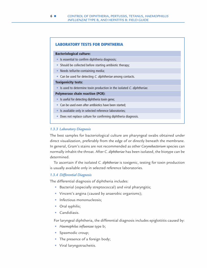
1.3.3 Laboratory Diagnosis
The best samples for bacteriological culture are pharyngeal swabs obtained underdirect visualization, preferably from the edge of or directly beneath the membrane.In general, Gram’s stains are not recommended as other Corynebacterium species cannormally inhabit the throat. After C. diphtheriae has been isolated, the biotype can bedetermined.
To ascertain if the isolated C. diphtheriae is toxigenic, testing for toxin productionis usually available only in selected reference laboratories.
1.3.4 Differential Diagnosis
The differential diagnosis of diphtheria includes:
• Bacterial (especially streptococcal) and viral pharyngitis;
• Vincent’s angina (caused by anaerobic organisms);
• Infectious mononucleosis;
• Oral syphilis;
• Candidiasis.
For laryngeal diphtheria, the differential diagnosis includes epiglottitis caused by:
• Haemophilus influenzae type b;
• Spasmodic croup;
• The presence of a foreign body;
• Viral laryngotracheitis.
LABORATORY TESTS FOR DIPHTHERIA
Bacteriological culture:
• Is essential to confirm diphtheria diagnosis;
• Should be collected before starting antibiotic therapy;
• Needs tellurite-containing media;
• Can be used for detecting C. diphtheriae among contacts.
Toxigenicity tests:
• Is used to determine toxin production in the isolated C. diphtheriae.
Polymerase chain reaction (PCR):
• Is useful for detecting diphtheria toxin gene;
• Can be used even after antibiotics have been started;
• Is available only in selected reference laboratories;
• Does not replace culture for confirming diphtheria diagnosis.
DIPHTHERIA 77
1.3.5 Complications
The severity of the signs and symptoms is usually proportional to the extent of the localinflammation, which is related to the production of the toxin in the diphtheritic mem-brane. Severe complications include respiratory obstruction, acute systemic toxicity,myocarditis, and neurological complications (usually neuritis) (1).
Local complications are related to the extension of the membrane:
• Laryngeal diphtheria and aspiration of the membrane (or part of it) can leadto respiratory obstruction;
• If the membrane extends downward, it can result in pneumonia and respira-tory obstruction;
• Sinusitis and otitis media are usually associated with nasopharyngeal diphthe-ria due to edema of the upper respiratory tract.
Systemic complications resulting from diphtheria toxin include:
• Myocarditis is the main cause of diphtheria-related mortality. It can be com-plicated by heart blocks and can progress to congestive heart failure. Earlymyocarditis occurs between the third and seventh day of infection and is usu-ally fatal. Less severe, late myocarditis usually occurs the second week afteronset and, occasionally, later.
• Neurological complications mostly manifest as a toxic peripheral neuropathythat primarily affects the motor nerves. These complications usually begin twoto eight weeks weeks after the onset of illness. Paralysis of eye muscles, limbs,and diaphragm can occur after the fifth week. Diaphragmatic paralysis can beserious, causing pneumonia or requiring mechanical ventilation. Normally,these neurological complications resolve completely.
The case-fatality rate for noncutaneous diphtheria is 5% to 10%, and hasremained at those levels for the last 50 years. Children aged less than 5 years andpersons over 40 years have a higher risk of death (1, 5).
1.3.6 Treatment
Prompt recognition and treatment of diphtheria are very important, as the early useof diphtheria antitoxin is associated with a better outcome. Complications aredirectly proportional to the number of days between the onset of illness and admin-istration of antitoxin. The patient should be isolated (strict isolation for pharyngealdiphtheria, and contact isolation for cutaneous diphtheria). Treatment should bestarted immediately after taking bacteriological specimens, without waiting for lab-oratory confirmation.

88 CONTROL OF DIPHTHERIA, PERTUSSIS, TETANUS, HAEMOPHILUS INFLUENZAE TYPE B, AND HEPATITIS B: FIELD GUIDE
Antitoxin. The use of diphtheria antitoxinc is the centerpiece of diphtheria treat-ment, and it should be administered when diphtheria is suspected. Antitoxin will neu-
tralize circulating (unbound) toxin, but nottoxin already fixed to the tissues. For this rea-son, the entire therapeutic dose should beadministered at one time. The antitoxin canbe given intramuscularly (IM) or intra-venously (IV); therapeutic levels of antitoxinin the blood can be achieved faster with IVadministration, and this method is usuallypreferred. The dose to be used ranges from20,000 to 120,000 units, depending on thesize of the lesions, as the amount of toxinproduced depends on the size of the mem-branes and the interval since the time ofonset (see Table 2). Since the antitoxin is pro-duced in horses, some experts suggest testingfor hypersensitivity to equine serum (8, 9).
The antitoxin is not indicated for prophylaxis.Antibiotics. Patients with diphtheria should also receive antibiotics to eliminate
the bacteria and thus reduce the duration of communicability and carriage. Howev-er, antibiotics are not a substitute for the antitoxin.
The recommended antibiotics are (8):
• Procaine penicillin G. It should be administered intramuscularly, at a dose of25,000–50,000 units/kg/day for children and 1.2 million units/day for adults,in two divided doses or
• Erythromycin. Parenteral erythromycin (40–50 mg/kg/day, with a maximumof 2 g per day) may be used until the patient can swallow, after which he orshe may be given erythromycin orally in four divided doses per day or oralpenicillin V (125–250 mg four times per day).
Treatment should continue for 14 days.Other Measures. Nonspecific supportive measures are indicated in addition to
antitoxin, isolation, and the use of antibiotics. Additionally, initiation or completionof active immunization against diphtheria is recommended for cases during the con-valescent period, because disease does not necessarily confer immunity.
Extensive disease of three or more days’ duration or diffuse swelling of the neck
80,000 to 120,000
Nasopharyngeal lesions 40,000 to 60,000
Pharyngeal or laryngeal disease of 48 hours’ duration or less
Cutaneous disease*20,000 to 40,000
IndicationDose (units)
* No consensus exists regarding the value of using antitoxin for cutaneous diphtheria.
Source: American Academy of Pediatrics. Diphtheria. In: Pickering LK, ed. Red book: 2003 report of the Committee on Infectious Diseases, 26th ed. Elk Grove Village, IL: American Academy of Pediatrics; ©2003:263-266, with permission.
Table 2. Suggested dose ranges for the use of diphtheria antitoxin
cFor Latin America and the Caribbean, diphtheria antitoxin can be obtained through thePAHO Revolving Fund for Vaccine Procurement by contacting the Immunization Unit at PAHOHeadquarters (Washington, D.C.).

DIPHTHERIA 99
1.3.7 Management of Contactsd
• Vaccination. The diphtheria vaccination status of case contacts should beassessed to complete the primary three doses of diphtheria vaccine in thosewho need it, give the fourth dose to children who have received the primaryseries, and give an age-appropriate diphtheria booster if no booster has beengiven in the previous five years.
• Antibiotics. Prophylactic antibiotics are also indicated for contacts: one doseof benzathine penicillin G, IM (600,000 units for persons aged less than 6years, and 1.2 million units for those aged 6 or older), or 7 to 10 days of oralerythromycin (40 mg/kg/day for children and 1 g/day for adults). If compli-ance cannot be guaranteed, one dose of benzathine penicillin is preferred forprophylaxis. If the contact is cultured and the result is positive, he or sheshould be treated as a case (8).
When feasible, management of close contacts also includes keeping them undersurveillance for seven days to detect disease, and taking nose and throat samples forculture before starting antibiotic prophylaxis.
11..44 VVAACCCCIINNAATTIIOONN AACCTTIIVVIITTIIEESS
1.4.1 Routine Immunization
The priority goal for diphtheria control for every country is to reach at least 95% cov-erage with three primary doses of pentavalent vaccine among 1-year-old children in
dA contact is defined as a person of any age living under the same roof as the case; if thepatient attends school, his/her classmates are also contacts. In overcrowded areas, contactsmay include close neighbors.
TREATMENT OF DIPHTHERIA CASES AND MANAGEMENT OF CONTACTS
Case management includes:
• Administration of antitoxin;
• Antibiotic therapy (after sample collection) with penicillin or erythromycin;
• Isolation of case;
• Supportive measures including vaccination with an age-appropriate diphtheria-containing vaccine.
Contact management includes:
• Vaccination to complete the primary series or use of an age-appropriate diphtheria booster;
• Prophylactic antibiotic treatment with penicillin or erythromycin;
• Close monitoring and follow-up for seven days.

1100 CONTROL OF DIPHTHERIA, PERTUSSIS, TETANUS, HAEMOPHILUS INFLUENZAE TYPE B, AND HEPATITIS B: FIELD GUIDE
each municipality (10). High levels of routine immunization coverage in appropriateage groups are crucial to maintaining high immunity levels in the community.
1.4.2 Other Vaccination Activities
Contacts of a diphtheria case should also be vaccinated according to their age andvaccination status (see Management of Contacts in Section 1.3.7). Routine use oftetanus-diphtheria (Td) vaccine, rather than monovalent tetanus toxoid, also helpsmaintain diphtheria immunity in adults.
1.4.3 Outbreak Control
To control diphtheria outbreaks, defined as the occurrence of a higher number ofcases than expected based on previous years, the priority should be intensified diph-theria vaccination through a combined approach of mass vaccination efforts andstrengthening of routine services. Vaccination efforts should target affected areasand areas with low coverage levels, covering the largest possible proportion of thepopulation group involved. Health officials should ensure that the populationreceives adequate diphtheria protection with the three primary doses of the vaccineand boosters in accordance with age. Several vaccination strategies can beemployed, such as door-to-door vaccination, fixed vaccination posts, and in-schoolvaccination.
In some countries of the Americas, the creation of ad hoc committees to reviewdiphtheria case data during an outbreak has proven a useful tool for improving casemanagement, case notification, and epidemiological investigation. These commit-tees can be composed of local health authorities, clinicians, epidemiologists, publichealth nurses, laboratory personnel, and others related to the outbreak. Theyshould meet on a regular basis (daily, weekly, or monthly) to review clinical, epidemi-ological, and laboratory data, including management of contacts, of each diphtheriacase reported.

2 PERTUSSIS (WHOOPING COUGH)
[Pertussis ICD-9 033.0; ICD-10 A37.0][Parapertussis ICD-9 033.1; ICD-10 A37.1]
22..11 IINNTTRROODDUUCCTTIIOONN
Pertussis, or whooping cough, is an acute disease of the respiratory tract caused bythe Gram-negative bacillus Bordetella pertussis. The disease is characterized by acough that becomes paroxysmal and can last for months. Infants and young chil-dren are more severely affected and can suffer paroxysms of cough that end in thecharacteristic, inspiratory “whoop.” Parents of children with pertussis are often dev-astated by the breathing difficulties their children experience with this life-threaten-ing disease.
Globally, the World Health Organization estimates that 20–40 million cases and200,000–400,000 deaths occur each year, making pertussis one of the leading causesof vaccine-preventable deaths in the world (11, 12). Vaccination is the main tool forprevention, and high routine vaccination coverage is crucial to controlling this disease.
22..22 EEPPIIDDEEMMIIOOLLOOGGYYaa
2.2.1 Infectious Agent
Pertussis is caused by the pertussis bacillus Bordetella pertussis. Bordetella parapertussiscauses a similar but usually milder illness, known as parapertussis.
2.2.2 Occurrence
Pertussis occurs worldwide, regardless of climate and latitude. Reported pertussiscases and deaths are more common among females. It is an endemic disease, withpeaks every two to five years (most commonly every three to four years). Thedecrease in incidence has not affected its periodicity, suggesting continued circulationof the infectious agent in the community. Outbreaks occur periodically (13, 14, 15).
2.2.3 Transmission
B. pertussis is transmitted from person to person via aerosolized droplets producedfrom a cough or sneeze, or by direct contact with secretions from the respiratorytract of infectious individuals. Pertussis is a highly contagious disease, with second-ary attack rates among susceptible household contacts as high as 90%, and50%–80% in school settings. In several studies, household members have been doc-umented to have been the source of pertussis in infants (13, 16).
aSee Annex 1, “Summary of the epidemiological characteristics of diphtheria, pertussis, tetanus,Haemophilus influenzae type b (Hib), and hepatitis B.”
1111

1122 CONTROL OF DIPHTHERIA, PERTUSSIS, TETANUS, HAEMOPHILUS INFLUENZAE TYPE B, AND HEPATITIS B: FIELD GUIDE
2.2.4 Reservoir
Humans are the only known host for B. pertussis. Adolescents and adults are animportant reservoir and a source of infection for infants (14, 15).
2.2.5 Incubation
The incubation period is usually seven to ten days (with a range of four to twenty-onedays) (14).
2.2.6 Communicability
Persons with pertussis are most infectious during the catarrhal period and the firsttwo weeks after cough onset (i.e., approximately 21 days) (14). Some individuals,such as infants who remain culture-positive for several weeks, can be infectious for alonger period. Persons not treated with antibiotics are considered contagious for upto three weeks after the onset of typical paroxysms (16). In persons treated witherythromycin, infectiousness is reduced to approximately five days after the begin-ning of the antibiotic therapy (15).
2.2.7 Immunity
Maternal protection of infants has not been demonstrated and infants are suscepti-ble to pertussis from the first weeks of life. Pertussis can occur at any age but is mostcommonly reported, and probably recognized, among children aged less than 5years. Older individuals infected with pertussis usually present a milder respiratorydisease that is often indistinguishable from other causes of cough. Vaccine-inducedimmunity can wane, explaining the occurrence of cases—mostly seen in industrial-ized countries—among previously immunized adolescents and adults.
2.2.8 Changing Epidemiology
Pertussis was one of the most common childhood diseases in the pre-vaccine era,and even though its incidence has decreased dramatically since the introduction ofpertussis vaccine, it remains a significant public health problem among children inthe developing world. Additionally, a rise in pertussis incidence has been seen incountries where anti-immunization movements have led to reductions in vaccinationcoverage, illustrating the importance of maintaining high vaccination coverage lev-els for pertussis control.
Recently, heightened recognition of outbreaks and cases among adolescents andadults has led to a better understanding of these persons’ role in introducing thepertussis bacillus into households with susceptible young children.
In the Americas, the incidence of pertussis has declined markedly (see Figure 4),but outbreaks still occur. Since the 1990s, an average of about 20,000 cases and 200deaths have been reported in the Region annually.b However, the actual numbers are like-
bData provided by the Immunization Unit, Family and Community Health Area, Pan AmericanHealth Organization, Washington, D.C. (www.paho.org).

PERTUSSIS 1133
ly to be much higher, since casesmay not be diagnosed and under-reporting may be considerable.
22..33 CCLLIINNIICCAALL AASSPPEECCTTSS
2.3.1 Pathogenesis
It is believed that B. pertussisattaches to ciliated cells in thenasopharynx, where it prolifer-ates and spreads into ciliatedcells in the trachea and bronchi,producing toxins that paralyzethe cilia and cause cell death.This causes inflammation of therespiratory tract that interfereswith normal clearing of pulmonary secretions. Bacteremia does not occur.
2.3.2 Clinical Features
Pertussis is characterized by spasms (paroxysms) of severe coughing, which are con-tinuous and without inspiration until the end of the spasm, when there is often acharacteristic inspiratory “whoop” and/or post-tus-sive vomiting (see Figure 5).
The clinical course of the disease can be dividedinto the following three stages:
• Catarrhal stage. The first stage of disease isinsidious and mimics a minor upper respira-tory tract infection. Manifestations includecoryza (runny nose), sneezing, low-gradefever, and a mild, occasional cough. Thecough progresses over one to two weeks and becomes paroxysmal.
• Paroxysmal stage. This stage is characterized by severe episodes of paroxysmalcough, apparently as a result of the difficulty in expelling thick mucus from thetracheobronchial tree. In this stage, a long inspiratory effort, usually accom-panied by a high-pitched whoop and/or vomiting, follows the paroxysm andthe patient can become cyanotic. Cyanosis and apnea following the paroxysmand a very ill appearance are more common in young infants during this stage(Figure 5). However, patients can appear normal between paroxysms.
• Convalescent stage. After reaching a peak of frequency and severity, the parox-ysms of cough gradually subside, rarely lasting more than two to six weeks.However, a nonparoxysmal cough can persist for several weeks (see Figure 6).
0
20,000
40,000
60,000
80,000
100,000
120,000
140,000
1978
1980
1982
1984
1986
1988
1990
1992
1994
1996
1998
2000
2002
0
20
40
60
80
100
Pertussis cases Vaccine coverage
Vaccine coverage (%)
Num
ber o
f cas
es
Year
Source: Pan American Health Organization, Family and Community Health Area, Immunization Unit.
2004
Figure 4. Number of pertussis cases notified and DPT3 vaccine coverage among children aged less than 1 year, Region of the Americas,1978–2004.
Cred
it:Co
urte
sy o
f Dep
artm
ent o
f Pae
diat
rics
and
Adol
esce
ntM
edic
ine,
Prin
cess
Mar
gare
t Hos
pita
l,Ho
ng K
ong.
Figure 5. Infant with pertussis.

1144 CONTROL OF DIPHTHERIA, PERTUSSIS, TETANUS, HAEMOPHILUS INFLUENZAE TYPE B, AND HEPATITIS B: FIELD GUIDE
“Classic” pertussis is more common among infants 6 months of age or older andin young children. Infants aged less than 6 months have the highest case-fatalityrates; cyanosis and dyspnea are more marked and, in some cases, are the main man-ifestations of the disease, as these infants may not present with pertussis paroxysms.Adolescents and adults may present with prolonged cough accompanied or not byparoxysms, sleep disturbance, expectoration, and vomiting. Mild and/or atypicalcases occur mostly among adolescents and adults (14, 17, 18).
2.3.3 Laboratory Diagnosis
The laboratory confirmation of pertussis cases remains a challenge, even during per-tussis outbreaks. Isolation of B. pertussis by culture is the standard and preferred lab-oratory test for the diagnosis of the disease. An elevated white blood cell count withlymphocytosis is usually present in severe cases among young infants (18).
Culture. B. pertussis is a fastidious bacterium that is usually difficult to isolate (19).The ability to obtain a positive culture result from a person with pertussis can beaffected by several factors:
| Communicability | Weeks 1 2 3 4 5 6 7 8 9 10
Catarrhal Paroxysmal Convalescent
Cough
Paroxysms
Lymphocytosis
Culture
Coryza
Source: Adapted from Morley D. Pediatric priorities in the developing world. The English Language Book Society and Butterworth. Butterworth & Co, Ltd: London, England,©1980:237, with permission from Elsevier.
Figure 6. Diagram of the clinical course of “classic” pertussis.

PERTUSSIS 1155
• How the specimen is handled;
• The stage of illness at the time of specimen col-lection;
• The use of antimicrobial therapy prior to cul-ture;
• Immunity derived from past infection or fromvaccination; and
• Age of the patient.
Specimen collection and handling ((1177)).. Specimensfrom the posterior nasopharynx, not the throat,should be collected using a swab or by aspiration. TheDacron or calcium alginate swab (non-cotton) shouldbe introduced slowly through the nostril and kept forabout 10 seconds in the pharynx before inoculatingthe media (Figure 7). Nasopharyngeal aspirates arepreferred, especially if the sample also will be used forthe polymerase chain reaction (PCR) test. The aspi-rates can be collected using a small tube (e.g., an infant feeding tube) connected toa mucous trap that is inserted into the nostril to the posterior pharynx, using a simi-lar approach to the one shown in Figure 7. Secretions are aspirated while the tube is inthat position, and while partly withdrawing it. Inoculating the secretions directly intoa specialized culture medium for pertussis (Bordet-Gengou) at the bedside increasesthe yield of positive cultures. If that is not possible, the Regan-Lowe medium can beused for transport, though the likelihood of isolation might be compromised.
Cultures of samples collected during the catarrhal stage have a higher successrate, as do samples collected prior to the use of antibiotics against pertussis (eryth-romycin or trimethoprim-sulfamethoxazole). Lower rates of positive cultures havebeen seen from vaccinated and older persons.
Polymerase Chain Reaction (PCR) Test. PCR for pertussis can be rapid, specific,and sensitive. Although bacteria cannot be cultured after five days of antibiotic ther-apy, PCR can remain positive for an additional week. If available, PCR may be usedin addition to culture.
Direct Fluorescent Antibody (DFA) Test. DFA testing is sometimes used as ascreening test for pertussis. However, DFA tests lack sensitivity for B. pertussis, lead-ing to false-negative results. They also have variable specificity, as there are somecross-reactions with normal nasopharyngeal flora. DFA testing should not be usedas a criterion for pertussis diagnosis.
Figure 7. Proper technique for obtaining a nasopharyngeal specimen for isolation of B. pertussis.
Source: Centers for Disease Control and Prevention.

1166 CONTROL OF DIPHTHERIA, PERTUSSIS, TETANUS, HAEMOPHILUS INFLUENZAE TYPE B, AND HEPATITIS B: FIELD GUIDE
Serologic Tests. Several tests to measure serum antibodies to B. pertussis, includ-ing enzyme-linked immunosorbent assays (ELISAs), have been used in investigation,but they are not yet widely available for routine clinical use. A significant rise in anti-body titers between an acute-phase specimen and a convalescent-phase specimensuggests infection. However, a significant rise in antibody titers may not be seenbecause the first specimen usually is taken late in the course of the infection due tothe insidious onset of pertussis (14).
2.3.4 Differential Diagnosis
The differential diagnosis for pertussis includes respiratory infections of various eti-ologies and parapertussis, though this latter disease is usually less severe. Thecatarrhal stage and pertussis among adolescents and adults can be indistinguish-able from other upper respiratory infections. In young infants, the differential diag-nosis includes other causes of episodic cyanosis or apnea.
2.3.5 Complications
Case-fatality rates for pertussis vary in different settings but are consistently higherin infants aged less than 6 months, children with enteric and respiratory infections,and malnourished children.
LABORATORY TESTS FOR PERTUSSIS
Bacteriological culture:
• Is the standard and preferred laboratory test for pertussis confirmation. However, isolation of Bordetella pertussis is difficult and is affected by several factors:
- Collection and handling of specimens;
- Stage of illness at specimen collection;
- Prior use of antibiotics;
- Age and vaccination status of the case.
Polymerase chain reaction (PCR) test:
• Is a rapid, specific, and sensitive test for detecting B. pertussis antigens;
• Is usually used in addition to bacteriologic culture because it is not widely available and not well standardized.
Direct fluorescent antibody (DFA) test:
• Is sometimes used as a screening test for pertussis. However, it lacks sensitivity and has variable specificity.
Serologic test:
• Is potentially useful if a significant rise in antibody titers is observed between acute and convalescent samples. However,this test is not widely available.

PERTUSSIS 1177
Complications include (14, 16):
• Pneumonia, which is the most common cause of pertussis-related deaths;
• Neurological alterations, including seizures and encephalopathy with alteredconsciousness;
• Nutritional problems and dehydration, mainly due to vomiting, but that canalso be due to increased caloric utilization and inadequate feeding practicesfor sick children;
• Complications resulting from the pressure of severe paroxysms of cough, suchas subconjuctival hemorrhages, epistaxis, edema of the face, pneumothorax,subdural hematomas, hernias, rectal prolapse, and, in adults, urinary incon-tinence and even rib fractures;
• Secondary bacterial infection which can cause pneumonia, otitis media, orsepsis.
2.3.6 Treatment
The management of pertussis cases consists of antibiotics, supportive measures,and isolation.
Antibiotics. The main value of using antibiotics is to limit the period of commu-nicability. Additionally, the antimicrobial treatment of pertussis cases may decreasethe severity of the symptoms if treatment is given in the catarrhal stage or earlyparoxysmal stage. Erythromycin (40–50 mg/kg/day, orally, divided in four doses;maximum 2 g/day) is the antibiotic of choice, and is given for 14 days.
Studies suggest that B. pertussis is susceptible to azithromycin (10–12 mg/kg/day,orally, in one dose for five days; maximum 600 mg/day) and clarithromycin (15–20mg/kg/day, orally, divided in two doses for seven days; maximum 1 g/day).Azithromycin and clarithromycin may be as effective as erythromycin and have bet-ter compliance (16).
Isolation. Known pertussis cases should be placed in respiratory isolation. Sus-pected cases should avoid contact with young children and infants, especially unim-munized ones. Isolation can be terminated after the first five days of anti-pertussisantibiotic therapy; otherwise, patients should be isolated for three weeks.
2.3.7 Management of Contacts
The main goal of managing pertussis contacts is to prevent the disease in infants. Management of close contacts includes antibiotics, vaccination, and quarantine.
• Antibiotics. A 14-day course of erythromycin or trimethoprim-sulfamethoxa-zole, regardless of immunization status and age (14). Azithromycin and clar-ithromycin are potential alternatives for persons who cannot tolerate erythro-mycin (16).

1188 CONTROL OF DIPHTHERIA, PERTUSSIS, TETANUS, HAEMOPHILUS INFLUENZAE TYPE B, AND HEPATITIS B: FIELD GUIDE
• Vaccination. Even though immunization against pertussis is not protective forcontacts of pertussis cases, vaccination is recommended to limit the spread ofthe disease in the affected community. Children aged less than 7 years who havenot received the primary series should be immunized, observing the minimumintervals between doses. Those children who have not received a dose within theprevious three years should be vaccinated as soon after exposure as possible (15).
• Quarantine. Inadequately vaccinated contacts aged less than 7 years shouldbe quarantined by excluding them from child-care centers, schools, and pub-lic gatherings for 21 days after exposure, or until cases and contacts havereceived five days of their 14-day antibiotic course (16).
22..44 VVAACCCCIINNAATTIIOONN AACCTTIIVVIITTIIEESS
2.4.1 Routine Immunization
The priority goal for pertussis control for every country should be to achieve at least95% coverage with three primary doses of pentavalent vaccine among 1 year oldchildren in each municipality (10).
2.4.2 Outbreak Control
Case-fatality rates from pertussis are high in infants aged less than 1 year and high-est in those aged less than 6 months. In the event of an outbreak, these children needto be, identified, monitored, and treated appropriately. Efforts should be made toconfirm an outbreak by obtaining laboratory confirmation in at least some of theprobable cases. To prevent the spread of the disease, antibiotic prophylaxis of allcontacts is essential. Closure of schools and other settings where children congre-gate may be necessary. During pertussis outbreaks, the vaccination status of all chil-dren aged between 1 and 6 years should be reviewed and vaccine given, if indicated.The benefits of accelerating the schedule for infants should be considered.
As for diphtheria outbreaks, the creation of ad hoc committees to review pertussiscase data during an outbreak could be useful to improve case management, casenotification, and epidemiological investigation.
3 TETANUS
[ICD-9 037; ICD-10 A35][Obstetrical tetanus: ICD-10 A34][Neonatal tetanus: ICD-9 771.3; ICD-10 A33]a
33..11 IINNTTRROODDUUCCTTIIOONN
Tetanus is an acute disease caused by the toxin produced by Clostridium tetani. It isfrequently fatal and is characterized by progressive muscular rigidity and convulsivespasms of the skeletal muscles.
Control of tetanus has been a general strategy pursued within the goals of theimmunization programs in the Americas. Although a goal has never been establishedfor its eradication, its incidence has declined significantly. Several factors have con-tributed to this reduction: the strengthening of the health services of the WesternHemisphere; the progressive increase in DPT coverage in childhood; vaccination ofchildren with tetanus toxoid at schools; and the effort to eliminate neonataltetanus, which was initiated in the 1980s. The strategy of eliminating neonataltetanus as a public health problem, in addition to promoting childbirth underhygienic conditions, gave rise to specific efforts to vaccinate all women of childbear-ing age who live in areas at risk for neonatal tetanus. The strategy consisted of iden-tifying municipalities with cases of neonatal tetanus, identifying the target popula-tion (women of childbearing age, usually between 15 and 49 years of age), and vac-cinating them with at least two doses of tetanus toxoid (20).
33..22 EEPPIIDDEEMMIIOOLLOOGGYYbb
3.2.1 Infectious Agent
C. tetani is an anaerobic, Gram-positive bacillus that can develop a terminal spore.The organism is sensitive to heat and cannot survive in the presence of oxygen. Thespores, however, are very resistant to heat and to antiseptics in daily use. They cansurvive in an autoclave at 121°C for 10 to 15 minutes. They are also very resistant tophenol and other chemical agents (21).
3.2.2 Occurrence
The spores are distributed worldwide in the soil and in the intestines of horses, cows,sheep, cats, rats, and chickens. Soil contaminated by excreta from these animals or
aFor neonatal tetanus, please consult Pan American Health Organization, Neonatal Tetanus Elimi-nation Field Guide (Scientific and Technical Publication No. 602; Washington, D.C.: PAHO, 2005).bSee Annex 1, “Summary of the epidemiological characteristics of diphtheria, pertussis, tetanus,Haemophilus influenzae type b (Hib), and hepatitis B.”
1199

2200 CONTROL OF DIPHTHERIA, PERTUSSIS, TETANUS, HAEMOPHILUS INFLUENZAE TYPE B, AND HEPATITIS B: FIELD GUIDE
treated with manure fertilizers contains large numbers of spores. In agriculturalareas, adults can also harbor spores in their digestive tracts. The spores have alsobeen found on skin and as contaminants of heroin (21).
3.2.3 Transmission
C. tetani usually enters the body through a wound (apparent or inapparent). How-ever, cases have been reported after surgeries, dental extractions, burns, otitismedia, animal bites, and abortions (20, 21).
3.2.4 Reservoir
The principal reservoirs of C. tetani are the intestinal tracts of humans and animalsand soil used for agriculture or livestock or products obtained from excreta of hors-es, cows, sheep, cats, rats, and chickens (21).
3.2.5 Incubation
The incubation period ranges from 3 to 21 days, and is usually around 8 days. Ingeneral, the farther the introduction site of the spores from the central nervous sys-tem, the longer the incubation period. Mortality is directly proportional to thelength of the incubation period, with shorter periods associated with higher mortal-ity. In neonatal tetanus, symptoms usually appear between 4 and 14 days afterbirth, with an average of 7 days.
3.2.6 Communicability
Tetanus cannot be transmitted from one person to another. It is one of the few vac-cine-preventable diseases that is infectious but not contagious (21).
3.2.7 Immunity
The disease does not confer immunity. Immunity is acquired passively throughmaternal antibodies or actively through injection of tetanus toxoid.
3.2.8 Changing Epidemiology
There have not been significant changes in the general epidemiology of tetanus inadults. The disease continuously occurs in unvaccinated populations at risk ofwounds and in places where there are C. tetani spores, that is, in rural, poor, live-stock-raising areas with populations that do not have adequate health services.
As mentioned earlier, the strategy employed to eliminate neonatal tetanus hascontributed to tetanus prevention in general. The reduction in incidence has beendramatic in the Americas. More than 7,000 cases of tetanus were reported in 1980,but only 598 cases were reported in 2004 (see Figure 8). Neonatal tetanus has beeneliminated as a public health problem in most of the Region, with most of these cas-es now occurring in Haiti.

TETANUS 2211
33..33 CCLLIINNIICCAALL AASSPPEECCTTSS
3.3.1 Pathogenesis
C. tetani usually enters the bodythrough a wound. Under anaero-bic conditions (very low levels ofoxygen), the spores germinate,producing toxins, includingtetanospasmin, that are dissemi-nated throughout the organismby blood and lymph. The toxinsact on different levels in the cen-tral nervous system, includingthe peripheral motor nerves,spinal cord, brain, and sympa-thetic nervous system. The typi-cal clinical manifestations of the disease begin when the toxin interferes with therelease of neurotransmitters, blocking those responsible for inhibiting nerve impuls-es. This leads to muscle contractions and spasms. Convulsions can also occur andthe autonomic nervous system can be affected (21).
3.3.2 Clinical Features
Three different forms of tetanus can be characterized: local, cephalic, and general-ized (21).
• Local tetanus is a very rare form of the disease in which the patients have per-sistent muscle contractions in the same anatomical area as the wound or injury.The contractions can persist for many weeks before disappearing. Local tetanuscan precede generalized tetanus. Approximately 1% of these cases are fatal.
• Cephalic tetanus is also a rare form of the disease, occurring occasionally inassociation with otitis media or following a head wound. The cranial nerves,especially facial ones, can be compromised.
• Generalized tetanus is the most common form of the disease, accounting forapproximately 80% of cases. The disease usually occurs with a descending pat-tern. The initial sign is trismus (“lockjaw,” or spasm of the jaw muscles), fol-lowed by rigidity of the neck, difficulty swallowing, and rigidity of the abdom-inal muscles. Other symptoms include an increase in temperature, perspira-tion, an increase in blood pressure, and episodes of tachycardia. Spasms canlast several minutes and may persist for three or four weeks. Total recovery cantake months.
Source: Pan American Health Organization, Family and Community Health Area, Immunization Unit.
0
1,000
2,000
3,000
4,000
5,000
6,000
7,000
8,000
1978
1979
1980
1981
1982
1983
1984
1985
1986
1987
1988
1989
1990
1991
1992
1993
1994
1995
1996
1997
1998
1999
2000
2001
2002
2003
2004
Year
Tetanus cases Vaccine coverage
0
20
40
60
80
100
Vaccine coverage (%)
Num
ber o
f cas
es
Figure 8. Annual incidence of tetanus and DPT3 vaccine coverage among children aged less than 1 year, Region of the Americas, 1978–2004.

2222 CONTROL OF DIPHTHERIA, PERTUSSIS, TETANUS, HAEMOPHILUS INFLUENZAE TYPE B, AND HEPATITIS B: FIELD GUIDE
In neonatal tetanus, which is a generalized form of tetanus, the clinical manifes-tations are seen within 3 to 28 days of birth and consist of (22):
• Cessation of breast-feeding (the first sign in a newborn) because ofdifficulty in grasping and sucking the nipple, usually beginning on atage 3 days;
• A characteristic position in which the legs are extended and the armsfolded toward the chest, with the hands kept closed due to difficul-ty in opening them;
• Generalized contractual crises leading to opisthotonus, which last afew minutes. Between these spasms, the child appears normal (seeFigure 9).
3.3.3 Laboratory Diagnosis
Laboratory confirmation of tetanus is generally difficult. The organism is rarelyrecovered from the site of infection and usually there is no detectable antibodyresponse.
3.3.4 Differential Diagnosis
The most common differential diagnoses are sepsis, meningoencephalitis, tetanywith other causes, peritonitis, and inflammatory processes of the external ear or ofthe oral region, accompanied by trismus.
3.3.5 Complications
Complications of tetanus include (21, 23):
• Laryngospasms (spasms of the vocal cords and/or the respiratory muscles);
• Fractures of the spinal column or of the tubular bones as a consequence ofprolonged contractions and convulsions;
• Hyperactivity of the autonomic nervous system, which can lead to hyperten-sion and tachycardia;
• Nosocomial infections, which are frequent due to prolonged hospitalization;
• Secondary infections, which can include sepsis, pneumonia, and decubitusulcers;
• Pulmonary embolisms, especially in the elderly;
• Pneumonias, including aspiration pneumonia; and
• Death, which in neonatal tetanus can occur in 10%–90% of cases that receivetreatment, and nearly 95% of untreated cases.
Figure 9. Child with opisthotonus.
Cred
it:Ce
nter
s fo
r Dis
ease
Con
trol
and
Pre
vent
ion
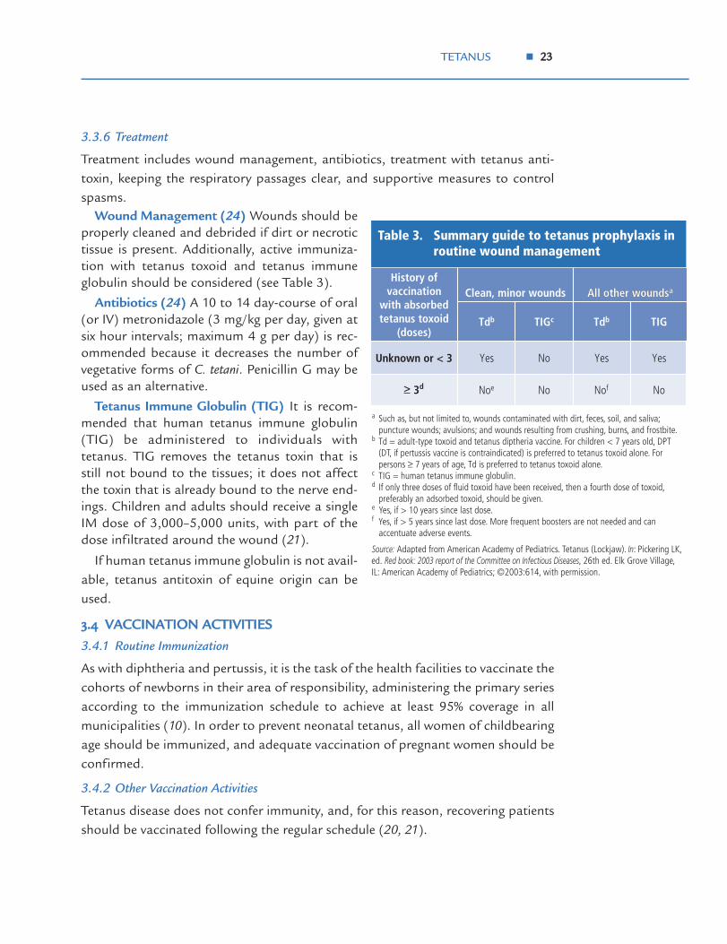
TETANUS 2233
3.3.6 Treatment
Treatment includes wound management, antibiotics, treatment with tetanus anti-toxin, keeping the respiratory passages clear, and supportive measures to controlspasms.
Wound Management (24) Wounds should beproperly cleaned and debrided if dirt or necrotictissue is present. Additionally, active immuniza-tion with tetanus toxoid and tetanus immuneglobulin should be considered (see Table 3).
Antibiotics (24) A 10 to 14 day-course of oral(or IV) metronidazole (3 mg/kg per day, given atsix hour intervals; maximum 4 g per day) is rec-ommended because it decreases the number ofvegetative forms of C. tetani. Penicillin G may beused as an alternative.
Tetanus Immune Globulin (TIG) It is recom-mended that human tetanus immune globulin(TIG) be administered to individuals withtetanus. TIG removes the tetanus toxin that isstill not bound to the tissues; it does not affectthe toxin that is already bound to the nerve end-ings. Children and adults should receive a singleIM dose of 3,000–5,000 units, with part of thedose infiltrated around the wound (21).
If human tetanus immune globulin is not avail-able, tetanus antitoxin of equine origin can beused.
33..44 VVAACCCCIINNAATTIIOONN AACCTTIIVVIITTIIEESS
3.4.1 Routine Immunization
As with diphtheria and pertussis, it is the task of the health facilities to vaccinate thecohorts of newborns in their area of responsibility, administering the primary seriesaccording to the immunization schedule to achieve at least 95% coverage in allmunicipalities (10). In order to prevent neonatal tetanus, all women of childbearingage should be immunized, and adequate vaccination of pregnant women should beconfirmed.
3.4.2 Other Vaccination Activities
Tetanus disease does not confer immunity, and, for this reason, recovering patientsshould be vaccinated following the regular schedule (20, 21).
a Such as, but not limited to, wounds contaminated with dirt, feces, soil, and saliva; puncture wounds; avulsions; and wounds resulting from crushing, burns, and frostbite.
b Td = adult-type toxoid and tetanus diptheria vaccine. For children < 7 years old, DPT (DT, if pertussis vaccine is contraindicated) is preferred to tetanus toxoid alone. For persons ≥ 7 years of age, Td is preferred to tetanus toxoid alone.
c TIG = human tetanus immune globulin.d If only three doses of fluid toxoid have been received, then a fourth dose of toxoid,
preferably an adsorbed toxoid, should be given.e Yes, if > 10 years since last dose.f Yes, if > 5 years since last dose. More frequent boosters are not needed and can
accentuate adverse events.
Source: Adapted from American Academy of Pediatrics. Tetanus (Lockjaw). In: Pickering LK, ed. Red book: 2003 report of the Committee on Infectious Diseases, 26th ed. Elk Grove Village, IL: American Academy of Pediatrics; ©2003:614, with permission.
NoNofNoNoe
YesYesNoYesUnknown or < 3
TIGTdbTIGcTdb
All other woundsaClean, minor woundsHistory of
vaccination with absorbed tetanus toxoid
(doses)
All other woundsa
Table 3. Summary guide to tetanus prophylaxis in routine wound management
> 3d

2244 CONTROL OF DIPHTHERIA, PERTUSSIS, TETANUS, HAEMOPHILUS INFLUENZAE TYPE B, AND HEPATITIS B: FIELD GUIDE
All groups who for occupational reasons are exposed to C. tetani are considered atrisk. All newborns also are considered at risk, since they can contract tetanus duringdelivery or during the first days of life if the mother is not adequately protected. It isrecommended that all women of childbearing age be vaccinated, as newborns can-not be vaccinated with tetanus toxoid and it is generally impossible to predict thecircumstances of delivery and postnatal care (22).
3.4.3 Outbreak Response
Outbreak response is not applicable to tetanus. However, if a cluster of cases ofneonatal tetanus is reported, active case search and investigation is indicated. Poorbirth attendant practices may increase the likelihood of additional cases in the com-munity where that birth attendant works.

4 HAEMOPHILUS INFLUENZAE TYPE B
[Haemophilus meningitis ICD-9 320.0; ICD-10 G00.0]
44..11 IINNTTRROODDUUCCTTIIOONN
Invasive infections by Haemophilus influenzae type b (Hib) are a major cause of bacte-rial meningitis and of lower respiratory infections in children, and a significant con-tributor to mortality in children aged less than 5 years. Worldwide, it is estimatedthat Hib infections cause at least three million cases of severe disease per year andbetween 400,000 and 700,000 children die annually due to Hib disease (25).
44..22 EEPPIIDDEEMMIIOOLLOOGGYYaa
4.2.1 Infectious Agent
Haemophilus influenzae type b (Hib) is a Gram-negative coccobacillus, usually aerobic,which requires chocolate agar as its culture medium for growth in the laboratory.
There are two varieties of Haemophilus: the encapsulated strains, among which Hibis the most pathogenic, and the acapsular strains, which are usually non-invasiveand less pathogenic but responsible for frequent otorhinolaryngeal infections (e.g.,otitis, epiglottitis, bronchitis, superinfections of the respiratory tract).
Haemophilus influenzae has six polysaccharide capsular serotypes but serotype b isresponsible for more than 95% of the invasive processes and the only one preventedby vaccination. Its external capsule consists of a polysaccharide—polyribosylribitolphosphate (PRP)—which is responsible for the organism’s virulence and for immu-nity (26).
4.2.2 Occurrence
Haemophilus influenzae type b is distributed worldwide and primarily affects childrenaged 2 months to 3 years. The disease is uncommon in children aged over 5 years.Secondary cases are sometimes observed in families and in child-care centers.
4.2.3 Transmission
Hib is transmitted from one person to another through the respiratory tract bymeans of aerosolized droplets.
4.2.4 Reservoir
Humans are the only reservoir of the bacteria.
4.2.5 Incubation
The exact incubation period is unknown but probably lasts from two to four days.
aSee Annex 1, “Summary of the epidemiological characteristics of diphtheria, pertussis, tetanus,Haemophilus influenzae type b (Hib), and hepatitis B.”
2255
2266 CONTROL OF DIPHTHERIA, PERTUSSIS, TETANUS, HAEMOPHILUS INFLUENZAE TYPE B, AND HEPATITIS B: FIELD GUIDE
4.2.6 Communicability
Hib is communicable while the microorganisms are present, even if there are nonasal secretions. The disease ceases to be communicable 24–48 hours after begin-ning treatment with antibiotics (27).
4.2.7 Immunity
Infection by Hib is age dependent. The maternal immunoglobulin G (IgG) antibod-ies transferred via the placenta and breast-feeding confer a certain degree of protec-tion during the first 2 to 6 months of life. At this age, an infant’s immune system istoo immature to generate an adequate response to the polysaccharide antigens ofthe external capsule of Hib, which explains the increase in the number of cases oncepassive natural protection declines. Starting at 2 years of age, the immune systemmatures and can generate protective responses.
During the pre-vaccine era, most children acquired natural immunity by 5 to 6 yearsof age through inapparent infection by Hib. In view of the fact that only a relativelysmall proportion of children of any age are carriers of Hib, it has been postulated thatexposure to organisms that have the same antigenic structure as the Hib capsule (i.e.,organisms that cross-react) promotes development of antibodies against that capsule.Natural exposure to Hib also induces formation of antibodies against the external proteinmembrane, lipopolysaccharides, and other antigens on the surface of the bacterium.
4.2.8 Changing Epidemiology
Before the introduction of effective vaccines against Hib, this organism was the lead-ing cause of bacterial meningitis and other invasive diseases, such as sepsis, pneu-monias, arthritis, and osteomyelitis in children aged less than 5 years. Estimationsof the Hib incidence rate during the pre-vaccine era ranged from 50 to 150 cases per100,000 children in this age group. When vaccination against Hib is introduced,most cases occur in infants and children who are not vaccinated or are incomplete-ly vaccinated. The highest rate of invasive disease caused by Hib tends to occuramong infants who have still not reached vaccination age (6 months). The incidenceamong children aged 1 to 4 years is much lower than among infants aged less than1 year. Case-fatality currently ranges from 2% to 5%, depending on the country, with15%–30% of survivors presenting with sequelae (28).
In countries of the Americas that introduced the vaccine in the mid-1990s, suchas Uruguay and Chile, a significant reduction of cases of invasive disease by H.influenzae type b has been observed (Figures 10 and 11). Chile uses a three-doseschedule and Uruguay a four-dose series, which, considering the effectiveness of thevaccine in relation to the number of doses administered, could explain some differ-ences in the degree of impact for each country (25, 29). Whether a three or four doseschedule is used, the impact has been extraordinary.
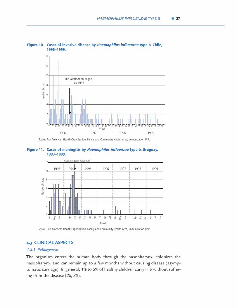
HAEMOPHILUS INFLUENZAE TYPE B 2277
44..33 CCLLIINNIICCAALL AASSPPEECCTTSS
4.3.1 Pathogenesis
The organism enters the human body through the nasopharynx, colonizes thenasopharynx, and can remain up to a few months without causing disease (asymp-tomatic carriage). In general, 1% to 5% of healthy children carry Hib without suffer-ing from the disease (28, 30).
Source: Pan American Health Organization, Family and Community Health Area, Immunization Unit.
0
2
4
6
8
10
12
14
1 7 13 19 25 31 37 43 49 3 9 15 21 27 33 39 45 51 4 10 16 22 28 34 40 46 52 6 12 18 24 30 36 42 48
Week
Num
ber o
f cas
es
Hib vaccination began July 1996
1996 1997 1998 1999
Figure 10. Cases of invasive disease by Haemophilus influenzae type b, Chile,1996–1999.
Source: Pan American Health Organization, Family and Community Health Area, Immunization Unit.
0
2
4
6
8
10
12
Jan
May
Sep
Apr
Aug
Dec
Mar Jul
Nov
Feb
Jun
Oct Jan
May
Sep
Apr
Aug
Dec
Mar Jul
Nov
Month
Num
ber o
f cas
es
1993 1994 1995 1996 1997 1998 1999
Vaccination began August 1994
Figure 11. Cases of meningitis by Haemophilus influenzae type b, Uruguay,1993–1999.

2288 CONTROL OF DIPHTHERIA, PERTUSSIS, TETANUS, HAEMOPHILUS INFLUENZAE TYPE B, AND HEPATITIS B: FIELD GUIDE
In the invasive processes, Hib passes from the nasopharynx to the blood, causinginfection at distant sites, especially the meninges. Viral infections are thought tofacilitate the invasion. Overcrowding, large households, attendance at child-carecenters, low socioeconomic level, low educational status, and school-age siblingsincrease a child’s risk of contracting Hib. Patients with chronic diseases and certainethnic groups (e.g., Eskimos and blacks) also are more susceptible to infection (26,28, 30).
4.3.2 Clinical Features
The invasive disease caused by Hib occurs in several clinical forms. Meningitis is theinfection of the membranes that cover the brain and is the most common clinicalmanifestation of invasive infections by Hib, accounting for approximately 50% to65% of cases of meningitis during the pre-vaccine era (25).
Other frequently occurring forms of invasive Hib are epiglottitis, septic arthritis,cellulitis (infection of the skin and soft tissues), and unifocal or multifocal pneumo-nia, with or without effusion. Osteomyelitis and pericarditis are less frequent.
Otitis media is usually caused by non-b strains of Haemophilus influenzae and can-not be prevented with the vaccine.
4.3.3 Laboratory Diagnosis
Laboratory diagnosis is made through isolation of Hib from normally sterile fluids,such as cerebrospinal fluid (CSF), pleural fluid, blood, and joint fluid, and aspiratefrom the middle ear. Positive culture of such samples in chocolate agar and the pres-ence of the Gram-negative coccobacillus confirm the diagnosis of disease caused byHib. Serotyping can be done in isolates of Haemophilus influenzae to determine if it istype b, the only serotype that is potentially vaccine-preventable.
Antigen detection in the aforementioned body fluids can help facilitate the diag-nosis in patients partially treated with antibiotics. Latex particle agglutination andcountercurrent immunoelectrophoresis are two useful serological methods for anti-gen detection; the former is more rapid, sensitive, and specific (31).
4.3.4 Differential Diagnosis
Because the incidence of bacterial meningitides is high, differential diagnosis ofmeningitis and Haemophilus influenzae type b should be conducted for every child withfever who presents altered mental state, irritability, or evidence of other neurologi-cal dysfunction. The most frequent differential diagnoses involve meningitidescaused by Neisseria meningitidis (meningococcus) and Streptococcus pneumoniae (pneu-mococcus), although several infectious agents can cause widespread infection of thecentral nervous system with clinical manifestations similar to those of Hib (27).
In any case of pneumonia with consolidation on the X-ray image, Haemophilusinfluenzae type b should be included in the differential diagnosis.

HAEMOPHILUS INFLUENZAE TYPE B 2299
Clinically, no difference can be established among meningitides or bacterial pneu-monias caused by different bacteria.
4.3.5 Complications
The most frequent complications of meningitis are convulsions, increase in intracra-nial pressure, paralysis of the facial nerves, cerebrovascular accidents, cerebral her-niation, transverse myelitis, ataxia, thrombosis in the venous sinuses, and subduralabscesses.
Epiglottitis caused by Hib can cause death due to obstruction of the respiratorytract.
Mortality in industrialized countries ranges from 2% to 5%, regardless of theantimicrobial treatment. Severe neurological sequelae (hearing disorders, delay inlanguage acquisition, developmental delay, motor abnormalities, visual impairment,and behavior abnormalities) occur in 15% to 30% of those who survive (28, 30).
4.3.6 Treatment (27, 32)
Specific treatment with antibiotics is necessary for invasive disease caused by Hib.Ceftriaxone, cefotaxime, or ampicillin combined with chloramphenicol should beadministered until the sensitivity of the organism is determined. Intravenous antibi-otics are usually given for 10 days, but longer therapy may be indicated for compli-cated cases. The drug of choice used to be ampicillin; however, considering that upto 30% of Hib strains are resistant through production of β-lactamase, ampicillinalone is no longer the first option. To ensure the elimination of the organism,rifampicin should be administered before the patient leaves the hospital.
Concurrent quarantine is not needed.
4.3.7 Management of Contacts (27, 32)
In investigating the contacts and the source of infection, all children less than 6 yearsof age should be observed carefully, especially infants. Observation should be atten-tive to the signs of the disease, especially fever.
Contacts should receive chemoprophylaxis, since it is effective in preventing sec-ondary transmission of Hib. In homes where, in addition to the index case, there areinfants aged less than 12 months, immunodeficient children, or inadequately immu-nized 1 to 3 year old children, treatment with oral rifampicin is recommended for allcontacts in the family unit (once a day for four days at a dosage of 20 mg/kg; a max-imum dose of 600 mg/day).
Employees and other children in the rooms or classes in child-care centers orkindergartens where a case occurs also should be treated as above. The protectivelevels of antibodies after vaccination are not reached until the end of the first week;therefore, a vaccinated child exposed to the disease during that time still runs therisk of acquiring the disease while he or she develops antibodies.

3300 CONTROL OF DIPHTHERIA, PERTUSSIS, TETANUS, HAEMOPHILUS INFLUENZAE TYPE B, AND HEPATITIS B: FIELD GUIDE
44..44 VVAACCCCIINNAATTIIOONN AACCTTIIVVIITTIIEESS
The principal objective of vaccination against Hib is to prevent the serious diseasescaused by the bacterium in infants and young children.
4.4.1 Routine Immunization
Universal vaccination of infants with the primary series of vaccine against Hibb hasproven to be effective and should be a priority in the routine activities of the EPI(25). The objective is to achieve at least 95% coverage in children aged less than 1year in all municipalities with Hib or pentavalent (10). Routine vaccination againstHib is generally given at the same age as DPT, either in combination or in separateinjections. Some countries give a booster following the primary three-dose series.
4.4.2 Other Vaccination Activities
Those at risk for Hib infection generally include people with underlying cardiopul-monary conditions; persons with chronic diseases, such as asthma, cystic fibrosis,cardiopulmonary malformations, immune deficiencies, renal insufficiency, and sick-le cell anemia; patients undergoing treatment for cancer; asplenics; and patientsinfected with the human immunodeficiency virus (HIV), although in these patientsimmunogenicity varies according to the degree of infection. Some patients with spe-cial conditions, such as malnutrition, tuberculosis, or debilitating diseases, couldalso be considered at risk for Hib infection and should benefit from timely, system-atic administration of the vaccine.
bFor more details on the Hib immunization schedule see Section 7.2 on Hib in Chapter 7 of thisguide.

5 HEPATITIS B
[ICD-9 070.3; ICD-10 B16]
55..11 IINNTTRROODDUUCCTTIIOONN
Infection with the hepatitis B virus (HBV) is estimated to affect some two billionpeople worldwide, of whom some 360 million have a chronic infection with a highrisk of sequelae, such as liver failure, cirrhosis, or liver cancer (33). HBV may be thecause of up to 80% of the cases of liver can-cer throughout the world, and is second totobacco among known carcinogens (34, 35).
55..22 EEPPIIDDEEMMIIOOLLOOGGYYaa
5.2.1 Infectious Agent
Hepatitis B virus is a DNA virus belonging tothe family Hepadnaviridae. Its principal com-ponents are the surface antigen (HBsAg),previously called Australia antigen; the coreantigen (HBcAg); and the e antigen (HBeAg).Figure 12 shows a diagram of HBV.
5.2.2 Occurrence
Hepatitis B is endemic throughout theworld, with few seasonal variations.According to the World Health Organiza-tion (36), approximately 45% of the world population lives in areas where the preva-lence of HBV is high (8% or more of the population is HBsAg-positive), 43% live inareas of intermediate endemicity (2%–7% are HBsAg-positive), and 12% live in areasof low endemicity (under 2% are HBsAg-positive) (see Figure 13).
The endemicity of HBV infection in the Americas varies from low to intermediate(37). In Latin America and the Caribbean, the highest prevalence rates are in theAmazon Basin (8%) and the lowest are in South America’s southern cone. Great het-erogeneity is observed in the proportion of carriers, which varies in relation to geo-graphic area and ethnic distribution.
Figure 12. Hepatitis B virus.
Surface protein HBs
Core proteinHBc
DNApolymerase
DNA
aSee Annex 1, “Summary of the epidemiological characteristics of diphtheria, pertussis,tetanus, Haemophilus influenzae type b (Hib), and hepatitis B.”
Source: Millinship S. Hepatitis B virology and immunology [Internet site]. Available at:http://www.hon.ch/Library/Theme/HepB/virology.html. Accessed on 24 December 2004.
3311

3322 CONTROL OF DIPHTHERIA, PERTUSSIS, TETANUS, HAEMOPHILUS INFLUENZAE TYPE B, AND HEPATITIS B: FIELD GUIDE
5.2.3 Transmission
Transmission of HBV occurs through infected blood or other body fluids, includingserous fluids; saliva; cerebrospinal fluid; peritoneal, pleural, pericardial, synovial, andamniotic fluids; and semen and vaginal secretions. The virus can survive one week ormore at room temperature, so contact with contaminated objects and by percuta-neous exposure, such as injection with unclean needles, are also transmission routes.
The principal modes of transmission are:
• Sexual contact;
• Household contact;
• Vertical transmission (e.g., from an infected mother to her infant);
• Use of injectable drugs; and
• Nosocomial exposure (exposure in health care settings).
HBsAg Prevalence≥ 8% - High
2 -7% - Intermediate
<2% - Low
Source: World Health Organization, Hepatitis B vaccine [Internet site]. Available at: www.who.int/vaccines/en/hepatitisb.shtml, data for the WesternHemisphere gathered from PAHO member countries.
Figure 13. World prevalence of hepatitis B, prior to mass vaccine introduction in Latin America and the Caribbean, 1997.

HEPATITIS B 3333
Hepatitis B is considered a sexually transmitted disease. There is no fecal-oraltransmission of HBV, nor is it transmitted by tears, sweat, or urine.
The likelihood of perinatal transmission of HBV to an infant is 10% in motherswho are HBsAg positive but HBeAg negative; it is 70% to 90% when the mother isboth HBsAg positive and HBeAg positive (35). Several studies indicate that trans-mission in early infancy is more likely to result in chronic infections. In the Americas,the low prevalence of HBsAg in children during the first year of life and the increasein the prevalence of HBsAg and anti-HBc after the first year indicate that transmis-sion in early infancy is more important than in the perinatal period.
5.2.4 Reservoir
Human beings are the reservoir for HBV.
5.2.5 Incubation
The incubation period usually is from six weeks to six months, with the average being60–90 days (34, 38).
5.2.6 Communicability
All HBsAg-positive individuals are infective. Infected individuals can transmit theinfection from one to two months before the first symptoms appear and are infec-tive throughout the acute course of the disease and in the chronic carrier phase,which can persist for years.
5.2.7 Immunity
Everyone is susceptible to hepatitis B. Immunity is lasting, whether obtained throughthe disease or through vaccine.
55..33 CCLLIINNIICCAALL AASSPPEECCTTSS
5.3.1 Pathogenesis
The cellular and humoral immune responses to HBV infection are complex. Moststudies suggest that HBV is not directly cytopathic to infected hepatocytes and thatthe cellular response to several viral proteins is correlated to the severity of the clin-ical disease and to virus elimination (35).
The chronic infection is believed to be related to a weak T-cell response againstthe viral antigens. Although neonatal immune tolerance to the viral antigens seemsto play an important role in the persistence of the infection in newborns, the mech-anism of the poor T-cell response in adults is not well understood (35).
5.3.2 Clinical Features
Hepatitis B is a viral disease that affects the liver. The virus produces an infectionthat can occur in various forms (Figure 14):
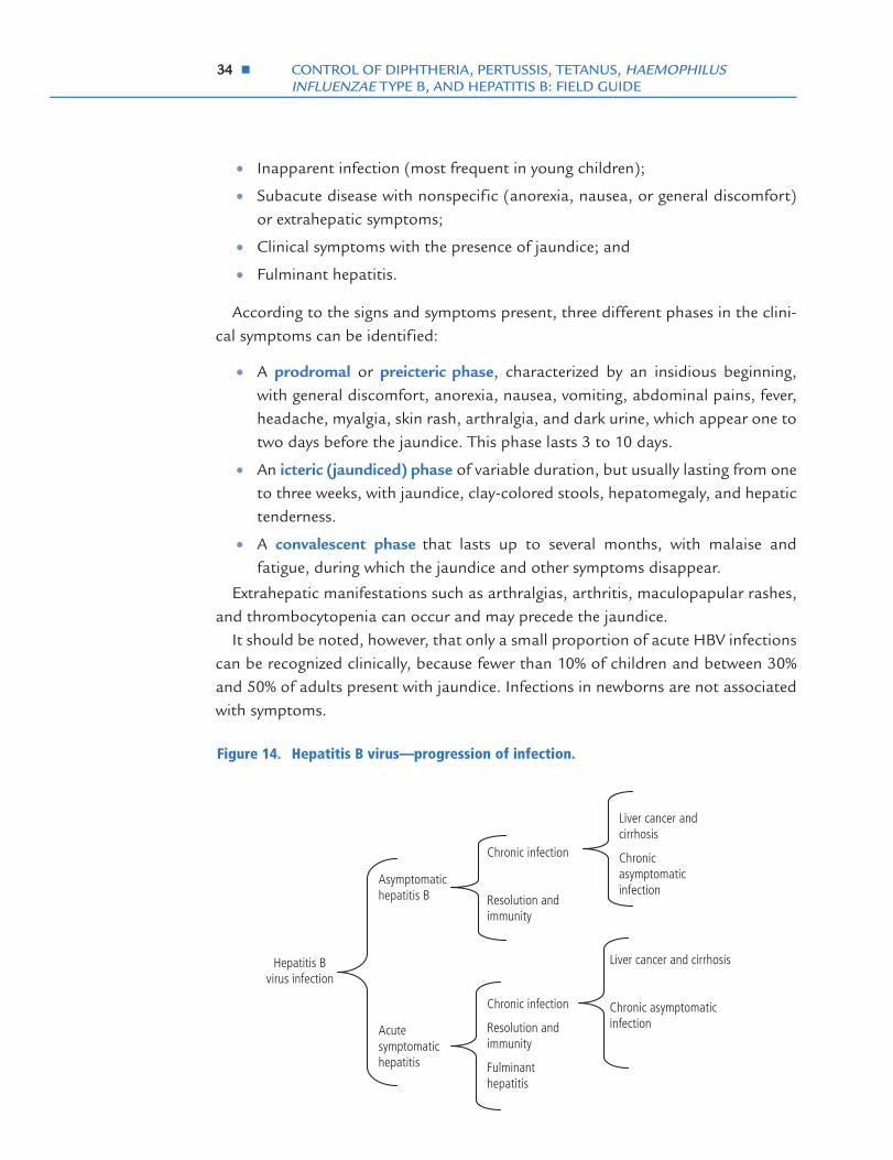
3344 CONTROL OF DIPHTHERIA, PERTUSSIS, TETANUS, HAEMOPHILUS INFLUENZAE TYPE B, AND HEPATITIS B: FIELD GUIDE
• Inapparent infection (most frequent in young children);
• Subacute disease with nonspecific (anorexia, nausea, or general discomfort)or extrahepatic symptoms;
• Clinical symptoms with the presence of jaundice; and
• Fulminant hepatitis.
According to the signs and symptoms present, three different phases in the clini-cal symptoms can be identified:
• A prodromal or preicteric phase, characterized by an insidious beginning,with general discomfort, anorexia, nausea, vomiting, abdominal pains, fever,headache, myalgia, skin rash, arthralgia, and dark urine, which appear one totwo days before the jaundice. This phase lasts 3 to 10 days.
• An icteric (jaundiced) phase of variable duration, but usually lasting from oneto three weeks, with jaundice, clay-colored stools, hepatomegaly, and hepatictenderness.
• A convalescent phase that lasts up to several months, with malaise andfatigue, during which the jaundice and other symptoms disappear.
Extrahepatic manifestations such as arthralgias, arthritis, maculopapular rashes,and thrombocytopenia can occur and may precede the jaundice.
It should be noted, however, that only a small proportion of acute HBV infectionscan be recognized clinically, because fewer than 10% of children and between 30%and 50% of adults present with jaundice. Infections in newborns are not associatedwith symptoms.
Hepatitis Bvirus infection
Asymptomatic hepatitis B
Acute symptomatic hepatitis
Chronic infection
Resolution and immunity
Chronic infection
Resolution and immunity
Fulminant hepatitis
Liver cancer and cirrhosis
Chronic asymptomatic infection
Liver cancer and cirrhosis
Chronic asymptomatic infection
Figure 14. Hepatitis B virus—progression of infection.

HEPATITIS B 3355
5.3.3 Laboratory Diagnosis
Diagnosis is based on clinical, epidemiologicalal, and laboratory findings. The infec-tion cannot be differentiated solely by clinical symptoms; definitive diagnosisdepends on the results of serologic tests (see Table 4) (34).
Detection of Hepatitis B Surface Antigen (HBsAg). Detection of HBsAg is themost common approach to diagnosing acute hepatitis B infection or detecting carri-ers. HBsAg in itself is not infectious, but its presence implies that the virus is present.This antigen can be detected from as early as one or two weeks to 12 weeks after expo-sure. Persistence of HBsAg for six or more months defines a chronic infection (chron-ic carrier). The presence of antibodies to HBsAg (anti-HBs) indicates immunity.
Detection of Hepatitis B Core Antigen (HBcAg) and Antibodies to HBcAg. HBcAgis detected only in liver tissue of infected individuals and not in serum. Anti-HBc (thecore antibody) indicates past infection. This antibody is not present in individualswho developed immunity through vaccination. The presence of IgM antibody toHBcAg (anti-HBc) indicates recent infection (four to six months). A negative test for
*The persistence of this marker for more than six months indicates chronicity.
Source: Adapted from Centers for Disease Control and Prevention. Epidemiology and prevention of vaccine-preventable diseases, Atkinson W, Hamborsky J, McIntyre L, Wolfe S, eds., 8th ed. Washington, D.C.: Public Health Foundation, 2005:191–199 and American Academy of Pediatrics. Hepatitis B. In: Pickering LK, ed. Red book: 2003 report of the Committee on Infectious Diseases, 26th ed. Elk Grove Village, IL: American Academy of Pediatrics; ©2003:322, with permission.
Table 4. Markers for the detection of hepatitis B infection and risk of transmission
Identification of carriers with high risk of transmitting HBsAg
Active viral replication Antigen E HBeAg
Detection of acute or recent hepatitis B infection, including in persons who are HBsAg - negative. Negative results for IgM anti - HBc and positive results for HBsAg indicate chronic infection.
Recent infection IgM antibodies against the virus core protein
IgM anti - HBc
Identification of people who have acquired immunity through illness or vaccine
Immunity Antibodies against the surface antigen (HBsAg)
Anti - HBs
Detection of carriers or indication of acute infection
Virus present Hepatitis B surface antigen HBsAg *
Usage Interpretation Antigen or antibody Marker
Identification of carriers with high risk of transmitting HBsAg
Active viral replication Antigen E HBeAg
Detection of acute or recent hepatitis B infection, including in persons who are HBsAg - negative. Negative results for IgM anti - HBc and positive results for HBsAg indicate chronic infection.
Recent infection IgM antibodies against the virus core protein
IgM
Identification of people who have acquired immunity through illness or vaccine
Immunity Antibodies against the surface antigen (HBsAg)
Anti - HBs
Detection of carriers or indication of acute infection
Virus present Hepatitis B surface antigen HBsAg *
Usage Interpretation Antigen or antibody Marker

3366 CONTROL OF DIPHTHERIA, PERTUSSIS, TETANUS, HAEMOPHILUS INFLUENZAE TYPE B, AND HEPATITIS B: FIELD GUIDE
IgM anti-HBc with a positive test forHBsAg indicates chronic infection.
Detection of Hepatitis B eAntigen (HBeAg) and Antibod-ies to HBeAg. HBeAg is detectedwhen there are high titers of viruscirculating, indicating a state ofhigh infectivity. Antibody toHBeAg (anti-HBe) indicates lowinfectivity.
When interpreting serologictests, it is important to take intoaccount the different results atthe same time. Table 5 includesthe most common combinationsof serological results and theirinterpretation.
5.3.4 Differential Diagnosis
Hepatitis is an inflammation ofthe liver which can be either infec-tious or non-infectious. Non-infec-tious causes of hepatitis includeautoimmune, genetic, metabolic,and drug-related chemical reac-tions. Infectious hepatitis can bethe only, or main, manifestationresulting from an infection withhepatotropic hepatitis A, B, C, D,
E, or G viruses. However, it can be also one of the manifestations of a systemic or com-municable illness due to agents such as cytomegalovirus, Epstein-Barr virus, adenovirus,and hemorrhagic fever viruses. In the Americas, many infectious diseases can result injaundice, such as leptospirosis and yellow fever, to name some. The various types ofhepatitis usually cannot be distinguished clinically, making laboratory detection of theserologic markers crucial.
5.3.5 Complications
Chronic Infection. Chronic infection is defined as the presence of HBsAg in serumfor at least six months. It is estimated that 10% of all HBV infections progress tochronic infection. However, the risk of developing chronic HBV infection is inverselyproportional to age (34), as follows:
*Postvaccination testing, when it is recommended, should be performed 1–2 months following dose #3.
Source: Adapted from Centers for Disease Control and Prevention, Epidemiology and prevention of vaccine-preventable diseases, Atkinson W, Hamborsky J, McIntyre L, Wolfe S, eds., 8th ed. Washington, D.C.: Public Health Foundation, 2005:194
May be recovering from acute HBV infection.May be distantly immune and the test is not sensitive
enough to detect a very low level of anti-HBs in serum.
May be susceptible with a false positive anti-HBc.May be chronically infected and have an undetectable
level of HBsAg present in the serum. Negativeanti-HBs
Positiveanti-HBc
NegativeHbsAg
Negativeanti-HBs
NegativeIgM anti-HBc
Positiveanti-HBcChronic infection
PositiveHBsAg
Negativeanti-HBs
PositiveIgM anti-HBc
Positiveanti-HBcAcute infection
PositiveHBsAg
Positiveanti-HBs
Positiveanti-HBc Immunity through natural infection
NegativeHBsAg
Positive (≥ 10 mIU/mL*)anti-HBs
Negativeanti-HBc Immunity through vaccination
NegativeHBsAg
Negativeanti-HBs
Negativeanti-HBc Susceptible
NegativeHBsAg
InterpretationResultTest
-HBs in
.
Table 5. Interpretation of serologic tests for hepatitis B

HEPATITIS B 3377
• 90% for infants infected at birth;
• 30% to 50% for children infected between 1 and 5 years old; and
• 6% to 10% for adults.
Individuals with Down syndrome or with a compromised immune system, such asthose with lymphoproliferative diseases, infected by HIV, or undergoing hemodialy-sis, apparently have a greater propensity for presenting chronic infection.
Cirrhosis and Hepatocellular Carcinoma. Cirrhosis of the liver and hepatocellular car-cinomas are long-term complications of hepatitis B. It is estimated that between 15% and25% of those with chronic HBV infection die of hepatocellular carcinoma or cirrhosis (39).
Fulminant Hepatitis. Fulminant hepatitis is defined as severe acute hepatitis that pro-gresses to liver necrosis, which can be fatal. The fulminant course is also seen in preg-nant women and newborns of infected mothers. Some 1%–2% of patients with acuteinfection present fulminant hepatitis, with mortality ranging from 63% to 93% (34).
5.3.6 Treatment
There is no specific treatment for acute HBV infection. Symptomatic supportivetherapy is recommended. Numerous antiviral agents to prevent chronic infectionhave been investigated, including interferon alpha-2b, which is successful in inter-rupting viral replication in 25%–40% of cases (39).
There is no need for quarantine, although concurrent disinfection of equipmentpotentially contaminated with blood or other body fluids is necessary.
55..44 VVAACCCCIINNAATTIIOONN AACCTTIIVVIITTIIEESS AANNDD PPAASSSSIIVVEE IIMMMMUUNNIIZZAATTIIOONN WWIITTHHHHEEPPAATTIITTIISS BB IIMMMMUUNNEE GGLLOOBBUULLIINN
The main objective of the strategies to combat hepatitis B is to prevent chronic HBVinfection and its serious consequences, including liver cirrhosis and hepatocellular cancer.
PAHO’s Technical Advisory Group on Vaccine-preventable Diseases recommends the following forthe prevention and control of hepatitis B infection (40):
• Routine universal infant immunization should be the primary strategy to prevent HBV trans-mission.
• Countries that have introduced hepatitis B (HepB) vaccine should consider using combinedvaccines, either DPT+HepB or, preferably, DPT+HepB+Haemophilus influenzae type b (Hib)(pentavalent vaccine).
• Health care workers who are at risk of being exposed to blood or other body fluids should beroutinely vaccinated.
• Vaccination coverage should be monitored on a regular basis and the impact of hepatitis Bvaccination measured through surveillance. Coverage levels for the third dose of hepatitis Bvaccine (HepB3) should equal those of the third dose of pentavalent.

3388 CONTROL OF DIPHTHERIA, PERTUSSIS, TETANUS, HAEMOPHILUS INFLUENZAE TYPE B, AND HEPATITIS B: FIELD GUIDE
5.4.1 Routine Immunization
Universal Vaccination of Infants. High coverage with the primary vaccine seriesamong infants aged < 12 months has the greatest overall impact on the prevalenceof chronic HBV infection in children (33). As with other vaccines used in childrenaged less than 1 year, the aim of hepatitis B vaccination should be to reach at least95% coverage in all municipalities (41, 42).
This strategy offers the following significant advantages:
• Ease of integrating HBV vaccine into the existing immunization schedules ineach country;
• Better compliance through the vaccination schedule for children aged lessthan 1 year;
• Possibility of combining vaccine antigens, as in DPT+HepB+Hib (pentavalent).
Booster injections after the infant schedule are usually not recommended becausealmost all children are protected following the primary vaccination schedule. Addi-tionally, PAHO does not recommend routine postvaccination serologic testing (42).
Prevention of Perinatal Transmission. Hepatitis B vaccination at birth preventsperinatal transmission of HBV from HBsAg-positive mothers to their infants in> 90% of cases. The decision to add a birth dose should be based on the prevalenceof carriers in the general population and the country’s resources. PAHO recom-mends that the birth dose be added when seroprevalence is > 8% (42). Only themonovalent hepatitis B vaccine should be used as the neonatal dose.
For schedule options that do and do not include the birth dose, refer to Chapter7, Section 7.3.3 (Schedule and Dosage).
5.4.2 Other Vaccination Activities
Vaccination of Adolescents. Countries with intermediate or low hepatitis Bendemicity may consider adding catch-up strategies targeted at older age groupssuch as adolescents with risk factors for acquiring HBV infection (42).
Vaccination of Health Care Workers and Other Groups at Risk. In addition touniversal infant immunization, PAHO recommends vaccinating health care workersto protect them from hepatitis B as an occupational hazard and to reduce the riskof virus transmission from infected workers to patients in clinical settings (42).
Serology prior to vaccination to determine immune status is not indicated forhealth professionals.
Countries may also choose to vaccinate other groups with risk factors for acquir-ing HBV infection, such as patients who undergo hemodialysis, receive frequenttransfusions, suffer chronic renal insufficiency, or who are infected with HIV; per-sonnel and patients of institutions for the mentally disabled or drug addicts; homo-sexual and bisexual individuals; intravenous drug users; persons with multiple sex
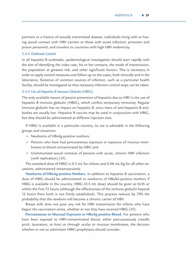
HEPATITIS B 3399
partners or a history of sexually transmitted disease; individuals living with or hav-ing sexual contact with HBV carriers or those with acute infection; prisoners andprison personnel; and travelers to countries with high HBV endemicity.
5.4.3 Outbreak Control
In all hepatitis B outbreaks, epidemiological investigation should start rapidly withthe aim of identifying the index case, his or her contacts, the mode of transmission,the population at greatest risk, and other significant factors. This is necessary inorder to apply control measures and follow up on the cases, both clinically and in thelaboratory. Existence of common sources of infection, such as a particular healthfacility, should be investigated so that necessary infection control steps can be taken.
5.4.4 Use of Hepatitis B Immune Globulin (HBIG)
The only available means of passive prevention of hepatitis due to HBV is the use ofhepatitis B immune globulin (HBIG), which confers temporary immunity. Regularimmune globulin has no impact on hepatitis B, since titers of anti-hepatitis B anti-bodies are usually low. Hepatitis B vaccine may be used in conjunction with HBIG,but they should be administered at different injection sites.
If HBIG is available in a particular country, its use is advisable in the followinggroups and situations:
• Newborns of HBsAg-positive mothers;
• Persons who have had percutaneous exposure or exposure of mucous mem-branes to blood contaminated by HBV; and
• Unimmunized sexual contacts of persons with acute, chronic HBV infection(with replication) (35).
The standard dose of HBIG is 0.5 mL for infants and 0.06 mL/kg for all other sit-uations, administered intramuscularly.
Newborns of HBsAg-positive Mothers. In addition to hepatitis B vaccination, adose of HBIG should be administered to newborns of HBsAG-positive mothers ifHBIG is available in the country. HBIG (0.5 mL dose) should be given at birth orwithin the first 72 hours (although the effectiveness of the immune globulin beyond12 hours from birth is not firmly established). This practice reduces by 70% theprobability that the newborn will become a chronic carrier of HBV.
Breast milk does not pose any risk for HBV transmission for infants who havebegun the vaccination series, whether or not they have received HBIG (35).
Percutaneous or Mucosal Exposure to HBsAg-positive Blood. For persons whohave been exposed to HBV-contaminated blood, either percutaneously (needleprick, laceration, or bite) or through ocular or mucous membranes, the decisionwhether or not to administer HBIG prophylaxis should consider:

4400 CONTROL OF DIPHTHERIA, PERTUSSIS, TETANUS, HAEMOPHILUS INFLUENZAE TYPE B, AND HEPATITIS B: FIELD GUIDE
• Whether the source of the blood is known;
• The source’s HBsAg levels; and
• Whether the exposed person has been vaccinated against HBV and, if so, hisor her response to the vaccine.
Table 6 shows the recommendations used in the United States for prophylaxisagainst hepatitis B after percutaneous, ocular, or mucosal exposure to the virus, asan example. Countries should refer to their national recommendations, if available.
Sexual Exposure to an HBsAg-Positive Person. HBIG should be used as postex-posure prophylaxis following sexual exposure to an infected person in addition tohepatitis B vaccination. HBIG given within seven days of exposure to an infectedpartner is about 75% effective in reducing acute hepatitis B disease and chronicinfection.
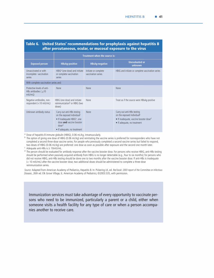
HEPATITIS B 4411
Immunization services must take advantage of every opportunity to vaccinate per-sons who need to be immunized, particularly a parent or a child, either whensomeone visits a health facility for any type of care or when a person accompa-nies another to receive care.
a Dose of hepatitis B immune globulin (HBIG), 0.06 mL/kg, intramuscularly. b The option of giving one dose of HBIG (0.06 mL/kg) and reinitiating the vaccine series is preferred for nonresponders who have not
completed a second three-dose vaccine series. For people who previously completed a second vaccine series but failed to respond, two doses of HBIG (0.06 mL/kg) are preferred: one dose as soon as possible after exposure and the second one month later.
c Adequate anti-HBs is ≥ 10mIU/mL. d The person should be evaluated for antibody response after the vaccine booster dose. For persons who receive HBIG, anti-HBs testing
should be performed when passively acquired antibody from HBIG is no longer detectable (e.g., four to six months); for persons who did not receive HBIG, anti-HBs testing should be done one to two months after the vaccine booster dose. If anti-HBs is inadequate
(< 10 mIU/mL) after the vaccine booster dose, two additional doses should be administered to complete a three-dose reimmunization series.
Source: Adapted from American Academy of Pediatrics, Hepatitis B. In: Pickering LK, ed. Red book: 2003 report of the Committee on Infectious Diseases , 26th ed. Elk Grove Village, IL: American Academy of Pediatrics; ©2003:335, with permission.
Unevaluated or unknown
HBsAg-negative HBsAg-positiveExposed person
Carry out anti-HBs testing on the exposed individualc
• If inadequate, vaccine booster dosed
• If adequate, no treatment
NoneCarry out anti-HBs testing on the exposed individualc
• If inadequate HBIGa , one dose and vaccine booster dosed
• If adequate, no treatment
Unknown antibody status
Treat as if the source were HBsAg-positive NoneHBIG (one dose) and initiate reimmunizationb or HBIG (two doses)
Negative antibodies, non-respondent (<10 mIU/mL)
NoneNoneNoneProtective levels of anti-HBs antibodies ( >10 mIU/mL)
With complete vaccination series and:
HBIG and initiate or complete vaccination seriesInitiate or complete vaccination series
HBIGa (one dose) and initiate or complete vaccination series
Unvaccinated or with incomplete vaccination series
Treatment when the source is:
c
••
)
Table 6. United States’ recommendations for prophylaxis against hepatitis B after percutaneous, ocular, or mucosal exposure to the virus

6 EPIDEMIOLOGICAL SURVEILLANCE
66..11 GGEENNEERRAALL CCOONNSSIIDDEERRAATTIIOONNSS
Conducting adequate surveillance is a major component of the control strategies forall five diseases covered in this field guide. The impact of control strategies is meas-ured by collecting surveillance data on disease incidence and vaccination coverage.Timely reporting allows for rapid investigation of cases of diphtheria, pertussis, Hib,and hepatitis B and satisfactory management of contacts. It also leads to earlydetection of outbreaks, which, in turn, allows for opportune implementation of out-break control measures. For vaccines recently included in national immunizationprograms, such as Hib, trend analysis permits an assessment of the impact of vac-cine introduction (43).
Specific actions and issues to be considered for the surveillance of the five dis-eases are outlined below.
Diphtheria
Critical activities for diphtheria surveillance in the Americas are:
• Routine case-based surveillance;
• Thorough case investigation; and
• Early investigation of outbreaks.
Pertussis
In the case of pertussis, routine surveillance can be case-based or consist of routinereporting of aggregated data from the local level to the central level, depending onthe epidemiological situation and pentavalent coverage in each country. Sentinelsurveillance to collect in-depth pertussis information, such as case-fatality rates byage groups, can be implemented in a few major hospitals; such surveillance shouldbe linked to developments in laboratory diagnostics and networks.
Surveillance for parapertussis is not a priority at present.
Tetanus
Besides monitoring the occurrence of tetanus, tetanus (non-neonatal) surveillanceshould include assessment of the use of prophylaxis with adult-type diphtheria andtetanus toxoids (Td vaccine) and use of TIG in the routine management of wounds.
Haemophilus influenzae type b (Hib)
Even though Hib surveillance focuses mainly on Hib meningitis, including Hib in thesurveillance for pneumococcus pneumonia is recommended due to the importanceof Hib as an etiologic agent for pneumonia. In developing countries, Hib has beenshown to be the cause of some 20% of pneumonias in children under 2 years old (44).
4422

EPIDEMIOLOGICAL SURVEILLANCE 4433
Epidemiological surveillance of pneumonias can be done through networks of sen-tinel hospitals, utilizing standardized radiological and clinical definitions. Countriesmay choose to incorporate other invasive Hib diseases (e.g., septic arthritis, epiglot-titis) into their Hib surveillance programs, as necessary.
Hepatitis B
Routine surveillance of hepatitis B only considers acute disease; however, to fullyunderstand the burden of hepatitis B, the sequelae of the disease such as chronicasymptomatic infections, cirrhosis, and primary cancer of the liver should also beconsidered. A report of low levels of acute viral hepatitis should not be misinterpret-ed as low incidence of HBV, as many of the infections occur asymptomatically. Datacollection from other sources not routinely used, including hospital surveillance,mortality from hepatitis B complications, and cancer registries, may be necessarywhen assessing disease burden.
Besides vaccine coverage, assessing the seroprevalence of HBsAg in young childrenshould assist in measuring the impact of hepatitis B vaccination (42).
Due to the characteristics and mode of transmission of hepatitis B, in addition tothe general population, surveillance may also include: health workers in contactwith blood samples and body fluids in the work environment; pregnant women; sexpartners of carriers; blood recipients, such as hemophiliacs; employees of and per-sons confined to prisons, barracks, and psychiatric hospitals; and travelers to areaswith high HBV prevalence.
66..22 CCAASSEE DDEEFFIINNIITTIIOONNSS
Diphtheria
Clinical Description: A person of any age presenting laryngitis, pharyngitis, or tonsilli-tis, and an adherent membrane covering the tonsils, pharynx, and/or nasal septum.
Case Classificationa
• Suspected case: Not applicable.
• Probable case: A case that meets the clinical description.
• Confirmed case: A probable case that has been laboratory-confirmed or epi-demiologically linked to a laboratory-confirmed case.
• Clinically confirmed case: A probable case in which no other diagnosis has beenlaboratory-confirmed.
aPersons with positive C. diphtheriae cultures who do not meet the clinical description (i.e.,asymptomatic carriers) should not be reported as probable or confirmed diphtheria cases.

4444 CONTROL OF DIPHTHERIA, PERTUSSIS, TETANUS, HAEMOPHILUS INFLUENZAE TYPE B, AND HEPATITIS B: FIELD GUIDE
Pertussis
Clinical Description: A case diagnosed by a health care worker, or a person presentingpersistent cough for two or more weeks, without another apparent cause, and hav-ing at least one of the following manifestations:
• Paroxysms (i.e., fits) of coughing;
• Inspiratory “whooping”; and
• Post-tussive vomiting (i.e., vomiting immediately after coughing) withoutanother apparent cause.
Case Classification:
• Suspected case: Not applicable.
• Probable case: A case that meets the clinical description.
• Confirmed case: A probable case that has been clinically confirmed where oth-er causes have not been identified, laboratory-confirmed, or epidemiological-ly linked to a laboratory-confirmed case.
• Laboratory-confirmed case: A case from which B. pertussis has been isolated orwith positive results by PCR.
• Clinically confirmed case: A probable case in which no other diagnosis has beenconfirmed.
Tetanus (non-neonatal tetanus)b
Clinical Definition: Tetanus is defined as the acute appearance of hypertonia orpainful muscle contractions (usually in the neck or jaw) and generalized musclespasms without another apparent medical cause.
• Confirmed case: A compatible clinical case reported by a health professional.
Haemophilus influenzae type b (Hib)
Hib surveillance should be integrated into meningitis surveillance. The definitionslisted below should be used for the surveillance of Hib meningitis.
Case Classification:
• Suspected case: Every child aged 1 month to 5 years who presents clinical symp-toms or signs of meningitis.
• Probable case: A suspected case whose cerebrospinal fluid (CSF) shows positiveresults with Gram’s stain (Gram-negative coccobacillus for Hib) or shows at
bFor neonatal tetanus case definitions please refer to Pan American Health Organization,Neonatal Tetanus Elimination Field Guide (Scientif ic and Technical Publication No. 602; Washing-ton, D.C.: PAHO, 2005).
EPIDEMIOLOGICAL SURVEILLANCE 4455
least one of the following alterations in cytochemistry suggestive of bacterialmeningitis:
− Pleocytosis: white blood cell count ≥ 10 per mm3, with a predominance ofneutrophils;
− Low CSF glucose: glucose ≤ 50% of blood glucose;− High CSF protein: proteins ≥ 45 mg/dL.
• Confirmed case: Every probable case with an isolation of H. influenzae type bfrom CSF or laboratory results indicating the presence of the bacteria by latexparticle agglutination, immunoelectrophoresis, or PCR.
Additionally, Hib should be integrated into each country’s existing local surveil-lance for pneumonia. As already mentioned, other forms of Hib invasive disease maybe included in local Hib surveillance systems as national health authorities deemnecessary.
Hepatitis B
Clinical Description: A person with jaundice who also presents choluria, acholic (clay-colored) stools, anorexia, asthenia, nausea, and liver enzyme levels 2.5 times higherthan normal, without other attributable causes.
Case Classification:
• Suspected case: Not applicable.
• Probable case: A case that meets the clinical description.
• Confirmed case: A probable case confirmed by laboratory (compatible serologyor histopathology).
• Discarded case: Laboratory results or clinical description not corresponding tothe disease.
• Carrier case: An asymptomatic case who is HBsAg-positive six months afteracquiring the infection.
66..33 FFLLOOWW OOFF SSUURRVVEEIILLLLAANNCCEE IINNFFOORRMMAATTIIOONN
The flow of information and activities in the surveillance of vaccine-preventable dis-ease is illustrated in Figure 15.
6.3.1 Case Reporting
Routine reporting is essential for the surveillance of most vaccine-preventable dis-eases. Public and private health practitioners should be aware of the characteristicsand the occurrence of diphtheria, pertussis, tetanus (in addition to neonataltetanus), Hib, and hepatitis B cases, and of the importance of reporting probableand confirmed cases through the routine surveillance system. Additionally, efforts

4466 CONTROL OF DIPHTHERIA, PERTUSSIS, TETANUS, HAEMOPHILUS INFLUENZAE TYPE B, AND HEPATITIS B: FIELD GUIDE
should be made to ensure that laboratories conducting microbiological testing forC. diphtheriae and B. pertussis report cases to the health authorities. For hepatitis B,laboratories and blood banks should maintain a registry on positive tests for HBsAgand report at least monthly.
6.3.2 Case Investigation
Public health workers should investigate reports of probable cases of vaccine-pre-ventable diseases to ensure that contacts are properly managed and that these dis-eases are adequately controlled. This is especially important when cases of diphthe-ria, pertussis, or Hib have been confirmed in institutions that cater to children, suchas child-care centers, kindergartens, or schools. In addition to investigating casesand contacts, vaccination coverage should be evaluated at the municipality level inorder to increase vaccination among those with low coverage.
For case-based surveillance, a unique identifier should be assigned to the person,and a case-investigation form should be used to obtain the following information:geographic location; age or date of birth; sex; immunization status and date of lastdose of the specific vaccine; clinical history, including date of onset; laboratoryresults; treatment; outcome (recovery or death); information on contacts and theirfollow-up; and final classification of the case. Obtaining information on socioeco-nomic status may be useful to better guide control strategies. (See Annexes 2 to 7for sample notification and investigation forms.)
Health
facility
Local
health unit
Central
level
PAHO
Lab
Case investigation
Reports
Technical Support
ResultSample
Notification
Not
ifica
tion
NotificationNotification
FeedbackFeedback
CONTROL & PREVENTION STRATEGIES
Case
PAHO
Laboratory
Contacts
Community
Surveillance-control-vaccination
Figure 15. Vaccine-preventable disease surveillance flow.

EPIDEMIOLOGICAL SURVEILLANCE 4477
6.3.3 Feedback
Feedback to health professionals participating in surveillance activities includes pro-viding them with information on the number of cases reported by geographic area,a summary of the characteristics of the cases, information on the impact of controlmeasures, and specific recommendations on how to improve case reporting. Theresults of analyses conducted at the national level should be shared periodically withthe local health authorities and services. Feedback on surveillance can be given forseveral vaccine-preventable diseases at once, for example in the form of periodic bul-letins.
66..44 DDAATTAA AANNAALLYYSSIISS AANNDD SSUURRVVEEIILLLLAANNCCEE IINNDDIICCAATTOORRSS
6.4.1 Data Analysis
Table 7 summarizes the type of analysis suggested for diphtheria, pertussis, tetanus,Hib, and hepatitis B.
6.4.2 Surveillance Indicators
Indicators that can be used to monitor the surveillance system in general include:
• Number of health units notifying regularly;
• Proportion of cases report-ed on time (e.g., within 24hours of diagnosis); and
• Proportion of cases ade-quately investigated.
Besides the indicators suggestedabove, the use of additional sur-veillance indicators depends onthe local status of each particulardisease. Suggested additional indi-cators for diphtheria, pertussis,and hepatitis B include:
Diphtheria
• Proportion of probable cas-es with nasopharyngeal orpharyngeal swab collected;
• Proportion of cases that arelaboratory-confirmed; and
• Proportion of contacts man-aged properly.
a See glossary for explanation and calculation of rates.b For specific neonatal tetanus data analysis and surveillance indicators refer to: Pan American Health Organization,
Neonatal tetanus elimination field guide. Scientific and Technical Publication No. 602. Washington, D.C.: PAHO, 2005.
Hepatitis B• Sex-specific incidence
Hib• Proportion of bacterial meningitides caused
by Hib
• Proportion of consolidated pneumonias with isolation or diagnosis of Hib by laboratory
Tetanusb
• Local standards and practices used for wound management
• Incidence rates by geographic area
• Specific incidence among vaccinated, unvaccinated, and incompletely vaccinated individuals
• Age-specific incidence rates
• Case-fatality rate and, if high, possible causes (e.g., poor case management, delays in starting specific treatment, patients not seeking care in time, etc.)
• Coverage rates with the primary series (i.e., DTP3, Hib3, HepB3 or pentavalent-3 in children < 1 year) by municipality
• Investigation of outbreaks in terms of person, place, and time (descriptive epidemiology), and determining risk factors for infection
SpecificGenerala
Hib
••
•
•
•
•
•
Table 7. Data analysis for diphtheria, pertussis, tetanus, Hib, and hepatitis B

4488 CONTROL OF DIPHTHERIA, PERTUSSIS, TETANUS, HAEMOPHILUS INFLUENZAE TYPE B, AND HEPATITIS B: FIELD GUIDE
Pertussis (outbreaks)
• Proportion of outbreaks that are laboratory-confirmed; and
• Proportion of probable cases with nasopharyngeal aspirate or swab collected.
Hepatitis B
• Proportion of cases and/or outbreaks investigated;
• Proportion of contacts investigated; and
• Proportion of laboratories and blood banks that report monthly.
66..55 OOUUTTBBRREEAAKK RREESSPPOONNSSEE
Outbreaks of vaccine-preventable diseases should be reported to the health author-ities and investigated as soon as they come to the attention of the health system.Outbreak investigation increases understanding of the epidemiology of vaccine-pre-ventable diseases and the reasons why the outbreak occurred (e.g., vaccine failure,failure to immunize, accumulation of susceptibles, waning immunity, and emer-gence of new toxigenic strains). Adequate outbreak investigation and responseensures the proper management of cases and contacts, and limits the spread of dis-ease in the community.
Specific activities for outbreak control for each disease are addressed in Chapters1 to 5.

7 VACCINES AGAINST DIPHTHERIA, PERTUSSIS, TETANUS,HAEMOPHILUS INFLUENZAE TYPE B, AND HEPATITIS B
77..11 DDIIPPHHTTHHEERRIIAA,, PPEERRTTUUSSSSIISS,, AANNDD TTEETTAANNUUSS VVAACCCCIINNEESS
7.1.1 Components
Vaccines against diphtheria, pertussis, and tetanus are commonly combined eitheras DPT, which contains diphtheria and tetanus toxoids and the whole-cell pertussis(wP) antigen, or as DTaP, which also contains diphtheria and tetanus toxoids, buthas an acellular pertussis (aP) component with purified and inactivated compo-nents of B. pertussis cells. Diphtheria-tetanus vaccines not including the pertussiscomponent are also available as DT for children and as Td, with a lower dose ofdiphtheria toxoid, for adults (45).
7.1.2 Immunity and Effectiveness
Diphtheria. After the administration of three doses of diphtheria toxoid, virtually allinfants and adults develop diphtheria antibody titers greater than 0.01 IU/mL (thelowest level conferring some degree of protection). DTaP (diphtheria-tetanus-acel-lular pertussis) produces significantly lower geometric mean titers than DPT, butwithout clear clinical differences. Effectiveness seems to be 90% or higher for threeor more doses and it has been seen that when diphtheria occurs in previously immu-nized persons, the disease tends to be less severe and has lower case-fatality rates.
Several serosurveys in Europe and the United States suggest a trend towardincreasing susceptibility with advancing age, raising concerns regarding the durationof diphtheria toxoid-induced immunity. For this reason, most countries recommendperiodical booster doses for adults with Td (tetanus-diphtheria for persons olderthan 7 years) (45).
Pertussis. There has been extensive experience with the use of wP vaccines. A pri-mary series of four doses confers 70%–90% protection against serious pertussis dis-ease. However, the immunity conferred by this vaccine wanes over time, and little orno protection is left 5–10 years after the last dose. The benefits of wP vaccine havebeen proven by the decline in morbidity and mortality from pertussis seen with theimplementation of vaccination programs and the recurrence of disease when cover-age levels have decreased (45).
Trials on aP vaccine have shown variable efficacies and it is not yet known if theduration of protection conferred by aP is comparable with wP vaccines. Some recentclinical trials comparing aP vaccine to wP vaccine have shown wP vaccine to be moreeffective, but the clinical significance of this finding is not clear (45).
4499

5500 CONTROL OF DIPHTHERIA, PERTUSSIS, TETANUS, HAEMOPHILUS INFLUENZAE TYPE B, AND HEPATITIS B: FIELD GUIDE
In 2005, two aceullular pertussis vaccines for use in adolescents and adults werelicensed in the United States. Both are formulated in ccoommbbiinnaattiioonn with Td and con-tain a lower dose of the acellular pertussis component than the vaccines licensedand uusseedd for infants and children aged < 7 years.
Tetanus. To provide active tetanus immunity, vaccines containing tetanus toxoidare used. Tetanus vaccine, when administered correctly and following the proposedschedule, is nearly 100% effective. Antitoxin levels higher than 0.01 IU/mL are con-sidered protective. Protective antitoxin levels decrease over time, so a booster doseevery 10 years is usually recommended.
7.1.3 Adverse Events and Contraindications
Diphtheria and tetanus toxoids
Diphtheria and tetanus toxoids are among the safest vaccines available. The reac-tions to vaccines containing diphtheria and tetanus toxoids can be classified as fol-lows (47):
• Local reactions: erythema and induration with or without tenderness. Thesereactions are usually self-limited and require no therapy;
• Exaggerated local (Arthus-like) reactions: extensive, painful swelling, typicallyfrom shoulder to elbow, that usually begins two to eight hours after vaccination.These reactions are infrequent and more often reported in adults, particularlyamong those who have received frequent doses of diphtheria or tetanus toxoid;
• Systemic reactions: fever and malaise are rare; more severe anaphylactoidreactions have been reported on a few occasions.
The frequency and intensity of adverse reactions following vaccination with diph-theria and tetanus toxoids increase with the dose number and also vary with the pre-vaccination diphtheria-tetanus antitoxin levels.
Diphtheria-tetanus vaccine is contraindicated for those with severe allergic reac-tions to a vaccine component or a previous dose. Also, it is recommended to deferdiphtheria and tetanus toxoid administration for persons experiencing moderate to
PAHO’s Technical Advisory Group (in 1997) and the World Health Organization (in its positionpaper of 1999 and 2005 update) recognized the effectiveness of whole cell pertussis (wP) vaccinesin reducing the incidence of pertussis worldwide. The wP vaccines have a considerably lower pricethan acellular pertussis (aP) vaccines; where resources are limited and the vaccine is well accept-ed by the local population, wP remains the vaccine of choice. In countries where the higher reac-togenicity of wP is an impediment to high vaccination coverage, aP may be used instead, at leastfor booster injections (11, 12, 46).
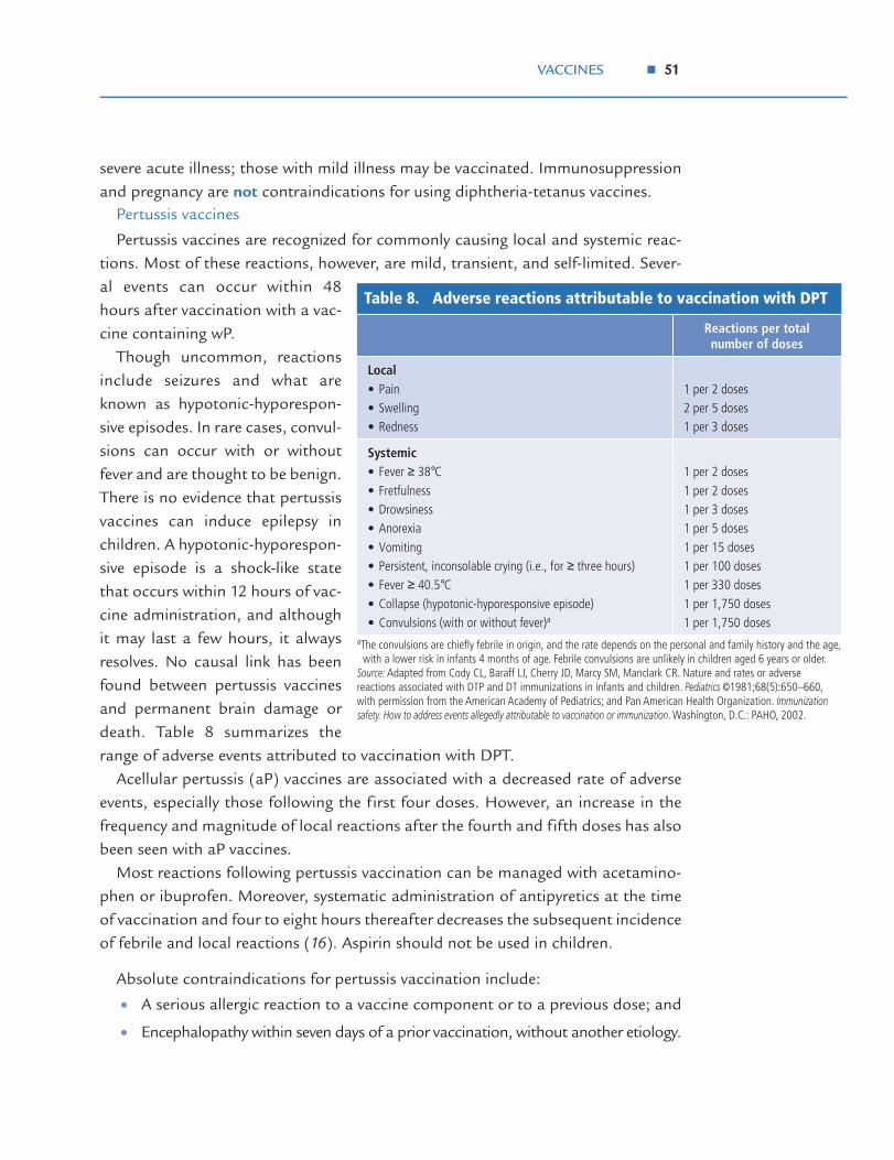
VACCINES 5511
severe acute illness; those with mild illness may be vaccinated. Immunosuppressionand pregnancy are not contraindications for using diphtheria-tetanus vaccines.
Pertussis vaccines
Pertussis vaccines are recognized for commonly causing local and systemic reac-tions. Most of these reactions, however, are mild, transient, and self-limited. Sever-al events can occur within 48hours after vaccination with a vac-cine containing wP.
Though uncommon, reactionsinclude seizures and what areknown as hypotonic-hyporespon-sive episodes. In rare cases, convul-sions can occur with or withoutfever and are thought to be benign.There is no evidence that pertussisvaccines can induce epilepsy inchildren. A hypotonic-hyporespon-sive episode is a shock-like statethat occurs within 12 hours of vac-cine administration, and althoughit may last a few hours, it alwaysresolves. No causal link has beenfound between pertussis vaccinesand permanent brain damage ordeath. Table 8 summarizes therange of adverse events attributed to vaccination with DPT.
Acellular pertussis (aP) vaccines are associated with a decreased rate of adverseevents, especially those following the first four doses. However, an increase in thefrequency and magnitude of local reactions after the fourth and fifth doses has alsobeen seen with aP vaccines.
Most reactions following pertussis vaccination can be managed with acetamino-phen or ibuprofen. Moreover, systematic administration of antipyretics at the timeof vaccination and four to eight hours thereafter decreases the subsequent incidenceof febrile and local reactions (16). Aspirin should not be used in children.
Absolute contraindications for pertussis vaccination include:
• A serious allergic reaction to a vaccine component or to a previous dose; and
• Encephalopathy within seven days of a prior vaccination, without another etiology.
aThe convulsions are chiefly febrile in origin, and the rate depends on the personal and family history and the age, with a lower risk in infants 4 months of age. Febrile convulsions are unlikely in children aged 6 years or older.
Source: Adapted from Cody CL, Baraff LJ, Cherry JD, Marcy SM, Manclark CR. Nature and rates or adverse reactions associated with DTP and DT immunizations in infants and children. Pediatrics ©1981;68(5):650–660, with permission from the American Academy of Pediatrics; and Pan American Health Organization. Immunization safety. How to address events allegedly attributable to vaccination or immunization. Washington, D.C.: PAHO, 2002.
1 per 2 doses 1 per 2 doses 1 per 3 doses 1 per 5 doses 1 per 15 doses 1 per 100 doses 1 per 330 doses 1 per 1,750 doses 1 per 1,750 doses
Systemic • Fever ≥ 38 ° C • Fretfulness • Drowsiness • Anorexia • Vomiting • Persistent, inconsolable crying (i.e., for ≥ three hours) • Fever ≥ 40.5 ° C • Collapse (hypotonic - hyporesponsive episode) • Convulsions (with or without fever) a
1 per 2 doses 2 per 5 doses 1 per 3 doses
Local • Pain • Swelling • Redness
Reactions per total number of doses
Table 8. Adverse reactions attributable to vaccination with DPT

5522 CONTROL OF DIPHTHERIA, PERTUSSIS, TETANUS, HAEMOPHILUS INFLUENZAE TYPE B, AND HEPATITIS B: FIELD GUIDE
Relative contraindications include the following reactions to a previous doseoccurring within 48 hours of vaccination:
• Fever ≥ 40.5°C with no other cause;
• A hypotonic-hyporesponsive episode;
• Persistent, uncontrollable crying lasting more than three hours; and
• Convulsions, with or without fever, within three days of receiving a previous dose.
Precautions include moderate or severe acute illness. This prevents fever or othersymptoms from being wrongly attributed to the vaccine.
Relative contraindications need to be weighed against the benefits in the event ofan outbreak in the community. Pertussis vaccines are not recommended for childrenaged 7 years or older. Vaccine reactions are thought to be more frequent in older agegroups and the burden of pertussis decreases with increasing age (45, 47, 48).
7.1.4 Schedule and Dosage
Three doses (the primary doses) of DPT given at least four weeks apart are neededto confer protection against diphtheria, tetanus, and pertussis. Booster doses, giv-en no less than six months after the third dose and not before 1 year of age, may benecessary to maintain immunity.
In the Americas, all countries recommend that the three primary doses of DPTvaccine (or combination vaccines containing DPT) be given during the first year oflife, usually at 2, 4, and 6 months, and most recommend at least one booster dose.
There is no need to restart the primary series, regardless of the time elapsedbetween doses. DPT vaccine is not recommended for children aged less than 6weeks, for whom responses to pertussis are suboptimal.
When pediatric DT vaccine is used (in children aged less than 7 years for whompertussis is contraindicated), the following schedule is recommended:
• Four doses should be used when the child has received the first dose before 12months of age;
• Three doses—two doses of DT vaccine, given 2 months apart, and a third dose6 to 12 months later—should be given for unimmunized children aged 1 to 7years.
Unimmunized persons aged 7 years or older should receive a three-dose, adult Tdvaccine series, with the second dose given 4 to 8 weeks after the first, and the thirdgiven 6 to 12 months later.
Booster doses with Td vaccine (for persons 7 years of age or older) to maintainimmunity are suggested every 10 years. Td vaccinations fewer than 10 years apart arenot recommended, as there are reports suggesting increased incidence and severityof adverse local reactions.

VACCINES 5533
Patients recovering from tetanus disease should be vaccinated following the regu-lar schedule for DPT or Td vaccine.
The standard pediatric dose of DPT and DT vaccines is 0.5 mL. DPT vaccine isusually purchased in multi-dose vials.
77..22 HHAAEEMMOOPPHHIILLUUSS IINNFFLLUUEENNZZAAEE TTYYPPEE BB VVAACCCCIINNEESS
Conjugate vaccines have proven to be an excellent tool to prevent the infections causedby Haemophilus influenzae type b. They consist of the capsular polysaccharide of the bac-terium, linked to a protein carri-er, which influences the immuno-logical mechanism, transformingan antigen-independent T-cellresponse to an antigen-depend-ent T-cell response. Unlike thefirst vaccines developed for useagainst Hib, conjugate vaccinesconfer immunity in infants.
Three Hib conjugate vaccinesa
are authorized for use in infantsand are interchangeable:
• HbOC (HibTITER®)
• PRP-OMP (PedvaxHIB®)
• PRP-T (ActHIB® andOmniHIB®).
A fourth Hib vaccine, PRP-D(ProHIBIT®), is only authorizedfor infants and children between 12 and 60 months of age and should not be usedfor the primary series.
Table 9 shows the types of Hib conjugate vaccines available and their recommend-ed vaccination series.
7.2.1 Immunity and Effectiveness
All three conjugate vaccines for use in infants are highly immunogenic. Effectivenesshas been estimated at 95% to 100%. Invasive Hib disease in a fully vaccinated childis rare (45).
aPRP-D is authorized as a booster dose after a primary series of another type of vaccine for infants aged 12 months and as a single dose for infants who at 15 months of age have not been previously vaccinated.
Source: Adapted from Centers for Disease Control and Prevention, Epidemiology and prevention of vaccine-preventable diseases, Atkinson W, Hamborsky J, McIntyre L, Wolfe S, eds., 8th ed. Washington, D.C.: Public Health Foundation, 2005:101–113.
Single dose or boostera
Diphtheria toxoidPRP-D (ProHIBIT®)
BoosterSecond dose
First doseOuter meningococcus membrane B protein
PRP-OMP (PedvaxHIB®)
BoosterThird doseSecond dose
First doseTetanus toxoidPRP-T (ActHIB® or OmniHIB®)
BoosterThird doseSecond dose
First doseMutant diphtheria toxin protein
HbOC(HibTITER®)
12–15 months6 months4 months2 months
Usual schedule
Protein carrier Vaccine (brand)
Table 9. Conjugate vaccines against Haemophilus influenzae type b (Hib) and recommended vaccination series
aHbOC refers to: oligosaccharides conjugated to diphtheria toxin protein; PRP-OMP refers to:polyribosylribitol phosphate polysaccharide conjugated to a meningococcal outer membraneprotein; PRP-T refers to: polyribosylribitol phosphate polysaccharide conjugated to tetanustoxoid.

5544 CONTROL OF DIPHTHERIA, PERTUSSIS, TETANUS, HAEMOPHILUS INFLUENZAE TYPE B, AND HEPATITIS B: FIELD GUIDE
Hib vaccine is also immunogenic in patients with increased risk for invasive dis-ease (patients with sickle-cell disease, leukemia or HIV infection). However, amongHIV-infected persons, the immunogenicity varies with the degree of immunocom-promise (26).
The effectiveness of Hib immunization is not only achieved by the direct protec-tion provided by Hib vaccine, but also through an important herd effect due to areduction in nasopharyngeal carriage of the microorganism in the population (49).
7.2.2 Adverse Events and Contraindications
Adverse events after vaccination with Hib conjugate vaccine are uncommon. Mod-erate fever tends to occur in 1 out of 20 vaccinated children. Edema, reddening, orpain at the injection site—or a combination of these—have been reported in one ofevery four cases. These symptoms usually resolve within 24 to 48 hours. Systemicreactions, such as high fever and irritability, are infrequent.
Vaccination with conjugate Hib is contraindicated for anyone who has experi-enced anaphylaxis following the administration of a previous dose of that vaccine.
7.2.3 Schedule and Dosage
All infants, including those born prematurely, should receive a primary series of Hibconjugate vaccine (separately or in a combination of vaccines) beginning at 2months of age. Recent data indicate that if Hib conjugate vaccine is administered toinfants aged less than 6 weeks, immunological tolerance to additional doses of Hibvaccine can be induced. Consequently, vaccines containing Hib conjugate shouldnot be administered to children aged less than 6 weeks (26).
The number of doses in the primary series depends on the type of vaccine used. Aprimary series of the vaccine PRP-OMP (PedvaxHIB®) consists of two doses; HbOC(HibTITER®) and PRP-T (ActHIB® and OmniHIB®) require a primary series ofthree doses (see Table 9). Most countries in the Americas use the PRP-T vaccine.
A booster is sometimes administered at 12 to 15 months of age, regardless ofwhich vaccine has been used for the primary series. Studies are being conducted infour Latin American countries to evaluate differences in vaccination effectiveness, ifany, between three-dose schedules with a booster and without a booster dose.
The optimal interval between doses is two months; the minimum interval betweenthem is one month. The booster dose, if used, should be given at least two monthsafter the previous dose.
Infants and children unvaccinated at 7 months of age or older may not require aseries totaling three or four doses. The number of doses that an infant needs inorder to complete the series depends mainly on his or her age at the time of con-sultation.

VACCINES 5555
Children between 15 and 60 months of age who have not received a previous doseof any Hib conjugate vaccine should receive a single dose. In general, children over60 months do not need to be vaccinated.
The Hib conjugate vaccine PRP-OMP is available in combination with hepatitis Bvaccine (Comvax®). In addition, PRP-T (ActHIB®) is available in combination withDTaP in a product called TriHIBit®. However, TriHIBit® is authorized for use onlyas the fourth dose of the Hib and DTaP series due to reduced immunogenicity of theHib component when used as a combination. Thus, TriHIBit® should not take theplace of the first, second, or third dose of the Hib series (26).
The standard pediatric dose is 0.5 mL; Hib conjugate vaccines are available in liq-uid or lyophilized (freeze-dried powder) form, in single- or multi-dose vials. They canbe utilized as monovalent vaccines or in combination with other vaccines, such asDPT and/or the hepatitis B vaccine (see Section 7.4 on combination vaccines in thischapter).
77..33 HHEEPPAATTIITTIISS BB VVAACCCCIINNEESS
The hepatitis B vaccine used in the Americas is an inactivated recombinant vaccineobtained through genetic engineering. This vaccine contains the subunit of the sur-face antigen (HBsAg) obtained from purified hepatitis B virus (HBV) through therecombinant DNA (rDNA) technique in Saccharomyces cerevisiae yeasts, in which thegene responsible for the synthesis of the HBsAg (S gene) is inserted. These recombi-nant vaccines are interchangeable.
7.3.1 Immunity and Effectiveness
Three doses of vaccine are required to induce an adequate response of protectiveantibodies (anti-HBs ≥ 10 milli-IU/mL in more than 90% of healthy adults and morethan 95% of children and adolescents). Protective titers begin to be reached twoweeks after the second dose, and the effectiveness of the vaccine is 95%–98%. The
Important issues regarding Hib conjugate vaccines (26):
• The three Hib conjugate vaccines authorized for use in infants are interchangeable.
• A series that includes vaccines of more than one type will still induce a level of protective anti-bodies.
• If it is necessary to change the type of vaccine, three doses of any combination constitute theprimary series.
• Any authorized conjugate vaccine may be used for the booster dose, regardless of the type usedin the primary series.

5566 CONTROL OF DIPHTHERIA, PERTUSSIS, TETANUS, HAEMOPHILUS INFLUENZAE TYPE B, AND HEPATITIS B: FIELD GUIDE
third dose confers optimal protection, acting as a booster dose. In a three-doseschedule, longer intervals between the last two doses (4–12 months) result in high-er final titers of anti-HBs (50, 51). Several studies have shown that the currentlylicensed vaccines produce high rates of seroconversion (>95%) and induce adequatelevels of anti-HBs when administered to infants at birth, 2 months, and 6 months ofage, or at 2, 4, and 6 months of age (52).
Some factors that influence seroconversion after a complete vaccination series are:
• Age: Individuals aged more than 40 years present a smaller proportion of sero-conversion;
• Type of host: Immunocompromised patients, such as those with chronic renalinsufficiency, those infected by HIV, and those being treated with immunosup-pressors, have a lower percentage of seroconversion (50%–70%);
• Application site: Application in the gluteal region yields lower levels of sero-conversion in adults due to decreased absorption of the immunizing antigenin that area.
There is disagreement about whether smoking, male sex, obesity, and diabetes mel-litus influence seroconversion. Some 10% of adults are considered non-responders:that is, they do not present an antibody response to the initial vaccination series.
Studies indicate that the duration of immunity as measured by serology varies.However, long-term studies in children and adults indicate that immunologicalmemory remains intact after up to 15 years of monitoring and that the antibodiesprotect against chronic HBV infection, although the concentrations of anti-HBs maybe low or undetectable (35).
7.3.2 Adverse Events and Contraindications
Hepatitis B vaccine is a safe vaccine, although it does produce some local effects in3%–9% of vaccinees, such as pain, erythema, and induration, which are generallytransitory. These reactions are more common in adults (13%–29%).
Some systemic symptoms, which are slight and transitory, have been reported:fatigue, headache, and irritability in 8% to 18% of vaccinated children, and feverabove 37.7°C in 0.4% to 8% of children. These reactions disappear spontaneously ina few days, and do not require interruption of the vaccination schedule.
Hepatitis B vaccine does not produce adverse effects in chronic carriers. Individ-uals who have had an unrecognized past infection with positive anti-HBsAg andreceive hepatitis B vaccine are not at greater risk of adverse reactions.
Recombinant DNA hepatitis B vaccine cannot transmit HBV or any other virus(e.g., HIV) because the vaccine is produced through genetic engineering.

VACCINES 5577
Prior anaphylaxis due to any component of this vaccine is a contraindication forits use. However, it can be administered without risk to pregnant women, since itcontains noninfectious particles of HBsAg.
7.3.3 Schedule and Dosage
Three-dose schedules for hepatitis B vaccine, not including the birth dose, usuallyfollow the DPT schedule and are given as pentavalent at 2, 4, and 6 months of age.
As mentioned in Chapter 5, some countries with high prevalence of hepatitis Binfection will choose to include a dose of hepatitis B vaccine at birth to prevent peri-natal transmission. Whether or not the birth dose is used in a particular country,there are several options for including hepatitis B vaccine in the immunizationschedule for children aged < 1 year.
Some countries that administer the birth dose of hepatitis B and have the pen-tavalent vaccine in their schedule, utilize the following schedule: monovalent hepa-titis B (is administered) at birth, followed by three doses of pentavalent, usually at2, 4, and 6 months. These countries find this approach to be programmatically eas-ier and administer four doses of hepatitis B antigen.
Others countries that use three-dose schedules of hepatitis B antigen, including abirth dose, usually follow the schedule used for children born to HBsAg-positivemothers, that is:
• First dose at birth (within 24 hours of birth);
• Second dose at 1–2 months of age; and
• Third dose at 6 months.
In this series, the last two doses are usually administered simultaneously with DPTand Hib vaccines.
In general, a minimum interval of four weeks is recommended between the firstand second doses of hepatitis B vaccine, and eight weeks between the second andthird. Studies have shown that longer intervals between the last two doses may in-crease the final anti-HBs titers but do not affect seroconversion rates (52).
Hepatitis B vaccine is presented in mono-dose or in multiple-dose vials that con-tain 2, 6, or 10 doses of vaccine. They contain aluminum hydroxide as adjuvant andoften thimerosal as preservative. Each dose of lyophilized vaccine contains 5, 10, or20 μg of antigen (HBsAg), reconstituted in 0.5 or 1 mL doses, according to the man-ufacturer’s instructions. Table 10 summarizes the number of doses and the antigenconcentration recommended for different groups.
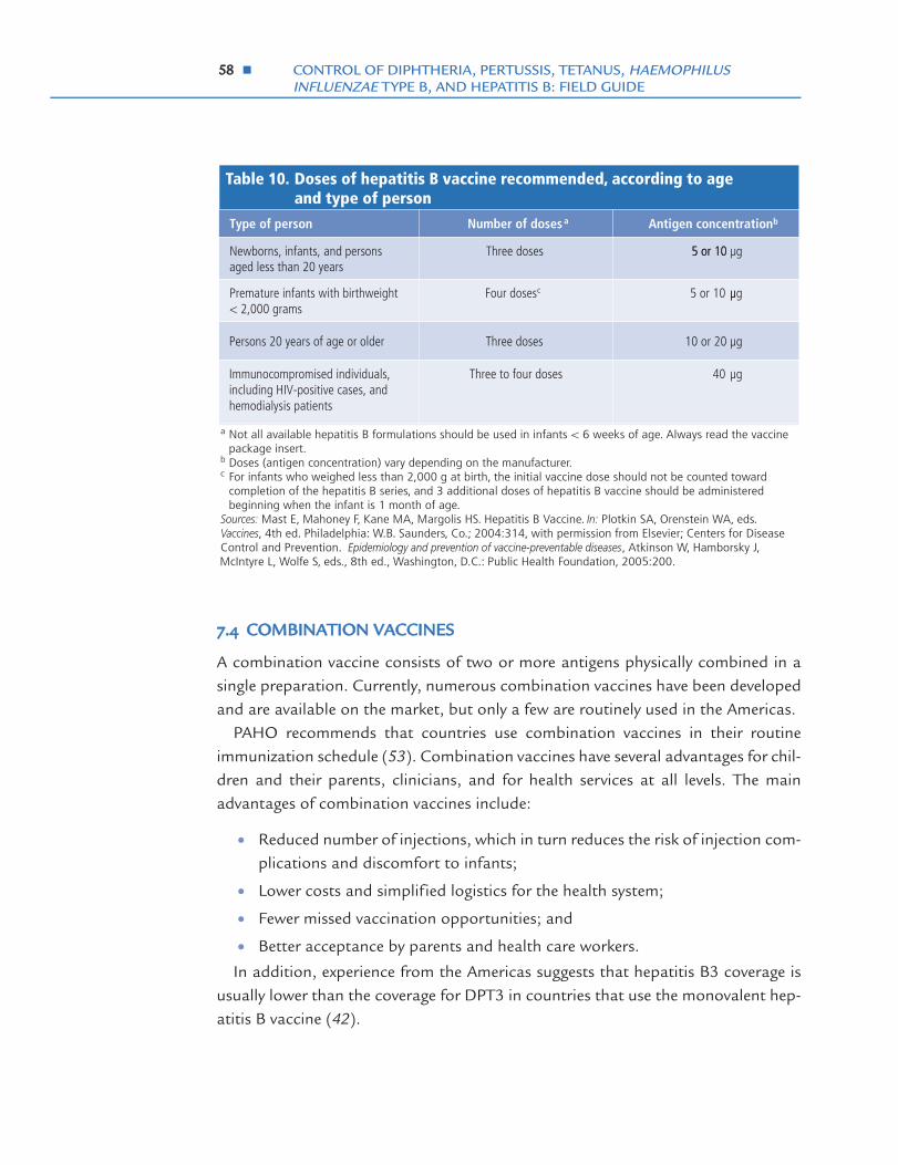
5588 CONTROL OF DIPHTHERIA, PERTUSSIS, TETANUS, HAEMOPHILUS INFLUENZAE TYPE B, AND HEPATITIS B: FIELD GUIDE
77..44 CCOOMMBBIINNAATTIIOONN VVAACCCCIINNEESS
A combination vaccine consists of two or more antigens physically combined in asingle preparation. Currently, numerous combination vaccines have been developedand are available on the market, but only a few are routinely used in the Americas.
PAHO recommends that countries use combination vaccines in their routineimmunization schedule (53). Combination vaccines have several advantages for chil-dren and their parents, clinicians, and for health services at all levels. The mainadvantages of combination vaccines include:
• Reduced number of injections, which in turn reduces the risk of injection com-plications and discomfort to infants;
• Lower costs and simplified logistics for the health system;
• Fewer missed vaccination opportunities; and
• Better acceptance by parents and health care workers.
In addition, experience from the Americas suggests that hepatitis B3 coverage isusually lower than the coverage for DPT3 in countries that use the monovalent hep-atitis B vaccine (42).
a Not all available hepatitis B formulations should be used in infants < 6 weeks of age. Always read the vaccine package insert.
b Doses (antigen concentration) vary depending on the manufacturer.c For infants who weighed less than 2,000 g at birth, the initial vaccine dose should not be counted toward
completion of the hepatitis B series, and 3 additional doses of hepatitis B vaccine should be administered beginning when the infant is 1 month of age.
Sources: Mast E, Mahoney F, Kane MA, Margolis HS. Hepatitis B Vaccine. In: Plotkin SA, Orenstein WA, eds. Vaccines, 4th ed. Philadelphia: W.B. Saunders, Co.; 2004:314, with permission from Elsevier; Centers for Disease Control and Prevention. Epidemiology and prevention of vaccine-preventable diseases, Atkinson W, Hamborsky J, McIntyre L, Wolfe S, eds., 8th ed., Washington, D.C.: Public Health Foundation, 2005:200.
40 µgThree to four dosesImmunocompromised individuals, including HIV-positive cases, and hemodialysis patients
10 or 20 µgThree dosesPersons 20 years of age or older
Four doses cPremature infants with birthweight< 2,000 grams
Three dosesNewborns, infants, and persons aged less than 20 years
Antigen concentration bNumber of doses aType of person
5 or 10 µgµ
5 or 10 µg5 or 10
Table 10. Doses of hepatitis B vaccine recommended, according to age and type of person
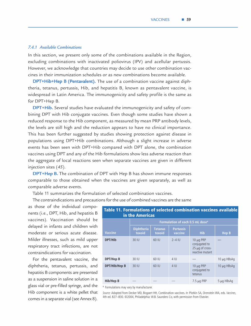
VACCINES 5599
7.4.1 Available Combinations
In this section, we present only some of the combinations available in the Region,excluding combinations with inactivated poliovirus (IPV) and acellular pertussis.However, we acknowledge that countries may decide to use other combination vac-cines in their immunization schedules or as new combinations become available.
DDPPTT++HHiibb++HHeepp BB ((PPeennttaavvaalleenntt)).. The use of a combination vaccine against diph-theria, tetanus, pertussis, Hib, and hepatitis B, known as pentavalent vaccine, iswidespread in Latin America. The immunogenicity and safety profile is the same asfor DPT+Hep B.
DPT+Hib. Several studies have evaluated the immunogenicity and safety of com-bining DPT with Hib conjugate vaccines. Even though some studies have shown areduced response to the Hib component, as measured by mean PRP antibody levels,the levels are still high and the reduction appears to have no clinical importance.This has been further suggested by studies showing protection against disease inpopulations using DPT+Hib combinations. Although a slight increase in adverseevents has been seen with DPT+Hib compared with DPT alone, the combinationvaccines using DPT and any of the Hib formulations show less adverse reaction thanthe aggregate of local reactions seen when separate vaccines are given in differentinjection sites (45).
DPT+Hep B. The combination of DPT with Hep B has shown immune responsescomparable to those obtained when the vaccines are given separately, as well ascomparable adverse events.
Table 11 summarizes the formulation of selected combination vaccines.The contraindications and precautions for the use of combined vaccines are the same
as those of the individual compo-nents (i.e., DPT, Hib, and hepatitis Bvaccines). Vaccination should bedelayed in infants and children withmoderate or serious acute disease.Milder illnesses, such as mild upperrespiratory tract infections, are notcontraindications for vaccination.
For the pentavalent vaccine, thediphtheria, tetanus, pertussis, andhepatitis B components are presentedas a suspension in saline solution in aglass vial or pre-filled syringe, and theHib component is a white pellet thatcomes in a separate vial (see Annex 8).
a Formulations may vary by manufacturer.
Source: Adapted from Decker MD, Bogaert HH, Combination vaccines. In: Plotkin SA, Orenstein WA, eds. Vaccines, 4th ed. 827–830. ©2004, Philadelphia: W.B. Saunders Co, with permission from Elsevier.
5 µg HBsAg7.5 µg PRP———Hib/Hep B
10 µg HBsAg10 µg PRP conjugated to tetanus
4 IU60 IU30 IUDPT/Hib/Hep B
10 µg HBsAg—4 IU60 IU30 IUDPT/Hep B
--10 µg PRP conjugated to 25 µg of cross-reactive mutant
2–4 IU60 IU30 IUDPT/Hib
Hep BHibPertussis vaccine
Tetanus toxoid
Diphtheria toxoid
Formulation of each 0.5 mL dosea
Vaccine
—
Table 11. Formulations of selected combination vaccines available in the Americas
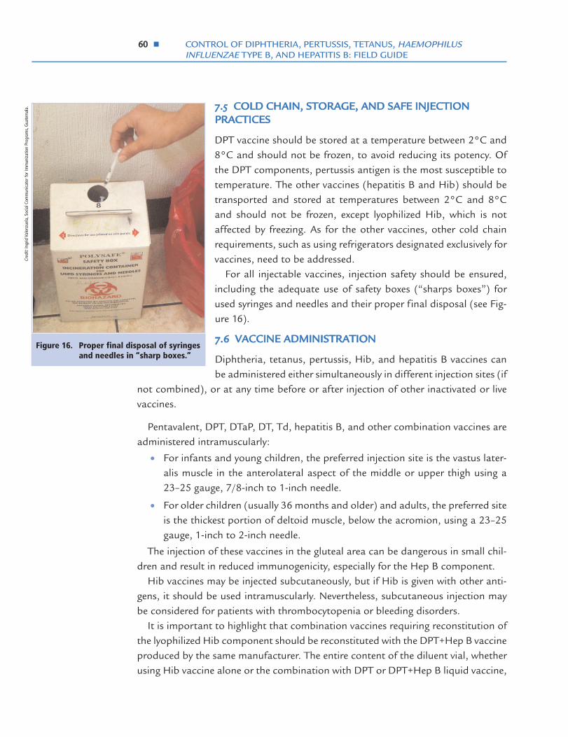
6600 CONTROL OF DIPHTHERIA, PERTUSSIS, TETANUS, HAEMOPHILUS INFLUENZAE TYPE B, AND HEPATITIS B: FIELD GUIDE
77..55 CCOOLLDD CCHHAAIINN,, SSTTOORRAAGGEE,, AANNDD SSAAFFEE IINNJJEECCTTIIOONNPPRRAACCTTIICCEESS
DPT vaccine should be stored at a temperature between 2°C and8°C and should not be frozen, to avoid reducing its potency. Ofthe DPT components, pertussis antigen is the most susceptible totemperature. The other vaccines (hepatitis B and Hib) should betransported and stored at temperatures between 2°C and 8°Cand should not be frozen, except lyophilized Hib, which is notaffected by freezing. As for the other vaccines, other cold chainrequirements, such as using refrigerators designated exclusively forvaccines, need to be addressed.
For all injectable vaccines, injection safety should be ensured,including the adequate use of safety boxes (“sharps boxes”) forused syringes and needles and their proper final disposal (see Fig-ure 16).
77..66 VVAACCCCIINNEE AADDMMIINNIISSTTRRAATTIIOONN
Diphtheria, tetanus, pertussis, Hib, and hepatitis B vaccines canbe administered either simultaneously in different injection sites (if
not combined), or at any time before or after injection of other inactivated or livevaccines.
Pentavalent, DPT, DTaP, DT, Td, hepatitis B, and other combination vaccines areadministered intramuscularly:
• For infants and young children, the preferred injection site is the vastus later-alis muscle in the anterolateral aspect of the middle or upper thigh using a23–25 gauge, 7/8-inch to 1-inch needle.
• For older children (usually 36 months and older) and adults, the preferred siteis the thickest portion of deltoid muscle, below the acromion, using a 23–25gauge, 1-inch to 2-inch needle.
The injection of these vaccines in the gluteal area can be dangerous in small chil-dren and result in reduced immunogenicity, especially for the Hep B component.
Hib vaccines may be injected subcutaneously, but if Hib is given with other anti-gens, it should be used intramuscularly. Nevertheless, subcutaneous injection maybe considered for patients with thrombocytopenia or bleeding disorders.
It is important to highlight that combination vaccines requiring reconstitution ofthe lyophilized Hib component should be reconstituted with the DPT+Hep B vaccineproduced by the same manufacturer. The entire content of the diluent vial, whetherusing Hib vaccine alone or the combination with DPT or DPT+Hep B liquid vaccine,
Figure 16. Proper final disposal of syringes and needles in “sharp boxes.”
Cred
it: In
grid
Val
enzu
ela,
Soci
al C
omm
unic
ator
for I
mm
uniz
atio
n Pr
ogra
ms,
Gua
tem
ala.

VACCINES 6611
should be added to the vial containing the lyophilized Hib vaccine; the vial mustthen be shaken until the pellet is completely dissolved (see Annex 8).
When using multidose vials, each dose should be withdrawn with a sterile needleand under strict aseptic conditions to avoid contaminating the vial.
77..77 FFIINNAALL CCOONNSSIIDDEERRAATTIIOONNSS
When planning to introduce Hib and/or hepatitis B vaccine into an immunizationprogram, it is important to consider the vaccine formulation (combination vs.monovalent) and presentation (1- vs. 10-dose vials). The formulation and presenta-tion selected will help determine the expense, wastage percentage, cold chain space,need for reconstitution, and training activities for health care workers.
Sustaining vaccination activities against diphtheria, pertussis, tetanus, Hib, andhepatitis B, and striving to reach vaccination coverage levels of 95% or more in eachmunicipality, are critical for keeping these diseases under control in the Americas.

6622 CONTROL OF DIPHTHERIA, PERTUSSIS, TETANUS, HAEMOPHILUS INFLUENZAE TYPE B, AND HEPATITIS B: FIELD GUIDE
REFERENCES
1. Wharton M, Vitek CR. Diphtheria toxoid. In: Plotkin SA, Orenstein WA, eds.Vaccines, 4th ed. Philadelphia: W.B. Saunders Co.; 2004:211–228.
2. Pan American Health Organization. Diphtheria incidence and coverage in theAmericas, 1978–2000. EPI Newsletter 2001;23(6):8.
3. Ohuabunwo CJ, Bisgard KM, Popovic T, Wharton M. Diphtheria. In: WhartonM, Hughes H, Reilly M, eds. Manual for the surveillance of vaccine-preventable dis-eases, 3rd ed. Atlanta: Centers for Disease Control and Prevention; 2002:1–10.Accessed on 10 August 2004 at http://www.cdc.gov/nip/publications/surv-manual/default.htm [online publication].
4. Vitek CR, Wharton M. Diphtheria in the former Soviet Union: reemergence ofa pandemic disease. Emerg Infect Dis 1998;4(4):539–550.
5. Centers for Disease Control and Prevention. Diptheria. In: Atkinson W, Ham-borsky J, McIntyre L, Wolfe S, eds. Epidemiology and prevention of vaccine-preventa-ble diseases, 8th ed. Washington, D.C.: Public Health Foundation; 2005;55–64.
6. Pan American Health Organization. Diphtheria outbreak in Paraguay. EPINewsletter 2002;24(3):6.
7. Ropero AM, Oliva O, Castillo-Solórzano C, Dietz V, Izurieta H, Carraso P, et al.Recent outbreaks of diphtheria in the Americas [abstract]. In: Technical Advi-sory Group on Vaccine-preventable Diseases, XV Meeting, 22–23 November2002, Washington, D.C. (Washington, D.C.: Pan American Health Organiza-tion; 2002).
8. Heymann DL, ed. Diphtheria. In: Control of communicable diseases manual, 18thed. Washington, D.C.: American Public Health Association; 2004:171–175.
9. American Academy of Pediatrics. Diphtheria. In: Pickering LK, ed. Red book:2003 report of the Committee on Infectious Diseases, 26th ed. Elk Grove Village, IL:American Academy of Pediatrics; 2003:263–266.
10. Pan American Health Organization. Final report (conclusions and recommenda-tions). Municipalities: improving immunization services. XV Meeting of the TechnicalAdvisory Group (TAG) on Vaccine-preventable Diseases. Washington, D.C.:PAHO; 2002.
11. World Health Organization. Pertussis vaccines. Wkly Epidemiol Rec 1999;74(18):137–143.
12. World Health Organization. Pertussis vaccines. Wkly Epidemiol Rec 2005;80(4):31–39.
13. Edwards KM, Decker MD. Pertussis vaccine. In: Plotkin SA, Orenstein WA, eds.Vaccines, 4th ed. Philadelphia: W.B. Saunders Co.; 2004:471–528.
14. Centers for Disease Control and Prevention. Pertussis. In: Atkinson W, Ham-borsky J, McIntyre L, Wolfe S, eds. Epidemiology and prevention of vaccine-preventa-ble diseases, 8th ed. Washington, D.C.: Public Health Foundation; 2005:75–88.

REFERENCES 6633
15. Heymann DL, ed. Pertussis and parapertussis. In: Control of communicable dis-eases manual, 18th ed. Washington, D.C.: American Public Health Association;2004:399–404.
16. American Academy of Pediatrics. Pertussis. In: Pickering LK, ed. Red book: 2003report of the Committee on Infectious Diseases, 26th ed. Elk Grove Village, IL: Amer-ican Academy of Pediatrics; 2003:472–486.
17. Bisgard K, Pascual FB, Tiwari T, Murphy TV. Pertussis. In: Wharton M, HughesH, Reilly M, eds. Manual for the surveillance of vaccine-preventable diseases, 3rd ed.Atlanta: Centers for Disease Control and Prevention; 2002. Accessed on 27August 2004 at http://www.cdc.gov/nip/publications/surv-manual/default.htm [online publication].
18. Cherry JD, Heininger U. Pertussis and other Bordetella infections. In: Feigin RD,Cherry J, Demmler GJ, Kaplan S, eds. Textbook of pediatric infectious diseases, 5thed. Philadelphia: W.B. Saunders Co.; 2004:1588–1608.
19. Centers for Disease Control and Prevention. Guidelines for the control of pertussisoutbreaks. Atlanta, GA: CDC; 2000.
20. Heymann DL, ed. Tetanus. In: Control of communicable diseases manual, 18th ed.Washington, D.C.: American Public Health Association; 2004:528–533.
21. Centers for Disease Control and Prevention. Tetanus. In: Atkinson W, Ham-borsky J, McIntyre L, Wolfe S, eds. Epidemiology and prevention of vaccine-preventa-ble diseases, 8th ed. Washington, D.C.: Public Health Foundation; 2005:65–73.
22. Pan American Health Organization. Neonatal tetanus elimination field guide. Scien-tific and Technical Publication No. 602. Washington, D.C.: PAHO; 2005.
23. Wassalik GF, Murphy TV, Roper MH, Orenstein WA. Tetanus toxoid. In:Plotkin SA, Orenstein WA, eds. Vaccines, 4th ed. Philadelphia: W.B. SaundersCo., 2004:745–781.
24. American Academy of Pediatrics. Tetanus (lockjaw). In: Pickering LK, ed. Redbook: 2003 report of the Committee on Infectious Diseases, 26th ed. Elk Grove Village,IL: American Academy of Pediatrics; 2003:611–616.
25. World Health Organization. Global Programme for Vaccines and Immuniza-tion (GPV)—The WHO position paper on Haemophilus influenzae type b conju-gate vaccines. Wkly Epidemiol Rec 1998;73(10):64–68.
26. Centers for Disease Control and Prevention. Haemophilus influenzae type b. In:Atkinson W, Hamborsky J, McIntyre L, Wolfe S, eds. Epidemiology and preventionof vaccine-preventable diseases, 8th ed. Washington, D.C.: Public Health Founda-tion, 2005:101–113.
27. Heymann DL, ed. Meningitis. In: Control of communicable diseases manual, 18th ed.Washington, D.C.: American Public Health Association; 2004:366–369.
28. Wenger JD, Ward JI. Haemophilus influenzae vaccine. In: Plotkin SA, OrensteinWA, eds. Vaccines, 4th ed. Philadelphia: W.B. Saunders Co.; 2004:229–268.
29. Pan American Health Organization. Impact of Uruguay’s introduction of theHaemophilus influenzae type b (Hib) vaccine. EPI Newsletter 1996;18:6.

6644 CONTROL OF DIPHTHERIA, PERTUSSIS, TETANUS, HAEMOPHILUS INFLUENZAE TYPE B, AND HEPATITIS B: FIELD GUIDE
30. Ward JI. Haemophilus influenzae. In: Feigin RD, Cherry J, Demmler GJ, Kaplan S,eds. Textbook of pediatric infectious diseases, 5th ed. Philadelphia: W.B. SaundersCo.; 2003:1636–1655.
31. Brammer L, Fukuda K, Klimov A, Cox N. Haemophilus influenzae type b invasivedisease. In: Wharton M, Hughes H, Reilly M, eds. Manual for the surveillance ofvaccine-preventable diseases, 3rd ed. Atlanta: Centers for Disease Control and Pre-vention; 2002. Accessed on 27 August 2004 at http://www.cdc.gov/nip/publi-cations/surv-manual/default.htm [online publication].
32. American Academy of Pediatrics. Haemophilus influenzae infections. In: PickeringLK, ed. Red book: 2003 report of the Committee on Infectious Diseases, 26th ed. ElkGrove Village, IL: American Academy of Pediatrics; 2003:293–301.
33. World Health Organization (WHO). Hepatitis B vaccines. Wkly Epidemiol Rec2004;79(28):255–263.
34. Centers for Disease Control and Prevention. Hepatitis B. In: Atkinson W, Ham-borsky J, McIntyre L, Wolfe S, eds. Epidemiology and prevention of vaccine-preventa-ble diseases, 8th ed. Washington, D.C.: Public Health Foundation; 2005:191–212.
35. Mast E, Mahoney F, Kane M, Margolis H. Hepatitis B. In: Plotkin SA, OrensteinWA, eds. Vaccines, 4th ed. Philadelphia: W.B. Saunders Co.; 2004:299–328.
36. World Health Organization (WHO). Hepatitis B vaccine [Internet site]. Accessedon 7 December 2004 at http://www.who.int/vaccines/en/hepatitisb.shtml.
37. Pan American Health Organization. Health in the Americas. Scientific and Tech-nical Publication No. 587. Washington, D.C.: PAHO; 2002:318.
38. Heymann DL, ed. Control of communicable diseases manual, 18th ed. Washington,D.C.: American Public Health Association; 2004:253–261.
39. American Academy of Pediatrics. Hepatitis B. In: Pickering LK, ed. Red book:2003 report of the Committee on Infectious Diseases, 26th ed. Elk Grove Village, IL:American Academy of Pediatrics; 2003:318–336.
40. Pan American Health Organization. Technical Advisory Group on Vaccine-pre-ventable Diseases. Final report XIV Meeting, 2–5 October 2000, Foz do Iguaçu, Brazil.Washington, D.C.: PAHO; 2000.
41. Pan American Health Organization. Final report (conclusions and recommenda-tions). Municipalities: improving immunization services. XV Meeting of the TechnicalAdvisory Group (TAG) on Vaccine-preventable Diseases. Washington, D.C.:PAHO;2002.
42. Ropero AM, Danovaro-Holliday C, Andrus JK . Progress in vaccination againsthepatitis B in the Americas. J Clin Virol [in press] 2005.
43. World Health Organization, Department of Vaccines and Biologicals. WHO-recommended standards for surveillance of selected vaccine-preventable diseases. Geneva:WHO; 2003. (WHO/V&B/03.01.)

REFERENCES 6655
44. Mulholland K, Hilton S, Adegbola R, Usen S, Oparaugo A, Omosigho C, et al.Randomised trial of Haemophilus influenzae type-b tetanus protein conjugatevaccine [corrected] for prevention of pneumonia and meningitis in Gambianinfants. Lancet 1997;349(9060):1191–1197. (Erratum in: Lancet 1997; 350(9076):524.
45. Decker MD, Bogaert HH. Combination vaccines. In: Plotkin SA, Orenstein WA,eds. Vaccines, 4th ed. Philadelphia: W.B. Saunders Co.; 2004:825–854.
46. Pan American Health Organization. Final report (conclusions and recommenda-tions). XII Meeting of the Technical Advisory Group (TAG) on Vaccine-prevent-able Diseases. Washington, D.C.: PAHO; 1997.
47. World Health Organization, Department of Vaccines and Biologicals. Supple-mentary information on vaccine safety. Part 2: Background rates of adverse events follow-ing immunization. Geneva: WHO; 2000. (WHO/V&B/00.36; 17–25.)
48. Pan American Health Organization. Immunization safety. How to address eventsallegedly attributable to vaccination or immunization. Washington, D.C.: PAHO;2002.
49. World Health Organization. Haemophilus influenzae type b vaccine [Internetsite]. Accessed on 21 March 2005 at http://www.who.int/vaccines/en/haeflub.shtml.
50. Jilg W, Schmidt M, Dienhardt F. Vaccination against hepatitis B: comparisonof three different vaccination schedules. J Infect Dis 1989;160:766–769.
51. Hadler SC, Monzon MA, Lugo DR, Perez M. Effect of timing of hepatitis B vac-cine dose on response to vaccine in Yucpa Indians. Vaccine 1989;7:106–110.
52. Centers for Disease Control and Prevention. Hepatitis B virus: a comprehensivestrategy for eliminating transmission in the United States through universalchildhood vaccination: recommendations of the Immunization Practices Advi-sory Committee (ACIP). MMWR 1995;40(RR13):1–19.
53. Pan American Health Organization. Final report (conclusions and recommenda-tions). Immunization: pan-Americanism and equity in action. XIII Meeting of the Tech-nical Advisory Group (TAG) on Vaccine-preventable Diseases. Washington,D.C.: PAHO; 1999.
BIBLIOGRAPHY
DDiipphhtthheerriiaa aanndd DDiipphhtthheerriiaa VVaacccciinneess
Centers for Disease Control and Prevention. Diphtheria epidemic—New independ-ent states of the former Soviet Union, 1990–1994. MMWR 1995;44(10):177–181.
Cody CL, Baraff LJ, Cherry JD, Marcy SM, Manclark CR. Nature and rates of adversereactions associated with DPT and DT immunizations in infants and children.Pediatrics 1981;68(5):650–660.
Farizo KM, Strebel PM, Chen RT, Kimbler A, Cleary TJ, Cochi SL. Fatal respiratorydisease due to Corynebacterium diphtheriae: case report and review of guidelines formanagement, investigation, and control. Clin Infect Dis 1993;16(1):59–68.
Galazka A. The changing epidemiology of diphtheria in the vaccine era. J Infect Dis2000;181(Suppl 1):S2–9.
Landazabal Garcia N, Burgos Rodriguez MM, Pastor D. Diphtheria outbreak in Cali,Colombia, August–October 2000. Epidemiol Bull 2001;22(3):13–15.
Pan American Health Organization. Diphtheria epidemic in Ecuador. EPI Newsletter1994;16(5):5–6, 8.
Ropero AM, Oliva O, Castillo-Solórzano C, Dietz V, Izurieta H, Carraso P, et al.Recent outbreaks of diphtheria in the Americas [abstract]. In: Technical Adviso-ry Group on Vaccine-preventable Diseases, XV Meeting, 22–23 November 2002,Washington, D.C. Washington, D.C.: Pan American Health Organization; 2002.
World Health Organization, Department of Vaccines and Biologicals. Supplementaryinformation on vaccine safety. Part 2: Background rates of adverse events following immuniza-tion. Geneva: WHO; 2000. (WHO/V&B/00.36; 17–25.)
World Health Organization. Immunization, Vaccines and Biologicals—Diphtheriatoxoid. Accessed on 2 August 2004 at http://www.who.int/vaccines/en/dipthe-ria.shtml [Web page].
PPeerrttuussssiiss aanndd PPeerrttuussssiiss VVaacccciinneess
Centers for Disease Control and Prevention. Guidelines for the control of pertussis out-breaks. Atlanta, GA: CDC; 2000.
Duclos P, Olivé JM. Invited commentary: pertussis, a forgotten killer. Am J Epidemiol2002;155(10):897–898.
Morley, D. Pediatric priorities in the developing world. The English Language Book Societyand Butterworth. London, England: Butterworth & Co, Ltd; 1980:237.
Pan American Health Organization. Immunization safety. How to address eventsallegedly attributable to vaccination or immunization. Washington, D.C.: PAHO;2002.
World Health Organization. Immunization, Vaccines and Biologicals. 2004. Pertus-sis. Accessed on 5 August 2004 at http://www.who.int/vaccines/en/pertussis.shtml [Web page].
6666

BIBLIOGRAPHY 6677
TTeettaannuuss aanndd TTeettaannuuss VVaacccciinneess
Cherry JD, Harrison RE. Tetanus. In: Feigin RD, Cherry J, Demmler GJ, Kaplan S, eds.Textbook of pediatric infectious diseases, 5th ed. Philadelphia: W.B. Saunders Co.;2004:1766–1775.
Galazaka A. Immunization of pregnant women against tetanus. Geneva: World HealthOrganization; 1983. (WHO/EPI/GEN/83/5).
Galazka A. Stability of vaccines. Geneva: World Health Organization; 1989. (WHO/EPI/GEN/89.8).
Gershon A, Hotez PJ, Katz S. Krugman’s infectious diseases of children, 11th ed. St. Louis:C.V. Mosby; 2004.
Jawetz E, Melnik JL, Adelberg EA. Review of medical microbiology, 18th ed. Los Altos,CA: Lange Medical Publications; 1989.
Nagachinta T, Cortese MM, Roper MH, Pascual FB, Murphy T. Tetanus. In: WhartonM, Hughes H, Reilly M, eds. Manual for the surveillance of vaccine-preventable diseases,3rd ed. Atlanta: Centers for Disease Control and Prevention; 2002. Accessed on27 August 2004 at http://www.cdc.gov/nip/publications/surv-manual/default.htm [online publication].
Pan American Health Organization. Neonatal tetanus control, defining high riskareas: the experience in the Americas. EPI Newsletter 1991;12(4):3.
Shulman ST, Phair JP, Peterson LR, Warren JR. The biologic and clinical basis of infectiousdiseases, 5th ed. Philadelphia: W.B. Saunders Co.; 1997.
Strassburg MA. Guidelines for the investigation and control of outbreaks of EPI target diseases.Geneva: World Health Organization; 1984. (EPI/GEN/84/7).
Vera Martínez A, Ramírez Boettner CM, Salinas VM, Zárate R. Tétanos: estudio clin-ico y epidemiológico de 2,337 casos. Bol Oficina Sanit Panam 1976;80(4):323–332.
World Health Organization and United Nations Children’s Fund. Planning principlesfor accelerated immunization activities: a joint WHO/UNICEF statement. Geneva: WHO;1985.
World Health Organization, Expanded Programme on Immunization. Prevention ofneonatal tetanus through immunization. Geneva: WHO; 1986. (WHO/EPI/GEN/86/9Rev 1).
World Health Organization, Expanded Programme on Immunization. Disease surveil-lance training for mid-level managers. Geneva: WHO; 1995.
World Health Organization. Replacing tetanus toxoid (TT) and diphtheria-tetanus toxoid(DT) with tetanus-diphtheria toxoid (Td). Geneva: WHO; 1998. (GPV-CVI/SAGE.98/WP.04.)
World Health Organization. Guía practica para la eliminación del tétanos neonatal.Cuaderno Técnico No. 35. Geneva: WHO; 1999. (WHO/V&B/99.14.)
6688 CONTROL OF DIPHTHERIA, PERTUSSIS, TETANUS, HAEMOPHILUS INFLUENZAE TYPE B, AND HEPATITIS B: FIELD GUIDE
HHaaeemmoopphhiilluuss iinnfflluueennzzaaee ttyyppee bb ((HHiibb)) aanndd HHiibb VVaacccciinneess
de Andrade AL, Brandileone MC, Di Fabio JL, Oliveira RM, Silva SA, Biaocchi SS.Haemophilus influenzae resistance in Latin America: systematic review of surveil-lance data. Microb Drug Resist 2001;7(4):403–411.
Di Fabio JL, Castillo-Solorzano C, Oliva O, et al. Monitoring diseases caused byHaemophilus influenzae type b after the introduction of the vaccine. Technical Advi-sory Group on Vaccine-preventable Diseases, XV meeting, 22–23 November2002, Washington, D.C.: Pan American Health Organization; 2002:55.
Di Fabio JL, de Quadros C. Considerations for combination vaccine developmentand use in the developing world. Clin Infect Dis 2001;33(Suppl 4):S340–345.
Lagos R, Horwitz I, Toro J, San Martin O, Abrego P, Bustamante C, et al. Large scale,postlicensure, selective vaccination of Chilean infants with PRP-T conjugate vac-cine: practicality and effectiveness in preventing invasive Haemophilus influenzaetype b infections. Pediatr Infect Dis J 1996;15(3):216–222.
Levine OS, Lagos R, Munoz A, Villaroel J, Alvarez AM, Abrego P, et al. Defining theburden of pneumonia in children preventable by vaccine against Haemophilusinfluenzae type b. Pediatr Infect Dis J 1999;18(12):1060–1064.
Pan American Health Organization. Impact of Uruguay’s introduction of theHaemophilus influenzae type b (Hib) vaccine. EPI Newsletter 1996;18:6.
Pan American Health Organization, Division of Vaccines and Immunization, VaccineTechnology Access Program. First workshop on standardization of chest X-rayinterpretation. Santiago, Chile, 2000. Washington, D.C. (Unpublished docu-ment PAHO/HVP/HVA/2000-000010.)
Peltola H. Haemophilus influenzae type b disease and vaccination in Latin America andthe Caribbean. Pediatr Infect Dis J 1997;16(8):780–787.
World Health Organization. Estimating the local burden of Haemophilus influenzaetype b (Hib) disease preventable by vaccination: A rapid assessment tool. Gene-va: WHO; 2001. (WHO/V&B/01.27.)
World Health Organization, Global Programme for Vaccines and Immunization. TheWHO position paper on Haemophilus influenzae type b conjugate vaccines. WklyEpidemiol Rec 1998;73(10):64–68.
World Health Organization. Haemophilus influenzae type b vaccine [Internet site].Available at: http://www.who.int/vaccines/en/haeflub.shtml. Accessed on 21March 2005.
World Health Organization. Standardization of interpretation of chest radiographs for thediagnosis of pneumonia in children. World Health Organization Pneumonia VaccineTrials Investigators’ Group. Geneva: WHO; 2001. (WHO/V&B/01.35.)
World Health Organization. The management of acute respiratory infection in children: prac-tical guidelines for outpatient care. Geneva: WHO; 1995.

BIBLIOGRAPHY 6699
World Health Organization. WHO-recommended standards for surveillance of selected vac-cine-preventable diseases. Geneva: WHO; 2003:4–9. (WHO/V&B/03.01.)
World Health Organization. WHO-recommended surveillance. Acute lower respiratory tractinfections (aLRTI) and pneumonia. Geneva: WHO; 1999:131–133. (WHO/CDS/CSR/ISR/99.2.)
HHeeppaattiittiiss BB aanndd HHeeppaattiittiiss BB VVaacccciinneess
American Academy of Pediatrics. Hepatitis and youth in corrections settings. In:Pickering LK, ed. Red book: 2003 report of the Committee on Infectious Diseases, 26thed. Elk Grove Village, IL: American Academy of Pediatrics; 2003:167–173.
Brazil. Ministério de Saúde. Fundação Nacional de Saúde. Centro Nacional de Epi-demiologia. Guia de vigilância epidemiológica, 4th ed. Rev. ampl. Brasília: FundaçãoNacional de Saúde; 1998.
Centers for Disease Control and Prevention. Guidelines for vaccinating pregnant women.Atlanta, GA: CDC; 2004. Accessed on 28 August 2004 athttp://www.cdc.gov/nip/publications/preg_guide.pdf [online publication].
Demmler GJ. Viral hepatitis. In: Feigin RD, Cherry J, Demmler GJ, Kaplan S, eds. Text-book of pediatric infectious diseases, 5th ed. Philadelphia: W.B. Saunders Co.;2003:658–667.
Fay OH. Hepatitis B in Latin America: epidemiological patterns and eradicationstrategy. The Latin American Regional Study Group. Vaccine 1990;(Suppl 8):S100–6; discussion S134–S139.
Fineli L, Alter M. Hepatitis B. In: Wharton M, Hughes H, Reilly M, eds. Manual for thesurveillance of vaccine-preventable diseases, 3rd ed. Atlanta: Centers for Disease Con-trol and Prevention; 2002. Accessed on 27 August 2004 athttp://www.cdc.gov/nip/publications/surv-manual/default.htm [online publica-tion].
Hadler SC, Monzon MA, Lugo DR, Perez M. Effect of timing of hepatitis B vaccinedose on response to vaccine in Yucpa Indians. Vaccine 1989;7:106–110.
León P, Venegas E, Bengoechea L, Rojas E, Lopez JA, Elola C, Echevarria JM. Preva-lence of infections by hepatitis B, C, D and E viruses in Bolivia. Rev Panam SaludPublica 1999;5(3):144–151.
Millinship S. Hepatitis B virology and immunology [Internet site]. Available at:http://www.hon.ch/Library/Theme/HepB/virology.html. Accessed on 24 Decem-ber 2004.
Pan American Health Organization. Technical Advisory Group on Vaccine-preventa-ble Diseases—Conclusions and Recommendations, XIII Meeting, 12–16 April1998, Ottawa, Canada. Washington, D.C.: PAHO; 1998.
Silveira TR, da Fonseca JC, Rivera L, Fay OH, Tapia R, Santos JI, Urdeneta E,Clemens SA. Hepatitis B seroprevalence in Latin America. Rev Panam Salud Publica1999;6(6):378–383.

7700 CONTROL OF DIPHTHERIA, PERTUSSIS, TETANUS, HAEMOPHILUS INFLUENZAE TYPE B, AND HEPATITIS B: FIELD GUIDE
Torres JR. Hepatitis B and hepatitis delta virus infection in South America. Gut1996;38(Suppl 2):S48–55.
World Health Organization. Introduction of hepatitis B vaccine into childhood immunizationservices: management guidelines, including information for health workers and parents.Geneva: WHO; 2001. (WHO/V&B/01.31.)
World Health Organization. Hepatitis B vaccine [Internet site]. Available at:http://www.who.int/vaccines/en/hepatitisb.shtml. Accessed on 7 December2004.
World Health Organization. Hepatitis B vaccines. Wkly Epidemiol Rec 2004; 79(28):255–263.
CCoommbbiinnaattiioonn VVaacccciinneess
GlaxoSmithKline Biologicals. Learning about Tritanrix vaccines. Rixensart, Belgium:GSK Biologicals; 2001. Accessed on 28 August 2004 at http://www.who.int/vac-cines-diseases/epitraining/TRITANRIX%20LearnAbout.pdf [online document].
Wood S, Steinfeld R, Wittet S. Immunizing children against Haemophilus influenzae type B(Hib). A training module for vaccinators. Seattle: Program for Appropriate Technolo-gy in Health; 2003. Accessed on 5 August 2004 at http://www.path.org/files/CVP-hib_training_module.pdf [online document].

ANNEXES
ANNEX 1. SUMMARY OF THE EPIDEMIOLOGICAL CHARACTERISTICS OF DIPHTHERIA, PERTUSSIS, TETANUS, HAEMOPHILUS INFLUENZAE TYPE B, AND HEPATITIS B
ANNEX 2. EXAMPLE OF DIPHTHERIA NOTIFICATION AND INVESTIGATION FORM
ANNEX 3. EXAMPLE OF A FORM FOR THE MANAGEMENT OF CONTACTS
ANNEX 4. EXAMPLE OF PERTUSSIS-LIKE ILLNESS NOTIFICATION AND INVESTIGATION
FORM
ANNEX 5. EXAMPLE OF TETANUS NOTIFICATION AND INVESTIGATION FORM
ANNEX 6. EXAMPLE OF HAEMOPHILUS INFLUENZAE TYPE B MENINGITIS
NOTIFICATION AND INVESTIGATION FORM
ANNEX 7. EXAMPLE OF HEPATITIS B NOTIFICATION AND INVESTIGATION FORM
ANNEX 8. HOW TO RECONSTITUTE AND ADMINISTER LYOPHILIZED DPT+HIB+HEPATITIS B (PENTAVALENT) VACCINE


ANNEX 1 7733
AN
NE
X 1
.Su
mm
ary
of t
he e
pide
mio
logi
cal c
hara
cter
isti
cs o
f dip
hthe
ria,
pert
ussi
s, t
etan
us, H
aem
ophi
lus
infl
uenz
ae t
ype
b, a
nd h
epat
itis
B
-Lo
ng-l
asti
ng
prot
ectio
n th
roug
h di
seas
e or
vac
cina
tion
-So
me
prot
ecti
on fr
om
mat
erna
l ant
ibod
ies
and
brea
st-f
eedi
ng u
ntil
2 m
onth
s of
age
-Fo
llow
ing
Hib
infe
ctio
n an
d po
ssib
ly s
ome
cros
s-re
actio
ns
to o
ther
org
anis
ms
-Fo
llow
ing
vacc
inat
ion
-D
oes
not
nece
ssar
ily r
esul
t fr
om d
isea
se (i
mm
uniz
atio
n is
indi
cate
d fo
r ca
ses)
-Fo
r at
leas
t 10
yea
rs
follo
win
g im
mun
izat
ion
with
te
tanu
s to
xoid
-In
fant
s of
act
ivel
y im
mun
ized
mot
hers
-M
ater
nal p
rote
ctio
n of
in
fant
s ha
s no
t be
en
dem
onst
rate
d -
Vac
cine
-indu
ced
imm
unity
may
wan
e
-So
me
prot
ectio
n fr
om m
ater
nal
antib
odie
s in
infa
nts
< s
ix m
onth
s -
Follo
win
g di
seas
e or
as
ympt
omat
ic in
fect
ion
(at
leas
t pa
rtia
l)-
Long
-last
ing
prot
ectio
n af
ter
thre
e do
ses
of t
oxoi
d(n
o pr
otec
tion
agai
nst
colo
niza
tion)
Imm
unity
All
pers
ons
who
are
H
BsA
gpo
sitiv
e. F
rom
w
eeks
bef
ore
onse
t th
roug
h th
e ac
ute
phas
e. T
he in
fect
ivity
of
chr
onic
ally
infe
cted
pe
rson
s va
ries
Whi
le t
he b
acte
ria r
emai
ns in
th
e re
spira
tory
tra
ct. A
ntib
ioti
c tr
eatm
ent
redu
ces
the
perio
d of
com
mun
icab
ility
Not
con
tagi
ous
as it
is n
ot
tran
smitt
ed fr
om p
erso
n-to
-pe
rson
Pert
ussi
sca
ses
are
mos
t in
fect
ious
dur
ing
the
cata
rrha
l per
iod
and
the
first
tw
o w
eeks
aft
er c
ough
on
set;
som
e in
divi
dual
s m
ay b
e in
fect
ious
for
a lo
nger
per
iod.
Ant
ibio
tic
trea
tmen
t re
duce
s th
e pe
riod
of c
omm
unic
abili
ty
Tran
smis
sion
can
occ
ur a
s lo
ng a
s th
e to
xige
nic
bact
eria
are
pre
sent
in
dis
char
ges
and
lesi
ons
(nor
mal
ly ≤
two
wee
ks a
nd s
eldo
m
mor
e th
an fo
ur w
eeks
). A
ntib
ioti
c th
erap
y te
rmin
ates
she
ddin
g.
Rare
chr
onic
car
riers
she
d th
e ba
cilli
for
six
mon
ths
or m
ore
Com
mun
icab
ility
60–9
0 da
ys(r
ange
: six
wee
ks t
o si
x m
onth
s)
Unk
now
n du
e to
hig
h ra
te o
f as
ympt
omat
ic c
arria
ge
(pro
babl
y tw
o to
four
day
s)
Eigh
t da
ys
(ran
ge: 3
–21
days
)7–
20 d
ays
(ran
ge: 4
–21
days
)Tw
o to
five
day
s(r
ange
: 1–1
0 da
ys)
Incu
batio
n pe
riod
Thro
ugh
body
flui
ds:
bloo
d, s
aliv
a, s
emen
, et
c.Se
xual
con
tact
, ho
useh
old
cont
act,
pe
rinat
alex
posu
re,
intr
aven
ous
drug
use
, an
d no
soco
mia
lex
posu
re
Pers
on-t
o-pe
rson
from
the
re
spira
tory
tra
ct v
ia
aero
soliz
ed d
ropl
ets
Spor
es c
onta
ined
in s
oil a
nd
fece
s ar
e us
ually
intr
oduc
ed
to t
he b
ody
thro
ugh
wou
nds
and
less
freq
uent
ly a
fter
su
rgic
al p
roce
dure
s,
incl
udin
g ci
rcum
cisi
on
Pers
on-t
o-pe
rson
via
ae
roso
lized
dro
plet
s pr
oduc
ed fr
om a
cou
gh o
r sn
eeze
or
by d
irect
con
tact
w
ith s
ecre
tions
from
the
re
spira
tory
tra
ct o
f in
fect
ious
indi
vidu
als
Pers
on-t
o-pe
rson
from
the
re
spira
tory
tra
ct o
f a c
ase
or fr
om
tran
sien
t ca
rrie
rs. R
arel
y fr
om
fom
ites
or s
kin
lesi
ons
Tran
smis
sion
Wor
ldw
ide
with
few
se
ason
al v
aria
tions
Wor
ldw
ide:
Mai
nly
affe
cts
child
ren
aged
2 t
o 3
year
sW
orld
wid
eW
orld
wid
e. M
ost
com
mon
in
you
ng c
hild
ren
Wor
ldw
ide.
Mos
t fr
eque
nt in
co
lder
mon
ths
in t
empe
rate
zon
esO
ccur
renc
e
Hum
ans
Hum
ans
Inte
stin
es o
f hor
ses
and
othe
r an
imal
s, in
clud
ing
hum
ans
Soil
or fo
mite
sco
ntam
inat
ed
with
fece
s co
ntai
ning
the
sp
ores
of
C. te
tani
Hum
ans
Hum
an c
arrie
rs a
re t
he r
eser
voir
for
C. d
ipht
heria
eRe
serv
oir
Hep
atiti
s B
viru
sD
NA
viru
s of
the
Hep
adna
virid
aefa
mily
Haem
ophi
lus i
nflu
enza
e ty
pe b
Clos
tridi
umte
tani
Bord
etel
lape
rtuss
isTo
xige
nic
stra
ins
of
Cory
neba
cter
ium
diph
ther
iae
Infe
ctio
us a
gent
--
- - -
- - -
- --
- - --
--
––
––
–
--
--
--
cont
amin
ated
Dip
hthe
ria
Pert
ussi
sTe
tanu
sH
ibH
epat
itis
B
Sour
ces:
Cen
ters
for D
isea
se C
ontr
ol a
nd P
reve
ntio
n (C
DC).
Epid
emio
logy
and
pre
vent
ion
of v
acci
ne-p
reve
ntab
le d
iseas
es. (
Atki
nson
W,H
ambo
rsky
J,M
cInt
yre
L,W
olfe
S,e
ds.),
8th
ed.W
ashi
ngto
n,D.
C.:
Publ
ic H
ealth
Fou
ndat
ion,
2005
;Hey
man
n DL
,ed.
,Con
trol o
f com
mun
icab
le d
iseas
es m
anua
l, 18
th e
d.W
ashi
ngto
n,D.
C.:A
mer
ican
Pub
lic H
ealth
Ass
ocia
tion;
2004
,rep
rinte
d w
ith p
erm
issi
on fr
om th
eAm
eric
an P
ublic
Hea
lth A
ssoc
iatio
n.

7744 CONTROL OF DIPHTHERIA, PERTUSSIS, TETANUS, HAEMOPHILUS INFLUENZAE TYPE B, AND HEPATITIS B: FIELD GUIDE
ANNEX 2. Example of Diphtheria Notification and InvestigationForm
[Name of institution] Notification and Investigation form – DIPHTHERIA Case number State/Province District Municipality Neighborhood/Landmarks Informant Telephone Service
I. CASE IDENTIFICATION First and last name Address Telephone Mother’s name Father’s name
Sex � Male � Female
Date of birth Day Month Year
If date of birth unavailable, age
Years ______ Months ______ Days ______
II. BACKGROUND
Date of symptom onset Day Month Year Consultation date Day Month Year
Notification date Day Month Year
Investigation date Day Month Year
Case identified by: � Spontaneous consultation (passive)
� Institutional search � Community search
Contact with confirmed case � Yes � No � Unk Attendance at school, kindergarten, or day care
� Yes � No � Unk
Number of diphtheria vaccine doses
� 0 � 1 � 2 � ≥ 3� Unknown
Date of last dose Day Month Year
Type of vaccine: � DTP � Pentavalent � Other____________
Vaccination information obtained by:
� Vaccination card � Health services � Parents or another adult
III. CLINICAL DATA, FOLLOW-UP AND TREATMENT Signs and symptoms Complications Fever (grade ________ ) � Yes � No � Unk Neurological � Yes � No � Unk Tonsillitis � Yes � No � Unk Cardiac � Yes � No � Unk Pharyngitis � Yes � No � Unk Renal � Yes � No � Unk Laryngitis � Yes � No � Unk Tracheotomy � Yes � No � Unk Membranes (where ______________) � Yes � No � Unk Other complications � Y e s � N o � U n k
Thoracic retraction � Yes � No � Unk Other symptoms and complications:
Hospitalization � Yes � No � Unk Admission date Day Month Year
Name of hospital Registry/history #
Final status
� Recovered � Transferred to ______ � Dead � Unknown
Date of discharge/death
Day Month Year
Antibiotics � Yes � No � Unk Type of antibiotics Duration of antibiotic therapy (days)
Date of last antibiotic dose Day Month Year
Antitoxin � Yes � No � Unk
Dose of antitoxin Date of antitoxin Day Month Year
Other treatment:
ANNEX 2 7755
IV. SAMPLES AND LABORATORY ANALYSIS SAMPLE 1 SAMPLE 2 SAMPLE 3 SAMPLE 4
Type of sample
� Nasopharyngeal � Membrane � Serum � Other: ________
� Nasopharyngeal � Membrane � Serum � Other: ________
� Nasopharyngeal � Membrane � Serum � Other: ________
� Nasopharyngeal � Membrane � Serum � Other: ________
Identification #
Date taken Day Month Year Day Month Year Day Month Year Day Month Year
Date sent FOR LABORATORY USE
Date received Day Month Year Day Month Year Day Month Year Day Month Year
Laboratory name
Identification # in laboratory
Type of test
Results
� Positive � Negative � Undetermined � Not processed
� Positive � Negative � Undetermined � Not processed
� Positive � Negative � Undetermined � Not processed
� Positive � Negative � Undetermined � Not processed
If C. diphtheriae was isolated, toxigenicity
� Positive � Negative � Undetermined � Not processed
� Positive � Negative � Undetermined � Not processed
� Positive � Negative � Undetermined � Not processed
� Positive � Negative � Undetermined � Not processed
Result dates Day Month Year Day Month Year Day Month Year Day Month Year
V. CLASSIFICATION
Final classification
� Laboratory confirmation � Confirmed by epidemiological association � Probable � Discarded, final diagnosis: _______________________________
Date classified
Day Month Year
Classified by (Name)
Investigator Telephone
Institution
Signature Date
Observations:
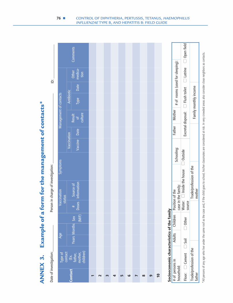
7766 CONTROL OF DIPHTHERIA, PERTUSSIS, TETANUS, HAEMOPHILUS INFLUENZAE TYPE B, AND HEPATITIS B: FIELD GUIDE
Age
Vacc
inat
ion
stat
us
Man
agem
ent o
f con
tact
s
Vacc
inat
ion
Antib
iotic
Co
ntac
t
Type
of
cont
act
(Ex.
fa
ther
, m
othe
r, ch
ildre
n)
Year
s
Mon
ths
Sex
(M/F
)
# Do
ses
Sour
ce o
f in
form
atio
n
Sym
ptom
s
Vacc
ine
Resu
lt of
cu
lture
Type
Da
teDa
te
Oth
er
med
ica-
tion
Com
men
ts
1
2
3
4
5
6
7
8
9
10
So
cioe
cono
mic
cha
ract
eris
tics
of
the
fam
ily
# of
per
sons
in
hous
ehol
d:
Adul
ts
Child
ren
Posi
tion
of th
e ca
se in
the
fam
ily:
Sc
hool
ing:
Fa
ther
M
othe
r #
of r
oom
s (u
sed
for s
leep
ing)
:
Floo
r: �
Cem
ent
�
Soi
l
�
Oth
er
Wat
er: �
Insi
de th
e ho
use
�
Out
side
so
urce
Ex
cret
al d
ispo
sal:
�
Flu
sh to
ilet
�
Lat
rine
�
Ope
n fie
ld
Trad
e/pr
ofes
sion
of t
he
fath
er
Tr
ade/
prof
essi
on o
f the
m
othe
r
Fam
ily m
onth
ly in
com
e
Date
of i
nves
tigat
ion:
____
____
____
__
Pe
rson
in c
harg
e of
inve
stig
atio
n: _
____
____
____
____
____
____
____
____
____
*All
pers
ons
of a
ny a
ge w
ho li
ve u
nder
the
sam
e ro
of a
s th
e ca
se a
nd, i
f the
chi
ld g
oes
to s
choo
l, hi
s/he
r cla
ssm
ates
are
con
sider
ed a
t risk
. In
very
cro
wde
d ar
eas
also
con
sider
clo
se n
eigh
bors
as
cont
acts
.
ID: _
____
____
____
____
_
AN
NE
X 3
.E
xam
ple
of a
form
for
the
man
agem
ent
of c
ont
acts
*

ANNEX 4 7777
[Name of institution] Notification and Investigation form – PERTUSSIS-LIKE ILLNESS Case number State/Province District Municipality Neighborhood/Landmarks Informant Telephone Service
I. CASE IDENTIFICATION First and last name Address Telephone Mother’s name Father’s name
Sex � Male � Female
Date of birth Day Month Year
If date of birth unavailable, age
Years ______ Months ______ Days ______
II. BACKGROUND
Date of symptom onset Day Month Year Consultation date Day Month Year
Notification date Day Month Year
Investigation date Day Month Year
Case identified by: � Spontaneous consultation (passive)
� Institutional search � Community search
Contact with confirmed case � Yes � No � Unk Attendance at school, kindergarten, or day care
� Yes � No � Unk
Number of pertussis vaccine doses
� 0 � 1 � 2 � ≥3 � Unknown
Date of last dose Day Month Year
Type of vaccine: � DTP � Pentavalent � Other____________
Vaccination information obtained by:
� Vaccination card � Health services � Parents or another adult
III. CLINICAL DATA, FOLLOW-UP AND TREATMENT Signs and symptoms Complications Cough Duration ______ days
� Yes � No � Unk Post-tussive vomiting (vomiting after coughing)
� Yes � No � Unk
Paroxysmal cough episodes � Yes � No � Unk Cyanosis � Yes � No � Unk Inspiratory whoop � Yes � No � Unk Unconsciousness � Yes � No � Unk Other symptoms and complications:
Hospitalization � Yes � No � Unk Admission date Day Month Year
Name of hospital Registry/history #
Final status
� Recovered � Transferred to ______ � Dead � Unknown
Date of discharge/death
Day Month Year
Antibiotics � Yes � No � Unk Type of antibiotics Duration of antibiotic therapy (days)
Date of last antibiotic dose Day Month Year
Antitoxin � Yes � No � Unk
Other treatment:
ANNEX 4. Example of Pertussis-like Illness Notification andInvestigation Form

7788 CONTROL OF DIPHTHERIA, PERTUSSIS, TETANUS, HAEMOPHILUS INFLUENZAE TYPE B, AND HEPATITIS B: FIELD GUIDE
IV. SAMPLES AND LABORATORY ANALYSIS SAMPLE 1 SAMPLE 2 SAMPLE 3 SAMPLE 4
Type of sample
� Nasopharyngeal aspirate � Nasopharyngeal swab � Other: ________
� Nasopharyngeal aspirate � Nasopharyngeal swab � Other: ________
� Nasopharyngeal aspirate � Nasopharyngeal swab � Other: ________
� Nasopharyngeal aspirate � Nasopharyngeal swab � Other: ________
Identification #
Date taken Day Month Year Day Month Year Day Month Year Day Month Year
Date sent FOR LABORATORY USE
Date received Day Month Year Day Month Year Day Month Year Day Month Year
Laboratory name
Identification # in laboratory
Type of test
Results
� Positive � Negative � Undetermined � Not processed
� Positive � Negative � Undetermined � Not processed
� Positive � Negative � Undetermined � Not processed
� Positive � Negative � Undetermined � Not processed
Result dates Day Month Year Day Month Year Day Month Year Day Month Year
V. CLASSIFICATION
Final classification
� Laboratory confirmation � Confirmed by epidemiological link � Probable � Discarded, final diagnosis: _______________________________
Date classified
Day Month Year
Classified by (Name)
Investigator Telephone
Institution
Signature Date
Observations:

ANNEX 5 7799
[Name of institution] Notification and Investigation form – TETANUS (Not neonatal) Case number State/Province District Municipality Neighborhood/Landmarks Informant Telephone Service
I. CASE IDENTIFICATION First and last name Address Telephone Mother’s name Father’s name
Sex � Male � Female
Date of birth Day Month Year
If date of birth unavailable, age
Years ______ Months ______ Days ______
II. BACKGROUND
Date of symptom onset Day Month Year Consultation date Day Month Year
Notification date Day Month Year
Investigation date Day Month Year
Wound present: � Yes � No � Unk Date of wound occurrence Day Month Year
Anatomic site and description of wound
Number of tetanus vaccine doses
� 0 � 1 � 2 � ≥3 � Unknown
Date of last dose Day Month Year
Type of vaccine: � DTP � Pentavalent � Other____________
Vaccination information obtained by:
� Vaccination card � Health services � Parents or another adult
III. CLINICAL DATA, FOLLOW-UP AND TREATMENT Signs and symptoms Complications Trismus (“lockjaw”) � Yes � No � Unk Opisthotonos � Yes � No � Unk Difficulty swallowing � Yes � No � Unk Abdominal rigidity � Yes � No � Unk Muscle spasms � Yes � No � Unk Other symptoms � Yes � No � Unk
Clinical type
� Generalized � Cephalic � Localized � Unknown
Other symptoms and complications:
Hospitalization � Yes � No � Unk Admission date Day Month Year
Name of hospital Registry/history #
Final status
� Recovered � Transferred to ______ � Dead � Unknown
Date of discharge/death
Day Month Year
Tetanus immunoglobulin � Yes � No � Unk
Dose of TIG Date of last TIG dose Day Month Year
Other treatment:
ANNEX 5. Example of Tetanus Notification and Investigation Form
8800 CONTROL OF DIPHTHERIA, PERTUSSIS, TETANUS, HAEMOPHILUS INFLUENZAE TYPE B, AND HEPATITIS B: FIELD GUIDE
V. CLASSIFICATION
Final classification
� Confirmed
� Discarded, final diagnosis:
_______________________________
Date classified
Day Month Year
Classified by (Name)
Investigator Telephone
Institution
Signature Date
Observations:

ANNEX 6 8811
I. CASE IDENTIFICATION First and last name Address Telephone Mother’s name Father’s name
Sex � Male � Female
Date of birth Day Month Year
If date of birth unavailable, age
Years ______ Months ______ Days ______
II. BACKGROUND
Date of symptom onset Day Month Year Consultation date Day Month Year
Notification date Day Month Year Investigation date Day Month Year
Case identified by: � Spontaneous consultation (passive)
� Institutional search � Community search
Contact with confirmed case � Yes � No � Unk Attendance at school, kindergarten, or day care
� Yes � No � Unk
Number of Hib vaccine doses � 0 � 1 � 2 � ≥3 � Unknown
Date of last dose Day Month Year
Type of vaccine: � DTP+Hep B � Pentavalent � Hib+Hep B � Other __________________________
Vaccination information obtained by:
� Vaccination card � Health services � Parents or another adult
III. CLINICAL DATA, FOLLOW-UP AND TREATMENT Signs and symptoms Complications Fever (grade ________ ) � Yes � No � Unk Vomiting � Yes � No � Unk Stiff neck � Yes � No � Unk Lethargy � Yes � No � Unk
Bulging fontanelle (infant) � Yes � No � Unk Irritability or alteration of mental alertness, stupor, or coma
� Yes � No � Unk
Difficulties suckling (infant) � Yes � No � Unk Convulsions (fits) � Yes � No � Unk Petechial rash � Yes � No � Unk Other meningeal signs
Other complications or sequelae
� Hearing loss � Visual impairment � Motor alterations � Other _________
Hospitalization � Yes � No � Unk Admission date Day Month Year
Name of hospital Registry/history #
Final status
� Recovered � Transferred to ______ � Dead � Unknown
Date of discharge/death
Day Month Year
Antibiotics � Yes � No � Unk Type of antibiotics Duration of antibiotic therapy (days)
Date of last antibiotic dose Day Month Year
Other treatment:
Case number State/Province District Municipality Neighborhood/Landmarks Informant Telephone Service
[Name of institution] Notification and Investigation form – HAEMOPHILUS INFLUENZAE TYPE B (Hib) MENINGITIS
ANNEX 6. Example of Haemophilus influenzae type b MeningitisNotification and Investigation Form

8822 CONTROL OF DIPHTHERIA, PERTUSSIS, TETANUS, HAEMOPHILUS INFLUENZAE TYPE B, AND HEPATITIS B: FIELD GUIDE
IV. SAMPLES AND LABORATORY ANALYSIS SAMPLE 1 SAMPLE 2 SAMPLE 3 SAMPLE 4
Type of sample � Cerebrospinal fluid (CSF) � Other: ________
� Cerebrospinal fluid (CSF) � Other: ________
� Cerebrospinal fluid (CSF) � Other: ________
� Cerebrospinal fluid (CSF) � Other: ________
Identification #
Date taken Day Month Year Day Month Year Day Month Year Day Month Year
Date sent FOR LABORATORY USE
Date received Day Month Year Day Month Year Day Month Year Day Month Year
Laboratory name
Identification # in laboratory
Type of test
Results
� Positive � Negative � Undetermined � Not processed
� Positive � Negative � Undetermined � Not processed
� Positive � Negative � Undetermined � Not processed
� Positive � Negative � Undetermined � Not processed
Result dates Day Month Year Day Month Year Day Month Year Day Month Year
Serotype result � ___________ � Undetermined � Not processed
� ___________ � Undetermined � Not processed
� ___________ � Undetermined � Not processed
� ___________ � Undetermined � Not processed
Result dates Day Month Year Day Month Year Day Month Year Day Month Year
V. CLASSIFICATION
Final classification
� Suspected � Laboratory confirmation � Probable � Discarded, final diagnosis: _______________________________
Date classified
Day Month Year
Classified by (Name)
Investigator Telephone
Institution
Signature Date
Observations:

ANNEX 7 8833
I. CASE IDENTIFICATION First and last name Address Telephone If patient under 18 yrs, Mother’s name
If patient under 18 yrs, Father’s name
Sex � Male � Female
Date of birth Day Month Year
If date of birth unavailable, age
Years ______ Months ______ Days ______
II. BACKGROUND
Date of symptom onset Day Month Year Consultation date Day Month Year
Notification date Day Month Year
Investigation date Day Month Year
Case identified by: � Spontaneous consultation (passive) � Institutional search � Community search
Born to carrier mother � Yes � No � Unk Place of birth of the case (city/province/country)
Household or other contact of a probable/confirmed Hep B case
� Yes � No � Unk If yes, specify contact � Household � Sexual partner � Other
Recipient of blood (or blood products)
� Yes � No � Unk Date of transfusion(s) Day Month Year
Has undergone hemodialysis � Yes � No � Unk Is/was health care worker � Yes � No � Unk
Has used IV drugs � Yes � No � Unk Has been institutionalized � Yes � No � Unk
Other known risk factors
Number of hepatitis B vaccine doses
� 0 � 1 � 2 � ≥3 � Unknown
Date of last dose Day Month Year
Type of vaccine: � DTP+Hep B � Pentavalent � Hib+Hep B � Other __________________________
Vaccination information obtained by:
� Vaccination card � Health services � Parents or another adult
III. CLINICAL DATA, FOLLOW-UP AND TREATMENT Signs and symptoms Complications Jaundice � Yes � No � Unk Nausea/vomiting � Yes � No � Unk Abdominal pain � Yes � No � Unk Fatigue � Yes � No � Unk Anorexia � Yes � No � Unk Fulminant hepatitis � Yes � No � Unk Other symptoms and complications:
Hospitalization � Yes � No � Unk Admission date Day Month Year
Name of hospital Registry/history #
Final status
� Recovered � Transferred to ______ � Dead � Unknown
Date of discharge/death
Day Month Year
Prior use of Hep B immunoglobulin
� Yes � No � Unk If yes, specify:
Treatment:
Case number State/Province District Municipality Neighborhood/Landmarks Informant Telephone Service
[Name of institution] Notification and Investigation form – HEPATITIS B
ANNEX 7. Example of Hepatitis B Notification and Investigation Form

8844 CONTROL OF DIPHTHERIA, PERTUSSIS, TETANUS, HAEMOPHILUS INFLUENZAE TYPE B, AND HEPATITIS B: FIELD GUIDE
IV. BLOOD SAMPLES AND LABORATORY ANALYSIS SAMPLE 1 SAMPLE 2 SAMPLE 3 SAMPLE 4
Identification #
Date taken Day Month Year Day Month Year Day Month Year Day Month Year
Date sent FOR LABORATORY USE
Date received Day Month Year Day Month Year Day Month Year Day Month Year
Laboratory name
Identification # in laboratory
RESULTS HBsAg
� Positive � Negative � Undetermined � Not processed
Anti-HBs
� Positive � Negative � Undetermined� Not processed
Anti-HBc � Positive
� Negative
� Undetermined
� Not processed
Anti-HBcAg � Positive
� Negative
� Undetermined
� Not processed
HBeAg
� Positive
� Negative
� Undetermined
� Not processed
Result dates Day Month Year Day Month Year Day Month Year Day Month Year
V. CLASSIFICATION
Final classification
� Confirmed � Probable � Discarded, final diagnosis: _______________________________
Date classified
Day Month Year
Classified by (Name)
Investigator Telephone
Institution
Signature Date
Observations:

ANNEX 8 8855
IMPO
RTA
NT
FACT
S TO
CO
NSI
DER
Lyop
hiliz
ed H
ib+
DTP+
hepa
titis
B va
ccin
e co
mes
in tw
o se
para
te v
ials
:
� O
ne v
ial c
onta
ins
liqui
d DT
P +
hep
atiti
s B
vacc
ine
(use
d as
a d
iluen
t) �
The
sec
ond
vial
con
tain
s a
lyop
hiliz
ed (f
reez
e-dr
ied)
Hib
vac
cine
• O
nly
use
the
DPT-
Hep
B va
ccin
e su
pplie
d w
ith th
e ly
ophi
lized
Hib
vac
cine
•
Nev
er u
se w
ater
or a
ny o
ther
dilu
ent t
o re
cons
titut
e th
e pe
ntav
alen
t vac
cine
•
Rem
embe
r tha
t the
dilu
ent I
S th
e DT
P-He
p B
com
pone
nt o
f the
vac
cine
RECO
NST
ITU
TIN
G
AD
MIN
ISTE
RIN
G
• U
se a
0.5
mL
syrin
ge a
nd n
eedl
e (d
ispo
sabl
e or
aut
o-di
sabl
e), t
he s
ame
type
of s
yrin
ge
and
need
le a
s ar
e ro
utin
ely
used
for D
TP in
ject
ions
•
Draw
0.5
mL
of re
cons
titut
ed (m
ixed
) vac
cine
into
the
inje
ctio
n sy
ringe
•
Adm
inis
ter a
s an
intr
amus
cula
r inj
ectio
n (IM
) in
the
infa
nt’s
out
er m
id-t
high
*:
* NEV
ER g
ive
intr
amus
cula
r inj
ectio
ns in
the
butt
ock
of in
fant
s as
ther
e is
risk
of d
amag
ing
nerv
es in
that
are
a. A
lso,
it w
ill re
sult
in a
re
duct
ion
in im
mun
ogen
icity
, esp
ecia
lly fo
r the
He
p B
com
pone
nt o
f the
vac
cine
.
•
Mak
e su
re y
ou h
ave
both
via
ls an
d 2
mL
mix
ing
(reco
nstit
utio
n) s
yrin
ges
• Ch
eck
the
expi
ry d
ate
of th
e DT
P +
hep
atiti
s B
vacc
ine:
�
Dis
card
vac
cine
that
is to
o ol
d or
has
bee
n ex
pose
d to
too
muc
h he
at
• U
se th
e sh
ake
test
to d
eter
min
e if
the
DTP
+ h
epat
itis
B va
ccin
e ha
s be
en fr
ozen
: �
Do
not u
se D
TP +
hep
atiti
s B
vacc
ine
that
has
bee
n fro
zen,
or t
hat y
ou s
uspe
ct
has
been
froz
en.
• U
sing
the
mix
ing
syrin
ge, d
raw
up
all o
f the
DTP
+ h
epat
itis
B va
ccin
e (u
sed
as
dilu
ent).
Inje
ct it
into
the
vial
con
tain
ing
the
lyop
hiliz
ed H
ib v
acci
ne.
• Re
mov
e th
e m
ixin
g sy
ringe
from
the
vacc
ine
vial
and
sha
ke th
e vi
al, o
r rol
l it
betw
een
your
pal
ms,
unt
il th
e po
wde
r is
fully
diss
olve
d an
d th
ere
are
no v
isib
le
part
icle
s in
the
vial
.
N
OTE
: A
ste
rile
syrin
ge a
nd n
eedl
e m
ust b
e us
ed fo
r eac
h in
ject
ion
and
disc
arde
d in
a
safe
ty b
ox. T
he s
yrin
ge a
nd n
eedl
e us
ed fo
r rec
onst
itutio
n sh
ould
not
be
used
for
givi
ng th
e in
ject
ion
REM
EMBE
R TH
E FO
LLO
WIN
G P
RECA
UTI
ON
S
IMPO
RTA
NT:
Dis
card
any
reco
nstit
uted
pe
ntav
alen
t vac
cine
afte
r six
hou
rs, o
r at t
he
end
of e
ach
sess
ion,
whi
chev
er c
omes
firs
t
To fa
cilit
ate
the
adeq
uate
reco
nstit
utio
n of
the
pent
aval
ent v
acci
ne, a
lway
s:
�
Log
the
vacc
ines
AN
D di
luen
ts in
the
stoc
k in
vent
ory
book
s �
Av
oid
keep
ing
the
lyop
hiliz
ed H
ib v
acci
ne a
nd th
e DT
P-He
p B
vacc
ine
(use
d as
di
luen
t) se
para
ted
Durin
g su
perv
isor
y vi
sits
, sup
ervi
sors
mus
t ens
ure
the
prop
er re
cons
titut
ion
and
adm
inis
trat
ion
of th
e pe
ntav
alen
t vac
cine
by:
�
Obs
ervi
ng th
e re
cons
titut
ion
and
inje
ctio
n pr
oces
s �
Ens
urin
g th
e av
aila
bilit
y of
the
sam
e nu
mbe
r of l
yoph
ilize
d H
ib a
nd D
TP-H
ep B
via
ls
__
____
____
____
__
Sour
ce: P
ATH
(Pro
gram
for A
ppro
priat
e Te
chno
logy
in H
ealth
). Im
mun
izin
g ch
ildre
n ag
ains
t Hae
moph
ilus i
nflue
nzae
type
b (H
ib).
A tr
aini
ng m
odul
e fo
r vac
cina
tors
. Ava
ilabl
e at
ht
tp://
ww
w.c
hild
rens
vacc
ine.
org/
htm
l/ip_
clin
ical
.htm
#tra
inin
g
AN
NE
X 8
.H
ow t
o r
eco
nsti
tute
and
adm
inis
ter
lyo
phili
zed
DT
P+H
ib+
Hep
atit
is B
(pe
ntav
alen
t) v
acci
ne