Contrast and Brightness Enhancement for Low …...Contrast and Brightness Enhancement for Low...
5
International Journal of Scientific & Engineering Research, Volume 4, Issue 5, May-2013 1519 ISSN 2229-5518 IJSER © 2013 http://www.ijser.org Contrast and Brightness Enhancement for Low Medical X-Ray Images Faisel G. Mohammed, Huda M. Rada, Sega G. Mohammed Abstract— Image enhancement techniques have been widely used in many fields, especially in medical scope, where the subjective quality of images is important for human interpretation (diagnosis). Denoising and contrast enhancement are important factors in any subjective evaluation of image quality. In the recent research work some techniques have been used to enhance medical images in the spatial domain using spatial filter (like, Sobel, Laplacian, and Smoothing). In additional, some logical and arithmetic operators have been used (like, AND, sum, subtract).A Hybrid strategy for image enhancement by combining spatial enhancement with power-law transforms function has been implemented. The effectiveness of this technique was evaluated using tested low medical x-ray images. The assistance criteria such as MAE (mean absolute error), and human visual was tested. The final results show that the suggested method is over tested images. Index Terms — arithmetic operator, contrast enhancement, transform function, medical images. —————————— —————————— 1 INTRODUCTION HE enhancement is widely used for medical image pro- cessing and as a preprocessing step in speech recognition, texture synthesis, and many other image/video processing applications. Exising enhancement approaches fall into two broad categories: spatial domain methods and frequency do- main methods [1]. As medical images are poorly illuminated, edg- es/boundaries are fuzzy/vague in nature; direct edge detection techniques will give discontinued and broken edges. So, pre- processing is required to obtain good and clear edge images. Pre processing is the image enhancement that plays an im- portant role in image processing. It highlights the important features or the features that are not properly visible and sup- presses unwanted information that is not relevant to image pro- cessing tasks. Enhancement may be edge enhancement or con- trast enhancement. Edge enhancement enhances the edg- es/boundaries of the image thereby suitable for edge detection where as contrast enhancement enhances the overall quality of the image. Median filtering is a good technique that preserves the boundaries and then on calculating the tohtal variation with respect to the central pixel of the filter window, an image with enhanced boundary is obtained. The image is then edge detect- ed using any edge detection methods [2]. Medical imaging is concerned with the development of the imaging devices that help to identify different aspects of the tissue and organs based on various properties and reveal new properties of the tissue and internal structure. Medical image processing is a field of science that is gaining wide ac- ceptance in healthcare industry due to its technological ad- vances and software breakthroughs. It plays a vital role in dis- ease diagnosis and in improved patient care. It also helps medical practitioners during decision making with regard to the type of treatment. Several state-of-the-art equipments pro- duce human organs in digital form which includes X-ray- based devices, Computed Tomography (CT), Magnetic Reso- nance Imaging (MRI), Ultrasound (US), Positron Emission Tomography (PET) and Single Photon Emission Computed Tomography (SPECT). [3] In the last several decades, because of dramatically devel- opment of making x-ray images, it has been widely applied to clinical diagnose. But it needs far more effort to attain the de- gree that x-ray image diagnose independently by computer can replace the work of physician. One reason is that the anal- ysis needs strong medic experience. The other is the x-ray im- age often not very clear which make the image processing can not distinguish all useful characteristic and make right judg- ment every time[4]. 2 PROCEDURE FOR P APER SUBMISSION The proposed scheme elements related to the current research work of interest are showed in this fig. 1. 2.1 Spatial Differentiation: 1 st Derivitive (Sobel Operators) First derivatives in image processing are implemented using the magnitude of the gradient. For a function f(x, y), the gradi- ent of f at coordinates (x, y) is defined as the two-dimensional column vector: T ———————————————— • Faisel G. Mohammed is currently assist prof. has PhD in Astronomy in Baghdad University, Iraq, PH-+9647903317690. E-mail: [email protected] • Huda M. Reda is currently programmer has higher diploma in Computer Science in Baghdad University, Iraq, PH-+946. E-mail: [email protected] • Sega G. Mohammed is currently programmer has higher diploma in Com- puter Science in Baghdad University, Iraq, PH-+946. E-mail: [email protected] IJSER
Transcript of Contrast and Brightness Enhancement for Low …...Contrast and Brightness Enhancement for Low...

International Journal of Scientific & Engineering Research, Volume 4, Issue 5, May-2013 1519 ISSN 2229-5518
IJSER © 2013 http://www.ijser.org
Contrast and Brightness Enhancement for Low Medical X-Ray Images Faisel G. Mohammed, Huda M. Rada, Sega G. Mohammed
Abstract— Image enhancement techniques have been widely used in many fields, especially in medical scope, where the subjective quality of images is important for human interpretation (diagnosis). Denoising and contrast enhancement are important factors in any subjective evaluation of image quality. In the recent research work some techniques have been used to enhance medical images in the spatial domain using spatial filter (like, Sobel, Laplacian, and Smoothing). In additional, some logical and arithmetic operators have been used (like, AND, sum, subtract).A Hybrid strategy for image enhancement by combining spatial enhancement with power-law transforms function has been implemented. The effectiveness of this technique was evaluated using tested low medical x-ray images. The assistance criteria such as MAE (mean absolute error), and human visual was tested. The final results show that the suggested method is over tested images.
Index Terms — arithmetic operator, contrast enhancement, transform function, medical images.
—————————— ——————————
1 INTRODUCTION HE enhancement is widely used for medical image pro-cessing and as a preprocessing step in speech recognition, texture synthesis, and many other image/video processing
applications. Exising enhancement approaches fall into two broad categories: spatial domain methods and frequency do-main methods [1].
As medical images are poorly illuminated, edg-es/boundaries are fuzzy/vague in nature; direct edge detection techniques will give discontinued and broken edges. So, pre-processing is required to obtain good and clear edge images. Pre processing is the image enhancement that plays an im-portant role in image processing. It highlights the important features or the features that are not properly visible and sup-presses unwanted information that is not relevant to image pro-cessing tasks. Enhancement may be edge enhancement or con-trast enhancement. Edge enhancement enhances the edg-es/boundaries of the image thereby suitable for edge detection where as contrast enhancement enhances the overall quality of the image. Median filtering is a good technique that preserves the boundaries and then on calculating the tohtal variation with respect to the central pixel of the filter window, an image with enhanced boundary is obtained. The image is then edge detect-ed using any edge detection methods [2].
Medical imaging is concerned with the development of the imaging devices that help to identify different aspects of the tissue and organs based on various properties and reveal new properties of the tissue and internal structure. Medical
image processing is a field of science that is gaining wide ac-ceptance in healthcare industry due to its technological ad-vances and software breakthroughs. It plays a vital role in dis-ease diagnosis and in improved patient care. It also helps medical practitioners during decision making with regard to the type of treatment. Several state-of-the-art equipments pro-duce human organs in digital form which includes X-ray-based devices, Computed Tomography (CT), Magnetic Reso-nance Imaging (MRI), Ultrasound (US), Positron Emission Tomography (PET) and Single Photon Emission Computed Tomography (SPECT). [3]
In the last several decades, because of dramatically devel-opment of making x-ray images, it has been widely applied to clinical diagnose. But it needs far more effort to attain the de-gree that x-ray image diagnose independently by computer can replace the work of physician. One reason is that the anal-ysis needs strong medic experience. The other is the x-ray im-age often not very clear which make the image processing can not distinguish all useful characteristic and make right judg-ment every time[4].
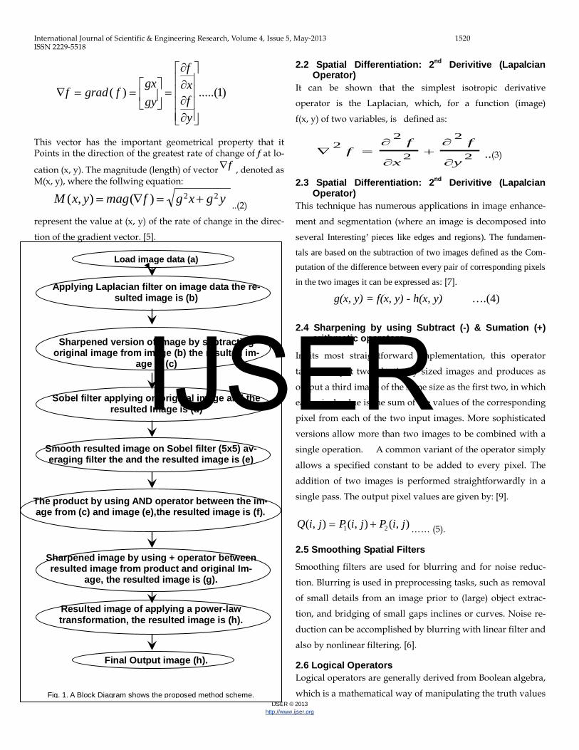
2 PROCEDURE FOR PAPER SUBMISSION The proposed scheme elements related to the current research work of interest are showed in this fig. 1.
2.1 Spatial Differentiation: 1st Derivitive (Sobel
Operators)
First derivatives in image processing are implemented using the magnitude of the gradient. For a function f(x, y), the gradi-ent of f at coordinates (x, y) is defined as the two-dimensional column vector:
T
———————————————— • Faisel G. Mohammed is currently assist prof. has PhD in Astronomy in
Baghdad University, Iraq, PH-+9647903317690. E-mail: [email protected]
• Huda M. Reda is currently programmer has higher diploma in Computer Science in Baghdad University, Iraq, PH-+946. E-mail: [email protected]
• Sega G. Mohammed is currently programmer has higher diploma in Com-puter Science in Baghdad University, Iraq, PH-+946. E-mail: [email protected]
IJSER
International Journal of Scientific & Engineering Research, Volume 4, Issue 5, May-2013 1520 ISSN 2229-5518
IJSER © 2013 http://www.ijser.org
)1.....()(
∂∂∂∂
=
==∇
yfxf
gygx
fgradf
This vector has the important geometrical property that it Points in the direction of the greatest rate of change of f at lo-
cation (x, y). The magnitude (length) of vector f∇ , denoted as M(x, y), where the follwing equation:
ygxgfmagyxM 22)(),( +=∇= ..(2)
represent the value at (x, y) of the rate of change in the direc-
tion of the gradient vector. [5].
2.2 Spatial Differentiation: 2nd Derivitive (Lapalcian Operator)
It can be shown that the simplest isotropic derivative
operator is the Laplacian, which, for a function (image)
f(x, y) of two variables, is defined as:
2
2
2
22
y
f
x
ff
∂
∂+
∂
∂=∇ ..(3)
2.3 Spatial Differentiation: 2nd Derivitive (Lapalcian Operator)
This technique has numerous applications in image enhance-
ment and segmentation (where an image is decomposed into
several Interesting’ pieces like edges and regions). The fundamen-
tals are based on the subtraction of two images defined as the Com-putation of the difference between every pair of corresponding pixels
in the two images it can be expressed as: [7].
g(x, y) = f(x, y) - h(x, y) ….(4) 2.4 Sharpening by using Subtract (-) & Sumation (+)
arithmetic operators
In its most straightforward implementation, this operator
takes as input two identically sized images and produces as
output a third image of the same size as the first two, in which
each pixel value is the sum of the values of the corresponding
pixel from each of the two input images. More sophisticated
versions allow more than two images to be combined with a
single operation. A common variant of the operator simply
allows a specified constant to be added to every pixel. The
addition of two images is performed straightforwardly in a
single pass. The output pixel values are given by: [9].
),(),(),( 21 jiPjiPjiQ += …… (5).
2.5 Smoothing Spatial Filters
Smoothing filters are used for blurring and for noise reduc-
tion. Blurring is used in preprocessing tasks, such as removal
of small details from an image prior to (large) object extrac-
tion, and bridging of small gaps inclines or curves. Noise re-
duction can be accomplished by blurring with linear filter and
also by nonlinear filtering. [6].
2.6 Logical Operators Logical operators are generally derived from Boolean algebra,
which is a mathematical way of manipulating the truth values
Load image data (a)
Sharpened version of image by subtracting original image from image (b) the resulted im-
age is (c)
Applying Laplacian filter on image data the re-sulted image is (b)
The product by using AND operator between the im-age from (c) and image (e),the resulted image is (f).
Final Output image (h).
Sobel filter applying on original image and the resulted Image is (d)
Sharpened image by using + operator between resulted image from product and original Im-
age, the resulted image is (g).
Resulted image of applying a power-law transformation, the resulted image is (h).
Smooth resulted image on Sobel filter (5x5) av-eraging filter the and the resulted image is (e)
Fig. 1. A Block Diagram shows the proposed method scheme.
IJSER

International Journal of Scientific & Engineering Research, Volume 4, Issue 5, May-2013 1521 ISSN 2229-5518
IJSER © 2013 http://www.ijser.org
of concepts in an abstract way without bothering about what
the concepts actually mean. The truth value of a concept in
Boolean value can have just one of two possible values: true or
false. Boolean algebra allows you to represent things like: The
block is both red and large by something like: A AND B where
A represents `The block is red', and B represents `The block is
large'. Now each of these sub-phrases has its own truth value
in any given situation: each sub-phrase is either true or false.
[8].
2.7 The Power-Law (Gamma) Transformation Power-law transformations have the basic form:
S = c rγ … (6) Where c and γ are positive constants. A variety of devices used for image capture, printing, and dis-
play respond according to a power law. By convention, the
exponent in the power-law equation is referred to as gam-
ma.The process used to correct this power-law response phe-
nomena is Gamma correction is important if displaying an
image accurately on a computer screen is of concern. . [6].
3. THE RESULTS OF SUGGESTED IMAGE ENHANCEMENT METHOD: The proposed algorithm is applied on tested low lumi-nance medical x-ray images. The evaluation was made in terms of statistical measures MAE. The evaluation of the results of image enhancement in spa-tial domain is performed by using object error criterion MAE (mean absolute error) as showing in following equation [10]:
),(),()/(11
0
1
0YXAYXBNMMAE
N
Y
M
X−×= ∑∑
−
=
−
=
… (7) So, for all experiments performed on the proposed project ta-ble (1), (2) summarize the results of the error measurement as an average of nine runs.
The results in tables (1), (.2) reflect the following behaviors:- 1- For, using power-law value =1, As it is shown it is the
best, Results, in spite of the value of result are greater
than the value of power-law which is equal of 0.9
2- When using the power value greater than 1, we shown
that the result is so Brightness and value of results is in-
creased.
3- For image filter size, we shown that the result value it is
increased, whenever increased the size of filter.
4- Finally, we shown that the best visual image and result is
applied, When the power-law is equal to 1, and the size of
filter is equal of 5x5.
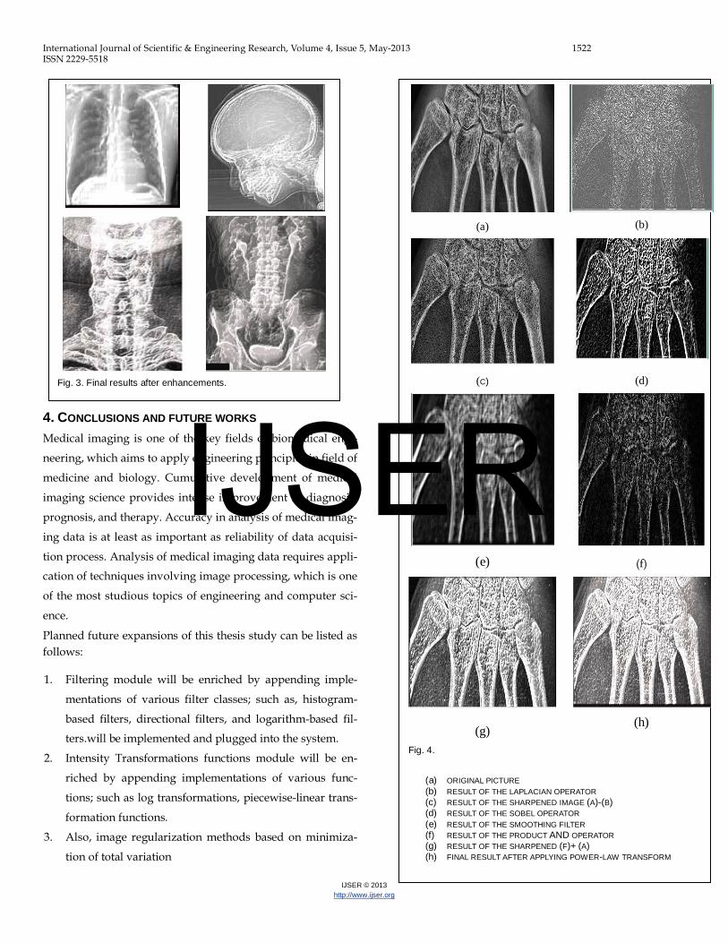
Figures (2&3) shows the original tested images and the result-
es after implemented the suggested system enhancement sys-
tem. While figure (4) show the detailed stages implementation
suggested enhancement method.
TABLE 1 PERFORMANCE OF IMAGE ENHANCEMENT ALGORITHM AP-
PLIED ON SKELETON IMAGE.
MAE Power-law value
Smooth filter size
1.87715 1 3x3 1.89421 1 5x5 1.94190 1 7x7 2.70291 1.05 3x3 2.72292 1.05 5x5 2.78251 1.05 7x7 1.50661 0.9 3x3 1.49691 0.9 5x5 1.52992 0.9 7x7
TABLE 2 PERFORMANCE OF IMAGE ENHANCEMENT ALGORITHM AP-
PLIED ON BONE IMAGE.
MAE Power-law value
Smooth filter size
1.87715 1 3x3 1.89421 1 5x5 1.94190 1 7x7 2.70291 1.05 3x3 2.72292 1.05 5x5 2.78251 1.05 7x7 1.50661 0.9 3x3 1.49691 0.9 5x5 1.52992 0.9 7x7
Fig. 2. Original tested x-ray dark images.
IJSER
International Journal of Scientific & Engineering Research, Volume 4, Issue 5, May-2013 1522 ISSN 2229-5518
IJSER © 2013 http://www.ijser.org
4. CONCLUSIONS AND FUTURE WORKS Medical imaging is one of the key fields of biomedical engi-
neering, which aims to apply engineering principles in field of
medicine and biology. Cumulative development of medical
imaging science provides intense improvement in diagnosis,
prognosis, and therapy. Accuracy in analysis of medical imag-
ing data is at least as important as reliability of data acquisi-
tion process. Analysis of medical imaging data requires appli-
cation of techniques involving image processing, which is one
of the most studious topics of engineering and computer sci-
ence.
Planned future expansions of this thesis study can be listed as follows:
1. Filtering module will be enriched by appending imple-
mentations of various filter classes; such as, histogram-
based filters, directional filters, and logarithm-based fil-
ters.will be implemented and plugged into the system.
2. Intensity Transformations functions module will be en-
riched by appending implementations of various func-
tions; such as log transformations, piecewise-linear trans-
formation functions.
3. Also, image regularization methods based on minimiza-
tion of total variation
Fig. 3. Final results after enhancements.
(a)
(b)
(C)
(d)
(e)
(f)
(g)
(h)
Fig. 4.
(a) ORIGINAL PICTURE (b) RESULT OF THE LAPLACIAN OPERATOR (c) RESULT OF THE SHARPENED IMAGE (A)-(B) (d) RESULT OF THE SOBEL OPERATOR (e) RESULT OF THE SMOOTHING FILTER (f) RESULT OF THE PRODUCT AND OPERATOR (g) RESULT OF THE SHARPENED (F)+ (A) (h) FINAL RESULT AFTER APPLYING POWER-LAW TRANSFORM
IJSER

International Journal of Scientific & Engineering Research, Volume 4, Issue 5, May-2013 1523 ISSN 2229-5518
IJSER © 2013 http://www.ijser.org
REFERENCES [1] Hasanul Kabir, Abdullah Al-Wadud, and Oksam Chae, “Brightness Pre-
serving Image Contrast Enhancement Using Weighted Mixture of Global and Local Transformation Functions”. The International Arab Journal of Information Technology, Vol. 7, No. 4, October 2010.
[2] Tamalika Chaira, “A rank ordered filter for medical image edge enhancement and detection using intuitionistic fuzzy set”. Journal of Applied Soft Computing, P.p.1259–1266, Dec. 2012.
[3] K. Karthikeyan and C. Chandrasekar, “Wavelet-based Image Enhancement Tech-niques for Improving Visual Quality of Ultrasonic Images”, International Journal of Computer Applications, P.p. 0975– 8887, Vol. 39, No.17, Feb. 2012.
[4] Jiao Feng, Naixue Xiong, Laurence T. Yang and Yan Yang, "A low distortion image enhancement scheme based on multi-resolutions analysis in next generation network", Journal Multimed Tools Apple, Vol. 56, P.p. 227–243, 2012.
[5] Gonzales, R. C. and Wintz, P., "Digital Image processing", Addison Wesley Pub. Company, 1992.
[6] Gonzalez, Rafel C., Richard E. Woods, "Digital Image processing", PEARSON (Prentice Hall), Third Edition, 2008.
[7] Jain, A.," Fundamentals of Digital Image Processing”, prentice Hall, pp240-241. [8] Boyle R. and R. Thomas, “Computer Vision”, A First Course, Blackwell Scien-
tific Publications, p 35, 1988. [9] BHOI .N," Development of Some Novel Spatial-Domain and Transform-Domain
Digital Image Filters" National Institute of Technology, Rourkela, INDIA Janu-ary 2009.
[10] Umbaugh. S. E.," Computer Vision and Image processing, A Practical Approach Using CVIP Tools", Prentice Hall, 1998. IJSER