Contraindication for Transplantation - Yonsei...
54
Kidney Transplantation Department of Surgery Yonsei University Wonju College of Medicine Kim Myoung Soo M.D. [email protected] http://gs.yonsei.ac.kr Current Kidney Transplantation
Transcript of Contraindication for Transplantation - Yonsei...

Kidney Transplantation
Department of SurgeryYonsei University Wonju College of Medicine
Kim Myoung Soo [email protected]
http://gs.yonsei.ac.kr
Current Kidney Transplantation
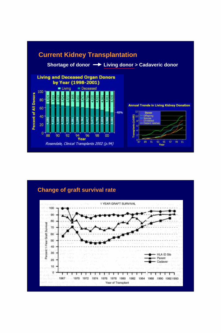
Current Kidney TransplantationShortage of donor Living donor > Cadaveric donor
Change of graft survival rate

Change of graft survival rate
Change of graft survival rate
Improvement of survival rate and half-life
Short-term survival Long-term survival

Comparison between dialysis and transplantationDialysis
Home-HD Center-HD CAPDKidney
TransplantationNormal social activity 59% 44% 48% 79%Normal, sometimes assist 25% 25% 25% 9%Self care only 9% 13% 12% 5%
Functionalsocial activityrecovery rate
Need assistance 7% 18% 15% 7%Quality of life Machine-dependent
2-3 times /week4-6 times/day
Life-longmedication
Quality of health
Only 20% of normal renal function Hgb: 7.0-8.0 / Hct: 20 – 22 BUN: 60 - 80 / Cr.: 6.0-8.0 Electrolyte imbalance(+) Bleeding tendency(+)
Near normalvalue
Comparison between dialysis and transplantation“ Quality of Life “
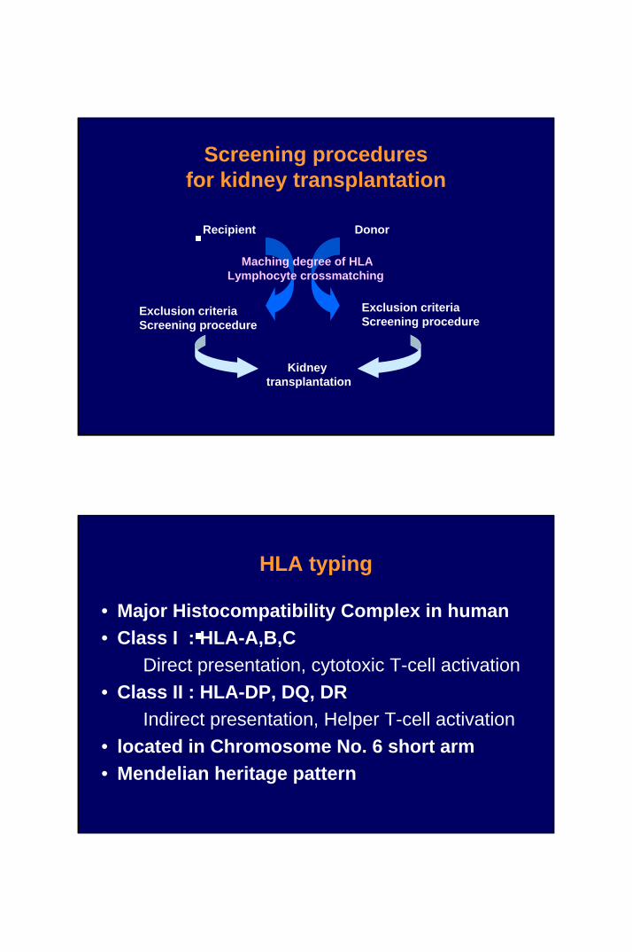
Screening procedures for kidney transplantation
Recipient Donor
Kidney transplantation
Exclusion criteria Screening procedure
Exclusion criteria Screening procedure
Maching degree of HLALymphocyte crossmatching
HLA typing
• Major Histocompatibility Complex in human• Class I : HLA-A,B,C
Direct presentation, cytotoxic T-cell activation• Class II : HLA-DP, DQ, DR
Indirect presentation, Helper T-cell activation• located in Chromosome No. 6 short arm• Mendelian heritage pattern
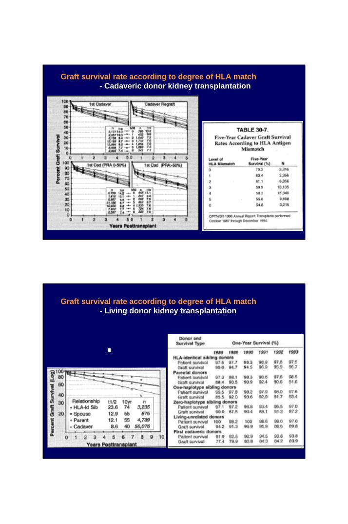
Graft survival rate according to degree of HLA match- Cadaveric donor kidney transplantation
Graft survival rate according to degree of HLA match- Living donor kidney transplantation
Lymphocyte Cross Matching
• Performed antibodies in recipient’s serum• Hyperacute rejection• Type
– T cell : Modified and Johnson’s method– B cell : Warm (37oC) and Cold (4oC)
• PRA (Panel Reactive Antibody) : screening test for sensitization to the common antigen
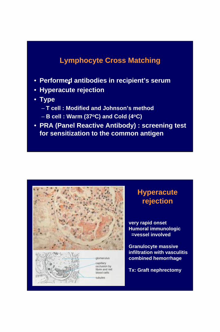
Hyperacuterejection
very rapid onsetHumoral immunologic =vessel involved
Granulocyte massive infiltration with vasculitiscombined hemorrhage
Tx: Graft nephrectomy
Contraindication for Transplantation
• Disseminated Malignancy• Infection, unresponsive to treatment• Refractory cardiac failure or respiratory failure• Progressive hepatic failure• Severe mental retardation• Severe congenital urinary tract abnormality• Extensive vascular disease
Recurred disease after kidney transplantation
• Diabetes : 100% but recurred late• FSGS : 30-70% with nephrotic syndrome• IgA nephropathy : 50% occasional graft failure• MPGN type 2 : > 90% slow graft failure• Oxalosis : 90%• HUS(Hemolytic uremic syndrome)• Amyloidosis, Cystinosis, Fabry’s,
Anti-GBM nephritis
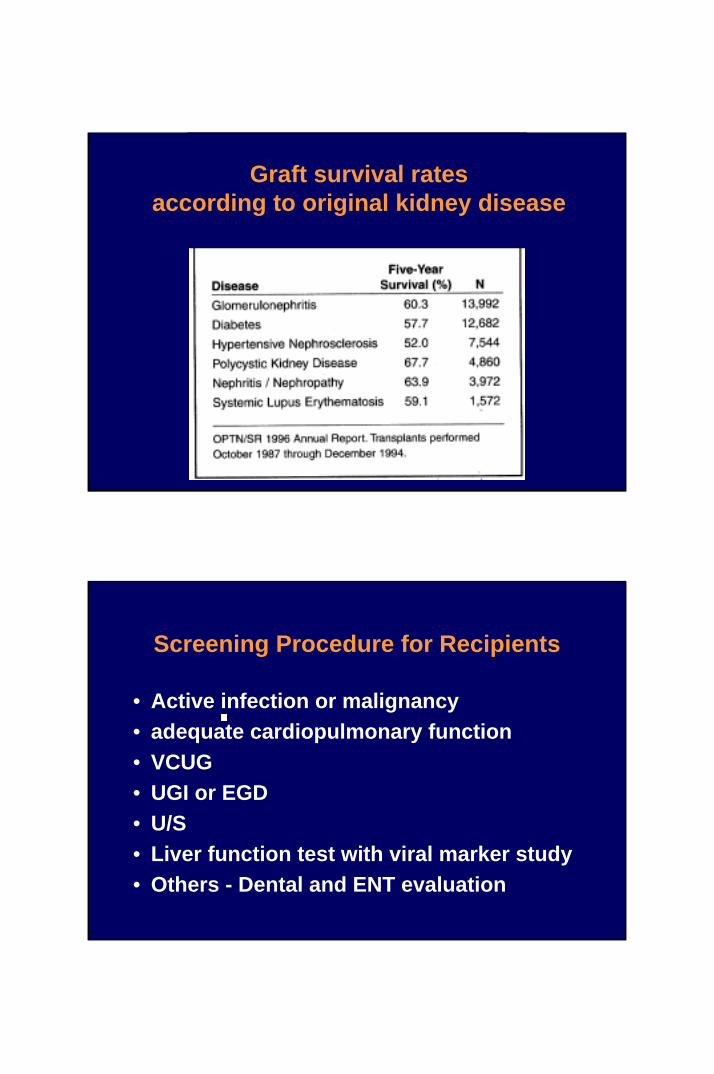
Graft survival rates according to original kidney disease
Screening Procedure for Recipients
• Active infection or malignancy• adequate cardiopulmonary function• VCUG• UGI or EGD• U/S• Liver function test with viral marker study• Others - Dental and ENT evaluation

Normal VCUG
Filling phase Post-voiding phase
Point of view)
Bladder contour ?VUR ?residual urine ?
VCUG- Grade III/IV VUR = indication of native nephrectomy
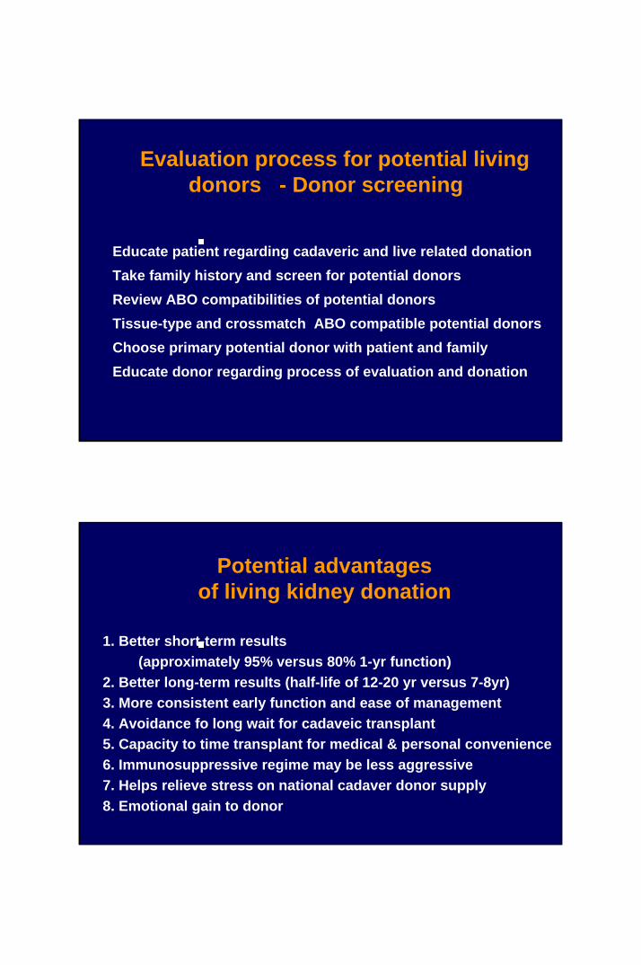
Evaluation process for potential living donors - Donor screening
Educate patient regarding cadaveric and live related donationTake family history and screen for potential donorsReview ABO compatibilities of potential donorsTissue-type and crossmatch ABO compatible potential donorsChoose primary potential donor with patient and familyEducate donor regarding process of evaluation and donation
Potential advantages of living kidney donation
1. Better short-term results (approximately 95% versus 80% 1-yr function)
2. Better long-term results (half-life of 12-20 yr versus 7-8yr)3. More consistent early function and ease of management4. Avoidance fo long wait for cadaveic transplant5. Capacity to time transplant for medical & personal convenience6. Immunosuppressive regime may be less aggressive7. Helps relieve stress on national cadaver donor supply8. Emotional gain to donor

Potential disadvantages of living kidney donation
1. Psychological stress to donor and family2. Inconvenience and risk of evaluation process
(I.e., IVP and angiogram)3. Operative mortality (approximately 1/2000)4. Major postoperative complications (approximately 2%)5. Minor post-operative complications (up to 50%)6. Long-term morbidity
(possibly mild hypertension and proteinuria)7. Risk of traumatic injury to remaining kidney
Contraindications to cadaveric donation Absolute Relative
Age > 70 Age > 60Chronic renal disease Age < 6Potentially metastasizing malignancy Mild hypertensionSevere hypertension Treated infectionBacterial sepsis Donor ATNIntravenous drug abuse Donor medical diseasePositive HBsAg or anti-HCV* ( diabetes, SLE )Positive HIV Prolonged cold ischemiaIntestinal perforationProlonged warm ischemia
ATN = acute tubular necrosis, HCV = hepatitis C vrius, HIV = human immunodeficiency virus, SLE = systemic lupus erythematosus

Exclusion criteria for living donors
Current trend, donor
Inferior survival rate
but
Donor shortage
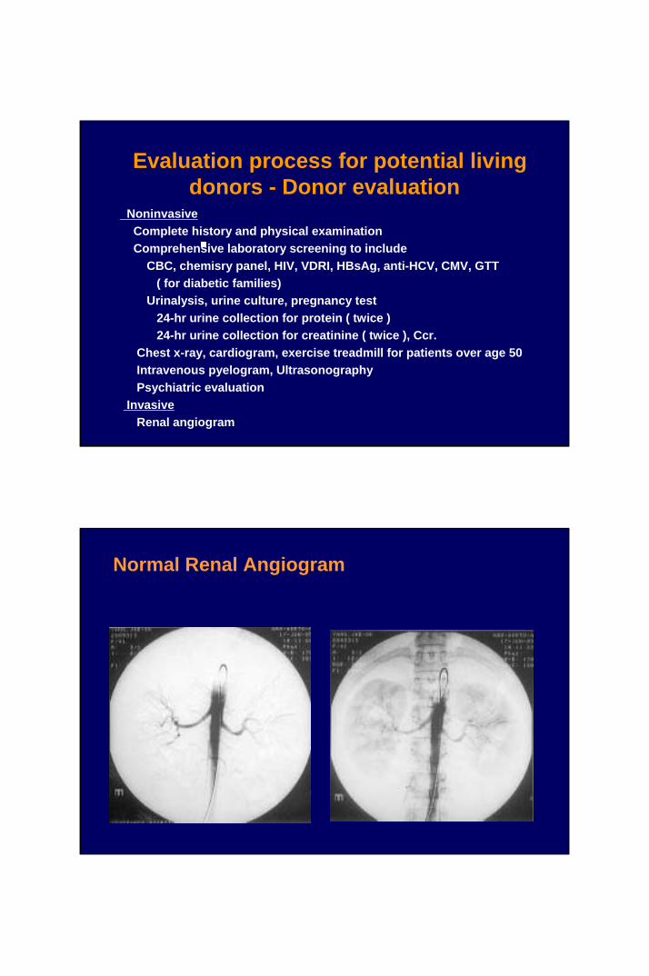
Evaluation process for potential living donors - Donor evaluation
NoninvasiveComplete history and physical examinationComprehensive laboratory screening to include
CBC, chemisry panel, HIV, VDRI, HBsAg, anti-HCV, CMV, GTT( for diabetic families)
Urinalysis, urine culture, pregnancy test24-hr urine collection for protein ( twice )24-hr urine collection for creatinine ( twice ), Ccr.
Chest x-ray, cardiogram, exercise treadmill for patients over age 50Intravenous pyelogram, UltrasonographyPsychiatric evaluation
InvasiveRenal angiogram
Normal Renal Angiogram
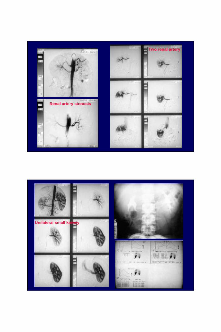
Renal artery stenosis
Two renal artery
Unilateral small kidney

Rt. : 1 Artery 1 VeinLt. : 2 Artery 1 Vein
Rt. double ureter & pelvis

Surgery of kidney transplantation
• Incision;Extraperitoneal vs. Transperitoneal approach
• Anastomosis of artery ;End-to-end vs. End-to-side anastomosis
• Ureteral anastomosis ;Ureteroneocystosotmy vs. Pyeloureterostomy
Alexis Carrel (1873-1944)

OP. Procedure (1)-- Op. field exposure
External iliac vein
Externaliliac
artery
Hockey-Stick incision
OP. Procedure (2)-- Vessel dissection
External iliac artery
External iliac vein
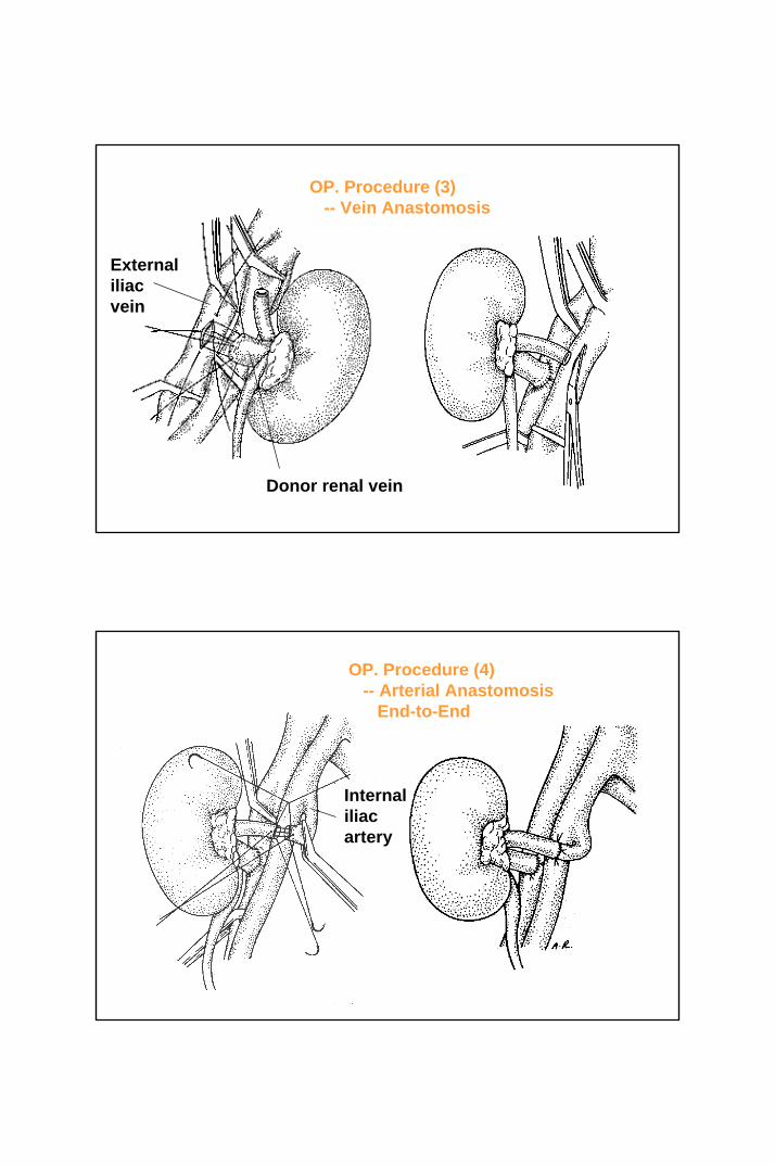
OP. Procedure (3)-- Vein Anastomosis
Donor renal vein
Externaliliac vein
OP. Procedure (4)-- Arterial Anastomosis
End-to-End
Internal iliac artery

OP. Procedure (4)-- Arterial Anastomosis
End-to-Side
External iliac vein & artery
OP. Procedure (5)-- Ureteroneocystostomy
ureter
Bladder wall
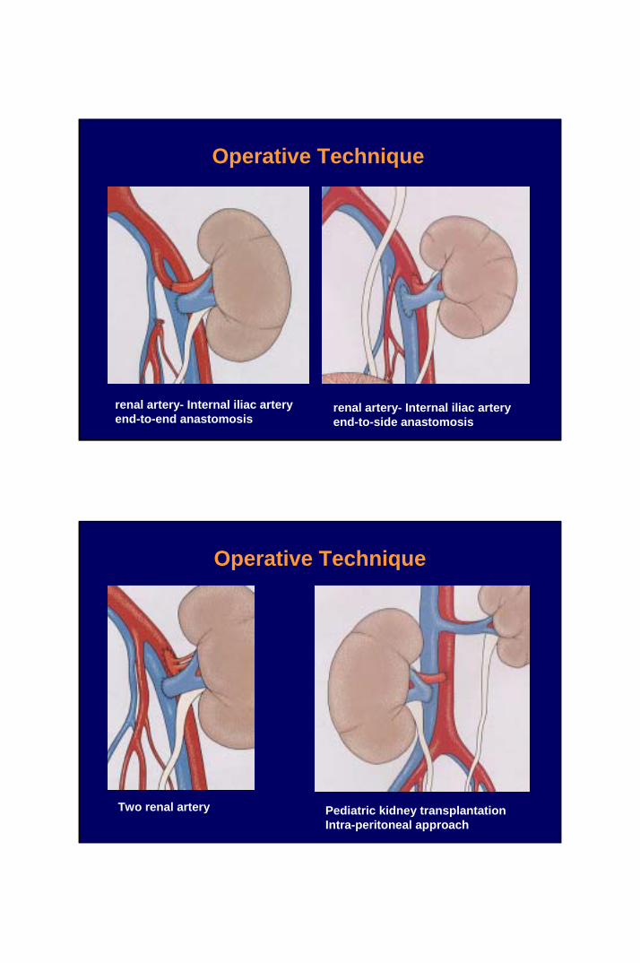
renal artery- Internal iliac artery end-to-end anastomosis
renal artery- Internal iliac artery end-to-side anastomosis
Operative Technique
Two renal artery Pediatric kidney transplantationIntra-peritoneal approach
Operative Technique
Indications of native nephrectomy
• Uncontrolled hypertension esp. renal origin• VUR Grade > III and refractory UTI• Large sized polycystic kidney• Persistent or recurrent pyelonephritis• Nephrostomy in place• Heavy proteinuria with edema
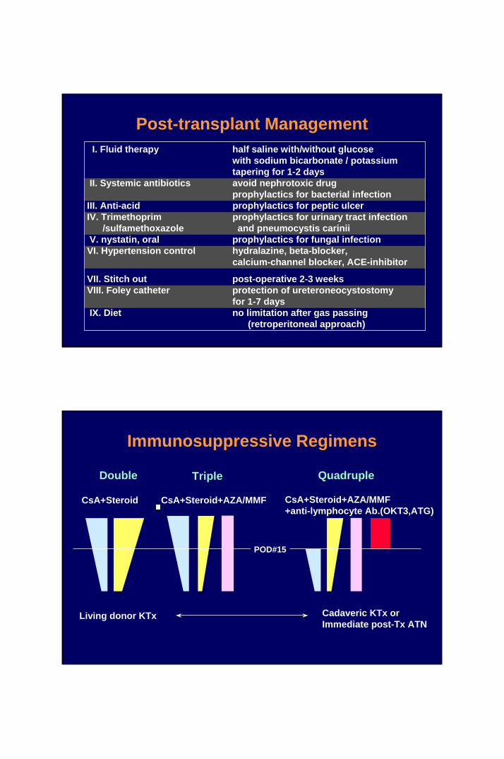
Post-transplant Management I. Fluid therapy half saline with/without glucose
with sodium bicarbonate / potassiumtapering for 1-2 days
II. Systemic antibiotics avoid nephrotoxic drugprophylactics for bacterial infection
III. Anti-acid prophylactics for peptic ulcerIV. Trimethoprim prophylactics for urinary tract infection
/sulfamethoxazole and pneumocystis cariniiV. nystatin, oral prophylactics for fungal infectionVI. Hypertension control hydralazine, beta-blocker,
calcium-channel blocker, ACE-inhibitor
VII. Stitch out post-operative 2-3 weeks VIII. Foley catheter protection of ureteroneocystostomy
for 1-7 daysIX. Diet no limitation after gas passing
(retroperitoneal approach)
Immunosuppressive Regimens
Double Triple Quadruple
CsA+Steroid CsA+Steroid+AZA/MMF
POD#15
CsA+Steroid+AZA/MMF+anti-lymphocyte Ab.(OKT3,ATG)
Cadaveric KTx orImmediate post-Tx ATN
Living donor KTx

MMF 1.5-2.0 g/day or AZA 1-2 mg/kg/day
Induction Immunosuppression
Imm
unos
uppr
essi
ve a
gent
s
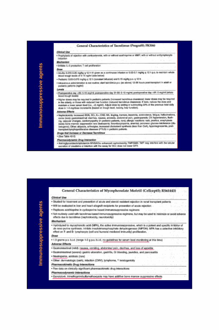
Imm
unos
uppr
essi
ve a
gent
sIm
mun
osup
pres
sive
age
nts

Causes of polyuria after transplantation
Ishemic injury of graft kidney= mild ATN
Hypervolemic status
Hyperosmolar status
polyuria
ineffective dialysisvolume expander during operation
elevated BUN/cr.Colloid fluid infusion during operation
Immediate/early post-transplant oliguria
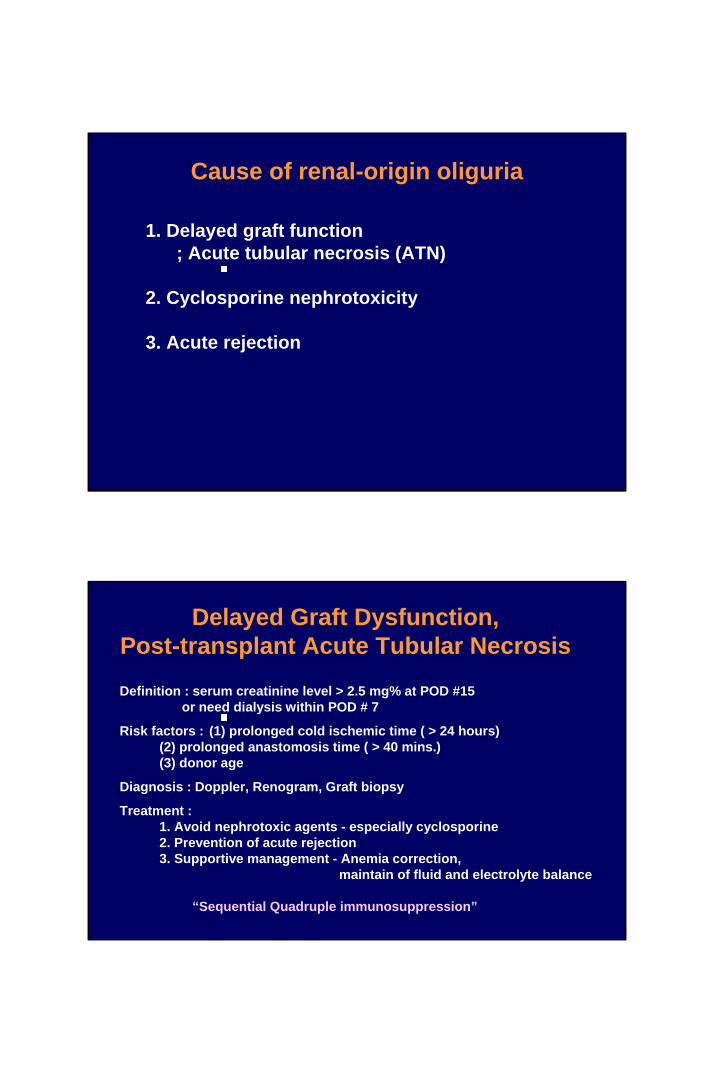
Cause of renal-origin oliguria
1. Delayed graft function ; Acute tubular necrosis (ATN)
2. Cyclosporine nephrotoxicity
3. Acute rejection
Delayed Graft Dysfunction, Post-transplant Acute Tubular Necrosis
Definition : serum creatinine level > 2.5 mg% at POD #15or need dialysis within POD # 7
Risk factors : (1) prolonged cold ischemic time ( > 24 hours) (2) prolonged anastomosis time ( > 40 mins.) (3) donor age
Diagnosis : Doppler, Renogram, Graft biopsy
Treatment :1. Avoid nephrotoxic agents - especially cyclosporine2. Prevention of acute rejection 3. Supportive management - Anemia correction,
maintain of fluid and electrolyte balance
“Sequential Quadruple immunosuppression”

Sequential Quadruple immunosuppression
Oliguria phase
Avoidance of cyclosporine nephrotoxicityPrevention of acute rejection
Cyclosporine nephrotoxocity
Clinical manifestation increment of serum-cr. : less than acute rejectionfluctuation of serum-cr. by whole blood CsA level reversal of serum-cr. after reduction of CsA dosage
Definitive diagnostic study = graft kidney biopsy ; arteriosclerosis

Rejection versus CsA nephrotoxicity
Biopsy
↓
Acute rejectionDiagnosis
Rapid onset ( within 6 months)Typical clinical symptoms
uncommon in CsA-treated groupclosed monitoring of s-cr.
Graft biopsy is definitive diagnostic study

Acute rejection
Incidencewithin post-transplant 6 months rare after post-transplant 1 year
“delayed acute rejection “ = acute rejection occurred after 1 year
Graft biopsy findings
Parenchyme, small vessel involvedCellular >> Humoral immunologic response
Mononuclear mixed cell inflammationInterstitial edema and hemorrhagetubulitis
Acute rejection
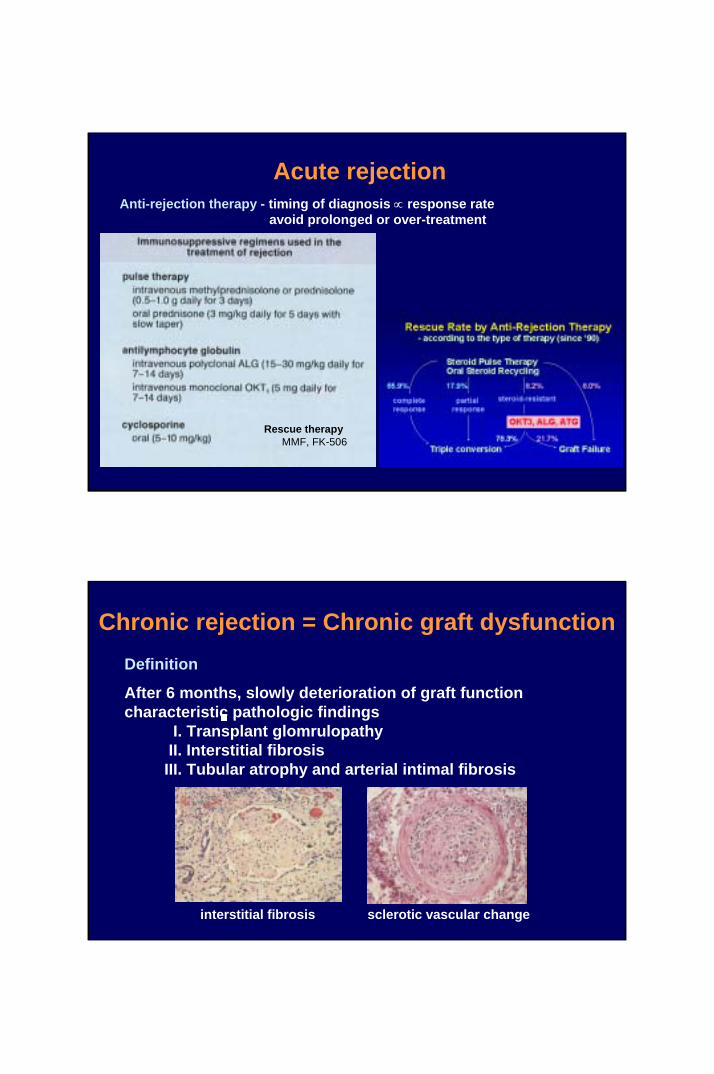
Acute rejection
Rescue therapy MMF, FK-506
Anti-rejection therapy - timing of diagnosis ∝ response rateavoid prolonged or over-treatment
Chronic rejection = Chronic graft dysfunctionDefinition
After 6 months, slowly deterioration of graft functioncharacteristic pathologic findings
I. Transplant glomrulopathyII. Interstitial fibrosisIII. Tubular atrophy and arterial intimal fibrosis
interstitial fibrosis sclerotic vascular change

Chronic rejection = Chronic graft dysfunctionPathogenesis
Immunologic causes Non-immunologic cause
1. Endothelial cell injury2. Release cytokines3. Proliferation of vascular smooth muscle cell4. Migration of vascular smooth muscle cell5. Proliferative obstructive vasculopathy6. Glomerulosclerosis / mesangial cell proliferation7. Ischemic injury
Pascual M et al. NEJM 2002; 346: 580
Risk factors affecting chronic allograft nephropathy

Chronic rejection = Chronic graft dysfunction
Immunologic factors
I. Immunologic injury = acute rejection episodespro: related with acute rejection history
especially multiple and delayed onset rejection(> 3 months)
con: lowering acute rejection ≠ chronic rejection
II. Histocompatibility differences
Chronic rejection = Chronic graft dysfunction
Non-immunologic factors
I. Ischemia/ Reperfusion injuryII. Infection
cytomegalovirus (CMV) infetion III. Immunosuppressive agents
cyclosporine/ FK-506 - focal glomerulosclerosisIV. Lipid abnormalities
hyperlipidemia, triglycerides, hypercholesterolemiaV. Hyperfiltration = relatively small numbers of nephron
donor-recipient size mismatching ex) gender, BMI difference, kidney weight/recipient weight
VI. Hypertension

Chronic rejection = Chronic graft dysfunction
Prevention
I. Avoid acute rejection & acute rejection sequaleII. Avoid under immunosuppressionIII. Prevention and Early management InfectionIV. Control Hypertension, Hypercholesterolemia, HyperglycemiaV. Maintain Ideal body weight
Treatment New immunosuppressive agent – MMF, RapamycinAgent for smooth muscle cell – dilatrend
effect is not confirmed !!!!!
Doppler; Resistant Index
PSV - DV
PSVRI =
Normal : 0.58 +/- 0.12Acute rejection: 0.78 +/- 0.14ATN, Infection: 0.70 +/- 0.05
Diagnostic Tool
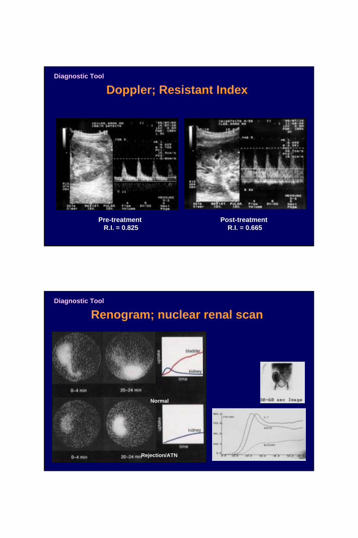
Pre-treatment R.I. = 0.825
Post-treatment R.I. = 0.665
Doppler; Resistant IndexDiagnostic Tool
Renogram; nuclear renal scan
Normal
Rejection/ATN
Diagnostic Tool
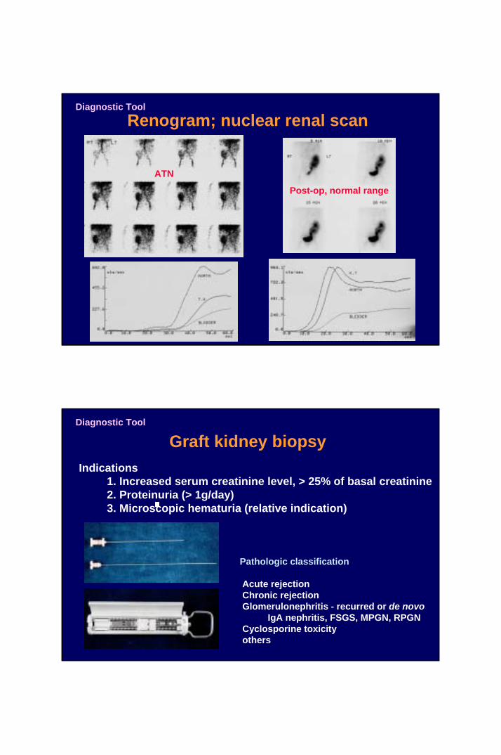
Renogram; nuclear renal scan
ATN
Post-op, normal range
Diagnostic Tool
Graft kidney biopsy Indications
1. Increased serum creatinine level, > 25% of basal creatinine2. Proteinuria (> 1g/day)3. Microscopic hematuria (relative indication)
Pathologic classification
Acute rejectionChronic rejectionGlomerulonephritis - recurred or de novo
IgA nephritis, FSGS, MPGN, RPGNCyclosporine toxicityothers
Diagnostic Tool

Graft kidney biopsy
Definitive diagnostic study
1. Classification of pathology2. Grade of pathology3. Combined pathology
Diagnostic Tool
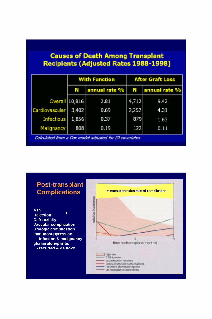
Immunsuppression related complicationPost-transplant Complications
ATNRejectionCsA toxicityVascular complicationUrologic complicationimmunosuppression
- infection & malignancyglomerulonephritis
- recurred & de novo
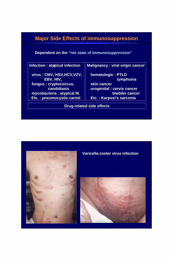
Major Side Effects of immunosuppression
Infection : atypical infection
virus : CMV, HSV,HCV,VZV,EBV, HIV,
fungus : cryptococcus, candidiasis
mycobacteria : atypical M.Etc. : pneumocystis carinii
Malignancy : viral origin cancer
hematologic : PTLD lymphoma
skin cancerurogenital : cervix cancer
bladder cancerEtc. : Karposi’s sarcoma
Drug-related side effects
Dependent on the “net state of immunosuppression”
Varicella-zoster virus infection

CMV pneumonitis
- CMV IgM(+) x4CMV-PCR (+)inclusion body
CMV enteritis
Inclusion body(+)

Mycobacterial Infection
- relatively common atypical mycobacteria- extra-pulmonary involvement
Intestinal Tbc.
Duodenal Tbc.
Soft tissue Tbc.
Spinal Tbc.

Esophageal candidiasis
Fungal Infection
Karposi’s sarcoma
Neck mass
lung metastasis

Cyclosporine-related side effects
Gum hyperplasia
s-Cr. K+
Total Bilirubin > SGOT/PTCholesterol Uric acidGlucose
Neurolgic – tremorHpertensionHypertrichosisGum hyperplasia
Urologic complication – VUR via neoureterocystostomy

KIDNEYRelated donorCadaver donorUnrelated living donor
LIVERHEARTPANCREAS-KIDNEYPANCREASLUNGBONE MARROW
403731332421191530
University of ColoradoUniversity of MinnesotaFMUSP-Sao PauloUniversity of ColoradoStanford UniversityUniv Hosp-ZurichUniversity of MinnesotaThe Toronto HospitalFHCRC-Seattle
Survyrs Hospital
Clinical Transplants 2002
Longest Surviving Transplants - 2002
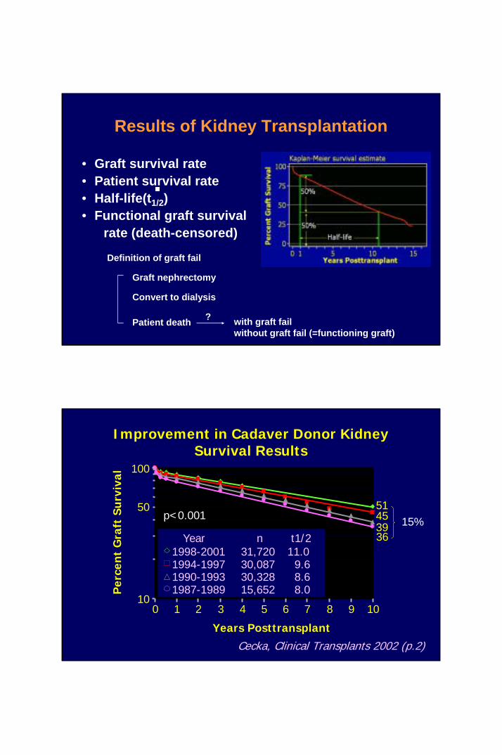
Results of Kidney Transplantation
• Graft survival rate • Patient survival rate• Half-life(t1/2)• Functional graft survival
rate (death-censored)
Patient death
Graft nephrectomy
Convert to dialysis
with graft failwithout graft fail (=functioning graft)
?
Definition of graft fail
Years Posttransplant
Per
cen
t G
raft
Su
rviv
al
10
100
0 1 2 3 4 5 6 7 8 9 10
p<0.00151453936
1998-20011994-19971990-19931987-1989
31,72030,08730,32815,652
11.09.68.68.0
n t1/2Year
Cecka, Clinical Transplants 2002 (p.2)
Improvement in Cadaver Donor Kidney Survival Results
5015%

Cecka, Clinical Transplants 2002 (p.13)
Improvement in Living Donor Kidney Survival Results
10
100
0 1 2 3 4 5 6 7 8 9 10Years Posttransplant
Per
cen
t G
raft
Su
rviv
al
68635755
1998-20011994-19971990-19931987-1989
Year14,16213,8479,8614,113
n18.615.713.512.3
t1/2
5013%
p<0.001
Risk factors affecting graft survival rate
HLA matchingPRA scoreType of immunosuppressionAcute rejection history
Delayed graft functionDonor type (living vs. cadaver)Recipient ageRace of recipientPreemptive transplant
Donor ageIschemic timeDonor medical illness (DM, hypertension)Recipient medical illness (DM, HBV,hypertension)Donor-recipient size mismatchingRecurrent original kidney disease
Immunologic non-immunologic
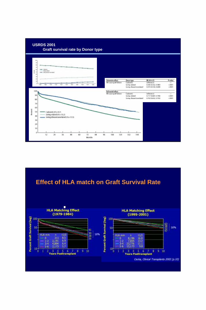
USRDS-2001
USRDS 2001Graft survival rate by Donor type
Effect of HLA match on Graft Survival Rate
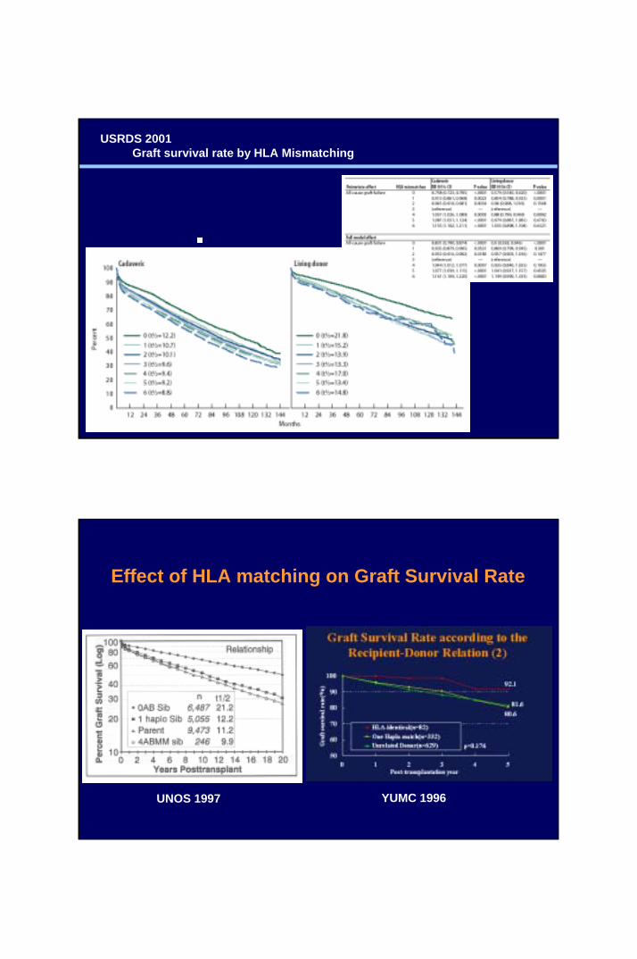
USRDS 2001Graft survival rate by HLA Mismatching
Effect of HLA matching on Graft Survival Rate
UNOS 1997 YUMC 1996

Effect of HLA matching on Graft Survival Rate

Effect of Acute rejection on Graft Survival Rate
YUMC 1996
USRDS 2001Graft survival rate by Donor age

Effect of Donor age on Graft Survival Rate
YUMC 1996
USRDS 2001Graft survival rate by Recipient age

Preemptive Transplant= Transplant without regular dialysis

USRDS 2001Graft survival rate by Cold ischemia time in cadaveric transplants
Thank You
Myoung Soo Kim M.D.
Department of SurgeryYonsei University Wonju College of [email protected]://gs.yonsei.ac.kr