Continuing Education Improving Outcomes for Patients with ...
10
HOURS Continuing Education 26 AJN ▼ March 2017 ▼ Vol. 117, No. 3 ajnonline.com CE 1.5 I n Part 1 of this article, which appeared last month, we offered an overview of chronic kid- ney disease (CKD), described its identification and etiology, and discussed ways to slow disease progression. Here, in part 2, we address disease complications and treatment for kidney failure. As in part 1, the case study of Anna Lowry, a 49-year- old woman with CKD, will be used for illustration, offering nurses specific guidance in helping patients to better understand and manage their CKD. (This case is a composite based on the authors’ experience.) COMPLICATIONS OF CKD As kidney function declines, fewer functioning neph- rons remain. The complications associated with CKD are complex, and may include anemia, hyperkalemia, hypoalbuminemia, metabolic acidosis, and abnormal mineral metabolism and bone disease. Laboratory work may show multiple metabolic abnormalities. Yet most people don’t feel any different until their CKD is quite advanced. As noted in part 1, dietary choices can affect many of the metabolic abnormalities associated with CKD. Thus referral to a registered dietitian who is knowledgeable about CKD may help in managing complications. There are many symptoms, signs, and laboratory values that must be tracked in patients with progres- sive CKD. The unique nature of the nurse–patient re- lationship may allow nurses to pick up on symptoms and signs that the primary care provider might have missed—particularly nonverbal behaviors and cues. It’s essential for nurses to alert primary care providers ABSTRACT: Coping with chronic kidney disease (CKD) is challenging for many people, since symptoms often don't appear until the disease is advanced and the patient is close to requiring dialysis. This two-part article aims to provide nurses with the basic information necessary to assess and manage patients with CKD. Part 1, which appeared last month, offered an overview of the disease, described identification and etiology, and discussed ways to slow disease progression. Part 2 addresses disease complications and treatment for kidney failure. Keywords: chronic kidney disease, collaborative care, end-stage kidney failure, end-stage renal failure, in- terdisciplinary care, kidney disease Addressing disease complications and treatment for kidney failure: a clinical review. Improving Outcomes for Patients with Chronic Kidney Disease: Part 2
Transcript of Continuing Education Improving Outcomes for Patients with ...

HOURSContinuing Education
26 AJN ▼ March 2017 ▼ Vol. 117, No. 3 ajnonline.com
CE 1.5
In Part 1 of this article, which appeared last month, we offered an overview of chronic kid-ney disease (CKD), described its identification
and etiology, and discussed ways to slow disease progression. Here, in part 2, we address disease complications and treatment for kidney failure. As in part 1, the case study of Anna Lowry, a 49-year-old woman with CKD, will be used for illustration, offering nurses specific guidance in helping patients to better understand and manage their CKD. (This case is a composite based on the authors’ experience.)
COMPLICATIONS OF CKDAs kidney function declines, fewer functioning neph-rons remain. The complications associated with CKD are complex, and may include anemia, hyperkalemia,
hypoalbuminemia, metabolic acidosis, and abnormal mineral metabolism and bone disease. Laboratory work may show multiple metabolic abnormalities. Yet most people don’t feel any different until their CKD is quite advanced. As noted in part 1, dietary choices can affect many of the metabolic abnormalities associated with CKD. Thus referral to a registered dietitian who is knowledgeable about CKD may help in managing complications.
There are many symptoms, signs, and laboratory values that must be tracked in patients with progres-sive CKD. The unique nature of the nurse–patient re-lationship may allow nurses to pick up on symptoms and signs that the primary care provider might have missed—particularly nonverbal behaviors and cues. It’s essential for nurses to alert primary care providers
ABSTRACT: Coping with chronic kidney disease (CKD) is challenging for many people, since symptoms often don't appear until the disease is advanced and the patient is close to requiring dialysis. This two-part article aims to provide nurses with the basic information necessary to assess and manage patients with CKD. Part 1, which appeared last month, offered an overview of the disease, described identification and etiology, and discussed ways to slow disease progression. Part 2 addresses disease complications and treatment for kidney failure.
Keywords: chronic kidney disease, collaborative care, end-stage kidney failure, end-stage renal failure, in-terdisciplinary care, kidney disease
Addressing disease complications and treatment for kidney failure: a clinical review.
Improving Outcomes for Patients with Chronic Kidney Disease: Part 2

[email protected] AJN ▼ March 2017 ▼ Vol. 117, No. 3 27
Since iron deficiency is common in CKD, iron sta-tus and hemoglobin levels should be checked before the addition of any iron supplements to the regimen.5 Supplemental iron is available in both oral and iv for-mulations. Absorption of oral iron supplements may be reduced by the intake of caffeinated beverages, supplemental calcium or calcium-containing antac-ids, and H2-receptor blockers or proton pump in-hibitors.16 Iron supplements may cause unwanted gastrointestinal effects such as heartburn or nausea. Beginning with half the recommended dosage and gradually increasing to the full dosage may help.16 Patients may also have fewer adverse effects with a different preparation, or by taking iron with food, in divided doses, or along with stool softeners.16 In-jectable erythropoiesis-stimulating agents are used infrequently in treating CKD.
Hyperkalemia. Potassium excretion is regulated by the renin−angiotensin−aldosterone system (RAAS). Perturbations of this system may result in hyperkale-mia. Potassium levels tend to increase as GFR de-clines.17 Nearly half of patients with an eGFR less than 30 mL/min/1.73 m2 have serum potassium lev-els of 4.5 mEq/L or greater.3 RAAS antagonists may increase the risk of hyperkalemia. Potassium-sparing medications, dietary intake, and transcellular shifts may also affect serum potassium levels.
By Jenna M. Norton, MPH, Eileen P. Newman, MS, RD, Gayle Romancito, RN, Stephanie Mahooty, DNP, MSN, Theresa Kuracina, MS, RD, CDE, LN, and Andrew S. Narva, MD, FACP, FASN
to any subtle changes in a patient’s condition, psycho-social issues, patient concerns, abnormal laboratory test results, or trends in laboratory values. (See Case Study: Metabolic Complications.)
Anemia. In the general population, a hemoglo-bin level of less than 13 g/dL in men and less than 12 g/dL in women indicates anemia.1, 2 But the opti-mal target hemoglobin level for people with CKD is currently unknown.
Anemia may occur early in CKD and is generally due to inadequate synthesis of the hormone eryth-ropoietin by the damaged kidneys. The prevalence of anemia increases as the glomerular filtration rate (GFR) declines, affecting nearly 50% of patients with an estimated GFR (eGFR) of less than 30 mL/min/1.73 m2.3 Among patients with advanced CKD, there is evidence that the prevalence of anemia is higher in those with diabetes than in those without diabetes.4
CKD-associated anemia is generally normochromic and normocytic.5 That said, identifying and correcting other causes of anemia (such as iron deficiency) is nec-essary.5 Assessing iron status in CKD requires a com-plete blood count and checking iron indices, including serum iron level and total iron-binding capacity (see Table 16-9). These two results are used to determine the transferrin saturation percentage, which reflects available iron. The serum ferritin level is used to as-sess stored iron. Optimal target levels of serum iron, total iron-binding capacity, and serum ferritin for peo-ple with CKD are unknown. Furthermore, they are af-fected by inflammation,10 which is common in CKD,11 making results more difficult to interpret. The abso-lute reticulocyte count may also be used to differen-tiate the cause of anemia or to monitor response to treatment.8
Ruling out other causes of anemia, including vitamin-deficiency anemia, may also be important. Although megaloblastic anemia is not commonly seen with CKD, some people with diabetes who have taken metformin for years may be vitamin B12 deficient. Metformin reportedly decreases absorption of this vitamin.12 Both B12 and folate levels may be lower than normal with metformin use.13 Metformin is contraindicated in patients with an eGFR below 30 mL/min/1.73 m2.14
Many other factors may contribute to inadequate iron stores in people with CKD. As their GFR declines, patients may lose interest in high-protein foods. Hepci-din, a hormone that plays a key role in controlling iron levels, regulating iron absorption from the gut, and mobilizing stored iron, accumulates in CKD,15 and this may result in reduced serum iron levels. Inflammation may also play a significant role in reducing iron ab-sorption.5 Pa
intin
g by
Viv
ian
Jay,
MSN
, FN
P-BC
, ww
w.e
bban
dflo
ww
ater
colo
rs.c
om.
28 AJN ▼ March 2017 ▼ Vol. 117, No. 3 ajnonline.com
Several factors can cause potassium to shift be-tween the intracellular and extracellular compart-ments. Insulin tends to move potassium into the cells; therefore, insulin deficiency can result in hyperkale-mia.18 Metabolic acidosis, characterized by an excess of hydrogen ions in the plasma, may drive potassium out of the cells, as hydrogen ions are buffered intracel-lularly.18 As a result, treating both hyperglycemia and acidemia may lower serum potassium.18 In some pa-tients with CKD, treating acidosis may allow the con-tinued use of RAAS antagonists.19
Patients with hyperkalemia should be counseled to limit foods that are higher in dietary potassium and to read ingredient lists in order to avoid foods that con-tain potassium chloride. Beginning in July 2018, food manufacturers will be required to include potassium on the Nutrition Facts label.20
Hypoalbuminemia occurs in CKD as a result of multiple factors. Both acute and chronic inflamma-tion are associated with reduced albumin synthesis.21 Loss of albumin in the urine in large quantities is asso-ciated with reduced serum albumin levels. Metabolic acidosis,22 insulin resistance,23 and a decrease in intake of high-protein foods24 may also contribute to low se-rum albumin. One large study of patients on mainte-nance hemodialysis found that a serum albumin level of greater than 3.8 g/dL was associated with reduced mortality risk, with the lowest such risk seen at levels of 4.4 g/dL or greater.25 Unfortunately, another study found that only 11% of new dialysis patients had se-rum albumin levels of 4 g/dL or greater.26
Metabolic acidosis is usually defined as a serum bicarbonate level of less than 22 mEq/L. The prev-alence of decreased serum bicarbonate increases as
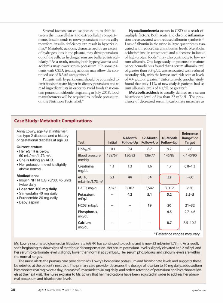
Case Study: Metabolic Complications
Anna Lowry, age 49 at initial visit, has type 2 diabetes and a history of gestational diabetes at age 30.
Current status:• Her eGFR is below
60 mL/min/1.73 m2. • She is taking an ARB. • Her potassium level is slightly
above normal.
Medications:• Insulin NPH/REG 70/30, 45 units
twice daily• Losartan 100 mg daily • Simvastatin 40 mg daily• Furosemide 20 mg daily• Baby aspirin
Test Initial6-Month
Follow-Up12-Month Follow-Up
18-Month Follow-Up
Reference Range* or
Target
HbA1c,% 10.1 9.4 8.7 9.2 < 8
Blood pressure, mmHg
138/67 150/92 136/77 145/83 < 140/90
Creatinine, mg/dL
1.1 1.3 1.6 1.7 0.8–1.3
eGFR, mL/min/1.73 m2
53 44 34 32 > 60
UACR, mg/g 2,823 3,107 3,542 3, 312 < 30
Potassium, mEq/L
-- 4.2 5.1 5.2 3.5–5
HC03, mEq/L -- -- 19 20 21–32
Phosphorus, mg/dL
-- -- -- 4.5 2.7–4.6
Calcium, mg/dL
-- -- -- 8.7 8.5–10.2
Ms. Lowry’s estimated glomerular filtration rate (eGFR) has continued to decline and is now 32 mL/min/1.73 m2. As a result, she’s beginning to show signs of metabolic decompensation. Her serum potassium level is slightly elevated at 5.2 mEq/L and her serum bicarbonate level is slightly lower than normal at 20 mEq/L. Her serum phosphorus and calcium levels are within the normal ranges.
The nurse alerts the primary care provider to Ms. Lowry’s borderline potassium and bicarbonate levels and suggests these be retested at the patient’s next visit. The primary care provider decreases the dosage of losartan to 50 mg daily, adds sodium bicarbonate 650 mg twice a day, increases furosemide to 40 mg daily, and orders retesting of potassium and bicarbonate lev-els at the next visit. The nurse explains to Ms. Lowry that her medications have been adjusted in order to address her abnor-mal potassium and bicarbonate levels.
* Reference ranges may vary.

[email protected] AJN ▼ March 2017 ▼ Vol. 117, No. 3 29
GFR declines.27 Nearly a quarter of patients with an eGFR below 30 mL/min/1.73 m2 have a serum bi-carbonate level of 20.5 mEq/L or less.3 Chronic meta-bolic acidosis is associated with accelerated muscle degradation, reduced albumin synthesis, exacerba-tion of metabolic bone disease, impaired glucose tol-erance, increased inflammation, and accelerated CKD progression.28 Animal protein is a source of acid load, while fresh fruits and vegetables are not. Serum bicar-bonate levels may increase as dietary protein intake decreases.29
Interventions to treat chronic metabolic acidosis include an adequate (but not excessive) intake of ani-mal protein and a supplemental base, such as sodium bicarbonate.28 It’s important to note that one 650-mg tablet of sodium bicarbonate has 178 mg of sodium. Reemphasize dietary salt restriction when sodium bi-carbonate is prescribed. Some patients may need an added diuretic to help remove the extra sodium.28
Abnormal mineral metabolism and bone dis-ease. Some CKD patients may have low levels of 25-hydroxyvitamin D,30 which can trigger complica-tions that affect bone strength and increase the risk of vascular calcification.31, 32 Serum calcium levels also decrease as a result of low vitamin D levels.33 Serum phosphorus levels may be within the normal range until CKD is advanced.33
As eGFR declines, the prevalences of hypocalce-mia and hyperphosphatemia increase.3, 33 Over 19% of patients with an eGFR below 30 mL/min/1.73 m2
have a serum calcium level of 8.9 mg/dL or less, and nearly 30% have a serum phosphorus level of
4.7 mg/dL or more.3 The systemic disorders of min-eral and bone metabolism associated with CKD are reflected in abnormalities in calcium, phosphorus, parathyroid hormone, and vitamin D metabolism. These derangements result in abnormalities in bone turnover, mineralization, volume, and linear growth or strength.33 They may be associated with vascular or other soft tissue calcification.34
Bone disease in people with CKD is complex, and interpreting laboratory test results is difficult. Although observational data support the correction of the meta-bolic abnormalities, there is limited high-quality evi-dence to support intervention. Phosphorus restriction may be implemented while trying to maintain ade-quate protein intake.30 Phosphorus-binding medica-tions are generally prescribed with meals in order to prevent phosphorus absorption. Low 25-hydroxyvi-tamin D levels may be treated with ergocalciferol or cholecalciferol, although further trials are needed to confirm the benefits.31 Elevated parathyroid hormone levels may be managed with vitamin D and phospho-rus restriction.31 Most adults exceed the recommended dietary allowance of 700 mg/day for phosphorus.35 According to 2011–2012 National Health and Nutri-tion Examination Survey (NHANES) data, the aver-age phosphorus intake was 1,393 mg/day.36
Depending on the food source, phosphorus ab-sorption varies. Between 10% and 60% of phos-phorus found naturally in protein-rich foods (such as meat, poultry, dairy, nuts, seeds, dried beans, and whole grains) is absorbed.37 Between 80% and 100% of inorganic phosphorus, which is added to many
a Reference ranges are for the general population unless otherwise noted.
Laboratory Parameter Reference Rangea Notes
Serum iron level 60–170 mcg/dL Level may decrease with iron deficiency and inflammation.
Total iron–binding capac-ity (TIBC)
240–450 mcg/dL • Level may increase with iron deficiency.
• Level may decrease with inflammation.
Transferrin saturation (TSAT) measures iron avail-able for erythropoiesis.
• Normal: 20%–50% • Early functional iron deficiency: < 16%
TSAT = serum iron/TIBC × 100
Serum ferritin level mea-sures the amount of stored iron.
Normal: • Men, 18–270 ng/mL • Women, 18–160 ng/mL
Depleted iron stores in patients with chronic kidney disease (both sexes):
• < 100 ng/mL in non−dialysis-dependent and peritoneal dialysis patients
• < 200 ng/mL in hemodialysis patients
Level may increase with inflam-mation or infection.
Table 1. Assessing Iron Status6-9

30 AJN ▼ March 2017 ▼ Vol. 117, No. 3 ajnonline.com
packaged and processed foods, is absorbed.37 For ex-ample, colas contain phosphoric acid. Sodium phos-phates are often added to poultry and pork products to enhance flavor or preserve tenderness. Cereals for-tified with calcium may contain calcium phosphate.
Patients should be counseled to check ingredient lists for words containing “phos”—such as phospho-rus, sodium phosphate, or pyrophosphate—and to avoid foods that contain such ingredients. Because
protein-rich foods tend to contain significant amounts of phosphorus, reducing consumption of such foods may also help in reducing phosphorus intake.
PREPARING FOR KIDNEY FAILURECoping with CKD and kidney failure is challenging for many people, since symptoms don’t often appear until the disease is advanced and the patient is close to requiring dialysis. Many patients exert great effort
Case Study: Preparing for Kidney Failure
Current status, two years after diagnosis:• Ms. Lowry is upset because a
month ago she learned she has progressive kidney disease.
• She reports frequent low blood glucose levels.
• She has stopped following any dietary restrictions and her HbA1C is lower.
• She is still taking her medications.
Test InitialTwo Years
LaterReference
Range* or Target
HbA1c,% 10.1 6.9 < 8
Blood pressure, mmHg 138/67 160/90 < 140/90
Creatinine, mg/dL 1.1 2.6 0.8–1.3
eGFR, mL/min/1.73 m2 53 26 > 60
UACR, mg/g 2,823 5,444 < 30
Hemoglobin, g/dL 12.1 11.6 12–16
Potassium, mEq/L 3.7 4.6 3.5–5
Albumin, g/dL 2.6 2.2 3.4–5
25-hydroxyvitamin D, ng/mL 12 28 20
Calcium, mg/dL 8.7 8.3 8.5–10.2
Phosphorus, mg/dL 3.7 4.9 2.7–4.6
iPTH, pg/mL -- 137 < 65
Over the course of two years, Ms. Lowry’s estimated glomerular filtration rate has dropped from 53 to 26 mL/min/1.73 m2. Upon learning that her disease has progressed despite her best efforts to adhere to the treatment regimen, she becomes very upset. Frustrated, she stops following the recommended dietary restrictions. Despite this, her glycated hemoglobin level con-tinues to decrease.
The nurse listens patiently as Ms. Lowry expresses her turmoil, despair, fear, and sadness about the progression of her chronic kidney disease. The nurse acknowledges these emotions, expresses empathy, and explains that Ms. Lowry’s feelings are normal. The nurse encourages her to learn more about kidney failure and its treatment so that she can regain some sense of control.
Knowing that Ms. Lowry has been reluctant to tell her husband and son about her illness, the nurse asks Ms. Lowry to in-vite them to the next office visit, so they can all discuss her care together. She reminds Ms. Lowry that her family will want to be there to support her and help her make difficult treatment decisions. The nurse also
• educates Ms. Lowry on kidney failure and its treatment options, helping her to understand how each treatment might affect her life.
• refers her to a booklet that explains kidney failure treatment options. (For a copy, visit www.niddk.nih.gov and search us-ing the phrase “Kidney Failure: Choosing a Treatment That’s Right for You.”)
• helps Ms. Lowry find a local kidney disease education class where she can learn more about treatment options and hear from patients who have been through the decision-making process.
• helps her schedule a visit to a nearby dialysis center so she can see firsthand what it’s like. • asks about her immunization status, emphasizing the importance of keeping vaccinations up to date.
* Reference ranges may vary.

[email protected] AJN ▼ March 2017 ▼ Vol. 117, No. 3 31
in adhering to all treatment recommendations, yet still experience CKD progression. Furthermore, both the disease itself and treatment with dialysis are com-plicated matters. Patients may have difficulty under-standing the implications of changes in their health status. It’s not uncommon for patients to experience grief, fear, or depression.
Historically, the health care community has not done a good job in educating CKD patients. An anal-ysis of 1994–1998 and 1999–2000 NHANES data showed that fewer than 20% of patients with both moderately decreased kidney function (an eGFR of 30 to 59 mL/min/1.73 m2) and albuminuria (a urine albumin-to-creatinine ratio greater than 30 mg/g) had ever been told by a physician that they had “weak or failing kidneys.”38 Such delayed awareness leaves many patients with little time to prepare for kidney failure, leaving them limited options when they face decisions about treatment. As one reflection of inade-quate preparation, consider that in 2013 more than 80% of people started hemodialysis with a tempo-rary vascular access (catheter).39
Many nurses aren’t comfortable discussing CKD or dialysis with patients,40, 41 and may defer such dis-cussions to the nephrologist. This is a missed oppor-tunity, because it’s both appropriate and helpful for nurses to initiate these conversations. A new provider may not know the patient well, whereas the nurse in the diabetes or hypertension clinic who has been in-volved in managing the patient’s conditions will have established a trust that can make education more ef-fective. Nurses can help patients to better understand CKD and begin accepting and coping with changes in their health status. (See Case Study: Preparing for Kidney Failure.)
TREATMENT CHOICES FOR KIDNEY FAILUREThere are four options for treating kidney failure. Three involve kidney replacement therapy, including kidney transplantation, peritoneal dialysis, or hemo-dialysis (either in a dialysis center or at home). The fourth option involves supportive care without trans-plantation or dialysis.
Transplantation. In general, a kidney transplant is associated with the best quality of life and survival (see Figure 142). To receive a kidney transplant, a pa-tient must be healthy enough to endure a surgery that can last several hours; have access to a donated kid-ney, either through a living donor or by being on the waiting list for a deceased donor kidney; and be will-ing to take antirejection medications daily and to have routine follow-up appointments for the rest of her or his life. Although antirejection medications suppress the immune system, organ rejection remains a possi-bility. With a functioning transplant, dialysis is not needed, and a near-normal diet can be followed.
Kidney transplantation is a treatment, not a cure. The transplanted kidney will likely work very well
for a time—according to recent data trends analyses, 92% of deceased donor and 97% of living donor kid-neys continue to work at one year following transplan-tation, and 47% of deceased donor and 62% of living donor kidneys are still working 10 years later39—but eventually it is likely to fail. The outcome improves if the donor and the recipient are ABO blood-type com-patible and a match for human leukocyte antigens. Pretransplantation evaluation can take several months and typically includes a comprehensive assessment to check for the presence of any conditions that might place the patient or the transplanted kidney at risk (such as severe coronary artery disease or cancer). Eli-gibility criteria vary from facility to facility, with some more willing to include patients with a higher body mass index.
Peritoneal dialysis may be a choice for a patient who has no contraindicating abdominal pathology (such as extensive abdominal surgery), wants to do in-home treatment, is willing to perform the treat-ment daily, and has room to store the necessary sup-plies. Patients using peritoneal dialysis usually don’t require vascular access, but do require minor surgery for abdominal catheter placement.
In peritoneal dialysis, the peritoneal membrane is used as a semipermeable filter, replacing the kidneys. In a peritoneal dialysis exchange, a dialysis solution (the dialysate) flows through the catheter into the abdominal cavity, where it remains for a prescribed
100
Num
ber
of R
ecip
ient
s 9080706050403020100
Alive at 1 Year Alive at 5 Years Alive at 10 Years
Transplant-living donorPeritoneal dialysis
Transplant-deceased donorHemodialysis
Figure 1. Survival Rates at One, Five, and 10 Years for Patients Opt-ing for Kidney Transplantation or Dialysis
Survival rates for recipients of a living donor kidney are higher at one, five, and 10 years following the transplant compared with those for recipients of a deceased donor kidney, peritoneal dialysis, or hemodialysis. Survival rates for recipients of a kidney from either type of donor are higher at one, five, and 10 years following the transplant compared with those for recipients of either peritoneal dialysis or hemodialysis. Disparities in survival rates increase over time. Adapted with permission from Johns Hopkins University, PREPARED (Providing Resources to Enhance Patients’ Readiness to Make Decisions About Kidney Disease) Study Research Team. Preparing for Kidney Treatment: You Have a Choice. 2011.42

32 AJN ▼ March 2017 ▼ Vol. 117, No. 3 ajnonline.com
period of time known as the dwell time. Through the process of diffusion, waste products move down the concentration gradient from the blood in the perito-neal capillaries into the dialysate. The efficiency of this clearance is determined by the concentration gradient; the size of the solute; and the permeability of the peri-toneal membrane, which can vary over time. The dial-ysate includes an osmotic agent that draws fluid into the peritoneal cavity, removing water and producing some additional clearance by bulk flow. At the end of the dwell time, the solution is drained out through the catheter. The continuous nature of peritoneal dialysis allows the patient to reach equilibrium, avoiding the up-and-down cycles of hemodialysis.
The dialysis prescription must be individualized for each patient. Generally, dextrose is used as the osmotic agent, with the concentration varying based on how much fluid must be removed. The higher the dextrose concentration, the more fluid will be removed—but more dextrose will also be absorbed, elevating blood sugar. Each dialysis exchange is generally two to three liters in volume. The dwell times and the number of exchanges per day vary depending on the patient and the characteristics of the peritoneal membrane.
There are several options for peritoneal dialysis. Continuous ambulatory peritoneal dialysis is per-formed manually four to five times during the day.
Continuous cycling peritoneal dialysis involves the use of a cycler, a small appliance that performs the exchanges automatically. With the cycler, many pa-tients can perform enough exchanges while asleep at night, such that they don’t need additional exchanges during the day. Some patients need one or two addi-tional manual exchanges during the day for adequate clearance. Most patients on peritoneal dialysis now use the cycler.
It’s important for patients on peritoneal dialysis to restrict their potassium intake. (Such restriction may be greater for patients on hemodialysis.) Amino acids that are lost during the exchanges must be replaced, and the patient’s dietary protein needs will be higher. Absorbed dextrose may cause the patient to gain weight. Because peritoneal dialysis is continuous, pa-tients are never in a fasting state, and this has particu-lar implications for those who have diabetes. Glucose levels may be harder to control, but insulin can be added to the dialysis solution. Some patients experi-ence body-image concerns associated with the catheter, and may need psychological and emotional support.
Hemodialysis. In hemodialysis, patients are treated with a hemodialysis machine three or more times a week. A dialyzer serves as the filter, replacing the kid-neys. The patient’s blood is pumped from the body through tubing, passes through the dialyzer, and is returned to the body. Along the way, blood pressure monitors and airflow detectors ensure the patient’s safety. (See Figure 2.)
The blood enters at the top of the dialyzer and is forced through multiple hollow filaments, each about the size of a human hair. Each filament acts as a semi-permeable membrane. As blood passes through the filaments, the dialysis solution flows around the out-side of the filaments. It takes less than one second for blood to pass from the top of the dialyzer to the bot-tom; as it does, waste products diffuse into the dialy-sate and are carried off, and the blood returns to the body. Diffusion efficiency depends on the size of the solute. Protein-bound substances usually aren’t re-moved; some amino acids, glucose, and water-soluble vitamins are removed. (See Figure 3.)
In-center hemodialysis may be a choice for a patient who can travel to a dialysis center three times a week for scheduled treatments, prefers that trained staff handle the treatments, doesn’t mind venipuncture, and is willing to follow a diet that in-cludes numerous restrictions. Advantages of in-center hemodialysis include the availability of facilities na-tionwide and the presence of trained staff to do the work. If they so choose, patients can be relatively pas-sive. The staff places the needles, monitors treatment, and maintains the equipment. As with many people with chronic illnesses, people with end-stage kidney disease may become socially isolated, and may enjoy the social setting of the dialysis center. Patients typi-cally spend three to four hours three times a week with
Figure 2. The Hemodialysis Process
During hemodialysis, blood is removed from the body through the tubing shown in red and is propelled along by a blood pump. The blood passes through the dialyzer and then is returned to the body through the tubing shown in gray. Along the way, blood pressure monitors, dialyzer inflow moni-tors, and air detectors help to ensure the patient’s safety during the procedure. A heparin pump prevents the blood from clotting. Figures 2−4 are reprinted courtesy of the National Kidney Disease Education Program, a program of the National Institute of Diabetes and Digestive and Kidney Diseases.
Venouspressure monitor
Dialyzer inflowpressure monitor
Heparin pump(to prevent
clotting)Dialyzer
Air detectorclamp
Dialyzed bloodreturned to
body
Bloodremoved for
dialysis
Arterialpressure monitor
Blood pump
Air trap and air detector

[email protected] AJN ▼ March 2017 ▼ Vol. 117, No. 3 33
the same relatively small group of other patients and providers. Disadvantages include more stringent di-etary restrictions, the loss of nutrients during hemo-dialysis, limited control over the procedure, and the burden of travel to and from the center. Furthermore, hemodialysis patients never reach equilibrium, experi-encing instead either a gradual increase in waste prod-ucts and fluids between treatments or a rapid decrease of these during treatment. These up-and-down cycles may fatigue patients.
Home hemodialysis may be a choice for a patient who wants to perform in-home treatments, has some-one to help in doing so, can perform treatments three or more times per week, has room for the machine and supplies, and doesn’t mind needlesticks and self-cannulation.
Home hemodialysis is becoming more popular.39 It requires training and support. As with in-center hemo-dialysis, home hemodialysis can be done three times per week. But it also permits other options, including daily dialysis for two to three hours, five to six times per week, and nocturnal dialysis for six to eight hours, three or more nights per week. More frequent home dialysis appears to be associated with significant ben-efits to the patient.43
Home hemodialysis has a different set of advan-tages and disadvantages. On the one hand, patients have more control over their schedules, travel isn’t re-quired, and the newer machines are smaller and easier to use than the older models were. The diet may be less restrictive, and phosphate binders may be less nec-essary or not needed. And if treatments are done more frequently, the ups and downs are less severe. On the other hand, home hemodialysis requires that a sec-ond person (often a partner or other family member) be present to assist, which may cause stress to the pa-tient, the other person, or both. Either the patient or the person assisting has to insert the needles; and the machine and supplies require space. The patient might have to take time off from work in order to get the initial training, which may not be offered locally. Pro-tein requirements are higher because of protein losses during treatment.
Vascular access. To perform hemodialysis, vascu-lar access must be created. In dialysis, blood usually flows at a rate of about 400 mL/min. Withdrawing blood at that rapid rate from any native peripheral vein would collapse that vein. A blood vessel that can withstand that withdrawal rate without collapse is required.
With permanent vascular access, an artificial con-nection between an artery and a vein is created, such that some blood is diverted to the vein. This connec-tion may be direct or indirect. An arteriovenous fistula establishes a direct connection, and is the preferred ac-cess method, as it’s less likely to become infected or to clot (see Figure 4A). Fistula maturation takes several weeks and involves dilation and thickening of the vein,
which occurs as a result of increased blood flow. Once the fistula is mature, access to blood flow for dialysis occurs through a percutaneous needlestick. If a direct connection cannot be created because of small vessel size or another mechanical problem, then an arterio-venous graft is the second option. This connection is made indirectly, using a synthetic tube (see Figure 4B).
Permanent vascular access is usually established in the nondominant arm. A large iv line (such as a peripherally inserted central catheter) placed in a pe-ripheral vein can destroy that vein for future dialysis use. Patients with CKD should be counseled to pro-tect the blood vessels in both arms by avoiding veni-puncture or iv catheter placement above the wrist, if possible. When emergent dialysis must be performed, temporary vascular access may be established using a central vein, usually by placing a catheter in the inter-nal jugular vein in the neck. However, this is only a temporary solution. Catheters are associated with in-adequate dialysis, increased infection rates, increased clotting, and inflammation.44
Figure 3. The Dialyzer Used in Hemodialysis
Blood enters at the top of the dialyzer and is forced through thin, hollow filaments made of semipermeable membrane. As blood passes through these filaments, the dialysis solution passes in the opposite direction on the outside of the filaments. Waste products diffuse from the blood into the dialysate. Filtered blood is returned to the patient.
From the patient
To the patient
Used dialysis fluid
Freshdialysis fluid

34 AJN ▼ March 2017 ▼ Vol. 117, No. 3 ajnonline.com
Supportive treatment without transplantation or dialysis. Opting for neither transplantation nor dialy-sis may be right for patients who feel that such treat-ments won’t improve their health; feel they’ve done what they wanted to do in life; and, ideally, have fam-ily and friends who support their decision. Support-ive treatment involves active medical management in which many complications can be treated. Medica-tions for CKD are usually continued and can be ad-justed. However, with supportive treatment there is no medical intervention aimed at replacing lost kid-ney function. Without clearance of uremic toxins, the patient will eventually become uremic.
It’s essential to provide comfort and palliative care to these patients. Patients who choose supportive ther-apy need to understand that without kidney replace-ment therapy, they will eventually die from uremia; it’s important that their family members understand this also. These facts must be presented in a manner that doesn’t question the patient’s decision, yet ensures that the decision is an informed one.
For additional information on caring for pa-tients with CKD, visit the Web site of the Na-tional Kidney Disease Education Program (http://bit.ly/2gaGy4w). ▼
Jenna M. Norton is the program manager of the National Kidney and Urologic Science Translation Program at the National Insti-tute of Diabetes and Digestive and Kidney Diseases (NIDDK), National Institutes of Health, Bethesda, MD. Andrew S. Narva is the director and Eileen P. Newman is the associate director of the National Kidney Disease Education Program in the Division of Kidney, Urologic, and Hematologic Diseases at the NIDDK. Gayle Romancito is a nurse at the Zuni Comprehensive Commu-nity Health Center, Indian Health Service, Zuni, NM. Stephanie Mahooty is an NP at Renal Medicine Associates and Desert Kid-ney Associates in Albuquerque, NM. Theresa Kuracina is a di-etitian at the Albuquerque Indian Health Center, Indian Health Service, Albuquerque, NM. Authors Narva, Newman, and Nor-ton are federal employees of the National Institutes of Health, and Romancito and Kuracina are federal employees of the Indian Health Service. Contact author: Andrew S. Narva, [email protected]. The authors and planners have disclosed no potential conflicts of interest, financial or otherwise.
REFERENCES1. [no author.] KDIGO 2012 clinical practice guideline for the
evaluation and management of chronic kidney disease. Kid-ney Int Suppl 2013;3(1):S1-S150.
2. World Health Organization. Haemoglobin concentrations for the diagnosis of anaemia and assessment of severity.
For eight additional continuing nursing education activities on topics related to kidney disease, go to www.nursingcenter.com/ce.
Figure 4. The Arteriovenous Fistula and the Arteriovenous Graft
An arteriovenous fistula (A) establishes a direct connection between an artery and a vein. Once matured, the fistula allows access to blood flow for dialysis through a percutaneous needlestick. An arteriovenous graft (B) uses a syn-thetic tube to connect the artery and the vein. This method is used when a direct connection can’t be established because of either small vessel size or another mechanical problem.
ArteryArtery
Arteriovenousfistula Looped graft
Vein
Vein
A B

[email protected] AJN ▼ March 2017 ▼ Vol. 117, No. 3 35
Geneva, Switzerland; 2011. WHO/NMH/NHD/MNM/11.1. Vitamin and Mineral Nutrition Information System (VMNIS); http://www.who.int/vmnis/indicators/haemoglobin.pdf.
3. United States Renal Data System. 2009 annual data report: atlas of chronic kidney disease and end-stage renal disease in the United States. Ann Arbor, MI: USRDS Coordinating Center 2009. https://www.usrds.org/atlas09.aspx.
4. Li Vecchi M, et al. Prevalence and severity of anaemia in pa-tients with type 2 diabetic nephropathy and different degrees of chronic renal insufficiency. Nephron Clin Pract 2007;105(2): c62-c67.
5. Babitt JL, Lin HY. Mechanisms of anemia in CKD. J Am Soc Nephrol 2012;23(10):1631-4.
6. Fischbach FT, Dunning MB, III. A manual of laboratory and diagnostic tests. 8th ed. Philadelphia: Wolters Kluwer Health/Lippincott Williams and Wilkins; 2009.
7. Institute of Medicine, Panel on Micronutrients. Iron. In: Di-etary reference intakes for vitamin A, vitamin K, arsenic, bo-ron, chromium, copper, iodine, iron, manganese, molybdenum, nickel, silicon, vanadium, and zinc. Washington, DC: National Academy Press; 2001. p. 290-393. https://www.nap.edu/read/ 10026/chapter/11.
8. O’Mara NB. Anemia in patients with chronic kidney disease. Diabetes Spectr 2008;21:12-9.
9. U.S. National Library of Medicine, MedlinePlus.gov. Total iron binding capacity. 2016. https://medlineplus.gov/ency/article/003489.htm.
10. Konijn AM. Iron metabolism in inflammation. Baillieres Clin Haematol 1994;7(4):829-49.
11. Silverstein DM. Inflammation in chronic kidney disease: role in the progression of renal and cardiovascular disease. Pedi-atr Nephrol 2009;24(8):1445-52.
12. Kibirige D, Mwebaze R. Vitamin B12 deficiency among pa-tients with diabetes mellitus: is routine screening and supple-mentation justified? J Diabetes Metab Disord 2013;12(1):17.
13. [no author.] Type 2 diabetes and metformin. First choice for monotherapy: weak evidence of efficacy but well-known and acceptable adverse effects. Prescrire Int 2014;23(154):269-72.
14. U.S. Food and Drug Administration. FDA drug safety commu-nication: FDA revises warnings regarding use of the diabetes medicine metformin in certain patients with reduced kidney function. Washington, DC; 2016 Apr 8. Drug safety and avail-ability; http://www.fda.gov/Drugs/DrugSafety/ucm493244.htm.
15. Taes YE, et al. Prohepcidin accumulates in renal insufficiency. Clin Chem Lab Med 2004;42(4):387-9.
16. Society for the Advancement of Blood Management. A physi-cian’s guide to oral iron supplements. Englewood, NJ; 2013. Iron corner; http://iron.sabm.org/overview/docs/Physician%20Guide%20to%20Oral%20Iron%20Nov%202013.pdf.
17. Musso CG. Potassium metabolism in patients with chronic kidney disease (CKD), Part I: patients not on dialysis (stages 3-4). Int Urol Nephrol 2004;36(3):465-8.
18. Lehnhardt A, Kemper MJ. Pathogenesis, diagnosis and man-agement of hyperkalemia. Pediatr Nephrol 2011;26(3):377-84.
19. Krikken JA, et al. Benefits of dietary sodium restriction in the management of chronic kidney disease. Curr Opin Nephrol Hypertens 2009;18(6):531-8.
20. U.S. Department of Health and Human Services, Food and Drug Administration. 21 CFR part 101. Food labeling: revi-sion of the nutrition and supplement facts labels. Final rule. Washington, DC: Federal Register 2016 33742-999.
21. Kaysen GA, et al. Inflammation and reduced albumin synthe-sis associated with stable decline in serum albumin in hemodi-alysis patients. Kidney Int 2004;65(4):1408-15.
22. Ballmer PE, et al. Chronic metabolic acidosis decreases albu-min synthesis and induces negative nitrogen balance in hu-mans. J Clin Invest 1995;95(1):39-45.
23. Danziger J. Importance of low-grade albuminuria. Mayo Clin Proc 2008;83(7):806-12.
24. Thalacker-Mercer AE, et al. Nutrient ingestion, protein in-take, and sex, but not age, affect the albumin synthesis rate in humans. J Nutr 2007;137(7):1734-40.
25. Kalantar-Zadeh K, et al. Revisiting mortality predictability of serum albumin in the dialysis population: time depen-dency, longitudinal changes and population-attributable fraction. Nephrol Dial Transplant 2005;20(9):1880-8.
26. Kaysen GA, et al. Trends and outcomes associated with se-rum albumin concentration among incident dialysis patients in the United States. J Ren Nutr 2008;18(4):323-31.
27. Moranne O, et al. Timing of onset of CKD-related meta-bolic complications. J Am Soc Nephrol 2009;20(1):164-71.
28. Kraut JA, Madias NE. Metabolic acidosis: pathophysiology, di-agnosis and management. Nat Rev Nephrol 2010;6(5):274-85.
29. Gennari FJ, et al. Effect of dietary protein intake on serum total CO2 concentration in chronic kidney disease: Modifi-cation of Diet in Renal Disease study findings. Clin J Am Soc Nephrol 2006;1(1):52-7.
30. National Kidney Foundation. National Kidney Foundation Kidney Disease Outcomes Quality Initiative (NKF KDOQI) for chronic kidney disease. n.d. https://www.kidney.org/ professionals/guidelines/guidelines_commentaries.
31. Garcia-Canton C, et al. Vascular calcification and 25- hydroxyvitamin D levels in non-dialysis patients with chronic kidney disease stages 4 and 5. Nephrol Dial Transplant 2011; 26(7):2250-6.
32. Lee SY, et al. 25-hydroxyvitamin D levels and vascular calci-fication in predialysis and dialysis patients with chronic kid-ney disease. Kidney Blood Press Res 2012;35(5):349-54.
33. Drüeke TB. Hyperparathyroidism in chronic kidney disease. In: DeGroot LJ, et al., editors. Endotext. South Dartmouth, MA: MDText.com, Inc.; 2000. https://www.ncbi.nlm.nih.gov/books/NBK278975.
34. Seiler S, et al. Clinical relevance of FGF-23 in chronic kidney disease. Kidney Int Suppl 2009(114):S34-S42.
35. Standing Committee on the Scientific Evaluation of Dietary Reference Intakes, Food, and Nutrition Board, Institute of Medicine. Dietary reference intakes for calcium, phosphorus, magnesium, vitamin D, and fluoride. Washington, DC: Na-tional Academy Press; 1997. https://www.nap.edu/catalog/5776/dietary-reference-intakes-for-calcium-phosphorus-magnesium-vitamin-d-and-fluoride.
36. Moshfegh AJ, et al. Phosphorus intake of Americans: what we eat in America, NHANES 2011-2012. Washington, DC: U.S. Department of Agriculture, Agricultural Research Ser-vice; 2016 Sep Food Surveys Research Group dietary data brief #15; https://www.ars.usda.gov/ARSUserFiles/80400530/pdf/DBrief/15_Phosphorus_intake_1112.pdf.
37. Noori N, et al. Organic and inorganic dietary phosphorus and its management in chronic kidney disease. Iran J Kidney Dis 2010;4(2):89-100.
38. Coresh J, et al. Chronic kidney disease awareness, preva-lence, and trends among U.S. adults, 1999 to 2000. J Am Soc Nephrol 2005;16(1):180-8.
39. United States Renal Data System. 2015 annual data report: epidemiology of kidney disease in the United States. Ann Ar-bor, MI: USRDS Coordinating Center; 2015. https://www.usrds.org/adr.aspx.
40. Ceccarelli CM, et al. Advance care planning for patients with chronic kidney disease—why aren’t nurses more in-volved? Nephrol Nurs J 2008;35(6):553-7.
41. Hopkins DJ, et al. End-of-life issues and the patient with renal disease: an evidence-based practice project. Nephrol Nurs J 2011;38(1):79-83.
42. Johns Hopkins University, PREPARED (Providing Resources to Enhance Patients’ Readiness to make Decisions about Kid-ney Disease) Study Research Team. Preparing for kidney treat-ment: you have a choice [multimedia flipbook]. 2011.
43. Tennankore K, et al. Intensified home hemodialysis: clinical benefits, risks and target populations. Nephrol Dial Trans-plant 2014;29(7):1342-9.
44. Ravani P, et al. Associations between hemodialysis access type and clinical outcomes: a systematic review. J Am Soc Nephrol 2013;24(3):465-73.