CONTAMINATION AND CURRENT PRACTICE IN …
110
CONTAMINATION AND CURRENT PRACTICE IN DECONTAMINATION OF NEBULISERS IN VENTILATED PATIENTS Lizl van Heerden A research report submitted to the Faculty of Health Sciences, University of the Witwatersrand, Johannesburg, in partial fulfilment of the requirements for the degree of Masters of Science (Physiotherapy) Johannesburg, 2015
Transcript of CONTAMINATION AND CURRENT PRACTICE IN …

CONTAMINATION AND CURRENT PRACTICE IN
DECONTAMINATION OF NEBULISERS IN
VENTILATED PATIENTS
Lizl van Heerden
A research report submitted to the Faculty of Health Sciences, University of the Witwatersrand,
Johannesburg, in partial fulfilment of the requirements for the degree of Masters of Science
(Physiotherapy)
Johannesburg, 2015

i
ABSTRACT
Background
Aerosol therapy is an important and frequently used method of delivering drugs to the patient on
mechanical ventilation (MV). Different types of aerosol devices are available to deliver drug therapy
during MV. These devices need to be used according to the manufacturer’s guidelines which include
methods for decontamination and application. The methods used to store these nebulisers and the
pathogens in the surrounding air may contribute to the contamination of these devices. Nebulisers
have been identified as a possible source of ventilator-associated pneumonia (VAP). The incidence of
contamination of nebulisers associated with current decontamination and storage protocols will lay the
foundation for the development of evidence based practice of aerosol therapy in MV.
Objectives
The aim of this study was to determine the current incidence of contamination of nebulisers used
within a ventilator circuit and surrounding air in the intensive care units (ICUs) of hospitals in Pretoria
and to determine the current practice regarding decontamination and storage of these devices.
Micro-organisms that colonise these contaminated nebulisers and the surrounding air were also
identified.
Methods
A cross-sectional observational analytical study was done in seven ICUs in Pretoria whereby 61
nebulisers and the surrounding air were sampled and assessed. The unit manager of each ICU was
asked questions to identify the current decontamination and storage protocols for nebulisers used
within a ventilator circuit. Swabs were taken from the chambers of nebulisers used within a ventilator
circuit and streaked on blood agar plates (BAPs). An air sampler was used to collect air samples from
the surrounding environment. The BAPs of nebulisers and air were incubated for possible bacterial
and fungal contamination. Species of the most recurrent colonies observed were identified in both air
and nebuliser samples.
Results
A total of 61 nebulisers were sampled including 37 Micro Mist nebulisers and 24 Aeroneb nebulisers.
The incidence of contamination found in the Micro Mist nebulisers were 51.4% (n=19) and the
Aeroneb nebulisers were 50% (n=12). Most of the Aeroneb nebulisers in the ventilator circuit were
wet which resulted in 50% bacterial contamination. All the ICUs in the hospitals in Pretoria had
decontamination and storage protocols for the Micro Mist nebuliser. These protocols differed between
ICUs and ICUs within the same hospital. Staff adherence to these protocols was low as the methods
ii
observed for storage and decontamination differed from the protocols stated to be used in the ICUs.
Contamination rate was the least when the Micro Mist nebuliser was rinsed with alcohol and left open
to the environment. Micro Mist nebulisers that were taken apart and left to dry under a sterile cloth
resulted in the most fungal and bacterial contamination. No contamination was found in Micro Mist
nebulisers that were used for Bisolvon aerosolisation. Coagulase-negative Staphylococcus species
(spp.) was mostly found in air and Aeroneb samples and Enterococcus spp. mostly in the Micro Mist
nebuliser. Both of these micro-organisms are common causes of VAP.
Conclusion
Both types of nebulisers presented with similar rates of contamination. Although the ICUs in the
hospitals had decontamination and storage protocols in place, the incidence of contamination in the
Micro Mist nebulisers was high. The rate of contamination in the Micro Mist nebulisers can be
associated with different decontamination and storage protocols. This is the first study to identify the
rate of contamination in the Aeroneb nebuliser. Most of the Aeroneb nebulisers were wet during the
time of MV which increased the possibility of contamination. The micro-organisms found in nebulisers
and air samples harbour pathogens that can cause VAP.

iii
DECLARATION
I, Lizl van Heerden, hereby declare that this research report is my original work. It is being submitted
for partial fulfilment of the degree of Masters of Science (Physiotherapy) in the University of the
Witwatersrand, Johannesburg. Neither the substance of any part of this work has been, or is being, or
is to be submitted for another degree at this or any other university.
Signed: ..........................................
Date: ..............day of ................ 2015

iv
ACKNOWLEDGMENTS
I am grateful to the following people who supported me through this research report:
Associate Professor Helena van Aswegen for her tremendous insight and valuable opinions
forcing me to develop and enhance my research skills.
Dr. Ronel Roos for her patience and encouragement through the entire research and especially
with the statistical section of this report.
Associate Professor Sandy van Vuuren for teaching and assisting me, with all the aspects of
swabbing, culturing and incubating as well as allowing me to use the laboratory at the Pharmacy
Department of the University of the Witwatersrand.
Adriano Duse Professor & HOD: Clinial Microbiology at the University of the Witswatersrand for
his assistance in the identification of bacterial colonies found in nebuliser and air samples.
Mr Emery Ngamasana for his assistance and input with the statistical data.
Unit managers and staff in the ICUs of the four private hospitals for allowing me to conduct my
research in their units and for their assisting me with the collection of information when needed.
My wonderful children for always supporting me and encouraging me to finish.
My mother for always being there when I am tired and taking care of my children.
I am thankful to the Funding Research Committee of the University of the Witwatersrand and the
Cardio Pulmonary Rehabilitation group of the South African Society of Physiotherapy for their
funding towards my research report.
God for giving me the ability to endure and creating in me a deep trust towards Him.

v
TABLE OF CONTENTS
Page
Abstract…………………………………...……………………………………………. i
Declaration…………………………...……………………………………………….... iii
Acknowledgements.......................………………………………………………….. iv
Table of Contents……………………….…………………………………………...... v
List of Figures ……………………………..………………………………………...... ix
List of Tables………………………………..…………………………………………. x
List of Abbreviations……………...…...………………………………..…………..... xi
1. Chapter 1: Introduction.........,........................................................................... 1
1.1 Background……………………………………………….......................................... 1
1.2 Problem Statement……………………………....................................................... 3
1.3 Justification for Research………….....……………………………...….................... 3
1.4 Research Questions………………………………………………............................ 3
1.4.1 What is the incidence of nebuliser contamination within a ventilator circuit in the ICUs of hospitals in Pretoria, South Africa?................................................... 3
1.4.2 What is the current practice regarding decontamination and storage of nebulisers after use in ventilator circuits in ICUs of hospitals in Pretoria?........... 4
1.4.3 What is the extent of air borne contamination around the area where these nebulisers are being kept?..…………………………………………….................... 4
1.4.3 Is there any correlation between airborne contaminants and bacteria found in nebulisers? ……………………………………………............................................. 4
1.5 Hypothesis………………………………………………..……………....................... 4
1.6 Research Aim……………………………………………….…………....................... 4
1.7 Research Objectives............................................................................................ 4
1.7.1 To examine which types of nebulisers and which nebulised medications are being used in ICUs in Pretoria............................................................................ 4
1.7.2 To determine whether staff in ICUs are informed regarding the correct application of nebulisers used in MV circuits……………………………………….. 4
1.7.3 To determine whether a formal nebuliser decontamination protocol exists in these ICUs and of the protocol is part of daily practice in these ICUs…………… 4
1.7.4 To determine the incidence of contamination of jet nebulisers after use within a ventilator circuit in ICUs in Pretoria………………………………………………….. 4
1.7.5 To identify which practices are associated with bacterial growth, higher concentrations of bacteria and multiple species within nebulisers that were used or re-used.................................................................................................... 5
1.7.6 To identify the micro-organisms in contaminated nebulisers used within ventilator circuits................................................................................................... 5

vi
Page
1.7.7 To identify the micro-organisms in selected air samples where patient’s nebulisers are kept............................................................................................... 5
1.7.8 To determine whether there is some correlation between micro-organisms cultured from contaminated nebulisers and the surrounding air…………………. 5
1.8 Significance of the Study……………………………………………......................... 5
2. Chapter 2: Literature Review……………………………………......................... 6
2.1 Introduction……………………………………………………………………….......... 6
2.2 Inhalation Therapy…………………………………………………………................. 6
2.2.1 Types of Nebuliser Devices used during Mechanical Ventilation......................... 7
2.2.2 Single-Use versus Single-Patient-Use Devices.................................................... 11
2.2.3 Medication used in Nebuliser Therapy during Mechanical Ventilation…………. 13
2.2.4 Storage Methods of Nebulisers used within a Ventilator Circuit………………… 17
2.3 Ventilator-Associated Pneumonia........................................................................ 18
2.3.1 Pathogens that Lead to the Development of Ventilator-Associated Pneumonia.. 19
2.3.2 The Role of Contaminated Nebuliser in the Development of Ventilator-Associated Pneumonia......................................................................................... 20
2.4 Hospital Air ………………..................................................................................... 21
2.5 Fungal Infections in the Hospital Setting……………...…...................................... 24
2.6 Infection Control and Disinfection Practices................……….............................. 24
3. Chapter 3: Methodology................................................................................... 28
3.1 Introduction………………………………………………..………………………….... 28
3.2 Study Design….…………….................................................................................. 28
3.3 Sample Selection………………………………………………………...................... 28
3.3.1 Hospitals.……………………….…………………………………….......................... 28
3.3.1.1 Inclusion Criteria for Hospitals.............................................................................. 28
3.3.1.2 Exclusion Criteria for Hospitals............................................................................. 29
3.3.2 Nebulisers………………….…………………..………………….............................. 29
3.3.2.1 Inclusion Criteria for Nebulisers............................................................................ 29
3.3.2.2 Exclusion Criteria for Nebulisers.......................................................................... 29
3.3.3 Sample Size……………………………………………………………....................... 30
3.4 Process of Obtaining Informed Consent…………………………........................... 30
3.4.1 Ethical Clearance…………………………….......................................................... 30
3.4.2 Hospital Manager/Chief Executive Manager/Infection Control Manager.............. 30
3.4.3 Intensive Care Unit Manager................................................................................ 30

vii
Page
3.4.4 Patients................................................................................................................. 31
3.5 Pilot Study............................................................................................................ 31
3.5.1 Swabbing and Streaking Method.......................................................................... 31
3.5.1.1 Micro Mist Small Volume Nebuliser...................................................................... 32
3.5.1.2 Aeroneb Solo®Nebuliser...................................................................................... 32
3.5.2 Method of Collecting Air Samples........................................................................ 33
3.5.3 Application of Audit Tools..................................................................................... 35
3.6 Data Collection……………………………………………………….......................... 36
3.6.1 Interview with Unit Manager/Shift Leader............................................................. 36
3.6.2 Assessment of Nebuliser and Environment.......................................................... 36
3.6.3 Collection of Nebuliser and Air samples............................................................... 36
3.7 Assessment and Identification Procedure.………..…………………..................... 36
3.7.1 Streaking Method on TSA for Identification………………………………..……….. 37
3.8 Data Analysis................................................................................................ 38
4. Chapter 4: Results............................................................................................. 40
4.1 Introduction……….…………………………………………….................................. 40
4.2 The Types of Nebulisers and Medication used for Aerosol Therapy in ICUs in Pretoria……………………..………………………………………………................. 40
4.3 Daily Applications of Nebulisers in the ICU…..…………………………………….. 44
4.4 Decontamination Protocols and Storage Practices of Nebulisers in the ICUs..... 45
4.5 The Incidence of Contamination of Nebulisers after use within a Ventilator Circuit................................................................................................................... 47
4.6 Decontamination and Storage Protocols Associated with Bacterial Growth……. 52
4.6.1 Rinsing Solutions.................................................................................................. 53
4.6.2 Drying Methods.................................................................................................... 54
4.6.3 Storage Methods.................................................................................................. 54
4.6.4 Aerosol Medication............................................................................................... 54
4.6.5 Wet and Dry Chambers........................................................................................ 55
4.6.5.1 Micro Mist Small Volume Nebuliser...................................................................... 55
4.6.5.2 Aeroneb Nebuliser................................................................................................ 56
4.7 Bacterial Micro-organisms Identified in Contaminated Nebulisers and Surrounding Air.………………………………………………………….................... 57
5. Chapter 5: Discussion…................................................................................... 60
5.1 Introduction……………………………………………………………....................... 60

viii
Page
5.2 The Types of Nebulisers and Medication used for Aerosol Therapy in ICUs in Pretoria…………………………………………………............................................. 61
5.3 Daily Application of Nebulisers in the ICU…..………………................................. 62
5.4 Decontamination Protocols and Storage Practices of Nebulisers in the ICUs….. 63
5.5 The Incidence of Contamination of Nebulisers after use within a Ventilator Circuit……………………………………………………………………...…............... 65
5.6 Bacterial Micro-organisms Identified in Contaminated Nebulisers and Surrounding Air…………………..……………...…................................................ 69
5.7 Similar Bacterial Micro-Organisms Cultured from Contaminated Nebulisers and Air Samples………..………………………………...………………………………… 69
6. Chapter 6: Conclusion...................................................................................... 71
6.1 Limitations of this Study........................................................................................ 72
6.2 Recommendation for Future Research................................................................ 73
7. References………………………………………………………..…......................... 75
Appendix 1 : Ethics Clearance Certificate Number M120514…………………………..... 82
Appendix 2 : Hospital Manager/Infection Control Manager Information Leaflet…............ 83
Appendix 3 : Hospital Manager/Infection Control Manager Informed Consent Leaflet..... 86
Appendix 4 : Unit Manager Information Leaflet……......................................................... 87
Appendix 5 : Unit Manager Informed Consent Leaflet….................................................. 90
Appendix 6 : Section A: Unit Audit Tool……………………………………........................ 91
Appendix 6 : Section B: Nebuliser Assessment Form...………………………................. 93
Appendix 7 : Turnit-In Plagiarism Scan………...…………………………………………….. 95

ix
LIST OF FIGURES
Page
Figure 2.1 : Function of the Jet Nebuliser….…................................................................. 8
Figure 2.2 : The Micro Mist Nebuliser with Tee Connector….…........................................ 8
Figure 2.3 : Aeroneb® Solo Nebuliser and Tee Connector….…....................................... 10
Figure 2.4 : Aeroneb® Aeroneb Pro-X Controller….…...................................................... 10
Figure 3.1 : Blood Agar Plate….….................................................................................... 31
Figure 3.2 : Air Sampler….…............................................................................................ 33
Figure 3.3 : Blood Agar Plate with Colonies….….............................................................. 35
Figure 3.4 : Colonies on BAPs of Nebulisers and Air Samples….…................................. 37/38
Figure 3.5 : A Diagrammatic Representation of the Processes Followed During the Data Collection Period….….................................................................................... 39
Figure 4.1 : Nebuliser Types Identified in Four Hospitals.................................................. 41
Figure 4.2 : Types of Nebulisers in the Different ICUs...........….…................................... 42
Figure 4.3 : Types of Aerosol Medication.….…................................................................. 42
Figure 4.4 : Aerosol Medication Used in the Different ICUs.......................….................... 43
Figure 4.5 : Aerosol Dosage for the Two Types of Nebulisers….….................................. 44
Figure 4.6 : Incidence of Contamination of the Micro Mist Small Volume Nebuliser........... 47
Figure 4.7 : Incidence of Contamination of the Aeroneb Nebuliser….…............................ 48
Figure 4.8 : Incidence of Contaminaton in Hospital One….…............................................ 49
Figure 4.9 : Incidence of Contamination in Hospital Two….…........................................... 49
Figure 4.10 : Incidence of Contamination in Micro Mist small volume Nebulisers in Hospital Three….….......................................….…........................................ 49
Figure 4.11 : Incidence of Contamination in Aeroneb Nebulisers in Hospital Three….……. 49
Figure 4.12 : Incidence of Contamination in Hospital Four….….......................................... 50
Figure 4.13 : Bacterial Contamination According to Unit Type….….................................... 51
Figure 4.14 : Fungal Contamination According to Unit Type……........................................ 52
Figure 4.15 : Incidence of Fungal Growth in Micro Mist small volume and Aeroneb Nebulisers...................................................................................................... 52
Figure 4.16 : Bacterial and Fungal Contamination of Micro Mist Small Volume Nebulisers Associated with Decontamination Protocols….…........................................... 53
Figure 4.17 : Visual Inspection of the Micro Mist Small Volume Nebuliser........................... 56
Figure 4.18 : Visual Inspection of the Aeroneb Nebuliser…………...................................... 57
Figure 4.19 : Bacterial Micro-organisms Identified in Contaminated Nebulisers and Air Samples………………..…......................................….................................... 58
Figure 4.20 : Bacterial and Fungal Contamination in Air Samples....................................... 59

x
LIST OF TABLES
Page
Table 4.1 : Nebuliser Days and Aerosolisation Sessions….…....................................... 45
Table 4.2 : Type of Decontamination and Storage Protocols reported by Unit Manager to be in Place for the Micro Mist small volume Nebuliser….......... 46

xi
LIST OF ABBREVIATIONS
BAP - Blood Agar Plate
CF - Cystic Fibrosis
CCF - Cystic Fibrosis Foundation
COPD - Chronic Obstructive Pulmonary Disease
CoNS - Coagulase-negative Staphylococcus aureus
DNA - Deoxyribonucleic acid
HAI - Healthcare-Associated Infection
HAP - Hospital-Acquired Pneumonia
HEPA - High efficiency particulate air
ICU - Intensive Care Unit
ICT - Infection Control Team
INICC - International Nosocomial Infection Control Consortium
MDI - Meter-dose inhaler
pMDI - pressurised Metered-Dose Inhaler
MDR - Multi-drug resistant
MHRA - Medicines and Healthcare Products Regulatory Agency
MRSA - Methicillin-resistant Staphylococcus aureus
MSSA - Methicillin-sensitive Staphylococcus aureus
MV - Mechanical Ventilation
NHS - National Health System
NICU - Neonatal Intensive Care
NP - Nosocomial Pneumonia
PML - Pharmaceutical Microbiological Laboratory
SARS - Severe Acute Respiratory Syndrome
SMLT - Surgical Materials Testing Laboratory
TSA - Tryptone Soya agar
UV - Ultraviolet
VAP - Ventilator-associated pneumonia
VMN - Vibrating-mesh nebuliser
SPP. - Species
SP. - A species

1
CHAPTER 1
1. INTRODUCTION
1.1 BACKGROUND
Delivery of aerosolised pharmacologic agents is described as one of the most important
adjunctive therapies related to patient care, during mechanical ventilation (MV) (Kallet, 2013).
Nebulisation is the process whereby liquid medications are aerosolised in order to enhance
their penetration into the lower respiratory tract of patients in need of symptom relief. A range
of aerosol devices are used for administration of medication to patients during the period of
MV. These devices include the jet pneumatic nebuliser, vibrating-mesh nebuliser, ultrasonic
nebuliser and pressurised metered-dose inhaler (pMDI) with spacer (Ari, Areabi, & Fink, 2010).
The jet nebuliser is mostly used in the ICU followed by the ultrasonic nebuliser and more
recently vibrating mesh nebulisers (Dhand, 2008; Robinson, Athota, & Branson, 2009).
Inhaled drug therapy is routinely employed by physiotherapists and ICU nursing staff for the
management of patients receiving MV. Bronchodilator agents are among the drugs most
frequently administered in the ICU (Dhand, 2007; Ellis, Van Aswegen, Roos, & Becker, 2013).
The main bronchodilator agents that are used in patients receiving MV are beta-adrenergic
agonists (Dhand, 2008). Inhaled drug therapy is also implemented for patients who suffer from
symptoms apart from bronchospasm and include corticosteroids, prostanoids, surfactant,
mucolytics and antibiotics (Dhand, 2007). The nebulisation of medication during MV, results in
rapid localised and systemic effects with little side-effects (Dolovich & Dhand, 2011).
Ellis et al. (2013) performed a study to determine the incidence of contamination and the
practice of decontamination of nebulisers used within a ventilator circuit in ICUs in
Johannesburg, South Africa. Results showed that nebulisation was mainly done through the
re-use of single-use jet nebulisers. It has been noted that 93% of all nebulisers assessed were
not being used in accordance with the manufacturer’s guidelines as they were marked as
single-use devices. Ultrasonic nebulisers were the only additional nebuliser devices used for
nebulisation within a ventilator circuit (Ellis et al., 2013).
More than half of the re-used jet nebulisers (52%) used within a ventilator circuit presented
with contamination. Protocols for decontamination and storage were absent in these ICUs
and healthcare providers’ lack of knowledge regarding the implications of re-using jet
nebulisers was evident (Ellis et al., 2013).

2
Ellis et al. (2013) reported that physiotherapists and nursing staff in the ICUs stored these jet
nebulisers after aerosolisation sessions without decontamination. Some nebulisers were stored
with residual medication and visible secretions within the chambers of these devices.
Secretions coughed up by patients can drain into the nebuliser chamber during aerosolisation
or when left in the ventilator circuit after an aerosolisation session. These devices were
disconnected from the ventilator circuit and stored next to the patient’s bed in a variety of
methods which included a) stored within a latex glove; b) covered with a sterile drape; c) stored
within a paper bag and d) open to the environment (Ellis et al., 2013). These practices could
potentially have contributed to the growth of bacteria in the nebuliser chambers. Eleven
percent of nebulisers were stored without protective covering, thus open to the air in the ICU.
It was suggested that this method of storage might also have contributed to contamination due
to contaminated air and should be investigated (Ellis et al., 2013).
Ventilator-associated pneumonia (VAP) is an infection that occurs 48 hours after intubation
and represents 86% of pneumonias acquired in ICUs in America (Rotstein et al., 2008).
Research regarding VAP in South Africa is limited and only two papers relating to nosocomial
infection in paediatric ICUs have been published in the last 10 years (Morrow et al., 2009).
Several studies have shown that contaminated nebulisers have been linked to the incidence of
VAP (Ball et al., 2005; Dhand, 2008). Contaminated nebulisers are able to deliver pathogens
deep into the lower respiratory tract and the depth of penetration depends on the particle size
generated by the aerosol device (Miller, Amin, Palmer, & al, 2003). Safdar, Crnich & Maki
(2005) reported that the main sources of epidemic VAP were contaminated respiratory
equipment and medical aerosols.
Contaminated hospital air and water are environmental reservoirs contributing to Nosocomial
Pneumonia (Safdar, Crnich & Maki, 2005). Airborne transmission is well recognized for many
human pathogens. Diseases in the air can be transmitted over small and large distances by
direct/indirect contact or a combination of routes (Beggs, Noakes, Sleigh, Fletcher, & Siddiqi,
2003). The survival of infectious agents in the air depends on environmental factors such as
temperature, humidity, ultraviolet (UV) light and other pollutants in the atmosphere (Eames et
al., 2009).
Huang et al. (2013) collected samples of the surrounding air and various surfaces in two ICUs
to investigate the extent of microbial contamination. It was noted that Pseudomonas
aeruginosa was the most frequently detected and abundant bacterium in both surface and air

3
samples. Samples were taken around the bedsides of patients and the surrounding air. The
ventilator represented the most heavily contaminated surface location in both total pathogenic
bacteria colony counts, and frequency of positive detection. The study also indicated that
bacterial counts after visitation periods were higher (Huang et al., 2013). Studies investigating
the correlation between surface-bound microbial contamination and airborne contamination
remain limited. There is no information regarding the correlation between micro-organisms
cultured from contaminated nebulisers and air samples taken from around the patient’s
bedside.
1.2 PROBLEM STATEMENT
There is currently no information regarding the type of nebulisers used within ventilator circuits
or the incidence of contamination of these devices in ICUs in Pretoria, South Africa. There is
no documentation regarding the current practices of decontamination and storage of nebulisers
in these ICUs. Research is limited regarding the possible correlation between organisms
identified in contaminated nebulisers stored at a patient’s bedside and organisms detected in
the surrounding air.
1.3 JUSTIFICATION FOR RESEARCH
Investigation into the contamination and decontamination of nebulisers used within ventilator
circuits in ICUs of hospitals is a novel area of research in South Africa. Ellis et al. (2013) were
the first to report in this field. Ellis et al. (2013) studied the current practice in decontamination
of nebulisers used with in a ventilator circuit in the ICUs of hospitals in Johannesburg. A
limitation of the study by Ellis et al. (2013) is that they showed bacterial growth in contaminated
nebulisers but did not identify the bacteria. They also didn’t collect air samples around
patients’ bedsides to determine if bacteria in the air contributed to nebuliser contamination.
This study sets out to determine if similar practice regarding nebuliser use and
decontamination of nebulisers, as reported by Ellis et al. (2013), will be found in ICUs of
hospitals in Pretoria. In addition air sampling was done to investigate its role in contamination
of nebulisers. Bacterial colonies, cultured from the air and nebuliser samples at the same
bedside were identified for possible correlation.
1.4 RESEARCH QUESTIONS
1.4.1 What is the incidence of nebuliser contamination within a ventilator circuit in the ICUs of
hospitals in Pretoria, South Africa?

4
1.4.2 What is the current practice regarding decontamination and storage of nebulisers after use in
ventilator circuits in ICUs of hospitals in Pretoria?
1.4.3 What is the extent of air borne contamination around the area where these nebulisers are
being kept?
1.4.4 Is there any correlation between airborne contaminants and bacteria found in nebulisers?
1.5 HYPOTHESIS
There is a high rate of contamination of nebulisers used within a ventilator circuit in ICUs of
hospitals in Pretoria, South Africa because nebulisers are not effectively decontaminated after
being used within a ventilator circuit.
The micro-organisms identified in contaminated nebulisers are similar to the micro-organisms
from air samples taken around the patient’s bedside where nebulisers are kept.
1.6 RESEARCH AIM
The aim of this study is to determine the current incidence of contamination of nebulisers used
within a ventilator circuit and surrounding air in ICUs in Pretoria and to determine the current
practice regarding decontamination and storage of such devices.
1.7 RESEARCH OBJECTIVES
1.7.1 To examine which types of nebulisers and which nebulised medications are being used in
ICUs in Pretoria.
1.7.2 To determine whether staff in ICUs are informed regarding the correct application of nebulisers
used in MV circuits.
1.7.3 To determine whether a formal nebuliser decontamination protocol exists in these ICUs and of
the protocol is part of daily practice in these ICUs.
1.7.4 To determine the incidence of contamination of jet nebulisers after use within a ventilator circuit
in ICUs in Pretoria.
5
1.7.5 To identify which practices are associated with bacterial growth, higher concentrations of
bacteria and multiple species within nebulisers that were used or re-used.
1.7.6 To identify the micro-organisms in contaminated nebulisers used within ventilator circuits.
1.7.7 To identify the micro-organisms in selected air samples where patient’s nebulisers are kept.
1.7.8 To determine whether there is some correlation between micro-organisms cultured from
contaminated nebulisers and the surrounding air.
1.8 SIGNIFICANCE OF THE STUDY
Determining the incidence of contamination of the different types of nebulisers used within
ventilator circuits by physiotherapists and nursing staff will highlight current practice relating to
nebuliser care in ICUs in Pretoria, South Africa.
Establishing current practice in nebuliser decontamination and storage protocols as well as
identifying micro-organisms contaminating nebulisers and surrounding air, will provide a
platform for development of evidence based protocols for in-line nebuliser usage,
decontamination and storage. The implementation of evidence based decontamination and
storage protocols may contribute significantly to the prevention of VAP in these ICUs as
previous studies have highlighted the link between contaminated nebulisers and the incidence
of VAP (Ball et al., 2005; Dhand, 2008).

6
CHAPTER 2
2. LITERATURE REVIEW
2.1 INTRODUCTION
The literature reviewed in this chapter is organised according to the following sections:
inhalation therapy, types of nebuliser devices, pathogens leading to VAP, role of hospital air
and infection control protocols to provide the background for this study. The main search
engines used to identify the literature included: Google scholar, Science Direct, PubMed,
Ebsco Host. Papers published between 2000 and 2013 were reviewed as well as references
within these papers and cited on the basis of their relevance.
In this literature review information on this study topic was identified by using the following
search terms: “ VAP”, “aerosol devices”, “contamination of respiratory devices”, ” jet nebuliser”,
“single-patient-use-devices”, “single-use-devices”, “aerosol therapy”, “decontamination of
respiratory devices”, “inhalation therapy”, “aeroneb device”, “inhalation drugs”, “position of
nebuliser in ventilator circuit”, “aerosol particle size of aerosol devices”, “prevention of VAP”,
“nebuliser re-use”, “microbial colonization of respiratory devices”, “airborne transmission of
organisms”, “microbial air contamination”, “healthcare environment”, “hospital air”, “pathogens
in hospital air”, “pathogens causing VAP”, “air-sampler”, “hospital surface environment”,
“airborne and surface-bound contamination”, “infection control”, “management of VAP”,
“ventilator circuit”, “aerosol therapy during mechanical ventilation”, “bronchodilator therapy in
mechanically ventilated patients”.
2.2 INHALATION THERAPY
The inhalation of aerosolised medications, is an ancient method of drug therapy delivery, that
was used for the treatment of respiratory tract diseases and dates back as far as 4 000 BC.
Ayuravedic literature indicates that inhalation therapy was used for the relief of asthma
symptoms (Rau, 2004).
Since the 20th century, inhalation therapy has become a very important method of delivering
drugs to the respiratory system (Rau, 2004). Extensive developments have been made
regarding the type of devices as well as the spectrum of medications that can be aerolised,
which includes respiratory and non-respiratory medications (Rau, 2004). The current methods
of delivering aerosol therapy to ventilated patients in South African ICUs are metered-dose
inhalers (MDIs) and nebulisation (Ellis et al., 2013).

7
The delivery of aerosolised medication is probably the most important adjunctive therapy for
patients with respiratory disease on MV. The goal of aerosol therapy is to reverse the
underlying pathology and/or to stabilize gas exchange (Kallet, 2013). A recent international
survey indicated that 95% of intensivists in the ICU implement aerosol therapy for the
pharmacological management of various pulmonary diseases during MV (Ehrmann et al.,
2013).
The most important advantage of aerosol therapy is the delivery of low doses of aerosolised
drugs to the airway surfaces for a localised effect that leads to a rapid clinical response
(Dolovich & Dhand, 2011). The risks however, may include local side effects such as
bronchospasm and delayed systemic effects, such as tachycardia and tremors depending on
the type of medication administered (Ellis et al., 2013).
2.2.1 Types of Nebuliser Devices used during Mechanical Ventilation
Nebulisation of medication in liquid form and inhalation of medication as a pressurised gas, are
the two primary methods of delivering aerosolised drugs to patients on MV. Three types of
devices are used for nebulisation of liquid medication within a ventilator circuit namely jet,
ultrasonic and vibrating-mesh nebuliser (VMN). The device adapted for inhalation of
medication as a pressurised gas within a ventilator circuit is the pMDI (Michalopoulos,
Metaxas, & Falagas, 2011). The jet nebuliser is the most frequently used device within a
ventilator circuit in the ICU, followed by the ultrasonic and more recently the VMN (Ehrmann et
al., 2013). These devices produce aerosols of different particle sizes and consequently result
in different depths of penetration within the respiratory tract (Dhand, 2008; Robinson, Athota &
Branson, 2009; Ari, Areabi & Fink, 2010). During the operation of a jet nebuliser, compressed
gas (usually oxygen) is responsible for the atomisation of the liquid medication in the nebuliser
chamber. The nebuliser is connected to a port on the ventilator, which diverts flow through the
nebuliser (Ellis et al., 2013). This pressurised oxygen is delivered as a jet stream through the
bottom of the nebuliser chamber, creating a region of negative pressure. The drug solution is
routed by the gas stream, in the form of a liquid film, towards the baffle at the top of the
nebuliser chamber. The unsteady film breaks into particles as it is projected against the baffle.
Small particles form an aerosol which is injected into the oxygen stream and exits at the top of
the nebuliser. The small aerosol particles are inhaled by the patient while the larger particles
are diverted back to the liquid in the chamber, where it can be re-nebulised into smaller
particles (Hess, 2000; Ari et al., 2010).

8
“Picture taken from Amazon webpage [http://www.amazon.in/Omron-NE-C28-Compressor-
Nebulizer/dp/B0074I7AYA] [accessed on - 08.02.2015]”
Figure 2.1: Function of the Jet Nebuliser
The MICRO MIST® small volume nebuliser manufactured by Hudson RCI is a jet nebuliser
and can be used for aerosol therapy in the ambulatory patient (hand-held) and during MV. The
nebuliser consists of a nebuliser cap, nebuliser chamber where medication is instilled and the
loose baffle in the basis of the chamber. The nebuliser is connected at the nebuliser air-inlet
connector with tubing to the ventilator port from where the compressed oxygen is transported
to the nebuliser. According to the Surgical Materials Testing Laboratory (SMLT), Hudson RCI
and Henleys have performed validation studies and have demonstrated that the MICRO
MIST® small volume nebulisers may be re-used on the same patient three times a day for 30
days. However, the medical and nursing staff clean the nebuliser according to instructions
after every treatment. Hudson RCI however emphasised that the cleaning procedure does not
sterilise the nebuliser and recommend that the nebuliser should be discarded and replaced by
a new nebuliser with every treatment, if the patient has an infectious disease (SMLT, 2000).
Figure 2.2: The MICRO MIST® Small Volume Nebuliser with Tee connector
Baffle
Compressed Oxygen
O
9
The nebuliser is connected to the ventilator circuit with a Tee Connector illustrated in Figure
2.2. The Neb-Tee is a different Tee Connector which is a spring-loaded, self-opening and
closing adaptor which allows the nebuliser to be connected without breaking the circuit or
interrupting ventilation. The Neb-Tee is instilled into the ventilator circuit and left in place and
therefore a single-patient-use device. The valve of the Neb-Tee adaptor opens/closes
automatically upon insertion/removal of the nebuliser and thus helps to prevent the patient’s
aerosol from leaking into the ICU environment (Teleflex, 2014).
The ultrasonic nebuliser uses a piezoelectric transducer to produce ultrasonic waves through
the use of electric current. This action results in the formation of standing waves. The crests
of these waves are transmitted to the nebuliser chamber in which the liquid medication is
housed and breaks the liquid into gas particles which are inhaled by the patient (Dolovich &
Dhand, 2011). An advantage of the ultrasonic nebuliser is the higher rate of aerosol output
and shorter duration of therapy compared to the jet nebuliser (Kallet, 2013).
The vibrating-mesh nebuliser is the latest nebuliser introduced to the aerosol therapy
generation. The VMN uses electricity to vibrate plates with multiple micrometre-sized
apertures through which liquid drugs are extruded to generate aerosols. Vibrating-mesh
nebulisers do not heat the liquid during atomisation and are classified as passively or actively
vibrating nebulisers (Elhissi et al., 2013). The Aeroneb Pro vibrating-mesh nebuliser is
specifically recommended for delivery of drugs during MV (Fink, Schmidt & Power, 2001a;
Pederson et al., 2006). The Aeroneb Pro vibrating-mesh nebuliser is an actively vibrating
nebuliser and operates via a micro-pump system which employs a mesh plate with up to 1000
dome-shaped apertures. This perforated plate is surrounded by a ceramic vibrational element
which contracts and expands upon application of electrical current. The result is upward and
downward movements of the perforated plate by a few micrometres, which extrudes the liquid
through the mesh pores, generating an aerosol which is inhaled by the patient (Ghazanfari et
al., 2007; Elhissi et al., 2013; Najlah et al., 2013). The VMN was used less than the jet
nebuliser and ultrasonic nebuliser in an international survey. Ehrmann et al. (2013) suggested
that cost could be the reason why they are used less. In South Africa the cost of the Aeroneb
nebuliser is almost ten times the price of the Micro Mist small volume nebuliser with Tee
Connector. The price according to a private hospital group is ±R500 for the Aeroneb®Pro
nebulisers and ±R50 for the Micro Mist® small volume nebuliser with Tee Connector.

10
The Aeroneb® Solo System is a VMN device manufactured by Aerogen that has a
combination of re-use and single-patient-use components. The Aeroneb® Solo System, which
consists of the Aeroneb® Solo nebuliser and the Aeroneb Pro-X controller, is a nebuliser
system designed for use within mechanical ventilators. It is used for aerolising physician-
prescribed medications for inhalation which are approved for use within general nebulisers.
The Aeroneb® Solo nebuliser is for single-patient-use and the Aeroneb® Pro-X controller is for
re-use. The Aeroneb® Solo nebuliser consists of a nebuliser unit (aerosol generator and
plug) and the Tee Connector and should be replaced between patients (Aerogen, 2014).
Figure 2.3: Aeroneb ®Solo Nebuliser and Tee Connector
Figure 2.4: Aeroneb®Pro-X controller
Pressurised metered-dose inhalers are convenient, portable and multi-dose devices that
employ a propellant under pressure to generate aerosol through an atomisation nozzle. In a
ventilator circuit the pMDIs are applied to the inspiratory limb of the ventilator, using a
compatible spacer device (Dolovich & Dhand, 2011). These devices are less expensive,

11
provide a reliable dose, require a shorter time to administer and do not pose a risk to bacterial
infection. Furthermore if a collapsible cylinder spacer is used within the ventilator circuit no
disconnection is necessary which decreases the possible risk of infection and development of
pneumonia (Dhand, 2007). In spite of the advantages of using MDIs in mechanically ventilated
patients, this method of drug administration has not gained universal approval among intensive
care unit physicians. It is believed that drug deposition in the ventilator circuit and
endotracheal tube makes this device less effective (Georgopoulos et al., 2000). Georgopoulos
et al. (2000) showed that with the correct technique of administration and the use of a spacer
when applied during MV, MDIs are as effective as nebulisers, despite a significant lower output
dose. Ehrmann et al. (2013) reported results of an international survey on ICU physicians’ use
of aerosol therapy during MV. The results showed that jet nebulisers (55%) were mostly used,
followed by ultrasonic (44%) and less frequently vibrating mesh nebulisers (14%). Results
indicated that 55% also used pMDIs and that only 2% used MDI exclusively.
2.2.2 Single-Use versus Single-Patient-Use Devices
Single-use nebulisers are intended for one treatment only and should be discarded after use
while single-patient-use nebulisers are devices that can be safely re-used by the same patient
(Therapeutic Goods Administration, 2006). Single-patient-use devices must never be
reprocessed or be used on another patient (NHS, 2012). When using a single-patient-use
nebuliser the manufacturer’s guidelines regarding the method of cleaning and the extent by
which the device can be used should be followed.
Reusable nebulisers can be used on different patients following the appropriate reprocessing
between patients as indicated by the manufacturer (Kendrick, Johns & Leeming, 2003).
Reprocessing is the process by which a device is made available for safe re-use and includes
any or a combination of the following methods; cleaning, disinfection, decontamination,
sterilisation, refurbishment and repackaging (MHRA, 2013).
The Medicines and Healthcare Products Regulatory Agency (MHRA) in the United Kingdom
has published a document on single-use medical devices, which states that a device
designated as “single-use” must not be re-used. The device should only be used on an
individual patient during a single procedure and then discarded. The re-use of single-use
devices can affect their safety, performance and effectiveness, exposing patients and staff to
avoidable risks (MHRA, 2013).

12
NON-STERILE
The re-use of single-use devices has legal implications and medical staff will bear the full
responsibility for the safety and effectiveness when re-using single-use devices. In the case
where the re-use or altered use of these devices results in an adverse reaction, the healthcare
provider will be liable (Allen et al., 2005). Therefore, the packaging of the device must be
checked for the symbol which means do not re-use/use only once/single use (MHRA, 2013).
“Do not re-use”, “single use”, or “use only once”
Single-use devices can also be marked as non-sterile which needs processing before use
following the appropriate manufacturer’s instructions (MHRA, 2013).
“Symbol indicating that the device has not been sterilized”
The “single-use only” label to nebulisers has caused a negative reaction from medical and
nursing staff in hospitals and has resulted in manufacturers of disposable nebulisers changing
their labelling from single-use only to single-patient-use only. The hospital staff’s current
practice of re-using single-use nebulisers were found to be unacceptable according to
manufacturers. With the change in labelling to single-patient-use, the nursing staff can
continue following the same application procedures of the nebulisers but with additional
cleaning instructions (SMTL, 2000).
According to the SMLT, nebulisers that are reusable are labelled as such, and come with
reprocessing instructions according to the reprocessing validation done by the manufacturer.
These devices can also be used for extended periods according to the instructions, making
costs lower. However, there is a cost associated with the reprocessing and that reprocessing
instructions might not be followed, which can lead to the users being liable for any adverse
consequences. Therefore hospital staff must be aware of the manufacturer’s cleaning
instructions for a single-patient-use nebuliser to ensure safe re-use on the same patient
(SMLT, 2000).
2
2

13
The Infection Control Team (ICT) of the Wirral Hospital National Health System (NHS) Trust in
the United Kingdom identified an increase in the number of methicillin-resistant
Staphylococcus aureus (MRSA) isolates in the sputum of patients in the respiratory ward in
February 2002. With investigation, it was found that a number of single-patient-use nebulisers
were contaminated with MRSA. These nebulisers were used for as long as aerosol therapy
was prescribed even up to four times a day and for several days to weeks. There was no
storage protocol in place and these nebulisers were left hanging open to the environment at
the patients’ bedsides. The ICT suggested that the wet nebulisers could have become
contaminated by MRSA from patients in the same room. The different types of nebulisers
under investigation were marked as single-use devices but were used as single-patient-use
devices. Staff was also unaware that labelling of some nebulisers from the same manufacturer
changed as older packaging were labelled differently. It has been noted that manufacturers
are not required to inform their users of any changes made to the labelling of devices (Allen et
al., 2005). The manufacturer’s recommendations of these various nebulisers included washing
and drying after each aerosolisation and usage for up to 30 days. However nebulisers used on
patients with respiratory infection as well as single-use nebulisers, should be discarded after
each use. Following this investigation protocols for cleaning were developed at the Wirral
Hospital NHS Trust and single-patient-use nebulisers were bought from one manufacturer and
replaced after every 24h. The re-use of single-use nebulisers was also not permitted.
Replacing the nebulisers every 24h were reported to be less expensive than the cost of
cleaning them as well as assuming less than the cost implications from possible outbreaks of
infection in the future (Allen et al., 2005).
It is important to educate healthcare workers in the appropriate usage and decontamination of
nebulisers (Parker, 2004). It is also suggested by Lester et al. (2004) that nebuliser cleaning/
disinfection/replacement education should be included in the curriculum of physiotherapy
institutions or persons who dispense medication for nebulisations.
2.2.3 Medication Used for Nebuliser Therapy during Mechanical Ventilation
Delivering drugs through aerosol therapy during MV is being complicated by the presence of
the endotracheal tube. The endotracheal tube causes a decrease in the efficiency of drug
delivery and drug losses also occur within the ventilator circuit. Nevertheless, optimal methods
used during the implementation of nebulisers or MDIs during MV, will ensure that drug delivery
is as efficient as that of the ambulant, non-intubated device, its configuration with the ventilator,
the patient’s position, synchronisation with the ventilator, ventilator circuit conditions and

14
ventilator settings (Dolovich & Dhand, 2011). Ehrmann et al. (2013) reported that evidence
from various studies have provided data for developing practices that are associated with an
increase in efficacy and/or safety of aerosol therapy during MV (Dhand, 2004; Dhand, 2008;
Dolovich & Dhand, 2011).
A long list of drugs are administered as aerosols to patients receiving MV that includes
bronchodilators, prostaglandins, corticosteroids, mucolytics, proteins, surfactant,
antibiotics, antibacterials, antifungals and a number of diverse agents for e.g. aerosolised
insulin (Dhand, 2007; Khilnani & Banga, 2008).
Bronchodilators are the most frequently used drugs in patients with asthma or chronic
obstructive pulmonary disease (COPD) receiving MV (Dhand, 2007). The administration of
bronchodilator therapy to patients with COPD during MV with either a nebuliser or pMDI has
shown to improve respiratory mechanics (Dhand, 2007). Indications for the use of
bronchodilator administration in MV include the following conditions; acute bronchospasm or
wheezing, increased airway resistance, dynamic hyperinflation, weaning difficulties or
ventilator dependence. The aims of bronchodilator therapy are to decrease the work of
breathing, alleviate bronchoconstriction and dyspnoea.
An international survey representing 611 departments in 70 countries, which included South
Africa, showed that bronchodilators were the most common delivered drugs followed by
steroid therapy in MV (Ehrmann et al., 2013). Aerolised bronchodilators and steroids are
recommended as supportive treatment for patients suffering from COPD. Aerolised antibiotics
were used by 30% of respondents in more than five patients a year and in some departments
this was a general practice specifically for colistin (Ehrmann et al., 2013). Ellis et al. (2013)
found that bronchodilators (76%) followed by mucolytics (21%) were mostly administered to
ventilated patients in ICUs in Johannesburg, South Africa. Combivent (bronchodilator) was the
medication mostly used and Bisolvon (mucolytic) was the only drug that contained
preservatives. It was reported in this study that a nebuliser that was stored wet due to
residual Bisolvon did not present with bacterial growth (Ellis et al., 2013). These findings
support the hypothesis put forward by Oie et al. (2006) that preservatives may assist in the
inhibition of bacterial growth within a nebuliser if no decontamination has been done prior to
nebulisation. However, Oie et al. (2006) noted that some aerosol drugs with preservatives
may have less antibacterial activity if diluted and that microbial contamination of the nebuliser
is still a possibility.

15
The use of aerosolised antimicrobial agents for the treatment of VAP has gained much
attention. This is mainly due to nosocomial micro-organisms developing quick resistance to
various systemic antimicrobials in ICUs. Aerosolised antimicrobials in MV can result in a
direct deposit at the point of infection (Safdar, Crnich & Maki, 2005). Aerosolised antibiotics
are increasingly prescribed for the treatment of VAP caused by multi-drug resistant (MDR)
Gram-negative bacteria (Abu-Salah & Dhand, 2011).
Infections caused by a range of multi-resistant Gram-negative bacteria, such as Acinetobacter
spp. or Pseudomonas aeruginosa spp. have been successfully treated with aerosolised
colistin and polymyxin B. The addition of aerosolised tobramycin to systemic therapy in the
treatment of respiratory-tract infections caused by Gram-negative bacilli has also shown more
rapid results (Safdar, Crnich & Maki, 2005).
Several evidence-based consensus groups have, however, recommended against routine use
of aerosolised antimicrobials for prevention of VAP due to the possible promotion of
antimicrobial resistance (Joseph et al., 2010). Authors have also raised their concern
regarding prophylactic aerosolised antimicrobials as early as 30 years ago (Safdar, Crnich &
Maki, 2005). The regular implementation of prophylactic aerosolised colistin to patients in a
centre for cystic fibrosis (CF) has led to an unusual resistance of a strain of Pseudomonas
aeruginosa to colistin and subsequently was transmitted to other patients in the unit. The route
of transmission was inconclusive and it was suggested that patient to patient transfer was a
possibility (Safdar, Crnich & Maki, 2005).
Aerosolized antibiotics can successfully kill bacteria in the initial stages of an infection limited
to the airway epithelium but evidence is lacking to indicate if this would be the same for VAP
(Joseph et al., 2010). Aerosolised antibiotics used in addition to intravenous antimicrobials
have shown positive results in the treatment of VAP as well as VAP caused by MDR
pathogens whereby intravenous antibiotics alone were not effective (Abu-Salah & Dhand,
2011). The routine use of aerosolised antibiotics can only be recommended when
intravenous antibiotics alone are unsuccessful (Abu-Salah & Dhand, 2011).
Disadvantages of aerosolised antibiotics include acute bronchoconstriction due to
preservatives and antioxidants when intravenous preparation is used instead of antibiotics
specially formulated for aerosol therapy (Michalopoulos, Metaxis & Falagas, 2011). Aerosol
antibiotics are often more expensive than systemic antibiotics (Rubin, 2008). The

16
disadvantage of using a nebuliser is the quantity of antibiotic solution that gets wasted.
During nebulisation a proportion is delivered to the lungs and the rest remains deposited in the
nebuliser and tubing or escapes into the environment (Michalopoulus, Metaxis & Falagas,
2011). Antibiotic resistance can develop due to insufficient antibiotic concentration with the
treatment of bacterial infections. The low concentration of antibiotics cannot successfully kill
bacteria and can lead to the development of resistant pathogens (MacIntyre & Rubin, 2007).
Bacteria are very capable of obtaining genetic information through various mechanisms to
survive in an environment where antimicrobials are being used. These mechanisms provide a
passage by which mobile deoxyribonucleic acid (DNA) elements can be transmitted to distant
related bacterial species which results in the rapid development and spread of MDR bacterial
pathogens (Mcdermott & Robert, White, 2003).
Nebulisers can become colonised with more than one species of bacteria or a strain of the
same species when exposed to the environment (Ellis et al., 2013). Nebulisers stored wet
and exposed to a warm environment can promote the transferral of plasmids between species
in the chamber of the nebuliser and can promote the development and transmission of
antibiotic resistance (Ellis et al., 2013). Additionally if the wet nebuliser was stored and
contained an antibiotic with multiple bacterial species, antibacterial resistance can develop in
a shorter period (Ellis et al., 2013). Contaminated nebulisers that were used to nebulise
antibiotics can result in micro-organisms and antibiotics nebulised back to the patient (Prober
et al., 2000).
Antibiotic aerosols can contaminate the ICU environment with prolonged administration of
aerosolised antibiotics in non-intubated patients and potentially lead to a selection of MDR
micro-organisms (Prober et al., 2000; Michalopoulos et al., 2011; Kallet, 2013). Antibiotic
contamination of the environment through aerosolisation is a simple process and occurs
frequently. The concentration of antimicrobials can also accumulate in the local environment
if doses are increased, which explains the presence of tobramycin that has been observed on
patients’ skin (Prober et al., 2000). Antibiotics that escape to the environment through the use
of nebulisers are, however, unable to eradicate bacteria due to low concentration, but can
promote resistance to environmental strains which can be transferred to other patients (Rubin,
2008). The contamination of the environment with aminoglycosides in the presence of
multiple resistant Gram-negative organisms can result in a significant increase in resistance to
tobramycin. It has been suggested therefore, that nebulisers exhaust circuit filters and vent-

17
free nebulisers should be implemented to minimise environmental contamination (Prober et
al., 2000).
2.2.4 Storage Methods of Nebulisers Used within a Ventilator Circuit
Ellis et al. (2013) found that most nebulisers in ICUs in Johannesburg, South Africa were
stored either in a sterile drape or covered by a latex glove. The nebulisers stored in a sterile
drape presented with higher concentrations of bacterial growth than nebulisers stored in a
glove, paper bag or opened to the environment. Although storage of nebulisers under sterile
drapes is common practice, no evidence could be found to support this storage method. Ellis
et al. (2013) suggested that the increase in bacterial growth could be contributed to the dark
environment under the drape and not the drape itself, as light inhibits the growth of some
bacteria such as MRSA (Sheldon, Kokjohn & Martin, 2006).
Storage of nebulisers in latex gloves allows for more light penetration but prevents the
nebuliser from drying and can potentially contribute to the growth of bacteria. This is important
as most of the nebulisers in Ellis’ study were stored wet in the ICUs. Only one nebuliser was
stored with visible secretions and as expected presented with bacterial growth (Ellis et al.,
2013).
Nebulisers that were stored still connected to running oxygen (1L/ml), presented with no
bacterial growth. Ellis et al. (2013) suggested that the reason for this finding could be either
that the oxygen assisted in drying the chamber of the nebuliser or that the running oxygen
inhibited bacterial growth. The role of paper bags (obtained after a sterile pack had been
opened) used to cover nebulisers in prevention of contamination could not be determined as
those nebulisers were also connected to oxygen. It has been reported elsewhere that paper
bags has a greater absorbency than latex gloves (Fisher et al., 1999). Ellis et al. (2013)
suggested that paper bags could be more beneficial than gloves and sterile drapes due to their
absorbency and increased exposure to light. Additionally, recordings can be made on the bag
regarding the patient’s name, date and time of aerosolisation sessions (Ellis et al., 2013).
As previously mentioned, nebulisers left open to the environment can become contaminated by
pathogens in the surrounding air (Allen et al., 2005). Nebulisers stored wet and open can
become contaminated with antibiotic aerosols which can result in the development of MDR
pathogens. Additionally, contaminated nebulisers stored wet with antibiotics solution still left in
the chambers after aerosolisation can have the same outcome (Ellis et al., 2013).
18
2.3 VENTILATOR-ASSOCIATED PNEUMONIA
Approximately one in seven patients admitted to hospitals in South Africa are at high risk of
acquiring a healthcare-associated infection (HAI) (Brink et al., 2006). Nosocomial pneumonia
is the most common infection in ICUs and the second most common nosocomial infection in
the world (Jadhav, Sahasrabudhe, Kalley, & Gandham, 2013). This type of pneumonia, also
referred to as hospital-acquired pneumonia (HAP), is defined as pneumonia that occurs ≥ 48
hours after admission to a hospital and that was neither present nor incubating at the time of
admission (Brink et al., 2006). Ventilator-associated pneumonia (VAP) is the secondary result
of intubation and MV and is preventable (Fields, 2008). Ventilator associated pneumonia is
further defined as early-onset (occurring in less than five days after intubation) and late-onset
(occurring five days or longer after intubation) (Gillepsie, 2009). The incidence of VAP differs
between units, hospitals and countries. The incidence of VAP also varies in different units
within the same hospital and is more prevalent in high density units (Klompas, 2007; Pieracci &
Barie, 2007; Rea-Neto et al., 2008). Mortality rates due to VAP vary from 20% to 50% and
may be as high as 70% in patients with multi-resistant invasive pathogens (Gillespie, 2009).
Endotracheal intubation inhibits the cough reflex, compromises mucociliary clearance, disturbs
the tracheal epithelial surface and provides direct access for bacteria into the lower respiratory
tract (Efrati et al., 2010). Micro-organisms gain access to the lower respiratory tracts in
patients receiving MV through four mechanisms: a) most frequently by aspiration of micro-
laden oropharyngeal, gastric or tracheal secretions around the cuffed endotracheal tube
(Crnich, Safdar & Maki, 2005); b) adjacent contribution such as a pleural space infection (Efrati
et al., 2010); c) inhalation of contaminated air or medication aerosols (Efrati et al., 2010); or d)
by hematogenous transport of micro-organisms to the lung (Safdar, Crnich & Maki, 2005).
Other mechanisms that can potentially contribute to the high prevalence of VAP are unclean
hands, apparel of healthcare workers, contaminated hospital surfaces and environment and
respiratory equipment (e.g. nebulisers, resuscitation bags, ventilator circuits, tracheal tube),
hospital water (Legionella spp. mostly implicated) and hospital air (e.g., Aspergillus spp. or the
severe acute respiratory syndrome (SARS) virus.

19
The endotracheal-tube acts as a reservoir for micro-organisms which adheres to the tube to
produce a biofilm. Biofilms are highly resistant to the effects of antibiotic therapy and play an
important role in the late-onset of VAP due to the persistent colonisation of resistant organisms
(Safdar, Crnich & Maki, 2005).
The architectural design of an ICU must ensure adequate space, easy to clean furniture and
convenient locations for sinks to promote compliance with hand hygiene. Evidence that the
current recommendation regarding ICU design can assist in decreasing of nosocomial infection
is greatly needed (Crnich, Safdar & Maki, 2005).
Finally, studies have shown that ICUs that are inadequately staffed or staffed by temporary
staff had increased rates of nosocomial pneumonia (NP). The increased rates are likely due to
increased patient-to-staff ratios with noncompliance to hand hygiene and temporary staff being
unfamiliar with the ICU’s policies and procedures (Crnich, Safdar & Maki, 2005). According to
the International Nosocomial Infection Control Consortium (INICC), (2010) report, a higher
number of days spent on ventilator in the medical-surgical ICU were associated with the high
percentage of VAP (Rosenthal et al., 2010). Ventilator-associated pneumonia is associated
with increased mortality and morbidity, increased ventilator days, prolonged length of stay in
ICUs and hospitals. Consequently this leads to high hospitalisation costs. In the public sector
a stay per day in an ICU costs a minimum of R5000 in South Africa (Gillepsie, 2009).
2.3.1 Pathogens that Lead to the Development of VAP
The early-onset of bacterial NP develops within the first four days in patients with no risk
factors for multidrug-resistant bacteria, and are usually caused by Streptococcus pneumoniae,
Haemophilus influenzae, methicillin-sensitive Staphylococcus aureus and Moraxella catarrhalis
(Brink et al., 2006). Antibiotic-sensitive enteric Gram-negative bacilli such as Enterobacter
spp., Escherichia coli, Klebsiella spp., Proteus spp. and Serratia marcescens are also
responsible for early-onset of bacterial NP. These pathogens can also occur in the late-onset
of bacterial NP, but are usually due to methicillin-resistant Staphylococcus aureus (MRSA) and
other multi-drug-resistant pathogens which include Pseudomonas, Acinetobacter and
Klebsiella spp. (including isolates producing extended-spectrum beta-lactamase) (Brink et al.,
2006). Pseudomonas aeruginosa and Acinetobacter anitratus occur slightly later in the
patient’s stay in ICU and are a serious cause of VAP. Specific risk factors are associated with
these pathogens such as prolonged hospital stay, prior antibiotic therapy and severe
underlying diseases. (Feldman, 2005). The frequency of MDR Gram-negative bacteria and
MRSA in VAP are increasing in the ICUs (Abu-Salah & Dhand, 2011). Other common causes

20
of VAP include infection by Enterococci and Coagulase-negative Staphylococci (CoNS)
(Joseph et al., 2010). Enterococcus spp. are generally seen as part of the normal flora of the
gastrointestinal and genitourinary tract of the body, however, they have emerged as one of the
leading causes of nosocomial infections and shows an increase in multi-drug resistance
(Prakash, Rao & Parija, 2005). Other pathogens that may also cause VAP, even though to a
lesser extent, include fungi e.g. Candida and Aspergillus spp. and pathogens such as
Legionella spp. (Feldman, 2005). Uncommon pathogens causing VAP includes the Herpes
simplex virus, Mycoplasma pneumonia, Neisseria spp., Pneumocystis jiroveci etc. (Joseph et
al., 2010). The approximate frequency with which these micro-organisms cause NP is; Gram-
negative bacilli (40-75%), Gram-positive cocci (5-30%) anaerobes (1-5%), fungi (1-5%) and
other pathogens which include Legionella spp. and Moraxella catarrhalis (0-5%) (Feldman,
2005). Other factors contributing to the development of nosocomial infections are patient
susceptibility and bacterial resistance (WHO, 2002).
2.3.2 The Role of Contaminated Nebulisers in the Development of Ventilator-Associated
Pneumonia
Various studies, as early as the 1960s, have identified aerosols produced by nebulisers as a
possible source of VAP (Reinarz et al, 1965; Edmondson, Reinarz & Pierce, 1966; Craven et
al., 1984). In addition, early systematic reviews and other reports indicated the relationship
between nosocomial infection and bacterial contamination of nebulisers and other apparatus
used for the treatment of respiratory infections in hospitals (Mertz, Scharer, & McClement,
1967; Phillips & Spencer, 1965; Ringrose et al., 1968).
Ellis et al. (2013) reported that 52% of nebulisers that were used within ventilator circuits in
ICUs in Johannesburg presented with bacterial growth. Single-use jet nebulisers were mostly
used and all were being re-used although they were marked as single-use devices.
Jadhav et al., (2013) studied the microbial contamination of respiratory equipment including
nebulisers used in the ICUs, wards, casualty, and outpatient departments. Bacteria were
isolated in 47.5% of the nebuliser swabs taken from their chambers. The total number of
bacterial isolates collected from the various respiratory equipment indicated that 87.14% were
Gram-negative and 31.14% were Gram-positive cocci. The micro-organisms identified in the
chambers of nebulisers included: Pseudomonas spp., Acinetobacter spp., Klebsiella
pneumoniae, Escherichia coli and Stenotrophomonas maltophila, MRSA, Methicillin sensitive
Staphylococcus aureus (MSSA), CoNS (Jadhav et al., 2013).

21
Oie et al. (2006) found that 26.3% of nebuliser solutions showed microbial contamination due
to the use of contaminated ultrasonic nebulisers. Microbial contamination was significantly
lower when disinfection of the ultrasonic nebulisers was performed at 24-h intervals.
Additionally the use of multi-dose medication vials and the rinsing of nebuliser chambers with
tap water may contribute to contamination of nebulisers and result in the development of VAP
(Tablan, 2003).
No research could be found regarding contamination rates of VMNs when used in ventilator
circuits. These nebulisers have only recently been introduced to ICUs in South Africa and
specifically to ventilator circuits. There also seems to be a lack of knowledge regarding the
role that physiotherapists should play in the decontamination of jet nebulisers, the protocols
they should use and their adherence to these protocols. This is of great importance as
physiotherapists use nebulisation as a source of medication on a regular basis as a technique
in patients receiving MV in ICUs in South Africa (Ellis et al., 2013).
2.4 HOSPITAL AIR
The indoor quality of air in hospitals has become an important part of hospital management
protocols. Healthcare-associated infections caused by transmission of pathogens through the
airborne route have gained much attention in the last two decades. Critically ill patients that
are exposed to airborne pathogens may be more susceptible to cross-infection and this can
result in a significant increase in morbidity and mortality (Huang et al., 2013). This is of great
concern especially in the hospitals in South Africa where human immunodeficiency virus (HIV)
and tuberculosis (TB) occurs in high numbers (Setlhare et al., 2014).
The main pathogenic micro-organisms resulting in healthcare-associated infections by airborne
transmission includes fungi such as Aspergillus flavus, Gram-negative bacilli such as Neisseria
meningitidis, Serratia mascescens, Staphylococcus aureus, Streptococcus pyogenes,
Streptococcus pneumoniae and Tuberculosis bacilli (Kim, Kim & Kim, 2010).
Pathogens in the air can be transported on particles derived from skin or droplets generated
from the upper or lower respiratory tract, mouth, nose or droplets generated through vomiting,
and diarrhoea (Eames, Tang, Li, & Wilson, 2009). Creamer et al. (2014) identified MRSA from
air samples mainly in ward bays where MRSA-positive patients were hospitalised. Samples
taken from patients or environment which included mattresses, bedrails and locker detected
MRSA in 50% cases which suggest dispersal from patient to the surrounding air. Mattresses

22
were the item from which MRSA was mostly isolated. Methicillin-resistant Staphylococcus
aureus are also able to survive on hospital surfaces for long periods and may be transmitted to
patients and/or environment. Patients appear to shed skin scales more during the night and
early mornings and can be dispersed over great distances with lengthy activities such as bed
making. Higher rates of MRSA were detected in both air and surface samples early in the
morning (Creamer et al., 2014.). Huang et al. (2013) studied airborne and surface-bound
microbial contamination in two ICUs. Pseudomonas aeruginosa was identified as the most
frequently isolated bacterium on surface and in air samples and was the only pathogen
indicating a positive correlation of mean counts between both samples. The ventilator
presented with the most contamination in both total pathogenic bacteria colony counts and
frequency of positive detection. The order of positive detection frequency for the four bacteria
identified in the air samples corresponded with both bacteria isolated from surface samples
and the percentage of patients infected by the same bacteria (42% for Pseudomonas
aeruginosa and Escherichia coli, 35% for Staphylococcus aureus, and 33% for Acinobacter
baumannii). Furthermore, mean airborne counts and detection frequencies of these bacteria
were higher after patient visitation periods (Huang et al., 2013).
Gaudart et al. (2013) studied the environmental variability of micro-organisms in a medical ICU
and surgical ICU by measuring the total viable count on surfaces and in the air. The total
viable counts of air and surface samples were generally higher in the medical ICU. Colony
counts in the air and surface samples varied between height, locations, bedside area and bed
occupancy. Severity of illness was a constant predictor of contamination. Surface
contamination is affected by aero-contamination as some pathogens will eventually settle on
surfaces. Pathogens in the air can be distributed across a room at different distances due to
air movement (Gaudart et al., 2013). Organisms that have settled on surfaces may be
transferred to other surfaces by touch or air currents generated by human movement in the
unit. The air and surfaces within bed spaces were found to be consistently highly
contaminated (Gaudart el al., 2013). Larger respiratory droplets can fall on the ground and
contribute towards dust particles which can be suspended and re-suspended by activities such
as dressing, sweeping or bed-making (Hobday & Dancer, 2013).
Filamentous fungi and moulds are the main pathogens commonly found in ambient air,
including hospital air (Safdar, Crnich & Maki, 2005). Aspergillus fumigatus, Aspergillus flavus,
Aspergillus terreus are being recognized as common causes of nosocomial infections (Jadhav
et al., 2013).

23
Nosocomial infections caused by filamentous fungi are mostly acquired through air
transmission (Crnich, Safdar & Maki, 2005). Outbreaks through this route have been related to
breakdowns in hospital air-handling systems (Lutz et al., 2003) and during periods of hospital
construction (Panackal et al., 2003).
The survival of pathogens in the air depends on multiple factors such as residence time in the
air, humidity, temperature, UV light and other atmospheric pollutants (Eames et al., 2009).
Furthermore, airborne droplets can be distributed by the movement of people and their clothes,
opening of doors, ventilation systems and electrical equipment producing thermal gradients
(Eames et al., 2009). Most of the pathogens that cause airborne infection cannot tolerate
sunlight. Streptococci can survive for only five minutes in direct sunlight and over an hour in
diluted daylight whereas sunlight kill Staphylococci within 70 minutes of direct exposure
(Hobday & Dancet, 2013). Streptococci and Staphylococci are both Gram-positive cocci and
are causes of early onset of VAP (Feldman, 2005).
In the pre-antibiotic era the hospitals were designed to allow fresh air and sunlight to reduce
the possible risk of infection. Today modern healthcare settings are sealed insulated
structures with forced ventilation that may favour the persistence and transmission of
pathogens (Eames, 2009; Hobday & Dancer, 2013). Further research is needed to evaluate
the risks associated with airborne pathogens in the healthcare setting and the potential
benefits of fresh air and sunlight on surface decontamination (Hobday & Dancer, 2013). The
lack of reports regarding the distribution and extent of microbial contamination of hospital air in
South Africa is a concern especially with the increase risk of acquiring hospital-acquired
infection (HAI) through airborne route and the emergence of drug resistance to infectious
diseases (Setlhare et., 2014).
Providing ventilation to buildings is the process whereby outdoor air are supplied and
distributed through the building and at the same time diluting and removing pollutants
emerging from within the building (Eames et al., 2009). The conservation of adequate air
exchange rates which refers to rate by which outside air replaces indoor air in a building
together with air filtering has become important methods used in ventilation systems to ensure
air quality in hospitals (Huang, 2013). Most hospitals use negative pressure isolation for areas
highly exposed to infection such as operating theatres and ICUs. This method of ventilation
assists in controlling and preventing the transmission of exogenously-acquired healthcare-
associated infections (Huang, 2013).

24
Air purifiers include filtration units and chemical oxidisers (e.g., ozone and ionised air) and
have also been used in clinical settings to control microbial contamination (Huang, 2013).
High-efficiency particulate air (HEPA) filters have been shown to reduce the airborne
concentration of Aspergillus spp. and fungal levels (Mahieu et al., 2000; Araujo et al., 2008).
Recently an automated air disinfection system has shown to reduce both airborne microbial
counts and environmental contamination in the hospital environment. This system produces
hydroxyl radicals which have shown disinfection characteristics. Ventilation systems can
become contaminated due to inadequate maintenance and poor design and have been
associated with outbreaks of tuberculosis and MRSA (Hobday & Dancer, 2013).
2.5 FUNGAL INFECTIONS IN THE HOSPITAL SETTING
Fungal infections are increasingly becoming common in hospitals today, and more prevalent in
ill patients especially in the ICUs. The National Nosocomial Infections Surveillance system
report indicated a constant increase in the rate of nosocomial infection from January 1992 to
June 2004. It is believed that fungi take the lead in infecting the immune-compromised patient
who is usually well covered with antibiotics (Jadhav et al., 2013).
Jadhav et al. (2013), indicated another potential risk of fungal cross-infection. In the study,
swabs were obtained from the inner surfaces of the oxygen humidifiers and Hudson nebuliser
chambers in the ICUs, general wards, casualty and outpatient departments. A total of 53/70
(75.71%) fungal isolates were identified of which the ICUs presented with the highest growth
23/33 (69.70%). The ICUs included the medical ICU, surgical ICU and neonatal ICU.
Aspergillus fumigatus (33.96%) was predominantly isolated, followed by Aspergillus niger
(18.86%). The following fungal isolates were identified in swabs taken from the chambers from
nebulisers: Aspergillus fumigatus, A. niger, Fusarium spp., Chaetomium spp., Streptomyces
spp. and Candida spp. There are several reports on the on-going prevalence of non-albicans
Candida among hospitalized patients (Jadhav et al., 2013).
As previously mentioned certain Aspergillus spp. have become familiar causes of nosocomial
infections. Aspergillus species are ever-present and it can also be found in unfiltered air,
ventilation systems, oxygen humidifiers, nebuliser tubing as well as dust from construction,
carpeting, floor and ornamental plants. Hyalohyphomycosis e.g. nondematiaceous moulds
have also recently been recognised as pathogens causing nosocomial infections (Jadhav et
al., 2013). Nebuliser chambers colonized by fungi can result in the direct delivery of fungal
pathogens to the patients’ airways (Jadhav et al., 2013).

25
2.6 INFECTION CONTROL AND DISINFECTION PRACTICES
Infection control in the ICU emerged from hospital-wide infection-control programs due to the
Staphylococcal pandemic of the late 1950s and early 1960s (Warren & Kollef, 2005). The
increasing rates of healthcare-related infections and antibiotic resistance today (Dettenkofer &
Spencer, 2007) and the importance of infection control is a field of great concern for healthcare
professionals (Ellis et al., 2013).
The implementation and quality of infection control programmes varies across healthcare
institutions in South Africa and the costs associated with HAIs drain the already limited
financial reserves assigned to healthcare (Brink et al., 2006). Health-care professionals in the
ICU need an infection-control policy that is well explained, evidence-based, and eagerly
accepted by all staff. Leadership in the ICU and hospital is responsible to set a culture of
cooperation (Warren & Kollef, 2005).
The hands of health-care professionals are the most common vehicle for the transmission of
healthcare-associated micro-organisms between patients and within the healthcare
environment. Hand hygiene is the most important measure for preventing the spread of
antimicrobial resistant micro-organisms and for reducing healthcare-associated infections
(Allangranzi & Pittet, 2009). Washing hands is therefore an important infection control and
safety measure. Hands need to be washed as meticulously and promptly between patients
and after contact with respiratory care equipment that are contaminated by them such as
nebulisers, ventilators and humidifiers (Jadhav et al., 2013). An investigation of an outbreak of
Candida tropicalis fungaemia in a neonatal intensive care unit (NICU) was reported in
neonates who received parenteral nutrition and antibiotic therapy. Hand washing is an
important infection control measure as Candida tropicalis was isolated from two NICU workers
and not from the NICU environment (Jadhav et al., 2013). Hand hygiene in the ICU is
especially important when handling critically ill patients as they are vulnerable to nosocomial
infection due to their immune-compromised state. Qushmaq et al. (2008) indicated that only
20% of health-care providers (clinicians, nurses, residents and physiotherapists) adhered to
the hand hygiene recommendations. The highest adherence to the current recommendation
was among the physiotherapists.
Studies have indicated the association between increased nursing workload and the
development of nosocomial infections amoung ICU patients (Dang et al., 2002; Crnich, Safdar

26
& Maki, 2005; Warren & Kollef, 2005). Therefore it is highly important for ICUs to be
adequately staffed to ensure compliance with fundamental infection control practices and the
performing of essential care (Crnich, Safdar & Maki, 2005). Studies worldwide have not
highlighted the role that the staffing levels of other healthcare-care professionals including
physiotherapists play in the incidence of nosocomial infection (Warren & Kollef, 2005).
Therefore, it is highly important for ICUs to be adequately staffed to ensure compliance with
fundamental infection control practices and the performing of essential care (Crnich, Safdar &
Maki, 2005).
Ramsey et al. (2001) indicated that physiotherapists sharing multidose albuterol vials among
several patients in a community hospital were associated with the outbreak of Burkholderia
cepacia. Burkholderia cepacia was grown from cultures taken from three previously opened
multidose vials. Nebulisers were not dried between aerosolisation sessions and probably
contributed to the outbreak. It was suggested that physiotherapy departments in the hospital
must emphasize the adherence to infection-control protocols, particularly among temporary
and new physiotherapy staff (Ramsey et al., 2001).
The four steps in nebuliser care according to the Cystic Fibrosis Foundation (CCF) includes;
cleaning, disinfecting, rinsing and air drying if permitted by the manufacturer (O’Malley, 2009).
There are several disinfection protocols, but the protocol followed must be permitted by the
manufacturer’s guidelines. Tap water can only be used for the cleaning process prior to
disinfection and not be used for the final rinse. Furthermore the final rinse must be done with
sterile water as distilled water is only regulated to prevent coliform bacteria (e.g. Klebsiella,
Enterobacter and E. coli) (O’Malley, 2009). O’Malley found that the hospital nebuliser cleaning
protocol used for patients with (CF) conflicted with the CCF recommendations. O’Malley,
(2009) studied the nebuliser protocol followed in the Children’s Memorial Hospital in Chicago
and found it was safe. There was no contamination found in nebulisers with Pseudomonas
aeruginosa, Staphylococcus aureus, Haemophilus influenza or Burkholderia cepacia after it
has been re-used without cleaning for 24h and then replaced.
Jadhav et al. (2013) found that contamination levels dropped significantly after nebuliser
chambers were washed with soap and distilled water prior to disinfection with 70% alcohol.
After implementing this protocol the fungi colonisation rate dropped from 70% to 15% and the
bacterial colonisation rate from 75% to 12%. This protocol was done with strict compliance to
hand hygiene (Jadhav et al., 2013).

27
Delivering care to the critically ill patient is complex, therefore policies and procedures are
needed to assist in the organisation of any ICU. These ICU policies should include evidence-
based infection control practices which demonstrate prevention or reduction of nosocomial
infections. Almost all international accreditation programs for private as well as state hospitals
base their review of ICUs not only on policies by law, but also whether these policies are
adhered to. To succeed in the implementation of ICU policies, the policies should be easy to
follow, easy to understand and realistic (Warren & Kollef, 2005).
Ellis et al. (2013) found that none of the ICUs in Johannesburg, South Africa had nebuliser
decontamination protocols in place and that ICUs differ in their methods of storing re-used jet
nebulisers. Physiotherapists play a vital role in the respiratory management of patients
receiving MV in the ICUs. It is recommended that nursing staff and especially physiotherapists
should be informed regarding the possible contamination of nebulisers used in ventilator
circuits as nebulisation is used to either assist in the clearance of secretions or relieve
bronchospasm while patients receive MV.

28
CHAPTER 3
3. METHODOLOGY
3.1 INTRODUCTION
The methodology followed in this study was based on a previous study undertaken by Ellis et
al, (2013) in ICUs in Johannesburg hospitals, South Africa.
In the current study, additional air samples were taken at the bed sides where nebulisers were
kept and apart from the jet nebuliser, samples were also taken from VMNs that were recently
introduced to ICUs in Pretoria, South Africa. This section describes the audit tools used to
gather information regarding nebuliser decontamination protocols and storage methods of
these nebulisers. Details of the study design, sample selection, instruments and apparatus
used to collect samples from nebulisers and the surrounding air to evaluate possible
contamination have been included. The process of identifying bacterial and fungal colonies,
multiple species and higher concentrations of bacteria as well as the methods used for
bacterial culture identifications have been described. Matters related to ethical clearance for
the study have also been included in this chapter.
3.2 STUDY DESIGN
A cross-sectional observational analytical study design was used to study the practise
surrounding the use of nebulisers within a ventilator circuit and contamination levels in ICUs in
Pretoria.
3.3 SAMPLE SELECTION
3.3.1 Hospitals
3.3.1.1 Inclusion criteria for hospitals
The ICU’s of private as well as government hospitals identified in Pretoria, South Africa were
approached for participation in this study. Three main private hospital groups were
represented in this sample selection which included, Netcare, Life Healthcare and Medi-Clinic.
These hospitals were selected within a geographical location to obtain a representative
sample. All the ICUs were randomly approached for participation within the hospitals that gave
consent to conduct this study.

29
Hospitals that were approached for participation in this study:
Private Hospitals:
Unitas Hospital (Netcare)
Medforum Hospital (Mediclinic)
The Wilgers Hospital (Life Healthcare)
Pretoria East Hospital (Netcare)
Eugene Marais Hospital (Life Healthcare)
Kloof Hospital (Mediclinic)
Zuid-Afrikaans Hospital (independent)
Little Company of Mary Hospital (Life Healthcare)
Meulmed Hospital (Mediclinic)
Jacaranda Hospital (Netcare)
Astrid Hospital
Moot Hospital (Netcare)
Government Hospitals:
Steve Biko Academic Hospital
Kalafong Hospital
One Military Hospital
Dr George Mukhari Hospital
3.3.1.2 Exclusion criteria for hospitals
Hospitals situated outside of Pretoria, South Africa were excluded.
3.3.2 Nebulisers
3.3.2.1 Inclusion criteria for nebulisers
Nebulisers in the ICU at the time of audit were selected for assessment. These nebulisers
were used within a ventilator circuit either for an endotracheal or a tracheostomy tube.
3.3.2.2 Exclusion criteria for nebulisers
Nebulisers that were kept at the patient’s bedside but not used for inhalation therapy within a
ventilator circuit.
30
3.3.3 Sample Size
Ellis et al (2013) conducted a similar survey in Johannesburg, South Africa and screened 269
ICU beds. The sample size in their study was calculated according to the estimated
prevalence of ventilated patients in ICU set at 50%. This calculation was based on the number
of ICU beds in Gauteng as reported by Bhagwanjee and Scribante (2007). They found that the
average number of ventilated patients in ICUs in Johannesburg was 4.43 (SD ± 2.94) and that
16.7% of these patients received nebuliser therapy; a total of 45 nebulisers were identified and
assessed. Based on these results and in concordance with a statistician the sample size for
the current study was set a minimum of 270 ICU beds to be screened to identify nebulisers
used within a ventilator circuit in Pretoria.
3.4 PROCESS OF OBTAINING INFORMED CONSENT
3.4.1 Ethical Clearance
Ethical clearance to perform this study was sought and obtained from the University of the
Witwatersrand, Human Research Ethics Committee (HREC) prior to the study. Ethics
clearance certificate number M120514 (Appendix 1). The names of the participating hospitals
were coded and kept confidential. The data collected was only accessible by the researcher.
Each hospital has been given the opportunity to access their results.
3.4.2 Hospital Manager/Chief Executive Manager/Infection Control Manager
The hospitals were approached and communication done via a letter addressed to the hospital
manager/chief executive officer (CEO) and the infection control manager. This letter included
an information sheet to explain the nature of the study as well as a consent form (Appendix 2,
3). Possible participation of the hospitals was established through phone calls and meetings
with the hospital manager/CEO/infection control manager of each hospital and confirmed by
them signing the consent form. The manager/CEO/infection control manager was allowed to
decline participation of their hospital in this study.
3.4.3 Intensive Care Unit Manager
The unit manager of the selected ICUs was approached after consent was obtained from the
hospital manager/CEO/infection control manager of that hospital.
The unit managers of each ICU were identified and approached regarding the study via letter
(Appendix 4, 5). This letter included an information sheet to explain the nature of the study as
well as a consent form. After consent was obtained from the unit manager an interview was

31
set up to discuss an appropriate time and date for audit. The unit manager was also given the
opportunity to ask questions and to highlight any concerns regarding the procedures that were
to be followed.
3.4.4 Patients
There was no direct contact with patients during this study and therefore no consent from the
patient was needed. The patient’s treatments and nebulisation schedules were not altered or
affected. Nebulisers were returned to the patient’s bedside, in its original position and
condition to reduce the impact on the patient’s treatment. The use of sterile swabs and sterile
procedures ensured minimal risk of possible contamination.
3.5 PILOT STUDY
Nebuliser and air swabbing protocols were established through a pilot study done at one of the
participating hospitals. Prior to the pilot study the researcher practiced methods for swabbing
and air sampling under supervision at the University of the Witwatersrand PML laboratory to
ensure consistency. The methods followed during the pilot study included the following:
3.5.1 Swabbing and Streaking Method
Swabbing of the blood agar plates (BAP) (Figure 3.1) with regards to the nebuliser was
undertaken with a sterile swab by streaking on the surface from side to side in parallel lines
across the plate without digging into the agar. The BAPs were pre-prepared at the University
of the Witwatersrand Pharmaceutical Microbiological Laboratory (PML). Mueller Hinton
powder (19g) was mixed with 500ml of distilled water and autoclaved. The solution was mixed
with 22.5ml (5%) sheep blood after it cooled down. An automatic pipette was used to pour the
blood mixture into the plates to set. Before storing the plates in the fridge each plate was
checked for purity, covered and stored upside down in steel baskets.
Figure 3.1: Blood Agar Plate
32
A culture medium, such as blood agar, supports the growth and development of various micro-
organisms. Sterile sheep blood is used to enrich microbiological culture media to demonstrate
the typical growth features of these micro-organisms such as haemolysis. Most clinical
microbiology laboratories use blood agar to identify the haemolytic reaction as well as colonial
morphology of primary cultures. This establishes a platform whereby the selection of colonies
from primary culture for further growth, can be made. Sheep blood is the most ideal used for
blood enriched agar (Magbojos et al., 2011). The following procedures and protocols to collect
nebuliser samples were established and followed during the pilot study:
The researcher followed a sterile procedure within the cubicle of the patient. A trolley was
used as a working station. The trolley was wiped with 70% alcohol. Hands were washed
using the appropriate technique and nitrile powder free latex gloves and plastic apron were
applied before each procedure. Procedures were specifically followed according to the type of
nebuliser sampled.
3.5.1.1Micro Mist small volume nebuliser
The Micro Mist small volume nebuliser was removed from the oxygen tubing and/or covering
and placed on sterile gauze on the trolley. The easy-seal threaded cap was removed and
placed on sterile gauze. The base plate was then removed without touching the sides of the
chamber and also placed on the sterile gauze. A sterile swab was dipped into the residual
solute within the reservoir of the nebuliser without touching the sides of the chamber. The swab
was immediately streaked across a BAP and covered. This procedure was repeated to collect
two sets of BAPs. If the reservoir was dry or there was less than 2ml of liquid in the reservoir,
2ml of 9% sodium chloride was added to the reservoir. The BAP was marked according to
date, time and hospital as well as type of organism to be cultured. Each nebuliser was
reassembled and returned to the patient’s bedside in its original position and condition.
3.5.1.2 Aeroneb solo® nebuliser
The same procedure was followed with the Aeroneb system. The Aeroneb system is
manufactured to stay attached within the ventilator circuit and only its plug needed to be
opened to do swabbing of the liquid within the reservoir. The sides of the chamber were not
touched during the swabbing procedure. After two sets of streaked BAPs were collected the
plug was closed.

33
3.5.2 Method of Collecting Air Samples
The air samples were collected with the use of an air sampler. The Surface Air System (SAS)
sampler (SAS International PBI, Milan, Italy) was used to collect the air samples. The SAS
(Figure 3.2) is considered the international standard for portable air microbiology samplers.
The SAS sampler makes use of several models which use the same principle. Air is aspirated
at a fixed speed for variable time through the aspirated head which consists of small holes of a
special design. The laminar air flow is directed onto a contact plate. The plate is removed,
covered and incubated after the chosen cycle has been completed. The organisms cultured
after incubation are then visible to the naked eye and can be counted and assessed (SAS,
2014).
Figure 3.2: Air Sampler
For this study the SAS sampler has been programmed to sample a constant 200 L of air for
each air sample taken which is in line with the procedures outlined by Perdelli et al. (2008).
The objective for collecting air samples at the bed sides of patients where nebulisers were kept
was to determine if colony counts were similar to that found in the nebulisers and to identifying
similar micro-organisms cultured from contaminated nebulisers and air samples.
The handling and use of the air sampler were practiced at the PML to ensure the correct
application. This included the cleaning of the body and the chamber of the air sampler. The
cleaning was done by disinfecting the aspirating metal head and chamber with 70% alcohol.
After allowing the air the SAS sampler to dry completely it was positioned upright on a stable
surface and the BAP inserted. The sampler were programmed and set at 200L. At the end of
the cycle, the agar plate was removed and the disinfecting procedure repeated. The following

34
procedures and protocol to collect air samples were established and followed during the pilot
study:
Two air samples were taken at the selected bedsides where nebuliser samples were taken.
The SAS sampler was positioned on the trolley no more than one meter away from the
ventilator at the bedside. An aseptic procedure was followed at all times. The program was
selected at 200 L. The procedure was repeated to collect two sets of air samples. Each BAP
was coded and the time and date was documented. After collecting the nebulisers and air
samples, each BAP was put in a zip lock bag and stored upside down in a cooler box for safe
transportation to the PML at the Wits University and to prevent fluctuation of temperature.
Cooler boxes were utilized during transportation to keep temperature constant. Microbial
cultures remain dormant in cooler temperatures and thus kept in this manner until incubations
were undertaken. The trolley was cleaned and the swabs, gloves, apron and gauze were
discarded before the next assessment commenced.
Four BAPs were collected at each bed side, two air samples and two nebuliser samples. All
samples were stored upside down in a cooler box and transported by car to the PML for
incubation. One air BAP and one nebuliser BAP were incubated for 25°C for seven days for
possible fungal contamination and the other BAPs incubated for 37°C for 24h for possible
bacterial contamination. These BAP were placed upside down in the incubator and time and
date were documented.
After incubation the plates were removed and the BAPs assessed with the assistance of an
expert in microbiology for fungal and bacterial growth. The BAP (Figure 3.3) were assessed
according to the number of colony forming units (CFU) and each colony was described
according to elevation, colour, shape, size, surface, margins, density, pigments and the
presence of haemolysis. The data collected during the pilot study were not included in the final
results.
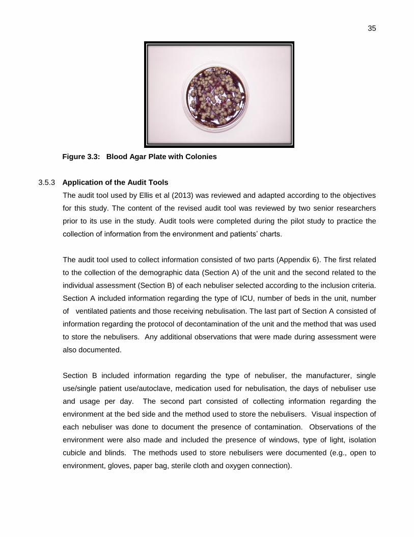
35
Figure 3.3: Blood Agar Plate with Colonies
3.5.3 Application of the Audit Tools
The audit tool used by Ellis et al (2013) was reviewed and adapted according to the objectives
for this study. The content of the revised audit tool was reviewed by two senior researchers
prior to its use in the study. Audit tools were completed during the pilot study to practice the
collection of information from the environment and patients’ charts.
The audit tool used to collect information consisted of two parts (Appendix 6). The first related
to the collection of the demographic data (Section A) of the unit and the second related to the
individual assessment (Section B) of each nebuliser selected according to the inclusion criteria.
Section A included information regarding the type of ICU, number of beds in the unit, number
of ventilated patients and those receiving nebulisation. The last part of Section A consisted of
information regarding the protocol of decontamination of the unit and the method that was used
to store the nebulisers. Any additional observations that were made during assessment were
also documented.
Section B included information regarding the type of nebuliser, the manufacturer, single
use/single patient use/autoclave, medication used for nebulisation, the days of nebuliser use
and usage per day. The second part consisted of collecting information regarding the
environment at the bed side and the method used to store the nebulisers. Visual inspection of
each nebuliser was done to document the presence of contamination. Observations of the
environment were also made and included the presence of windows, type of light, isolation
cubicle and blinds. The methods used to store nebulisers were documented (e.g., open to
environment, gloves, paper bag, sterile cloth and oxygen connection).

36
The time of assessment and additional observations of the environment were documented for
e.g. if the patient was isolated and the various air filtering systems for each ICU. The
additional information collected from the environment were not included in the final results.
There were no discrepancies found regarding the methods of sample collection. During the
pilot study the routine of the procedures were established and documented.
3.6 DATA COLLECTION
3.6.1 Completion of Section A of the audit tool with Unit Manager/Shift Leader
The unit manager or shift leader of the ICU was asked questions in order to complete Section
A of the audit tool on the day of audit. Thereafter the unit manager/shift leader identified the
beds in the unit of patients receiving MV. The unit manager/shift leader assisted in identifying
nebulisers at the bedsides of patients in the ICU which was then listed for assessment.
3.6.2 Assessment of Nebuliser and Environment
The second part of the audit tool (Section B) was completed for nebulisers at the bed sides of
patients that were identified by the unit manager for assessment. The nurse at the bedside
was approached to discuss the nature of the study and to answer any questions arising from
the discussion. The researcher ensured that the research procedure did not interfere in the
patient’s care at the time of audit. The first part of Section B was completed by gathering
information from the ICU chart or with the assistance of the nurse. The second part consisted
of observations made regarding the environment and the nebuliser at the bed.
3.6.3 Collection of Nebuliser and Air Samples
Nebuliser swabbing and air samples were done by following the protocols established and
practiced through the pilot study mentioned above.
3.7 ASSESMENT AND IDENTIFICATION PROCEDURE
The BAPs were assessed for fungal and bacterial growth after the incubation time was
completed as practiced at the PML. All information collected during the assessment of each
BAPs was written up on a Microsoft Excel spreadsheet and filed. Similar colonies cultured from
the air samples and nebulisers were grouped according to their visual description for each
hospital. The most frequently observed colonies which were similar and identified in both
nebuliser and air samples for each hospital were documented.
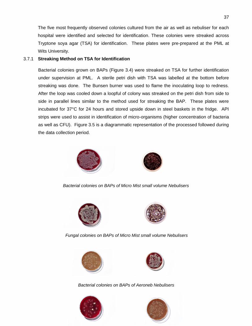
37
The five most frequently observed colonies cultured from the air as well as nebuliser for each
hospital were identified and selected for identification. These colonies were streaked across
Tryptone soya agar (TSA) for identification. These plates were pre-prepared at the PML at
Wits University.
3.7.1 Streaking Method on TSA for Identification
Bacterial colonies grown on BAPs (Figure 3.4) were streaked on TSA for further identification
under supervision at PML. A sterile petri dish with TSA was labelled at the bottom before
streaking was done. The Bunsen burner was used to flame the inoculating loop to redness.
After the loop was cooled down a loopful of colony was streaked on the petri dish from side to
side in parallel lines similar to the method used for streaking the BAP. These plates were
incubated for 37°C for 24 hours and stored upside down in steel baskets in the fridge. API
strips were used to assist in identification of micro-organisms (higher concentration of bacteria
as well as CFU). Figure 3.5 is a diagrammatic representation of the processed followed during
the data collection period.
Bacterial colonies on BAPs of Micro Mist small volume Nebulisers
Fungal colonies on BAPs of Micro Mist small volume Nebulisers
Bacterial colonies on BAPs of Aeroneb Nebulisers

38
Fungal colonies on BAPS of Aeroneb Nebulisers
Bacterial colonies on BAPs of Air Samples
Fungal colonies on BAPs of Air Samples
Figure 3.4: Colonies on BAPs of Nebulisers and Air Samples
3.8 DATA ANALYSIS
Descriptive statistics was used to analyse data relating to ICU and nebuliser
demographics. Demographic information described by categorical data such as nebuliser
type, aerosol medication and nebuliser dose was summarized using numbers and
percentages. Descriptive statistics relating to days and frequency of nebuliser use was
summarized by using means and standard deviation. The Fischer exact test was used to
ascertain the association of contamination of the Micro Mist small volume nebuliser to the
following variables: stored wet, stored in a glove, stored under a glove and open to the

39
environment. Statistical significance was set at 5% (p-value < 0.05) for all inferential statistics.
Percentages and numbers were used to describe the association of nebuliser contamination
with different storage and decontamination practices. This information is included in the text
and presented in graphs and tables. Data analysis was conducted using IBM SPSS 22 and
Stata13.1. Data from the participating hospitals were pooled together in one data set and
analysed as such to maintain hospital anonymity.

40
Figure 3.5: A Diagrammatic Representation of the Processes Followed During the
Data Collection Period
Obtaining consent from the Hospital Manager/Chief Executive Manager/Infection Control Manager
Obtaining consent from the Unit Manager of each ICU
Interview with the Unit managers
Four private hospitals
Seven ICUs
Beds screened = 440
Patients receiving MV = 132
Patients receiving aerosol therapy = 92
Beds selected and sampled = 61
Hospital 1 Beds sampled 12
Hospital 2 Beds sampled 12
Hospital 3 Beds sampled 32
Hospital 4 Beds sampled 5
61 BAP sampled from nebulisers for bacterial contamination
61 BAP sampled from the
surrounding air for fungal
contamination
61 BAP sampled from the
surrounding air for bacterial
contamination
61 BAP sampled from nebulisers
for fungal contamination
Selected bacterial samples streaked on TSA for identification
37 Micro Mist small volume nebulisers 24 Aeroneb nebulisers
Data collection period: 27 days
Samples taken at each bed side: 2 nebuliser and 2 air samples

41
CHAPTER 4
4. RESULTS
4.1 INTRODUCTION
This chapter presents the results obtained through data collected from both audit tools and
samples taken. This includes information collected through visual inspection of the nebuliser,
the storage method of the nebuliser and the surrounding ICU environment. Storage and
decontamination protocols for each ICU were obtained by completing Section A of the audit
tool with the unit manager. The type of aerosol medication and the frequency of application of
this medication through the nebuliser was obtained from the patient’s chart.
Consent was obtained from four private hospitals in Pretoria. Seven ICUs within these
hospitals were selected for sampling. The types of ICUs included in the study were: cardiac
care, medical, surgical, trauma, neurology and mixed units. A total of 440 beds were screened
in the seven ICUs. Of these beds 132 (30.0%) patients received MV and 92 (69.7%) aerosol
therapy. A total of 61 nebulisers and their surrounding air were sampled. The total nebuliser
sample included 24 Aeroneb and 37 Micro Mist small volume nebulisers. The remaining
nebulisers and their surrounding air were not sampled due to isolation of the patients and the
risks of cross-infection or time constraints. Due to isolation of patients, the number of beds in
the ICUs selected changed during the course of data collection.
The results obtained in this study will be discussed in line with the research objectives outlined
in Chapter one.
4.2 THE TYPES OF NEBULISERS AND MEDICATION USED FOR AEROSOL THERAPY IN
ICUs OF HOSPITALS IN PRETORIA
Two types of nebulisers were identified in the four hospitals during the study, namely the
Aeroneb nebuliser, a new addition to the ventilator circuit and the Micro Mist small volume
nebuliser. The Aerosol Tee connector that forms part of Micro Mist small nebuliser was the
only type of Tee connector identified in the ICUs in Pretoria. Figure 4.1 provides information on
the nebuliser types identified in the four hospitals.

42
Figure 4.1: Nebuliser Types Identified in the Four Hospitals
In hospital one only the Micro Mist small volume nebuliser was used. In hospital two only the
Micro Mist small volume nebuliser was sampled although both nebulisers were used. In
hospital two the Aeroneb nebuliser was used to a lesser extent and only one was observed
during the data collection period. In hospital three both devices were in use. In hospital four
only Aeroneb nebulisers were sampled although both types of nebulisers were used to
administer aerosol medication.
Figure 4.2 represents the different ICUs where the two types of nebulisers were sampled. The
eight units represent different types of ICUs. Unit zero represents the cardiac ICU, unit one the
medical ICU, unit two the surgical ICU, unit 3 the surgical and trauma ICU, unit four the trauma
and neurology ICU, unit five the mixed ICU, unit six the surgical and medical ICU and unit
seven the cardiac and surgical ICU. The mixed ICU [unit 5] used the most Aeroneb (n=11) and
Micro Mist small volume nebulisers (n=17) when administering aerosol medication.
19
5
12 12 13
0
5
10
15
20
Hospital 1 Hospital 2 Hospital 3 Hospital 4
Nu
mb
er
of
ne
bu
lise
rs
HOSPITALS
Aeroneb Hudson T-piece

43
Figure 4.2: Type of Nebulisers in the Different ICUs
Four types of aerosol medication were identified namely, bronchodilators (e.g. Atrovent,
Ventolin), corticosteroids (e.g. Pulmicort), saline (0.9%) and mucolytics (e.g. Bisolvon). Figure
4.3 provides information regarding aerosol medications used in the ICUs.
Figure 4.3: Types of Aerosol Medication
1 1
3
11
1
7
3 2 2
17
7 6
0
2
4
6
8
10
12
14
16
18
Unit 0 Unit 1 Unit 2 Unit 3 Unit 4 Unit 5 Unit 6 Unit 7
Nu
mb
er
of
ne
bu
lise
rs
Type of ICU
Aeroneb nebuliser Micro Mist small volume nebuliser
22
14
2
23
2
8
14
Bronchodilators Mucolytics other thansaline
Corticosteroids Saline
Nu
mb
er o
f n
ebu
liser
s
Aeroneb nebuliser
Micro Mist small volume nebuliser

44
The aerosol medication mostly used in the ICUs was bronchodilators (n=45) and mode of
application was similar (Aeroneb, n=22 and Micro Mist, n=23). The aerosol medication least
used was mucolytics (n=2) other than saline. Saline aerosolisation was usually used with
physiotherapy treatments (n=16) and the mode of application was mainly the Micro Mist small
volume nebuliser.
Bronchodilators in order mostly used were; Combivent, Atrovent, Duolin and Ventolin.
Pulmicort was the only corticosteroid used for aerosolisation and Bisolvon the only mucolytic
other than saline. Pulmicort was also mainly used in conjunction with bronchodilators. The four
types of aerosol medication used for aerosolisation are displayed according to their application
in the eight types of units in Figure 4.4.
F
igure
4.4:
Aero
sol
Medi
catio
n
used
in the Different ICUs
Bronchodilation therapy was used the most across all units and with higher frequencies noted
in the mixed ICU (n=20) [unit 5], cardiac and surgical ICU (n=8) [unit 7] and lastly the surgical
and medical ICU (n=6) [unit 6]. Corticosteroid therapy was the most used in the mixed ICU
(n=9) [unit 5]. Saline administration as aerosol medication was the second most frequently
used across ICUs as it was in use by six units. Mucolytic therapy (n=2) was only in use in the
cardiac and surgical ICU [unit 7].
4
1 2
3
1
20
6
8
2 3
1
9
4 5
1
1
1
7
2
4
0
5
10
15
20
25
Unit 0 Unit 1 Unit 2 Unit 3 Unit 4 Unit 5 Unit 6 Unit 7
Nu
mb
er
of
ne
bu
lise
rs
Units
Bronchodilator Mucolitics Corticosteroids saline

45
4.3 DAILY APPLICATION OF NEBULISERS IN THE ICU
The nebuliser dose describes the frequency of daily application and is illustrated in Figure 4.5.
Eleven categories of nebuliser dose were identified. Categories that consisted of two time
periods were due to two types of aerosol medications used within the nebuliser at different time
intervals. The categories are as follows: zero: four hourly, one: as needed, two: six hourly,
three: twice a day, four: eight hourly, five: twice a day and four hourly, six: six and eight hourly,
seven: four and six hourly, eight: four and eight hourly, nine: two and three hourly, ten: eight
and twelve hourly, eleven: six and twelve hourly.
Figure 4.5: Aerosolisation Dosage for the Two Types of Nebulisers
The most frequently used dose to administer aerosol medication was a dose of six hourly
(n=25) [category two] with the Aeroneb in use the most (n=14) and the Micro Mist less (n=11)
in use in this dose category. The second most frequently prescribed dose was four hourly
(n=9) as depicted by category zero.
Table 4.1 provides information regarding the mean number of days the different types of
nebulisers were used as well as the mean number of aerosolisation sessions.
14
1 1 1 4
1 1 1
9
2
11
7
2 5
2
1 1 0
5
10
15
20
25
30
Nu
mb
er o
f n
ebu
liser
s
Nebuliser dose
Aeroneb nebuliser Micro Mist small volume nebuliser

46
Table 4.1: Nebuliser Days and Aerosolisation Sessions
The mean number of days that the Aeroneb nebuliser was in use was 15.0 (±14.6) days and
the mean number of aerosolisation sessions 75 (±74.9). The mean number of days the Micro
Mist small volume nebuliser was in use was 11.6 (±13.9) and the mean number of
aerosolisation sessions 56.2 (±90.1).
Six of the Micro Mist small volume nebulisers was used for more than 90 aerosolisation
sessions and three Micro Mist small volume nebulisers was used for more than 30 days.
4.4 DECONTAMINATION PROTOCOLS AND STORAGE PRACTICES OF NEBULISERS IN
THE ICUs
Six decontamination storage protocols were identified in the seven ICUs during the course of
the study. A summary of the protocol is given in Table 4.2.
The ICU in hospital one had a decontamination storage protocol [protocol one], hospital two
had two ICUs and both had the same protocol for decontamination and storage [protocol two].
Hospital three had two ICUs with different decontamination and storage protocols [protocol
three and four] and hospital four had two ICUs with different decontamination protocols but the
same storage protocol [protocol five and six].
None of the Micro Mist small volume nebulisers were discarded after use. All methods for
decontamination differed in the type of rinsing solution used. Three types of rinsing solutions
were identified namely, 70% alcohol, wash with bioscrub and sterile water. Most protocols
stated that the Micro Mist small volume nebulisers be dried with a paper towel (n=4). Only one
protocol had no method of decontamination however, nebulisers were being dried with a paper
Type of Nebulisers n Minimum Maximum Mean ±SD
Aeroneb 24
Nebuliser days 24 2.0 66.0 15.0 ±14.6
Aerosolisation sessions 24 8.0 264.0 75 ±74.9
Micro Mist 37
Nebuliser days 37 1.0 62.0 11.6 ±13.9
Aerosolisation sessions 37 2.0 496.0 56.2 ±90.1

47
towel after use. Decontamination protocols differed between ICUs, and between the two ICUs
within Hospital three. There were no decontamination and storage protocols reported by the
unit managers for the Aeroneb nebuliser. The Aeroneb nebuliser is developed to remain in the
ventilator circuit during the period of MV and does not need to be decontaminated or stored
separately from the MV circuit.
Storage protocol one was the only protocol whereby the Micro Mist small volume nebuliser was
left open to the environment. However it was observed that the nebulisers were not stored on
a hook at the bed side but left open in a petri dish. Storage protocols four to six stated that the
Micro Mist small volume nebulisers be stored inside a latex glove while connected to the
oxygen output of the ventilator. In protocol two, used in two ICUs, the Micro Mist small volume
nebulisers were not dried before they were stored under a sterile cloth. According to the unit
manager these sterile cloths are replaced every week. In protocol three the Micro Mist small
volume nebulisers were stored inside an acceptor bag, however, no acceptor bag was
observed during data collection in that ICU. Storage protocols differed between ICUs, and
between the two ICUs within Hospital three.
Table 4.2: Type of Decontamination and Storage Protocols Reported by Unit
Managers to be in Place for the Micro Mist small Volume Nebuliser
Protocol Discarded Rinsed Method of Decontamination Dried
Nebuliser Drying Method Method of Storage
1 No Yes 70 % alcohol Yes Paper towel
Connected to the oxygen output of the ventilator, stored on a hook at the bed side and open to the environment
2 No Yes Provac water No None
Connected to oxygen, taken apart and left to dry under a sterile cloth
3 No Yes Wash with bioscrub once a day
Yes Paper towel Stored in acceptor(sterile) bag
4 No No None Yes Paper towel Stored in a glove connected to the oxygen output
5 No Yes Saline of Sterile water No None Stored in a glove connected to the oxygen output
6 No Yes Wash with bioscrub after aerosolisation
Yes Paper towel Stored in a glove connected to the oxygen output

48
4.5 THE INCIDENCE OF CONTAMINATION OF NEBULISERS AFTER USE WITHIN A
VENTILATOR CIRCUIT
A total of 61 nebuliser samples were collected and a total of 31 (50.8%) nebuliser swabs
presented with contamination. The incidence of contamination of both types of nebulisers was
similar: Micro Mist small volume (n=19; 51.4%) and Aeroneb (n=12; 50%). Figure 4.6
illustrates the incidence of contamination of the Micro Mist small volume nebulisers.
A total of 37 Micro Mist small volume nebulisers were included in this study. A combination of
bacterial and fungal growth was seen the most in the Micro Mist small volume nebulisers
(n=11; 29.7%). The total fungal growth (n=17) were more than the total bacterial growth (n=13)
in the Micro Mist small volume nebulisers.
Figure 4.6: Incidence of Contamination of the Micro Mist small Volume Nebuliser
Figure 4.7 provides information regarding the incidence of contamination of the Aeroneb
nebulisers. A total of 24 Aeroneb nebulisers were included in this study. A combination of
bacterial and fungal growth (n=9; 37.5%) was mostly observed. The total bacterial growth
(n=11) was almost similar to the total fungal growth (n=10) in the Aeroneb nebulisers.
2
6
11
18
Bacterial Fungal Bacterial and Fungal No growth

49
Figure 4.7: Incidence of contamination of the Aeroneb Nebuliser
The following figures present the incidence of contamination found in the different nebulisers in
the four hospitals.
Hospital One
A total of 12 Micro Mist small volume nebuliser swabs were collected in Hospital one. The
incidence of contamination in the Micro Mist small volume nebulisers in this hospital is shown
in Figure 4.8. Only three Micro Mist small volume nebulisers (n=3; 25%) presented with
contamination in Hospital one with an even distribution of bacterial (n=1), fungal (n=1) and
bacterial and fungal (n=1) contamination.
Hospital Two
A total of 12 Micro Mist small volume nebuliser swabs were collected in hospital two. The
incidence of contamination in the Micro Mist small volume nebuliser in this hospital is shown in
Figure 4.9. In Hospital two, eight (66.7%) Micro Mist small volume nebulisers presented with
bacterial and fungal growth (n=4; 33.3%).
Hospital Three
A total of 32 nebuliser swabs which includes, Aeronebs (n=19) and Micro Mist small volume
nebulisers (n=13) were collected in Hospital three. The incidence of contamination of
nebulisers in this hospital is shown in Figure 4.10 and 4.11. Eight (61.5%) Micro Mist small
volume nebulisers in hospital three presented with contamination of which a combination of
bacterial and fungal growth (n=6; 46.2%) was mostly observed. The total fungal growth (n=8)
was more than the total bacterial growth (n=6) in the Micro Mist small volume nebulisers. Less
2 1
9
12
Bacterial Fungal Bacterial and Fungal No Growth

50
contamination was observed in the Aeroneb nebulisers in Hospital three, with nine (47.4%)
nebulisers presenting growth where a combination of bacterial and fungal contaminants (n=6;
31.6%) were observed. The total bacterial growth (n=8) was more than the total fungal growth
(n=7) in the Aeroneb nebulisers.
Hospital Four
A total of five Aeroneb nebuliser swabs were collected in Hospital four. The incidence of
contamination of nebulisers in this hospital is shown in Figure 4.12. Three (60%) Aeroneb
nebulisers presented with bacterial and fungal contamination (n=3).
Hospital two presented with the most contamination in the Micro Mist small volume nebulisers
(66.7%) and Hospital four with the most Aeroneb contamination (60%).
Figure 4.8: Incidence of contamination Figure 4.9: Incidence of contamination
in Hospital One in Hospital Two
Figure 4.10: Incidence of contamination in Figure 4.11: Incidence of contamination
Micro Mist small volume Aeroneb Nebulisers in
Nebulisers in Hospital Three in Hospital Three
1
3
4
4
Bacterial Fungal
Bacterial and Fungal No growth
2 1
6 10
Bacterial Fungal
Bacterial and Fungal No growth
0
2
6
5
Bacterial Fungal
Bacterial and Fungal No growth
1 1
1
9
Bacterial Fungal
Bacterial and Fungal No growth
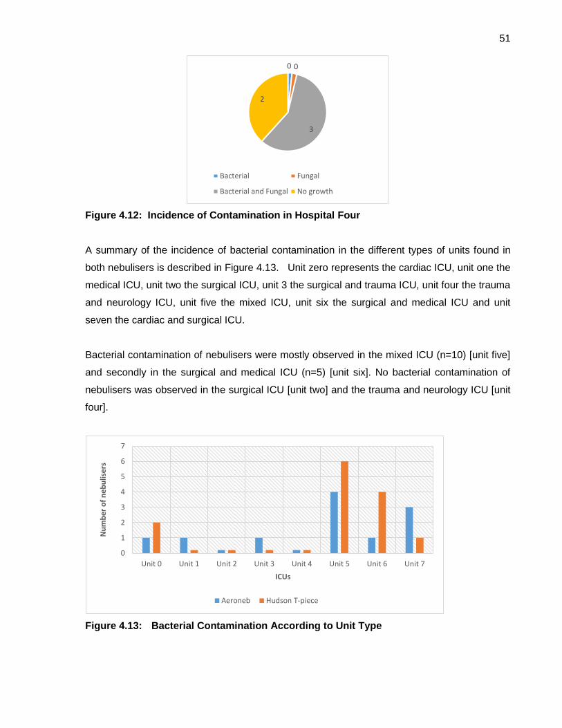
51
Figure 4.12: Incidence of Contamination in Hospital Four
A summary of the incidence of bacterial contamination in the different types of units found in
both nebulisers is described in Figure 4.13. Unit zero represents the cardiac ICU, unit one the
medical ICU, unit two the surgical ICU, unit 3 the surgical and trauma ICU, unit four the trauma
and neurology ICU, unit five the mixed ICU, unit six the surgical and medical ICU and unit
seven the cardiac and surgical ICU.
Bacterial contamination of nebulisers were mostly observed in the mixed ICU (n=10) [unit five]
and secondly in the surgical and medical ICU (n=5) [unit six]. No bacterial contamination of
nebulisers was observed in the surgical ICU [unit two] and the trauma and neurology ICU [unit
four].
Figure 4.13: Bacterial Contamination According to Unit Type
0 0
3
2
Bacterial Fungal
Bacterial and Fungal No growth
0
1
2
3
4
5
6
7
Unit 0 Unit 1 Unit 2 Unit 3 Unit 4 Unit 5 Unit 6 Unit 7
Nu
mb
er
of
ne
bu
lise
rs
ICUs
Aeroneb Hudson T-piece

52
A summary of the incidence of fungal contamination in the different types of units found in both
nebulisers is described in Figure 4.14. The highest number of fungal contamination of
nebulisers was observed in the mixed ICU (n=10) [unit 5] and secondly in the surgical and
medical ICU (n=5) [unit 6]. No fungal contamination of nebulisers was observed in the surgical
ICU [unit two].
Figure 4.14: Fungal Contamination According to Unit Type
Ten of the 24 Aeronebs and 17 of the 37 Micro Mist small volume nebulisers presented with
fungal contamination which can be divided into moulds and yeasts. These findings are
presented in Figure 4.15. The nebulisers presented with yeast contamination were mostly
observed in both Aeroneb (7/24; 29.2%) and Hudson Micro Mist small volume nebulisers
(15/37; 40.5%). Less contamination with moulds and a combination of moulds and yeasts
were observed in both Aeronebs and Micro Mist small volume nebulisers.
1 1 1
3
1
3
2 2
7
4
2
0
1
2
3
4
5
6
7
8
Unit 0 Unit 1 Unit 2 Unit 3 Unit 4 Unit 5 Unit 6 Unit 7
Nu
mb
er
of
ne
bu
lise
rs
ICUs
Aeroneb Hudson T-piece
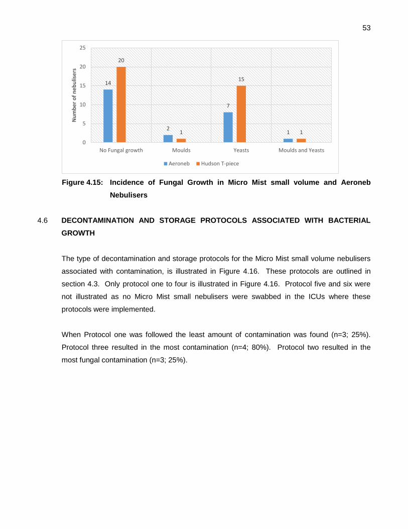
53
Figure 4.15: Incidence of Fungal Growth in Micro Mist small volume and Aeroneb
Nebulisers
4.6 DECONTAMINATION AND STORAGE PROTOCOLS ASSOCIATED WITH BACTERIAL
GROWTH
The type of decontamination and storage protocols for the Micro Mist small volume nebulisers
associated with contamination, is illustrated in Figure 4.16. These protocols are outlined in
section 4.3. Only protocol one to four is illustrated in Figure 4.16. Protocol five and six were
not illustrated as no Micro Mist small nebulisers were swabbed in the ICUs where these
protocols were implemented.
When Protocol one was followed the least amount of contamination was found (n=3; 25%).
Protocol three resulted in the most contamination (n=4; 80%). Protocol two resulted in the
most fungal contamination (n=3; 25%).
14
2
7
1
20
1
15
1
0
5
10
15
20
25
No Fungal growth Moulds Yeasts Moulds and Yeasts
Nu
mb
er
of
ne
bu
lise
rs
Aeroneb Hudson T-piece

54
Figure 4.16: Bacterial and Fungal Contamination of Micro Mist small volume
Nebulisers Associated with Decontamination and Storage Protocols
The following incidence of contamination was observed with different rinsing solutions and
drying methods as part of the decontamination storage protocols reported by the unit
managers.
4.6.1 Rinsing Solutions
Three types of rinsing solutions were identified in the decontamination protocols. Solution one:
70% alcohol; solution two: sterile water and solution three: washed with bioscrub. Solution
one resulted in the least bacterial contamination of Micro Mist small volume nebuliser (n=2;
16.7%) and solution three resulted in the most bacterial contamination (n=3; 75%). Solution
two resulted in bacterial contamination of (n=5; 41.7%) and using no rinsing solution, resulted
in three out of the nine nebulisers presenting with bacterial contamination (33.3%). Solution
one resulted in the least fungal contamination of the Micro Mist small volume nebulisers (n=2;
16.7%) and solution three resulted in the most fungal contamination (n=4; 100%). Solution two
resulted in fungal contamination of (n=7; 58.3%) and using no rinsing solution, resulted in four
out of the nine Micro Mist small volume nebulisers with fungal contamination (n=4; 44.4%).
1 1 1
3
1 1 1
4
3 3
9
4
1
4
Protocol 1 Protocol 2 Protocol 3 Protocol 4
Nu
be
r o
f N
eb
ulis
ers
Bacterial Fungal Bacterial and Fungal No contamination

55
4.6.2 Drying Methods
Two different methods were used to dry the Micro Mist small volume nebulisers as part of the
decontamination protocols and are as follows, method one: dry the nebuliser with a paper
towel; method two: take the nebuliser apart and leave to dry under a sterile cloth. Method two
resulted in the most bacterial contamination (n=5; 41.6%) and fungal contamination (n=7;
58.3%). Method one resulted in the least bacterial contamination (n=8; 32%) and fungal
contamination (n=10; 40.0%).
4.6.3 Storage Methods
Four types of storage methods were evaluated during visual inspection on the day of audit and
are as follows: method one: stored in a glove; method two: stored under a sterile cloth; method
three: stored open to the environment and method four: stored in a paper bag. Most Micro
Mist small volume nebulisers were stored in a latex glove (n=20). More than a third of the
Micro Mist small volume nebulisers, stored in a glove presented with bacterial growth (n=7;
35%) and half with fungal contamination (n=9; 47,4%). Since the p-value is 0.717 (>0.05),
there is evidence of no relationship (association) between method one and bacterial growth.
Method two resulted in the most bacterial contamination (n=4; 44.4%) and the most fungal
contamination (n=6; 66.7%). Since the p-value is 0.624 (>0.05), there is evidence of no
relationship (association) between method two and bacterial growth.
Method three resulted in the least bacterial (n=2; 28.6%) and fungal contamination (n=2;
28.6%). No paper bags were observed during the sampling procedures. Since the p-value is
0.594 (>0.05), there is evidence of no relationship (association) between method three and
bacterial growth.
One nebuliser was left in the ventilator circuit and presented with fungal contamination and no
bacterial contamination. Five Micro Mist small volume nebulisers were stored connected to the
oxygen port of the ventilator. Only one presented with bacterial and fungal contamination (n=1;
20%).
4.6.4 Aerosol Medication
Contamination associated with aerosol medication used in the Micro Mist small volume and
Aeroneb nebulisers were observed. Bronchodilators were mostly used for aerosol nebulisation
in Micro Mist small volume nebulisers followed by saline, corticosteroid and mucolytics (other

56
than saline). Bronchodilators were also mostly used in Aeroneb nebulisers followed by
corticosteroid and saline. No mucolytics were used in Aeroneb nebulisers. The highest
bacterial contamination was found in Micro Mist small volume (n=4; 50%) and Aeroneb
nebulisers (n=8; 57.1%) associated with corticosteroids aerosolisation. Bronchodilator
aerosolisation was associated with (n=9; 39.1%) contamination in the Micro Mist small volume
nebuliser and (n=10; 45.5%) in the Aeroneb nebuliser. Saline inhalation was associated with
(n=5; 35.7%) bacterial contamination. No contamination was found in Micro Mist small volume
nebulisers used for mucolytics (n=2; 0%). No contamination was found in Aeroneb nebulisers
used for saline aerosolisation.
4.6.5 Wet and Dry Chambers
Visual inspection of each nebuliser on the day of audit was performed to establish if the
chambers were stored wet or dry. In this study a nebuliser was described as “wet” when fluid
was observed in base of the chamber and the inner surface of the chamber. The results
obtained through visual inspection of the Micro Mist small volume nebuliser is illustrated in
Figure 4.17.
4.6.5.1 Micro mist small volume nebuliser
Most of the Micro Mist small volume nebulisers were stored wet (n=33; 89.2%). Only four
nebulisers were stored dry (n=4; 10.8%). Bacterial contamination was found in 11 of the 33 wet
Micro Mist small volume nebulisers (n=11; 33.3%) and fungal contamination found in 16 (n=16;
48.5%). One dry Micro Mist small volume nebuliser presented with (n=1; 25%) bacterial and
(n=1; 25%) fungal contamination. Three out of the four dry Micro Mist small volume nebulisers
presented with no contamination (n=3; 75%). One species was identified in seven wet Micro
Mist small volume nebulisers (7/11) and in one dry Micro Mist small volume nebuliser (1/4).
Two species were identified in three nebulisers (3/11) and three species in one nebuliser
(1/11). Most of the wet Micro Mist small volume nebulisers presented with more than 50
colonies (6/11) as well as one dry nebuliser (1/4). Two Micro Mist small volume nebulisers
presented with colonies between one and 10 (2/11) and two nebulisers with colonies between
21 and 30 (2/11). Although the odds ratio (0.67 and 95% Constant Interval = 0 – 5.42) shows
a 33% reduced risk of bacterial growth in dried as compared to wet nebulisers, there is
evidence of no association between dryness of the nebuliser chamber and bacterial growth (p-
value=0.7367) because the p-value is not significant.

57
Three out of the six Micro Mist small volume nebulisers that was used for more than 90
aerosolisation sessions presented with both bacterial and fungal contamination (50%). Nine
out of the 31 Micro Mist small volume nebulisers used for less than 90 sessions presented with
bacterial contamination (29%) and eleven with fungal contamination (35.5%).
Figure 4.17: Visual Inspection of the Micro Mist small volume Nebuliser
4.6.5.2 Aeroneb nebuliser
The results obtained through visual inspection of the Aeroneb nebuliser are illustrated in Figure
4.18. Most of the Aeroneb nebulisers were found to be wet with visual inspection (n=20;
83.3%).
Only four were dry (n=4; 16.7%). Bacterial contamination was found in 10 of the 20 wet
Aeroneb nebulisers (n=10; 50%) and in one dry nebuliser (n=1; 25%). Fungal contamination
was found in 8 of the 20 wet Aeroneb nebulisers (n=8; 40%) and two dry nebulisers (n=2;
50%). One species was identified in all the wet Aeroneb nebulisers (10/10) and in one dry
nebuliser (1/4). Colonies between one and 10 were identified in six wet Aeroneb nebulisers
(6/10) and more than 50 colonies were identified in three wet Aeroneb nebulisers (3/10) and in
one dry nebuliser (1/10).
4
33
Dry Wet

58
Figure 4.18: Visual Inspection of the Aeroneb Nebuliser
4.7 BACTERIAL MICRO-ORGANISMS IDENTIFIED IN CONTAMINATED NEBULISERS AND
SURROUNDING AIR
Ten types of micro-organisms were identified in the nebulisers and surrounding air. This is
shown in Figure 4.19. Micro-organisms were identified in 19 air samples, six Micro Mist small
nebuliser and seven Aeroneb nebuliser samples. The different micro-organisms included;
Enterococcus spp., Coagulase-negative Staphylococcus spp., Staphylococcus aureus,
Neisseria spp.; Bacillus spp., Micrococcus and related species, Pseudomonas stutzeri,
Brevundimonas vesicularis, Empedobacter (F.) brevis and Stenotrophomonas spp.
Coagulase-negative Staphylococcus was identified the most in air samples (n=8; 42.1%)
followed by Pseudomonas Stutzeri (n=4; 21.1%) and Neisseria spp. (n=3; 15.8%). Coagulase-
negative Staphylococcus (CoNS) was also the most identified in Aeroneb nebulisers (n=4;
57.1%). Enterococcus spp. was the most identified in Micro Mist small volume nebulisers
(n=2; 33.3%) and also identified in all three types of samples.
The micro-organisms identified in the samples included seven contaminated nebulisers and
their corresponding air samples. The same bacterial micro-organisms were identified in two
nebulisers and their corresponding air samples (n=2; 28.6%). An Enterococcus sp. was
identified in one Micro Mist small volume nebuliser and its corresponding air sample and a
Coagulase-negative Staphylococcus sp. was identified in one Aeroneb nebuliser and its
corresponding air sample.
4
20
Dry Wet

59
A=Enterococcus spp., B=Coagulase-negative Staphylococcus spp., C=Staphylococcus
aureus, D=Neisseria spp.; E=Bacillus spp., F=Micrococcus and related species,
G=Pseudomonas stutzeri, H=Brevundimonas vesicularis, I=Empedobacter (F.) brevis,
J=Stenotrophomonas spp.
Figure 4.19: Bacterial Micro-organisms Identified in Contaminated Nebulisers and Air
Samples
Figure 4.20 illustrates the bacterial and fungal contamination of the surrounding air where
these nebulisers were kept. A high incidence of contamination was identified in air samples
(n=60; 98.4%). Most of the air samples had bacterial and fungal contamination (n=43; 70.5%)
followed by fungal contamination (n=12; 19.7%) and bacterial contamination (n=5; 8.2%). The
total fungal contamination (n=55; 90.2%) in air samples was higher than the total bacterial
contamination (n=48; 78.7%). The various ventilation systems were identified by questioning
the unit manager in the hospitals and were as follows; Hospital one- HEPA filter; Hospital two-
Chilled Water Fan Coil Unit and Direct Expansion System; Hospital three - Air Handling Unit;
Hospital four- Chilled Water Fan Coil Unit.
2
4
1
2
1
1
2
1
2
8
1
3
1
1
4
1
0
1
2
3
4
5
6
7
8
9
A B C D E F G H I J
Nu
mb
er
of
sam
ple
s
Types of organisms Aeroneb Nebuliser Hudson T-piece Nebuliser Air Samples
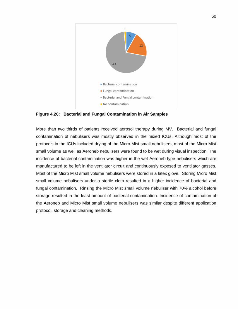
60
Figure 4.20: Bacterial and Fungal Contamination in Air Samples
More than two thirds of patients received aerosol therapy during MV. Bacterial and fungal
contamination of nebulisers was mostly observed in the mixed ICUs. Although most of the
protocols in the ICUs included drying of the Micro Mist small nebulisers, most of the Micro Mist
small volume as well as Aeroneb nebulisers were found to be wet during visual inspection. The
incidence of bacterial contamination was higher in the wet Aeroneb type nebulisers which are
manufactured to be left in the ventilator circuit and continuously exposed to ventilator gasses.
Most of the Micro Mist small volume nebulisers were stored in a latex glove. Storing Micro Mist
small volume nebulisers under a sterile cloth resulted in a higher incidence of bacterial and
fungal contamination. Rinsing the Micro Mist small volume nebuliser with 70% alcohol before
storage resulted in the least amount of bacterial contamination. Incidence of contamination of
the Aeroneb and Micro Mist small volume nebulisers was similar despite different application
protocol, storage and cleaning methods.
5
12
43
1
Bacterial contamination
Fungal contamination
Bacterial and Fungal contamination
No contamination

61
CHAPTER 5
5. DISCUSSION
5.1 INTRODUCTION
The incidence of contamination as well as the current practice of decontamination and storage
of nebulisers used within a ventilator circuit in ICUs in Pretoria is unknown. A similar study
was done in the ICUs in Johannesburg and the results of that study will be compared with the
current study to assist in gaining an overall perspective of contamination and nebuliser
practice in ICUs in Gauteng.
The main finding from the current study was the incidence of bacterial and fungal
contamination of the Micro Mist small volume nebuliser and the Aeroneb nebuliser when used
in a ventilator circuit. The incidence of contamination of the Micro Mist small volume nebuliser
in this study corresponds with the incidence of contamination found in the same aerosol device
in ICUs in Johannesburg (Ellis et al., 2013). The Aeroneb nebuliser is a new device that was
recently introduced to clinical practice as a means of administration medication to a
mechanically ventilated patient. No previous research regarding the incidence of contamination
in the Aeroneb nebuliser could be sourced in the literature. The incidence of contamination
found in the Aeroneb nebuliser was similar to the incidence of contamination in the Micro Mist
small volume nebuliser. In the current study both of these nebulisers were used as single-
patient use devices which is in contrast to the information reported by Ellis et al. (2013) as the
Micro Mist small volume nebuliser was reclassified by the manufacturer from single-use to
single-patient use device. Secondly, all the ICUs had decontamination and storage protocols
in place for the Micro Mist small volume nebuliser, however, all of these protocols differed
between hospitals and different ICUs within the same hospital. Thirdly, more Micro Mist small
volume nebulisers that were stored under a sterile drape presented with bacterial
contamination which corresponds with Ellis et al. (2013) study. Lastly, almost 90% of Micro
Mist small volume nebulisers were stored wet. More than two thirds of ventilated patients,
received aerosol therapy in the ICUs in Pretoria, which indicated that aerosol therapy was an
important treatment during MV. This finding is supported by an international survey which
found that 95% of intensivists prescribe aerosol therapy to patients receiving MV (Ehrmann et
al., 2013).

62
5.2 THE TYPES OF NEBULISERS AND MEDICATION USED FOR AEROSOL THERAPY IN
ICUs IN PRETORIA
The Micro Mist small volume nebuliser is a jet nebuliser which connects to a port on the
ventilator and the Aeroneb nebuliser is a VMN which operates electronically within the
ventilator circuit. The Aeroneb nebuliser was not identified as one of the nebulisers used in the
ICUs in Johannesburg at the time of the study done by Ellis et al. (2013). The Micro Mist small
volume nebuliser was mostly used in the ICUs in Pretoria. This corresponds with Ellis et al.
(2013) study in ICUs in Johannesburg and an international survey conducted by Erhmann et
al. (2013). The Aeroneb nebuliser was used less frequently in the ICUs of hospitals in
Pretoria. A factor that could influence its usage might be the cost of this new device as the
cost is markedly higher than the Micro Mist small volume nebuliser. The Aeroneb device is
almost 10 times the cost of the Micro Mist small volume nebuliser. An international survey by
Ehrmann et al. (2013) supports this assumption and showed that only 14% of respondents
used VMN in MV whereby cost was suggested as a limiting factor.
According to the Unit managers in the ICUs that were surveyed, the Aeroneb nebuliser is used
when a patient requires continuous positive end-expiratory pressure or if a patient is on long
term ventilation. This is in contrast to the Micro Mist small volume nebuliser which is used
more frequently in patients requiring ventilation following surgical procedures who are
anticipated to be extubated from the ventilator soon.
Most Aeroneb samples were collected in the mixed ICUs. It could be hypothesized that there
were more long-term ventilated patients in the mixed ICUs who required aerosol therapy. A
report done by the International Nosocomial Infection Control Consortium (INICC) between
2003 and 2008 supports this hypothesis whereby the average length of stay (LOS) for patients
in an ICU consisting of mixed conditions e.g. medical and surgical cases was found to be much
higher than other types of ICUs (INICC, 2010).
Four types of aerosol medication were identified during the course of this study namely,
bronchodilators, corticosteroids, saline and mucolytics. Bronchodilators were the most frequent
aerosol medication used for nebulisation in the seven ICUs followed by corticosteroids, saline
and mucolytics. This corresponds with the study done by Ellis et al. (2013) whereby
bronchodilators were ranked the highest. Mucolytics however were the second highest ranked
medication administered in ICUs in Johannesburg while in Pretoria ICUs these ranked last in

63
the group of aerosol medications. Mucolytics and saline were grouped together in the study
done by Ellis et al., (2013) and therefore the higher rank of mucolytics. The mucolytics used in
the ICUs in Johannesburg were Bisolvon, which was only used in one nebuliser, and saline,
which was used in eight nebulisers. In the current study administration of bronchodilators were
high in both the Micro Mist small volume and Aeroneb nebuliser. An international survey
showed similar results whereby bronchodilators were administered the most frequent followed
by steroids (Erhmann et al., 2013). No antibiotics were used for nebulisation during this study
while in an international survey antibiotics and especially Colistin were regularly aerolised
(Ehrmann et al., 2013). Most of respondents in this survey done by Ehrmann et al. (2013)
worked in an adult mixed ICU setting and reported a positive viewpoint towards inhaled
antibiotics as an adjunctive or primary treatment method for pulmonary infections.
5.3 DAILY APPLICATION OF NEBULISERS IN THE ICU
The Micro Mist small volume nebuliser and the Aeroneb nebuliser are single-patient use
devices which indicate that they should be used on the same patient for a period of time
according to the manufacturer’s recommendations. The six hourly (four times a day)
aerosolisation of medication were used the most in both Aeroneb and Micro Mist small volume
nebulisers. Information regarding nebuliser dose and the date when aerosolisation
commenced were collected from the patient’s prescription chart or physiotherapy notes to
calculate the number of aerosolisation sessions over a period of time. The mean number of
days as well as the number of aerosolisation sessions that the Micro Mist small volume
nebuliser was used, was less than that of the Aeroneb nebuliser. The Micro Mist small volume
nebuliser can be used three times a day for 30 days which calculates to 90 aerosolisation
sessions (SMLT, 2000). Allen et al. (2005) also recommended that the Micro Mist small
volume nebuliser should be replaced every 24h if the patient had a respiratory infection. In the
current study, the mean number of aerosolisation sessions 56.2 (±90.1) and days 11.6 (±13.9)
in which the Micro Mist small volume nebuliser was used, was in accordance with what is
recommended by the SMLT (SMLT, 2000). Only six of the Micro Mist small volume nebulisers
were used for more than 90 aerosolisation sessions and three for more than 30 days. No
recent literature could be sourced regarding the frequency in which the Micro Mist small
volume nebuliser is used in South Africa or internationally. Replacing the Micro Mist small
volume nebuliser could be costly and time consuming. The Valved Neb Tee, is a different type
of Tee connector manufactured by Hudson RCI that is a single-patient use device and can be
left in the ventilator circuit when the nebuliser is removed. A valve in the Neb Tee connector
opens/closes upon insertion/removal of the nebuliser. The valve prevents loss of ventilation

64
pressure and the release of patient’s aerosols in the surrounding environment (Teleflex, 2014).
This could result in lower costs and less time spend to remove the Micro Mist small volume
nebuliser from the circuit. The Aeroneb nebuliser in contrast is left in the ventilator circuit and
can be set on an intermittent or continuous cycle. There is no specified limit to the frequency
with which the Aeroneb nebuliser may be used (Aerogen, 2014).
During this study no documentation could be found on the ICU cardex or in the patient files to
indicate how long the Micro Mist small volume nebuliser were in use or if it was replaced. The
study design didn’t allow interviews with nursing staff at the bed side and therefore the
researcher speculates that the ICU personnel were not informed regarding the preferred
application of the Micro Mist small volume nebuliser and/or Tee connector.
5.4 DECONTAMINATION PROTOCOLS AND STORAGE PRACTICES IN THE ICUS
Six different decontamination and storage protocols for the Micro Mist small volume nebuliser
were identified in the seven ICUs assessed in Pretoria. In contrast, none of the ICUs assessed
in Johannesburg had nebuliser decontamination and storage protocols in place, however,
different methods of storage were observed by Ellis et al. (2013). In the current study the
decontamination and storage protocols for the Micro Mist small volume nebulisers differed
between hospitals and between ICUs within the same hospitals in Pretoria. The researcher
speculated that the ICU staff were not aware of the manufacturer’s recommendations regarding
decontamination and storage of the Micro Mist small volume nebuliser. There were no
decontamination and storage protocols found for the Aeroneb nebuliser in the ICUs of hospitals
in Pretoria. The Aeroneb nebuliser is manufactured to be left in the ventilator circuit during the
period of MV. The Aeroneb Pro-X controller is for re-use and should be cleaned between
patients according to the manufacturer’s recommendations (Aerogen, 2014)
Staff nurses at the patient’s bed side where nebuliser sampling was done were not asked
specifically if they were aware of these protocols and if they applied the protocols. No clinical
documentation was found at the bed side regarding the application of a decontamination
protocol of the Micro Mist small volume nebuliser after each aerosolisation in the ICUs in
Pretoria. Different manufacturers have slightly different approaches in cleaning methods but
all state that washing and drying should be done after each use (Allen et al., 2005).
Three types of rinsing solutions were identified in the six decontamination protocols namely,
70% alcohol, sterile water and bioscrub. A cleaning protocol for nebulisers developed by
65
Jadhav et al. (2013) included hand hygiene, washing the nebuliser with soap and distilled
water and lastly cleaning it with 70% alcohol. This protocol was followed after high rates of
bacterial and fungal contamination was found in the chambers of their nebulisers (Jadhav et
al., 2013). Only one protocol in the current study used 70% alcohol for cleaning the Micro Mist
small volume nebulisers. Hand hygiene was not evaluated or identified as part of the
decontamination and storage protocols in the ICUs in Pretoria. The CFF’s cleaning and
disinfecting protocol for home nebulisers recommend that sterile water should be used for final
rinsing after cleaning and that it should be air dried completely (O'Malley, 2009). Two
protocols used sterile water for rinsing after remaining fluid were thrown out without prior
washing of the Micro Mist small volume nebulisers. No literature could be found regarding the
use of bioscrub for washing nebulisers which were included in two of the protocols identified in
this study. Bioscrub is a disinfectant scrub specifically formulated for hands and the body to
help kill micro-organisms and prevent skin infections. The use of bioscrub for the
decontamination of Micro Mist small volume nebulisers has not been documented in
international studies and could be totally ineffective as it’s made of skin sanitation (GHSdirect,
2011).
During an outbreak of Staphylococcus aureus the ICT at the Wirral Hospital NHS Trust
developed a protocol whereby the nebulisers were only drained after each use and stored
covered with a paper towel, following the manufacturer’s guidelines were impractical (Allen et
al., 2005). The ICT also decided to replace nebulisers after 24h (Allen et al., 2005). O’Malley
(2009) found that in-hospital nebulisers used for cystic fibrosis patients can be safely used for
24-hours and replaced without cleaning. No Micro Mist small volume nebulisers were replaced
according to the cardex and files at the bedsides of patients in the ICUs in Pretoria.
In two of the protocols, drying was not done before storage which is in contrast with
manufacturer’s guidelines, specifically for Hudson RCI (SMTL, 2000). They recommend that
the Micro Mist small volume nebuliser must be re-assembled after cleaning and run as empty
to remove any remaining water and store in a dry clean place. Only in protocol one were the
nebulisers dried and left open to the environment. The remaining storage protocols used
methods to cover the Micro Mist small volume nebuliser which is in contrast with the
manufacturer’s guidelines (SMTL, 2000). It can be assumed that the nebulisers covered and
not dried or dried thoroughly can remain wet. Nebulisers stored open to the environment have
a better chance to dry completely. Storing nebulisers by using a glove was the method most
included in the protocols (n=3) which corresponds with the method mostly used to store Micro

66
Mist small volume nebulisers in ICUs in Johannesburg (Ellis et al., 2013). The methods of
storage used for the Micro Mist small volume nebulisers in the ICUs in Johannesburg
corresponded with the storage methods stated in the storage protocols of ICUs in Pretoria.
5.5 THE INCIDENCE OF CONTAMINATION OF NEBULISERS AFTER USE WITHIN A
VENTILATOR CIRCUIT
In this study the incidence of contamination found in the Micro Mist small volume nebulisers in
the ICUs in Pretoria (51.3%) corresponds with the incidence of contamination found in the
Micro Mist small volume nebulisers in the ICUs in Johannesburg (52%) (Ellis et al., 2013). Half
of the Aeroneb nebulisers presented with contamination. No previous studies could be found
regarding the contamination of Aeroneb nebulisers. The total level of contamination in both
types of nebulisers in this study (50.8%) also corresponded with the contamination levels
found in the nebulisers used in the ICUs in Johannesburg (Ellis et al., 2013). The results
showed that the Aeroneb nebuliser can become contaminated and therefore is not the ultimate
solution as previously assumed by Ellis et al. (2013). A study done by Jadhav et al. (2013)
found that Micro Mist small volume nebulisers used in the ICU, wards and outpatient
department presented with 47.5% bacterial contamination. This rate is close to what was
found in the ICUs in Pretoria and Johannesburg. Furthermore, 75.1% of swabs taken from
various sites presented with fungal growth which included swabs from Micro Mist small volume
nebulisers (Jadhav et al., 2013). Our study showed similar results whereby swabs taken from
the Micro Mist small volume nebulisers presented with both bacterial and fungal contamination.
Fungal (45.9%) contamination was also higher than bacterial (35.1%) contamination in the
Micro Mist small volume nebulisers, however, in the Aeroneb nebuliser bacterial contamination
was slightly higher than fungal contamination. Hospital one had the least contamination and
hospital two the most contamination. Three out of the four hospitals showed that bacterial
together with fungal contamination were more prevalent in both nebulisers and corresponds
with the study done by Jadhav et al. (2013).
In the mixed ICU [unit five] bacterial and fungal contamination was observed the most in both
Micro Mist small volume and Aeroneb nebulisers, followed by the medical-surgical ICU.
According to the INICC, the average LOS for patients in an ICU consisting of mixed conditions
was found to be much higher than other types of ICUs. The increased LOS in the ICU with
mixed conditions places the patient at higher risk to develop nosocomial infections and
possible increase in ventilator time and risk for VAP (INICC, 2010). Both bacterial pathogens

67
and fungal pathogens (to a lesser extent) play a role in the incidence of VAP (Brink et al,
2006). These micro-organisms gain access to the lower respiratory tract of patients through
various mechanisms and mainly due to the endotracheal tube which provides direct access for
bacteria into the lower respiratory tract (Crnich, Safdar & Maki, 2005; Efrati et al., 2010)
In this study six Micro Mist small volume nebulisers (n=6) were used for more than 90
aerosolisation sessions, which is higher than the manufacturer’s recommendations (SMLT,
2000). Half of those nebulisers presented with bacterial as well as fungal contamination. The
31 nebulisers used for less than 90 aerosolisation sessions presented with less bacterial
contamination and higher fungal contamination. As previously stated the fungal contamination
found in various sites which included Hudson nebulisers in the ICU were high and could
contribute to the increase of fungal infections in the critically ill patient (Jadhav et al., 2013).
However, it was outside the scope of the current study to test this assumption.
Bronchodilator medication was associated with the most bacterial contamination in both the
Micro Mist small volume nebuliser and the Aeroneb nebulisers in this study. Furthermore
mucolytics and specifically Bisolvon were used in two Micro Mist small volume nebulisers and
presented with zero contamination. This corresponds with results obtained from the ICUs in
Johannesburg and further supports Oie et al. (2013) hypothesis that preservatives in residual
aerosol medication after usage could assist in reducing bacterial growth.
In this study there was more yeast than mould contamination in both types of nebulisers.
Certain moulds have recently been recognised as emerging nosocomial pathogens and
Candida spp. (yeast) causes a majority of nosocomial infections (Jadhav et al., 2013). In the
study done by Jadhav et al., (2013) more moulds (Aspergillus fumigatus & Aspergillus niger)
were isolated in the chambers of Micro Mist small volume nebulisers which included nebulisers
from the ICU.
The decontamination and storage protocol that resulted in the least fungal and bacterial
contamination was protocol one. This protocol consisted of rinsing nebulisers with 70%
alcohol, drying with a paper towel and leaving them open to the environment. Jadhav et al.
(2013) found that the fungal and bacterial contamination rate of Micro Mist small volume
nebulisers decreased significantly after disinfecting with 70% ethanol which could explain the
low rate of contamination found in the current study with protocol one. As previously stated,
nebulisers stored open to the environment have a better chance to dry completely. Drying is a

68
very important part of a decontamination protocol as devices left wet can result in multiplication
of bacteria (O’ Malley, 2009). Ellis et al. (2013) found that two dry Micro Mist small volume
nebulisers with residual solute observed during assessment presented with no bacterial
growth. These two nebulisers were left connected to oxygen running at 1l/min and were
completely dry. The flow of oxygen could have assisted in drying the nebulisers in a shorter
period of time (Ellis et al., 2013). High concentrations of oxygen have also been shown to
reduce the growth of certain bacteria (Schobert and Tielen, 2010). Certain bacteria grow more
prolifically in moist environments and therefore it can be assumed that nebulisers stored dry
have a lesser chance of presenting with bacterial growth (Ellis et al., 2013). In this study the
oxygen flow stopped after the ventilator’s aerosolisation cycle was completed and therefore
could not assist in drying the chambers of the Micro Mist small volume nebulisers even though
it was still connected to the ventilator.
In this study decontamination and storage protocol two resulted in a high incidence of
contamination (n=8; 66.7%) especially fungal contamination. In this protocol, no drying of the
Micro Mist small volume nebuliser was done as the nebulisers were rinsed with Provac water
and left to dry under a sterile cloth. According to the unit manager these sterile cloths are
replaced every week. Similar results were found by Ellis et al. (2013) whereby Micro Mist
small volume nebulisers that were stored under a sterile drape presented with higher
concentrations of bacterial growth than nebulisers stored in a latex/non-sterile glove or open to
the environment. The dark environment created by the sterile drape could enhance the growth
of certain bacteria as light inhibits the growth of these bacteria (Sagripanti et al., 2013).
Protocol three resulted in the highest rate of bacterial (n=3; 60%) and fungal (n=5; 80%)
contamination respectively. These Micro Mist small volume nebulisers were reported to be
stored in an acceptor/sterile bag after being washed with bioscrub and dried. During data
collection no paper bags were identified during assessment of the twelve Micro Mist small
volume nebulisers where protocol three was in place. Nine of the 12 Micro Mist small volume
nebulisers were stored in a glove, two open to the environment and one left in the ventilator
circuit. Therefore it can be assumed that the storage protocol for that ICU was not followed.
Ellis et al. (2013) found that two nebulisers stored in paper bags retrieved from sterile packs
resulted in zero contamination. However these two nebulisers were left with 1l/min oxygen
running through them while they were stored in these paper bags. Although paper has a
greater absorbency than non-sterile/latex gloves the reason for the absence of contamination
in the light of the results found in this study could be contributed to the continuous flow of
oxygen. Therefore the results from the study done by Ellis et al., (2013) and from the current

69
study regarding the association of contamination with paper bags as storage method were
inconclusive. No evidence regarding this type on storage method could be sourced from the
literature.
As previously stated, drying the nebuliser after decontamination is an important step in the
cleaning protocol. When nebulisers were dried with a paper towel the least contamination was
found. Nebulisers taken apart and left to dry under a sterile cloth with no prior drying resulted
in the most fungal and bacterial contamination. The high rate of contamination that resulted
from method two could be due to the moist and dark environment created by the covering with
the sterile drape as highlighted by Ellis et al. (2013).
The contamination associated with decontamination and storage protocols of each unit
corresponded with the contamination resulted from methods of storage observed at the bed
side. Most Micro Mist small volume nebulisers that were assessed were stored in a non-
sterile/latex glove. As previously stated this also corresponded with the method used mainly to
store Micro Mist small volume nebulisers in the ICUs in Johannesburg (Ellis et al., 2013).
However the most bacterial and fungal contamination were found in the Micro Mist small
volume nebulisers left to dry under a sterile cloth.
Most of the Micro Mist small volume nebulisers were stored wet and most of the Aeroneb
nebulisers left in the ventilator circuit were also wet with assessment. More bacterial and
fungal contamination was found in wet Micro Mist small volume and Aeroneb nebulisers. One
dry Micro Mist small volume nebuliser presented with fungal and bacterial contamination and
one dry Aeroneb with bacterial and two with fungal contamination. In this study there was a
profound difference in the contamination levels between nebulisers stored wet and nebulisers
stored dry. The assumption made by Ellis et al. (2013) that nebulisers that are stored dry can
reduce the bacterial growth of certain strains is supported by the results of this study. The
current practice of decontamination contributed to the high rate of contamination found in Micro
Mist small nebulisers and supports the hypothesis stated in this study. Aeroneb nebulisers
were left in the ventilator circuit and no decontamination protocol was applied. Therefore the
hypothesis is rejected for this type of nebuliser.
Most of the BAPs of the Micro Mist small volume nebulisers (n=7) as well as Aeroneb (n=10)
nebulisers presented with one bacterial species during visual assessment. More colonies
(CFU) were found on the BAPs sampled from wet Micro Mist small volume nebulisers than

70
from wet Aeroneb nebulisers. Interestingly, one BAP of a dry Micro Mist small volume
nebuliser and one BAP of a dry Aeroneb nebuliser presented with one species but with more
than 50 colonies. Most BAPs of Micro Mist small volume nebulisers in the ICUs in
Johannesburg presented with colonies (CFU) between one and two (Ellis et al. (2013) which
stands in contrast with the findings found in Pretoria whereby most BAPs presented with more
than 50 colonies.
5.6 BACTERIAL MICRO-ORGANISMS IDENTIFIED IN CONTAMINATED NEBULISERS AND
SURROUNDING AIR
Ten types of organisms were identified in the samples taken from both types of nebulisers and
their surrounding air. The predominant organism identified in air samples and Aeroneb
nebuliser were CoNS. Only one Micro Mist small volume nebuliser presented with CONS (n=1;
16.7%). Staphylococcus aureus was identified in one Aeroneb nebuliser (n=1; 14.3%) and one
air sample (n=1; 5.2%). Jadhav et al. (2013) identified MRSA, Methicillin-sensitive
Staphylococcus aureus (MSSA) and CoNS in the chambers of Micro Mist small volume
nebulisers, this included nebulisers used in the ICUs. Staphylococcus aureus and CoNS are
pathogens known to lead to the development of VAP (Joseph et al., 2010). Enterococcus spp.
were identified in two Micro Mist small volume nebulisers (n=2; 33.3%) and were also identified
in Aeroneb nebulisers and air samples. Enterococcus species are also a common cause of
VAP (Joseph et al., 2010). Other species identified by Jadhav et al. (2013) included
Pseudomonas spp., Acinetobacter spp., E. coli and Klebsiella spp. which were not identified in
the nebulisers in this study. Gram negative bacteria presented 68.9% of the total bacterial
isolates and gram positive cocci 31.1% (Jadhav et al., 2013). In the current study, gram
positive and negative cocci were equally prevalent. Furthermore, Stenotrophomonas spp. and
Neisseria spp. were identified in one Micro Mist small volume nebuliser and Neisseria spp. in
three air samples. These micro-organisms are an unusual cause of VAP (Joseph et al., 2010).
5.7 SIMILAR BACTERIAL MICRO-ORGANISMS CULTURED FROM CONTAMINATED
NEBULISERS AND AIR SAMPLES
High contamination levels were identified in air samples (n=60; 98.4%). Fungal contamination
was more prevalent than bacterial contamination in the surrounding air of the ICUs in Pretoria.
In this study CoNS was the micro-organism most detected in air samples (n=8). These
findings correlate with the study done by Qudiesat et al. (2009) whereby CoNS was one of the
71
micro-organisms most detected in the air in a private and governmental hospital. The other
two micro-organisms detected in the air were Staphylococcus aureus and Micrococcus
(Qudiesat et al., 2009). In this study Staphylococcus aureus and Micrococcus were only
detected in one air sample respectively. In contrast with our study Huang et al. (2013) found
that Pseudomona aeruginosa was the most frequent and abundant micro-organism found in air
samples in two ICUs and showed a positive correlation of mean counts between samples taken
from the air and surface.
The micro-organism identified in the chamber of a nebuliser and its surrounding air was
identical in two instances in the current study. Enterococcus spp. was identified in a Micro Mist
small volume nebuliser and its surrounding air and CoNS was identified in an Aeroneb
nebuliser and its surrounding air. Although identical micro-organisms were identified in both
nebulisers and its surrounding air, it cannot be assumed that the surrounding air was the only
contributor as results showed that different decontamination and storage methods play an
important role in the possible contamination of Micro Mist small volume nebulisers.
The findings do not support the second hypothesis stated for the study. Although the Aeroneb
nebulisers were less exposed to handling and surrounding air, they still had contamination
rates similar to the contamination rates found in the Micro Mist small volume nebulisers. It can
be assumed that contamination found in the Aeroneb resulted from possible contamination of
the ventilator circuit and that the moist environment encouraged the growth of micro-
organisms. Therefore the hypothesis was rejected as the micro-organisms found in the
nebulisers and the surrounding air were only identical in two instances.

72
CHAPTER 6
6. CONCLUSIONS
Two types of nebulisers used within a ventilator circuit in the ICUs in Pretoria were the Micro
Mist small volume nebuliser and a fairly new device the Aeroneb nebuliser. The majority of
nebulisers used within the ICUs were Micro Mist small volume nebulisers. All four private
hospitals reported to have decontamination and storage protocols in place for the Micro Mist
small volume nebuliser. The Aeroneb nebuliser is manufactured to stay in the ventilator circuit
and therefore decontamination and storage is not applicable.
This study found that both types of nebulisers presented with fungal and bacterial
contamination. The contamination rates found in the Micro Mist small volume and Aeroneb
nebulisers were similar when compared to the contamination rate reported for the Micro Mist
small volume nebulisers in the ICUs of hospitals in Johannesburg. Although the ICUs in
Pretoria had decontamination and storage protocols the incidence of contamination were the
same in the ICUs of hospitals in Johannesburg where no decontamination and storage
protocols were implemented. Evaluation of staff adherence to the different protocols was not a
specific aim of the current study as staff adherence to reported protocols may have played a
role in contamination of nebulisers.
Most of the Micro Mist small volume nebulisers were stored wet and therefore it could be
assumed that decontamination protocols were not followed. To further bring weight to this
assumption is that the method used to store the nebulisers as observed during assessment did
not correspond with the storage protocol reported by the ICU on the day of audit. Of these
wet Micro Mist small volume nebulisers almost a third presented with bacterial contamination
and almost half with fungal contamination. Most of the wet nebulisers presented with higher
concentration of bacteria but only one type of species. Micro Mist small volume nebulisers that
were stored under a sterile cloth presented with a higher percentage of fungal and bacterial
contamination which corresponded with the results found in the Micro Mist small volume
nebulisers the ICUs of hospitals in Johannesburg. The nebulisers that were stored open to the
environment and rinsed with 70% alcohol had the least bacterial growth.
Air samples in this study revealed very high rates of contamination in the ICUs in Pretoria.
Fungal contamination was more prevalent than bacterial contamination. Bacterial and fungal
pathogens in the air can be distributed across distances in the hospital units and come to settle

73
on surfaces or transferred to other surfaces. Clearly nebulisers left open to the environment
can become contaminated with pathogens in the air. This risk increases, when nebulisers are
not properly dried. In this study two micro-organisms identified in the nebulisers were similar to
the micro-organisms found in the air sample taken at the bed side where the nebuliser was
kept. A coagulase-negative Staphylococcus sp. was identified in one Aeroneb nebuliser and its
corresponded air sample and an Enterococcus sp. was observed in one Micro Mist small
volume nebuliser and its corresponding air sample. Coagulase-negative Staphylococcus sp.
was also mostly identified in air and Aeroneb nebulisers samples.
This study found support that aerosolisation of medication is an important and frequent therapy
in the treatment of patients receiving MV. The development of more aerosol medication due to
absorption benefits makes it necessary to develop protocols that will ensure the safety of
aerosol therapy delivery to the critically ill patient.
6.1 LIMITATIONS OF THIS STUDY
No government sector hospitals were included in the study as none gave consent in the
time period set aside for acquiring consent and data collection.
The ICU staff were not evaluated regarding their adherence to implementation of the
decontamination and storage protocols in their ICUs as this was not the particular aim of
the current study. The rate of contamination associated with the adherence to
decontamination and storage protocols could have indicated the difference in the incidence
of contamination of application and number of applications of these protocols.
Pathogens identified within the respiratory system of patients where sampling were done,
were not always documented on the cardex. Correlation of pathogens in the nebuliser and
the patients’ respiratory system could be done. This type of correlation could assist further
in identifying the method by which the nebulisers could have become contaminated.
Identification of the bacterial cultures grown on the BAPs, was costly and therefore BAPs of
air and nebuliser samples were chosen through visual inspection of the most recurrent
colonies for identification. Similar micro-organisms could have been found between air and
nebuliser with the identification of more samples and could assist in identifying the role that
environmental contamination can play in the contamination of Micro Mist small volume
nebulisers and their method of storage.

74
6.2 RECOMMENDATIONS FOR FUTURE RESEARCH
The incidence of contamination of nebulisers used within a ventilator circuit and
decontamination protocols used in the ICUs in the government sector hospitals in Pretoria
should be investigated and the findings compared with the results found in this study and
the study done in Johannesburg, South Africa. This can further assist in the development of
a national decontamination and storage protocol for nebulisers that is cost effective,
practical and evidence based.
This study and previous studies regarding contamination of nebulisers should form the
platform for the development of evidence based decontamination and storage protocols.
Nursing staff and physiotherapists in the ICU are the health professionals that give aerosol
therapy on a regular basis to patients receiving MV and therefore must take the lead in the
development of these protocols. The role that the surrounding air plays in the
contamination of the ICU environment must be taken into account when developing these
protocols.
Unit managers should take the responsibility to inform ICU nursing staff and
physiotherapists regarding the decontamination and storage protocols in their units and the
importance of adherence to these protocols. Individual ICU staff and physiotherapists must
aim to give aerosol therapy safely and effectively and therefore normal infection control
activities like hand washing and gloving should be done. Furthermore nursing staff and
physiotherapists in the ICU must be informed regarding the potential danger of
contaminated nebulisers and the role they can play in the development of antibiotic
resistant VAP.
The results from this study support the development of an evidence based decontamination
and storage protocol for jet nebulisers. Staff adherence to such an evidence based
decontamination and storage protocol should be evaluated to assess the feasibility of such
protocols.
The future incidence of contamination after implementation of evidence based
decontamination and storage protocol for jet nebulisers used within a ventilator circuit
should be investigated.

75
The incidence of contamination of Micro Mist small volume nebulisers in the High Care
Units and general wards, in a South African context, is unknown. The existence of
decontamination and storage protocols in these wards is also unknown. Patients who are
discharged from the ICU to other units continue to use the same nebulisers and therefore
investigation into the level of contamination of such nebulisers should be done.
The rate of contamination of Micro Mist small volume nebuliser with the use of the Valved
Neb Tee (tee connector) is unknown. The Valved Neb Tee that connects the Micro Mist
small volume nebuliser to the ventilator circuit is manufactured to be left in the ventilator
circuit. When the Micro Mist small volume nebuliser is removed from the Valved Neb Tee,
the valve seals the opening, preventing the circuit from being exposed to the surrounding
air. Less handling of the circuit is required which will promote the time to spend applying
decontamination protocols.
The application of other single-patient use respiratory devices with regards to
contamination and storage e.g. Intermitted Positive Pressure Breath (IPPB) circuits used in
IPPB machines could be investigated.
The indoor quality of air must be investigated to identify the possible risks associated with
airborne pathogens in the healthcare setting and especially in the ICUs where a critically ill
patient is more susceptible to infections.

76
REFERENCES
Abdu-Salah, T., & Dhand, R. (2011). Inhaled Antibiotic Therapy for Ventilator-Associated Tracheobronchitis and Ventilator-Associated Pneumonia: an Update. Advances in Therapy, 28(9), 728-747. Aerogen 2014 [Online]. Available: http://www.aerogen.com/ [Accessed: 08.04.2014] Allangranzi, B., & Pittet, D. (2009). Role of hand hygiene in healthcare-associated infection prevention. Journal of Hospital Infection, 73(4), 305-315. Allen, J., Cunniffe, J., Edwards, C., Kretzer, D., Ledgerton, A., Mackintosh, C., & Marray, A. (2005). Nebuliser Decontamination. Journal of Hospital Infection, 59(1), 72-74. Araujo, R., Cabral, J., & Rodrigues, A. (2008). Air filtration systems and restrictive access conditions improve indoor air quality in clinical units: Penicillium as a general indicator of hospital indoor fungal levels. American Journal of Infection Control 36(2), 129-134. Ari, A., Areabi, H., & Fink, B. (2010). Evaluation of Aerosol Generator Devices at 3 Locations in Humidified and Non-humidified Circuits During Adult Mechanical Ventilation. Repiratory care, 55(7), 837-844. Ari, A., Atalay, O., Harwood, R., Sheard, M., Aljamhan, E., & Fink, B. (2010). Influence of Nebulizer Type, Position, and Bias Flow on Aerosol Drug Delivery in Simulated Pediatric and Adult Lung Models During Mechanical Ventilation. Respiratory Care, 55(10), 845-851. Ball, C., Cox, D., Engelebretson, K., Hill, C., & Thacker, M. (2005). Medical Devices and Their Role in the Incidence of Ventilator-Associated Pneumonia - Challenging Some Sacred Cows! Intensive and Critical Care Nursing. Intensive and Critical Care Nursing,21(3), 131-134.
Beggs, C., Noakes, C., Sleigh, P., Fletcher, L., & Siddiqi, K. (2003). The transmission of tuberculosis in confined spaces: an analytical review of alternative epidemiological models. Internation Journal of Tuberculosis and Lung Disease, 7, 1015-1026.
Bhagwanjee, S., & Scribante, J. (2007). National audit of critical care resources in South Africa - unit and bed distribution. South African Medical Journal, 97((12 Pt3)), 1311-1314. Brink, A., Feldman, C., Duse, A., Gopalan, D., Grolman, D., Mer, M., Naicker, S., Paget, G., Perovic, O., Richards, G. (2006). Guideline for the management of nosocomial infections in South Africa. The Southern African Journal of Epidemiology and Infection, 21(4), 152-160. Craven, D., Lichtenberg, R., Goularte, T., Make, B., & McCabe, W. (1984). Contaminated medication nebulisers in mechanical ventilator circuits; source of bacterial aerosols. The American Journal of Medicine 77(5), 834-838. Creamer, E., Shore, A., Deasy, E., Galvyn, S., Dolan, A., Walley, N., McHugh, S., Fitzgerald-Hughes, D., Sullivan, DJ., Cunney, R., Coleman, DC., Humphreys, H. (2014). Air and surface contamination patterns of meticillin-resistand Staphylococcus aureus on eight acute hospital wards. Journal of Hospital Infection, 86, 201-208.
77
Crnich, C., Safdar, N., & Maki, D. (2005). The Role of the Intensive Care Unit Environment in the Pathogenesis and Prevention of Ventilator-Associated Pneumonia. Respiratory Care, 50(6), 813-836.
Dang, D., Johantgen, M., Pronovost, P., Jenckes, M., & Bass, E. (2002). Postoperative complications: does intensive care unit staff nursing make a difference? Heart Lung, 31(3), 219-228.
Dettenkofer, M., & Spencer, R. (2007). Importance of environmental decontamination - a critical view. Journal of Hospital Infection, 65(S2), 55-57. Dhand, R. (2004). New frontiers in aerosol delivery during mechanical ventilation. Respiratory Care, 49, 666-677. Dhand, R. (2007). Inhalation therapy in invasive and noninvasive mechanical ventilation. Current Opinion in Critical Care, 13, 27-38. Dhand, R. (2008). Aerosol Delivery During Mechanical Ventilation: From Basic Techniques to New Devices. Journal of Aerosol Medicine and Pulmonary Drug Delivery, 21(1), 45-60. Dolovich, M., & Dhand, R. (2011). Aerosol drug delivery: developments in device design and clinical use. Lancet, 377, 1032-1045. Eames, I., Tang, J., Li, Y., & Wilson, P. (2009). Airborne transmission of disease in hospitals. Journal of The Royal Society Interface, 6, S697-S702. Edmondson, E., Reinarz, E., & Pierce, A. (1966). Nebulization equipment. A potential source of infection in gram-negative pneumonias. The American Journal of Diseases of Children, 111(4), 357-360. Efrati, S., Deutsch, I., Antonelli, M., Hockey, P., Rozenblum, R., & Gurman, G. (2010). Ventilator-Associated Pneumonia: Current Status and Future Recommendations. Journal of Clinical Monitoring and Computing, 24, 161-168. Ehrmann, S., Roche-Campo, F., Papa, G., Isabey, D., Brochard, L., & Apiou-Sbirlea, G. (2013). Aerosol therapy during mechanical ventilation: an international survey. Intensive Care Medicine, 39, 1048-1056. Elhissi, A., Hidayat, K., Phoenix, D., Mwesigwa, E., Crean, S., Ahmed, W., Faheem, A., Taylor, K. (2013). Air-jet and vibrating-mesh nebulization of niosomes generated using a particulate-based proniosome technology. International Journal of Pharmaceutics, 444, 193-199. Ellis, A., Van Aswegen, H., Roos, R., & Becker, P. (2013). Contamination and Current Practice in Decontamination of Nebulisers in Ventilated Patients. The South African Journal of Physiotherapy 10-14. Feldman, C. (2005). An overview of nososcomial pneumonia. The Southern African Journal of Epidemiology and Infection, 20(2), 49-57. Fields, L. (2008). Oral Care Intervention to ReduceIincidence of Ventilator-Associated Pneumonia in the Neurologic Intensive Care Unit. Journal of Neuroscience Nursing, 40(5), 291-298.

78
Fink, B., Schmidt, D., & Power, J. (2001a). Comparison of a nebulizer using a novel aerosol generator with a standard ultrasonic nebulizer designed for use during mechanical ventilation. Paper presented at the American Thoracic Society 97th International Conference, San Francisco, CA. Fisher, M., Reddy, V., Williams, F., Lin, K., Thacker, J., & Edlich, R. (1999). Biomechanical Performance of Powder-free Examination Gloves. The Journal of Emergency Medicine, 17(6), 1011-1018. Gaudart, J., Cloutman-Green, E., Guillas, S., D'Arcy, N., Hartley, J., Gant, V., & Klein, N. (2013). Healthcare Environments and Spatial Variability of Healthcare Associated Infections Risk: Cross-Sectional Surveys. PLoS ONE, 8(9), 1-8. Georgopoulos, D., Mouloudi, E., Kondili, E., & Klimathianaki, M. (2000). Bronchodilator delivery with metered-dose inhaler during mechanical ventilation. Critical Care 4, 227-234. Ghazanfari, T., Elhissi, A., Ding, Z., & Taylor, K. (2007). The influence of fluid physicochemical properties on vibrating-mesh nebulization. International Journal of Pharmaceutics, 339, 103-111. GHSdirect 2011 [Online]. Available: https://www.ghs-direct.com/products/view.php?id=244&c=4 [Accessed: 05.12.2014]. Gillespie, R. (2009). Prevention and management of ventilator-associated pneumonia - the Care Bundle approach. South African Journal of Critical Care, 25(2), 44-51. Hess, D. (2000). Nebulizers: principles and performance. Respiratory Care, 45(6), 609-622. Hobday, R., & Dancer, S. (2013). Roles of sunlight and natural ventilation for controlling infection: historical and current perspectives. Journal of Hospital Infection, 84, 271-282. Huang, P., Shi, Z., Chen, C., Den, W., Huang, H., & Tsai, J. (2013). Airborne and Surface-Bound Microbial Contamiantion in Two Intensive Care Units of a Medical Center In Central Taiwan. Aerosol and Air Quality Research, 13, 1060-1069. INICC. (2010). International Nosocomial Infection Control Consortium (INICC) report, data summary for 2003-2008, issued June 2009. American Journal of Infection Control, 38, 95-106. Jadhav, S., Sahasrabudhe, T., Kalley, V., & Gandham, N. (2013). The microbial colonization profile of respiratory devices and the significance of the role of disinfection: a blinded study. Journal of Clinical and Diagnostic Research, 7(6), 1021-1026. Joseph, N., Sistla, S., Dutta, T., Badhe, A., & Parija, S. (2010). Ventilator-associated pneumonia: a review. European Journal of Internal Medicine, 21, 360-368. Kallet, R. (2013). Adjuct Therapies During Mechanical Ventilation: Airway Clearance Techniques, Therapeutic Aerosols, and Gases. Respiratory Care, 58(6), 1053-1071. Kendrick, A., Johns, D., & Leeming, J. (2003). Infection control of lung function equipment: a practical approach. Repiratory Medicine, 97, 1163-1179.

79
Khilnani, G., & Banga, A. (2008). Aerosol Therapy. Indian Journal of Chest Diseases and Allied Sciences, 50, 209-219. Kim, K., Kim, Y., & Kim, D. (2010). Distribution Characteristics of Airborne Bacteria and Fungi in the General Hospitals of Korea. Industrial Health, 48, 236-243. Klompas, M. (2007). Does the patient have ventilator-associated pneumonia? Journal of the American Medical Association, 297(14), 1583-1593. Lester, M., Flume, P., Gray, S., Anderson, D., & Bowman, M. (2004). Nebuliser Use and Maintenance by Cystic Fibrosis Patients: A Survey Study. Repiratory Care, 49(12), 1504-1508. Lutz, B., Jin, J., Rinaldi, M., Wickes, B., & Huycke, M. (2003). Outbreak of Invasive Aspergillus Infection in Surgical Patients, Associated with Contaminated Air-Handling System. Clinical Infectious Diseases, 37(6), 786-793. Macintyre, N., & Rubin, B. (2007). Should Aerosolized Antibiotics be Administered to Prevent or Treat Ventilator-Associated Pneumonia in Patients Who Do Not Have Cystic Fibrosis? Respiratory Care, 52, 416-421. Magbojos, C., Aro, R., Caringal, M., Castillo, M., Llanes, D., & Sumaray, K. (2011). Preparation of the Blood-Enriched Agar with the Use of Red Cell Suspension. Asian Journal of Health, 1(1), 259-275. Mahieu, L., De Dooy, J., Van Laer, F., Jansens, H., & Leven, M. (2000). A prospective study on factors influencing aspergillus spore load in the air during renovation works in a neonatal intensive care unit. Journal of Hospital Infection, 45(3), 191-197. McDermott, P., Walker, R., & White, D. (2003). Antimicrobials: Modes of Action and Mechanisms of Resistance. International Journal of Toxicology 22, 135-143. Mertz, J., Scharer, L., & McClement, J. (1967). A hospital outbreak of Klebsiella pneumonia from inhalation therapy and contaminated aerosol solutions. The American Review of Respiratory Disease, 95(3), 454-460. MHRA. (2013). Single-use medical devices: implications and consequences of reuse. London: Medical and Healthcare Products Regulatory Agency. Michalopoulos, A., Metaxas, I., & Falagas, M. (2011). Aerosol Delivery of Antimicrobial Agents During Mechanical Ventilation: Current Practice and Perspectives. Current Drug Delivery, 8, 208-212. Miller, D., Amin, M., Palmer, L., & al, e. (2003). Aerosol delivery and modern mechanical ventilation: in vitro/in vivo evaluation. American Journal of Respiratory and Critical Care, 168, 1205-1209. Morrow, BM., Argent, AC., Jeena, PM., Green, RJ. (2009). Guidelines for the diagnosis, prevention and treatment of pediatric ventilator-associated pneumonia. The South African Medical Journal, 99(4), 253-268. Najlah, M., Vali, A., Taylor, M., Arafat, B., Ahmed, W., Phoenix, D., Taylor, K., Elhissi, A. (2013). A study of the effects of sodium halides on the performance of air-jet and vibrating-mesh nebulizers. International Journal of Pharmaceutics, 456, 520-527. NHS. (2012). Decontamination Policy. Royal Cornwall Hospital NHS, Cornwall.

80
O'Malley, C. (2009). Infection control in cystic fibrosis: cohorting, cross-contamination, and the respiratory therapist. Respiratory Care, 54(5), 641-655.
Oie, S., Makieda, D., Ishida, S., Okano, Y., & Kamiya, A. (2006). Microbial Contamination of Nebulization Solution and its Measures. Biological & Pharmaceutical Bullitin, 29(3), 503-507.
Panackal, A., Dahlman, A., Keil, K., Peterson, C., Mascola, L., Mirza, S., Phelan, M., Lasker, B., Brandt, M., Carpenter, J., Bell, M., Warnock, D., Hajjeh, R., Morgan, J. (2003). Outbreak of invasive aspergillosis among renal transplant recipients. Transplantation, 75(7), 1050-1053. Parker, L. (2004). Decontamination of medical devices: legislation and compliance to practice. British Journal of Nursing, 13(17), 1028-1032. Pederson, K., Handlos, V., Heslet, L., & Kristensen, H. (2006). Factors influencing in the in vitro depostion of tobramycin aerosol: a comparison of an ultrasonic nebulizer and a high-frequency vibrating mesh nebulizer. Journal of Aerosol Medicine 19(2), 175-183. Perdelli, F., Dallera, M., Luisa, C., Sartini, M., Ottria, G., Spangnolo, A., & Orlando, P. (2008). A new microbiological problem in intensive care units: environmental contamination by MRSA with reduced susceptibility to glycopeptides. Internation Journal of Hygiene and Environmental Health, 211((1-2)), 213-218. Phillips, I., & Spencer, G. (1965). Pseudomonas aeruginosa cross-infection due to contaminated respiratory apparatus. Lancet, 25(2), 1325-1327. Pieracci, F., & Barie, P. (2007). Strategies in the prevention and management of ventilator-associated pneumonia. American Journal of Surgery, 73(5), 421-432. Prakash, V., Rao, S., & Parija, S. (2005). Emergence of unusual species of enterococci causing infections, South India. BioMed Central Infectious Diseases, 5(14). doi: 10.1186/1471-2334-5-14 Prober, C., Walson, D., Jones, J., & Drugs, C. o. I. a. C. o. (2000). Technical report: Precautions Regarding the Use of Aerosolized Antibiotics. Pediatrics, 106(6), 1-6. Qudiesat, K., Abu-Elteen, K., Elkarmi, A., Hamad, M., Abussaud, M. (2009). Assessment of Airbone Pathogens in Healthcare Settings. African Journal of Microbiology, 3, 66-67. Qushmaq, I., Heels-Ansdell, D., Cook, D., Loeb, M., & Meade, M. (2008). Hand hygiene in the intensive care unit: prospective observations of clinical practice. Polish Archives of Internal Medicine, 118(10), 545-547. Ramsey, A., Skonieczny, P., Coolidge, D., Kurzynsk, T., Proctor, M., & Davis, J. (2001). Burkholderia cepacia lower respiratory tract infection associated with exposure to a respiratoy therapist. Infection Control and Hospital Epidemiology, 22(7), 423-426. Rau, J. (2004). The Inhalation of Drugs: Advantages and Problems. Respiratory Care, 50(3), 367-382.

81
Rea-Neto, A., Youssef, N., Tuche, F., Brunkhorst, F., Ranieri, V., Reinhart, K., Sakr, Y. (2008). Diagnosis of ventilator-associated pneumonia: a systematic review of the literature. Critical Care, 12 (2). doi: doi:10.1186/cc6877
Reinarz, A., Pierce, A., Mays, B., & Sanford, J. (1965). The Potential Role of Inhalation Therapy Equipment in Nosocomial Pulmonary Infections. Journal of Clinical Investigation, 44, 831-839.
Ringrose, R., McKown, B., Felton, F., Barclay, B., Muchmore, H., & Rhoades, E. (1968). A Hospital Outbreak of Serratia Marcescens Associated with Ultrasonic Nebulizers. Annals of Internal Medicine, 69(4), 719-729. Robinson, B., Athota, K., & Branson, R. (2009). Inhalational Therapies for the ICU. Current Opinion in Critical Care, 15, 1-9. Rosenthal, V., Maki, D., Jamulitrat, S., Medeiros, E., Todi, S., Gomez, D., Leblebicioglu, H.,Abu Khader, I., Novales, M., Berba, R., Wong, F., Barkat, A., Pino, O., Dueňas, L., Mitrev, Z., Bijie, H., Gurskis, V., Kanj, S., Mapp, T., Hidalgo, R., Jaballah, N., Raka, L., Gikas, A., Ahmed, A., Thu, L., Guzmán Spiritt, M., INICC Members. (2010). International nosocomial infection control consortium (INICC) report, data summary for 2003-2008, issued June 2009. American Journal of Infection Control, 38, 95-106. Rotstein, C., Evans, G., Born, A., Grossman, R., Light, R., Magder, S., McTaggert, B., Weiss, K., Zhanel, G. (2008). Clinical practice guidelines for hospital-acquired pneumonia and ventilator-associated pneumonia in adults. Canadian Journal of Infectious Diseases and Medical Microbiology, 19(1), 19-53. Rubin, B. (2008). Aerosolized Antibiotics for Non-Cystic Fibrosis Bronchiectasis. Journal of Aerosol Medicine and Pulmonary Drug Delivery, 21(1), 71-76. Safdar, N., Crnich, C., & Maki, D. (2005). The Pathogenesis of Ventilator-Associated Pneumonia: Its Relevance to Developing Effective Strategies for Prevention. Respiratory Care, 50(6), 725-739. Sagripanti, J., Grote, G., Niederwӧhrmeier, B., Marschall, H. (2013). Inactivation of Pseudomonas aeruginosa by Direct Sunlight. Photochemistry and Photobiology, 89(4), 1000-1003. SAS 2014 [Online]. Available: http://adelab.com.au/?manufacturer=pbi-international [Accessed: 20.05.2014] Setlhare, G., Malebo, N., Shale, K., & Lues, R. (2014). Identification of airborn microbiotica in selected ares in a helath-care setting in South Africa. BMC Microbiology, 14, 100. doi: http://www.biomedcentral.com/1471-2180/14/100 Sheldon, J., Kokjohn, T., & Martin, E. (2006). The Effects of Salt Concentration and Growth Phase on MRSA Solar and Germicidal Radiation Resistance. Ostomy Wound Management, 51(1). Schobert, M., Tielen, P. (2010). Contribution of oxygen-limiting conditions to persistent infection of Pseudomonas aeruginosa. Future Microbiology, 5, 603-621. SMLT. (2000). Nebuliser reuse. Retrieved 15.02.2014 from Medidex

82
Tablan, O., Anderson, L., Besser, R., Bridges, C., Hejjeh, R., CDC, & Committee, H. I. C. P. A. (2003). Guidelines for preventing health-care-associated pneumonia, 2003: recommendations of CDC and the Healthcare Infection Control Practices Advisory Committee. Morbidity and Morality Weekly Report Recommendations and Reports, 26(53), 1-36. Teleflex® 2014 [Online]. Available: www.teleflex.com/en/usa/productAreas/respiratory/index.html [Accessed: 12.04.2014] Therapeutic Goods Administration, D. o. H. a. a., Australian Government. (2006). Guidance Regarding the Re-manufacture of Single Use Medical Devices for Reuse. Last updated 28.10.2014. Acessed 17.11.2014: Internet: http://www.tga.gov.au/news-room. Warren, D., & Kollef, M. (2005). Prevention of hospital infection. Microbes and Infection 7, 268-274. WHO. (2002). Prevention of hospital-acquired infections, a practical guide World Health Organization, 2nd edition.
83
APPENDIX 1

84
APPENDIX 2
85
INFORMATION LEAFLET: HOSPITAL MANAGER/CEO/INFECTION CONTROL MANAGER
CONTAMINATION AND CURRENT PRACTICE IN DECONTAMINATION OF NEBULISERS IN
VENTILATED PATIENTS
I, Mrs L van Heerden, am a registered postgraduate student in the physiotherapy department of the
University of the Witwatersrand and am currently doing a study on jet nebuliser decontamination
practices in ventilated patients. This study will assist further research into evidence-based, cost-
effective and practical nebuliser decontamination protocols. Incorrect nebuliser cleaning, storage and
usage may increase the risk of patients on mechanical ventilation developing ventilator-associated
pneumonia.
1. Introduction
Your institution is invited to consider participating in this research study, which is entirely
voluntary. Before agreeing to participate, it is important that you read and understand the
following explanation of the purpose of the survey, the survey procedures, benefits and risks,
as well as your right to withdraw your institution from the survey at any time. If you have any
questions, do not hesitate to ask me.
You should not agree for your institution to take part unless you are satisfied about all the
procedures involved. If you decide that your institution can take part in this survey, you will be
asked to sign this document to confirm that you understand the survey. You will be given a
copy to keep for your own records.
2. Purpose of the Study
The purpose of this survey is to determine nebuliser contamination rate and the current
practice in decontamination of jet nebulisers in intensive care units (ICUs) in Pretoria.
Secondly to identify the micro-organisms that contaminate nebulisers used and the air
around the beds of patients who received nebulisation in the ICU.
3. Length of the Study and Number of Participants
Sixteen hospitals in Pretoria have been selected to participate in this survey.
Data collection will occur in the period July - September 2012.
There will only be one site visit at your institution.
4. Procedures

86
If you agree that your institution may take part in this survey, the unit manager of the ICU
will be asked questions regarding the demographics of the ICU and the nebuliser
decontamination protocol that is currently in place. This will be followed by an audit of the
unit to identify nebulisers currently used in ventilator circuits. The nebulisers will be
examined by me as they have been stored. Each nebuliser will be swabbed according to
the protocol for the study. Staff activity in the unit will not be disturbed.
Air samples at the bedside of patients who had received nebuliser therapy will be taken
thereafter
There will be no record kept of which staff member is responsible for which nebuliser.
5. Risks
While every effort has been made to ensure limitation of the publication of the names of the
hospitals who participated in the survey, the names will only be published in the final
research report that will be submitted for examination to the University of the
Witwatersrand. However, there will be no link made between the names of the hospitals
and the results obtained from the unit.
6. Benefits
The potential benefit derived from your institution’s participation in this survey is that
knowledge will be obtained regarding possible current nebuliser contamination and air
contamination around the patient’s bedside. The absence or presence of current nebuliser
decontamination protocols will also be identified.
Based on this information the survey will provide a basis for future research into the
implementation of evidence based decontamination protocols that are cost effective and
practical within the SA ICU context.
7. Financial Arrangements
There will be no financial re-imbursement for participation in this survey.
8. Ethics Approval
This research protocol has been submitted to the University of the Witwatersrand, Human
Research Ethics Committee (HREC) and written approval has been granted by that
committee to proceed with the study.

87
If you want any information regarding your rights as a research participant, or have any
complaints regarding this research study, you may contact Prof. Cleaton-Jones,
Chairperson of the University of the Witwatersrand, Human Research Ethics Committee
(HREC), which is an independent committee, established to help protect the rights of
research participants at (011) 717 2229.
The survey has been structured in accordance with the Declaration of Helsinki (last
updated: October 2000), which deals with the recommendations guiding doctors in
biomedical research involving human participants. A copy may be obtained from me should
you wish to review it.
9. Confidentiality
All information obtained during the course of this survey, including hospital names,
personnel data and research data will be kept strictly confidential. Data that may be
reported in scientific journals will not include any information that identifies you or your
hospital as a participant in this survey. The names of hospitals that have participated in the
study will be stored in a general list, and there will be no link with the data collected.
The information might also be inspected by the University of the Witwatersrand, HREC).
These records will be utilised by them only in connection with carrying out their obligations
relating to this survey.

88
APPENDIX 3
INFORMED CONSENT: HOSPITAL MANAGER/CEO/INFECTION CONTROL MANAGER
CONTAMINATION AND CURRENT PRACTICE IN DECONTAMINATION OF NEBULISERS IN
VENTILATED PATIENTS
I hereby confirm that I have been informed by the researcher, Lizl van Heerden, about the nature,
conduct, benefits and risks of the survey titled: Nebuliser contamination and current practice in
decontamination of nebulisers in ventilated patients, Pretoria, South Africa.
I have also received, read and understood the above-written information regarding the survey.
I am aware that the results of the study, including personal and professional details will be
anonymously processed into a study report.
I may, at any stage, without prejudice, withdraw my consent for my institution’s participation in the
study.
I have had sufficient opportunity to ask questions and (of my own free will) declare myself, in the
capacity of Hospital Manager/CEO/Infection Control Manager to allow my institution to participate
in the study.
HOSPITAL MANAGER/CEO/INFECTION CONTROL MANAGER
_____________________________________________________________________
Printed Name Signature Date and Time
I, Lizl van Heerden, herewith confirm that the above participant has been fully informed about the
nature, conduct and risks of the above study.
RESEARCHER:
______________________________________________________________________
Printed Name Signature Date and Time

89
APPENDIX 4
INFORMATION LEAFLET: UNIT MANAGER
Dear ICU Unit Manager
Your ICU has been selected to participate in the study described below. Consent for the study has
been obtained from the Hospital Manager/CEO/Infection Control Manager of the hospital.
Study Title: Contamination and current practice in decontamination of nebulisers in ventilated
patients.
Investigators: Mrs Lizl van Heerden
Institution: University of the Witwatersrand
1. Purpose of the study:
The purpose of this survey is to determine nebuliser contamination rate and the current
practice in decontamination of jet nebulisers in intensive care units (ICUs) in Pretoria. Secondly
to identify the micro-organisms that contaminates nebulisers used and air around the beds of
patients who received nebulisation in the ICU.
2. Length of the study and number of participants:
Sixteen hospitals in Pretoria have been selected to participate in this survey.
Data collection will occur in July 2012 – September 2012.
There will only be one site visit to your ICU.
3. Procedures:
The unit manager, CEO/Hospital manager and infection control manager have given
consent for this survey. You will not be asked any personal information. You will be asked
questions regarding the demographics of your ICU and your nebuliser decontamination
protocol in your capacity as unit manager. This will be followed by an audit of the unit. The
nebulisers will be examined as they have been stored. Each nebuliser will be swabbed
according to the protocol for the study. Staff activity in the unit will not be disturbed.
Air samples at the bedside of patients who had received nebuliser therapy will be taken
thereafter

90
There will be no record kept of which staff member is responsible for which nebuliser.
The only information that will be obtained from the patient’s ICU nursing chart is that
relating to nebuliser use as stipulated in Appendix 1 section B. No additional information
will be obtained from patient records.
4. Risks:
While every effort has been made to ensure limitation of the publication of the names of the
hospitals who participated in the survey, the names will only be published in the final
research report that will be submitted for examination to the university. However, there will
be no link made between the names of the hospitals and the results obtained from the audit
of the unit.
5. Benefits:
The potential benefit derived from your ICU’s participation in this survey is that knowledge
will be obtained regarding possible current nebuliser contamination and air contamination
around the patient’s bedside. The absence or presence of current nebuliser
decontamination protocols will also be identified.
Based on this information the survey will provide a basis for future research into the
implementation of evidence based decontamination protocols that are cost effective and
practical within the SA ICU context.
6. Financial arrangements:
There will be no financial re-imbursement for participation in this survey.
7. Ethical approval:
This survey protocol has been submitted to the University of the Witwatersrand, Human
Research Ethics Committee (HREC) and written approval has been granted by that
committee.
If you want any information regarding your rights as a research participant, or have any
complaints regarding this research study, you may contact Prof. Cleaton-Jones,
Chairperson of the University of the Witwatersrand, Human Research Ethics Committee
(HREC), which is an independent committee, established to help protect the rights of
research participants at (011) 717 2229.

91
The survey has been structured in accordance with the Declaration of Helsinki (last
updated: October 2000), which deals with the recommendations guiding doctors in
biomedical research involving human participants. A copy may be obtained from me should
you wish to review it.
8. Confidentiality
All information obtained during the course of this survey, including hospital names,
personnel data and research data will be kept strictly confidential. Data that may be
reported in scientific journals will not include any information that identifies you or your
hospital as a participant in this survey.
The information might also be inspected by the University of the Witwatersrand, Human
Research Ethics Committee HREC.
These records will be utilised by them only in connection with carrying out their obligations
relating to this survey.
92
APPENDIX 5
INFORMED CONSENT: UNIT MANAGER
CONTAMINATION AND CURRENT PRACTICE IN DECONTAMINATION OF NEBULISERS IN
VENTILATED PATIENTS
I hereby confirm that I have been informed by the researcher, Lizl van Heerden, about the nature,
conduct, benefits and risks of the survey titled: Nebuliser contamination and current practice in
decontamination of nebulisers in ventilated patients, Pretoria, South Africa.
I have also received, read and understood the above-written information regarding the survey.
I understand that only information pertaining to nebuliser use will be obtained from the ICU charts.
I am aware that the results of the study, including personal and professional details will be
anonymously processed into a study report.
I may, at any stage, without prejudice, withdraw my consent for my ICU’s participation in the study.
I have had sufficient opportunity to ask questions and (of my own free will) declare myself, in the
capacity of the unit manager to allow my ICU to participate in the study.
UNIT MANAGER
_____________________________________________________________________
Printed Name Signature Date and Time
I, Lizl van Heerden, herewith confirm that the above participant has been fully informed about the
nature, conduct and risks of the above study.
RESEARCHER:
______________________________________________________________________
Printed Name Signature Date and Time

93
APPENDIX 6
SECTION A
UNIT AUDIT TOOL: CURRENT PRACTICE IN DECONTAMINATION OF NEBULISERS IN
VENTILATED PATIENTS, PRETORIA, SOUTH AFRICA
Hospital code: _____________________________________________________________________
Number of beds in unit: ______________________________________________________________
Number of patients on a ventilator: _____________________________________________________
Number of ventilated patients receiving nebulisation: _______________________________________
What kind of ICU: Cardio____, Medical____, Surgical____. Trauma____, Neuro____, Mixed_______
Nurses:
Number of staff on duty: _____________________________________________________________
Number of permanent staff: __________________________________________________________
Number of agency staff: _____________________________________________________________
Physiotherapists:
Number of staff on duty: _____________________________________________________________
Number of permanent staff: __________________________________________________________
Number of locum staff: ______________________________________________________________
Is there a ventilator nebuliser decontamination protocol for this unit? YES NO
Nebuliser discarded after each use? YES NO
Nebuliser rinsed with a solution? YES NO
Solution?.........…………………………………………………………………………………………………..…
.
Nebuliser dried? YES NO
Material used to dry nebuliser?………………………………………….……………………………………….
Nebuliser stored:
Glove YES NO
Sterile cloth YES NO
Open to the environment YES NO

94
Paper bag YES NO
With residual solute YES NO
Attached to oxygen tubing YES NO
Nebuliser autoclaved YES NO
If yes, method used: ………………………………………………………………………………………………
Other observations made:
………………………………………………………………………………………………………………………
………………………………………………………………………………………………………………………
………………………………………………………………………………………………………………………
………………………………………………………………………………………………………………………
………………………………………………………………………………………………………………………

95
APPENDIX 6
SECTION B
NEBULISER ASSESSMENT FORM
Nebuliser number:__________________________________________________________________
Hospital number:___________________________________________________________________
Manufacturer:______________________________________________________________________
Single use:____________Single patient use:________________ Autoclave:____________________
Prescribed medication:_______________________________________________________________
Date when nebulisation started__________________________ /____hourly___________________
Time of last nebulisation:_________________________________________________
Time of assessment:_____________________________________________________
Days on Ventilation:______________________________________________________
Environment at bedside
Windows YES NO
Natural light YES NO
Artificial light YES NO
Cubicle isolated YES NO
Blinds YES NO
Nebuliser discarded after use YES NO
Nebuliser dry YES NO
If yes, is there dry solute in the chamber? YES NO
If no, appearance of solution Clear Opaque
Nebuliser stored:
Glove YES NO
Sterile cloth YES NO
Open to the environment YES NO
Paper bag YES NO
Removed from oxygen tubing YES NO
Nebuliser sent to be autoclaved YES NO

96
Other observations made
………………………………………………………………………………………………………………………
………………………………………………………………………………………………………………………
………………………………………………
Air sample at bedside
Persons at bedside:.....................................................................................................................
Sample one: Time..............
Sample two: Time..............
97
APPENDIX 7
Turn-it-In Plagiarism Scan

98