Construction of a novel phagemid to produce custom DNA origami … · Phagemids can be used to...
12
Construction of a novel phagemid to produce custom DNA origami scaffolds Parsa M. Nafisi, Tural Aksel, Shawn M. Douglas Department of Cellular and Molecular Pharmacology, University of California, San Francisco, San Francisco, CA 94158, USA Abstract DNA origami, a method for constructing nanoscale objects, relies on a long single strand of DNA to act as the "scaffold" to template assembly of numerous short DNA oligonucleotide "staples". The ability to generate custom scaffold sequences can greatly benefit DNA origami design processes. Custom scaffold sequences can provide better control of the overall size of the final object and better control of low-level structural details, such as locations of specific base pairs within an object. Filamentous bacteriophages and related phagemids can work well as sources of custom scaffold DNA. However, scaffolds derived from phages require inclusion of multi-kilobase DNA sequences in order to grow in host bacteria, and thus cannot be altered or removed. These fixed-sequence regions constrain the design possibilities of DNA origami. Here we report the construction of a novel phagemid, pScaf, to produce scaffolds that have a custom sequence with a much smaller fixed region of only 381 bases. We used pScaf to generate new scaffolds ranging in size from 1,512 to 10,080 bases and demonstrated their use in various DNA origami shapes and assemblies. We anticipate our pScaf phagemid will enhance development of the DNA origami method and its future applications. Introduction DNA origami builds tiny shapes using long single-stranded DNA (ssDNA) scaffolds and short ssDNA staples (1). A key strength of this method is that one scaffold sequence can be reused with different staples to create many different shapes. However, although shapes generated from a single scaffold sequence can vary in nanometer-scale geometry, it is difficult to engineer sub-nanometer-scale details of shapes using only a single scaffold, or to create structures that vary in size from a single scaffold precursor (2). Thus, it is important to generate new scaffolds to expand the space of possible DNA origami designs. For example, new scaffolds could help create larger structures, or shapes with novel multimeric assemblies, or could help elucidate the principles of DNA origami self-assembly. Here, our goal was to create new scaffolds of almost arbitrary custom sequence, up to 10 kilobases (kb) long, which are suitable for production in milligram (mg) quantities. Many methods for producing scaffolds of custom sequence have been reported previously (3–17). Typically, ssDNA scaffolds are derived from double-stranded DNA (dsDNA) sources via combinations of selective amplification, isolation, or degradation of one of the two strands. While reported methods offer excellent sequence customizability, they can be difficult to scale up in sequence length and production yield due to limitations in various in vitro enzymatic processing steps. We set out to develop a general approach for easily generating new scaffolds that would be scalable in both length and production yield, thus overcoming a significant hurdle in expanding their usability. reuse, remix, or adapt this material for any purpose without crediting the original authors. preprint (which was not peer-reviewed) in the Public Domain. It is no longer restricted by copyright. Anyone can legally share, The copyright holder has placed this . http://dx.doi.org/10.1101/309682 doi: bioRxiv preprint first posted online Apr. 27, 2018;
Transcript of Construction of a novel phagemid to produce custom DNA origami … · Phagemids can be used to...
Construction of a novel phagemid to produce custom DNA origami scaffolds
Parsa M. Nafisi, Tural Aksel, Shawn M. Douglas
Department of Cellular and Molecular Pharmacology, University of California, San Francisco,
San Francisco, CA 94158, USA
Abstract
DNA origami, a method for constructing nanoscale objects, relies on a long single strand of DNA to act as
the "scaffold" to template assembly of numerous short DNA oligonucleotide "staples". The ability to
generate custom scaffold sequences can greatly benefit DNA origami design processes. Custom scaffold
sequences can provide better control of the overall size of the final object and better control of
low-level structural details, such as locations of specific base pairs within an object. Filamentous
bacteriophages and related phagemids can work well as sources of custom scaffold DNA. However,
scaffolds derived from phages require inclusion of multi-kilobase DNA sequences in order to grow in
host bacteria, and thus cannot be altered or removed. These fixed-sequence regions constrain the
design possibilities of DNA origami. Here we report the construction of a novel phagemid, pScaf, to
produce scaffolds that have a custom sequence with a much smaller fixed region of only 381 bases. We
used pScaf to generate new scaffolds ranging in size from 1,512 to 10,080 bases and demonstrated their
use in various DNA origami shapes and assemblies. We anticipate our pScaf phagemid will enhance
development of the DNA origami method and its future applications.
Introduction
DNA origami builds tiny shapes using long single-stranded DNA (ssDNA) scaffolds and short ssDNA
staples (1). A key strength of this method is that one scaffold sequence can be reused with different
staples to create many different shapes. However, although shapes generated from a single scaffold
sequence can vary in nanometer-scale geometry, it is difficult to engineer sub-nanometer-scale details
of shapes using only a single scaffold, or to create structures that vary in size from a single scaffold
precursor (2). Thus, it is important to generate new scaffolds to expand the space of possible DNA
origami designs. For example, new scaffolds could help create larger structures, or shapes with novel
multimeric assemblies, or could help elucidate the principles of DNA origami self-assembly. Here, our
goal was to create new scaffolds of almost arbitrary custom sequence, up to 10 kilobases (kb) long,
which are suitable for production in milligram (mg) quantities.
Many methods for producing scaffolds of custom sequence have been reported previously (3–17). Typically, ssDNA scaffolds are derived from double-stranded DNA (dsDNA) sources via combinations of
selective amplification, isolation, or degradation of one of the two strands. While reported methods
offer excellent sequence customizability, they can be difficult to scale up in sequence length and
production yield due to limitations in various in vitro enzymatic processing steps. We set out to develop
a general approach for easily generating new scaffolds that would be scalable in both length and
production yield, thus overcoming a significant hurdle in expanding their usability.
reuse, remix, or adapt this material for any purpose without crediting the original authors. preprint (which was not peer-reviewed) in the Public Domain. It is no longer restricted by copyright. Anyone can legally share,
The copyright holder has placed this. http://dx.doi.org/10.1101/309682doi: bioRxiv preprint first posted online Apr. 27, 2018;

Figure 1. DNA origami design would benefit from custom scaffolds. A Many DNA origami shapes can be folded from a single scaffold. B New scaffolds will expand the space of possible designs. C Phagemids are excellent sources of scaffolds, but have multi-kilobase sequence constraints. D, E Previous studies offer hints of how these constraints could be circumvented. Dotto et al. (18) used phagemids with modified origins to show that f1-ori ssDNA initiation and termination functions overlap, but can be inactivated separately by modifying distinct sequences. Specthrie et al. (19) produced phage-like particles with ssDNA as short as 292 bases using a truncated f1-ori that acts as a terminator (f1-ori∆29).
We selected filamentous bacteriophages as a platform that offers mg-scale yields in shake flasks, and
whose yield can be boosted with bioreactors if needed (20) . Filamentous phages, such as M13, are
bacteria-specific viruses that package and export their single-stranded genomes into rod-like particles
that have a protein coat. The phages can be recovered from the culture media and the ssDNA purified by
standard molecular biology techniques (21) . Custom sequences up to 2.5 kb can be inserted reliably into
the M13 genome (22) , though schemes to create much longer scaffolds have been reported (13) . However, sequence customizability in the M13 phage is limited because most of its genome (>6 kb)
consists of protein-coding and regulatory sequences that cannot be easily modified without disrupting
phage growth.
reuse, remix, or adapt this material for any purpose without crediting the original authors. preprint (which was not peer-reviewed) in the Public Domain. It is no longer restricted by copyright. Anyone can legally share,
The copyright holder has placed this. http://dx.doi.org/10.1101/309682doi: bioRxiv preprint first posted online Apr. 27, 2018;
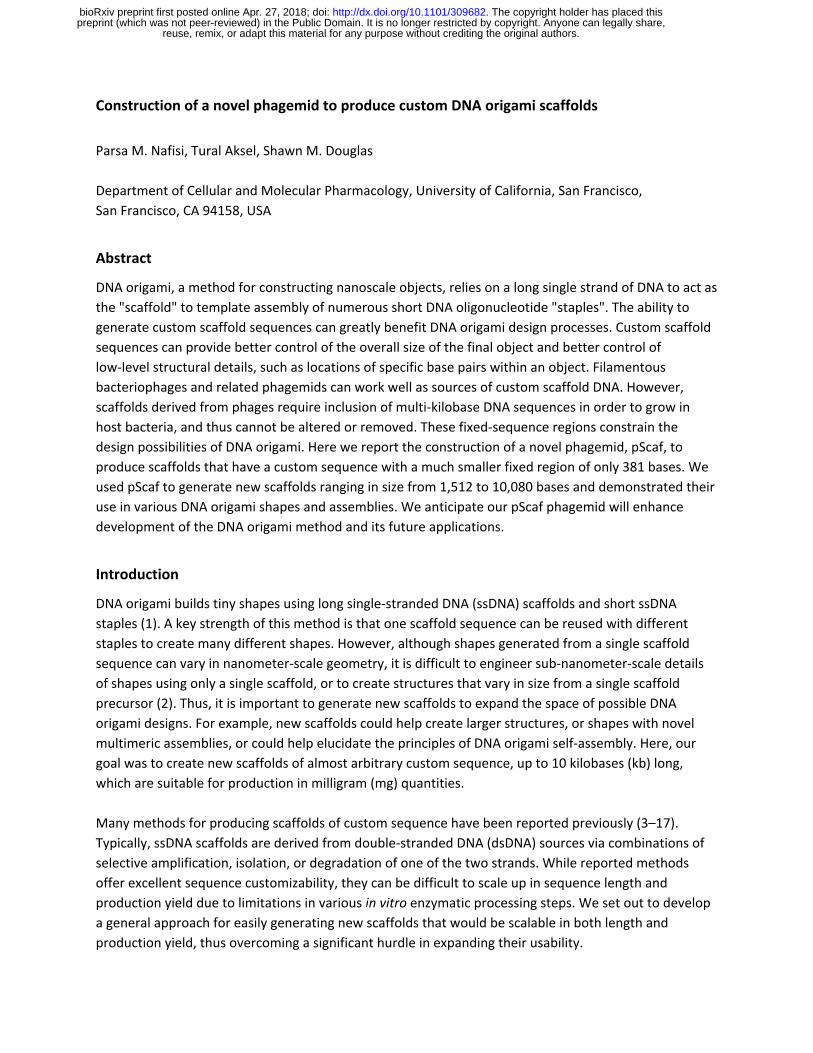
Phagemids can be used to create scaffolds that have improved sequence customizability compared to
M13 (7, 15, 23) . These plasmids typically contain a host origin of replication (ori) sequence, a phage ori
from M13 or relative such as f1, and an antibiotic resistance gene. Phagemid ssDNA can be exported in
phage-like particles if the host cell is co-infected with a "helper phage” or transformed with a “helper
plasmid” to express the necessary viral proteins (24) . Phagemids can accommodate custom inserts
several kb in size, but include a 2–3 kb fixed region that limits their usefulness in producing custom
origami scaffolds.
Figure 2: Construction and Optimization of the pScaf phagemid . A The pScaf phagemid is derived from the pUC18 plasmid and M13mp18 phage vectors. The phagemid includes an M13 origin of replication (M13ori) that contains ssDNA start site S1 and ssDNA termination site T2, a KpnI and BamHI cloning site, a packaging sequence (PS), and a terminator of ssDNA synthesis (M13term) containing ssDNA termination site T1 and ssDNA start site S2. The M13term sequence was adapted from the M13ori sequence by selectively deactivating the S2 ssDNA initiation function. B The ssDNA synthesis initiation and termination functions of the origin overlap. Therefore, we used a mutational screen of the δ region to optimize the M13term performance. C Three primary species were observed: S1T2, representing failed termination at terminator T1, S2T2, representing spurious initiation at initiator S2, and S1T1, the desired product. D We analyzed the variants by agarose gel. Substituting 0–2 thymines yielded the S2T2 species (*). Substituting 4–6 bases yielded the S1T2 species (**). Substituting 3 thymines yielded the best balance between spurious S2 initiation and failed T1 termination, producing the most pure S1T1 species (***).
To increase phagemid sequence customizability, we sought to create a scaffold that could be produced
using established preparation methods but would be able to package and export custom ssDNA
sequences that have a relatively small fixed region. We were inspired by two papers that reported
making use of modified f1-ori sequences to manipulate ssDNA synthesis (Fig. 1 D and E). In 1982, Dotto
et al . used phagemids with modified origins to show that f1-ori ssDNA synthesis initiation and
reuse, remix, or adapt this material for any purpose without crediting the original authors. preprint (which was not peer-reviewed) in the Public Domain. It is no longer restricted by copyright. Anyone can legally share,
The copyright holder has placed this. http://dx.doi.org/10.1101/309682doi: bioRxiv preprint first posted online Apr. 27, 2018;
termination functions overlap, but can be inactivated separately by modifying distinct sequences (18) . In
1992, Specthrie et al . packaged ssDNA as short as 292 bases into phage-like particles they called
microphages (19) . They were able to build small ssDNA strands using a phagemid that included an f1-ori,
a packaging sequence (PS), and a truncated f1-ori that acts as a terminator (f1-ori∆29). The terminator
interrupts ssDNA synthesis of the full phagemid sequence, leading to packaging and export of only the
region flanked by the ori and terminator.
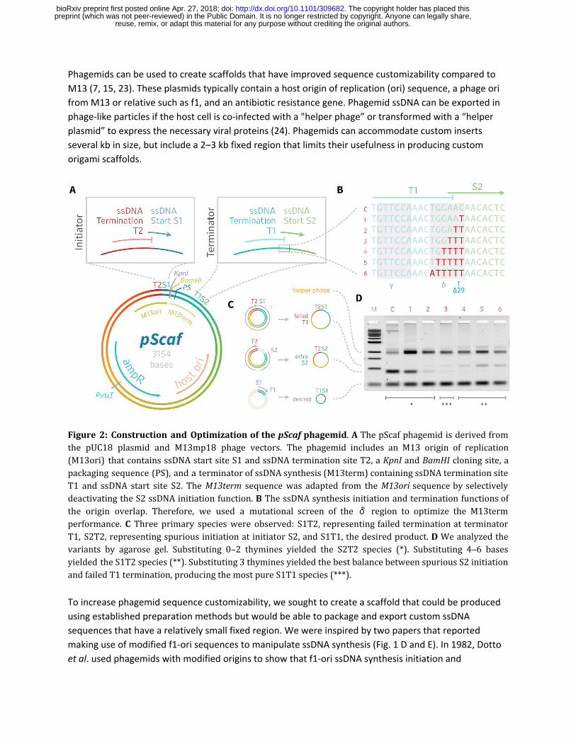
Figure 3: Cloning scheme and gel analysis of new scaffolds. A Custom sequence inserts are PCR-amplified with a forward primer containing KpnI and BglII sites and reverse primer containing a BamHI site. Inserts up to 3 kb in length were directly transformed into E.coli bearing helper plasmid. Larger scaffolds can be assembled by iterative PvuI+BamHI digestion of the vector containing the 5’ fragment, and PvuI+BglII digestion of the vector containing the 3’ fragment, followed by ligation, transformation, and miniprep. B Twelve inserts (A–L) were cloned into pScaf vector at the KpnI-BamHI site. Inserts A, B, and C were used to produce scaffolds with lengths of 1512, 2268, and 3024 bases. Larger scaffolds were assembled in multiple rounds as shown. C All scaffolds were grown in XL1-Blue cells containing helper plasmid M13cp, recovered and analyzed by agarose gel electrophoresis to determine purities ranging from 46% to 83%.
Materials and Methods
Construction of pScaf vector
We initially converted the pUC18 vector into a phagemid by cloning the M13 origin from M13mp18 at
the NdeI and KpnI restriction sites. Digested fragments were ligated with T4 DNA ligase (NEB) and
transformed into XL1-Blue MR competent cells (Agilent). We grew the pUC18-M13ori phagemid and
recovered the DNA with a miniprep. Next, we used M13mp18 as a template to PCR-amplify the ssDNA
synthesis terminator, based on the ∆29 design from Specthrie et al. (19) The terminator was then
inserted into the BamHI and EcoRI sites of the pUC18-M13ori. All subsequent variants of the terminator
(Fig 1) were assembled by PCR and cloned into the same BamHI and EcoRI sites, and verified for
reuse, remix, or adapt this material for any purpose without crediting the original authors. preprint (which was not peer-reviewed) in the Public Domain. It is no longer restricted by copyright. Anyone can legally share,
The copyright holder has placed this. http://dx.doi.org/10.1101/309682doi: bioRxiv preprint first posted online Apr. 27, 2018;

correctness using DNA sequencing. We used the variant with 3 thymine bases (TTT) as the final pScaf
vector in all subsequent experiments in this work.
Cloning scheme
We used KpnI and BamHI restriction sites between the ssDNA synthesis initiator and terminator regions
to clone inserts into the pScaf vector (Figure 3). In one step we cloned short PCR-amplified insert
sequences (A, B, C) directly into pScaf vectors that then generated the three smallest scaffolds (1512,
2268, and 3024). To create longer custom scaffolds, we adapted a multi-step cloning scheme previously
used for cloning repeat protein cDNA constructs (25) . We amplified custom sequence inserts in a
polymerase chain reaction (PCR) using forward and reverse primers designed to incorporate into the
product a 5’ KpnI and BglII site, and a 3’ BamHI site, respectively. We cloned PCR-amplified sequence
inserts of lengths up to 2.5 kb into separate pScaf vectors (D–L). To combine two inserts to create one
larger insert, we digested the pScaf vector containing the intended 5’ region with PvuI and BamHI, and
digested the vector containing the intended 3’ region with PvuI and BglII. BamHI and BglII digestions
create compatible sticky-end overhangs that can be used to ligate the two inserts. The bases adjacent to
the compatible sticky ends are different, so the ligation product does not reconstitute an internal BamHI
or BglII site. However, the ligated fragment maintains a 5’ BglII site and a 3’ BamHI site for subsequent
cloning rounds. We digest each plasmid at the PvuI restriction site to split the ampicillin-resistance gene
bla into two parts that are only reconstituted by successful ligation of the full-length product. Thus, we
generated three vectors with tandem inserts (DE, FG, IJ), and then combined FG with H to create FGH.
We combined IJ with K to create IJK, which was subsequently combined with L to create IJKL. The pScaf
vectors containing inserts DE, FGH, and IJKL were then ready to produce the larger scaffolds (5544,
8064, and 10080).
Scaffold amplification and purification
We transformed XL1-Blue-MRF’ using the M13cp helper plasmid (24) and then made it chemically
competent with TSS (10% PEG-8000, 30 mM MgCl2, 5% DMSO, in 2xYT, pH 6, filtered) in order to
produce a competent XL1-Blue_Helper strain. XL1-Blue_Helper was transformed with the desired pScaf
construct to create a custom-phage-producing strain. We selected and grew a single colony for 18 hours
in an incubator-shaker (30°C, 225 rpm) in 3-mL 2xYT media (1.6% tryptone, 1% yeast extract, .25% NaCl)
containing kanamycin, carbenicllin, and chloramphenicol. Subsequently, the culture was transferred to a
shake flask containing 100 mL 2xYT, 10 mL phosphate buffer (7% potassium phosphate dibasic, 3%
sodium phosphate monobasic, pH 7, autoclaved), 1 mL 50% glucose, 0.5 mL 1 M MgCl 2, and the
appropriate antibiotics. The flask was then grown for 24 hours on a shaker (30°C, 225 rpm). The flask
was harvested by transferring the culture to a 500-mL ultracentrifuge bottle, stored on ice for 30
minutes, and then centrifuged at 7000g for 15 minutes at 4°C, pelleting the bacteria while allowing the
phage to remain in the supernatant. The phage-containing supernatant was transferred to a clean
500-mL ultracentrifuge bottle, where 4 g PEG-8000 and 3g NaCl were added. The bottle was then
incubated on ice for 30 minutes prior to centrifugation at 9000g for 15 minutes at 4°C, allowing the
phage to form a pellet. The supernatant waste was decanted and the phage pellet was resuspended in 3
mL TE buffer. The bottle was then centrifuged at 15000g for 15 minutes at 4°C to pellet any residual
bacteria. The supernatant containing the concentrated phage was then transferred to a 50-mL
ultracentrifuge tube. 6 mL lysis buffer (0.2M NaOH, 1% SDS) was added and the tube was mixed. Next,
reuse, remix, or adapt this material for any purpose without crediting the original authors. preprint (which was not peer-reviewed) in the Public Domain. It is no longer restricted by copyright. Anyone can legally share,
The copyright holder has placed this. http://dx.doi.org/10.1101/309682doi: bioRxiv preprint first posted online Apr. 27, 2018;
4.5 mL neutralization buffer (3M KOAc, pH 5.5) was added and the tube was again mixed. The tube was
incubated on ice for 15 minutes and then centrifuged at 15000g for 15 minutes at 4°C. The resulting
supernatant was then decanted into a new 50-mL ultracentrifuge tube and 27 mL pure ethanol was
added to it. The tube was capped and mixed by inversion prior to incubation at -20°C for 18 hours. After
incubation, the tube was centrifuged at 16000g for 15 minutes at 4°C to pellet the DNA. The supernatant
waste was decanted leaving the DNA pellet at the bottom of the tube. The pellet was washed with 9 mL
ice-cold 70% ethanol and centrifuged at 16000g for 5 minutes at 4°C. The supernatant waste was again
decanted. The pellet was subsequently dried by gently blowing air into the tube, and finally the dried
pellet was resuspended in up to 1 mL TE, depending on the desired concentration.
DNA origami preparation
Custom scaffold and staples were mixed at final concentrations of 20 nM and 200 nM, respectively, in a
buffer containing 5 mM Tris, 1 mM EDTA and 13 mM MgCl2. The solution was then subject to the
following temperature ramp: denaturation at 65°C for 15 minutes, followed by cooling from 60°C to
38°C with a decrease of 1°C per 50 minutes.
Agarose gel analysis
Purified scaffolds and folded origami products were analyzed using 2% agarose gel electrophoresis in
TBE (45 mM tris-borate and 1 mM EDTA) supplemented with 11 mM MgCl2 and SYBR Safe. Upon
sample-loading, gels were run for 2 hours at 80V and subsequently scanned using a Typhoon FLA imager.
DNA origami products folded from 1512-, 2268-, and 3024-base scaffolds were subsequently purified by
extracting the desired gel band using a razor blade, muddled to break down the agarose, and then
centrifuged through a Freeze ‘N Squeeze column.
PEG purification
DNA origami products folded from scaffolds of 5544 bases and larger were purified using PEG
precipitation. Assembly products were mixed with an equal volume of PEG precipitation buffer (15%
PEG-8000, 10 mM Tris, 20 mM MgCl2, 500 mM NaCl) and immediately centrifuged at 16000g at 20°C for
25 minutes. The supernatant was removed and the pellet (which is often invisible) was resuspended in
1x folding buffer with magnesium (5 mM Tris, 1 mM EDTA and 13 mM MgCl2).
Transmission electron microscopy
Purified origami structures were diluted to approximately 25 ng/μL prior to imaging. 5 μL of the diluted
origami was applied to glow-discharged, carbon-coated, 400-mesh formvar grids (Ted Pella) for 1.5
minutes. The grid was then blotted dry on filter paper (Whatman). Washing and staining was performed
by preparing a pierce of parafilm with two 15-μL droplets of 1x folding buffer and two 15- μL droplets of
2% aqueous uranyl formate stain solution. The grid was dipped onto the first buffer droplet, blotted dry,
dipped onto the next buffer droplet, blotted dry, dipped onto the first stain droplet, blotted dry, and
then held onto the final stain droplet for 45 seconds before being blotted dry. Grids were then allowed
to air-dry for 10 minutes prior to imaging. Electron micrographs were collected using an FEI TECNAI T12
transmission electron microscope using a using a 4k × 4k charge-coupled device camera (UltraScan
4000, Gatan) at 26000× and 52000× magnifications. Class averages were obtained using EMAN2
software.
reuse, remix, or adapt this material for any purpose without crediting the original authors. preprint (which was not peer-reviewed) in the Public Domain. It is no longer restricted by copyright. Anyone can legally share,
The copyright holder has placed this. http://dx.doi.org/10.1101/309682doi: bioRxiv preprint first posted online Apr. 27, 2018;

Figure 4: Folding Custom Scaffolds into DNA Origami Shapes and Assemblies. Six scaffolds of varying sizes were folded into DNA origami shapes and assemblies. Folding reactions were analyzed by gel electrophoresis, and origami analyzed negative stain transmission electron microscopy. A Brick-like DNA origami shapes were folded from scaffolds of lengths 1512, 2268, 3024, 5544, 8064, and 10080 bases. Each brick was PEG-purified and imaged by negative stain TEM. Class averages for each brick are shown, along with a 20-nm scale bar. B Scaffolds of lengths 1512, 2268, and 3024 were folded separately into six-helix-bundle (6hb) nanotubes. All three scaffolds and 6hb-staple sets were combined into one-pot reaction mixtures. When connector staples are included, the three scaffolds assemble into a 6hb trimer. TEM micrographs of the individual 6hb nanotubes and combined trimer are shown with 100-nm scale bars.
Results
We constructed a phagemid, pScaf, that can be used to create custom DNA origami scaffolds (Figure 1).
We converted pUC18 into a phagemid for custom ssDNA production by adding four components: a
full-length M13 origin for ssDNA initiation, KpnI and BamHI restriction sites for insert cloning, the M13
packaging sequence for phage particle export, and a modified M13 origin to serve as the ssDNA
synthesis terminator. When we tested the “∆29” terminator used by Specthrie et al. (19) , we were able
to produce our desired custom ssDNA scaffolds. However, our phage cultures also yielded an off-target
ssDNA species consistent in size with residual packaging of the region downstream of our custom insert.
We suspected that the ∆29 terminator may not fully abolish initiation of ssDNA synthesis, resulting in
multiple ssDNA species produced from a single phagemid similar to constructs reported by Dotto et al.
(18) . Thus, we sought to optimize the terminator to reduce spurious initiation and produce a more pure
ssDNA scaffold product (Figure 2).
reuse, remix, or adapt this material for any purpose without crediting the original authors. preprint (which was not peer-reviewed) in the Public Domain. It is no longer restricted by copyright. Anyone can legally share,
The copyright holder has placed this. http://dx.doi.org/10.1101/309682doi: bioRxiv preprint first posted online Apr. 27, 2018;

Terminator optimization
We started by truncating the last 50 bases of the 381-base M13 ori sequence so it included the core
origin of plus-strand replication (α,β, γ, and δ regions) but omitted the downstream initiation
enhancer region (26) . We performed a mutational screen starting at the 3’ end of the δ region, near
the site of the Specthrie et al. ∆29 truncation. We cloned variants of the phagemid with up to six
δ-region bases substituted with thymines (T), except for one thymine base which we replaced with
adenine (Figure 2c). We tested ssDNA production of each variant in E.coli culture co-infected with helper
phage. We observed that 0-, 1-, or 2-base substitutions permitted ssDNA initiation, leading to synthesis
of an off-target species of ssDNA. Meanwhile, 4-, 5-, or 6-base substitutions interfered with ssDNA
termination, leading to an alternate off-target ssDNA species. We achieved the best balance between
target and off-target ssDNA using the 3-base substitution, which resulted in relatively low amounts of
each off-target species. We subsequently used the 3-base substitution in all pScaf constructs.
Custom scaffolds and test origami
We created custom scaffolds with lengths of 1512, 2268, 3024, 5544, 8064, and 10080 bases (Figure 3).
Each scaffold was verified by sequencing and analyzed by agarose-gel electrophoresis. We used Cadnano
(27) to design a series of DNA origami shapes that approximate rectangular cuboids or brick-like
structures. We folded and analyzed the resulting DNA origami shapes using agarose gel electrophoresis
and negative-stain TEM (Figure 4).
One-pot folding of multi-scaffold assemblies
The ability to synthesize custom scaffolds with relatively little sequence overlap affords the possibility of
folding DNA origami from multiple scaffolds in a one-pot reaction. To validate this concept, we designed
a six-helix-bundle nanotube comprised of 3 scaffolds with lengths of 1512, 2268, and 3024 bases. The
scaffolds were folded in a one-pot reaction both with and without connector staples to join them into a
single nanotube.
Discussion
Our approach has significant advantages over pre-existing methods for making custom scaffolds to build
DNA origami nanostructures. First, we have demonstrated that custom scaffolds can be generated with
lengths of up to 10 kb. Thus, the method is compatible with nearly all origami shapes that have been
published to date, and will enable the creation of additional large shapes. Second, although cloning large
(>3kb) custom insert sequences into phagemids presents difficult challenges, the cloning scheme we
report here circumvents some of them. We found the insertion of long sequences is much more reliable
with a multi-step assembly approach that cuts the antibiotic resistance gene during each round, and
includes a positive selection for full-length constructs that reconstitute the gene. We typically only
needed to screen a handful of colonies of transformed E.coli for each cloning round to locate the desired
sequence. Third, because we rely on phagemids to grow custom scaffolds in E.coli, the method can be
used to produce milligram-scale quantities of custom scaffolds using flasks and a shaker-incubator.
Bacterial production offers a potentially reliable and feasible method for scaling up production to much
larger quantities using bioreactors, as has been demonstrated for other scaffolds (20, 28) .
reuse, remix, or adapt this material for any purpose without crediting the original authors. preprint (which was not peer-reviewed) in the Public Domain. It is no longer restricted by copyright. Anyone can legally share,
The copyright holder has placed this. http://dx.doi.org/10.1101/309682doi: bioRxiv preprint first posted online Apr. 27, 2018;

With our method, it will now be possible to rapidly generate large DNA origami shapes with highly
customized scaffold sequences. We recognized the need for easier methods to make custom scaffolds
when making DNA origami shapes that fold from limited sets of reusable staple sequences (29) . Here,
we chose scaffold lengths to be multiples of 42, a convenient repeat length for designs using the
honeycomb lattice. For convenience, we incorporated sequences from pre-existing plasmids such as
pFastBac-GFP-dynein-2(D1091–Q4307). Additional scaffolds of various lengths can be generated quickly
using a similar approach. Sequences also might be ordered from gene synthesis vendors and combined
with our approach to generate scaffolds tailored for answering specific questions about DNA origami
design principles, or to incorporate protein-binding sites.
Custom scaffolds will be useful for studying the DNA origami method itself. For example, new scaffolds
can be used to examine sequence-level determinants of folding yield. Sequences of particular base
compositions (high-GC, high-AT, 3-letter alphabets) with well-defined repetition or self-complementarity
can be constructed and used to test hypotheses explaining the role of melting temperature or secondary
structure in origami folding. Additionally, custom scaffolds can have practical benefits for routine
origami design. We have often desired to create large DNA origami shapes with specific custom
sequences at multiple non-contiguous locations within the scaffold. Generating these custom scaffolds is
now much easier compared to cloning large inserts directly into M13mp18 in a single step.
It is also useful to be able to produce multiple unique scaffolds with very little sequence overlap. Sets of
multiple unique custom sequences can be used for facile one-pot assembly of multimeric asymmetric
structures. We demonstrated the one-pot assembly of small six-helix-bundle nanotube shapes, but
expect our approach can extend to reliably making larger shapes. For example, it should now be possible
to design three custom 10-kb scaffolds that can assemble into a 30kb origami in which nearly the entire
multimeric structure is uniquely addressable and can incorporate modified staples.
Before we settled on the pScaf design, we attempted to generate custom scaffolds using a phagemid
with a terminator based on the ∆29 truncation used by Specthrie et al. (19) . Our observations of an
off-target ssDNA species led us to attempt to optimize the terminator sequence. To guide our approach
and select the site for our mutational screen, we reviewed previous studies of the initiation and
termination of filamentous phage plus-strand synthesis (18, 26, 30) . A better understanding of the
fundamental biology of filamentous phages will likely lead to further improvements to the pScaf
phagemid.
Although the TTT terminator variant greatly improved the scaffold purity, we were not able to
completely eliminate incomplete termination or spurious initiation in our scaffold preps. We also
observed some prep-to-prep variation in scaffold quality and production of off-target species when
using the helper plasmid. We analyzed the scaffold gel image with ImageJ and estimated that the purity
of correct-length scaffold products ranged from 46% to 83% of total integrated intensity of the sample
lane (Fig 3c). Our maximum purity is similar to commercially available M13-derived scaffold (31) . Our
DNA origami gel analysis and TEM images show that we obtained a relatively homogeneous population
of folded origami structures, so the current levels of purity will be suitable for many applications. Future
reuse, remix, or adapt this material for any purpose without crediting the original authors. preprint (which was not peer-reviewed) in the Public Domain. It is no longer restricted by copyright. Anyone can legally share,
The copyright holder has placed this. http://dx.doi.org/10.1101/309682doi: bioRxiv preprint first posted online Apr. 27, 2018;
optimization of the ori and terminator sequences, helper plasmid, host strain, or growth conditions may
yield cleaner target ssDNA products.
The sequence insertion locus used KpnI and BamHI restriction sites, which would need modification if
those sites are to be incorporated into a custom scaffold. A multiple cloning site would provide greater
flexibility for cloning sequence inserts. We did not attempt to reduce the size of the terminator region
that is incorporated into the scaffold ssDNA, so a further reduction in size of the fixed region of 381
bases may be possible with additional optimization. While we produced scaffolds up to 10 kb in length,
we note that this length does not represent an upper limit. We have not yet attempted to make longer
scaffolds, but it should be possible following our scheme.
In summary, we have developed and validated a novel phagemid, pScaf, for creating highly customized
ssDNA DNA origami scaffolds of 10-kb lengths that can be produced at milligram-scale yields. Our
approach removes a long-standing constraint that has held back progress in the use of large DNA
origami scaffolds, which is that researchers have had to rely on the genome of M13 phage which cannot
be easily customized. The ability to generate many long, unique custom scaffolds will enable researchers
to resolve many unanswered questions about what the optimal methods are to create and design DNA
origami sequences, how to easily incorporate functional sequences, such as protein-binding sites, into
multiple sites within each shape, and how to better adapt these methods in future applications.
Acknowledgements
We thank P. Rothemund and L. Bienen for comments on the manuscript. We are grateful to A. Bradbury
for providing the M13cp helper plasmid. This work was supported by the Army Research Office
(W911NF-14-1-0507), Del E. Webb Foundation (13-2-28), and National Science Foundation (CCF-
1317640). T.A. holds a Ruth L. Kirschstein NRSA Postdoctoral Fellowship (F32GM119322). S.M.D holds a
Career Award at the Scientific Interface from the Burroughs Wellcome Fund (1010247).
Author Contributions
P.M.N., T.A., and S.M.D. design the research. P.M.N. performed the experiments. P.M.N. and T.A.
collected and analyzed the TEM data. All authors contributed to data interpretation and to writing the
manuscript.
reuse, remix, or adapt this material for any purpose without crediting the original authors. preprint (which was not peer-reviewed) in the Public Domain. It is no longer restricted by copyright. Anyone can legally share,
The copyright holder has placed this. http://dx.doi.org/10.1101/309682doi: bioRxiv preprint first posted online Apr. 27, 2018;
References
1. Rothemund,P.W.K. (2006) Folding DNA to create nanoscale shapes and patterns. Nature, 440, 297–302.
2. Pinheiro,A.V., Han,D., Shih,W.M. and Yan,H. (2011) Challenges and opportunities for structural DNA nanotechnology. Nat. Nanotechnol., 6, 763–772.
3. Pound,E., Ashton,J.R., Becerril,H.A. and Woolley,A.T. (2009) Polymerase chain reaction based scaffold preparation for the production of thin, branched DNA origami nanostructures of arbitrary sizes. Nano Lett., 9, 4302–4305.
4. Douglas,S.M., Dietz,H., Liedl,T., Högberg,B., Graf,F. and Shih,W.M. (2009) Self-assembly of DNA into nanoscale three-dimensional shapes. Nature, 459, 414–418.
5. Zhang,H., Chao,J., Pan,D., Liu,H., Huang,Q. and Fan,C. (2012) Folding super-sized DNA origami with scaffold strands from long-range PCR. Chem. Commun. , 10.1039/c2cc32204h .
6. Yang,Y., Han,D., Nangreave,J., Liu,Y. and Yan,H. (2012) DNA origami with double-stranded DNA as a unified scaffold. ACS Nano, 6, 8209–8215.
7. Zadegan,R.M., Jepsen,M.D.E., Thomsen,K.E., Okholm,A.H., Schaffert,D.H., Andersen,E.S., Birkedal,V. and Kjems,J. (2012) Construction of a 4 zeptoliters switchable 3D DNA box origami. ACS Nano, 6, 10050–10053.
8. Ducani,C., Kaul,C., Moche,M., Shih,W.M. and Högberg,B. (2013) Enzymatic production of ‘monoclonal stoichiometric’ single-stranded DNA oligonucleotides. Nat. Methods, 10, 647–652.
9. Marchi,A.N., Saaem,I., Tian,J. and LaBean,T.H. (2013) One-pot assembly of a hetero-dimeric DNA origami from chip-derived staples and double-stranded scaffold. ACS Nano, 7, 903–910.
10. Said,H., Schüller,V.J., Eber,F.J., Wege,C., Liedl,T. and Richert,C. (2013) M1.3 — a small scaffold for DNA origami. Nanoscale, 5, 284–290.
11. Li,W., Yang,Y., Jiang,S., Yan,H. and Liu,Y. (2014) Controlled Nucleation and Growth of DNA Tile Arrays within Prescribed DNA Origami Frames and Their Dynamics. J. Am. Chem. Soc., 136, 3724–3727.
12. Nickels,P.C., Ke,Y., Jungmann,R., Smith,D.M., Leichsenring,M., Shih,W.M., Liedl,T. and Högberg,B. (2014) DNA origami structures directly assembled from intact bacteriophages. Small, 10, 1765–1769.
13. Marchi,A.N., Saaem,I., Vogen,B.N., Brown,S. and LaBean,T.H. (2014) Toward larger DNA origami. Nano Lett., 14, 5740–5747.
14. Erkelenz,M., Bauer,D.M., Meyer,R., Gatsogiannis,C., Raunser,S., Saccà,B. and Niemeyer,C.M. (2014) A Facile Method for Preparation of Tailored Scaffolds for DNA-Origami. Small, 10, 73–77.
15. Brown,S., Majikes,J., Martínez,A., Girón,T.M., Fennell,H., Samano,E.C. and LaBean,T.H. (2015) An
reuse, remix, or adapt this material for any purpose without crediting the original authors. preprint (which was not peer-reviewed) in the Public Domain. It is no longer restricted by copyright. Anyone can legally share,
The copyright holder has placed this. http://dx.doi.org/10.1101/309682doi: bioRxiv preprint first posted online Apr. 27, 2018;

easy-to-prepare mini-scaffold for DNA origami. Nanoscale, 7, 16621–16624.
16. Veneziano,R., Ratanalert,S., Zhang,K., Zhang,F., Yan,H., Chiu,W. and Bathe,M. (2016) Designer nanoscale DNA assemblies programmed from the top down. Science, 10.1126/science.aaf4388.
17. Krieg,E. and Shih,W.M. (2018) Selective Nascent Polymer Catch-and-Release Enables Scalable Isolation of Multi-Kilobase Single-Stranded DNA. Angew. Chem. Int. Ed Engl., 57, 714–718.
18. Dotto,G.P., Horiuchi,K. and Zinder,N.D. (1982) Initiation and termination of phage f1 plus-strand synthesis. Proc. Natl. Acad. Sci. U. S. A., 79, 7122–7126.
19. Specthrie,L., Bullitt,E., Horiuchi,K., Model,P., Russel,M. and Makowski,L. (1992) Construction of a microphage variant of filamentous bacteriophage. J. Mol. Biol., 228, 720–724.
20. Kick,B., Praetorius,F., Dietz,H. and Weuster-Botz,D. (2015) Efficient Production of Single-Stranded Phage DNA as Scaffolds for DNA Origami. Nano Lett., 15, 4672–4676.
21. Sambrook,J. and Russell,D.W. (2001) Molecular Cloning: A Laboratory Manual, 3rd edition. Cold Spring Harbor Laboratory Press.
22. Messing,J. (1983) New M13 vectors for cloning. Methods Enzymol., 101, 20–78.
23. Shih,W.M., Quispe,J.D. and Joyce,G.F. (2004) A 1.7-kilobase single-stranded DNA that folds into a nanoscale octahedron. Nature, 427, 618–621.
24. Chasteen,L., Ayriss,J., Pavlik,P. and Bradbury,A.R.M. (2006) Eliminating helper phage from phage display. Nucleic Acids Res., 34, e145.
25. Aksel,T., Majumdar,A. and Barrick,D. (2011) The contribution of entropy, enthalpy, and hydrophobic desolvation to cooperativity in repeat-protein folding. Structure, 19, 349–360.
26. Horiuchi,K. (1997) Initiation mechanisms in replication of filamentous phage DNA. Genes Cells, 2, 425–432.
27. Douglas,S.M., Marblestone,A.H., Teerapittayanon,S., Vazquez,A., Church,G.M. and Shih,W.M. (2009) Rapid prototyping of 3D DNA-origami shapes with caDNAno. Nucleic Acids Res., 37, 5001–5006.
28. Praetorius,F., Kick,B., Behler,K.L., Honemann,M.N., Weuster-Botz,D. and Dietz,H. (2017) Biotechnological mass production of DNA origami. Nature, 552, 84.
29. Niekamp,S., Blumer,K., Nafisi,P.M., Tsui,K., Garbutt,J. and Douglas,S.M. (2016) Folding complex DNA nanostructures from limited sets of reusable sequences. Nucleic Acids Res., 44, e102.
30. Dotto,G.P. and Horiuchi,K. (1981) Replication of a plasmid containing two origins of bacteriophage f1. J. Mol. Biol., 153, 169–176.
31. Kuzyk,A., Yang,Y., Duan,X., Stoll,S., Govorov,A.O., Sugiyama,H., Endo,M. and Liu,N. (2016) A light-driven three-dimensional plasmonic nanosystem that translates molecular motion into reversible chiroptical function. Nat. Commun., 7, 10591.
reuse, remix, or adapt this material for any purpose without crediting the original authors. preprint (which was not peer-reviewed) in the Public Domain. It is no longer restricted by copyright. Anyone can legally share,
The copyright holder has placed this. http://dx.doi.org/10.1101/309682doi: bioRxiv preprint first posted online Apr. 27, 2018;