consensus sTATeMenT on THRoMBoeMBoLIc · PDF fileCONSENSUS STATEMENT ON THROMBOEMBOLIC...
17
CONSENSUS Director Dr. Jorge Ubaldini Editorial Committee Dr. Julio Chertcoff Dr. Eduardo Sampó Dr. Marcelo Casey Dr. José Ceresetto Dr. Roberto Boughen Dr. Miguel Veltri, Dr. Luis Flores Dr. Mario Kenar Dr. Ramón Suasnabal Dr. Salvador De Francesca Dr. Marcelo Pérez Dr. Cecilia Perel CONSENSUS STATEMENT ON THROMBOEMBOLIC DISEASE TABLE OF CONTENTS 1. Grades of recommendation .................................................................................................................. 412 2. Levels of evidence................................................................................................................................... 412 3. Background............................................................................................................................................ 412 4. Physiopathological considerations ........................................................................................................ 412 5. Risk factors............................................................................................................................................. 413 6. Prevention of DVT ................................................................................................................................ 413 6.1 DVT prophylaxis strategies .................................................................................................................... 413 a. Mechanical methods ............................................................................................................................. 413 - Intermittent pneumatic compression ................................................................................................... 413 - Graduated compression stockings ........................................................................................................ 413 b. Pharmacological methods ..................................................................................................................... 414 - Subcutaneous unfractionated heparin ................................................................................................. 414 - Low-molecular-weight heparin ............................................................................................................ 414 - Pentasaccharides ................................................................................................................................... 414 - Oral anticoagulant agents..................................................................................................................... 414 - Antiplatelet agents ............................................................................................................................... 414 6.2 General recommendations for prevention of DVT............................................................................... 415 6.3 Prophylaxis in special situations ............................................................................................................ 415 a. Coronary care unit and cardiovascular surgery .................................................................................... 415 b. Intensive care unit ................................................................................................................................. 416 c. Burn patients ......................................................................................................................................... 416 d. Spinal cord injury ................................................................................................................................... 416 e. Major trauma......................................................................................................................................... 416 f. Neurosurgery ......................................................................................................................................... 416 g. Hip surgery............................................................................................................................................. 416 h. Cancer .................................................................................................................................................... 416 i. Long distance travel ............................................................................................................................. 417 j. Extended prophylaxis ............................................................................................................................ 417 k. Prophylaxis and neuraxial analgesia ..................................................................................................... 417 7. Diagnosis of DVT ................................................................................................................................... 417 8. Diagnosis of PE ...................................................................................................................................... 417 8.1 Clinical presentation .............................................................................................................................. 417 8.2 Routine laboratory tests ........................................................................................................................ 418 8.3 Chest X-ray ............................................................................................................................................. 418 8.4 Electrocardiogram ................................................................................................................................. 418 8.5 Arterial blood gases and alveolar-arterial O2 gradient ........................................................................ 418 8.6 D-dimer .................................................................................................................................................. 419 8.7 Troponins ............................................................................................................................................... 419 8.8 Transthoracic echocardiography ........................................................................................................... 419 8.9 Tranesophageal echocardiography....................................................................................................... 419 8.10 Ventilation-perfusion (V/Q) scintigraphy.............................................................................................. 419 8.11 Helical computed tomography scan ..................................................................................................... 419 8.12 Diagnosis of DVT ................................................................................................................................... 420 8.13 Pulmonary angiography ........................................................................................................................ 420 9. Recommendations of diagnostic imaging tests .................................................................................... 420 10. Diagnostic and therapeutic algorithm .................................................................................................. 420 10.1 Models for clinical stratification ............................................................................................................ 420 11. High-risk (massive) PE ............................................................................................................................ 421 12. Treatment of thromboembolic disease ................................................................................................ 422 12.1 High-risk patient .................................................................................................................................... 422 12.2 Low-risk patient ..................................................................................................................................... 423 a. Anticoagulant therapy .......................................................................................................................... 423 b. Unfractionated heaprin ......................................................................................................................... 423 c. Low-molecular-weight heparin ............................................................................................................ 423 d. Pentasaccharides ................................................................................................................................... 423 e. Thrombolytic therapy ............................................................................................................................ 424 f. Mechanical and pharmacological treatment in the catheterization laboratory ................................ 425 g. Surgical embolectomy ........................................................................................................................... 425 13. Therapeutic recommendations ............................................................................................................. 425 14. Vena caval filters ................................................................................................................................... 425 15. Coumarin therapy.................................................................................................................................. 425 16. Recommendations for secondary prevention....................................................................................... 426 17. Pregnancy .............................................................................................................................................. 426
-
Upload
nguyenminh -
Category
Documents
-
view
218 -
download
2
Transcript of consensus sTATeMenT on THRoMBoeMBoLIc · PDF fileCONSENSUS STATEMENT ON THROMBOEMBOLIC...

CONSENSUS STATEMENT ON THROMBOEMBOLIC DISEASE 411consensus
Director
Dr. Jorge Ubaldini
Editorial Committee
Dr. Julio Chertcoff
Dr. Eduardo Sampó
Dr. Marcelo Casey
Dr. José Ceresetto
Dr. Roberto Boughen
Dr. Miguel Veltri,
Dr. Luis Flores
Dr. Mario Kenar
Dr. Ramón Suasnabal
Dr. Salvador De Francesca
Dr. Marcelo Pérez
Dr. Cecilia Perel
consensus sTATeMenT on THRoMBoeMBoLIc DIseAse
TABLE OF CONTENTS
1. Grades of recommendation .................................................................................................................. 4122. Levels of evidence ...................................................................................................................................4123. Background ............................................................................................................................................ 4124. Physiopathological considerations ........................................................................................................ 4125. Risk factors ............................................................................................................................................. 4136. Prevention of DVT ................................................................................................................................ 4136.1 DVT prophylaxis strategies .................................................................................................................... 413a. Mechanical methods ............................................................................................................................. 413- Intermittent pneumatic compression ................................................................................................... 413- Graduated compression stockings ........................................................................................................ 413b. Pharmacological methods ..................................................................................................................... 414- Subcutaneous unfractionated heparin ................................................................................................. 414- Low-molecular-weight heparin ............................................................................................................ 414- Pentasaccharides ................................................................................................................................... 414- Oral anticoagulant agents ..................................................................................................................... 414- Antiplatelet agents ............................................................................................................................... 4146.2 General recommendations for prevention of DVT ............................................................................... 4156.3 Prophylaxis in special situations ............................................................................................................ 415a. Coronary care unit and cardiovascular surgery .................................................................................... 415b. Intensive care unit ................................................................................................................................. 416c. Burn patients ......................................................................................................................................... 416d. Spinal cord injury ................................................................................................................................... 416e. Major trauma......................................................................................................................................... 416f. Neurosurgery ......................................................................................................................................... 416g. Hip surgery ............................................................................................................................................. 416h. Cancer .................................................................................................................................................... 416i. Long distance travel ............................................................................................................................. 417j. Extended prophylaxis ............................................................................................................................ 417k. Prophylaxis and neuraxial analgesia ..................................................................................................... 4177. Diagnosis of DVT ................................................................................................................................... 4178. Diagnosis of PE ...................................................................................................................................... 4178.1 Clinical presentation .............................................................................................................................. 4178.2 Routine laboratory tests ........................................................................................................................ 4188.3 Chest X-ray ............................................................................................................................................. 4188.4 Electrocardiogram ................................................................................................................................. 4188.5 Arterial blood gases and alveolar-arterial O2 gradient ........................................................................ 4188.6 D-dimer .................................................................................................................................................. 4198.7 Troponins ............................................................................................................................................... 4198.8 Transthoracic echocardiography ........................................................................................................... 4198.9 Tranesophageal echocardiography ....................................................................................................... 4198.10 Ventilation-perfusion (V/Q) scintigraphy .............................................................................................. 4198.11 Helical computed tomography scan ..................................................................................................... 4198.12 Diagnosis of DVT ................................................................................................................................... 4208.13 Pulmonary angiography ........................................................................................................................ 4209. Recommendations of diagnostic imaging tests .................................................................................... 42010. Diagnostic and therapeutic algorithm .................................................................................................. 42010.1 Models for clinical stratification ............................................................................................................ 42011. High-risk (massive) PE ............................................................................................................................ 42112. Treatment of thromboembolic disease ................................................................................................ 42212.1 High-risk patient .................................................................................................................................... 42212.2 Low-risk patient ..................................................................................................................................... 423a. Anticoagulant therapy .......................................................................................................................... 423b. Unfractionated heaprin ......................................................................................................................... 423c. Low-molecular-weight heparin ............................................................................................................ 423d. Pentasaccharides ................................................................................................................................... 423e. Thrombolytic therapy ............................................................................................................................ 424f. Mechanical and pharmacological treatment in the catheterization laboratory ................................ 425g. Surgical embolectomy ........................................................................................................................... 42513. Therapeutic recommendations ............................................................................................................. 42514. Vena caval filters ................................................................................................................................... 42515. Coumarin therapy .................................................................................................................................. 42516. Recommendations for secondary prevention....................................................................................... 42617. Pregnancy .............................................................................................................................................. 426

412 REVISTA ARGENTINA DE CARDIOLOGÍA / VOL 77 Nº 5 / SEpTEMBER-OCTOBER 2009
1. GRADES OF RECOMMENDATION
Class I: conditions for which there is evidence and/or general agreement that a given procedure or treatment is useful, and effective.
Class II: conditions for which there is conflicting evidence and/or a divergence of opinion about the use-fulness/efficacy of a procedure or treatment.
Class IIa: weight of evidence/opinion is in favor of usefulness/efficacy.
Class IIb: usefulness/efficacy is less well estab-lished by evidence/opinion.
2. LEVELS OF EVIDENCE
Level of evidence A: consistent evidence from randomized clinical trials or meta-analyses.
Level of evidence B: data derived from a single randomized trial.
Level of evidence C: data derived from consensus opinion of experts, retrospective studies or registries.
3. BACKGROUND
Pulmonary embolism (PE) is one of the most frequent causes of death in hospitalized patients and constitutes a clinical presentation of thromboembolic disease or venous thromboembolism together with deep venous thrombosis (DVT). (1) The risk of post-thrombotic syndrome, pulmonary hypertension and recurrences of thromboembolic events exists after the acute phase.
Thromboembolic disease is the third cause of car-diovascular morbidity after ischemic heart disease and cerebrovascular disease. The annual incidence is about 100 cases per 100000 person-years and the prevalence in hospitalized patients is 1%. In 1975, Dalen et al. (1) summarized the natural history of venous throm-boembolism on the basis of several epidemiological factors and pathological findings. Most deaths which occur in the first hours (Figure 1) can be prevented by
prophylaxis. For patients who have received treatment, mortality ranges from 6% to 10%, and reaches 25% - 30% in those cases without diagnosis and treatment. Currently, these figures are similar to those published in 1975, demonstrating that little progress has been achieved in the early diagnosis of this disease. About 70% of cases are still not diagnosed. This situation strengthens the need to improve prevention measures and to intensify early diagnosis strategies starting by a change in physicians’ attitudes, as the clinical suspicion of the disease is the first link in this chain. (2)
4. PHYSIOPATHOLOGICAL CONSIDERATIONS
The severity of embolic obstruction depends on the embolus size; in this way, symptomatic embolism usu-ally arises from thrombi originating in the deep venous system of the lower extremities, especially from the iliac and femoral veins. They rarely originate in the pelvic or upper extremity veins, inferior vena cava or the right heart chambers.
Emboli located proximally or producing severe vascular obstruction are associated with severe hemo-dynamic compromise, which has an inverse correlation with the cardiopulmonary reserve. In occasions, friable thrombi are likely to be fragmented by the pulsatile flow of blood in smaller thrombi that can travel more distally, occluding smaller vessels in the lung periphery where the section area is greater, producing less he-modynamic impairment. More than 50% of pulmonary emboli are multiple, and the lower lobes are involved more commonly than the upper lobes, unilaterally or bilaterally. (3)
Smaller thrombi are lodged directly in the lung pe-riphery producing less hemodynamic impact; they are more likely to produce pleuritic chest pain by initiat-ing an inflammatory response adjacent to the parietal pleura that does not necessarily imply a pulmonary infarction.
Fig. 1. Incidence of pulmo-nary embolism per year in the United States.

CONSENSUS STATEMENT ON THROMBOEMBOLIC DISEASE 413
Larger thrombi may be temporarily trapped in the right atrium or among the tendinous cords of the tricuspid valve and the papillary muscles of the right ventricle (thrombi in transit) and can be detected by echocardiography. (4)
Thrombi in transit may pass through a patent fora-men ovale from the right atrium into the left atrium and thus embolize to the arterial circulation. This migration is facilitated by the increased right atrium pressure in the presence of pulmonary hypertension induced by previous pulmonary embolisms.
5. RISK FACTORS
The primary and secondary known risk factors are related to traditional Virchow’s triad described in the 19th century (venous stasis, hypercoagulability and endothelial injury); the most important factors are summarized in Table 1.
Although the genetic predisposition for thrombosis is relatively frequent, the real incidence in our envi-ronment is unknown. Unexplained DVT should be suspected in persons < 40 years with recurrent PE or DVT and/or a positive family history. (6)
The incidence of thrombotic events increases with the age until the 8th decade and is more frequent in men than in women.
Recurrences are also more frequent in men.Thromboembolic disease has been reported in 30 -
60% of patients with stroke, 5- 35% of patients with acute myocardial infarction and 12% of patients with conges-tive heart failure who did not receive prophylaxis.
Immobilization during short periods of time (> 3 days) predisposes to DVT. The incidence of DVT after a simple surgery of inguinal hernia is about 5% and in-creases to 15% - 30% after major abdominal surgeries,
50% - 70% in hip surgeries and 50% - 100% in severe spinal cord injuries.
It should be noted that 25% of postoperative embo-lisms may occur after hospital discharge.
Approximately 70% - 90% of thrombi are located in the inferior vena cava, especially at the level of the femoral or iliac veins. Some recent studies have shown that the compromise of the pelvic veins (periprostatic or periuterine veins) has increased.
In 10% - 20% of cases, thrombi originate in the territory of the superior vena cava and are generally associated with diagnostic and therapeutic invasive procedures (insertion of catheters, pacemakers, che-motherapy, etc).
6. PREVENTION OF DVT (7-10)
The information provided by registries from many countries demonstrates that 50% of patients at risk for DVT do note receive prophylaxis at all or receive ineffective prophylaxis.
6.1 DVT prophylaxis strategies (Table 2)
The methods used for DVT prophylaxis are mechanical or pharmacological. The latter have been extensively studied and are the first indication. Mechanical meth-ods are used when the pharmacological strategies are contraindicated or are associated with them in cases of greater risk.
a. Mechanical methodsThese methods are used very little in our environment despite their relatively proved usefulness. Several on-going studies are evaluating them. Mechanical meth-ods are not as effective as pharmacological methods; for this reason, pharmacological prophylaxis should be initiated again once the risk of complications has disappeared.
- Intermittent pneumatic compressionThe way intermittent pneumatic compression (IPC) exerts its action is not completely clear. Some authors think that it increases the blood flow in the lower extremities veins. Another explanation is the release of fibrinolytic substances due to venous walls com-pression.
- Graduated compression stockingsGraduated compression stockings (GCS) exert a decreasing degree of pressure that is greatest at the ankle and reduces further up the leg, thus increasing the venous return. They offer adequate prophylaxis for moderate-risk patients and may be combined with pharmacological methods in high-risk patients.
Graduated compression stockings have some limita-tions: a) there is no evidence that their use reduces the risk of lethal PE; b) there is little experience in patients not undergoing surgery; c) they are not well-tolerated
Table 1. Risk factors (5)
Primary risk factors secondary risk factors
Antithrombin III deficiency Trauma
Anti-cardiolipin antibodies Bed rest (>3 days)
Factor V Leiden Surgery
prothrombin 20210 mutation Stroke
protein C deficiency Increasing age
protein S deficiency Heart failure
Factor XII deficiency Chronic obstructive pulmonary
Fibrinogen deficiency Malignancy
plasminogen deficiency Nephrotic syndrome
Hyperhomocysteinemia Crohn’s disease
prolonged travel
pregnancy/puerperium/oral
contraceptive therapy
414 REVISTA ARGENTINA DE CARDIOLOGÍA / VOL 77 Nº 5 / SEpTEMBER-OCTOBER 2009
in occasions; and, d) they should be used with care in patients with lower limb ischemia.
They are very useful to prevent recurrences and post-thrombotic syndrome. All patients with throm-boembolic disease should wear graduated compression stockings from the moment they start to walk and for two years.
b. Pharmacological methods- Subcutaneous unfractionated heparin (UFH)Subcutaneous UFH is the method most often used for DVT prophylaxis.
Prophylaxis with subcutaneous UFH increases the risk of postoperative hematomas but not of major or le-thal bleeding. The contraindications to heparin therapy are: previous bleeding disorders, active bleeding and conditions with potential risk for increased bleeding (active ulcer, esophageal varices, severe hypertension, infective endocarditis). Heparin administration should be delayed for a couple of hours after epidural anesthe-sia. Heparin-induced thrombocytopenia may occur in 0.3% of patients under prophylaxis.
The recommended dose is 5000 units every 12 hours for low-risk patients and every 8 hours for intermedi-ate-risk patients.
Prophylaxis with UFH remains the drug of choice in patients with kidney failure and in elder patients > 75 years.
A complete blood count including platelet count should be taken in all patients before receiving heparin of any kind (UFH, LMWH, etc.). An additional platelet count should be performed 24 hours after starting heparin therapy, on the 4th day, and then every 2 to 4 days until the end of treatment in patients who have received heparin within the last 100 days.
- Low-molecular-weight heparin (LMWH)Low-molecular-weight heparin has important ad-vantages: 1) once-daily dosing; 2) lower incidence of bleeding complications; and, 3) smaller risk of heparin-induced thrombocytopenia.
Some studies have demonstrated the superiority of LMWH over UFH, especially in high-risk patients.
However, its high cost is the most important dis-advantage.
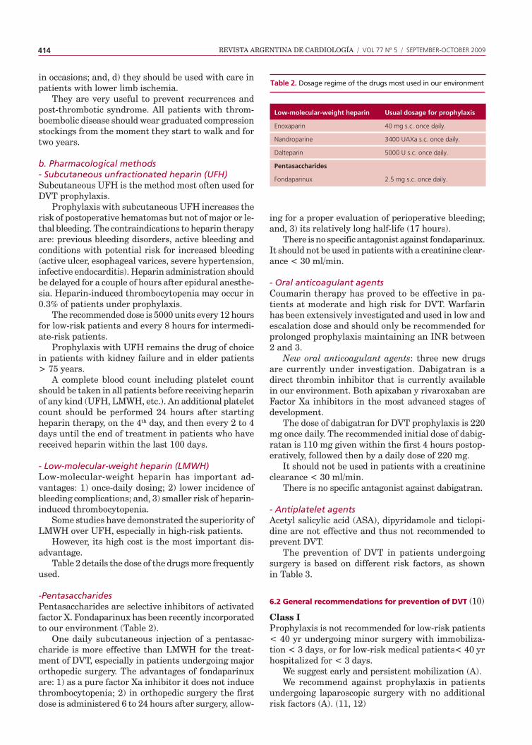
Table 2 details the dose of the drugs more frequently used.
-PentasaccharidesPentasaccharides are selective inhibitors of activated factor X. Fondaparinux has been recently incorporated to our environment (Table 2).
One daily subcutaneous injection of a pentasac-charide is more effective than LMWH for the treat-ment of DVT, especially in patients undergoing major orthopedic surgery. The advantages of fondaparinux are: 1) as a pure factor Xa inhibitor it does not induce thrombocytopenia; 2) in orthopedic surgery the first dose is administered 6 to 24 hours after surgery, allow-
ing for a proper evaluation of perioperative bleeding; and, 3) its relatively long half-life (17 hours).
There is no specific antagonist against fondaparinux. It should not be used in patients with a creatinine clear-ance < 30 ml/min.
- Oral anticoagulant agentsCoumarin therapy has proved to be effective in pa-tients at moderate and high risk for DVT. Warfarin has been extensively investigated and used in low and escalation dose and should only be recommended for prolonged prophylaxis maintaining an INR between 2 and 3.
New oral anticoagulant agents: three new drugs are currently under investigation. Dabigatran is a direct thrombin inhibitor that is currently available in our environment. Both apixaban y rivaroxaban are Factor Xa inhibitors in the most advanced stages of development.
The dose of dabigatran for DVT prophylaxis is 220 mg once daily. The recommended initial dose of dabig-ratan is 110 mg given within the first 4 hours postop-eratively, followed then by a daily dose of 220 mg.
It should not be used in patients with a creatinine clearance < 30 ml/min.
There is no specific antagonist against dabigatran.
- Antiplatelet agents Acetyl salicylic acid (ASA), dipyridamole and ticlopi-dine are not effective and thus not recommended to prevent DVT.
The prevention of DVT in patients undergoing surgery is based on different risk factors, as shown in Table 3.
6.2 General recommendations for prevention of DVT (10)
Class IProphylaxis is not recommended for low-risk patients < 40 yr undergoing minor surgery with immobiliza-tion < 3 days, or for low-risk medical patients< 40 yr hospitalized for < 3 days.
We suggest early and persistent mobilization (A).We recommend against prophylaxis in patients
undergoing laparoscopic surgery with no additional risk factors (A). (11, 12)
Table 2. Dosage regime of the drugs most used in our environment
Low-molecular-weight heparin usual dosage for prophylaxis
Enoxaparin 40 mg s.c. once daily.
Nandroparine 3400 UAXa s.c. once daily.
Dalteparin 5000 U s.c. once daily.
Pentasaccharides
Fondaparinux 2.5 mg s.c. once daily.

CONSENSUS STATEMENT ON THROMBOEMBOLIC DISEASE 415
Class I- Low-molecular-weight heparin (A)– Fondaparinux (A).– UFH 5000 U bid for low-risk patients (A)– UFH 5000 tid for high-risk patients (A).– For patients with multiple clinical or surgical risk
factors (highest-risk patients), we recommend UFH 5000 U tid or LMWH or fondaparinux plus mechani-cal prophylaxis with GCS or IPC(C).
We recommend consideration of renal impairment when deciding on doses that are cleared by the kidneys such as LMWH and fondaparinux. We suggest UFH in patients with a creatinine clearance < 30 ml/min or plasma creatinine level > 2.5 mg/dl
Class I– Intermittent pneumatic compression (B)– Graduated compression stockings (B).
We recommend that mechanical methods of pro-phylaxis be used primarily in patients in whom anti-thrombotic drugs are contraindicated or in those who are at high risk for bleeding.
We recommend against the use of aspirin alone as prophylaxis against VTE for any patient group (A).
6.3 Prophylaxis in special situations
a. Coronary care unit (13) and cardiovascular surgery (14)Prophylaxis with antithrombotic agents is recom-mended in patients with decompensated heart failure, acute pulmonary edema and in patients with heart diseases who are not receiving anticoagulants and will be immobilized for more 3days.
Class I– UFH, LMWH (B), fondaparinux (C).
The incidence of catheter-associated thrombosis is greater with fondaparinux than with enoxaparin. An estimated dose of 5000 U of UGH should be adminis-tered in patients undergoing cardiac catheterization.
For patients undergoing vascular surgery who do not have additional thromboembolic risk factors we suggest early and persistent mobilization.
For patients undergoing major vascular surgical procedures who have additional thromboembolic risk factors, we recommend prophylaxis with antithrom-botic agents.
Class I– UFH, LMWH (B), fondaparinux (C).
For patients undergoing cardiac surgery (coronary artery bypass graft surgery) or thoracic surgery we recommend prophylaxis.
Level of risk calf Proximal clínical Fatal successful prevention strategies
Low risk 2% 0.4% 0.2% < 0.01% Early Minor surgery in patients mobilization 40 yr with no additionalrisk factors
Moderate risk 10-20% 2-4% 1-2% 0.1-0.4% UFH (q12h)Minor surgery in patients LMWH oncewith additional risk factors dailySurgery in patients aged GCS40–60 yr with no additional IpCrisk factors
High risk 20-40% 4-8% 0.4-1% 0.4-1% UFH (q8h)Surgery in patients > LMWH 60 yr, or age 40–60 once daily with additional risk actors Fondaparinux(prior VTE, cancer, molecular IpChypercoagulability)
Highest risk 40-80% 10-20% 4-10% 0.2-5 LMWHSurgery in patients with multiple once daily risk factors (age > 40 yr, cancer, Fondaparinux, prior VTE) UFH/Hip or knee arthroplasty, LMWH + fracture surgery IpC/GCS Major trauma; spine cord injury
UFH: Unfractionated heparin LMWH: Low-molecular-weight heparin IPC: Intermittent pneumatic compression GCS: Graduated compression stockingsGeerts, et al. Chest 2004;126:338S-400S.
Table 3. Levels of thromboem-bolism risk in surgical patients without prophylaxis (9)
416 REVISTA ARGENTINA DE CARDIOLOGÍA / VOL 77 Nº 5 / SEpTEMBER-OCTOBER 2009
Class I– LMWH, UFH (C).– IPC or GCS for patients with contraindications to
antithrombotic agents (C).
b. Intensive care unit (15)Patients admitted to a critical care unit with recent major surgery, major trauma, sepsis, stroke, conges-tive heart failure, respiratory failure, history of DVT, extensive burns or elder patients have multiple risk factors for DVT. Additional risk factors are acquired during hospitalization: prolonged immobilization, se-dation or paralysis, central venous lines, mechanical ventilation, dialysis, etc. The incidence of DVT ranges between 10% and 90%, reflecting the great variability in the critically ill patients.
Class I– UFH (A), LMWH (A), fondaparinux (A).– Mechanical methods for patients with contraindica-
tions to antithrombotic agents (C).
c. Burn patients (16)Burn patients are at increased risk for VTE because of the presence of a profound systemic hypercoagulable state, as well as prolonged bed rest, performance of repeated surgical procedures, venous catheter inser-tion, and recurrent bouts of sepsis.
Class I– HUF (A), LMWH (A).
Class I– We recommend the use of IPC or GCS when anti-
coagulant prophylaxis is contraindicated (A).
d. Spinal cord injury (17)More than 50% of spinal cord injury (SCI) patients who are subjected to routine screening have DVT. Despite an increased awareness of DVT as a complication of SCI, PE remains the third leading cause of death and its incidence has not decreased in the recent years.
Prophylaxis with UFN, IPC and/or GSC alone has not proved to be effective. We recommend the use of LMWH alone or associated with mechanical methods.
Class I– LMWH once primary hemostasis is evident (A).– UHF combined with a mechanical method (B).– We recommend mechanical prophylaxis in patients
at high risk for hemorrhage or with spinal hema-toma (C).
e. Major trauma (18)Patients recovering from major trauma have the high-est risk of developing DVT. Without prophylaxis, these patients have a DVT risk exceeding 50%, with PE being the third leading cause of death in those who survive
beyond the first day of hospitalization.Based on a variety of studies, factors that were
associated with an increased risk of VTE include the following: spinal cord injury; lower extremity or pelvic fracture; need for a surgical procedure; femoral venous line insertion and prolonged immobility.
Class I– LMWH once primary hemostasis is evident
(A).– We recommend mechanical prophylaxis in
patients at high risk for bleeding until LMWH can be used safely(C).
– We recommend LMWH associated with a me-chanical method in high-risk patients (B).
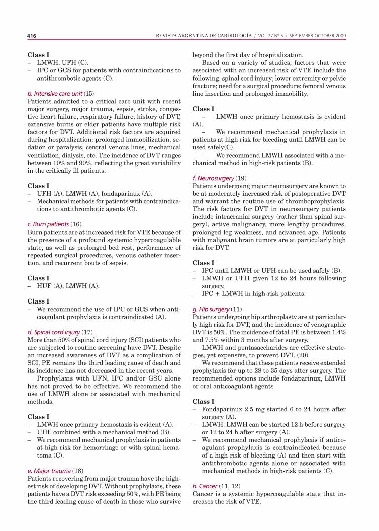
f. Neurosurgery (19)Patients undergoing major neurosurgery are known to be at moderately increased risk of postoperative DVT and warrant the routine use of thromboprophylaxis. The risk factors for DVT in neurosurgery patients include intracranial surgery (rather than spinal sur-gery), active malignancy, more lengthy procedures, prolonged leg weakness, and advanced age. Patients with malignant brain tumors are at particularly high risk for DVT.
Class I– IPC until LMWH or UFH can be used safely (B).– LMWH or UFH given 12 to 24 hours following
surgery.– IPC + LMWH in high-risk patients.
g. Hip surgery (11)Patients undergoing hip arthroplasty are at particular-ly high risk for DVT, and the incidence of venographic DVT is 50%. The incidence of fatal PE is between 1.4% and 7.5% within 3 months after surgery.
LMWH and pentasaccharides are effective strate-gies, yet expensive, to prevent DVT. (20)
We recommend that these patients receive extended prophylaxis for up to 28 to 35 days after surgery. The recommended options include fondaparinux, LMWH or oral anticoagulant agents
Class I– Fondaparinux 2.5 mg started 6 to 24 hours after
surgery (A).– LMWH. LMWH can be started 12 h before surgery
or 12 to 24 h after surgery (A).– We recommend mechanical prophylaxis if antico-
agulant prophylaxis is contraindicated because of a high risk of bleeding (A) and then start with antithrombotic agents alone or associated with mechanical methods in high-risk patients (C).
h. Cancer (11, 12)Cancer is a systemic hypercoagulable state that in-creases the risk of VTE.

CONSENSUS STATEMENT ON THROMBOEMBOLIC DISEASE 417
The pathogenic mechanisms of thrombosis in the patient with cancer involve a complex interaction between the tumor cells and the hemostatic system that produces activation of the coagulation system, inhibition of anticoagulant factors and fibrinolytic system, and vascular injury, leading to hypercoagu-lability. In addition, extrinsic factors such as surgery, chemotherapy and insertion of intravenous catheters play an important role.
Patients with cancer have a sixfold increased risk of VTE compared to those without cancer. Active can-cer accounts for almost 20% of all new VTE events occurring in the general population. Cancer patients undergoing surgery have at least twice the risk of post-operative DVT and more than three times the risk of fatal PE than noncancer patients who are undergoing similar procedures.
Cancer is also an independent predictor of lack of response to prophylaxis.
i. Long distance travel (21)We recommend the following general measures for long-distance travelers, whether by flight or by bus, who travel > 8 h: avoidance of constrictive clothing around the lower extremities or waist; avoidance of dehydration and frequent calf muscle stretching (Class I –C-). For high-risk patients we suggest the use of GCS (Class II -C-), or a single dose of LMWH, injected prior to departure (Class II -C-).
j. Extended prophylaxisSome patients may require prophylaxis beyond the hospital stay:– Patients with medical conditions or surgery patients
that must stay in bed after hospital discharge should receive prophylaxis as long as immobiliza-tion lasts (Class I -A-).
– Patients undergoing complex orthopedic surgery: total hip replacement, total knee replacement (Class I -A-)
– Abdominal or pelvic cancer surgery patients (Class I -B-).
Extended prophylaxis is recommended for 2 to 4 weeks with any of the antithrombotic agents previ-ously mentioned.
k. Prophylaxis and neuraxial analgesiaThe use of extended postoperative neuraxial anes-
thesia is more frequent. Removal of an epidural cath-eter should be done when the anticoagulant effect is at a minimum (usually just before the next scheduled subcutaneous injection). Anticoagulant prophylaxis should be delayed for at least 2 h after spinal needle or epidural catheter removal.
7. DIAGNOSIS OF DVT (22)The diagnosis of DVT based exclusively on clinical presentation is not reliable. More than 70% of DVT are asymptomatic and 50% of clinical diagnoses of DVT are wrong. Pain and edema are the most frequent yet unspecific findings. On the contrary, unilateral edema, the most specific clinical sign, is present in only 10% of patients. For these reasons, additional studies are necessary for diagnostic confirmation. In addition, unilateral edema may appear in lower extremity palsy in the absence of thrombosis.
Venography has been described as the gold standard method for the diagnosis of DVT; yet, it is an invasive strategy that is not readily available at all hospitals. The different methods for the diagnosis of DVT and their advantages, disadvantages, sensitivities and specificities are compared in Table 4.
High-speed multidetector computed tomography allows visualization of pulmonary arteries and of the entire venous system with same injection of contrast agent. However, radiation exposure is greater as more images are acquired. In this sense, the alarm has been raised, especially when young patients have to be studied.
8. DIAGNOSIS OF PE
8.1 Clinical presentation
The sensitivity and specificity of symptoms and signs of pulmonary embolism are low (table 5). Yet, the diagnosis of PE is strongly based on the clinical assess-ment (high, moderate or low clinical probability) that results from the combination of symptoms and signs suggestive of the condition.
Even considering their low predictive value, we should not forget that dyspnea, pleuritic pain, tachy-
Table 4. Methods for diag-nosis of DVT Method sensitivity specificity Finantial cost Desventajas
Venography 100% 100% Low risk Invasive Use of contrast agent
Doppler 66-90% 85-95% Very low Interobserver variability in its interpretation
LLHCT 95-100% 95-100% Moderate risk Use of contrast agent performed with chest CT
MRI 90-97% 100% High risk Scarcely accessible
LLHCT: lower limb helical computed tomography scan. MRI: Magnetic Resonance Imaging.

418 REVISTA ARGENTINA DE CARDIOLOGÍA / VOL 77 Nº 5 / SEpTEMBER-OCTOBER 2009
pnea and tachycardia are the most frequent symptoms and signs. On the contrary, the presence of a fourth heart sound or an increased pulmonary component of the second sound is uncommon yet more specific. The clinical probability of PE is higher in the presence of greater number of symptoms and signs. Complemen-tary tests do not modify the predictive value of clinical manifestations. (23)
Naturally, it is important to correlate the clinical presentation with the presence of risk factors.
The probability of PE is high in the presence of three clinical syndromes which may occur isolated or combined:
Acute unexpected dyspnea. These patients present sudden onset dyspnea, tachypnea and tachycardia. The electrocardiogram and chest X-ray may be normal. Dyspnea may be intermittent and thus may be absent when the patient is examined.
Hemoptisis and/or pleuritic pain. These patients usually present at least three of the following mani-festations: pleuritic pain, dyspnea, hemoptisis and radiographic evidence of parenchymal infiltrates. Fever, pleural friction rub and leukocytosis may also be present and a diagnosis of pneumonia should be ruled out.
Cardiogenic shock. Cardiogenic shock is the criti-cal manifestations of massive PE. Patients usually present some degree of loss of consciousness, anxiety, pronounced dyspnea, oppressive chest pain suggestive of myocardial infarction, increased pulmonary compo-nent of the second sound and signs of shock.
8.2 Routine laboratory tests (24)
Their usefulness is limited. Increased erythrocyte sedimentation rate and leukocytosis are nonspecific findings.
8.3 Chest X-ray
An abnormal chest X-ray has little value in patients with previous cardiac and respiratory conditions.
Table 6 shows the most common radiographic findings in patients with PE with no previous cardiac disease as described in the PIOPED I study.
8.4 Electrocardiogram
ECG findings depend on the level of pulmonary hypertension and on the presence of previous cardiac and pulmonary disease.
The PIOPED study included 117 patients with no previous cardiac and pulmonary disease. The ECG was normal in 30% of patients and 70% presented nonspecific abnormalities. Sinus tachycardia was the most frequent abnormality.
A S1 Q3 T3 y S1 S2 S3 pattern was described in less than 12% of patients. ST-segment and T-wave abnor-malities were observed in 66% of patients. The presence
of T-wave inversion in the anterior leads and acute RBBB correlated with the magnitude of PE, with an increase in pulmonary artery pressure and with the severity of right ventricular dysfunction. Occasionally, the magnitude and the morphology of ST-segment el-evation in leads V1, V2 and V3 may mimic an anterior myocardial infarction. In fact, these changes represent right ventricular injury and ischemia.
The presence of acute changes in the ECG - ar-rhythmias, right bundle branch block, S1Q3 pattern or ST-segment changes - is associated with greater 30-day mortality (29% versus 11%).
8.5 Arterial blood gases and alveolar-arterial O2 gradient
Pulmonary embolism is generally associated with hypoxemia, hypocapnia, respiratory alkalosis and in-creased alveolar-arterial O2 gradient. This combination is suggestive of PE in the absence of any alternative diagnosis.
However, a normal PaO2 does not exclude the di-agnosis of PE.
Table 6. Chest radiograph abnormalities in PE patients
Abnormalities %(n = 2.315)
Cardiomegaly 27
pleural effusion 23
Elevated diaphragm 20
prominent central 19
pulmonary artery
Atelectasis 18
Interstitial infiltrate 17
pulmonary congestion 14
Decreased pulmonary 8
vascularity
pulmonary infarction 5
Hyperinflation 5
Table 5. Diagnostic value of isolated symptoms and signs in pul-monary embolism*
sign or symptom sensitivity (%)specificity (%) PPV nVP (%) (%)
Dyspnea 73 28 32 31
pleuritic pain 66 41 34 66
Rales (crakles) 51 60 38 28
Fourth heart sound 24 86 45 29
Second heart sound 23 87 45 30
Modified from Stein, et al. Chest 1991;100:598-603.*Patients with no pre-existing cardiac or pulmonary disease.PPV: Positive predictive value. NPV: Negative predictive value.

CONSENSUS STATEMENT ON THROMBOEMBOLIC DISEASE 419
In massive PE with shock and ventilatory failure, hypercapnic respiratory acidosis may be combined with metabolic acidosis secondary to severe heart failure.
8.6 D-dimer (25)
Plasma D-dimer is the specific derivative of cross-linked fibrin, which is produced when fibrin is degraded by plasmin. D-dimer levels are elevated in plasma in the presence of an acute clot under a process of fibri-nolysis.
Most patients with PE have elevated levels of D-dimer (>500 U/ml). High plasma levels of D-dimer may also be found in elder patients, hospitalized pa-tients, cancer and recent surgery. For this reason, this sensitive marker has low specificity (< 40%) for the diagnosis of VTE (DVT - PE). Thus, the usefulness of the test is low is hospitalized patients as an elevated percentage of patients will have a positive test even in the absence of VTE.
Despite its low specificity, several authors have demonstrated that D-dimer has high sensitivity, PPV and NPV for DVT - PE.
There are a number of available assays with differ-ent characteristics:1. High sensitivity D-dimer assays: these methods
have a sensitivity ≥ 98% and a very high negative predictive value.
2. Moderately sensitive D-dimer assays: the sensitivity of these methods ranges from 85% to 98%. Their negative predictive value is not high enough to rule out PE and a negative result requires another diagnostic test.
The conventional latex test used in our environ-ment has low sensitivity (about 70%) and specificity.
In conclusion, a moderately sensitive assay excludes PE only in patients with a low clinical probability, while a negative D-dimer result in a highly sensitive assay has a negative predictive value high enough to be used as a single study.
Recent studies have shown that the magnitude of the increase has a prognostic value and that patients with abnormal D-dimer test have increased risk of re-current thromboembolism. (26) It has been suggested to continue anticoagulation therapy as long as D-dimer values remain elevated.
8.7 Troponins (27)
Although serum levels of troponin T and I are not use-ful for the diagnosis of PE due to low specificity, their value as markers of risk is increasing.
Cardiac troponins are elevated in 30-50% of patients with moderate or severe PE.
High cardiac troponin elevation is associated with a 5-fold higher risk of complications compared to moder-ate elevations, with a greater incidence of cardiogenic shock and in-hospital mortality.
8.8 Transthoracic echocardiography
Transthoracic echocardiography is currently an essen-tial tool for the evaluation of patients with suspected PE. A high percentage of patients with PE have ab-normalities in the right heart chambers that can be detected by transthoracic echocardiography.
The Mc Connell sign is the most specific finding: depressed contractility of the RV free wall compared with its apex. Other indirect signs are the presence of tricuspid regurgitation, systolic pulmonary hyperten-sion (not greater than 40 to 50 mm Hg in a previously healthy heart) without apparent cause or right ven-tricular dilatation with absence of inspiratory collapse of the inferior vena cava. (28)
In patients presenting with shock or hypotension, a normal transthoracic echocardiogram practically excludes PE as a cause of hemodynamic instability.
Once the diagnosis of PE has been confirmed, transthoracic echocardiography is useful for prognosis assessment. Right ventricular compromise is a strong independent predictor of mortality.
The presence of thrombi in transit (4% of echocar-diograms) is another indicator of poor outcomes. In this case, the association with a patent foramen ovale increases the risk.
8.9 Transesophageal echocardiography (29)
Bedside transesophageal echocardiography is being used with more frequency. The use of multiplanar transducers increases its performance. An additional advantage of this method is the ability to rule out other severe cardiovascular conditions as aortic dissection.
8.10 Ventilation-perfusion scintigraphy (V/Q scan)
When properly performed, ventilation-perfusion scintig-raphy has diagnostic value. The perfusion images should be acquired in the six views established by the PIOPED study (anterior, posterior and four oblique views).
A normal V/Q scan rules out EP, yet this situation is not common. On the other hand, only 10% of PE have a high-probability V/Q scan, while most PE (65-78%) have intermediate-probability scans. For this reason, further studies are necessary to make a diagnosis. In addition, most patients with previous cardiac and pulmonary diseases have intermediate-probability scans. Currently, V/Q scan has been replaced by heli-cal computed tomography (HCT) scan. However, when HCT is not available or is considered inconvenient, V/Q scan is still a useful tool, especially for patients with no pre-existent cardiac and pulmonary conditions. (30) An abnormal chest X-ray markedly increases the specificity of the method.
8.11 Helical computed tomography scan (31)
Although helical computed tomography scan has been used in the last years, it has been recently added to

420 REVISTA ARGENTINA DE CARDIOLOGÍA / VOL 77 Nº 5 / SEpTEMBER-OCTOBER 2009
the diagnostic algorithm. Yet, some aspects should be considered:1. The positive predict value of HCT to detect intralu-
minal defects in lobe arteries or in pulmonary artery trunk is 85%, equivalent to a high-probability V/Q scan
2. Subsegmental pulmonary defects are more difficult to visualize with this method.
3. A normal single-slice CT scan reduces the possibility of PE but does not exclude the diagnosis, (similar to a low-probability V/Q scan). In case of high clinical probability of PE, further studies should be performed.
4. Recent studies have determined that a normal HCT rules out the diagnosis of PE without need of additional studies.
It has been shown that 10% to 30% of patients with documented PE present with clots only in subsegmen-tal and smaller arteries. Controversy also exists about the clinical significance of small emboli; yet, they are thought to have prognostic relevance in sujects with a decreased cardiopulmonary reserve.
The most important advantage of multidetector-row spiral CT is improved diagnosis of small peripheral emboli. In addition, it is also useful to evaluate the presence of old, organized emboli and the territory of the inferior vena cava with the same injection of contrast agent.
Its diagnostic yield is equivalent to that of pulmo-nary angiography.
8.12 Deep venous thrombosis
The diagnosis of DVT is an indirect way to determine the probability of PE, as both treatments are similar.
In the presence of DVT, contrast venography detects DVT in 50% to 70% of patients. In addition, 50% of patients with DVT have asymptomatic PE that is evi-dent if investigated. Even more, in patients with DVT and clinical probability of PE, the latter is confirmed in 90% of patients.
Color-Doppler ultrasound of the lower extremities is the most useful, available and easiest tool fort the diagnosis of DVT; yet, some considerations should be mentioned:1. A normal ultrasound does not exclude PE in pa-
tients with a nondiagnostic V/Q scan or HCT, but it reduces its probability.
2. The absence of DVT diagnosed by Doppler ultra-sound is associated with low-risk of PE recurrence.
8.13 Pulmonary angiography (7, 25, 28)Pulmonary angiography is the gold standard diagnostic test for PE, but is invasive, costly and is not always available at all hospitals. It is currently indicated in critically ill high-risk patients when the results of non-invasive imaging are negative or equivocal. Pulmonary
angiography should be performed when mechanical ap-proaches or local pharmacological therapy are indicated. This test may be negative in 1-5% of patients with PE.
9. RECOMMENDATIONS OF DIAGNOSTIC IMAGING TESTS
Deep venous thrombosisClass I
– Contrast venography (A)– Venous Doppler ultrasound (A).– Combined venography and multislice helical
computed tomography pulmonary angiography (A).
Pulmonary embolismClass I– Pulmonary angiography (A).– Multislice helical computed tomography (A).
Class IIa– V/Q scintigraphy scan (A).– Helical single-slice computed tomography (A).– Transesophageal echocardiography (A). Transesophageal echocardiography (A).
10. DIAGNOSTIC AND THERAPEUTIC ALGORITHM (32)
The diagnosis of PE is not simple. Pulmonary embolism is one of the most common unexpected findings at autop-sies. Conversely, the diagnosis of PE is frequently em-pirical and unnecessary treatments are often initiated, increasing the risk of bleeding complications. Moreover, PE is a frequent clinical problem in daily practice.
Diagnostic algorithms specify a particular a set of medical tests to order so as to allow an accurate in-clusion and exclusion of patients with this condition. Algorithms should adapt to the specific hospital and provide adequate cost-benefit ratio.
Clinical suspicion is the initial step of any diagnostic strategy and should guide which studies to order. D-dimer (DD), ventilation-perfusion scintigraphy scan (V/Q scan), helical computed tomography (HCT) and 2D-echocardiography should be performed for initial stratification of clinical risk.
Naturally, in patients with clinical signs of deep venous thrombosis (DVT), Doppler ultrasound of the lower limbs should be the initial test as it provides high sensitivity and specificity, is easy to perform and cost-effective.
10. 1 Models for clinical stratification
The initial clinical suspicion may be guided by the clini-cal experience or ruled by preestablished algorithms. In this sense, there are several models that may be implemented as an initial approach.
Models based on scoresThe most frequently used clinical prediction rule is the one developed by Wells, based on symptoms, signs, the presence of an alternative diagnosis and risk factors.

CONSENSUS STATEMENT ON THROMBOEMBOLIC DISEASE 421
This scheme classifies patients as having low, moderate and high probability of PE (Table 7).
Wells developed this score for the emergency depart-ment and found a prevalence of PE of 2% in the low probability group (40% of the patients studied), 19% in the moderate probability group (45% of patients studied), and 50% in the high probability group (8% of patients).
In summary, according to the different studies, there is enough evidence supporting that empiric use of clinical parameters or standardized rules is useful for risk stratification of patients. The expected prevalence of PE is about 10% in patients with low clinical prob-ability, 25% in moderate clinical probability and 60% in those with high clinical probability. (Table 7)
Special situations modifying the diagnostic strategies1. DD may be positive in hospitalized surgery patients
(specificity 7% versus el 47% in outpatients).2. Cancer patients: 85% with positive DD even in the
absence of VTE.3. Elder patients are more likely to have positive DD
and abnormal V/Q scan.4. PE treatment: heparin significantly decreases the
sensitivity of DD due to reduction in plasma levels.5. A negative high sensitivity DD does not rule out
PE in patients with high clinical probability.6. Previous cardiovascular surgery modifies the result
of V/Q scans.
Based on the aforementioned studies, we believe that the last algorithm published by Goldhaber in 2005 is an interesting approach for this condition (Figure 2).
11. HIGH-RISK PE (34)
High-risk PE deserves a special consideration. The current concept of high-risk massive PE is functional rather than anatomical. Massive PE is defined as sus-
tained systolic hypotension (< 90 mm Hg or a reduction > 40 mm Hg in hypertensive patients) for at least 30 minutes, unresponsive to intravenous fluid expansion, that cannot be explained by other coexistent conditions (sepsis, active bleeding, arrhythmias).
The mortality rate within the first hours is ex-tremely high without appropriate treatment.
These massive emboli reduce the pulmonary cross-sectional area and produce an abrupt rise in pulmonary artery pressure, leading to acute right ventricular dilatation. Pulmonary artery pressure increases with more than 50% obstruction of the pulmonary artery bed in patients without coexistent cardiopulmonary disease and with 25% of obstruction or even less in those with prior cardiopulmonary disease.
Patients without prior pulmonary hypertension are unable to generate systolic or mean pulmonary artery pressure of ≥ 50 mm Hg or ≥ 40 mm Hg, respectively, which appear to be the maximal pressure that a healthy ventricle can generate. When the pulmonary artery bed obstruction is > 75% (or less in patients with prior cardiopulmonary disease), right ventricular failure develops, leading to ischemia by increasing myocardial oxygen demand while limiting supply. In turn, cardiac output and systemic blood pressure decrease and in-tramural pressure elevates. The presence of coronary artery disease worsens the outcomes. Death is then the consequence of unmanageable systolic overload, associated with ischemia and eventually with right ventricular infarction.
The sequence of events leading to acute decom-pensation of the RV is shown in Figure 3. Acute right ventricular dilatation produces pericardial restraint and leftward shift of the interventricular septum, re-ducing left ventricular preload and cardiac output, and coronary artery perfusion decrease. These phenomena occur when the right ventricle requirements are in-creased, needing maximal coronary artery flow.
These concepts explain the elevation of troponin levels in critically ill patients. (28)
12. TREATMENT OF THROMBOEMBOLIC DISEASE (35)
12.1 High-risk patient (Figure 4)
These patients are admitted to the Intensive Care Unit with hemodynamic impairment and/or severe hypox-emia and require a rapid and efficient management. The first step is to provide hemodynamic support. The combination of hypoxemia and increased pulmonary resistance is very dangerous. The initial treatment of patients in shock is rapid volume expansion (500 to 1000 cm3). However, in hypotensive patients with severe RV dysfunction excess fluid administration may precipitate further RV deterioration.
Ensuring adequate oxygenation is also important in stabilizing their condition. Mechanical ventilation is indicated for patients with severe hypoxia unresponsive to the initial measures.
Table 7. Clinical prediction rules for PE: Wells scoring system. Score and pretest probability (33)
score
Active cancer 1.0
Hemoptysis 1.0
Recent surgery 1.5
previous DVT 1.5
HR > 100 1.5
Clinical signs of DVT 3.0
Alternative diagnosis less likely than pE 3.0
Pretest probability
High ≥ 6
Moderate 2.5-5.5
Low ≤ 2

422 REVISTA ARGENTINA DE CARDIOLOGÍA / VOL 77 Nº 5 / SEpTEMBER-OCTOBER 2009
Inotropic and/or vasoactive agents are added if hemodynamic instability persists, following the guide-lines for treatment of cardiogenic shock.
Undoubtedly, thrombolysis or emboli fragmentation are the treatment of choice in hemodinamically unsta-ble patients with PE. When possible, the patient should be transferred to the catheterization lab for diagnostic and therapeutic purposes. If cath lab facilities are not available, intravenous thrombolysis is indicated.
12.2 Low-risk patient:
a. Anticoagulant therapy (28)Anticoagulant therapy for DVT and PE are similar as both conditions are manifestations of the same disease.
However, mortality is significantly higher and re-currences are three times greater in PE versus DVT without PE. In addition, about 4% of patients with PE will develop significant pulmonary hypertension in the medium term. (36)
The goal of the treatment is to interrupt the progres-sion of the thrombotic phenomena; yet it does not exert any action on the existent clots which are degraded by endogenous fibrinolysis in the medium term.
The duration of anticoagulation with LMWH or UFH is currently 5 days. In the absence of contraindica-tions, therapy with oral vitamin K antagonists starts at the first or second day. An adequate anticoagulation is usually achieved by the fifth day, and heparin can then be discontinued.
In patients with clinical suspicion of thromboem-bolic disease, and in the absence of contraindications, we suggest starting any heparin treatment until the diagnosis can be ruled out or confirmed.
Fig. 2. Diagnostic algorithm. (Adapted from Piazza G, God-haber SZ. Acute Pulmonary Embolism Part I. Circulation 2006;114:e28-e32.)
Fig. 4. Diagnostic-therapeutic approach in the patient with critical hemodynamic instability
Fig. 3. Physiopathology of massive PE
CONSENSUS STATEMENT ON THROMBOEMBOLIC DISEASE 423
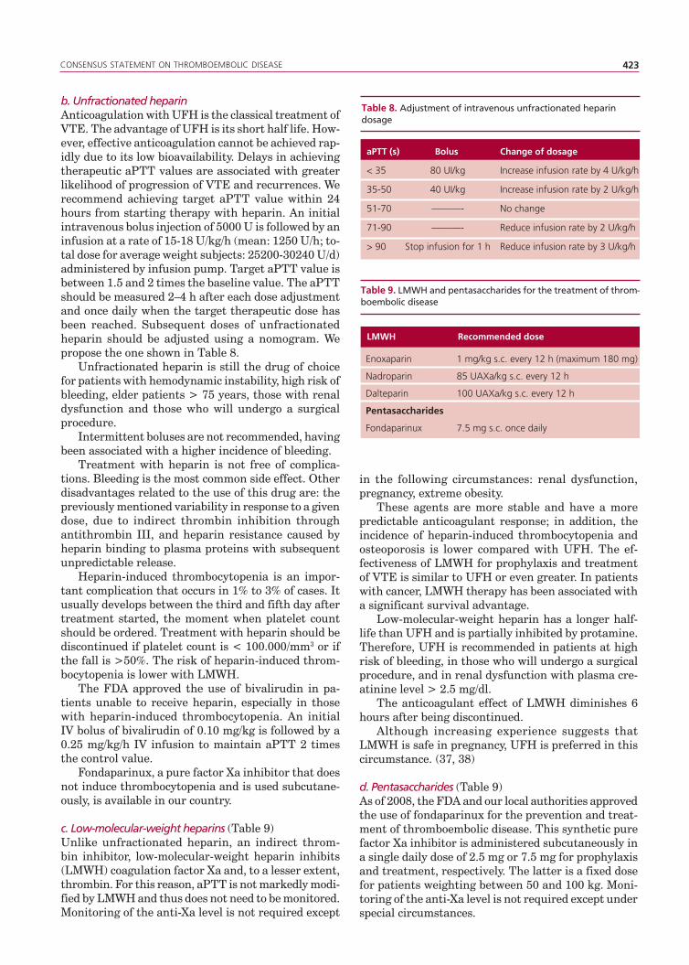
b. Unfractionated heparinAnticoagulation with UFH is the classical treatment of VTE. The advantage of UFH is its short half life. How-ever, effective anticoagulation cannot be achieved rap-idly due to its low bioavailability. Delays in achieving therapeutic aPTT values are associated with greater likelihood of progression of VTE and recurrences. We recommend achieving target aPTT value within 24 hours from starting therapy with heparin. An initial intravenous bolus injection of 5000 U is followed by an infusion at a rate of 15-18 U/kg/h (mean: 1250 U/h; to-tal dose for average weight subjects: 25200-30240 U/d) administered by infusion pump. Target aPTT value is between 1.5 and 2 times the baseline value. The aPTT should be measured 2–4 h after each dose adjustment and once daily when the target therapeutic dose has been reached. Subsequent doses of unfractionated heparin should be adjusted using a nomogram. We propose the one shown in Table 8.
Unfractionated heparin is still the drug of choice for patients with hemodynamic instability, high risk of bleeding, elder patients > 75 years, those with renal dysfunction and those who will undergo a surgical procedure.
Intermittent boluses are not recommended, having been associated with a higher incidence of bleeding.
Treatment with heparin is not free of complica-tions. Bleeding is the most common side effect. Other disadvantages related to the use of this drug are: the previously mentioned variability in response to a given dose, due to indirect thrombin inhibition through antithrombin III, and heparin resistance caused by heparin binding to plasma proteins with subsequent unpredictable release.
Heparin-induced thrombocytopenia is an impor-tant complication that occurs in 1% to 3% of cases. It usually develops between the third and fifth day after treatment started, the moment when platelet count should be ordered. Treatment with heparin should be discontinued if platelet count is < 100.000/mm3 or if the fall is >50%. The risk of heparin-induced throm-bocytopenia is lower with LMWH.
The FDA approved the use of bivalirudin in pa-tients unable to receive heparin, especially in those with heparin-induced thrombocytopenia. An initial IV bolus of bivalirudin of 0.10 mg/kg is followed by a 0.25 mg/kg/h IV infusion to maintain aPTT 2 times the control value.
Fondaparinux, a pure factor Xa inhibitor that does not induce thrombocytopenia and is used subcutane-ously, is available in our country.
c. Low-molecular-weight heparins (Table 9)Unlike unfractionated heparin, an indirect throm-bin inhibitor, low-molecular-weight heparin inhibits (LMWH) coagulation factor Xa and, to a lesser extent, thrombin. For this reason, aPTT is not markedly modi-fied by LMWH and thus does not need to be monitored. Monitoring of the anti-Xa level is not required except
Table 8. Adjustment of intravenous unfractionated heparin dosage
aPTT (s) Bolus change of dosage
< 35 80 UI/kg Increase infusion rate by 4 U/kg/h
35-50 40 UI/kg Increase infusion rate by 2 U/kg/h
51-70 ———- No change
71-90 ———- Reduce infusion rate by 2 U/kg/h
> 90 Stop infusion for 1 h Reduce infusion rate by 3 U/kg/h
Table 9. LMWH and pentasaccharides for the treatment of throm-boembolic disease
LMWH Recommended dose
Enoxaparin 1 mg/kg s.c. every 12 h (maximum 180 mg)
Nadroparin 85 UAXa/kg s.c. every 12 h
Dalteparin 100 UAXa/kg s.c. every 12 h
Pentasaccharides
Fondaparinux 7.5 mg s.c. once daily
in the following circumstances: renal dysfunction, pregnancy, extreme obesity.
These agents are more stable and have a more predictable anticoagulant response; in addition, the incidence of heparin-induced thrombocytopenia and osteoporosis is lower compared with UFH. The ef-fectiveness of LMWH for prophylaxis and treatment of VTE is similar to UFH or even greater. In patients with cancer, LMWH therapy has been associated with a significant survival advantage.
Low-molecular-weight heparin has a longer half-life than UFH and is partially inhibited by protamine. Therefore, UFH is recommended in patients at high risk of bleeding, in those who will undergo a surgical procedure, and in renal dysfunction with plasma cre-atinine level > 2.5 mg/dl.
The anticoagulant effect of LMWH diminishes 6 hours after being discontinued.
Although increasing experience suggests that LMWH is safe in pregnancy, UFH is preferred in this circumstance. (37, 38)
d. Pentasaccharides (Table 9)As of 2008, the FDA and our local authorities approved the use of fondaparinux for the prevention and treat-ment of thromboembolic disease. This synthetic pure factor Xa inhibitor is administered subcutaneously in a single daily dose of 2.5 mg or 7.5 mg for prophylaxis and treatment, respectively. The latter is a fixed dose for patients weighting between 50 and 100 kg. Moni-toring of the anti-Xa level is not required except under special circumstances.

424 REVISTA ARGENTINA DE CARDIOLOGÍA / VOL 77 Nº 5 / SEpTEMBER-OCTOBER 2009
It is used once a day due to its long half-life (17 hours). There is no specific antagonist against fondaparinux.
e. Thrombolytic therapyThrombolysis seems to be an excellent option, at least in theory; it has a lytic effect in both venous thrombi and pulmonary emboli, which is beneficial in terms of immediate hemodynamic and clinical improvement. However, the number of patients included in random-ized trials comparing thrombolytic agents versus placebo is not more than 800.
The MAPPET trial is the most important study recently published that evaluated thrombolysis plus heparin versus heparin alone. (39) The investigators used rtPA and included patients with right ventricular dysfunction. The study was favorable to thrombolytic therapy, yet the methodology used was strongly criti-cized.
The ICOPER registry (40) included 2454 patients and 304 were treated with thrombolytic drugs. In this study these agents did not improve the survival. Yet, the incidence of intracranial hemorrhage was 3%.
Nine studies with a total of 461 patients were in-cluded in a meta-analysis published in 2002. Only three of the trials were double-blind. Most of these studies showed that thrombolytic agents were more effective and rapid than heparin to dissolve the thrombi, espe-cially during the first 24 hours. The most important finding of this meta-analysis was that thrombolysis did not provide any benefit to unselected patients with pulmonary embolism (RR 0.63, 95% CI 0.32-1.93).
There is well established evidence to indicate thrombolysis to patients with severe hemodynamic impairment (systolic blood pressure < 90 mm Hg or a reduction > 40 mm Hg in hypertensive patients) who are unresponsive to intravenous fluid expansion for 30-60 minutes, and require the use of vasopressive drugs, and to patients with refractory hypoxemia. In these high-risk patients, rapid improvement in pul-monary circulation may make the difference between life and death.
Thrombolysis may be indicated in patients with thrombi in transit, especially associated with a patent foramen ovale.
Yet, the indication in patients with right ventricular dysfunction or severe PE with preexistent cardiopul-monary diseases is controversial. In these cases, the decision should be based on the individual patient considering the risk-benefit ratio. (28)
All patients treated with thrombolysis should receive continuous infusion of UFH in the absence of contraindications, following the controls previously mentioned. In patients previously treated with heparin, we recommend to discontinue anticoagulation during thrombolytic therapy. The aPTT should be monitored at the end of thrombolysis and then every two hours. Therapy with heparin is reinitiated when aPTT reaches < 80 s.
Table 10 describes the different therapeutic sched-ules usually used. The main contraindications are shown in Table 11.
It is recommended to discontinue infusion after 12 hours, as prolonged thrombolytic periods are not effective and are associated with increased risk of bleeding.
The following precautions should be taken into ac-count when thrombolysis is used to treat pulmonary embolism:– Avoid invasive procedures such as punctures in
vessels that cannot be compressed (e.g. subclavian vein) or arterial punctures.
– No coagulation tests are necessary after throm-bolysis because the doses of thrombolytic agent are fixed. This treatment should be performed under strict control in an intensive care unit. Infusion should be discontinued if disorders of conscious-ness suggestive of brain hemorrhage or signs of intraabdominal bleeding develop.
The route of administration is another controver-sial issue. Traditionally, the intravenous route has been considered as effective as the direct infusion in the pulmonary artery. Yet, some experimental stud-ies suggest that, in patients with massive pulmonary embolism, direct local infusion of a thrombolytic agent via a catheter in the pulmonary artery, at a reduced dosage (10-20% the intravenous dose), might offer better outcomes than systemic infusion, with a lower
Table 10. Dosage regime of the drugs used in our environment
rtpA 100 mg over 2 h in continuous infusion The first 10 mg may be given in bolus
Streptokinase 1500000 U over 2 h in continuous infusion
rtpA 0.6 mg/kg over 5-15 min (maximum dose 60 mg) in critically ill patients
Table 11. Contraindications to thrombolytic therapy
Absolute Relative
Hemorrhagic stroke Bleeding disorders or thrombo cytopenia < 100000/mm3
Central nervous Refractory hypertension system neoplasms (systolic/diastolic blood pressure >180/110 mmHg)
Recent head surgery Ischemic stroke in the precedingor trauma (within 6 monthspreceding 2 months)
Internal bleeding in the Surgery in the preceding 10 dayspreceding 6 months

CONSENSUS STATEMENT ON THROMBOEMBOLIC DISEASE 425
risk of bleeding. This therapeutic strategy has been used in small studies in critically ill patients, with encouraging results.
Time window: classically, there is a 14-day “win-dow” for administration of PE thrombolysis. In fact, in daily practice, most deaths occur within the first hours after the onset of PE.
f. Mechanical and pharmacological treatment in the ca-theterization laboratory
Nowadays, patients with pulmonary embolism are not referred to the catheterization laboratory only for diagnostic purposes.
Most patients with massive PE undergo cardiac catheterization to confirm the diagnosis and for per-cutaneous embolectomy and fragmentation to open a partially occluded pulmonary trunk or major pulmo-nary arteries. The procedure may be performed using specially designed pulmonary catheters or conventional cardiac catheters (e.g. pigtail catheter). Thrombolytic therapy can be administered as adjunctive therapy in doses equivalent to 10% to 20% of the systemic dose.
g. Surgical embolectomyTraditionally, pulmonary embolectomy has been
reserved for patients in extremis with PE. The Tren-delemburg procedure is no longer used and most centers use cardiopulmonary bypass. Surgical embo-lectomy is associated with elevated mortality, even in referral centers worldwide. Recently, a group of investigators from Boston has revived this technique with favorable outcomes in patients with severe though not critical PE. We believe that this technique is still an option for tertiary care centers with well-trained surgical staff.
13. THERAPEUTIC RECOMMENDATIONS
Class IPatients with PE should initiate anticoagulation for 5 days, until achieving target INR levels of > 2 for 24 hours with any of the following drugs:– Unfractionated heparin in continuous infusion (A).– Low-molecular-weight heparin (A)– Pentassacharides: fondaparinux (A).
Anticoagulation with unfractionated heparin should be initiated in patients with shock and in those in whom subcutaneous drug absorption cannot be warranted.
In the absence of contraindications, all patients with suspected PE and a high pretest probability should be treated with anticoagulant agents until the diagnosis is confirmed or excluded (C).
Class IIa– Use of bivalirudin or fondaparinux is the recom-
mended form of initial treatment for patients in whom heparin is contraindicated (B).
Class I– Fibrinolytic therapy in patients with shock (C).- rtPA 100 mg over 2 h.- Streptokinase 1500000 IU over 90 minutes.
Class IIa– Fibrinolytic therapy in patients with refractory
hypoxemia under mechanical ventilation and high fraction of inspired O2 (C).
– Fibrinolytic therapy in patients with thrombi in transit (C).
– Mechanical therapy with or without fibrinolytic therapy associated in PE (A)
– Surgical embolectomy (B).– rtPA in bolus injection of 50 mg over 15 minutes in
patients with impending circulatory arrest (C).
Class IIb– Fibrinolytic therapy in PE patients with signs of
right ventricular dysfunction on echocardiography without hemodynamic impairment. (B)
14. VENA CAVAL FILTERSRecommendations for vena caval filters
Class I– Patients with active bleeding (C)– Patients with recurrent embolism despite adequate
anticoagulation treatment (C).– Patients with bleeding during adequate anticoagu-
lation treatment (C).
Other indications: the indication of vena caval fil-ters for prevention of recurrent embolism in patients with massive PE, inadequate cardiopulmonary reserve or in those undergoing a previous surgical or percuta-neous embolectomy is still under debate. Under these special circumstances, clinical management decisions will continue to be individualized and based on updated guidelines.
Exceptionally, venous filters have been inserted in the superior vena cava.
Retrievable devices, currently available may be removed within 2 weeks of implantation or left in situ for longer periods of time.
The percentage of patients who receive vena caval filters is low (3% to 5%). At present, there is no evidence showing a significant reduction on mortality; yet, a moderate reduction in short-term morbidity has been demonstrated.
15. COUMARIN THERAPY
Coumarin therapy is indicated for long-term anticoagu-lation treatment to prevent recurrences. These drugs are vitamin K antagonists. These drugs prolong the prothrombin time; yet the maximum effect occurs five days after the initial dose when all the factors involved in the coagulation cascade are effectively inhibited.

426 REVISTA ARGENTINA DE CARDIOLOGÍA / VOL 77 Nº 5 / SEpTEMBER-OCTOBER 2009
Therefore, in patients with PE or DVT heparin and coumarin therapies should overlap for approximately four to five days; heparin is discontinued thereafter and vitamin K antagonists are administered alone. The duration of oral anticoagulation has not been precisely established by reliable trials. In consequence, we sug-gest the guidelines recommended by the American College of Chest Physicians (Table 12).
Proper duration of therapy is unclear in first event with:– Factor V Leiden, homocystinemia, deficiency of
protein C or S;– Thrombophilias with reversible risk factors (Hyers
TM, et al. Chest 2001;169:176S).
If severe hemorrhage develops, infusion of cryo-precipitates or 2 units of fresh frozen plasma should be rapidly initiated. Intravenous injection of 10 mg of vitamin K reverses oral anticoagulant therapy in 6 to 12 hours; refractoriness to subsequent anticoagulant therapy persists for 10 to 15 days.
16. RECOMMENDATIONS FOR SECONDARY PREVENTION
– Early mobilization is indicated in patients with DVT, if tolerated (Class I -B-).
– We recommend starting therapy with vitamin K antagonists together with heparin therapy, and over-lapping both drugs for 3 to 5 days (Class I -B-).
– Anticoagulant agents should be given at doses adjusted to maintain a target INR of 2 to 3 (Class I -A-).
– We recommend lifetime anticoagulation in patients with 2 or more thromboembolic events (Class II -A-).
– We recommend anticoagulation therapy for 3 months in patients with DVT and reversible or time-limited risk factor (Class I -A-).
– In cancer patients with DVT we recommend antico-agulation therapy with LMWH for 3 to 6 months and lifetime anticoagulation until resolved (Class I -C-).
– In patients with anticardiolipin antibodies or mul-tiple thrombophilias we recommend anticoagula-tion therapy for 12 months (Class I -C-).
Lifetime anticoagulation is also suggested (Class II -C-).
– Lifetime anticoagulation should be considered in idiopathic DVT (Class II -A-).
– Recommendations for lifetime anticoagulation are subject to modification considering the individual risk-benefit ratio (Class I -C-).
17. PREGNANCY
Pregnant women, those in the post-partum period or under hormone therapy, are at high risk of thromboem-bolic disease. The risk of VTE in the puerperal period increases 5-fold as compared to the antepartum period, and the risk of PE is 15 times higher.
The diagnostic tools should not interfere with the fetus; CT scanning may be used providing effective protection to the fetus.
Low molecular weight heparins are recommended in the usual dose. Thrombolytic therapy may be indicated in life-threatening PE (reports from isolated cases, with favorable outcomes) Low-molecular-weight heparin is indicated as coumarins have teratogenic effects.
BIBLIOGRAPHY
1. Dalen JE. Pulmonary Embolism: What have we learned since Virchow? Natural history, pathophisiologhy and diagnosis .Chest 2002;122:1440-56.2. Kearon C. Diagnosis of pulmonary embolism.CMAJ 2003;168:183-94.3. Benotti JR, Dalen JE. The natural history of pulmonary embolism. Chest 1984;5:403-10.4. Veltri MA, Perez MH, Soloaga ED, Chertcoff FJ, Manuale O, Ubaldi-ni JE. Impending paradoxical embolism. Medicina 2006;66:558-60.5. Stein PD, Beemath A, Matta F, Weg JG, Yusen RD, Hales CA, et al. Clinical characteristics of patients with acute pulmonary embolism: data from PIOPED II. Am J Med 2007;120:871-9.6. Rosendaal FR. Venous thrombosis: A multicausal disease. Lancet 1999;353:1167-73.7. Guidelines on diagnosis and management of acute pulmonary embolism. Task Force on pulmonary embolism, European Society of Cardiology. Eur Heart J 2000;21:1301-36.8. Geerts WH, Bergqvist D, Pineo GF, Heit JA, Samama CM, Las-sen MR; American College of Chest Physicians. Prevention of venous thromboembolism: American College of Chest Physicians. Evidence-Based Clinical Practice Guidelines (8th Edition). Chest 2008;133:381S-453s.9. Geerts WH, Pineo GF, Heit JA, Bergqvist D, Lassen MR, Colwell CW, et al. Prevention of Venous Thromboembolism: the Seventh ACCP Conference on Antithrombotic and Thrombolytic Therapy. Chest 2004;126:338S-400S.10. Francis CW. Clinical Practice. Prophylaxis for thromboembolism in hospitalized medical patients. N Engl J Med 2007;356:1438-44.11. Kakkar VV, Boeckl O, Boneu B, Bordenave L, Brehm OA, Brücke P, et al. Efficacy and safety of a low-molecular-weight heparin and standard unfractionated heparin for prophylaxis of postoperative venous thromboembolism: European multicenter trial. World J Surgery 1997;21:2-8.12. Rassweiler J, Seemann O, Schulze M, Teber D, Hatzinger M, Frede T. Laparoscopic versus open radical surgery prostatectomy: a comparative study at a single institution. J Urol 2003;169:1689-93.13. Haas SK. Venous thromboembolic risk and its prevention in hos-pitalized medical patients. Semin Thromb Hemost 2002;28:577-84.
Table 12. Duration of anticoagulant therapy
3-6 monthsFirst event with reversible or time-limited risk factor
6 monthsIdiopathic VTE, first event
12 months to lifetimeFirst event with cancer, until resolved– Anticardiolipin antibody– Recurrent event, idiopathic– Recurrent event with thrombophilia

CONSENSUS STATEMENT ON THROMBOEMBOLIC DISEASE 427
14. Hannan EL, Racz MJ, Walford G, Ryan TJ, Isom OW, Bennett E, et al. Predictors of readmission for complications of coronary artery bypass graft surgery. JAMA 2003;290:773-80.15. Geerst WH, Pineo GF, Heit JA, Bergqvist D, Lassen MR, Colwell CW. Prevention of venous thromboembolism: the Seventh ACCP Conference on Antithrombotic and Thrombolytic Therapy. Chest 2004;126:338S-400S.16. Barret JP, Dziewulski PG. Complications of the hypercoagulable status in burn injury. Burns 2006;32:1005-8.17. Prevention of thromboembolism in spinal cord injury. Consortium for Spinal Cord Medicine. J Spinal Cord Med 1997;20:259-83.18. Knudson MM, Ikossi DG, Khaw L, Morabito D, Speetzen LS. Thromboembolism after trauma: an analysis of 1602 episodes from the American College of Surgeons National Trauma Data Bank. Ann Surg 2004;240:490-6.19. Iorio A, Agnelli G. Low- molecular-weight und unfractionated heparin for prevention of venous thromboembolism in neurosurgery: a meta-analysis. Arch Inter Med 2000;160:2327-32.20. Turpie AG, Bauer KA, Eriksson BI, Lassen MR. Superiority of fondaparinux over enoxaparin in preventing venous thromboembo-lism in major orthopedic surgery using different efficacy end points. Chest 2004;126:501-8.21. Clarke M, Hopewell S, Juszcazak E, Eisinga A, Kjeldstrøm M. Compression stockings for preventing deep vein thrombosis in airline passengers. Cochrane database Syst Rev 2006;CD004002.22. Piazza G, Goldhaber SZ. Acute Pulmonary Embolism. Part I: epidemiology and diagnosis. Circulation 2006;114:e28-e32.23. Stein PD, Terrin ML, Hales CA, Palevsky HI, Saltzman HA, Thomp-son BT. Clinical, laboratory, roentgenographic, and electrocardiographic findings in patients with acute pulmonary embolism and no pre-existing cardiac or pulmonary disease. Chest 1991;100:598-603.24. Tapson VF. Acute Pulmonary Embolism. N Engl J Med 2008;358:1037-52.25. Wittram C, Waltman AC, Shepard JA, Halpern E, Goodman LR. Discordance between CT and angiography in the PIOPED II study. Radiology 2007;242:883-9.26. Palareti G, Cosmi B, Legnani, Tosetto A, Brusi C, Iorio A. D-Dimer testing to determine the duration of anticoagulation therapy. N Engl J Med 2006;355:1780-9.27. Becattini C, Vedovati MC, Agnelli G. Prognostic values of tro-ponins in acute pulmonary embolism: a meta-analysis. Circulation 2007;116:427-33.28. Goldhaber SZ, Haire WD, Feldstein ML, Miller M, Toltzis R, Smith JL. Alteplase versus heparin in acute pulmonary embolism: randomised trial assessing right-ventricular function and pulmonary
perfusion. Lancet 1993;341:507-11.29. Pruszczyk P, Torbicki A, Pacho R, Chlebus M, Kuch-Wocial A, Pruszynski B. Noninvasive diagnosis of suspected severe pulmonary embolism: transesophageal echocardiography vs spiral CT. Chest 1997;112:722-8.30. Sostman HD, Stein PD, Gottschalk A, Matta F, Hull R, Good-man L. Acute pulmonary embolism: sensitivity and specificity of ventilation-perfusion scintigraphy in PIOPED II study. Radiology 2008;246:941-6.31. Goldhaber SZ. Multislice computed tomography for pulmonary embolism– a technological marvel. N Engl J Med 2005;352:1812-4.32. Goldhaber SZ. Pulmonary embolism. Lancet 2004;363:1295-305.33. Wells PS, Anderson DR, Rodger M, Stiell I, Dreyer JF, Barnes D, et al. Excluding pulmonary embolism at the bedside without diag-nostic imaging: management of patients with suspected pulmonary embolism presenting to the emergency department by using a simple clinical model and d-dimer. Ann Intern Med 2002;135:98-107.34. Wood KE. Major pulmonary embolism: Review of pathophysi-ologic approach to the golden hour of hemodynamically significant pulmonary embolism. Chest 2002;121:877-905.35. Kearon C, Kahn SR, Agnelli G, Goldhaber S, Raskob GE, Comerota AJ; American College of Chest Physicians. Antithrombotic Therapy for Venous Thromboembolic Disease: American College of Chest Physicians Evidence-Based Clinical Practice Guidelines (8th Edition). Chest 2008;133:454s-5s.36. Segal JB, Streiff MB, Hofmann LV, Thornton K, Bass EB. Manage-ment of venous thromboembolism: a systematic review for a practice guideline. Ann Intern Med 2007;146:211-22.37. Simonneau G, Sors H, Charbonnier B, Page Y, Laaban JP, Azarian R. A comparison of low-molecular-weight heparin with unfractionated heparin for acute pulmonary embolism. The THESEE Study Group. Tinzaparine ou Heparine Standard: Evaluations dans l’Embolie Pulmonaire. N Engl J Med 1997;337:663-9.38. Büller HR, Davidson BL, Decousus H, Gallus A, Gent M, Piovella F; Matisse Investigators. Subcutaneous fondaparinux versus unfrac-tionated heparin in the initial treatment of pulmonary embolism. N Engl J Med 2003;349:1695-702.39. Konstantinides S, Geibel A, Heusel G, Heinrich F, Kasper W; Management Strategies and Prognosis of Pulmonary Embolism-3 Trial Investigators. Heparin plus alteplase compared with heparin alone in patients with submassive pulmonary embolism. N Engl J Med 2002;347:1143-50.40. Goldhaber SZ, Visan L, De Rosa M. Acute pulmonary embolism: clinical outcomes in the International Cooperative Pulmonary Em-bolism Registry (ICOPER). Lancet 1999;353:1386-9.