Connection of the upper limb - continuation · Conjugata diagonalis 12.5 ... Length of superior...
54
Connection of the upper limb - continuation
Transcript of Connection of the upper limb - continuation · Conjugata diagonalis 12.5 ... Length of superior...
Connection of the upper limb - continuation
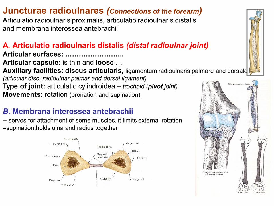
Juncturae radioulnares (Connections of the forearm) Articulatio radioulnaris proximalis, articulatio radioulnaris distalis
and membrana interossea antebrachii
A. Articulatio radioulnaris distalis (distal radioulnar joint) Articular surfaces: ……………………..
Articular capsule: is thin and loose …
Auxiliary facilities: discus articularis, ligamentum radioulnaris palmare and dorsale
(articular disc, radioulnar palmar and dorsal ligament)
Type of joint: articulatio cylindroidea – trochoid (pivot joint)
Movements: rotation (pronation and supination).
B. Membrana interossea antebrachii – serves for attachment of some muscles, it limits external rotation
=supination,holds ulna and radius together
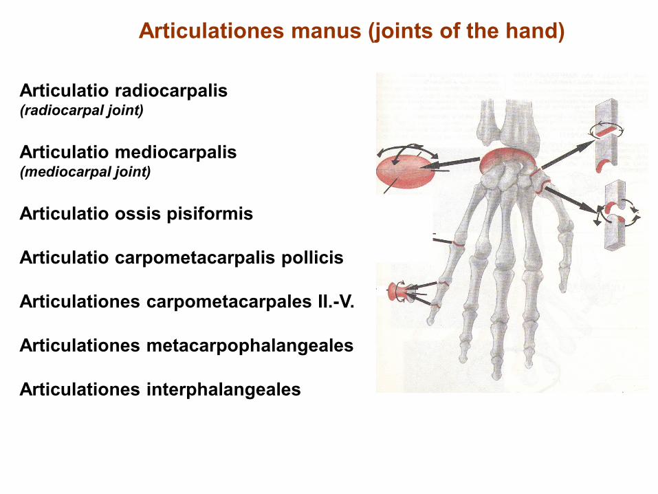
Articulationes manus (joints of the hand)
Articulatio radiocarpalis (radiocarpal joint)
Articulatio mediocarpalis (mediocarpal joint)
Articulatio ossis pisiformis
Articulatio carpometacarpalis pollicis
Articulationes carpometacarpales II.-V.
Articulationes metacarpophalangeales
Articulationes interphalangeales

4. Articulationes manus - Joints of the hand
A. Articulatio radiocarpalis (radiocarpal joint)
Articular surfaces: facies articularis carpea radii (carpal articular facet of radius) and
os scaphoideum (scaphoid bone), lunatum and triquetrum
Articular capsule: shares together with articulatio mediocarpalis (midcarpal joint).
Auxiliary facilities: ulna is separated from carpal bones by discus articularis
(articular disc). Ligaments shares with articulatio mediocarpalis (midcarpal joint).
Type of joint: articulatio ellipsoidea (ellipsoidal joint)
Movements: shares together with articulatio mediocarpalis (midcarpal joint)

B. Articulatio mediocarpalis - Midcarpal joint Articular surfaces: …………………….
Articular capsule: shares together with art. radiocarpalis (radiocarpal joint).
Auxiliary facilities: lig. radiocarpeum dorsale and palmare (dorsal and palmar radiocarpal
ligament), lig. ulnocarpeum palmare (palmar ulnocarpal ligament), lig. carpi radiatum (carpal
radial ligament), ligg. intercarpea dorsalia, palmaria and interossea (intercarpal dorsal,
palmar and interosseal ligaments).
Type of joint: ellipsoid, movements together with radiocarpal joint – palmar and dorsal
flexion, radial and ulnar duction and circumduction.

C. Articulatio ossis pisiformis (Articulation of pisiforme bone) Articular surfaces:
Articular capsule:
Auxiliary facilities: articular capsule is reinforced by lig. pisohamatum and lig.
pisometacarpeum.
Type of joint: amphiarthrosis
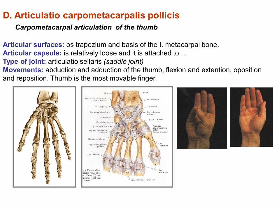
D. Articulatio carpometacarpalis pollicis
Carpometacarpal articulation of the thumb
Articular surfaces: os trapezium and basis of the I. metacarpal bone.
Articular capsule: is relatively loose and it is attached to …
Type of joint: articulatio sellaris (saddle joint)
Movements: abduction and adduction of the thumb, flexion and extention, oposition
and reposition. Thumb is the most movable finger.
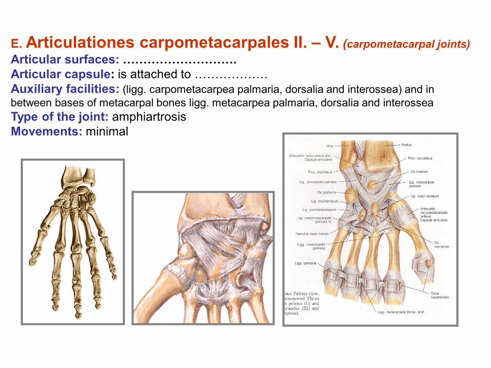
E. Articulationes carpometacarpales II. – V. (carpometacarpal joints)
Articular surfaces: ……………………….
Articular capsule: is attached to ………………
Auxiliary facilities: (ligg. carpometacarpea palmaria, dorsalia and interossea) and in
between bases of metacarpal bones ligg. metacarpea palmaria, dorsalia and interossea
Type of the joint: amphiartrosis
Movements: minimal
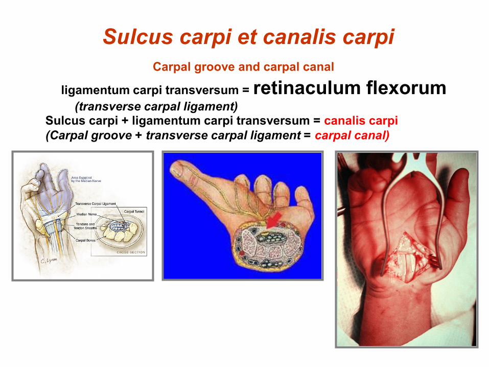
Sulcus carpi et canalis carpi
Carpal groove and carpal canal ligamentum carpi transversum = retinaculum flexorum (transverse carpal ligament)
Sulcus carpi + ligamentum carpi transversum = canalis carpi
(Carpal groove + transverse carpal ligament = carpal canal)

F. Articulationes metacarpophalangeae (metacarpophalangeal joints)
Articular surfaces:
Articular capsule:
Auxiliary facilities: laminae fibrocartilagineae palmares (palmar fibrocartilagineous
plates) and ligg.collateralia (collateral ligaments). Sesamoid bones (thumb).
II. to V. metacarpals are joined by ligamentum metacarpeum transversum profundum
(transverse metacarpal profound ligament).
Type of joint: articulatio ellipsoidea (ellipsoidal).
Movements: flexion, extension, abduction and adduction.
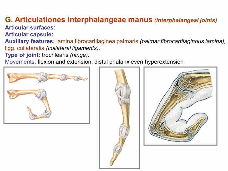
G. Articulationes interphalangeae manus (interphalangeal joints)
Articular surfaces:
Articular capsule:
Auxiliary features: lamina fibrocartilaginea palmaris (palmar fibrocartilaginous lamina),
ligg. collateralia (collateral ligaments).
Type of joint: trochlearis (hinge).
Movements: flexion and extension, distal phalanx even hyperextension.

Connection of the lower limb bones (juncturae ossium extremitatis inferioris)
includes connection of pelvic girdle and free part of lower limb
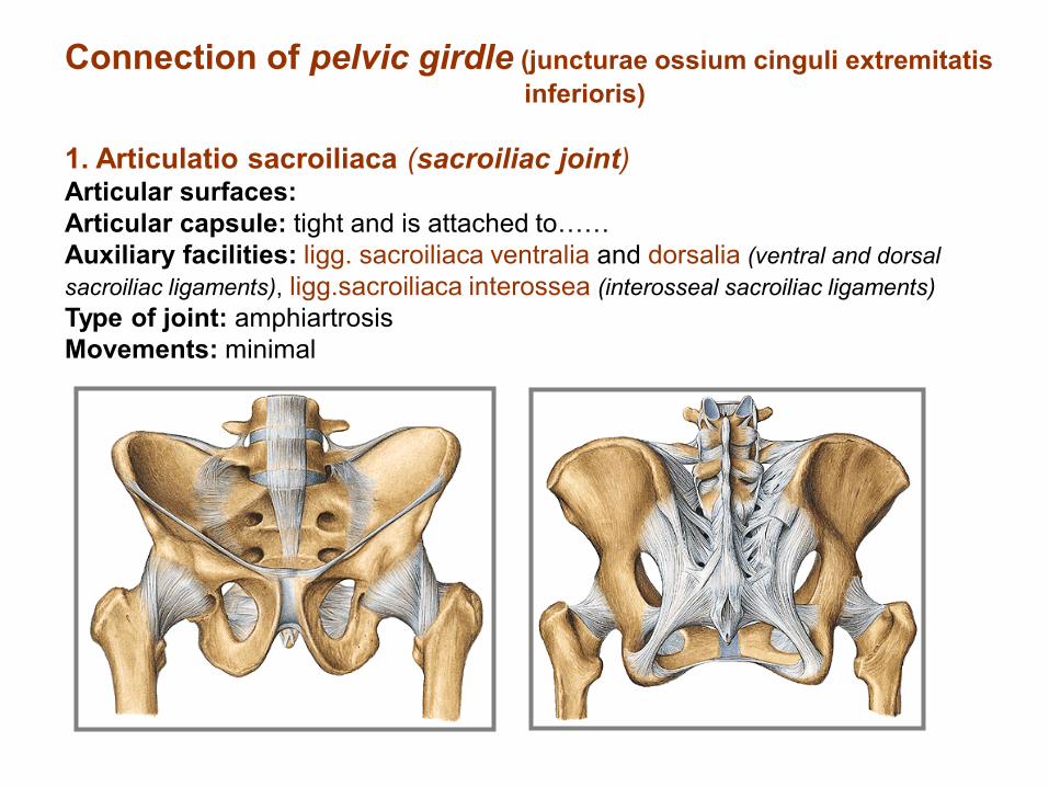
Connection of pelvic girdle (juncturae ossium cinguli extremitatis
inferioris)
1. Articulatio sacroiliaca (sacroiliac joint) Articular surfaces:
Articular capsule: tight and is attached to……
Auxiliary facilities: ligg. sacroiliaca ventralia and dorsalia (ventral and dorsal
sacroiliac ligaments), ligg.sacroiliaca interossea (interosseal sacroiliac ligaments)
Type of joint: amphiartrosis
Movements: minimal

2. Symphysis pubica cartilagenous discus interpubicus connects both pubic bones. Symphysis
pubica is 4,5 – 5 cm in height.
lig. pubicum superius and stronger lig. arcuatum pubis (arcuate pubic ligament)
3. Membrana obturatoria a stiff membrane which closes foramen obturatum (canalis obturatorius)
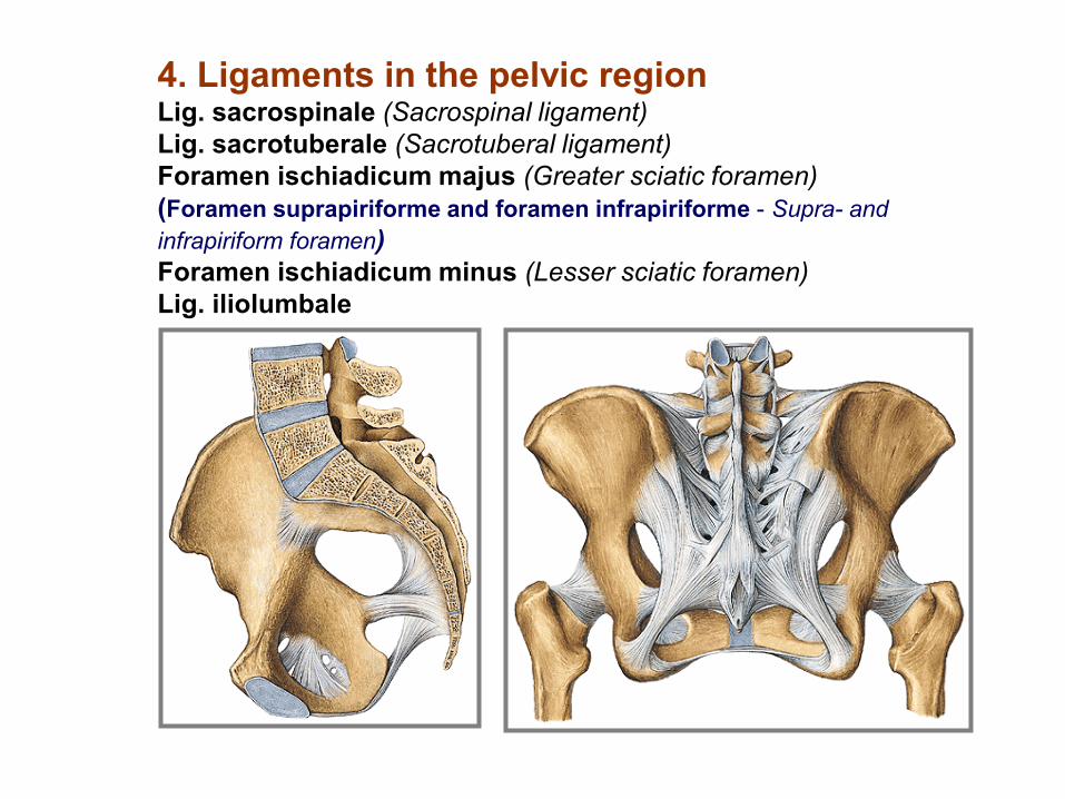
4. Ligaments in the pelvic region Lig. sacrospinale (Sacrospinal ligament)
Lig. sacrotuberale (Sacrotuberal ligament)
Foramen ischiadicum majus (Greater sciatic foramen)
(Foramen suprapiriforme and foramen infrapiriforme - Supra- and
infrapiriform foramen)
Foramen ischiadicum minus (Lesser sciatic foramen)
Lig. iliolumbale
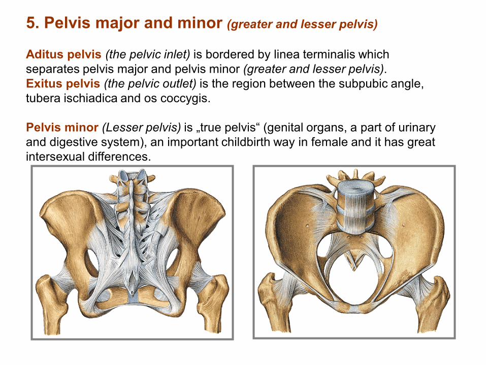
5. Pelvis major and minor (greater and lesser pelvis)
Aditus pelvis (the pelvic inlet) is bordered by linea terminalis which
separates pelvis major and pelvis minor (greater and lesser pelvis).
Exitus pelvis (the pelvic outlet) is the region between the subpubic angle,
tubera ischiadica and os coccygis.
Pelvis minor (Lesser pelvis) is „true pelvis“ (genital organs, a part of urinary
and digestive system), an important childbirth way in female and it has great
intersexual differences.

Inclinatio pelvis
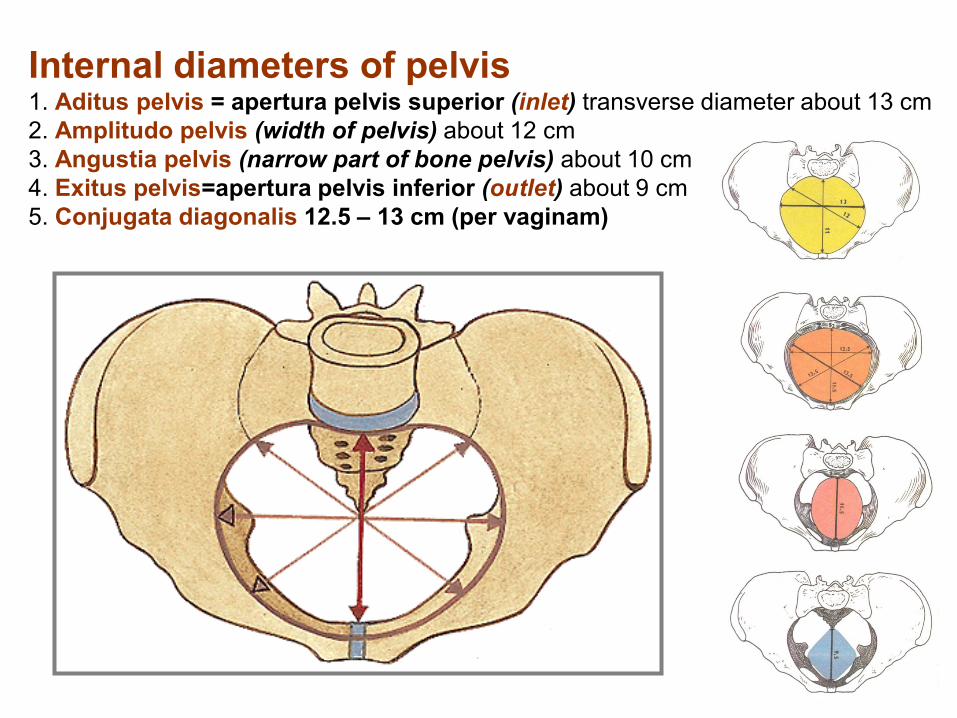
Internal diameters of pelvis 1. Aditus pelvis = apertura pelvis superior (inlet) transverse diameter about 13 cm
2. Amplitudo pelvis (width of pelvis) about 12 cm
3. Angustia pelvis (narrow part of bone pelvis) about 10 cm
4. Exitus pelvis=apertura pelvis inferior (outlet) about 9 cm
5. Conjugata diagonalis 12.5 – 13 cm (per vaginam)
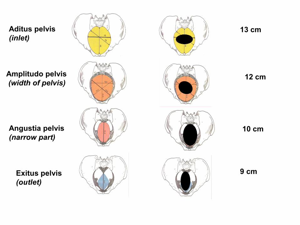
Exitus pelvis
(outlet)
Angustia pelvis
(narrow part)
Amplitudo pelvis
(width of pelvis)
Aditus pelvis
(inlet) 13 cm
12 cm
10 cm
9 cm
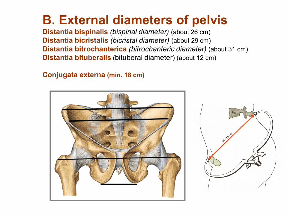
B. External diameters of pelvis Distantia bispinalis (bispinal diameter) (about 26 cm)
Distantia bicristalis (bicristal diameter) (about 29 cm)
Distantia bitrochanterica (bitrochanteric diameter) (about 31 cm)
Distantia bituberalis (bituberal diameter) (about 12 cm)
Conjugata externa (min. 18 cm)

Textbook of anatomy
Atlas of anatomy
Personal notes, drawings
10 minutes pause

Connection of free part of lower limb (juncturae ossium extremitatis liberae inferioris)

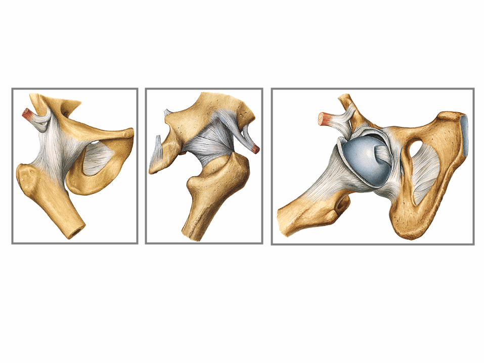
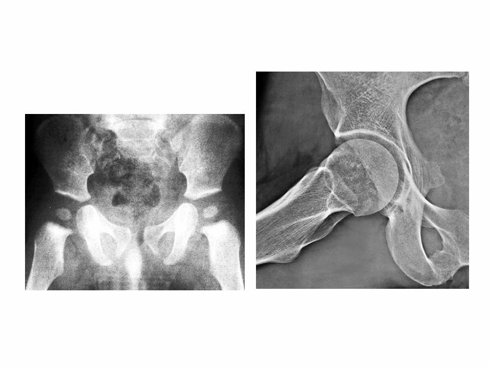
1. Articulatio coxae (hip joint)
Articular surfaces: facies lunata of the acetabulum only !!!!!..........
Articular capsule: is attached to the margins of acetabulum. It reaches ventrally
linea intertrochanterica of femur, dorsally is attached to the collum femoris (neck of
femur) medially away from fossa trochanterica.
Auxiliary facilities:
a) Labrum acetabulare formed by cartilage and fibrous tissue
b) Lig. transversum acetabuli runs through incisura acetabuli.
c) Lig. iliofemorale (the strongest lig.)
d) Lig. pubofemorale
e) Lig. ischiofemorale
f) Zona orbicularis
g) Lig. capitis femoris
Type of joint: typical spheroid joint (ball-and-socket) with restricted movements
(enarthrosis).
Movements: abduction, adduction, flexion, extention, pronation, supination
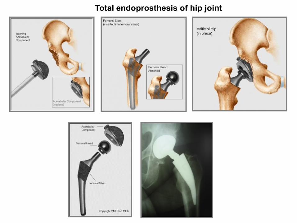
Total endoprosthesis of hip joint
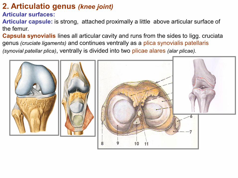
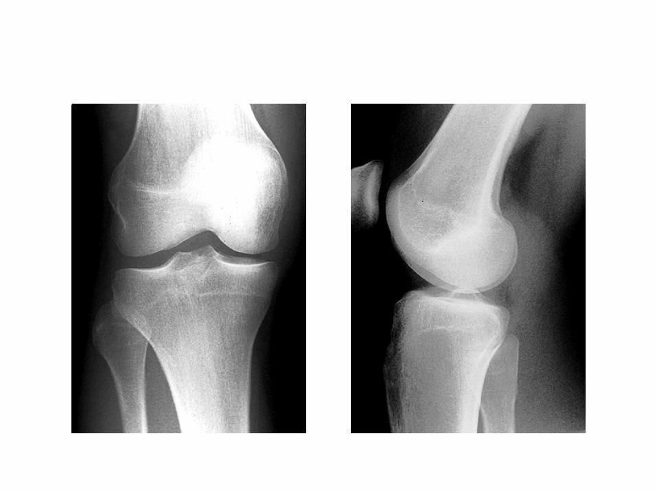
2. Articulatio genus (knee joint) Articular surfaces:
Articular capsule: is strong, attached proximally a little above articular surface of
the femur.
Capsula synovialis lines all articular cavity and runs from the sides to ligg. cruciata
genus (cruciate ligaments) and continues ventrally as a plica synovialis patellaris
(synovial patellar plica), ventrally is divided into two plicae alares (alar plicae).

I. Intraarticular auxiliary facilities of the articulatio genus (knee joint):
1. Meniscus – Medial and lateral
2. Ligamenta cruciata genus (cruciate ligaments of knee):
– anterius - limits extension and medial rotation
– posterius - limits extension and keeps stability of the joint
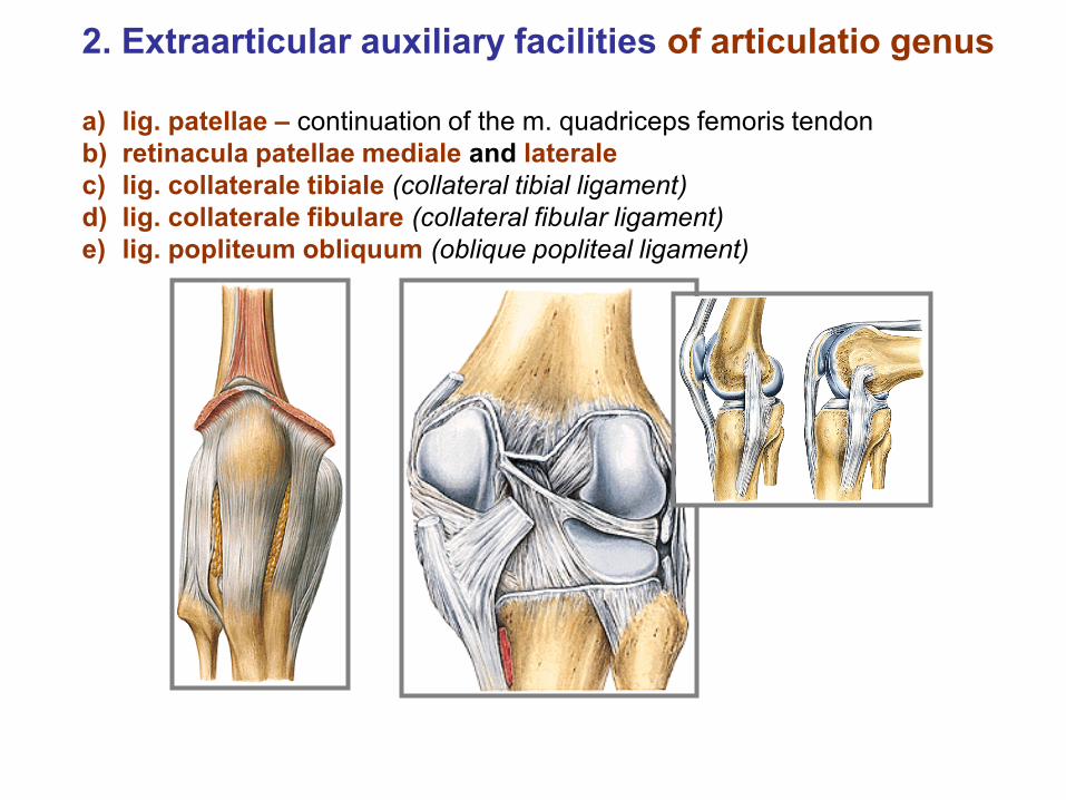
2. Extraarticular auxiliary facilities of articulatio genus
a) lig. patellae – continuation of the m. quadriceps femoris tendon
b) retinacula patellae mediale and laterale
c) lig. collaterale tibiale (collateral tibial ligament)
d) lig. collaterale fibulare (collateral fibular ligament)
e) lig. popliteum obliquum (oblique popliteal ligament)

Arthroscopy
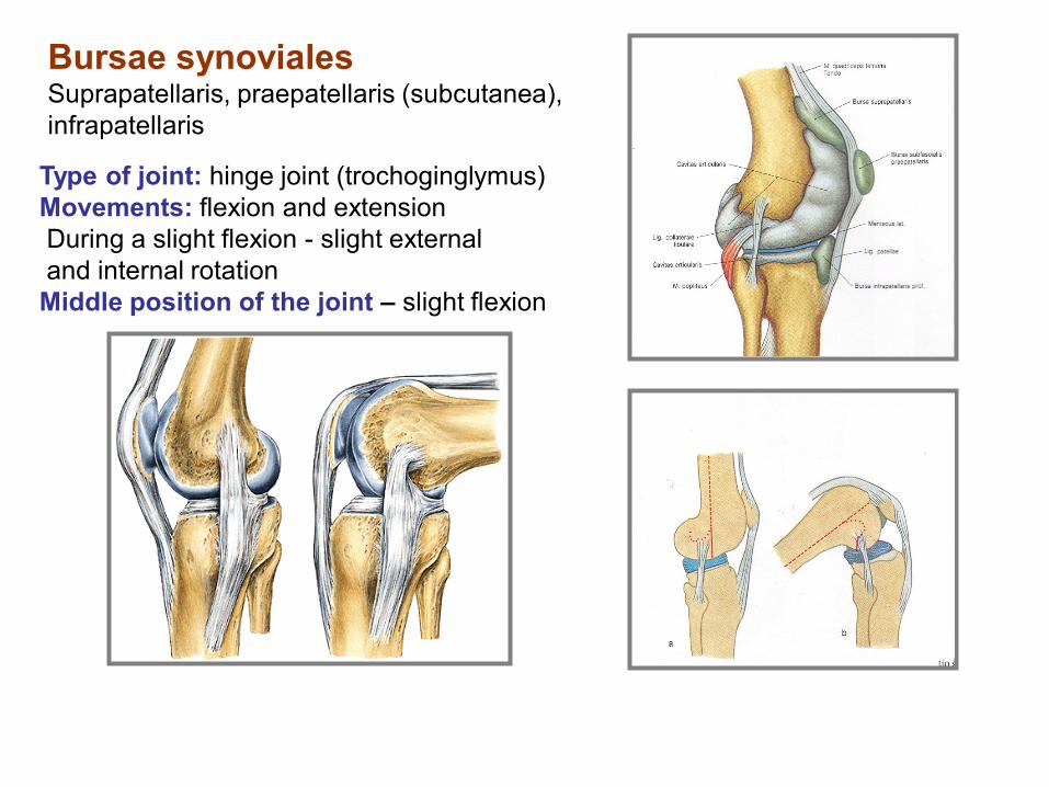
Type of joint: hinge joint (trochoginglymus)
Movements: flexion and extension
During a slight flexion - slight external
and internal rotation
Middle position of the joint – slight flexion
Bursae synoviales Suprapatellaris, praepatellaris (subcutanea),
infrapatellaris
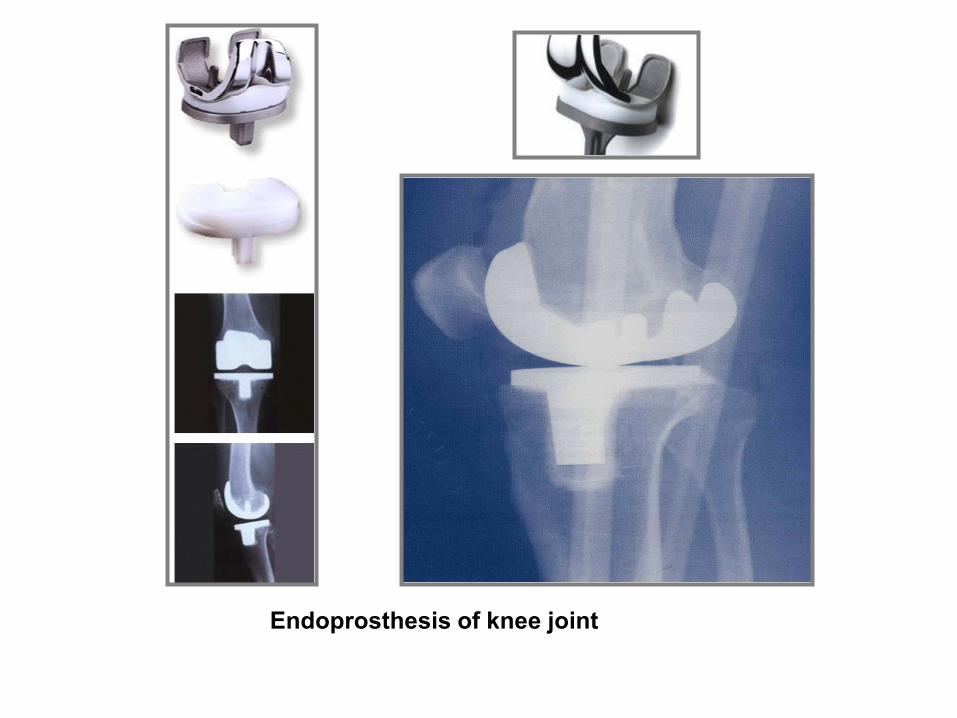
Endoprosthesis of knee joint

3. Juncturae tibiofibulares (tibiofibular connections) Articulatio tibiofibularis, membrana interossea cruris and syndesmosis
tibiofibularis.
A. Articulatio tibiofibularis (tibiofibular joint)
Articular surfaces:
Articular capsule: is short, stiff and attached to….
Auxiliary facilities: lig. capitis fibulae anterius and posterius
Type of joint kloubu: plane
Movements: slight movements ahead and back.
B. Membrana interossea cruris stiff membrane. Serves as a site for insertion of some muscles.
C. Syndesmosis tibiofibularis = fibrous joint between distal ends
of tibia and fibula
Syndesmosis is reinforced by lig. tibiofibulare anterius and lig. tibiofibulare
posterius (tibiofibular anterior and posterior ligaments).

Articulationes pedis (Joints of foot)
1. Articulatio talocruralis (Talocrural joint)
2. Articulationes intertarseae (Intertarsal joints)
Articulatio subtalaris (subtalar joint)
Art. talocalcaneonavicularis (talocalcanear joint)
Art. calcaneocuboidea (calcaneocuboid joint)
„Articulatio tarsi transversa“ (Chopart´s joint)
Articulatio cuneonavicularis (Cuneonavicular joint)
Articulatio cuneocuboidea (Cuneocuboid joint)
3. Articulationes tarsometatarseae (Tarsometatarsal
joints) – Lisfranck´s joint)
4. Articulationes metatarsophalangeae (Metatarsophalangeal joints)
5. Articulationes interphalangeae pedis (Interphalangeal joints)
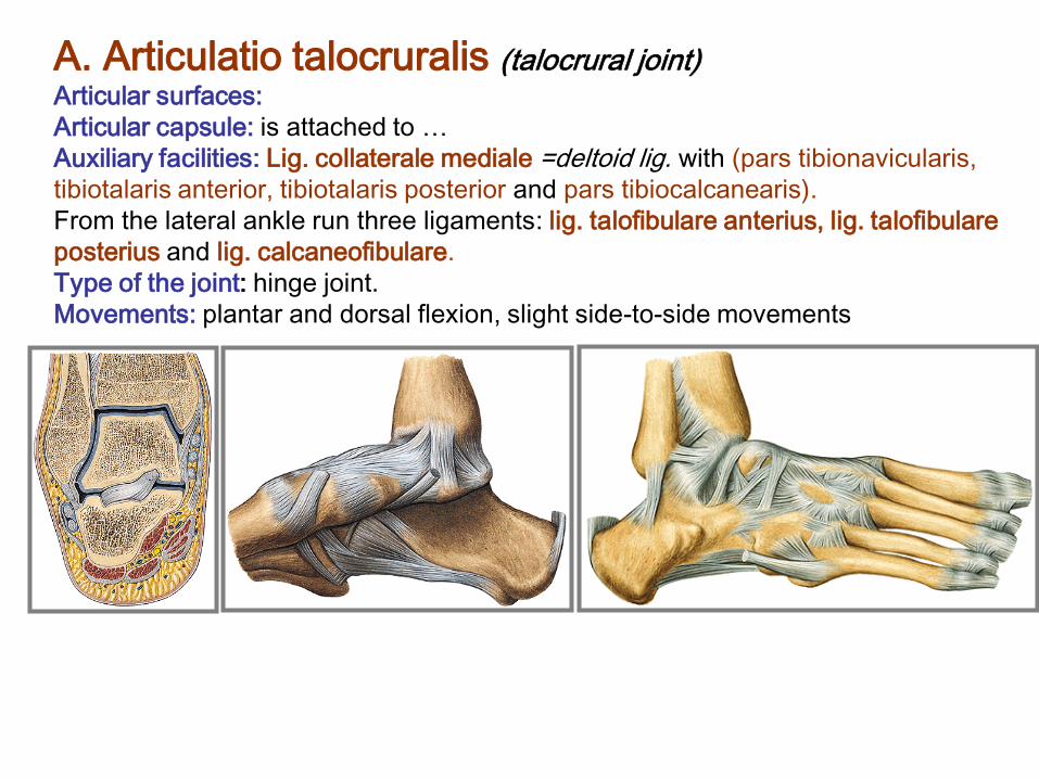
A. Articulatio talocruralis (talocrural joint) Articular surfaces:
Articular capsule: is attached to …
Auxiliary facilities: Lig. collaterale mediale =deltoid lig. with (pars tibionavicularis,
tibiotalaris anterior, tibiotalaris posterior and pars tibiocalcanearis).
From the lateral ankle run three ligaments: lig. talofibulare anterius, lig. talofibulare
posterius and lig. calcaneofibulare.
Type of the joint: hinge joint.
Movements: plantar and dorsal flexion, slight side-to-side movements.

d) Chopart´s joint – articulatio tarsi transversa – a fissure between talus
and os naviculare medially and in between calcaneus and os cuboideum laterally.
Opening of this joint - by cut of lig. bifurcatum (lig. calcaneonaviculare and lig.
calcaneocuboideum).
e) Articulationes cuneonavicularis, cuneocuboidea and
intercuneiformia are joints between adjacent tarsal bones;
dorsal, plantar and interosseous ligaments.
Springing movements

C. Articulationes tarsometatarseae (Lisfranc
s joint) Articular surfaces:
Articular capsule: is thin and is attached to …
Auxiliary facilities: lig. tarsometatarsea dorsalia, plantaria and interossea
Type of joint: amphiarthrosis
Slight movements, specially during loading of plantar arch.
Between bases of metatarsal bones - (articulationes intermetatarseae)
– lig. metatarsea dorsalia, plantaria and interossea.
In the fissure of Lisfranc´s joint could be exarticulated toes of the foot.
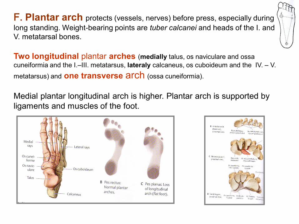
F. Plantar arch protects (vessels, nerves) before press, especially during
long standing. Weight-bearing points are tuber calcanei and heads of the I. and
V. metatarsal bones.
Two longitudinal plantar arches (medially talus, os naviculare and ossa
cuneiformia and the I.–III. metatarsus, lateraly calcaneus, os cuboideum and the IV. – V.
metatarsus) and one transverse arch (ossa cuneiformia).
Medial plantar longitudinal arch is higher. Plantar arch is supported by
ligaments and muscles of the foot.

Longitudinal plantar arch
Lig. plantare longum, m. tibialis posterior et anterior, flexors of toes, aponeurosis plantaris
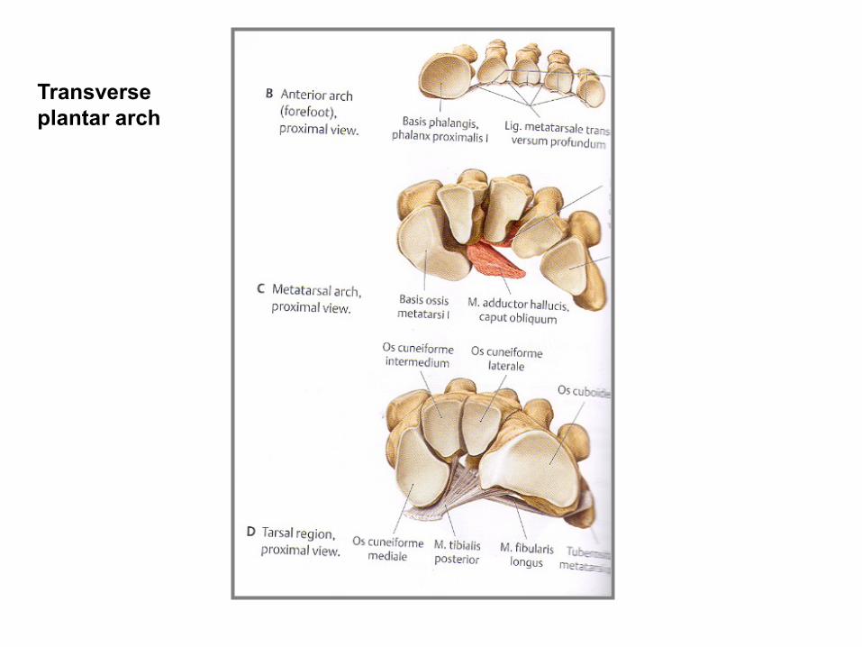
Transverse
plantar arch

Position of calcaneus – normal foot
and flatfoot („Flatfoot“ – pes planus – severe pain
in the foot and leg occurs, due to overstretching
of the long muscles and nerves and vessels of the sole.
Pes planus
Pes planovalgus Pes cavus
Used pictures come from:
Moore, K. L. (1992): Clinical oriented anatomy. Third edition.
Williams&Wilkins, A Waverly Company.
Gilroy, A. M. et all. (2009): Atlas of Anatomy. Thieme New York, Stuttgart.
Putz, R. (2008):
Atlas of Human Anatomy Sobotta. Elsevier Books.
Platzer, W., Kahle, W., Leonhardt H. (1992):
Locomotor system. Georg Thieme Verlag, Stuttgart,
New York, 4th edition.
Čihák, R. (1987): Anatomie 1. Avicenum, Zdravotnické nakladatelství.


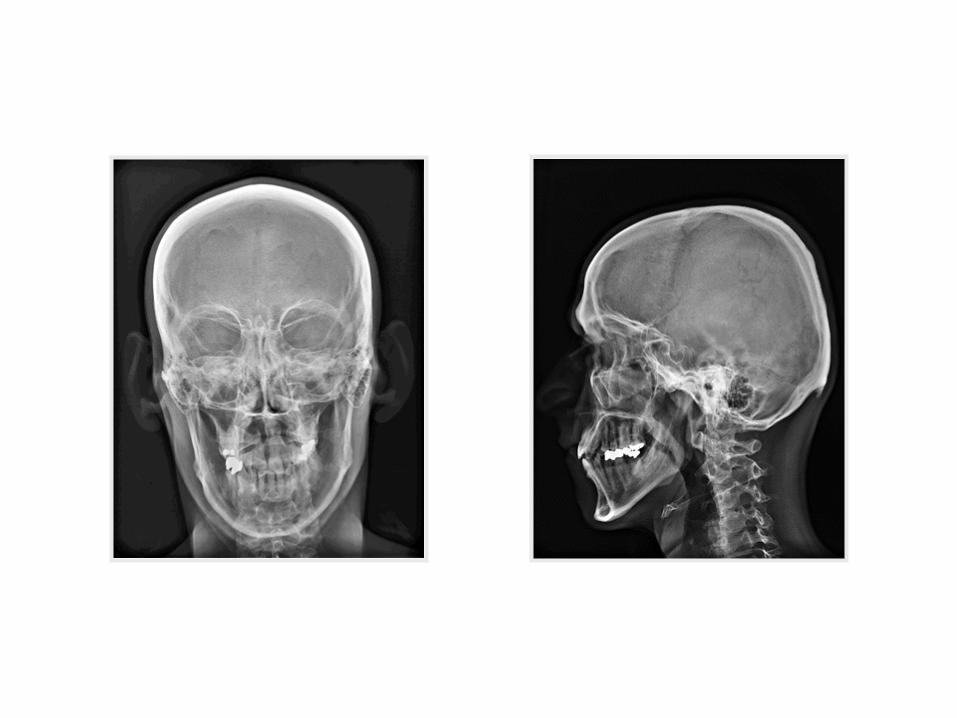
X-rays
Radiology and anatomy
1. Anatomy is essential for understanding radiology.
2. You will see anatomical structures this way much more frequently than during operation or autopsy.
3. Familiarity with normal radiographs allows you to recognize abnormalities (e.g. tumors, fractures).
4. You must be able to visualize the diseased organ and its associated structures.

X–ray (K. Roentgen 1895 – awarded by Nobel price in physics)
A highly penetrating beam of x-rays „transluminates“ the pacient,
showing tissues of differing densities on x-ray film.
A tissue or organ that is relatively dense absorbs (stops) more x-rays
than a less dense tissue. Relatively fewer x-rays reach the silver
emulsion in the film therefore only fewer grains of silver are developed
at this area when the film is processed – „white area of bones“.

1. Simple X – ray
2. X– ray with contrast materials
a) positive (iodide preparations, barium meal)
b) negative (air, gases)
4. Projection according to the course of x-ray (anteroposterior, lateral)
5. New methods (sonografy, CT (computerized tomography – using CT
scanners, shows sections of the body – a small beam of x-rays is passed
through a plane of the body while the x-ray tube moves in an arc or a circle
around the body), MRI

X-ray of the thorax
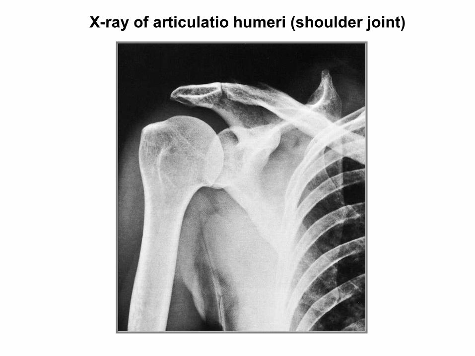
X-ray of articulatio humeri (shoulder joint)
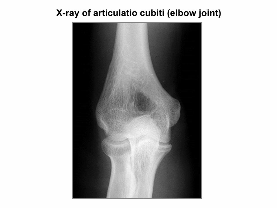
X-ray of articulatio cubiti (elbow joint)
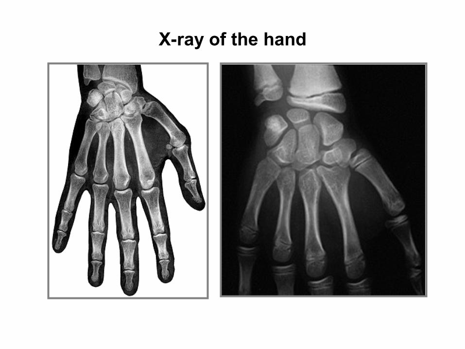
X-ray of the hand

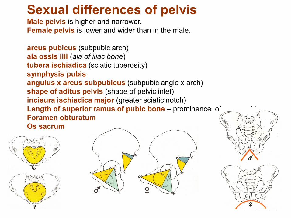
Sexual differences of pelvis Male pelvis is higher and narrower.
Female pelvis is lower and wider than in the male.
arcus pubicus (subpubic arch)
ala ossis ilii (ala of iliac bone)
tubera ischiadica (sciatic tuberosity)
symphysis pubis
angulus x arcus subpubicus (subpubic angle x arch)
shape of aditus pelvis (shape of pelvic inlet)
incisura ischiadica major (greater sciatic notch)
Length of superior ramus of pubic bone – prominence of mons pubis
Foramen obturatum
Os sacrum