Congenital Midline Nasal Masses: Diagnosis and Management
12
Congenital Midline Nasal Masses: Diagnosis and Management CHARLES D. KENNARD, M.D. JAMES E. RASMUSSEN, M.D. I Abstract. Congenital midline nasal masses (CMNMs) are rare lesions most commonly caused by dermoids, heman- giomas, nasal gliomas or encephaloceles. We report a case of nasal glioma and discuss the embryologic development, evaluation, and management of such CMNMs. J Dermatol Surg Oncol 1990; 16:1025-1036. HONORABLE MENTION of such lesions are critical to their safe and effective management. CASE REPORT Congenital midline nasal masses (CMNMs) occur in only 1 of every 20,000 to 40,000 live births.' They are usually obvious at birth, but some CMNMs may not be recognized until well into adulthood.2 The most common lesions producing CMNMs are, in order of decreasing frequency: dermoids, hemangioma, nasal glioma, and encephalocele.3 Nasal gliomas are rare, benign tumors of heterotopic brain tissue thought to arise from the abnormal extracranial se- questration of neural elements during embryologic development. Since nasal gliomas and other CMNMs may have intracranial extensions, familiarity with the differential diagnosis and appropriate evaluation Charles D. Kennard, M.D., Fellow, Dermatologic Surgery and Cutaneous Oncology, is from the Department of Derma- tology, University of Iowa Hospitals and Clinics, Iowa City, Iowa. James E. Rasmussen, M.D.,is from the Department of Derma- tology, University of Michigan, Ann Arbor, Michigan. Reprints are not available. 0 1990 by Elsevier Science Publishing Co., Inc. 0148-0812/90/$3.50 LULJ A 1-week-old male infant was referred to the Depart- ment of Dermatology for evaluation of a congenital mass at the root of the nose that had not changed since birth. There was no history of nasal blockage, discharge, bleeding, seizures, or meningitis. The pa- tient's mother believed that the lesion transiently in- creased in size when the child was crying. He was a full-term child, the product of a normal labor and delivery and no other abnormalities were identified. There was no history of congenital defects in three older siblings and the family history was unremark- able. Physical examination revealed a 1-cm, firm, mo- veable, nonpulsatile mass located at the root of the nose just slightly left of midline (Fig. 1). There was a slight prominence to the vascularity of the overlying skin, but no other changes in the skin surface were seen. There were no intranasal masses and hyperte- lorism was not present. The remainder of the physi- cal examination was unremarkable. The clinical differential diagnosis was nasal glioma, encephalocele, and cavernous hemangioma. The patient was referred to the Pediatric Neurosur- gery service, which favored the diagnosis of ence- phalocele. An MRI scan (Figs. 2 and 3) showed a soft tissue mass in the left nasal region. There was no evi- dence of fluid or blood in the mass. There was no alteration of the parenchyma of the brain and no transcranial extension of the mass (Fig. 4). The patient was referred to the Plastic Surgery ser- vice, where the mass was excised under general anes- 1 nqE
Transcript of Congenital Midline Nasal Masses: Diagnosis and Management

Congenital Midline Nasal Masses: Diagnosis and Management CHARLES D. KENNARD, M.D. JAMES E. RASMUSSEN, M.D.
I
Abstract. Congenital midline nasal masses (CMNMs) are rare lesions most commonly caused by dermoids, heman- giomas, nasal gliomas or encephaloceles. We report a case of nasal glioma and discuss the embryologic development, evaluation, and management of such CMNMs. J Dermatol Surg Oncol 1990; 16:1025-1036.
HONORABLE MENTION
of such lesions are critical to their safe and effective management.
CASE REPORT
Congenital midline nasal masses (CMNMs) occur in only 1 of every 20,000 to 40,000 live births.' They are usually obvious at birth, but some CMNMs may not be recognized until well into adulthood.2 The most common lesions producing CMNMs are, in order of decreasing frequency: dermoids, hemangioma, nasal glioma, and encephalocele.3 Nasal gliomas are rare, benign tumors of heterotopic brain tissue thought to arise from the abnormal extracranial se- questration of neural elements during embryologic development. Since nasal gliomas and other CMNMs may have intracranial extensions, familiarity with the differential diagnosis and appropriate evaluation
Charles D. Kennard, M.D., Fellow, Dermatologic Surgery and Cutaneous Oncology, is from the Department of Derma- tology, University of Iowa Hospitals and Clinics, Iowa City, Iowa. James E. Rasmussen, M.D., is from the Department of Derma- tology, University of Michigan, Ann Arbor, Michigan. Reprints are not available.
0 1990 by Elsevier Science Publishing Co., Inc. 0148-0812/90/$3.50 LULJ
A 1-week-old male infant was referred to the Depart- ment of Dermatology for evaluation of a congenital mass at the root of the nose that had not changed since birth. There was no history of nasal blockage, discharge, bleeding, seizures, or meningitis. The pa- tient's mother believed that the lesion transiently in- creased in size when the child was crying. He was a full-term child, the product of a normal labor and delivery and no other abnormalities were identified. There was no history of congenital defects in three older siblings and the family history was unremark- able.
Physical examination revealed a 1-cm, firm, mo- veable, nonpulsatile mass located at the root of the nose just slightly left of midline (Fig. 1). There was a slight prominence to the vascularity of the overlying skin, but no other changes in the skin surface were seen. There were no intranasal masses and hyperte- lorism was not present. The remainder of the physi- cal examination was unremarkable.
The clinical differential diagnosis was nasal glioma, encephalocele, and cavernous hemangioma. The patient was referred to the Pediatric Neurosur- gery service, which favored the diagnosis of ence- phalocele. An MRI scan (Figs. 2 and 3) showed a soft tissue mass in the left nasal region. There was no evi- dence of fluid or blood in the mass. There was no alteration of the parenchyma of the brain and no transcranial extension of the mass (Fig. 4).
The patient was referred to the Plastic Surgery ser- vice, where the mass was excised under general anes-
1 nqE

CONGENITAL MIDLINE NASAL MASSES
Y
FIGURE 1. Nasal glioma.
thesia. An eliptical excision exposed a cystic lesion within the dermis that, upon dissection, was found to have eroded the distal nasal bone on the left and to have partially eroded the nasal septum and nasal cartilage with involvement of the nasal mucosa. The lesion was removed intact and no extensions were
FIGUi extra] nasal tracra
RE 2. MRI sagittal scan demonstrz iasal mass with involvement bones. No communication with
lnial contents is seen.
detected. Postoperative course was unremarkable, and the patient was well at 1 month postoperatively.
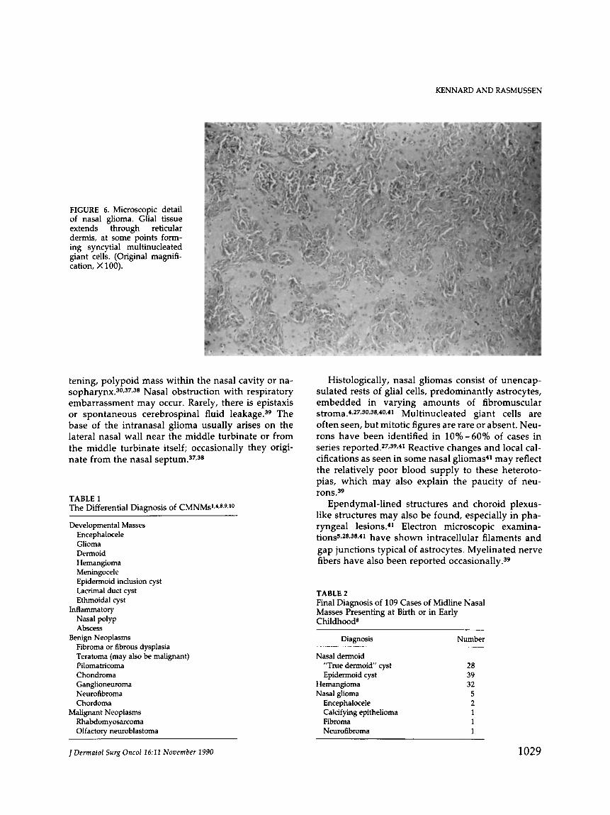
Histologic exam showed that the epidermis was near normal for the area. The density of adnexal structures was decreased and the dermis extensively altered by the inclusion of glial tissue arranged in ir- regular and somewhat angular islands (Fig. 5). The combination of glial tissue and collagen extended to the deep fascia. Large, syncytial, multinucleated giant cells were formed by syncytial aggregates of glial cells (Fig. 6 ) . These changes were interpreted as diagnostic of nasal glioma. Ganglion cells were not identified.
DISCUSSION
Up to 50% of congenital malformations involve the cerebrospinal axis.3 The majority of these are found caudally, but the cranium may also be affected; CMNMs are rarer still.
Patients with CMNMs are usually managed by neurosurgeons, otorhinolaryngologists, and plastic surgeons. However, dermatologists may be consulted for the evaluation of unusual subcutaneous masses
ites of
in-
1026 1 Dermatol Surg Oncol 16:11 November 1990
KENNARD AND RASMUSSEN
FIGURE 3. MRI coronal scan shows soft tissue mass distorting nasal cartilage and expanding soft tissue.
of the nose in an infant or young child.‘-’ The differ- ential diagnosis of such lesions has been assembled by several authors8-10 and may include developmen- tal anomalies, inflammatory lesions, and benign or malignant neoplasms (Table 1). In a series of 109 con- secutive admissions to two children’s hospitals over a 20-year period for evaluation of nasal masses present from birth or with onset in early childhood, Bradley and Singhe found 67 nasal dermoids, 32 hemangio- mas, 5 nasal gliomas, 2 encephaloceles and 3 other entities (Table 2). During the same time period, a total of 304,000 admissions had been made to the same hospitals, demonstrating the rarity of CMNMs.
Embryologic Development Since the majority of CMNMs result from an alter- ation of the embryologic events that produce the nose, frontal-basal skull, and intracranial con- tents,lJ1J2 a review of the normal sequence will be useful in understanding the results of aberrant devel- opmental pathways. The neuroectoderm first ap- pears in the human embryo at about 18 days after conception; folding of this neural plate and fusion to produce the neural tube follows. The anterior portion of this neural tube, the anterior neuropore, fuses at about 24 days of gestation. At the same time, meso-
Dermatol Surg Oncol 16:11 November 1990
FIGURE 4. MRI coronal scan demonstrating normal cribi- form plate and crista galli.
dermal tissues begin to form the bone, cartilage, ves- sels, and meninges of the skull. A space called the fonticulus frontalis, or nasofrontal fontanel, lies be- tween the frontal and nasal bones. A cartilagenous capsule, which ultimately will form the upper lateral nasal cartilage, is formed in the anterior wall of the external nose. The prenasal space extends from the brain to the nasal tip lying deep to the nasal bone and skin, and superficial to the nasal cartilage capsule. These relationships are shown in Figure 7A.
As development proceeds, the nasofrontal fon- tanel is altered by extensions of the frontal bones to form a canal (foramen caecum) through which a pro- jection of dura-mater extends to traverse the prenasal space and contact the nasal skin (Fig. 7B). Progres- sively, the frontal, nasal, sphenoid, and ethmoid bones fuse to form the base of the skull and cribiform plate. The dural tract in the prenasal space separates from the nasal skin and is obliterated (Fig. 7C).
Alterations in this cycle can include faulty closure of the anterior neuropore with defects in the fonticu- lus frontalis, foramen caecum, cribiform plate, or
1027
CONGENITAL MIDLINE NASAL MASSES
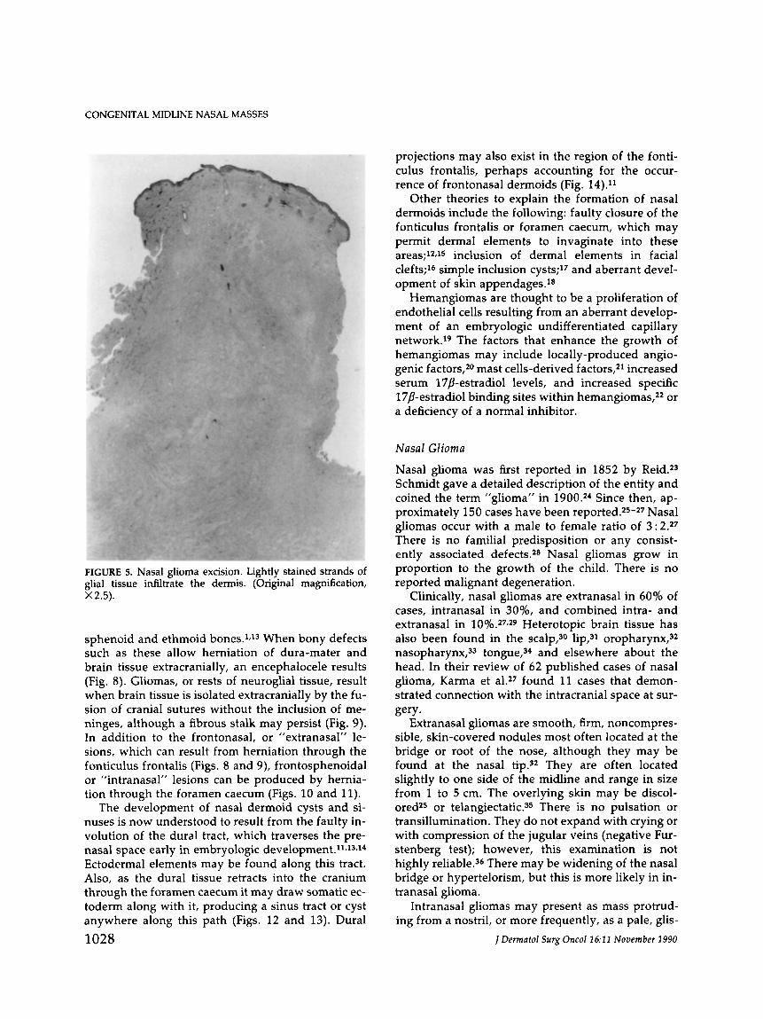
FIGURE 5. Nasal glioma excision. Lightly stained strands of glial tissue infiltrate the dermis. (Original magnification, X 2.5).
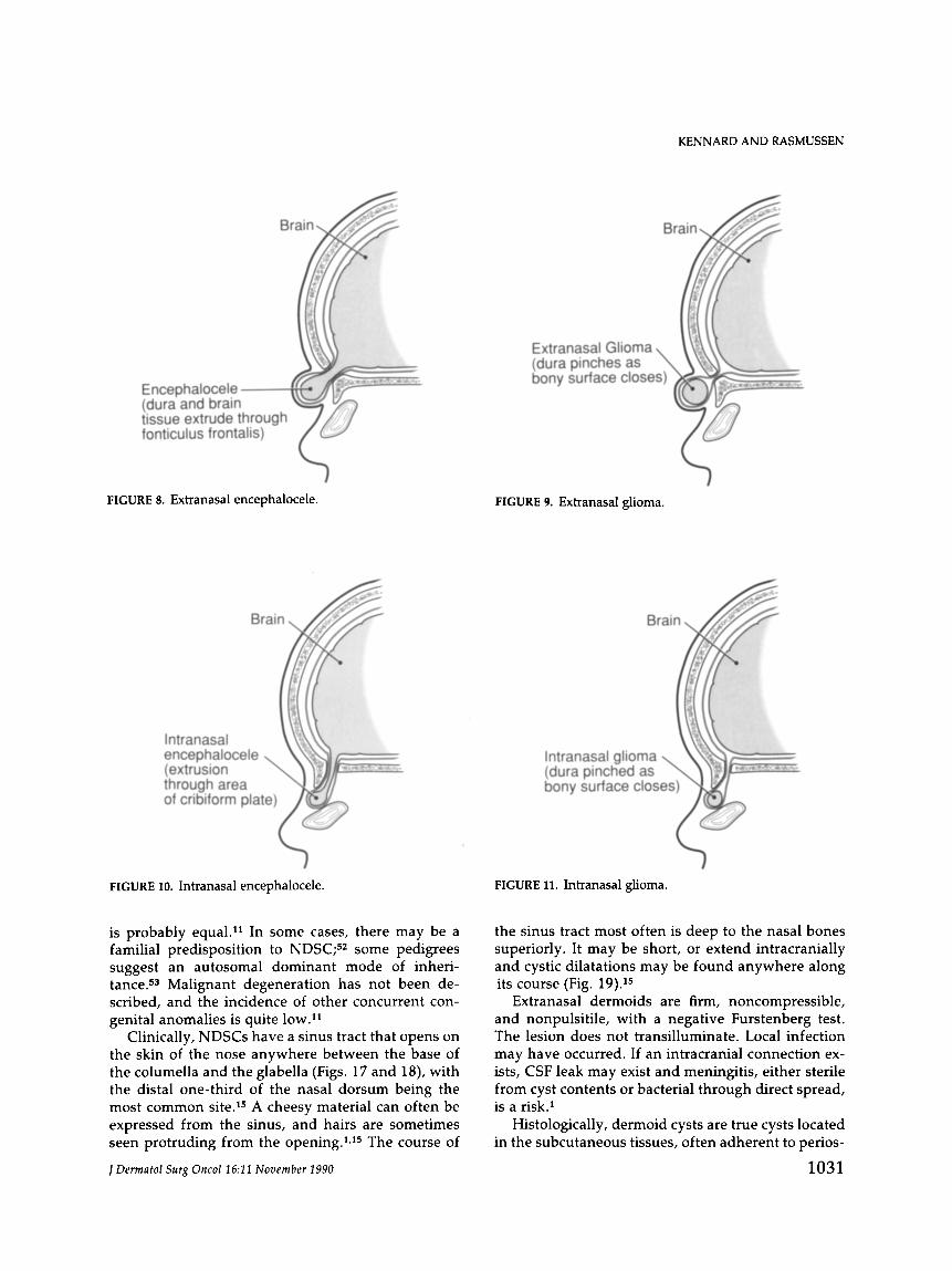
sphenoid and ethmoid bones.',l3 When bony defects such as these allow herniation of dura-mater and brain tissue extracranially, an encephalocele results (Fig. 8). Gliomas, or rests of neuroglial tissue, result when brain tissue is isolated extracranially by the fu- sion of cranial sutures without the inclusion of me- ninges, although a fibrous stalk may persist (Fig. 9). In addition to the frontonasal, or "extranasal" le- sions, which can result from herniation through the fonticulus frontalis (Figs. 8 and 9), frontosphenoidal or "intranasal" lesions can be produced by hernia- tion through the foramen caecum (Figs. 10 and l l).
The development of nasal dermoid cysts and si- nuses is now understood to result from the faulty in- volution of the dural tract, which traverses the pre- nasal space early in embryologic de~elopment .~~J3J~ Ectodermal elements may be found along this tract. Also, as the dural tissue retracts into the cranium through the foramen caecum it may draw somatic ec- toderm along with it, producing a sinus tract or cyst anywhere along this path (Figs. 12 and 13). Dural 1028
projections may also exist in the region of the fonti- culus frontalis, perhaps accounting for the occur- rence of frontonasal dermoids (Fig. 14).11
Other theories to explain the formation of nasal dermoids include the following: faulty closure of the fonticulus frontalis or foramen caecum, which may permit dermal elements to invaginate into these areas;12J5 inclusion of dermal elements in facial clefts;16 simple inclusion cysts;17 and aberrant devel- opment of skin appendages.18
Hemangiomas are thought to be a proliferation of endothelial cells resulting from an aberrant develop- ment of an embryologic undifferentiated capillary netw0rk.1~ The factors that enhance the growth of hemangiomas may include locally-produced angio- genic factors,20 mast cells-derived factors,21 increased serum 17p-estradiol levels, and increased specific 17p-estradiol binding sites within hemangiomas,22 or a deficiency of a normal inhibitor.
Nasal Glioma
Nasal glioma was first reported in 1852 by Reid.23 Schmidt gave a detailed description of the entity and coined the term "glioma" in 1900.24 Since then, ap- proximately 150 cases have been reported.*5-27 Nasal gliomas occur with a male to female ratio of 3 : ZZ7 There is no familial predisposition or any consist- ently associated defects.28 Nasal gliomas grow in proportion to the growth of the child. There is no reported malignant degeneration.
Clinically, nasal gliomas are extranasal in 60% of cases, intranasal in 30%, and combined intra- and extranasal in 10%.27,29 Heterotopic brain tissue has also been found in the scalp,30 lip,31 o r o p h a r y n ~ , ~ ~ nasopharynx,33 tongue,34 and elsewhere about the head. In their review of 62 published cases of nasal glioma, Karma et al.27 found 11 cases that demon- strated connection with the intracranial space at sur- gery *
Extranasal gliomas are smooth, firm, noncompres- sible, skin-covered nodules most often located at the bridge or root of the nose, although they may be found at the nasal t i ~ . 3 ~ They are often located slightly to one side of the midline and range in size from 1 to 5 cm. The overlying skin may be discol- ored25 or telangiectatic.35 There is no pulsation or transillumination. They do not expand with crying or with compression of the jugular veins (negative Fur- stenberg test); however, this examination is not highly reliable.36 There may be widening of the nasal bridge or hypertelorism, but this is more likely in in- tranasal glioma.
Intranasal gliomas may present as mass protrud- ing from a nostril, or more frequently, as a pale, glis-
1 Dermatol Surg Oncol 16:11 November 1990
KENNARD AND RASMUSSEN
FIGURE 6. Microscopic detail of nasal glioma. Glial tissue extends through reticular dermis, at some points form- ing syncytial multinucleated giant cells. (Original magnifi- cation, X 100).
tening, polypoid mass within the nasal cavity or na- sopharyn~.30,3~,38 Nasal obstruction with respiratory embarrassment may occur. Rarely, there is epistaxis or spontaneous cerebrospinal fluid leakage.39 The base of the intranasal glioma usually arises on the lateral nasal wall near the middle turbinate or from the middle turbinate itself; occasionally they origi- nate from the nasal ~eptum.3~~38
TABLE 1 The Differential Diagnosis of CMNMs1,4,8,9,'0
Developmental Masses Encephalocele Glioma Dermoid Hemangioma Meningocele Epidermoid inclusion cyst Lacrimal duct cyst Ethmoidal cyst
Inflammatory Nasal polyp Abscess
Fibroma or fibrous dysplasia Teratoma (may also be malignant) Pilomatricoma C hondroma Ganglioneuroma Neurofibroma Chordoma
Malignant Neoplasms Rhabdomyosarcoma Olfactory neuroblastoma
Benign Neoplasms
Histologically, nasal gliomas consist of unencap- sulated rests of glial cells, predominantly astrocytes, embedded in varying amounts of fibromuscular stroma.4,27,30,38,40,41 Multinucleated giant cells are often seen, but mitotic figures are rare or absent. Neu- rons have been identified in 10%-60% of cases in series reported.27.39.41 Reactive changes and local cal- cifications as seen in some nasal gliomas41 may reflect the relatively poor blood supply to these heteroto- pias, which may also explain the paucity of neu- rons.39
Ependymal-lined structures and choroid plexus- like structures may also be found, especially in pha- ryngeal lesions." Electron microscopic examina- tions5,28,3*,41 have shown intracellular filaments and gap junctions typical of astrocytes. Myelinated nerve fibers have also been reported occasionally.39
TABLE 2 Final Diagnosis of 109 Cases of Midline Nasal Masses Presenting at Birth or in Early Childhoods
Diagnosis Number
Nasal dermoid "True dermoid' cyst Epidermoid cyst
Hemangioma Nasal ghoma
Encephalocele Calcifying epithelioma Fibroma Neurofibroma
28 39 32
5 2 1 1 1
1 Dermatol Surg Oncol 16:11 November 1990 1029

CONGENITAL MIDLINE NASAL MASSES
FIGURE 7. Sequence of normal em- bryologic development of the fron- tobasal skull and nose. (A) Early de- velopmental stage illustrating the relationships between external nasal cartilage, nasal bone and frontal bone, and the two potential spaces of the fonticulus frontalis and the prenasal space. (8) Formation of for- amen caecum by partial fusion of nasal bone and frontal and basal skull, demonstrating the dural pro- cess extending to nasal skin. (C) Complete closure of foramen cae- cum and obliteration of nasal neuro- ectodermal elements. (After Ses- sions.")
Encephalocele
An encephalocele is a herniation of cranial contents through a defect in the skull. The first medical report of encephalocele may have appeared in the 16th cen- tury.42 Encephaloceles occur in about 1-3 out of 10,000 live births,43,44 and affect the sexes equally. Of these, 75% are located at the occiput; 15% are sinci- pital, or extranasally situated about the dorsum of the nose, orbits, and forehead; and 10% are basal, or found in the intranasal, sphenomaxillary, or pharyn- geal regions. There is a 6% risk of CNS abnormality in subsequent siblings.44 No malignant degeneration is found.
The classification of encephaloceles may also be made on the contents of the extracranial herniation. Meningoceles contain only meninges; meningoence- phaloceles, or simply encephaloceles, contain neural tissue as well as meninges; and meningoencephalo- cystoceles contain meninges and neural tissue as well as a portion of the ventricular system.'s45 The follow- ing discussion will utilize the term encephalocele to encompass these entities as they relate to CMNMs.
The etiology of encephaloceles is uncertain. Appar- ently, most lesions are sporadic developmental de- fects. However, the occurrence of encephaloceles in families with other CNS malformations, in associa- tion with known genetic syndromes such as fronto- nasal dysplasia, and in the HARD + E syndrome (hydrocephalus, agyria, retinal dysplasia, and en- cephalocele) suggests that genetic influences may occur in some cases.46.47 Additionally, teratogens such as trypan blue and excess doses of vitamin A can cause encephaloceles in experimental animals.48
Clinically, both basal and sincipital encepahlo- celes are softer or more compressible than nasal glio- mas, and they often have a bluish tint.' They may
1030
at
ura in contact
range in size from less than 1 cm in diameter to a large mass containing nearly half the brain.46 They may spontaneously leak CSF, and a history of menin- gitis may be obtainable. The Furstenberg test is usu- ally positive and spontaneous pulsation may be seen.' Transillumination may be decreased if neural elements are present in the herniated sac.
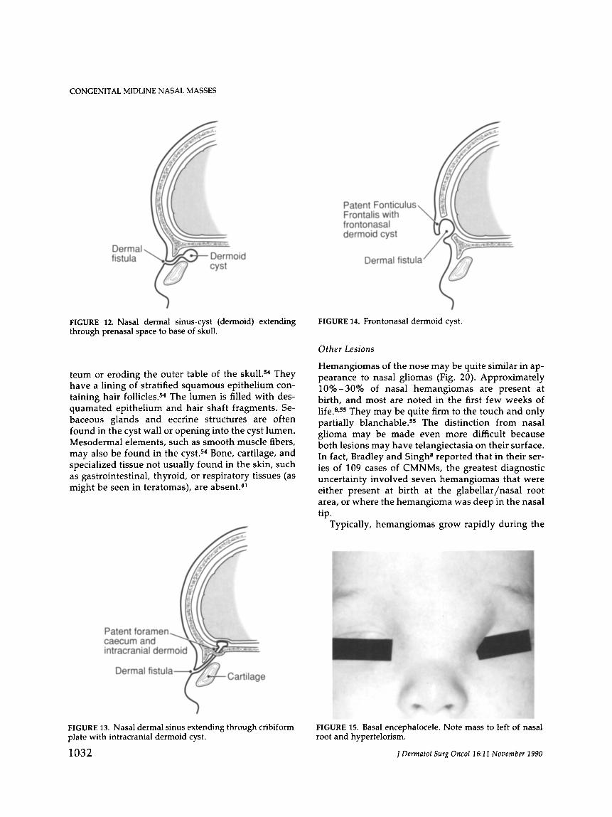
Hypertelorism is frequent (Fig. 15) and associated facial and cerebral malformations such as facial cleft- ing (Fig. 16), depressed nasal tip, and agenesis of the corpus callosum may be n ~ t e d . ~ ~ , * ~ There is always an underlying defect of the cranium.*,46 Intranasal ence- phaloceles may produce obstruction, and such le- sions may be confused with a nasal polyp-a lesion which is usually not found in infants or young chil- dren.8
Histologically, the differentiating feature between an encephalocele and a nasal glioma is the presence of dura and lep t~meninges .~~ Mature neural tissue with varying degrees of organization may be found in both entities, and unfortunately, leptomeninges are not always recognizable. In such cases, the dis- tinction between nasal glioma and encephalocele can be made only with clinicopathologic correlation.40
Nasal Dermoid
In 1982, Sessions'' proposed the term "nasal dermal sinus-cyst" (NDSC) for a group of rare lesions of so- matic ectodermal or mixed somatic and neuroectoder- ma1 derivation arising on the nose with occasional growth to and rarely through the cranium. Such NDSCs, or nasal dermoids as they are more fre- quently called, account for approximately 10% of all dermal sinuses of the head and neck, and for 1% of all such cysts throughout the b ~ d y . ~ ~ , ~ ' The sex ratio
1 Dermntol Surg Oncol 16:11 November 1990
KENNARD AND RASMUSSEN
Brain,/
Encephalocele /&+ tissue (dura and extrude brain-y! through
fonticulus frontalis)
FIGURE 8. Extranasal encephalocele.
FIGURE 10. Intranasal encephalocele.
is probably equal." In some cases, there may be a familial predisposition to NDSC;52 some pedigrees suggest an autosomal dominant mode of inheri- tance.53 Malignant degeneration has not been de- scribed, and the incidence of other concurrent con- genital anomalies is quite low."
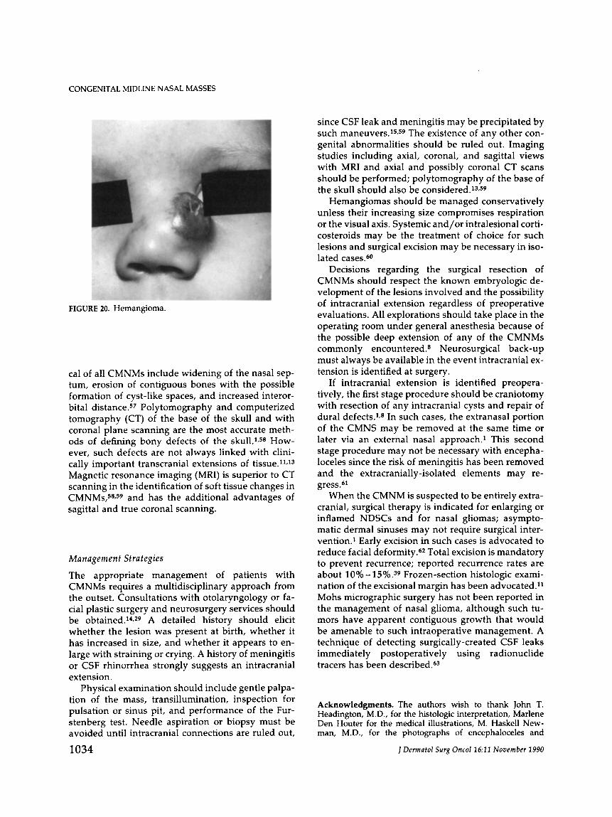
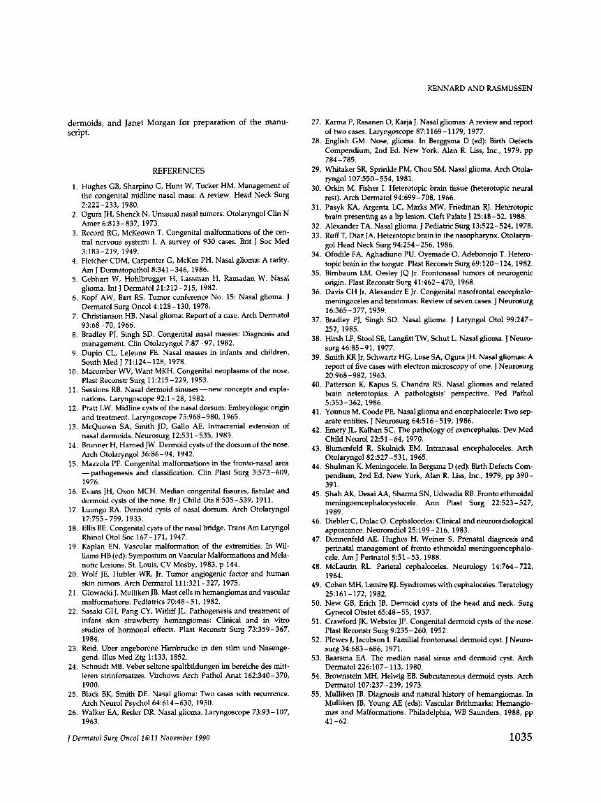
Clinically, NDSCs have a sinus tract that opens on the skin of the nose anywhere between the base of the columella and the glabella (Figs. 17 and 18), with the distal one-third of the nasal dorsum being the most common site.15 A cheesy material can often be expressed from the sinus, and hairs are sometimes seen protruding from the opening.1,15 The course of
1 Dermatol Surg Oncol 16:11 November 1990
Extranasal Glioma (dura pinches as bony surface
FIGURE 9. Extranasal glioma.
lntranasal glioma (dura pinched as bony surface
FIGURE 11. Intranasal ghoma.
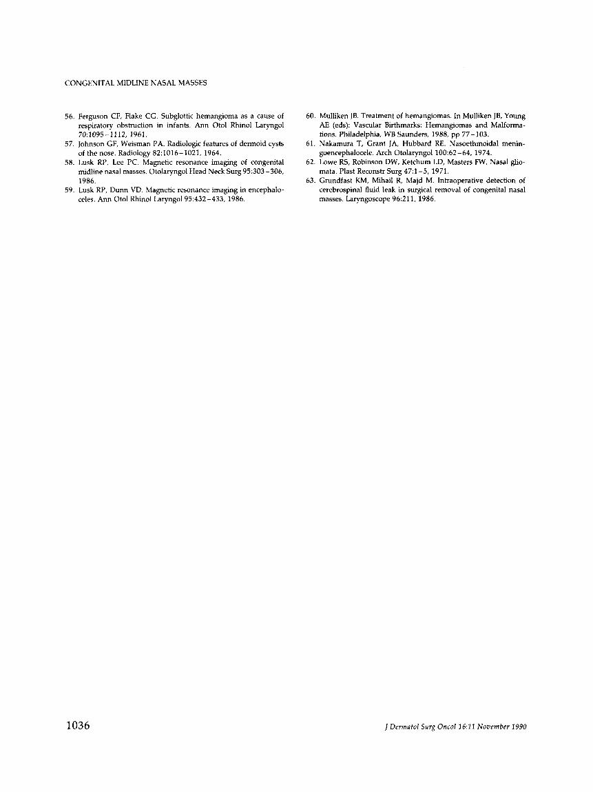
the sinus tract most often is deep to the nasal bones superiorly. It may be short, or extend intracranially and cystic dilatations may be found anywhere along its course (Fig. 19).15
Extranasal dermoids are firm, noncompressible, and nonpulsitile, with a negative Furstenberg test. The lesion does not transilluminate. Local infection may have occurred. If an intracranial connection ex- ists, CSF leak may exist and meningitis, either sterile from cyst contents or bacterial through direct spread, is a risk.'
Histologically, dermoid cysts are true cysts located in the subcutaneous tissues, often adherent to perios-
1031
CONGENITAL MIDLINE NASAL MASSES
Dermal fistula
FIGURE 12. Nasal dermal sinus-cyst (dermoid) extending through prenasal space to base of skull.
teum or eroding the outer table of the They have a lining of stratified squamous epithelium con- taining hair follicles.54 The lumen is filled with des- quamated epithelium and hair shaft fragments. Se- baceous glands and eccrine structures are often found in the cyst wall or opening into the cyst lumen. Mesodermal elements, such as smooth muscle fibers, may also be found in the cyst.54 Bone, cartilage, and specialized tissue not usually found in the skin, such as gastrointestinal, thyroid, or respiratory tissues (as might be seen in teratomas), are absent.41
Patent foramen caecum and intracranial dermoid
Dermal fistula- /
FIGURE 13. Nasal dermal sinus extending through cribiform plate with intracranial dermoid cyst.
1032
frontonasal dermoid cyst
Dermal fistula
FIGURE 14. Frontonasal dermoid cyst.
Other Lesions Hemangiomas of the nose may be quite similar in ap- pearance to nasal gliomas (Fig. 20). Approximately 10% -30% of nasal hemangiomas are present at birth, and most are noted in the first few weeks of life.8~55 They may be quite firm to the touch and only partially blanchable.55 The distinction from nasal glioma may be made even more difficult because both lesions may have telangiectasia on their surface. In fact, Bradley and Singhe reported that in their ser- ies of 109 cases of CMNMs, the greatest diagnostic uncertainty involved seven hemangiomas that were either present at birth at the glabellar/nasal root area, or where the hemangioma was deep in the nasal tip.
Typically, hemangiomas grow rapidly during the
FIGURE 15. Basal encephalocele. Note mass to left of nasal root and hypertelorism.
Dermatol Surg Oncol 16:11 November 1990
KENNARD AND RASMUSSEN
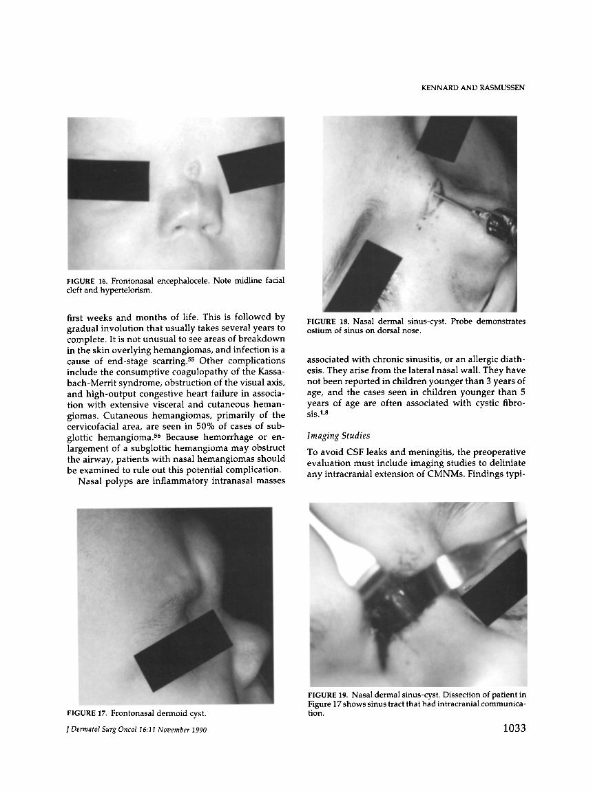
FIGURE 16. Frontonasal encephalocele. Note midline facial cleft and hypertelorism.
first weeks and months of life. This is followed by gradual involution that usually takes several years to complete. It is not unusual to see areas of breakdown in the skin overlying hemangiomas, and infection is a cause of end-stage scarring.55 Other complications include the consumptive coagulopathy of the Kassa- bach-Merrit syndrome, obstruction of the visual axis, and high-output congestive heart failure in associa- tion with extensive visceral and cutaneous heman- giomas. Cutaneous hemangiomas, primarily of the cervicofacial area, are seen in 50% of cases of sub- glottic hemangioma.56 Because hemorrhage or en- largement of a subglottic hemangioma may obstruct the airway, patients with nasal hemangiomas should be examined to rule out this potential complication.
Nasal polyps are inflammatory intranasal masses
FIGURE 17. Frontonasal dermoid cyst.
1 Dermatol SUrg Oncol 16 : l l November 1990
FIGURE 18. Nasal dermal sinus-cyst. Probe demonstrates ostium of sinus on dorsal nose.
associated with chronic sinusitis, or an allergic diath- esis. They arise from the lateral nasal wall. They have not been reported in children younger than 3 years of age, and the cases seen in children younger than 5 years of age are often associated with cystic fibro- sis.1.8
Imaging Studies
To avoid CSF leaks and meningitis, the preoperative evaluation must include imaging studies to deliniate any intracranial extension of CMNMs. Findings typi-
FIGURE 19. Nasal dermal sinus-cyst. Dissection of patient in Figure 17 shows sinus tract that had intracranial communica- tion.
1033
CONGENITAL MIDLINE NASAL MASSES
FIGURE 20. Hemangioma.
cal of all CMNMs include widening of the nasal sep- tum, erosion of contiguous bones with the possible formation of cyst-like spaces, and increased interor- bital distance.57 Polytomography and computerized tomography (CT) of the base of the skull and with coronal plane scanning are the most accurate meth- ods of defining bony defects of the sku11.',58 How- ever, such defects are not always linked with clini- cally important transcranial extensions of t iss~e.~'J3 Magnetic resonance imaging (MRI) is superior to CT scanning in the identification of soft tissue changes in C M N M S , ~ ~ , ~ ~ and has the additional advantages of sagittal and true coronal scanning.
Management Strategies
The appropriate management of patients with CMNMs requires a multidisciplinary approach from the outset. Consultations with otolaryngology or fa- cial plastic surgery and neurosurgery services should be obtained.'*J9 A detailed history should elicit whether the lesion was present at birth, whether it has increased in size, and whether it appears to en- large with straining or crying. A history of meningitis or CSF rhinorrhea strongly suggests an intracranial extension.
Physical examination should include gentle palpa- tion of the mass, transillumination, inspection for pulsation or sinus pit, and performance of the Fur- stenberg test. Needle aspiration or biopsy must be avoided until intracranial connections are ruled out,
1034
since CSF leak and meningitis may be precipitated by such maneuver^.'^,^^ The existence of any other con- genital abnormalities should be ruled out. Imaging studies including axial, coronal, and sagittal views with MRI and axial and possibly coronal CT scans should be performed; polytomography of the base of the skull should also be ~ o n s i d e r e d . ' ~ , ~ ~
Hemangiomas should be managed conservatively unless their increasing size compromises respiration or the visual axis. Systemic and/or intralesional corti- costeroids may be the treatment of choice for such lesions and surgical excision may be necessary in iso- lated cases.60
Decisions regarding the surgical resection of CMNMs should respect the known embryologic de- velopment of the lesions involved and the possibility of intracranial extension regardless of preoperative evaluations. All explorations should take place in the operating room under general anesthesia because of the possible deep extension of any of the CMNMs commonly encountered.8 Neurosurgical back-up must always be available in the event intracranial ex- tension is identified at surgery.
If intracranial extension is identified preopera- tively, the first stage procedure should be craniotomy with resection of any intracranial cysts and repair of dural defects.l.8 In such cases, the extranasal portion of the CMNS may be removed at the same time or later via an external nasal approach.' This second stage procedure may not be necessary with encepha- loceles since the risk of meningitis has been removed and the extracranially-isolated elements may re- gress.61
When the CMNM is suspected to be entirely extra- cranial, surgical therapy is indicated for enlarging or inflamed NDSCs and for nasal gliomas; asympto- matic dermal sinuses may not require surgical inter- vention.' Early excision in such cases is advocated to reduce facial deformity.62 Total excision is mandatory to prevent recurrence; reported recurrence rates are about 10% - 15'30.~~ Frozen-section histologic exami- nation of the excisional margin has been advocated." Mohs micrographic surgery has not been reported in the management of nasal glioma, although such tu- mors have apparent contiguous growth that would be amenable to such intraoperative management. A technique of detecting surgically-created CSF leaks immediately postoperatively using radionuclide tracers has been d e ~ c r i b e d . ~ ~
Acknowledgments. The authors wish to thank John T. Headington, M.D., for the histologic interpretation, Marlene Den Houter for the medical illustrations, M. Haskell New- man, M.D., for the photographs of encephaloceles and
] Dermatol Surg Oncol 16:11 November 1990
KENNARD AND RASMUSSEN
dermoids, and Janet Morgan for preparation of the manu- script.
REFERENCES
1. Hughes GB, Sharpino G, Hunt W, Tucker HM. Management of the congenital midline nasal mass: A review. Head Neck Surg
2. Ogura JH, Shenck N. Unusual nasal tumors. Otolaryngol Clin N Amer 6:813-837, 1973.
3. Record RG, McKeown T. Congenital malformations of the cen- tral nervous system: I. A survey of 930 cases. Brit J SOC Med
4. Fletcher CDM, Carpenter G, McKee PH. Nasal glioma: A rarity.
5. Gebhart W, Hohlbrugger H, Lassman H, Ramadan W. Nasal
6. Kopf AW, Bart RS. Tumor conference No. 15: Nasal glioma. J
7. Christianson HB. Nasal glioma: Report of a case. Arch Dermatol
8. Bradley PJ, Singh SD. Congenital nasal masses: Diagnosis and management. Clin Otolaryngol787-97, 1982.
9. Dupin CL, LeJeune FE. Nasal masses in infants and children. South Med J 71:124-128, 1978.
10. Macomber WV, Want MKH. Congenital neoplasms of the nose. Plast Reconstr Surg 11215-229, 1953.
11. Sessions RB. Nasal dermoid sinuses-new concepts and expla- nations. Laryngoscope 921 -28, 1982.
12. Pratt LW. Midline cysts of the nasal dorsum: Embryologic origin and treatment. Laryngoscope 75:968-980, 1965.
13. McQuown SA, Smith JD, Gallo AE. Intracranial extension of nasal dermoids. Neurosurg 12531 -535, 1983.
14. Brunner H, Hamed JW. Dermoid cysts of the dorsum of the nose. Arch Otolaryngol 36:86-94, 1942.
15. Mazzola PF. Congenital malformations in the fronto-nasal area -pathogenesis and classification. Clin Plast Surg 3:573-609, 1976.
16. Evans JH, Oxon MCH. Median congenital fissures, fistulae and dermoid cysts of the nose. Br J Child Dis 8:535-539, 1911.
17. Luongo RA. Dermoid cysts of nasal dorsum. Arch Otolaryngol
18. Ellis BE. Congenital cysts of the nasal bridge. Trans Am Laryngol Rhino1 Otol SOC 167-171, 1947.
19. Kaplan EN. Vascular malformation of the extremities. In Wil- liams HB (ed): Symposium on Vascular Malformations and Mela- notic Lesions. St. Louis, CV Mosby, 1983, p 144.
20. Wolf JE, Hubler WR, Jr. Tumor angiogenic factor and human skin tumors. Arch Dermatol 111:321-327, 1975.
21, Glowacki J, Mulliken JB. Mast cells in hemangiomas and vascular malformations. Pediatrics 70:48-51, 1982.
22. Sasaki GH, Pang CY, Witliff JL. Pathogenesis and treatment of infant skin strawberry hemangiomas: Clinical and in vitro studies of hormonal effects. Plast Reconstr Surg 73:359-367, 1984.
23. Reid. Uber angeborene Himbrucke in den stim und Nasenge- gend. Illus Med Ztg 1:133, 1852.
24. Schmidt MB. Veber seltene spaltbildungen im bereiche des mitt- leren strinforsatzes. Virchows Arch Pathol Anat 162340-370, 1900.
25. Black BK, Smith DE. Nasal glioma: Two cases with recurrence. Arch Neurol Psycho1 64:614-630, 1950.
26. Walker EA, Resler DR. Nasal glioma. Laryngoscope 73:93-107, 1963.
2~222-233, 1980.
3x183-219, 1949.
Am J Dermatopathol 8:341-346, 1986.
glioma. Int J Dermatol 21:212-215, 1982.
Dermatol Surg Oncol4:128-130, 1978.
93:68-70, 1966.
17:755-759, 1933.
1 Dermatol Surg Oncol 26:11 November 1990
27. Karma P, Rasanen 0, Karja J. Nasal gliomas: A review and report of two cases. Laryngoscope 87:1169-1179, 1977.
28. English GM. Nose, glioma. In Berggsma D (ed): Birth Defects Compendium, 2nd Ed. New York, Alan R. Liss, Inc., 1979, pp
29. Whitaker SR, Sprinkle PM, Chou SM. Nasal glioma. Arch Otola-
30. Orkin M, Fisher I. Heterotopic brain tissue (heterotopic neural
31. Pasyk KA, Argenta LC, Marks MW, Friedman RJ. Heterotopic
32. Alexander TA. Nasal glioma. J Pediatric Surg 13:522-524,1978. 33. Ruff T, Diaz JA. Heterotopic brain in the nasopharynx. Otolaryn-
go1 Head Neck Surg 94254-256,1986. 34. Ofodile FA, Aghadiuno PU, Oyemade 0, Adebonojo T. Hetero-
topic brain in the tongue. Plast Reconstr Surg 69:120- 124, 1982. 35. Bimbaum LM, Oesley JQ Jr. Frontonasal tumors of neurogenic
origin. Plast Reconstr Surg 41:462-470, 1968. 36. Davis CH Jr, Alexander E Jr. Congenital nasofrontal encephalo-
meningoceles and teratomas: Review of seven cases. J Neurosurg
37. Bradley PJ, Singh SD. Nasal glioma. J Laryngol Otol 99247- 252,1985.
38. Hush LF, Stool SE, Langfitt TW, Schut L. Nasal glioma. J Neuro- surg 46:85-91, 1977.
39. Smith KR Jr, Schwartz HG, Luse SA, Ogura JH. Nasal gliomas: A report of five cases with electron microscopy of one. J Neurosurg
40. Patterson K, Kapus S, Chandra RS. Nasal gliomas and related brain neterotopias: A pathologists’ perspective. Ped Pathol
41. Younus M, Coode PE. Nasal glioma and encephalocele: Two sep- arate entities. J Neurosurg 64:516-519, 1986.
42. Emery JL, Kalhan SC. The pathology of exencephalus. Dev Med Child Neurol22:51-64, 1970.
43. Blumenfeld R, Skolnick EM. Intranasal encephaloceles. Arch Otolaryngol 82527-531, 1965.
44. Shulman K. Meningocele. In Bergsma D (ed): Birth Defects Com- pendium, 2nd Ed. New York, Alan R. Liss, Inc., 1979, pp 390- 391.
45. Shah AK, Desai AA, Sharma SN, Udwadia RB. Fronto ethmoidal meningoencephalocystocele. Ann Plast Surg 22523-527, 1989.
46. Diebler C, Dulac 0. Cephaloceles: Clinical and neuroradiological appearance. Neuroradiol25:199-216, 1983.
47. Donnenfeld AE, Hughes H, Weiner S. Prenatal diagnosis and perinatal management of fronto ethmoidal meningoencephalo- cele. Am J Perinatol5:51-53, 1988.
48. McLaurin RL. Parietal cephaloceles. Neurology 14:764- 722, 1964.
49. Cohen MH, Lemire RJ. Syndromes with cephaloceles. Teratology
50. New GB, Erich JB. Dermoid cysts of the head and neck. Surg Gynecol Obstet 65:48-55, 1937.
51. Crawford JK, Webster JP. Congenital dermoid cysts of the nose. Plast Reconstr Surg 9935-260, 1952.
52. Plewes J, Jacobson I. Familial frontonasal dermoid cyst. J Neuro- surg 34:683-686, 1971.
53. Baarsma EA. The median nasal sinus and dermoid cyst. Arch Dermatol 226:107-113, 1980.
54. Brownstein MH, Helwig EB. Subcutaneous dermoid cysts. Arch Dermatol 107237-239, 1973.
55. Mulliken JB. Diagnosis and natural history of hemangiomas. In Mulliken JB, Young AE (eds): Vascular Brithmarks: Hemangio- mas and Malformations. Philadelphia, WB Saunders, 1988, pp
784 - 785.
ryngol 107:550-554, 1981.
rest). Arch Dermatol94:699-708, 1966.
brain presenting as a lip lesion. Cleft Palate J 25:48-52, 1988.
16~365-377, 1959.
20~968-982, 1963.
5:353-362, 1986.
25:161-172, 1982.
41 -62.
1035
CONGENITAL MIDLINE NASAL MASSES
56. Ferguson CF, Flake CG. Subglottic hemangioma as a cause of respiratory obstruction in infants. Ann Otol Rhinol Laryngol
57. Johnson GF, Weisman PA. Radiologic features of dermoid cysts of the nose. Radiology 82:1016-1021, 1964.
58. Lusk RP, Lee PC. Magnetic resonance imaging of congenital midline nasal masses. Otolaryngol Head Neck Surg 95:303- 306, 1986.
59. Lusk RP, Dunn VD. Magnetic resonance imaging in encephalo- celes. Ann Otol Rhinol Laryngol 95:432-433, 1986.
70:1095-1112, 1961.
1036
60. Mulliken JB. Treatment of hemangomas. In Mulliken JB, Young AE (eds): Vascular Birthmarks: Hemangiomas and Malforma- tions. Philadelphia, WB Saunders, 1988, pp 77- 103.
61. Nakamura T, Grant JA, Hubbard RE. Nasoethmoidal menin- goencephalocele. Arch Otolaryngol 100:62 -64, 1974.
62. Lowe RS, Robinson DW, Ketchum LD, Masters FW. Nasal glio- mata. Plast Reconstr Surg 47:l-5, 1971.
63. Grundfast KM, Mihail R, Majd M. Intraoperative detection of cerebrospinal fluid leak in surgical removal of congenital nasal masses. Laryngoscope 96:211, 1986.
1 Dermatol Surg Oncol 16:11 November 1990