Congenital diaphragmatic hernia: A rare cause of ...ijhbr.com/pdf/April 2015 113-116.pdfCongenital...
4
International J. of Healthcare and Biomedical Research, Volume: 03, Issue: 03, April 2015, Pages 113-116 113 www.ijhbr.com ISSN: 2319-7072 Case report Congenital diaphragmatic hernia: A rare cause of obstructive jaundice Dr Manjusha M. litake , Dr Sunil B. Tarode Sassoon General Hospital and B J Medical College Pune , India Corresponding author : Dr Manjusha M. litake Abstract Congenital Diaphragmatic Hernia in adults are exceedingly rare. They have been reported to cause dyspnea, gastric reflux and intestinal obstruction .We present the case of a young male with obstructive jaundice secondary to boch- dalek hernia of the right hemi-diaphragm. We discuss the aetiologies ,presentation and treatment of the disorder. Case History A 29 year old male presented to hospital with complains of recurrent jaundice (serum bilirubin 27 direct 17 ),icterus, nausea with on and off abdominal pain. There was no history of trauma. A plain chest xray taken on admission demonstrated a right lung collapse and an elevated right hemi-diaphragm. Ultrasonography of abdomen performed on the same day demonstrated intrahepatic bile duct dilatation and herniation of liver, gall bladder, intestines in right side of thorax. Due to the dual findings of the chest x-ray and ultrasonography, double contrast computed tomography (CT) of the abdomen and thorax was performed (fig 1). This demonstrated that the right lobe of liver, gall bladder, kidney and intestinal loops , mesentery was within the right chest cavity with kinking of bile duct with collapse of right lung .It also demonstrated intrahepatic duct dilatation as a result of anatomical distortion. There was no obvious pathology in the spleen, left kidney , pancreas ,left hemi-diaphragm. Computed tomography of the abdomen and thorax demonstrating Mild prominence of intra-hepatic biliary radicals in right lobe of liver, with altered signal intensity on right lobe of liver suggestive of obstructive biliopathy. Dilated left hepatic duct with abrupt narrowing of right hepatic duct at the confluence suggestive of biliary stricture MRI Abdomen
Transcript of Congenital diaphragmatic hernia: A rare cause of ...ijhbr.com/pdf/April 2015 113-116.pdfCongenital...
International J. of Healthcare and Biomedical Research, Volume: 03, Issue: 03, April 2015, Pages 113-116
113
www.ijhbr.com ISSN: 2319-7072
Case report
Congenital diaphragmatic hernia: A rare cause of obstructive jaundice
Dr Manjusha M. litake , Dr Sunil B. Tarode
Sassoon General Hospital and B J Medical College Pune , India
Corresponding author : Dr Manjusha M. litake
Abstract
Congenital Diaphragmatic Hernia in adults are exceedingly rare. They have been reported to cause dyspnea, gastric
reflux and intestinal obstruction .We present the case of a young male with obstructive jaundice secondary to boch-
dalek hernia of the right hemi-diaphragm. We discuss the aetiologies ,presentation and treatment of the disorder.
Case History
A 29 year old male presented to hospital with
complains of recurrent jaundice (serum bilirubin 27
direct 17 ),icterus, nausea with on and off abdominal
pain. There was no history of trauma. A plain chest
xray taken on admission demonstrated a right lung
collapse and an elevated right hemi-diaphragm.
Ultrasonography of abdomen performed on the same
day demonstrated intrahepatic bile duct dilatation and
herniation of liver, gall bladder, intestines in right
side of thorax.
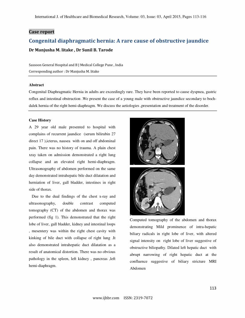
Due to the dual findings of the chest x-ray and
ultrasonography, double contrast computed
tomography (CT) of the abdomen and thorax was
performed (fig 1). This demonstrated that the right
lobe of liver, gall bladder, kidney and intestinal loops
, mesentery was within the right chest cavity with
kinking of bile duct with collapse of right lung .It
also demonstrated intrahepatic duct dilatation as a
result of anatomical distortion. There was no obvious
pathology in the spleen, left kidney , pancreas ,left
hemi-diaphragm.
Computed tomography of the abdomen and thorax
demonstrating Mild prominence of intra-hepatic
biliary radicals in right lobe of liver, with altered
signal intensity on right lobe of liver suggestive of
obstructive biliopathy. Dilated left hepatic duct with
abrupt narrowing of right hepatic duct at the
confluence suggestive of biliary stricture MRI
Abdomen

International J. of Healthcare and Biomedical Research, Volume: 03, Issue: 03, April 2015, Pages 113-116
114
www.ijhbr.com ISSN: 2319-7072
MRI Abdomen showing large diaphragmatic hernia on right side with herniation of bowel loops, fat right
lobe of liver, gall bladder and right kidney with underlying collapse of lung.
• Obstructive jaundice reduced within a week
of admission without any intervention.It was
decided elective repair of diaphragmatic
hernia was needed due the risk of bowel
strangulation or further billiary
complications.
Intra-operative findings
• Intra-operatively thoracotomy and
paramedian incision was taken for better
exposure .Right lobe of liver was found to
be cirrhotic with left lobe hypertrophy.
• E/o large defect of size 10×7cm through
which right lobe of liver, gall bladder and
right kidney were seen to be herniating into
thorax. Right lobe of liver was cirrhotic.
• E/o kink in right hepatic duct just proximal
to junction with left hepatic duct causing
biliary obstruction.
• Right lobe of liver , right kidney & bowel
loops reduced into abdominal cavity,
• Cholecystectomy done .
• After reduction of the hernial contents,
defect was closed and reinforced with an on
laying mesh ,primary closure of the
abdominal cavity was done.
Medworld asia
Dedicated for quality research
www.medworldasia.com
International J. of Healthcare and Biomedical Research, Volume: 03, Issue: 03, April 2015, Pages 113-116
114
www.ijhbr.com ISSN: 2319-7072
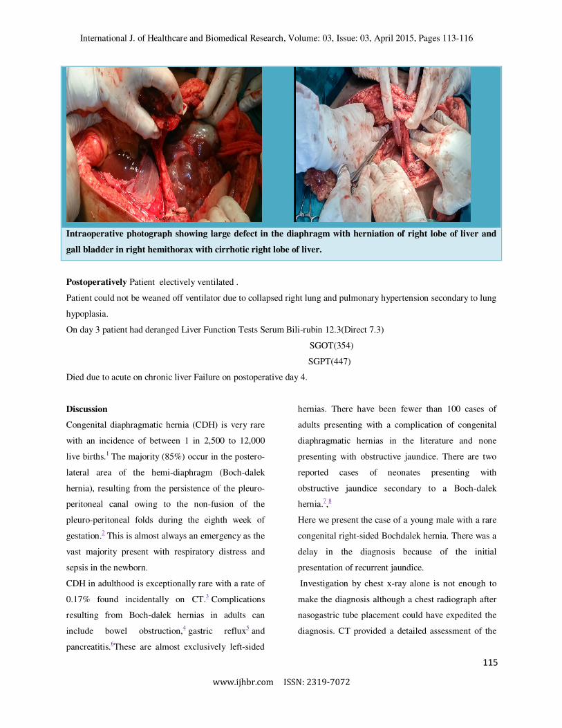
Intraoperative photograph showing large defect in the diaphragm with herniation of right lobe of liver and
gall bladder in right hemithorax with cirrhotic right lobe of liver.
Postoperatively Patient electively ventilated .
Patient could not be weaned off ventilator due to collapsed right lung and pulmonary hypertension secondary to lung
hypoplasia.
On day 3 patient had deranged Liver Function Tests Serum Bili-rubin 12.3(Direct 7.3)
SGOT(354)
SGPT(447)
Died due to acute on chronic liver Failure on postoperative day 4.
Discussion
Congenital diaphragmatic hernia (CDH) is very rare
with an incidence of between 1 in 2,500 to 12,000
live births.1 The majority (85%) occur in the postero-
lateral area of the hemi-diaphragm (Boch-dalek
hernia), resulting from the persistence of the pleuro-
peritoneal canal owing to the non-fusion of the
pleuro-peritoneal folds during the eighth week of
gestation.2 This is almost always an emergency as the
vast majority present with respiratory distress and
sepsis in the newborn.
CDH in adulthood is exceptionally rare with a rate of
0.17% found incidentally on CT.3 Complications
resulting from Boch-dalek hernias in adults can
include bowel obstruction,4 gastric reflux5 and
pancreatitis.6These are almost exclusively left-sided
hernias. There have been fewer than 100 cases of
adults presenting with a complication of congenital
diaphragmatic hernias in the literature and none
presenting with obstructive jaundice. There are two
reported cases of neonates presenting with
obstructive jaundice secondary to a Boch-dalek
hernia.7,8
Here we present the case of a young male with a rare
congenital right-sided Bochdalek hernia. There was a
delay in the diagnosis because of the initial
presentation of recurrent jaundice.
Investigation by chest x-ray alone is not enough to
make the diagnosis although a chest radiograph after
nasogastric tube placement could have expedited the
diagnosis. CT provided a detailed assessment of the
115

International J. of Healthcare and Biomedical Research, Volume: 03, Issue: 03, April 2015, Pages 113-116
114
www.ijhbr.com ISSN: 2319-7072
anatomy and a cause for the obstructive jaundice was
established.
Even though CDH has been well described in the
literature, the incidence of clinical presentations in
adulthood is exceedingly rare and this is the first case
of it leading directly to obstruction of the biliary
system. Open mesh repair of the hernia is the gold
standard treatment option with evidence to back up
its safety and efficacy. There are very few
documented cases of successful laparoscopic repair
of an adult CDH in the literature. One case series
suggests that this is a safe alternative treatment
modality for CDH presenting past infancy.9
Current recommendations are that all adults with a
CDH undergo repair in order to avoid
complications.10Possible postoperative complications
include abdominal compartment syndrome although
there is no evidence in the literature to support this.
Conclusions
CDH is exceedingly rare in adulthood and has been
reported to become symptomatic in only a handful of
cases. However, since its presence can lead to serious
adverse events such as acute intestinal obstruction, or
in this case obstruction of the biliary system, it
should be investigated fully and repaired rapidly. The
diagnosis should be considered in any patient
presenting with abdominal pain and an unexplained
consolidation on a chest x-ray.
References
1. García-Muñoz F, Santana C, Reyes D, et al. Early sepsis, obstructive jaundice and right-sided diaphragmatic
hernia in the newborn. Acta Paediatr. 2001;90:96–98
2. Kanazawa A, Yoshioka Y, inoi o, et al. Acute respiratory failure caused by an incarcerated right-sided adult
Bochdalek hernia: report of a case. Surg Today. 2002;32:812–815.
3. Mullins ME, Saini S. Imaging of incidental Bochdalek hernia. Semin Ultrasound CT MR. 2005;26:28–36
4. Oliver MJ, Wilson AR, Kapila L. Acute pancreatitis and gastric volvulus occurring in a congenital diaphragmatic
hernia. J Pediatr Surg. 1990;25:1,240–1,241
5. Sigalet DL, Nguyen LT, Adolph V, et al. Gastroesophageal reflux associated with large diaphragmatic hernias. J
Pediatr Surg. 1994;29:1,262–1,265.
6. Rout S, Foo FJ, Hayden JD, et al. Right-sided Bochdalek hernia obstructing in an adult: case report and review of
the literature. Hernia. 2007;11:359–362.
7. Schiffer M, Rescorla FJ, Fitzgerald J, Grosfeld JL. Obstructive jaundice. An unusual delayed presentation of
congenital diaphragmatic hernia. Arch Surg. 1988;123:780–781
8. Allen JL, Petrovich JA, Gooley NA. Congenital foramen of Bochdalek’s hernia in an infant with obstructive
jaundice. Surgery. 1989;105:224–226
9. Palanivelu C, Rangarajan M, Rajapandian S, et al. Laparoscopic repair of adult diaphragmatic hernias and
eventration with primary sutured closure and prosthetic reinforcement: a retrospective study. Surg
Endosc.2009;23:978–985.
10. Schumacher L, Gilbert S. Congenital diaphragmatic hernia in the adult. Thorac Surg Clin. 2009;19:469–472.
116 116