Cone beam computed tomography diagnostic …this kind of tumor from an odontogenic cyst or...
4
J Clin Exp Dent. 2017;9(9):e1158-61. CBCT and diagnostic imaging e1158 Journal section: Oral Medicine and Pathology Publication Types: Case Report Cone beam computed tomography diagnostic imaging of intra-osseous mucoepidermoid carcinoma in the mandible Andre-Luiz-Ferreira Costa 1 , Thasia-Luiz-Dias Ferreira 1 , Haroldo-Arid Soares 2 , Ana-Carla-Raphaelli Nahas- Scocate 1 , Gonzalo-André-Parra Montesinos 3 , Paulo-Henrique Braz-Silva 4 1 PhD,DDS, Department of Orthodontics and Radiology, University of City of Sao Paulo, Sao Paulo, SP, Brazil 2 PhD, DDS, Regional Hospital Dr. Carmino Caricchio, Division of Stomatology, Sao Paulo, SP, Brazil 3 MSc, Department of Orthodontics and Radiology, University of City of Sao Paulo, Sao Paulo, SP, Brazil 4 PhD, DDS, Division of General Pathology, Department of Stomatology, School of Dentistry, University of Sao Paulo, Sao Paulo, SP, Brazil Correspondence: Department of Orthodontics and Radiology UNICID, Rua Cesário Galeno 448 Bloco A. Tatuapé, São Paulo, SP, 03071-000 [email protected] Received: 14/02/2017 Accepted: 12/07/2017 Abstract Intra-osseous mucoepidermoid carcinoma in the mandible is a rarely reported entity, comprising only 2-3% of all mucoepidermoid carcinomas. Unilocular and/or multilocular radiolucency is characteristic of mucoepidermoid carcinoma, but has a radiographic appearance similar to that of odontogenic benign and malignant tumors and thus cannot be accurately diagnosed on plain films. This article describes a case of a 36-year-old man with intra-osseous mucoepidermoid carcinoma in the left mandible. For a detailed analysis, both panoramic radiograph and cone beam computed tomograph were taken. Important clinico-pathological and imaging features, differential diagnosis and review of the literature are described. Key words: Diagnosis, Cone Beam Computed Tomography, Head and Neck Tumors. doi:10.4317/jced.53785 http://dx.doi.org/10.4317/jced.53785 Introduction Intra-osseous mucoepidermoid carcinoma (IMC) in the mandible is a rare bone malignant neoplasm, compri- sing 2-4% of all mucoepidermoid carcinomas diagnosed (1). For some authors, these lesions are classified more appropriately as odontogenic tumors rather than as sali- vary gland neoplasms (2). Imaging plays an important role in the detection and differentiation of IMC becau- se its sclerotic periphery and mixed internal structure, consisting of unilocular and/or multilocular pattern with imaging characteristics similar to those of other lesions, including ameloblastoma, glandular odontogenic cyst and keratocystic odontogenic tumour (1). This case re- port intends to highlight the value of cone beam compu- ted tomography (CBCT) for the diagnosis of a patient with IMC in mandible as it helps to delineate the lesion and plan the treatment properly. Case report A 37-year-old male patient presented to the Division of Article Number: 53785 http://www.medicinaoral.com/odo/indice.htm © Medicina Oral S. L. C.I.F. B 96689336 - eISSN: 1989-5488 eMail: [email protected] Indexed in: Pubmed Pubmed Central® (PMC) Scopus DOI® System Costa ALF, Ferreira TLD, Soares HA, Nahas-Scocate ACR, Montesinos GAP, Braz-Silva PH. Cone beam computed tomography diagnostic imag- . Cone beam computed tomography diagnostic imag- ing of intra-osseous mucoepidermoid carcinoma in the mandible. J Clin Exp Dent. 2017;9(9):e1158-61. http://www.medicinaoral.com/odo/volumenes/v9i9/jcedv9i9p1158.pdf
Transcript of Cone beam computed tomography diagnostic …this kind of tumor from an odontogenic cyst or...
J Clin Exp Dent. 2017;9(9):e1158-61. CBCT and diagnostic imaging
e1158
Journal section: Oral Medicine and Pathology Publication Types: Case Report
Cone beam computed tomography diagnostic imaging of intra-osseous mucoepidermoid carcinoma in the mandible
Andre-Luiz-Ferreira Costa 1, Thasia-Luiz-Dias Ferreira 1, Haroldo-Arid Soares 2, Ana-Carla-Raphaelli Nahas-Scocate 1, Gonzalo-André-Parra Montesinos 3, Paulo-Henrique Braz-Silva 4
1 PhD,DDS, Department of Orthodontics and Radiology, University of City of Sao Paulo, Sao Paulo, SP, Brazil2 PhD, DDS, Regional Hospital Dr. Carmino Caricchio, Division of Stomatology, Sao Paulo, SP, Brazil3 MSc, Department of Orthodontics and Radiology, University of City of Sao Paulo, Sao Paulo, SP, Brazil4 PhD, DDS, Division of General Pathology, Department of Stomatology, School of Dentistry, University of Sao Paulo, Sao Paulo, SP, Brazil
Correspondence:Department of Orthodontics and RadiologyUNICID, Rua Cesário Galeno 448Bloco A. Tatuapé, São Paulo, SP, [email protected]
Received: 14/02/2017Accepted: 12/07/2017
Abstract Intra-osseous mucoepidermoid carcinoma in the mandible is a rarely reported entity, comprising only 2-3% of all mucoepidermoid carcinomas. Unilocular and/or multilocular radiolucency is characteristic of mucoepidermoid carcinoma, but has a radiographic appearance similar to that of odontogenic benign and malignant tumors and thus cannot be accurately diagnosed on plain films. This article describes a case of a 36-year-old man with intra-osseous mucoepidermoid carcinoma in the left mandible. For a detailed analysis, both panoramic radiograph and cone beam computed tomograph were taken. Important clinico-pathological and imaging features, differential diagnosis and review of the literature are described.
Key words: Diagnosis, Cone Beam Computed Tomography, Head and Neck Tumors.
doi:10.4317/jced.53785http://dx.doi.org/10.4317/jced.53785
IntroductionIntra-osseous mucoepidermoid carcinoma (IMC) in the mandible is a rare bone malignant neoplasm, compri-sing 2-4% of all mucoepidermoid carcinomas diagnosed (1). For some authors, these lesions are classified more appropriately as odontogenic tumors rather than as sali-vary gland neoplasms (2). Imaging plays an important role in the detection and differentiation of IMC becau-se its sclerotic periphery and mixed internal structure, consisting of unilocular and/or multilocular pattern with
imaging characteristics similar to those of other lesions, including ameloblastoma, glandular odontogenic cyst and keratocystic odontogenic tumour (1). This case re-port intends to highlight the value of cone beam compu-ted tomography (CBCT) for the diagnosis of a patient with IMC in mandible as it helps to delineate the lesion and plan the treatment properly.
Case reportA 37-year-old male patient presented to the Division of
Article Number: 53785 http://www.medicinaoral.com/odo/indice.htm© Medicina Oral S. L. C.I.F. B 96689336 - eISSN: 1989-5488eMail: [email protected] in:
PubmedPubmed Central® (PMC)ScopusDOI® System
Costa ALF, Ferreira TLD, Soares HA, Nahas-Scocate ACR, Montesinos GAP, Braz-Silva PH. Cone beam computed tomography diagnostic imag-. Cone beam computed tomography diagnostic imag-ing of intra-osseous mucoepidermoid carcinoma in the mandible. J Clin Exp Dent. 2017;9(9):e1158-61.http://www.medicinaoral.com/odo/volumenes/v9i9/jcedv9i9p1158.pdf

J Clin Exp Dent. 2017;9(9):e1158-61. CBCT and diagnostic imaging
e1159
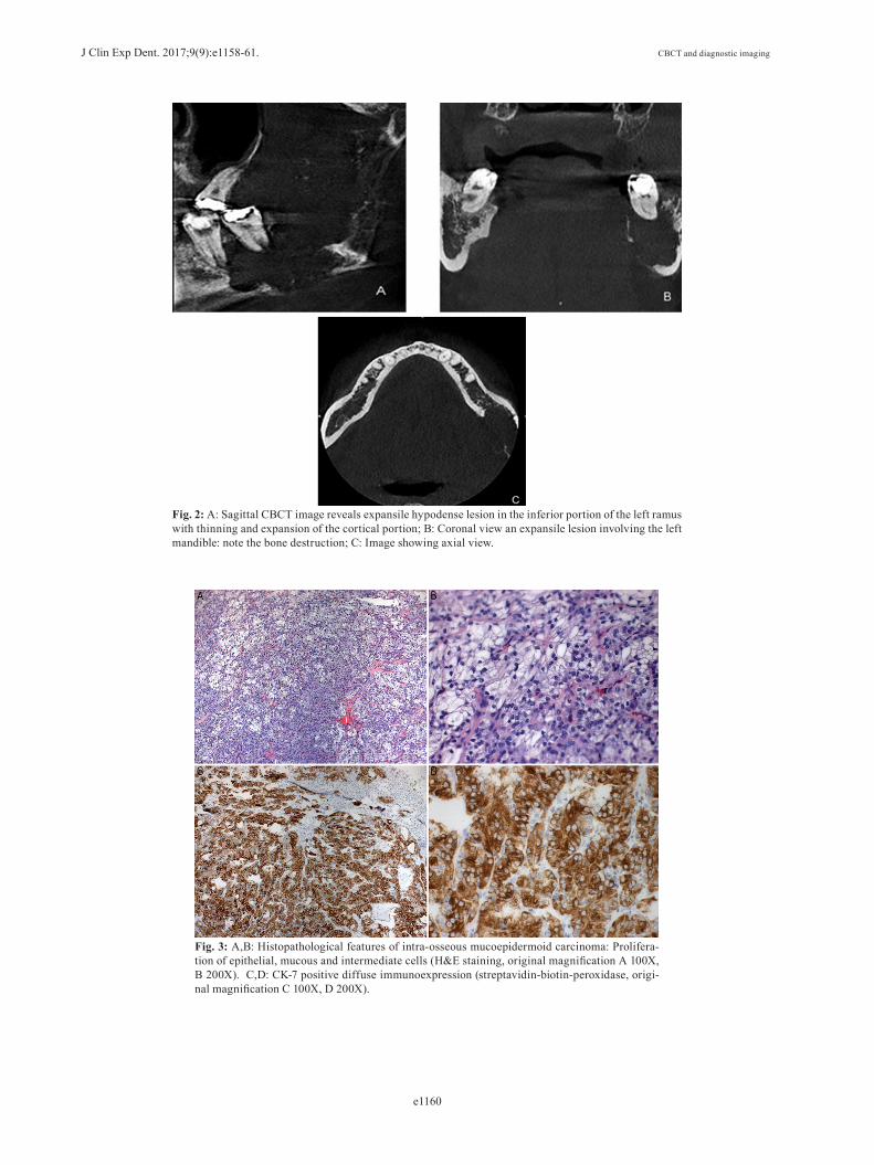
Stomatology of the Carmino Carrichio Regional Hospi-tal, São Paulo, SP, Brazil, complaining of a painful left-cheek swelling for approximately three months. Trismus and paraesthesia were observed. The patient’s past medi-cal history was unremarkable as he denied smoking and consuming alcohol. On extra-oral examination, a facial asymmetry with a smooth, hard and fixed swelling at the left mandible was noticed (Fig. 1A). Clinical intraoral examination showed a large ulcerated lesion with buccal and lingual bone expansion around the left body and ra-mus of the mandible. The associated teeth were sound, but with noticeable mobility. The panoramic radiograph revealed irregular unilocular radiolucency extending ho-rizontally from posterior region of the body to the whole ramus and angle of the mandible (Fig. 1B). Erosion of the inferior border of the mandible with irregular, ill-defined areas of bone destruction was observed. The pa-tient was also submitted to CBCT examination for better evaluation of both extension and form of the mandibular lesion. CBCT scan images (Kodak CS8100 3D system, Kodak Carestream Health, Paris, France) showed an ex-pansive lesion in the left mandible with destruction of posterior body and extend into ramus. Irregular hypo-density was observed with diffuse limits, covering the posterior region of the body, angle and ramus as well as lateromedial expansion and bone erosion of the ramus of the mandible (sagittal view, Fig. 2A), compromising the alveolar supporting bone of teeth #36 and #37. Co-ronal CBCT view showed an expansile hypodense le-sion, erosion of lingual cortical plate (Fig. 2B), whereas axial CBCT view (Fig. 2C) showed an ill-defined soft tissue density mass, and extension of the lesion within the mandible with break in lingual cortexBased on clinical and radiographic aspects, the diffe-rential diagnosis included ameloblastic carcinoma and primary intra-osseous carcinoma. Under local anesthe-sia, incisional biopsy was performed. Histopathological sections revealed cystic and solid patterns, with proli-
Fig. 1: A: Extra-oral image of the affected area, with marked facial swelling on the left mandibu-lar side and trismus; B: An irregular, ill-defined diffuse radiolucency located in the posterior region of the mandibular body, affecting teeth 37 and 36 and almost the whole left mandibular ramus with destruction of cortical basal bone.
feration of epidermoid, mucous and intermediate cells (Fig. 3A). Neither significant pleomorphism nor mitotic activity was observed (Fig. 3B). Cytokeratin 7 (CK-7) immunohistochemistry showed a diffuse expression, which confirms the glandular epithelial differentiation of the tumor (Fig. 3C,D). Based on the histopathological features, the final diagnosis was an intermediate-grade intra-osseous mucoepidermoid carcinoma. The patient was referred to the State of São Paulo Can-cer Institute (ICESP) for treatment, with complete re-section of the lesion and adjuvant radiotherapy being performed. The patient has been followed up since then and feeling well.
DiscussionThe aetiology of IMC is still unclear (3). Some studies suggest three origins (3,4), namely: embryonic salivary gland remnants included within the mandible or mucous glands entrapped within the bone; transformation of mucous cells found in odontogenic cysts; and submu-cosal mucous glands with intra-osseous extension. The tumour is usually located in the mandible (premolar-mo-lar-angle region), occurring three times more often in the maxilla (5). Also, it affects women twice more than men (5,6) and it has been reported to occur in all ages, most commonly in the 4th and 5th decades of life (3,5).Reports of IMC originating from jaws are remarkably uncommon and often involve swelling and pain, whe-reas trismus, paraesthesia and tooth mobility are less frequently noticed (7). He et al. (3) reported a total of 24 cases and Bell et al. (5) reviewed 25 cases. Only one report focused on diagnostic imaging features (1). Ra-diographically, these lesions may present unilocular or multilocular radiolucency (7) with diffuse bone resorp-tion and extensive loss of cortical bone (8).Radiographic examination is important for categoriza-tion of a central jaw lesion (7,9). Brookstone and Huvos (10) proposed a three-grade classification for IMC:
J Clin Exp Dent. 2017;9(9):e1158-61. CBCT and diagnostic imaging
e1160
Fig. 2: A: Sagittal CBCT image reveals expansile hypodense lesion in the inferior portion of the left ramus with thinning and expansion of the cortical portion; B: Coronal view an expansile lesion involving the left mandible: note the bone destruction; C: Image showing axial view.
Fig. 3: A,B: Histopathological features of intra-osseous mucoepidermoid carcinoma: Prolifera-tion of epithelial, mucous and intermediate cells (H&E staining, original magnification A 100X, B 200X). C,D: CK-7 positive diffuse immunoexpression (streptavidin-biotin-peroxidase, origi-nal magnification C 100X, D 200X).

J Clin Exp Dent. 2017;9(9):e1158-61. CBCT and diagnostic imaging
e1161
- Grade 1: without expansion and rupture of cortical plate; - Grade 2: with expansion but without rupture of cortical plate; - Grade 3: with rupture of cortical plates or presence of regional metastasis.The present case was classified as grade 3, that is, with no metastasis being detected. The radiographic features of IMC typically show marked variation in the appea-rance of the borders, and consequently it is worth regar-ding them as a differential diagnosis of jaw radiolucency (8,9). Panoramic radiography and conventional compu-ted tomography (CT) are routinely used as diagnostic tools for evaluating the maxillofacial area.However, panoramic radiography does not allow as-sessment of destructive bone lesions with non-uniform border or with degrees of extension and invasion into the surrounding tissues (8), nor enough to differentiate this kind of tumor from an odontogenic cyst or amelo-blastoma. On the other hand, CT offers a large amount of information on size, location and area of the tumor in the region (11). In 2013, Chan et al. (1) analysed the radiographic fea-tures of four cases using CT imaging. The internal pat-tern comprises disseminated foci of amorphous sclerotic bone, which appear as irregular masses with cortical bone-like structure without any organization into tra-beculae or septa. Multiple small radiolucent loculations measuring less than 8 mm with and without rough bor-ders in the septum were also observed.This case report describes the value of using CBCT in the diagnoses of IMC. CBCT provides images of the lesion in all planes and thus enables localization, exten-sion and internal structure of it. In fact, CBCT confirmed the sclerotic periphery and mixed internal structure of the lesion by highlighting border expansion and thinning of the cortical plate with erosion through the cortex.These lesions are treated with radical resection of the margin of the adjacent normal bone. Neck dissection and post-operative radiation therapy may be required to control spread to lymph nodes (2).The lesion in our case was treated with radical resection and adjuvant radio-therapy. To the best of our knowledge, the present case report illustrates the first use of CBCT for diagnostic approach of this rare malignancy.
References1. Chan KC, Pharoah M, Lee L, Weinreb I, Perez-Ordonez B. In-traosseous mucoepidermoid carcinoma: a review of the diagnos-tic imaging features of four jaw cases. Dentomaxillofac Radiol. 2013;42:20110162.2. Pires FR, Paes de Almeida O, Lopes MA, Elias da Cruz Perez D, Kowalski LP. Central mucoepidermoid carcinoma of the mandible: report of four cases with long-term follow-up. Int J Oral Maxillofac Surg. 2003;32:378-82.3. He Y, Wang J, Fu HH, Zhang ZY, Zhuang QW. Intraosseous mucoe-pidermoid carcinoma of jaws: report of 24 cases. Oral Surg Oral Med Oral Pathol Oral Radiol. 2012;114:424-9.
4. Johnson B, Velez I. Central mucoepidermoid carcinoma with an aty-pical radiographic appearance. Oral Surg Oral Med Oral Pathol Oral Radiol Endod. 2008;106:e51-3.5. Bell D, Lewis C, El-Naggar AK, Weber RS. Primary intraosseous mucoepidermoid carcinoma of the jaw: Reappraisal of The MD Ander-son Cancer Center experience. Head Neck. 2016;38 Suppl 1:E1312-7.6. Eversole LR, Sabes WR, Rovin S. Aggressive growth and neoplas-tic potential of odontogenic cysts: with special reference to central epi-dermoid and mucoepidermoid carcinomas. Cancer. 1975;35:270-82.7. Raut D, Khedkar S. Primary intraosseous mucoepidermoid carcino-ma of the maxilla: a case report and review of literature. Dentomaxi-llofac Radiol. 2009;38:163-8.8. Adachi M, Inagaki T, Ehara Y, Azuma M, Kurenuma A, Motohashi M, et al. Primary intraosseous carcinoma arising from an odontogenic cyst: A case report. Oncol Lett. 2014;8:1265-8.9. Kaffe I, Ardekian L, Peled M, Machtey E, Laufer D. Radiologi-cal features of primary intra-osseous carcinoma of the jaws. Analysis of the literature and report of a new case. Dentomaxillofac Radiol. 1998;27:209-14.10. Brookstone MS, Huvos AG. Central salivary gland tumors of the maxilla and mandible: a clinicopathologic study of 11 cases with an analysis of the literature. J Oral Maxillofac Surg. 1992;50:229-36.11. Sukovic P. Cone beam computed tomography in craniofacial ima-ging. Orthod Craniofac Res. 2003;6 Suppl 1:31-6; discussion 179-82.
Conflicts of interestThe authors declare they have no conflicts of interest.