CONCURRENT MONITORING OF monitoring of Trichinella and Toxoplasma infections in pigs from controlled...
154
CONCURRENT MONITORING OF TRICHINELLA AND TOXOPLASMA INFECTIONS IN PIGS FROM CONTROLLED HOUSING SYSTEMS Gertie Bokken 2017
Transcript of CONCURRENT MONITORING OF monitoring of Trichinella and Toxoplasma infections in pigs from controlled...

CONCURRENT MONITORING OF TRICHINELLA AND TOXOPLASMA INFECTIONS IN PIGS FROM
CONTROLLED HOUSING SYSTEMS
Gertie Bokken
2017

Concurrent monitoring of Trichinella and Toxoplasma infections in pigs from controlled housing systems
PhD thesis, Utrecht University, the Netherlands ISBN: 978-94-6295-656-8 Cover ontwerp: Gertie Bokken, Tagxedo, Proefschriftmaken Lay out: Gertie Bokken Printing: Proefschriftmaken

Concurrent monitoring of Trichinella and Toxoplasma infections in pigs from
controlled housing systems
Gelijktijdige monitoring van Trichinella en Toxoplasma infecties in varkens van gecontroleerde huisvestingsomstandigheden
systemen
(met een samenvatting in het Nederlands)
Proefschrift
Ter verkrijging van de graad van doctor aan de Universiteit Utrecht op gezag van de rector magnificus, prof. dr. G.J. van der Zwaan,
ingevolge het besluit van het college voor promoties in het openbaar te verdedigen op maandag 26 juni 2017 des middags te 4.15 uur
door
Gertruda Cornelia Antonia Maria Bokken geboren op 20 december 1966 te Wamel

Promotor: prof. dr. Frans. van Knapen
Co-promotor: dr. Aldert A. Bergwerff


Beoordelingscommissie Prof. dr. D.J.J. Heederik Prof. dr. A. Kijlstra Prof. dr. J.A. Stegeman Prof. dr. A.G.M. Tielens Prof. dr. B. Urlings Paranimfen Angèle Timan Gerdit Greve

TABLE OF CONTENTS:
Chapter 1 9
General Introduction
Chapter 2 45
A novel bead-based assay to detect specific antibody responses against Toxoplasma gondii and Trichinella spiralis simultaneously in sera of experimentally infected swine
Chapter 3 63
Refined assay parameters of a bead-based detection method for Trichinella spiralis infections in pigs
Chapter 4 71
Bayesian estimation of diagnostic accuracy of a new bead-based antibody detection test to reveal Toxoplasma gondii infections in pig populations
Chapter 5 85
Specific serum antibody responses following a Toxoplasma gondii and Trichinella spiralis co-infection in swine
Chapter 6 101
Potential association between Trichinella and Toxoplasma gondii infection in a naturally infected pig population
Chapter 7 113
General discussion
Summary 135
Nederlandse samenvatting 141
Curriculum vitae en publicaties 147
Dankwoord 151


Chapter 1
GENERAL INTRODUCTION

Chapter 1
10
DISCOVERY OF TRICHINELLA AND T. GONDII
The nematode Trichinella was discovered by James Paget in London in 1834 (1) in a patient who died from trichinellosis. However, it was Richard Owen who first described the parasite (2). Around 1860 individual efforts of Rudolf Leuckart, Rudolf Virchow and Friedrich Zenker clarified the different aspects of the Trichinella life cycle. After recognition of pork as the main contributor of human trichinellosis, methods of detection of the parasite followed by control measures of pork have been introduced in large parts of the world.
Trichinoscopy, essentially the microscopic analysis of meat samples between pressurized glass plates (3), was first introduced in 1863 in Germany. In 1877, the many outbreaks of human trichinellosis cases and related deaths led to the introduction of this test to control Trichinella transmission via pork. The test was first made mandatory in Prussia soon followed by other German states, and became common in much of western Europe by the end of the nineteenth century (4). Nowadays in the EU a regulation which controls Trichinella infection in pork is in action since 1963. Currently, trichinellosis cases in the European population are largely contained.
The apicomplexan Toxoplasma gondii (T. gondii) was first observed in 1908 in a rodent by Nicolle and Manceaux (5). It took until 1939 before Wolf, Cowen and Paige identified the parasite in a child who had developed encephalomeyelitis and retinitis died one month after birth, and (6). They also showed that infection could be continued in mice and rabbits after intracerebrally infection of homogenates of the child’s cortex and spinal cord. During the sixties of last century, the coccidian phase of T. gondii was identified by several studies done by Hutchison (7), Frenkel (8), Overdulve (9-11) and Sheffield and Melton (12). During the same time the role of Felidae in the sexual replication phase of the parasite was first described (7, 8, 12, 13).
Meat-associated infections are one of the routes of transmission towards humans. Even though T. gondii has been recognized as one of the currently most prominent disease causing pathogens (14), no control measures of food-associated infections have been introduced to contain toxoplasmosis in the EU population (15).

General introduction
11
TRICHINELLA SPP. AND T. GONDII LIFE CYCLES
Trichinella spp.
The life cycle of Trichinella spp. is continued from one host to the next host after the passage of the stomach (Figure 1, arrow 1). The muscle larvae are freed from their capsule by the activity of gastric acid and enzymes where after they migrate to the small intestine (Figure 1, arrow 2). At two days after ingestion, the larvae develop into adult worms after four subsequent moults, where after male and female worms copulate (Figure 1, arrow 3). Around 7 days after infection, new born larvae (NBL) are produced in the mucosa of the intestine (Figure 1, arrow 4). After passage of the intestinal mucosa, NBL migrate through the body by the fluidic system to striated muscle cells where they form nurse cells and encysts (Figure 1, arrow 5). Encysted parasites can reside for a long time within the tissue of the host, which in case of pigs kept for meat production can continue during its entire life span. Continuation of the parasites life cycle is initiated by uptake of these encysted parasites by a new meat-eating host. Within several weeks after infection the production of NBL stops after intestinal worm expulsion caused by an intestinally immune-mediated host response.
Trichinella spp. infections are observed in mammals, birds and reptiles (16). T. spiralis, T. britovi, T. nativa and T. pseudospiralis, are the most common Trichinella species present in Europe (16). Of these species, T. spiralis and T. britovi are the most observed species in the studies performed in EU foxes (Table 1). T. britovi is predominantly associated to the sylvatic cycle, while T. spiralis is more associated to the domestic cycle (17). T. nativa is associated to the neartic and artic zone’s, and in the EU, this species is predominantly present in Scandinavia (16). Only 1.6% of the observed Trichinella spp. isolates in EU wildlife were T. pseudospiralis, which indicates that this species is rather uncommon (18). Of the four Trichinella species observed in Europe, T. pseudospiralis is the only one associated to both avian and mammalian species. Additionally, T. pseudospiralis belongs to the non-encapsulated clade which do not form capsulated tissue cysts, while T. spiralis, T. britovi and T. nativa belong to the encapsulated clade (18).

Chapter 1
12
T. gondii
The hexogeneous life cycle of T. gondii (Figure 2) is complex and consists of three distinct pathways; a pathway where parasites replicate sexually in the end host, Felidae, a pathway where they replicate asexually in intermediate hosts and a pathway where new hosts are infected through vertical transmission. One of the stages of the T. gondii life cycle, here named the oocysts infection route, starts after the infection of cats by T. gondii parasites (Figure 2, arrow 4). After ingestion of oocysts or tissue cyst, the cell wall of the infectious vehicle is dissolved by proteolytic enzymes produced in stomach and small intestines of the cat. In the end host, freed sporozoites or bradyzoites attach and penetrate the small intestines epithelial cells, ultimately resulting in the formation of immature oocysts (19). Once these oocysts are discharged through the cats faces, they sporulate under certain humidity and temperature conditions (Figure 2, arrow 1) (13) into mature oocysts. Sporulated oocysts contain two sporocysts, each consisting out of four sporozoites (19). Oocysts are produced during a short excretory period, however, due to
Figure 1: Trichinella spp. life cycle Source: https://www.cdc.gov/parasites/images/trichinellosis/trichinella_lifecycle.gif

General introduction
13
the large production of oocysts, high quantities can be shed (13). The environmental spread oocysts can infect mammalian and avian species (figure 2, arrows 2 and 5), and can be found on vegetables, fruit and in surface water (Figure 2, arrow 7). Sporulated oocysts are resistant to extreme weather conditions and can remain infectious for up to 18 months (20).
Figure 2: T. gondii life cycle Source: https://www.cdc.gov/parasites/images/toxoplasmosis/toxoplasma_lifecycle_bam1.gif
Another stage of the T. gondii life cycle is running through the formation of tissue cysts (Figure 2, arrows 3 and 6). Upon oral ingestion of parasites by intermediate hosts, i.e. mammals, birds and some reptiles, bradyzoites from tissue cysts or sporozoites from oocysts are transformed to tachyzoites. Thereupon, the tachyzoites penetrate nucleated host cells. During the acute phase of infection, in which tachyzoites rapidly multiply, the parasites eventually burst out of their confinements, whereupon they can infect other host cells. By not fully understood processes (21), the fast replicating and highly metabolic tachyzoite differentiate after 6-7 days to bradyzoite stage parasites within tissue cysts (19). The majority of these tissue cysts are found within neural and muscular tissue.

Chapter 1
14
Studies in pigs showed that brain tissue, tongue and heart are more frequently infected than other parts of the pigs’ body (19). Additionally, blood transfusion and the transplantation of organs from an infected donor can induce toxoplasmosis in the recipient (Figure 2, arrows 8 and 10.). Tachyzoites are also able to cross the placental membrane, thereby inducing prenatal infections in human and other mammals alike (Figure 2, arrow 11) (22).
One of the most noticeable differences between oocysts and tissue cyst routes is the time span of tissue cyst formation in intermediate hosts. While new tissue cysts can be found as early as 6 days after infection with tissue cysts, generation of tissue cyst following an oocysts infection takes an additional day (19). Tissue cysts can remain present for the remainder of the life of the host.
DISEASE
Trichinellosis and toxoplasmosis are diseases which are caused by infection of Trichinella spp. or T. gondii, respectively. Infection leading to the diseases of trichinellosis and acquired toxoplasmosis are majorly associated to the consumption of parasitic units in food and from oral uptake of environmentally spread oocysts. To a lesser extent, infection is transmitted by tissue transplants from an infected individual to the recipient. Additionally, congenital toxoplasmosis in unborn children is caused by horizontal infection during an acute infection of the mother to the child during gestation.
Trichinella spp. infection
Human trichinellosis has been characterized by a widespread of clinical signs ranging from abdominal pain, nausea, diarrhea, and vomiting at 1-2 weeks after infection, to symptoms like fever and fatigue, or more extreme, symptoms associated to problems due to damage of organs like the central nervous system, heart and lungs. In extreme circumstances people die from this infection, however, in the majority of human infections the disease runs asymptomatically (23).
The measure of extremity of the infection is related to the quantity of infectious larvae ingested in humans (23) and animals alike (24, 25). Furthermore, the severity of disease in humans is Trichinella strain dependent (23). A recent study has estimated that only a few Trichinella parasites of either sex may have a considerable risk of development of

General introduction
15
(a)symptomatic infection (26). In most cases, the clinical signs associated to the mild and moderate infections will pass away within a few months.
Human trichinellosis is mainly associated to the consumption of infected meat. Historically, the main source of human trichinellosis infections was through consumption of infected pork. Current status of infection in pigs indicates a low prevalence of less than 0.01 % on EU level (27). Furthermore, the number of Trichinella induced human deaths and disease in the EU have decreased dramatically to the point that in the period between 2011 and 2015 only a very small percentage of the human EU population becomes infected (between 0.03 and 0.07 confirmed cases per 100,000 people) (27). These cases can be contributed to consumption of EU-produced or imported pork, horse meat, wild hunted or farmed boar, bear meat or other meat from game, or otherwise, could have been contracted outside EU countries (28).
T. gondii infection
Disease associated to T. gondii infections in humans are characterized by the time-point of infection. Postnatally acquired toxoplasmosis can remain unnoticed because the disease runs asymptomatic or the symptoms are non-descriptive and are not recognized as toxoplasmosis. Cases of infection can lead to symptoms like mild illness with fever, enlarged lymph nodes, muscle pain and sour throat. Especially immunosupressed patients can get extreme diseases like encephalitis and neurological diseases.
The disease burden due to congenital toxoplasmosis is associated to the moment of infection during gestation via the haematogenous transplacental route (29). Infections acquired early in gestation can lead to serious disease, like hydrocephalus or microcephaly, intracranial calcification, and chorioretinitis, in the offspring. In some cases this infection will lead to spontaneous abortion or intrauterine death. Transmission of the parasite in late phase of pregnancy can result in potential vision loss, mental disability, and seizures later in the life of the offspring.
It has been estimated that about 50% of the human toxoplasmosis cases can be related to foodborne infections (30). Furthermore, 30% to 63% of cases of toxoplasmosis in pregnant women could be attributed to consumption of undercooked, raw or cured infected meat, while up to 17% could be attributed to intake of environmentally spread oocysts (31). A more recent American study estimated that 8% of foodborne associated illness which is caused by T. gondii result in hospitalization, while death from foodborne infections was in 24% of the cases caused by T. gondii (32). Another study estimated that, of all meat-borne human infections in the Netherlands, beef was contributing the most while pork and

Chapter 1
16
lamb/mutton were contributing to a lesser extent (33). This either indicates that the T. gondii prevalence is higher in cattle than in pigs and sheep, and/or, differences in the method of food preparation of the three meat groups play a vital role in the transmission. Another Dutch study estimates that T. gondii, obtained via food or environmental route, is the number one pathogen in humans with the highest associated burden on population basis and on individual level (14), indicating the importance of further prevention of infection through foodborne transmission. Even though these figures may be variable from region to region, they indicate that T. gondii has a serious impact on consumers’ safety.
Foodborne related disease
According to the WHO, a foodborne acquired T. gondii infection has a larger impact on public health as compared to a Trichinella infection. For example the foodborne Disability Adjusted Life Years (DALY) score in the Western part of Europe is 2 (95%UI 1-3) and 6 (95%UI 4-10) for respective congenitally and acquired T. gondii related infections, while Trichinella spp. related infections leads to 0.04 (95%UI 0.02-0.07) DALYs (34). However, the measure of foodborne related disability for both infections within this region is estimated to be almost equal, i.e. 0.06 (0.05-0.08) and 0.1 (0.06-0.1) foodborne DALYs per case for T. gondii and Trichinella spp., respectively (34). The difference between the DALY scores can be explained by the higher number of toxoplasmosis diseased persons and T. gondii caused deaths. Indeed, Trichinella spp. does not pose a real risk on human health in most EU countries. Human incidences of Trichinella spp. infections are observed rarely, and the number of deaths due to this parasite are not reported. While on the other hand, both T. gondii caused incidences of disease and deaths are more heavily reported (34). These figures indicate that in comparison to Trichinella, there is a higher need to control the human foodborne transmissions of T. gondii.
TRICHINELLA SPP. AND T. GONDII CONTROL MEASURES
Trichinella spp.
In the EU, present control of Trichinella spp. has been directed through EU 2075/2005 (35) and amending regulation EU 216/2014 (36). In a nutshell these directives describe the mandatory inspection of pig carcasses at slaughter for Trichinella larvae by use of artificial digestion. Alternatively, the amending regulation EU 216/2014 describes the conditions to

General introduction
17
holdings (farms) and controlling agencies should comply in order to achieve a Trichinella spp. free status of holdings (farms). Holdings within the Member State with a Trichinella spp. free status would then be able to produce Trichinella free animals and could by this way circumvent the mandatory individual inspection at slaughter. In 1991, the Netherlands tried to apply for a Trichinella free status, unfortunately, this was too early as politic was not ready for that at the time (15, 37). These days, the Trichinella free national status has been acknowledged in Denmark (38) and Belgium. However, under the recent adaptation of EU 216/2014, the national Trichinella free status in these countries is no longer recognized. Under the condition that holdings within these countries comply to the prerequisites of Controlled Housing Conditions (CHS), the Trichinella free status is granted.
T. gondii
Although T. gondii has been marked as a threat to human health since 1938, until now, no real measure of control of T. gondii has been implemented to reduce the risk of transmission via consumption of meat. As compared to other food-related pathogens, the existence and transmission routes of this parasite were discovered relatively late in history. Most likely the non-descriptive signs during human disease, flu like symptoms, and the relatively small parasitic size complicated the recognition and detection in the past. However, during the last decade the impact of this parasite to human health was studied more elaborately. These studies calculated the severity of human T. gondii infections and that of other pathogens by estimation of the disability-adjusted life years (DALYs) (39, 40) in the Netherlands. The direct health care costs caused by toxoplasmosis are estimated around 20 billion euros, the third highest infection associated costs in the Netherlands in 2011(41). From this increased knowledge it became clear that, at least in the Netherlands, T. gondii was one of the largest human food-related microbiological threats at present time. It is assumed that pork is one of the major contributors to transmission of this parasite to humans (31). A Dutch study estimated that 11.2% and 7.1% of meat consumers are infected with T. gondii by consumption of infected pork and minced beef/pork products, respectively (33). This is a clear indication that some form of T. gondii infection control in pork should be implemented in order to avoid transmission to its consumers. Unfortunately, little information is available on T. gondii prevalence within the EU pig population. Therefore, testing these animals for T. gondii presence would generate a better insight in order to control the issues of transmission.

Chapter 1
18
TRICHINELLA SPP. AND T. GONDII TRANSMISSION PREVENTION OF PIGS
Given the information of the presence and impact of pork related T. gondii infections in EU citizens, it is of importance that a strategy is implemented to avoid future infections. For implementation of a strategy, costs and risk factors are weighed. From a cost-wise perspective, it would be logic to follow a strategy which protects public health from a combination of zoonotic pathogens in pigs/pork. For such an approach, the similarities in the Trichinella spp. and T. gondii transmission routes from pig preceding host, to pigs and subsequently human, could be used.
Combined Trichinella spp. and T. gondii infection vectors
Potential meat-associated infection vectors for T. spiralis and T. gondii for domesticated pigs are intermediate hosts which are edible by pigs (Figure 3). Most likely, intermediate hosts are small mammals, or in the case of T. gondii, birds, which habitat and/or feed locations are situated on the premises of the piggery, as well. However, larger game or cadavers cannot be ruled out as transmission vectors to pigs. Additionally for T. gondii, the transmission route can run through the oral uptake of environmental oocysts shed by cats.
The Trichinella spp. and T. gondii transmission routes towards pigs can be distinguished in two different pathways; a sylvatic (Figure 3, dark grey boxes) and a domestic cycle (Figure 3, light grey boxes).
Continuation of Trichinella spp. and T. gondii infections within domesticated pigs is characterized by the uptake of infected meat via feeding of offal from slaughtered sylvatic animals (Figure 3, box G) and farm animals (Figure 3, box F), tail and ear biting within the herd (Figure 3, box A), scavenging or preying on small mammals like mice or rats (Figure 3, box B), birds (Figure 3, box C) and insects (Figure 3, box E). Within this picture, cats (Figure 3, box D) are exceptional as they can be infected by both parasitic species, but they are unlikely prey for pigs. The development of T. gondii oocysts by cats, however, is a major transmission vector for not only pigs, but also for many other animal species.
Sylvatic animals
In wildlife, carnivores, like red foxes, raccoon dogs and wolves but also omnivores, like polar, black and grizzly bears and wild boars, and herbivores, like reindeer, are known

General introduction
19
species which can harbour Trichinella spp. (42). The feeding of offal from any of these animals to pigs is prohibited by EU 2075/2005. Furthermore, it is highly unlikely that confined pigs can get in contact with animals from the wild and is therefore not expected that this group will contribute to the two parasitic infections of EU pigs.
Figure 3: Trichinella spp. and T. gondii transmission routes to pig
Farm animals
Amongst animals in the domestic cycle, pigs are the predominant species infected with T. spiralis; however, occasionally this parasite is also found in species like horses and sheep (43). T. gondii infections in farm animals are predominantly found in cow, sheep and pigs (33). Feeding of offal from these animals to other farm animals is prohibited by EU 2075/2005. Therefore, this route will not contribute to an increase of Trichinella spp. and T. gondii infections amongst farm animals. Furthermore, in cases that animal species are housed indoors, strictly isolated from other animal species, the contribution of other animals to the transmission of parasites will be negligible.

Chapter 1
20
Pigs
Cannibalism is not uncommon amongst pigs when carcasses of dead pigs are not removed from the premises (44, 45). However, EU 2075/2005 specifies that all dead animals should be removed directly from the direct environment of pigs, which will minimize the probability of transmission. Furthermore, continuation of infection via this route is dependent on the presence of the parasite in the herd. Therefore, the probability of transmission is likely to be associated to the presence of infection via the other routes. Additionally, vertical transmission of T. gondii from mother to offspring, which has been shown to occur in three successive generations of mice (46), may contribute to the overall infection pressure.
Rodents and other small mammals
Studies of sylvatic or synantropic living micromammals like rodents and insectivores have indicated that these species are potential carriers of T. gondii and T. spiralis. The most notable infection causing species is the brown Norway rat (Rattus norvegicus). However, other species like house mouse (Mus musculus), voles (Microtus spp.), hispid cotton rats (Sigmodon hispidus), greater white-toothed shrews (Crocidura russula) and many others have been found to contain one or both parasitic species (47-53). The habitats and feeding habits of these species differ from those of brown rats and it is logic to assume that they are accidental infection vectors for pigs.
Brown rats
The role of brown rats as transmitter for T. spiralis has been studied elaborately. These animals live in groups, and as omnivores are known to forage on a broad range of foods. In an abundance of food, brown rats will sample all these foods and consume as much as is necessary to fulfil their energy needs (54). As they may consume 2 to 3 grams of food per feeding, and they are known to eat meat, the chance of parasite transmission from a dead infected carcass is plausible. Evidence of brown rats acting as an intermediary to T. spiralis infections for pigs has been described in two studies (45, 55). These studies observed that T. spiralis infected rat to pigs transmission took place in the zone with the intermediate and highest rate of contact between species (45). Furthermore, 16 months after removal of infected dead pigs, the in origin suspected cause of infection in rats, T. spiralis infection was still observed in rats (55). Because, mortality amongst wild rats is around 95% per year (56), it is likely that T. spiralis parasites in these rats were transmitted within the rat population by cannibalism.

General introduction
21
In rats, transmammally and potentially congenitally transmission of Trichinella from mother to offspring has been reported (57-59). The exact route of vertical transmission towards the offspring is unclear. However, hypothetically these routes can run via an intra uterus infection with NBL, via infection of mammalian glands with NBL and shedding into milk or via expulsed intestinal larvae in the faeces from the mother. Moreover, vertical transmission occurs in other species than rats alone as was demonstrated by an Trichinella in utero infected case of an unborn child (60).
Unfortunately little data has been published which sheds light on the prevalence ratio of Trichinella spp. and T. gondii in brown rats. However, red foxes are the sentinels of these parasitic infections in the wildlife reservoir. A collection of Trichinella spp. and T. gondii infection data of foxes illustrates that on average a 10 times higher prevalence for T. gondii as compared to Trichinella can be found (see Table 1 and 2). In particular, studies which used the same foxes to determine the infection status of both showed that the T. gondii prevalence was around 62 times higher as that of T. spiralis (61, 62). A T. spiralis and T. gondii prevalence study in feral pigs (63-65) show the same trend: T. gondii infections are found more often than that of Trichinella spp. These data show that the there is a higher T. gondii pressure within the sylvatic system as compared to Trichinella spp.
Extra T. gondii causalities in synantropic living animals can be expected from oocysts shed by cats, which live in the neighbourhood of the farm. Additionally, the property of this parasite to transmit infection by vertical transmission may ultimately result in a higher T. gondii prevalence amongst synantrophic living animals as compared to Trichinella spp. in the same animals. Assuming that T. gondii and Trichinella spp. infections in pigs originate from infected synantrophic living animals and environmentally spread oocysts; again a higher T. gondii prevalence as compared to Trichinella spp. prevalence is expected. This line of thought was endorsed by several studies within the same pig population, where the T. gondii prevalence was higher than the Trichinella spp. prevalence (Table 3) (63-75). Also the preliminary studies of several member states showed the same tendency on national level (76).
The exact role of rats as vector organism for Trichinella transmission is still under discussion (43, 77-80). Even though the true nature of rats as reservoir is still unknown, there is substantial evidence that they can play a role in transmission of infections to pigs. The likelihood of this route depends on the prevalence and presence of rats in the pig housing facilities.

Chapter 1
22
Table 1: Studies of Trichinella spp. prevalence determined by digestion in red foxes from European countries in period 1985 to 2015
Country study Era Number of animals
Prevalence (%) Trichinella spp.
Belgium Flanders Wallonia
(132) 1996-1999 1998-2000
179 639
0Ø
0
Denmark (133) 1995-1996 1997-1998
3,133 3,008
0.001 0
N.D.
Denmark (134) 2009-2012 384 0
France (Corsica) (135) 2006-2008 74 0
France (136) 2006-2009 108 2.7 2
Germany (137) 2002-2011 3,154 0.3 1, 2, 3, 4
Hungary (138) 2002 100 3.0 2
Hungary (139) 2006 2,116 1.8 1, 2, 3
Hungary (140) 2006-2013 3,304 2.1 1, 2
Ireland*** (61) 2003 454 0.9 1
Italy (Tuscany) (141) 2004-2006 129 0
Italy (Liguria, Piedmont) (142) 2009-2012 165 0
Italy (Abruzzo) (142) 2004-2014 24 5.0 N.D
Northern Ireland (143) 2003-2004 443 0.2 N.D
Norway** (144) 1994-1995 2002-2005
393 4.8 2, 4
Poland (137) 2011-2012 1,634 2.7 1, 2, 4
Poland (145) 2012 24 16.7 2, 3
Poland (146) 2010-2015 1,447 10.0 1, 2, 3, $
Portugal (147) 2008-2010 47 2.1 2
Romania (148) 2012-2014 121 21.5 1, 2
Slovakia (149) 2000-2007 5,270 11.5 1, 2, 3
UK* (150) 1999-2000 587 0 N.D.
Spain (Soria) (151) 4 years# 400 15.5 1, 2
Sweden (152) 1985-2003 1,800 4.5 1, 2, 4
Switzerland (153) 2006-2007 1,298 1.6 2
The Netherlands (154) 2010-2013 369 0.27 $
In comparison to Table 5: * same study; ** partly the same animals; *** same animals; Ø: tested by Trichinoscopy; $: unclassified subspecies; #: time period not mentioned. Where 1: Trichinella spiralis, 2: Trichinella britovi, 3: Trichinella pseudospiralis, 4: Trichinella nativa, N.D.: not determined, N.I.: not identified.

General introduction
23
Birds
The role of birds as a transmission vector is limited to Trichinella pseudospiralis and T. gondii. Although T. pseudospiralis infections in pigs are uncommon (81), as such they can pose a threat to public health (42, 82-84). T. gondii has been reported to be present in many different avian species (85, 86). However, the prevalence of both parasites in birds is largely unknown (18), due to limited numbers of studies and the large numbers of different avian species. Furthermore, it is unknown to what extent any of the avian species come in contact with pigs. Because of the limited data, it is difficult to estimate the contribution of birds towards the parasitic prevalence in pigs.
Table 2: Studies of T. gondii prevalence in red foxes from European countries in period 1990 to 2014
Country study Era Test method Number Prevalence (%)
Austria (155) 1999 IFAT 94 35 Belgium (156) # PCR 304 18.8 Czech Republic (157) 2012 Indirect ELISA/IFAT 80 100/100 Germany (Brandenburg) (Saxony Anhalt)
(158) 2009-2010 Immunoblot/PCR 204
74.5/84.7 18.4/13.4
Hungary (159) 2003 DAT 337 68 Ireland (160) 1999-2000 LAT 51 24 Ireland*** (62) 2003 IFAT 454 56 Italy (Piedmont) (161) 2009-2012 PCR 94 20.2 Italy (Pisa) (162) 2009-2011 IFAT 191 53.4 Norway** (163) 1994-1995
2002-2005 DAT 275 67
Portugal* (164) 2008-2010 Indirect ELISA 6 Ø 100 Romania (165) 2012 PCR 182 6 UK* (150) 1999-2000 PCR 61Ø 0 UK (166) # IFAT 500 20 UK (Scotland) (167) # PCR 83 6.0 Slovakia (168) 2010-2014 Indirect ELISA 303 62.7 Spain (169) 1990-2006 MAT 102 64.7 Spain (170) 2009-2011 PCR 41 51.2 Sweden (171) 1991-1999 DAT 221 38 In comparison to Table 4: * same study; ** partly the same animals; *** same animals; # not mentioned; Ø same animals were used for Trichinella prevalence. Where test method IFAT: indirect immunofluorescent antibody test; PCR: polymerase chain reaction: DAT, direct agglutination test and LAT: latex agglutination test, MAT:
modified agglutination test.

Chapter 1
24
Table 3: Studies between 1990 and 2015 reporting the prevalence of Trichinella spp. and T. gondii in swine/wild boar populations
Estimated prevalence %(95%CI) (test)
Country Pig origin year Medium N T. gondii Trichinella spp. study
Nether- lands
finishing pigs 1991 Sera/ diaphragm
23,348 2.1(1) 0(4) (66)
USA pigs 1992 sera 509 48.5(2) 2.1(5) (67)
USA finishing pigs 1998 sera 2,238 0.58 (0.31-0.99) (2) 0.046 (0-0.255) (5) (68)
USA feral swine 2002 sera 227 49(1) 39(5) (63)
Nether- lands
fattening pigs (indoor/outdoor)
2007 sera 845 2.6(1) 0.12(5) (69)
USA pigs (indoor/outdoor)
2008 sera 616 4.1(3) 0.3(5) (70)
USA feral swine 2011 sera 83 27.7(1) 13.3(5) (64)
Finland farmed wild boar 2012 sera 197 33 (27-40) (1) 2 (1-5) (5) (71)
Nepal pigs 2013 sera 742 11.7 (5.2–17.5) (1) 0.1 (0–0.7) (5) (72)
Spain fattening pigs (outdoor)
2013 sera/ diaphragm
709 27(1) 0(5) (73)
Finland finishing pigs 2014 meat-juice 1,353 3.2 (2.4-4.3) (1) 0 (0-0.3) (5) (74)
USA feral swine 2014 sera 3,247 17.7(1) 3.0(5) (65)
Germany finishing pigs 2014 meat-juice 50 10 (8.9–11.0) (1) 0 (0–0.1) (5) (75)
Where test methods 1: diverse T. gondii ELISA’s; 2: T. gondii direct agglutination test (DAT); 3: T. gondii modified agglutination test (MAT); 4: Trichinella digestion; 5: diverse Trichinella ELISA’s
Insects
Flies and other insects in diverse stadia of development have been reported as carriers of Trichinella spp. and T. gondii. For example cockroaches are potential transmission vector for both pathogens (87, 88). Furthermore, Trichinella larvae can survive in maggots and have been proven infectious for mice (89). T. gondii can survive in maggots and pupa, but they are not detectable in the developed flies (90). However, these stages may be of less importance to pig infection as the dispersal of the larval and pupal stage is limited to dead animals on which they feed. Moreover, in case the pig consumes the dead animal, the function of infection vector by the maggot and pupa is minimalized. Nevertheless, the presence of flies and mosquitos were associated to T. gondii infections in pigs (91) indicating that these animals may be the transmission vectors to the parasite. It remains unclear whether bradyzoites in tissue cysts or potentially tachyzoites can survive in flies/mosquitos, however, all insects are potential mechanical carriers of T. gondii oocysts. Some scientists however, doubt the role of insects as a source of infection as initially the

General introduction
25
route of T. gondii transmission was believed to run through insects, but evidence for this was never found (92).
METHODS OF INFECTION DETECTION IN PIGS
Prevention of infection through the consumption of infected meat can be achieved by the determination of the infectious status of the animal, followed by adequate measures as freezing to inactivate the parasite. The infectious status can be determined by direct tests, tests which detect the parasite, or indirect tests, tests which detect the immunological responses to the presence of the parasite.
Direct tests
Direct tests are tests which are based on the detection of the intact parasite or of (multiple) parasitic constituents or products. This detection can be done by visualization of the morphological shape of the whole parasite within tissue, or by visualization of parasite parts, like for example, cell wall structures, secreted or excreted proteins or glycoproteins, or genetic factors like DNA. Because a direct test determines the presence of a current infection, this method of detection is preferred above indirect tests, which are hampered by a so-called immune-window of antibody production.
Trichinella spp.
The magnetic stirrer method for pooled sample digestion is the current reference method in the EU (35). Pooling of samples up to 100 samples per digestion reduces labour and costs of the procedure; however, the sensitivity of this test is reduced alongside of this. Estimates made by Forbes and Gahjadhar (93), report that the overall sensitivity of the artificial digestion method lies around 80-100%. However, in cases that the parasitic load within the infected host is low, for example between 0.01 and 0.09 larvae per gram (LPG), the sensitivity of artificial digestion of 1 g meat sample is significantly reduced to 40% and could lead to infected meat entering the human food chain. Furthermore, because the pepsin and hydrochloric acid concentrations and incubation times of the solution used for artificial digestion are optimized for detection of encapsulated muscle larvae in meat samples, this testing method may be less optimal for detection of parasites which are in a pre-stadium of encapsulation (94).

Chapter 1
26
Other direct tests, like PCR, have been described in literature; however, these testing methods have their value for epidemiological purposes but not for the benefit of meat inspection for reasons of relatively high costs, long time-to-results and relative insensitivity.
T. gondii
One of the direct tests available is a T. gondii bioassay. This bioassay uses T. gondii naïve mice (mouse bioassay) and/or cats (cat bioassay) to prolong the infection by feeding the animals tissues of possibly infected meat sources. Upon infection, mice then produce tissue cysts and cats additionally produce oocysts in their faecal shedding. Infection is determined by checking homogenized mice brains for tissue cysts by use of a microscope and/or counting oocysts in the faces of cats (51, 95), respectively.
Other direct testing methods might include Polymerase Chain Reactions (PCR), a technique which multiplies T. gondii specific DNA fragments. This technique is sensitive, however, the challenge lies in harvesting sufficient parasitic DNA from infected tissue in order to determine the infection. As was shown in several studies, the sensitivity of this testing method is less as compared to the bioassay (96-98). Possibly, by introducing a parasite extraction and purification method before PCR the test sensitivity may be increased (99); however, the laboriousness of the whole procedure would make testing on large scale unattainable for large screening purposes.
Indirect tests
Indirect tests, tests which determine the animals’ responses to an infectious agent, may be seen as an alternative to direct tests. These indirect tests, often referred as serological tests, are quick, simple to perform and economically attractive. One of the most well-known indirect tests are Enzyme Linked Immuno Sorbent Assays (ELISA’s). Other tests have been developed which differ from the ELISA by the structure of the surface matrixes whereupon immune complexes are formed, and subsequent equipment which is needed for the detection of immune complex formation.
Recent technical development showed new innovative high-throughput micro array tests which can determine antibody responses simultaneously for combinations of antigens (100-102). These kinds of serological tests can have the same specifications as other indirect tests; however, their ability to determine responses for multiple antigens at the same time makes them more desirable than single antibody tests like ELISA´s.

General introduction
27
Test accuracy
Foremost, the sensitivity and specificity of the binding dynamics of antigens and antibody are important to determine a previous infection in animals using sera. To calculate the specificity and sensitivity of a test, samples from animals with a known infection status, the so-called true infection status, are related to the test outcomes (Table 4). The true infection status of animals is often unknown. To determine the true infections status so called gold standard tests can be used.
Table 4: Classical method for calculation of SE, SP, PPV and NPV of a test
True infection status infected not infected
Test + True Positives (TP) False Positives (FP) Positive predictive value (PPV) - False Negatives (FN) True Negatives (TN) Negative predictive value (NPV)
Sensitivity (Se) Specificity (Sp)
To calculate sensitivity (Se), specificity (Sp), positive predictive value (PPV) and negative predictive value (NPV) of a test the following formulas can be used:
Se = TP / (TP + FN) * 100%
Sp = TN / (TN + FP) * 100%
PPV = TP / (TP + FP) * 100%
NPV = TN / (TN + FN) * 100%
Furthermore, apparent prevalence (AP) = (TP+FP)/(TP+FP+FN+TN) and, true prevalence (π) = (TP+FN)/(TP+FP+FN+TN).
Sensitivity and specificity should be further specified by the terms “analytical” or “diagnostic”. Analytical sensitivity is used when the test is addressing the minimum limits of material quantity which is detectable by the system, and analytical specificity is based upon the degree of cross-reactivity of antibodies and antigen. Diagnostic sensitivity indicates the percentage of tested positives in relation to infected subjects, while diagnostic specificity indicates the percentage of tested negatives in relation to non-infected subjects.
Indirect tests may be applicable for determination of the infection status in meat-producing animals. However, the time points of infection of the animal and the subsequent production of specific antibodies against parasitic antigens are spatially distributed. Serological testing the antibodies during this frame could lead to a falsely qualification of meat safety, subsequently resulting in infected pork entering the food

Chapter 1
28
chain. For Trichinella infections, this is the reason why indirect tests cannot be used as end point indicators of infection (103). However, as was indicated in the European Trichinella regulation (35), serology can be implemented for monitoring purposes when this test is validated and approved by the Community Reference Laboratory (CRL). At this moment no serological Trichinella test is accepted by the CRL.
Trichinella spp.
Thus far, many Trichinella antigens have been described in literature. Initially, crude extract of whole muscle larvae was used to develop ELISA tests (104, 105). However, due to high cross-reactivity with other non Trichinella sera, the International Commission of Trichinellosis (ICT) does not recommend to use this antigen in serological tests. Instead, the organization advices the use of Excretory/Secretory (ES) product actively secreted by first stage muscular larvae (106), also referred to as L1 stage.
ES product offers the advantage that it can detect antibodies against all Trichinella species and furthermore, the material can be produced in vitro in a consistent way. All currently commercially available Trichinella serological tests use Trichinella ES antigen in the indirect test. ES consists of a mixture of a family of (glyco)-proteins (107, 108), whereof 5 epitopes are recognized as specific T. spiralis markers when tested with positive pig sera on Western Blot (109). Because the Trichinella migration phase occurs around 10 days after infection, ES is produced relatively late after infection. Therefore, IgG antibody responses which are generated after 7 to 10 days after antigen production may ultimately be developed after three to four weeks post infection. Experimental infections in pigs have shown that the time point of seroconversion is prolonged by decreasing parasitical loads within muscle tissue (110-112).
T. gondii
Many testing methods based on the detection of antibody responses to determine T. gondii infections have been described. The most commonly used tests are: The Sabin Feldman test (113), the direct agglutination test (DAT) (114, 115), the modified agglutination test (MAT) (116), the latex agglutination test (LAT) (117), the indirect hemagglutination test (IHAT), the indirect fluorescent assay test (IFAT) and various ELISA’s. Also newer techniques such as a microarray assay which detect serum antibodies using combined microbial antigens printed on glass slides have been described (118). These tests have been based on the use of active tachyzoites (Sabin Feldman test), inactivated tachyzoites (DAT, MAT, LAT) and tachyzoite lysate (ELISA). Antigens of other ELISA’s are

General introduction
29
recombinant proteins which, like the peptides of the microarray, are based on epitopes of parasitic proteins. The feasibility of a test to differentiate between high and low avide IgM and IgG antibodies in serum can help to determine the acute and chronic status of infection in pregnant women (119).
Until now, many different proteins from T. gondii parasite organelles, like surface proteins (e.g. SAG 1, SAG2 and SAG3), dense granule proteins (e.g. GRA1, GRA2 and GRA7), rhoptrie proteins (ROP1 and ROP2), and microsome proteins (e.g. MIC1) have been described. From these potential antigens, the SAG1 (P30) (120, 121), could function as a candidate for serologic diagnostic purposes, since serum antibody responses to this protein remained detectable until after 50 weeks post infection in pigs (120). Further development of production of combinations of various parasitic immune reactive epitopes in one recombinant protein, like the antigens composed of 3, 5 and 6 immunoreactive epitopes (119, 122-126) of T. gondii proteins, may improve diagnostic testing for human and animal alike.
Comparison studies between the diverse tests have been performed (127, 128). However, because none of the compared tests were standardized and test results are very dependent on the quality of the antigen used, the value of these tests to determine infection could not be assessed. Furthermore, comparison of recombinant antigens to native protein as antigen in EIA showed that little differences were observed between SAG-1 and tachyzoite lysate (120). Seroconversion can be expected two weeks after infection, something which is apparent for both antigens and even very low infection doses of T. gondii (120).
PREDICTION
Presence of T. gondii within pig production systems can be seen as a hygiene indicator which expresses the contact of pigs with the environment outside the production system. Presence of T. gondii can therefore be used to assess the level of good farming practices (129). As Trichinella is similarly an indicator of contact with the environment, the same is true for this parasite. However, the pressure of T. gondii from the natural environment, in wildlife or in synantropic living animals, is expected to be higher than that of Trichinella spp. Evidence of this expectancy is maybe best illustrated in red foxes, sentinels of both T. gondii and Trichinella spp. infections within the sylvatic system. Indeed, studies performed between 1991 and 2012 in different countries in Europe (Tables 3 and 4) showed that a higher prevalence for T. gondii. Even more illustrative, in two studies with

Chapter 1
30
the same batch of foxes again T. gondii prevalence was higher than that of Trichinella spp. (61, 62). Moreover, in all 13 studies performed since 1995 which studied Trichinella spp. and T. gondii prevalence’s in the same batch of pigs, all studies reported a higher T. gondii prevalence than that of Trichinella (Table 3). Although these figures are no concrete proof of such an proportional distribution in pigs and other animals in the close environment of piggeries, these number indicate that it is highly likely that T. gondii is more omnipresent than Trichinella spp. Therefore, it would be more logic to use T. gondii as a contact indicator of pigs to the external environment.
Due to the partial mutual infection route of T. gondii and T. spiralis to pigs (Figure 3) a correlation between the two parasitic infestations in these hosts may be expected. In case a correlation is present and T. gondii prevalence is higher as compared to T. spiralis, the presence or absence of T. spiralis may be correlated upon the to the presence or absence of T. gondii, respectively. Therefore, the following probability categories can be classified (Table 5):
p(Tri+|Tox+); probability of Trichinella infection given T. gondii infection
p(Tri-|Tox+); probability of no Trichinella infection given T. gondii infection
p(Tri+|Tox-); probability of Trichinella infection given no T. gondii infection
p(Tri-|Tox-); probability of no Trichinella infection given no T. gondii infection
Table 5: Relationship between Trichinella spp. and T. gondii test results op population level T. gondii test + -
Trichinella spp. test + Tri+∩Tox+ (a) Tri+∩Tox- (b) APtri - Tri-∩Tox+ (c) Tri-∩Tox- (d) 1-APtri
APtox 1-APtox (N)
where: a, b, c, and d are the numbers of animals falling within the test category; N = a + b + c + d; APtri = p(Tri+) = (a+b)/N and APtox = p(Tox+) = (a+c)/N. And where further: p(Tri+|Tox+) = p((Tri+∩Tox+)/APtox) = a/(a+c); p(Tri-|Tox+) = p((Tri- ∩Tox+)/APtox) = c/(a+c); p(Tri+|Tox-) = p((Tri+∩Tox-)/(1-APtox)) = b/(b+d) and p(Tri-|Tox-) = p((Tri-∩Tox-)/(1-APtox)) = d/(b+d)
Given that there is a correlation in transmission mode (Figure 3) between the two parasitic species towards pigs, an association between the two infections might be expected. Consequently, the probability of Trichinella infection in the animal coincides with a T. gondii infection and no Trichinella infection coincides with no T. gondii infection. In other words, the presence or absence of T. gondii may relate to the presence or absence of Trichinella spp., respectively. As a consequence, given that the T. gondii prevalence is higher than that of Trichinella spp., the presence of Trichinella spp. may predict the

General introduction
31
presence of T. gondii, and alternatively, the absence of T. gondii may predict the absence of Trichinella spp.
Currently, in the EU pig population, Trichinella spp. infections are virtually absent (27). The T. gondii prevalence is rather unclear as data collection amongst these animals was limited. However, it is clear that T. gondii infections are present in the pig population (27, 130, 131). Trichinella spp. absence prediction from T. gondii absence may be of practical use in the determination of Trichinella free status in pig populations where T. gondii and Trichinella spp. infections are virtually absent. Potentially, this prediction method could be used as an alternative method to determine the absence of T. spiralis infection on individual basis or within pig herds, thereby overruling the necessity to test for Trichinella spp. infection.
In terms of risk benefit, the control of T. gondii infections in pork has a double function. First, it determines the absence of T. gondii infection within the tested animal or population, and additionally, it may predict the absence of Trichinella spp. Further measures which restrict the entry of the meat from these animals into the food-chain reduces the transmission of the T. gondii, thereby contributing towards decreased numbers of human infections.
To test this hypothesis, the potentiality of the correlation between T. gondii and Trichinella spp. infections need to be addressed. On the basis of this potential relation, the value of the prediction system within the population of interest should be judged. In order to test the hypothesis, the Trichinella spp. and T. gondii infection statuses in pigs should be determined.
STUDY OBJECTIVE
The subject of this thesis is to test the applicability of the, in this chapter described, prediction system. The method is intended as an alternative and/or addition to the present EU Trichinella monitoring and control program. Furthermore, the method introduces an T. gondii monitoring system. In short, the hypothesis is that the absence of T. gondii in pigs predicts the absence of Trichinella spp.
To be able to determine the Trichinella spp. and T. gondii infection statuses, an indirect test was developed which was capable of detecting antibodies to both T. gondii and Trichinella spp. antigens simultaneously in a single sample (Chapter 2). This bead-based test was optimized for its intended use. The test performances were further specified by

Chapter 1
32
the use of serum of natural infected animals or serum of pigs from experimental infections with different doses, strains and time windows between infection and the collection of the sample (Chapters 3 and 4).
Because the hypothesized prediction system uses the absence of T. gondii as an indicator for the absence of Trichinella spp., the infection by the two parasites should not have an interactive effect on the status-determining parameters; the parasitic presence and specific antibodies. To be able to disprove the potential interactive effect, T. spiralis and T. gondii co-infections in pigs were performed (Chapter 5).
To be able to test the functionality for the prediction method, the correlation between parasitic infections in pigs using animals which were potentially exposed to both parasites in a natural environment were tested (Chapter 6). Because a valid statistical analysis demands a certain parasitic prevalence and sample size, the study should be performed in a population with a higher parasitic prevalence. The Trichinella spp. prevalence in EU in-house pigs are very low and would therefore not be the suitable population to test the correlation.

General introduction
33
REFERENCES
1. Paget J, Wilks S. On the discovery of Trichina. The Lancet. 1866 3/10;87(2219):269-70.
2. Owen R. Description of a microscopic entozoon infesting the muscles of the human body. The Transactions of the Zoological Society of London. 1835;1(4):315-24.
3. Ostertag R. Handbuch der Fleischbeschau für Tierärzte, Ärzte und Richter. first ed. Stuttgard: Verlag von Ferdinand Enke; 1892.
4. Slayton Blair L, Bullick GA, Campbell WC, Castro GA, Denham DA, Despommier DD, et al. Trichinella and trichinosis. first ed. Campbell WC, editor. New York: Plenum Press; 1983.
5. Nicolle C, Manceaux L. Sur une infection a corps de Leishman (ou organismes voisins) du gondi. Comptes rendus hebdomadaires des séances de l'Académie des sciences. 1908;147:763-6.
6. Wolf A, Cowen D, Paige B. Human toxoplasmosis: Occurrence in infants as an encephalomyelitis verification by transmission to animals. Science. 1939 Mar. 10;89(2306):pp. 226-227.
7. Hutchison WM, Dunachie JF, Siim JC, Work K. Coccidian-like nature of Toxoplasma gondii. Br Med J. 1970;1(5689):142-4.
8. Frenkel JK, Dubey JP, Miller NL. Toxoplasma gondii in cats: fecal stages identified as coccidian oocysts. Science. 1970;167(3919):893-6.
9. Overdulve JP. The identity of Toxoplasma Nicolle and Manceaux, 1909 with Isospora Schneider, 1881. I. Proc K Ned Akad Wet C. 1970;73(1):129-41.
10. Overdulve JP. The identity of Toxoplasma Nicolle and Manceaux, 1909 with Isospora Schneider, 1881. II. Proc K Ned Akad Wet C. 1970;73(1):142-51.
11. Ovedulve. The probable identity of Toxoplasma and Isospora and the role of the cat in the transmission of toxoplasmosis. Tijdschr Diergeneeskd. 1970;95:149-55.
12. Sheffield HG, Melton ML. Toxoplasma gondii: the oocyst, sporozoite, and infection of cultured cells. Science. 1970;167(3919):892-3.
13. Frenkel JK, Dubey JP. Toxoplasmosis and its prevention in cats and man. J Infect Dis. 1972;126(6):664-73.
14. Havelaar AH, Haagsma JA, Mangen MJJ, Kemmeren JM, Verhoef LPB, Vijgen SMC, et al. Disease burden of foodborne pathogens in the Netherlands, 2009. Int J Food Microbiol. 2012;156(3):231-8.
15. van Knapen F, Rommel M. [International measures for the control of toxoplasmosis]. DTW Dtsch Tierarztl Wochenschr. 1994;101(7):293-5.
16. Pozio E, Zarlenga D. New pieces of the Trichinella puzzle. Int J Parasitol. 2013;43(12-13):983-97.
17. Pozio E. Trichinellosis in the European union: epidemiology, ecology and economic impact. Parasitol Today (Regul Ed ). 1998;14(1):35-8.

Chapter 1
34
18. Pozio E. Trichinella pseudospiralis an elusive nematode. Vet Parasitol. 2016 11/15;231:97-101.
19. Dubey JP, Lindsay DS, Speer CA. Structures of Toxoplasma gondii tachyzoites, bradyzoites, and sporozoites and biology and development of tissue cysts. Clin Microbiol Rev. 1998;11(2):267-99.
20. Frenkel JK, Ruiz A, Chinchilla M. Soil survival of Toxoplasma oocysts in Kansas and Costa Rica. Am J Trop Med Hyg. 1975;24(3):439-43.
21. Skariah S, McIntyre MK, Mordue DG. Toxoplasma gondii: Determinants of tachyzoite to bradyzoite conversion. Parasitol Res. 2010;107(2):253-60.
22. Dubey JP, Beattie CP. Toxoplasmosis in humans (homo sapiens). In: Toxoplasmosis of animals and humans. 2nd ed. Boca Raton: CRC Press; 2010. p. 73-94.
23. Capo V, Despommier DD. Clinical aspects of infection with Trichinella spp. Clin Microbiol Rev. 1996;9(1):47-54.
24. Jungersen G, Jensen L, Riber U, Heegaard PMH, Petersen E, Poulsen JSD, et al. Pathogenicity of selected Toxoplasma gondii isolates in young pigs. Int J Parasitol. 1999 22-25 October 1998;29(8):1307-19.
25. Franssen FFJ, Fonville M, Takumi K, Vallée I, Grasset A, Koedam M, et al. Antibody response against Trichinella spiralis in experimentally infected rats is dose dependent. Vet Res. 2011;42:113-.
26. Teunis PFM, Koningstein M, Takumi K, Van Der Giessen JWB. Human beings are highly susceptible to low doses of Trichinella spp. Epidemiol Infect. 2012;140(2):210-8.
27. EFSA and ECDC. The European Union summary report on trends and sources of zoonoses, zoonotic agents and food-borne outbreaks in 2015. EFSA Journal [Internet]. 2016;14(12)(4634):231.
28. Pozio E. Trichinella spp. imported with live animals and meat. Vet Parasitol. 2015;213(1-2):46-55.
29. Carlier Y, Truyens C, Deloron P, Peyron F. Congenital parasitic infections: A review. Acta Trop. 2012;121(2):55-70.
30. Slifko TR, Smith HV, Rose JB. Emerging parasite zoonoses associated with water and food. Int J Parasitol. 2000 11;30(12–13):1379-93.
31. Cook AJC, Gilbert RE, Buffolano W, Zufferey J, Petersen E, Jenum PA, et al. Sources of Toxoplasma infection in pregnant women: European multicentre case-control study. BMJ (Clinical Research edition). 2000;321(7254):142-7.
32. Scallan E, Hoekstra RM, Angulo FJ, Tauxe RV, Widdowson M-, Roy SL, et al. Foodborne illness acquired in the United States - Major pathogens. Emerg Infect Dis. 2011;17(1):7-15.
33. Opsteegh M, Prickaerts S, Frankena K, Evers EG. A quantitative microbial risk assessment for meatborne Toxoplasma gondii infection in The Netherlands. Int J Food Microbiol. 2011 11/1;150(2–3):103-14.
34. WHO estimate of the global burden of foodborne diseases 2007-2015. report. Geneva, Switserland: WHO; 2015. Report No.: ISBN 978 92 4 156516 5.

General introduction
35
35. Council European Parliament. Commission Regulation 2075/2005 of 5 December 2005 Laying Down Specific Rules on Official Controls for Trichinella in Meat. Official Journal of the European Union [Internet]. 2005 22.12.2005 [cited 1 December 2010];L 338(60).
36. European Commission. Commission Regulation (EU) No 216/2014 of 7 March 2014 amending Regulation (EC) No 2075/2005 laying down specific rules on official controls for Trichinella in meat. Official Journal of the European Union. 2014;69(08.03.2014):85-92.
37. Knapen Fv. Absence of trichinellosis in the Netherlands. Bilthoven: RIVM; 1991. Report No.: 188802002.
38. Alban L, Boes J, Kreiner H, Petersen JV, Willeberg P. Towards a risk-based surveillance for Trichinella spp. in Danish pig production. Prev Vet Med. 2008;87(3-4):340-57.
39. Havelaar AH, Kemmeren JM, Kortbeek LM. Disease burden of congenital toxoplasmosis. Clin Infect Dis. 2007;44(11):1467-74.
40. Kortbeek LM, Hofhuis A, Nijhuis CDM, Havelaar AH. Congenital toxoplasmosis and DALYs in the Netherlands. Mem Inst Oswaldo Cruz. 2009;104(2):370-3.
41. Mangen MJ, Bouwknegt M, Friesema IHM, Haagsma JA, Kortbeek LM, Tariq L, et al. Cost-of-illness and disease burden of food-related pathogens in the Netherlands, 2011. Int J Food Microbiol. 2015 3/2;196:84-93.
42. Pozio E. Factors affecting the flow among domestic, synanthropic and sylvatic cycles of Trichinella. (Special issue: Trichinella and trichinellosis). Vet Parasitol. 2000;93(3/4):241-62.
43. Pozio E. New patterns of Trichinella infection. Vet Parasitol. 2001;98(1-3):133-48.
44. Hanbury RD, Doby PB, Miller HO, Murrell KD. Trichinosis in a herd of swine: cannibalism as a major mode of transmission. J Am Vet Med Assoc. 1986;188(10):1155-9.
45. Schad GA, Duffy CH, Leiby DA. Trichinella spiralis in an agricultural ecosystem: Transmission under natural and experimentally modified on-farm conditions. J Parasitol. 1987;73(1):95-102.
46. Beverley JKA. Congenital transmission of toxoplasmosis through successive generations of mice. Nature. 1959;183(4671):1348-9.
47. Holliman RB, Meade BJ. Native trichinosis in wild rodents in Henrico County, Virginia. J Wildl Dis. 1980;16(2):205-7.
48. Tizard IR, Harmeson J, Lai CH. The prevalence of serum antibodies to Toxoplasma gondii in Ontario mammals. Can J Comp Med. 1978;42(2):177-83.
49. DeFeo ML, Dubey JP, Mather TN, Rhodes III RC. Epidemiologic investigation of seroprevalence of antibodies to Toxoplasma gondii in cats and rodents. Am J Vet Res. 2002;63(12):1714-7.
50. Hejlíček K, Literák I, Nezval J. Toxoplasmosis in wild mammals from the Czech Republic. J Wildl Dis. 1997;33(3):480-5.
51. Dubey JP, Weigel RM, Siegel AM, Thulliez P, Kitron UD, Mitchell MA, et al. Sources and reservoirs of Toxoplasma gondii infection on 47 swine farms in Illinois. J Parasitol. 1995;81(5):723-9.

Chapter 1
36
52. Kijlstra A, Meerburg B, Cornelissen J, De Craeye S, Vereijken P, Jongert E. The role of rodents and shrews in the transmission of Toxoplasma gondii to pigs. Vet Parasitol. 2008;156(3-4):183-90.
53. Meerburg BG, De Craeye S, Dierick K, Kijlstra A. Neospora caninum and Toxoplasma gondii in brain tissue of feral rodents and insectivores caught on farms in the Netherlands. Vet Parasitol. 2012;184(2-4):317-20.
54. Barnett SA. The rat: a study on behavior. first edition ed. Barnett SA and Lofland LH, editors. Chicago: University of Chicago Press; 1975.
55. Leiby DA, Schad GA, Duffy CH, Murrell KD. Trichinella spiralis in an agricultural ecosystem. III. Epidemiological investigations of Trichinella spiralis in resident wild and feral animals. J Wildl Dis. 1988;24(4):606-9.
56. David Sibley L, Khan A, Ajioka JW, Rosenthal BM. Genetic diversity of Toxoplasma gondii in animals and humans. Philos T Roy Soc B. 2009;364(1530):2749-61.
57. Matenga E, Mukaratirwa S, Bhebhe E, Willingham Iii AL. Evidence of congenital and transmammary transmission of Trichinella zimbabwensis in rats (Rattus norvegicus) and its epidemiological implications. Int J App Res Vet M. 2006;4(4):307-12.
58. Denham DA. Infections with Trichinella spiralis passing from mother to filial mice pre- and post-natally. J Helminthol. 1966;40(3/4):201-96.
59. Mukaratirwa S, Magwedere K, Matenga E, Foggin CM. Transmission studies on Trichinella species isolated from Crocodylus niloticus and efficacy of fenbendazole and levamisole against muscle L1 stages in Balb C mice. Onderstepoort J Vet Res. 2001;68(1):21-5.
60. Dubinský P, Böör A, Kinceková J, Tomasovicová O, Reiterová K, Bielik P. Congenital trichinellosis? Case report. Parasite. 2001;8(2 Suppl):180-2.
61. Rafter P, Marucci G, Brangan P, Pozio E. Rediscovery of Trichinella spiralis in red foxes (Vulpes vulpes) in Ireland after 30 years of oblivion. J Infect. 2005;50(1):61-5.
62. Murphy TM, Walochnik J, Hassl A, Moriarty J, Mooney J, Toolan D, et al. Study on the prevalence of Toxoplasma gondii and Neospora caninum and molecular evidence of Encephalitozoon cuniculi and Encephalitozoon (Septata) intestinalis infections in red foxes (Vulpes vulpes) in rural Ireland. Vet Parasitol. 2007;146(3-4):227-34.
63. Gresham C, Duffy M, Faulkner C, Patton S. Increased prevalence of Brucella suis and pseudorabies virus antibodies in adults of an isolated feral swine population in coastal South Carolina. J Wildl Dis. 2002;38(3):653-6.
64. Sandfoss M, dePerno C, Patton S, Flowers J, Kennedy-Stoskopf S. Prevalence of antibody to Toxoplasma gondii and Trichinella spp. in feral pigs (Sus Scrofa) of Eastern North Carolina. J Wildl Dis. 2011;47(2):338-43.
65. Hill DE, Dubey JP, Baroch JA, Swafford SR, Fournet VF, Hawkins-Cooper D, et al. Surveillance of feral swine for Trichinella spp. and Toxoplasma gondii in the USA and host-related factors associated with infection. Vet Parasitol. 2014 10/15;205(3–4):653-65.

General introduction
37
66. Berends BR, Smeets JF, Harbers AH, van Knapen F, Snijders JM. Investigations with enzyme-linked immunosorbent assays for Trichinella spiralis and Toxoplasma gondii in the Dutch 'Integrated Quality Control for finishing pigs' research project. Vet Q. 1991;13(4):190-8.
67. Dubey JP, Gamble HR, Rodrigues AO, Thulliez P. Prevalence of antibodies to Toxoplasma gondii and Trichinella spiralis in 509 pigs from 31 farms in Oahu, Hawaii. Vet Parasitol. 1992 6;43(1–2):57-63.
68. Davies PR, Morrow WEM, Deen J, Gamble HR, Patton S. Seroprevalence of Toxoplasma gondii and Trichinella spiralis in finishing swine raised in different production systems in North Carolina, USA. Prev Vet Med. 1998 7/17;36(1):67-76.
69. van der Giessen J, Fonville M, Bouwknegt M, Langelaar M, Vollema A. Seroprevalence of Trichinella spiralis and Toxoplasma gondii in pigs from different housing systems in The Netherlands. Vet Parasitol. 2007;148(3-4):371-4.
70. Gebreyes WA, Bahnson PB, Funk JA, McKean J, Patchanee P. Seroprevalence of Trichinella, Toxoplasma, and Salmonella in antimicrobial-free and conventional swine production systems. Foodborne Pathog Dis. 2008;5(2):199-203.
71. Jokelainen P, Näreaho A, Hälli O, Heinonen M, Sukura A. Farmed wild boars exposed to Toxoplasma gondii and Trichinella spp. Vet Parasitol. 2012;187(1-2):323-7.
72. Devleesschauwer B, Pruvot M, Joshi D, De Craeye S, Jennes M, Ale A, et al. Seroprevalence of zoonotic parasites in pigs slaughtered in the Kathmandu Valley of Nepal. Vector Borne Zoonotic Dis. 2013;13(12):872-6.
73. Hernández M, Gómez Laguna J, Tarradas C, Luque I, García Valverde R, Reguillo L, et al. A serological survey of Brucella spp., Salmonella spp., Toxoplasma gondii and Trichinella spp. in Iberian fattening pigs reared in free-range systems. Transbound Emerg Dis. 2014;61(5):477-81.
74. Felin E, Jukola E, Raulo S, Fredriksson-Ahomaa M. Meat juice serology and improved food chain information as control tools for pork-related public health hazards. Zoonoses Public Health. 2015;62(6):456-64.
75. Meemken D, Tangemann AH, Meermeier D, Gundlach S, Mischok D, Greiner M, et al. Establishment of serological herd profiles for zoonoses and production diseases in pigs by “meat juice multi-serology”. Prev Vet Med. 2014 3/1;113(4):589-98.
76. EFSA and ECDC. The European Union summary report on trends and sources of zoonoses, zoonotic agents and food-borne outbreaks in 2013. EFSA Journal. 2015;13(1):3991:1-162.
77. Stojcevic D, Zivicnjak T, Marinculic A, Marucci G, Andelko G, Brstilo M, et al. The epidemiological investigation of Trichinella infection in brown rats (Rattus norvegicus) and domestic pigs in Croatia suggests that rats are not a reservoir at the farm level. J Parasitol. 2004;90(3):666-70.
78. Hill DE, Pierce V, Darwin Murrell K, Ratliffe N, Rupp B, Fournet VM, et al. Cessation of Trichinella spiralis transmission among scavenging mammals after the removal of infected pigs from a poorly managed farm: Implications for trichinae transmission in the US. Zoonoses Public Health. 2010;57(7-8):e116-23.
79. Ribicich M, Gamble HR, Bolpe J, Sommerfelt I, Cardillo N, Scialfa E, et al. Evaluation of the risk of transmission of Trichinella in pork production systems in Argentina. Vet Parasitol. 2009;159(3-4):350-3.

Chapter 1
38
80. Takumi K, Franssen F, Fonville M, Grasset A, Vallée I, Boireau P, et al. Within-host dynamics of Trichinella spiralis predict persistent parasite transmission in rat populations. Int J Parasitol. 2010;40(11):1317-24.
81. Beck R, Beck A, Lučinger S, Florijančić T, Bošković I, Marinculić A. Trichinella pseudospiralis in pig from Croatia. Vet Parasitol. 2009 2/23;159(3–4):304-7.
82. Ranque S, Faugère B, Pozio E, La Rosa G, Tamburrini A, Pellissier JF, et al. Trichinella pseudospiralis outbreak in France. Emerg Infect Dis. 2000;6(5):543-7.
83. Jongwutiwes S, Chantachum N, Kraivichian P, Siriyasatien P, Putaporntip C, Tamburrini A, et al. First outbreak of human trichinellosis caused by Trichinella pseudospiralis. Clin Infect Dis. 1998;26(1):111-5.
84. Pozio E. Trichinella pseudospiralis an elusive nematode. Vet Parasitol. 2016;231:97-101.
85. Dubey JP. A review of toxoplasmosis in wild birds. Vet Parasitol. 2002;106(2):121-53.
86. Alvarado Esquivel C, Rajendran C, Ferreira LR, Kwok OCH, Choudhary S, Alvarado Esquivel D, et al. Prevalence of Toxoplasma gondii infection in wild birds in Durango, Mexico. J Parasitol. 2011;97(5):809-12.
87. Chan OTM, Lee EKW, Hardman J, Navin J. The cockroach as a host for Trichinella and Enterobius vermicularis: implications for public health. Hawaii Med J. 2004;63(3):74-7.
88. Wallace GD. Experimental transmission of Toxoplasma gondii by cockroaches. J Infect Dis. 1972;126(5):545-7.
89. Maroli M, Pozio E. Influence of temperature on the survival and infectivity of Trichinella spiralis larvae in Sarcophaga argyrostoma (Diptera, Sarcophagidae) maggots. J Parasitol. 2000;86(3):633-4.
90. Wallace GD. Experimental transmission of Toxoplasma gondii by filth-flies. Am J Trop Med Hyg. 1971;20(3):411-3.
91. Tao Q, Wang Z, Feng H, Fang R, Nie H, Hu M, et al. Seroprevalence and risk factors for Toxoplasma gondii infection on pig farms in central China. J Parasitol. 2011;97(2):262-4.
92. Ferguson DJP. Identification of faecal transmission of Toxoplasma gondii: Small science, large characters. Int J Parasitol. 2009 7/1;39(8):871-5.
93. Forbes LB, Gajadhar AA. A validated Trichinella digestion assay and an associated sampling and quality assurance system for use in testing pork and horse meat. J Food Prot. 1999;62(11):1308-13.
94. Jiang P, Wang Z-, Cui J, Zhang X. Comparison of artificial digestion and baermann's methods for detection of Trichinella spiralis pre-encapsulated larvae in muscles with low-level infections. Foodborne Pathog Dis. 2012;9(1):27-31.
95. Dubey JP. Unexpected oocyst shedding by cats fed Toxoplasma gondii tachyzoites: In vivo stage conversion and strain variation. Vet Parasitol. 2005;133(4):289-98.
96. Garcia JL, Gennari SM, Machado RZ, Navarro IT. Toxoplasma gondii: Detection by mouse bioassay, histopathology, and polymerase chain reaction in tissues from experimentally infected pigs. Exp Parasitol. 2006;113(4):267-71.

General introduction
39
97. Hill DE, Chirukandoth S, Dubey JP, Lunney JK, Gamble HR. Comparison of detection methods for Toxoplasma gondii in naturally and experimentally infected swine. Vet Parasitol. 2006;141(1-2):9-17.
98. Bezerra RA, Carvalho FS, Guimaraes LA, Rocha DS, Silva FL, Wenceslau AA, et al. Comparison of methods for detection of Toxoplasma gondii in tissues of naturally exposed pigs. Parasitol Res. 2012;110(2):509-14.
99. Opsteegh M, Langelaar M, Sprong H, den Hartog L, De Craeye S, Bokken G, et al. Direct detection and genotyping of Toxoplasma gondii in meat samples using magnetic capture and PCR. Int J Food Microbiol. 2010;139(3):193-201.
100. Thomas ME, Klinkenberg D, Bergwerff AA, van Eerden E, Stegeman JA, Bouma A. Evaluation of suspension array analysis for detection of egg yolk antibodies against Salmonella enteritidis. Prev Vet Med. 2010;95(1-2):137-43.
101. Jani IV, Janossy G, Brown DW, Mandy F. Multiplexed immunoassays by flow cytometry for diagnosis and surveillance of infectious diseases in resource-poor settings. Lancet Infect Dis. 2002 4;2(4):243-50.
102. Bokken GCAM, Bergwerff A, van Knapen F. A novel bead-based assay to detect specific antibody responses against Toxoplasma gondii and Trichinella spiralis simultaneously in sera of experimentally infected swine. BMC Vet Res. 2012;8:36-.
103. International Commission on Trichinellosis: Recommendations on the use of serological tests for the detection of Trichinella in animal and man [Internet]. []. Available from: http://www.trichinellosis.org/uploads/ICT_Serological_guidelines-final.pdf.
104. Ruitenberg EJ, van Knapen F. The enzyme-linked immunosorbent assay and its application to parasitic infections. J Infect Dis. 1977;136(4 ,suppl.):267-73.
105. Bolas-Fernandez F, Albarran-Gomez E, Navarrete I, Martinez-Fernandez AR. Dynamics of porcine humoral responses to experimental infections by Spanish Trichinella isolates: comparison of three larval antigens in ELISA. Zentralblatt fur Veterinarmedizin.Reihe B.J Vet Med. Series B. 1993;40(4):229-38.
106. Gamble HR, Pozio E, Bruschi F, Nöckler K, Kapel CMO, Gajadhar AA. International commission on trichinellosis: Recommendations on the use of serological tests for the detection of Trichinella infection in animals and man. Parasite. 2004;11(1):3-13.
107. Appleton JA, Bell RG, Homan W, Van Knapen F. Consensus on Trichinella spiralis antigens and antibodies. Parasitol Today. 1991;7(8):190-2.
108. Ortega-Pierres MG, Yepez-Mulia L, Homan W, Gamble HR, Lim PL, Takahashi Y, et al. Workshop on a detailed characterization of Trichinella spiralis antigens; a platform for future studies on antigens and antibodies to this parasite. Parasite Immunol. 1996;18(6):273-84.
109. Nöckler K, Reckinger S, Broglia A, Mayer-Scholl A, Bahn P. Evaluation of a Western Blot and ELISA for the detection of anti-Trichinella-IgG in pig sera. Vet Parasitol. 2009;163(4):341-7.
110. van der Leek ML, Dame JB, Adams CL, Gillis KD, Littell RC. Evaluation of an enzyme-linked immunosorbent assay for diagnosis of trichinellosis in swine. Am J Vet Res. 1992;53(6):877-82.

Chapter 1
40
111. Gamble HR. Sensitivity of artificial digestion and enzyme immunoassay methods of inspection for trichinae in pigs. J Food Prot. 1998;61(3):339-43.
112. Serrano FJ, PerezMartin JE, Carron A, Navarrete I. Comparison of IgM, IgG1 and IgG2 responses to Trichinella spiralis and Trichinella britovi in swine. Parasite. 2001 20-24 August, 2000;8(2(Supp):S133-5.
113. Sabin AB, Feldman HA. Dyes as microchemical indicators of a new immunity phenomenon affecting a protozoon parasite (Toxoplasma). Science. 1948;108(2815):660-3.
114. Fulton JD. Micro-agglutination test for Toxoplasma antibodies. Immunology. 1965;9(5):491-5.
115. Desmonts G, Remington JS. Direct agglutination test for diagnosis of Toxoplasma infection: Method for increasing sensitivity and specificity. J Clin Microbiol. 1980;11(6):562-8.
116. Dubey JP, Desmonts G. Serological responses of equids fed Toxoplasma gondii oocysts. Equine Vet J. 1987;19(4):337-9.
117. Kwantes W, Payne RA, Ludlam GB, Bridges JB, Fleck DG. An assessment of a latex agglutination slide test for Toxoplasma antibody. J Clin Pathol. 1972;25(4):359-60.
118. Bacarese-Hamilton T, Mezzasoma L, Ardizzoni A, Bistoni F, Crisanti A. Serodiagnosis of infectious diseases with antigen microarrays. J Appl Microbiol. 2004;96(1):10-7.
119. Beghetto E, Buffolano W, Spadoni A, Del Pezzo M, Di Cristina M, Minenkova O, et al. Use of an Immunoglobulin G avidity assay based on recombinant antigens for diagnosis of primary Toxoplasma gondii infection during pregnancy. J Clin Microbiol. 2003;41(12):5414-8.
120. Gamble HR, Andrews CD, Dubey JP, Webert DW, Parmley SF. Use of recombinant antigens for detection of Toxoplasma gondii infection in swine. J Parasitol. 2000;86(3):459-62.
121. Pardini L, Maksimov P, Herrmann DC, Bacigalupe D, Rambeaud M, Machuca M, et al. Evaluation of an in-house TgSAG1 (P30) IgG ELISA for diagnosis of naturally acquired Toxoplasma gondii infection in pigs. Vet Parasitol. 2012.
122. Beghetto E, Nielsen HV, Del Porto P, Buffolano W, Guglietta S, Felici F, et al. A combination of antigenic regions of Toxoplasma gondii microneme proteins induces protective immunity against oral infection with parasite cysts. J Infect Dis. 2005;191(4):637-45.
123. Buffolano W, Beghetto E, Del Pezzo M, Spadoni A, Di Cristina M, Petersen E, et al. Use of recombinant antigens for early postnatal diagnosis of congenital toxoplasmosis. J Clin Microbiol. 2005;43(12):5916-24.
124. Beghetto E, Spadoni A, Bruno L, Buffolano W, Gargano N. Chimeric antigens of Toxoplasma gondii: Toward standardization of toxoplasmosis serodiagnosis using recombinant products. J Clin Microbiol. 2006;44(6):2133-40.
125. Dai J, Jiang M, Wang Y, Qu L, Gong R, Si J. Evaluation of a recombinant multiepitope peptide for serodiagnosis of Toxoplasma gondii infection. Clin Vaccine Immunol. 2012;19(3):338-42.
126. Ferra B, Holec Gasior L, Kur J. Serodiagnosis of Toxoplasma gondii infection in farm animals (horses, swine, and sheep) by enzyme-linked immunosorbent assay using chimeric antigens. Parasitol Int. 2015;64(5):288-94.

General introduction
41
127. Dubey JP, Thulliez P, Weigel RM, Andrews CD, Lind P, Powell EC. Sensitivity and specificity of various serologic tests for detection of Toxoplasma gondii infection in naturally infected sows. Am J Vet Res. 1995;56(8):1030-6.
128. Mondesire RR, Charlton DE, Tizard IR. A standardized enzyme-linked immunosorbent assay (ELISA) for the detection of antibodies to Toxoplasma gondii. J Immunoassay. 1981;2(1):45-57.
129. van Knapen F, Kremers AF, Franchimont JH, Narucka U. Prevalence of antibodies to Toxoplasma gondii in cattle and swine in The Netherlands: towards an integrated control of livestock production. Vet Q. 1995;17(3):87-91.
130. EFSA and ECDC. The European Union summary report on trends and sources of zoonoses, zoonotic agents and food-borne outbreaks in 2010. EFSA Journal [Internet]. 2012;10(3):2597:1-442.
131. EFSA and ECDC. The European Union summary report on trends and sources of zoonoses, zoonotic agents and food-borne outbreaks in 2011. EFSA Journal. 2013;11(4): 3129:1-250.
132. Vercammen F, Vervaeke M, Dorny P, Brandt J, Brochier B, Geerts S, et al. Survey for Trichinella spp. in red foxes (Vulpes vulpes) in Belgium. Vet Parasitol. 2002;103(1-2):83-8.
133. Enemark HL, Bjørn H, Henriksen SA, Nielsen B. Screening for infection of Trichinella in red fox (Vulpes vulpes) in Denmark. Vet Parasitol. 2000;88(3-4):229-37.
134. Al-Sabi MNS, Chriél M, Jensen T, Enemark H. Endoparasites of the raccoon dog (Nyctereutes procyonoides) and the red fox (Vulpes vulpes) in Denmark 2009-2012 - A comparative study. Int J Parasitol Parasites Wildl. 2013;2:144-51.
135. Richomme C, Lacour SA, Ducrot C, Gilot Fromont E, Casabianca F, Maestrini O, et al. Epidemiological survey of trichinellosis in wild boar (Sus scrofa) and fox (Vulpes vulpes) in a French insular region, Corsica. Vet Parasitol. 2010;172(1-2):150-4.
136. Aoun O, Lacour S, Levieuge A, Marié J, Vallée I, Davoust B. Screening for Trichinella britovi infection in red fox (Vulpes vulpes) and wild boar (Sus scrofa) in southeastern France. J Wildl Dis. 2012;48(1):223-5.
137. Chmurzyńska E, Różycki M, Bilska-Zając E, Nöckler K, Mayer-Scholl A, Pozio E, et al. Trichinella nativa in red foxes (Vulpes vulpes) of Germany and Poland: Possible different origins. Vet Parasitol. 2013 11/15;198(1–2):254-7.
138. Sréter T, Széll Z, Marucci G, Pozio E, Varga I. Extraintestinal nematode infections of red foxes (Vulpes vulpes) in Hungary. Vet Parasitol. 2003;115(4):329-34.
139. Széll Z, Marucci G, Bajmóczy E, Cséplo A, Pozio E, Sréter T. Spatial distribution of Trichinella britovi, T. pseudospiralis and T. spiralis in red foxes (Vulpes vulpes) in Hungary. Vet Parasitol. 2008;156(3-4):210-5.
140. Tolnai Z, Széll Z, Marucci G, Pozio E, Sréter T. Environmental determinants of the spatial distribution of Trichinella britovi and Trichinella spiralis in Hungary. Vet Parasitol. 2014;204(3-4):426-9.
141. Magi M, Macchioni F, Dell'omodarme M, Prati MC, Calderini P, Gabrielli S, et al. Endoparasites of red fox (Vulpes vulpes) in central Italy. J Wildl Dis. 2009;45(3):881-5.

Chapter 1
42
142. Magi M, Guardone L, Prati MC, Mignone W, Macchioni F. Extraintestinal nematodes of the red fox (Vulpes vulpes) in north-west Italy. J Helminthol. 2015;89(4):506-11.
143. Zimmer IA, Fee SA, Spratt Davison S, Hunter SJ, Boughtflower VD, Morgan CP, et al. Report of Trichinella spiralis in a red fox (Vulpes vulpes) in Northern Ireland. Vet Parasitol. 2009;159(3-4):300-3.
144. Davidson RK, Gjerde B, Vikøren T, Lillehaug A, Handeland K. Prevalence of Trichinella larvae and extra-intestinal nematodes in Norwegian red foxes (Vulpes vulpes). Vet Parasitol. 2006;136(3-4):307-16.
145. Moskwa B, Gozdzik K, Bien J, Borecka A, Gawor J, Cabaj W. First report of Trichinella pseudospiralis in Poland, in red foxes (Vulpes vulpes). Acta Parasitol. 2013;58(2):149-54.
146. Cybulska A, Kornacka A, Bien J, Gozdzik K, Kalisinska E, Lanocha Arendarczyk N, et al. The occurrence of Trichinella spp. in red foxes (Vulpes vulpes) in different regions of Poland: Current data. Vector Borne Zoonotic Dis. 2016.
147. Lopes A, Vila Viçosa M, Coutinho T, Cardoso L, Gottstein B, Müller N, et al. Trichinella britovi in a red fox (Vulpes vulpes) from Portugal. Vet Parasitol. 2015;210(3-4):260-3.
148. Imre K, Pozio E, Tonanzi D, Sala C, Ilie M, Imre M, et al. The red fox (Vulpes vulpes) plays a minor role in the epidemiology of the domestic cycle of Trichinella in Romania. Vet Parasitol. 2015;212(3-4):448-50.
149. Hurníková Z, Dubinský P. Long-term survey on Trichinella prevalence in wildlife of Slovakia. Vet Parasitol. 2009;159(3-4):276-80.
150. Smith GC, Gangadharan B, Taylor Z, Laurenson MK, Bradshaw H, Hide G, et al. Prevalence of zoonotic important parasites in the red fox (Vulpes vulpes) in Great Britain. Vet Parasitol. 2003;118(1-2):133-42.
151. Lledó L, Giménez Pardo C, Saz J, Serrano J. Wild red foxes (Vulpes vulpes) as sentinels of parasitic diseases in the province of Soria, Northern Spain. Vector Borne Zoonotic Dis. 2015;15(12):743-9.
152. Pozio E, Christensson D, Stéen M, Marucci G, La Rosa G, Bröjer C, et al. Trichinella pseudospiralis foci in Sweden. Vet Parasitol. 2004;125(3-4):335-42.
153. Frey CF, Schuppers ME, Müller N, Ryser-Degiorgis MP, Gottstein B. Assessment of the prevalence of Trichinella spp. in red foxes and Eurasian lynxes from Switzerland. Vet Parasitol. 2009;159(3-4):295-9.
154. Franssen F, Deksne G, Esíte Z, Havelaar A, Swart A, van der Giessen J. Trend analysis of Trichinella in a red fox population from a low endemic area using a validated artificial digestion and sequential sieving technique. Vet Res. 2014;45:120.
155. Wanha K, Edelhofer R, Gabler-Eduardo C, Prosl H. Prevalence of antibodies against Neospora caninum and Toxoplasma gondii in dogs and foxes in Austria. Vet Parasitol. 2005;128(3-4):189-93.
156. De Craeye S, Speybroeck N, Ajzenberg D, Dardé ML, Collinet F, Tavernier P, et al. Toxoplasma gondii and Neospora caninum in wildlife: common parasites in Belgian foxes and Cervidae? Vet Parasitol. 2011;178(1-2):64-9.
157. Bártová E, Slezáková R, Nágl I, Sedlák K. Neospora caninum and Toxoplasma gondii antibodies in red foxes (Vulpes vulpes) in the Czech Republic. Ann Agric Environ Med. 2016;23(1):84-6.

General introduction
43
158. Herrmann DC, Maksimov P, Maksimov A, Sutor A, Schwarz S, Jaschke W, et al. Toxoplasma gondii in foxes and rodents from the German Federal States of Brandenburg and Saxony-Anhalt: seroprevalence and genotypes. Vet Parasitol. 2012;185(2-4):78-85.
159. Jakubek E, Farkas R, Pálfi V, Mattsson JG. Prevalence of antibodies against Toxoplasma gondii and Neospora caninum in Hungarian red foxes (Vulpes vulpes). Vet Parasitol. 2007 3/15;144(1–2):39-44.
160. Wolfe A, Hogan S, Maguire D, Fitzpatrick C, Vaughan L, Wall D, et al. Red foxes (Vulpes vulpes) in Ireland as hosts for parasites of potential zoonotic and veterinary significance. Vet Rec. 2001;149(25):759-63.
161. Ferroglio E, Bosio F, Trisciuoglio A, Zanet S. Toxoplasma gondii in sympatric wild herbivores and carnivores: epidemiology of infection in the Western Alps. Parasit Vectors. 2014;7:196.
162. Verin R, Mugnaini L, Nardoni S, Papini R, Ariti G, Poli A, et al. Serologic, molecular, and pathologic survey of Toxoplasma gondii infection in free-ranging red foxes (Vulpes vulpes) in central Italy. J Wildl Dis. 2013;49(3):545-51.
163. Åkerstedt J, Lillehaug A, Larsen I-, Eide NE, Arnemo JM, Handeland K. Serosurvey for canine distemper virus, canine adenovirus, Leptospira interrogans, and Toxoplasma gondii in free-ranging canids in Scandinavia And Svalbard. J Wildl Dis. 2010;46(2):474-80.
164. Lopes A, Sargo R, Rodrigues M, Cardoso L. High seroprevalence of antibodies to Toxoplasma gondii in wild animals from Portugal. Parasitol Res. 2011;108(5):1163-9.
165. Suteu O, Mihalca A, Pastiu A, Györke A, Matei I, Ionica A, et al. Red foxes (Vulpes vulpes) in Romania are carriers of Toxoplasma gondii but not Neospora caninum. J Wildl Dis. 2014;50(3):713-6.
166. Hamilton CM, Gray R, Wright SE, Gangadharan B, Laurenson K, Innes EA. Prevalence of antibodies to Toxoplasma gondii and Neospora caninum in red foxes (Vulpes vulpes) from around the UK. Vet Parasitol. 2005;130(1-2):169-73.
167. Burrells A, Bartley PM, Zimmer IA, Roy S, Kitchener AC, Meredith A, et al. Evidence of the three main clonal Toxoplasma gondii lineages from wild mammalian carnivores in the UK. Parasitology. 2013;140(14):1768-76.
168. Reiterová K, Špilovská S, Cobádiová A, Hurníková Z. Prevalence of Toxoplasma gondii and Neospora caninum in red foxes in Slovakia. Acta Parasitol. 2016;61(4):762-8.
169. Sobrino R, Cabezón O, Millán J, Pabón M, Arnal MC, Luco DF, et al. Seroprevalence of Toxoplasma gondii antibodies in wild carnivores from Spain. Vet Parasitol. 2007;148(3-4):187-92.
170. Calero Bernal R, Saugar J, Frontera E, Pérez Martín J, Habela M, Serrano F, et al. Prevalence and genotype identification of Toxoplasma gondii in wild animals from southwestern Spain. J Wildl Dis. 2015;51(1):233-8.
171. Jakubek E-, Bröjer C, Regnersen C, Uggla A, Schares G, Björkman C. Seroprevalences of Toxoplasma gondii and Neospora caninum in Swedish red foxes (Vulpes vulpes). Vet Parasitol. 2001 12/3;102(1–2):167-72

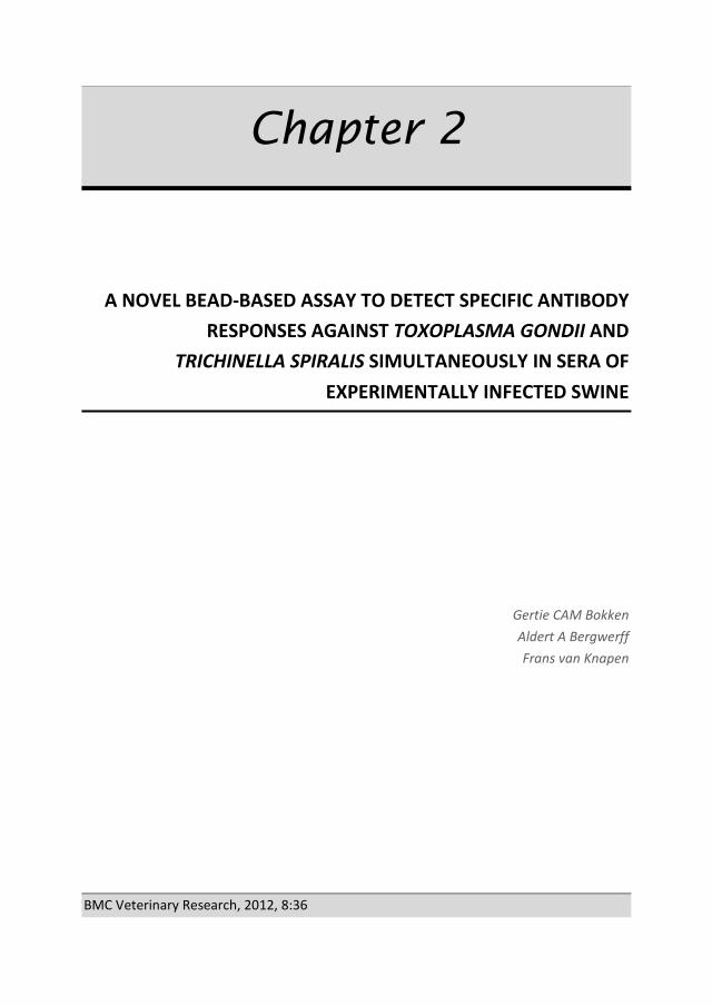
Chapter 2
A NOVEL BEAD-BASED ASSAY TO DETECT SPECIFIC ANTIBODY RESPONSES AGAINST TOXOPLASMA GONDII AND
TRICHINELLA SPIRALIS SIMULTANEOUSLY IN SERA OF EXPERIMENTALLY INFECTED SWINE
Gertie CAM Bokken Aldert A Bergwerff
Frans van Knapen
BMC Veterinary Research, 2012, 8:36

Chapter 2
46
ABSTRACT
Background
A novel, bead-based flow cytometric assay was developed for simultaneous determination of antibody responses against Toxoplasma gondii and Trichinella spiralis in pig serum. This high throughput screening assay could be an alternative for well-known indirect tests like ELISA. One of the advantages of a bead-based assay over ELISA is the possibility to determine multiple specific antibody responses per single sample run facilitated by a series of antigens coupled to identifiable bead-levels. Furthermore, inclusion of a non-coupled bead-level in the same run facilitates the determination of, and correction for non-specific binding. The performance of this bead-based assay was compared to one T. spiralis and three T. gondii ELISAs. For this purpose, sera from T. gondii and T. spiralis experimentally infected pigs were used. With the experimental infection status as gold standard, the area under the curve, Youden Index, sensitivity and specificity were determined through receiver operator curve analysis. Marginal homogeneity and inter-rater agreement between bead-based assay and ELISAs were evaluated using McNemar’s Test and Cohen’s kappa, respectively.
Results
Results indicated that the areas under the curve of the bead-based assay were 0.911 and 0.885 for T. gondii and T. spiralis, respectively, while that of the T. gondii ELISAs ranged between 0.837 and 0.930 and the T. spiralis ELISA was 0.879. Bead-based T. gondii assay had a sensitivity of 86% and specificity of 96%, while the ELISAs ranged between 64-84% and 93-99%, respectively. The bead-based T. spiralis assay had a sensitivity of 68% and specificity of 100% while the ELISA scored 72% and 95%, respectively. Marginal homogeneity was found between the T. gondii bead-based test and one of the T. gondii ELISAs. Moreover, in this test combination and between T. spiralis bead-based assay and respective ELISA, an excellent inter-rater agreement was found. When results of samples before expected seroconversion were removed from evaluation, notably higher test specifications were found.
Conclusions
This new bead-based test, which detects T. gondii and T. spiralis antibodies simultaneously within each sample, can replace two indirect tests for the determination of respective antibodies separately, while performing equally well or better.

A novel bead-based assay to detect specific antibody responses against Toxoplasma gondii and Trichinella spiralis simultaneously in sera of experimentally infected swine
47
BACKGROUND
Trichinella spiralis and Toxoplasma gondii are well known zoonoses which can pass from pigs to humans by consumption of raw or undercooked infected pork. In humans, most cases of T. gondii and T. spiralis infections go undetected; however, some cases can lead to mild disease. Other cases of trichinellosis can be very severe and may lead to myocarditis, encephalitis or pneumonia. Post-natal acquired toxoplasmosis can incidentally lead to encephalitis and necrotizing retinochoroiditis, while congenital transmitted toxoplasmosis can lead to mental retardation, convulsions, spasticity, cerebral palsy, deafness and severely impaired vision in the offspring. In rare occasions, both these infections can lead to death.
These days, in Europe, trichinellosis is rarely reported in association with the consumption of pork from conventionally raised pigs (1). An EU regulation (2) directs inspection of T. spiralis in each pig carcass at slaughter by direct parasitological methods. This regulation also states that serological tests may be implemented as a supplement for monitoring purposes.
Unlike T. spiralis, no such regulations exist for T. gondii, although the prevalence of this parasite in pigs is higher and health consequences of toxoplasmosis can be, like those of trichinellosis, rather serious. For example, in a Dutch survey in 2004, T. gondii infection was found in 2.6% of the studied pigs (3), while in that year none of the over 13 million slaughtered pigs were found T. spiralis positive (4).
Consumption of raw or undercooked T. gondii infected pork may cause toxoplasmosis in humans. Obviously, determination of the T. gondii status of the meat producing pigs, subsequently followed by precautionary methods, like freezing of pork to kill the parasite or altogether removal of this meat from the food chain, could contribute to fewer infections in humans. A Dutch study which assessed the epidemiology and impact of, amongst others, T. gondii infections in humans indicated that this parasite is one of the major contributors of disease through zoonotic transmission (5). Similarly, an American study indicated that T. gondii in pork ranked second on the list of the zoonotic micro-organisms with the greatest impact on annual disease burden in that country (6) and was only surpassed by Campylobacter in chicken. In a scientifically opinion to the European Food Safety Authority (EFSA) it is recommended that standardized methods should be used on T. gondii pre-harvest monitoring of, amongst others, pigs (7).
Like T. spiralis, the T. gondii infection status of animals can be examined by serological tests in order to produce T. gondii controlled pork. Testing serum samples of finisher pigs

Chapter 2
48
requires an automated and easy to perform test method with a high sensitivity (Se) and specificity (Sp). Enzyme-linked immunosorbent assays (ELISAs) are such test methods which are commonly used.
Bead-based assays (BBA) are a new dimension in the determination of specific antibody responses. The test is performed on beads which are available in different sizes and levels. During flow cytometric analysis individual beads are distinguishable by size and intrinsic fluorescence intensity level. The bead surface is carboxylate modified, which allows covalent coupling of protein. The great advantage of these tests over ELISA is the possibility of simultaneous detection of specific responses against multiple antigens per single serum sample. More specifically, by individual coupling of antigens to specific bead levels, and combination of these bead levels per test sample, a multitude of specific responses can be determined simultaneously per sample. Furthermore, by the use of a non-coupled bead, non-specific binding (NSB) can be monitored and corrected for. The use of T. gondii and T. spiralis antigens on two bead levels in a combined bead-based test to determine the serological status of swine would provide a new innovative assay which could be used as an alternative to ELISA in a T. gondii and T. spiralis monitoring system.
In this report, the specifications of a bead-based array test, with combined T. gondii and T. spiralis antigen bead levels to determine specific antibodies in serum of experimentally infected swine, are evaluated and compared to commercial and non-commercial ELISAs.
METHODS
Porcine sera
Experimental infection sera
Swine serum samples originated from an experimental co-infection of pigs with T. gondii and T. spiralis (8). Before infection, animals used in the experiment were assumed T. gondii and T. spiralis free on basis of the post-partum determined negative serological status of sows which gave birth to these animals (8). Briefly, eight to nine week old animals had been singly (T. gondii n=8, T. spiralis n=10), simultaneously (n=10), or successively (T. gondii/T. spiralis n=9, T. spiralis/T. gondii n=10) orally inoculated with either 2,700 or 2,000 T. gondii tissue cysts (strain DX) and/or 5,000 T. spiralis muscle larvae (strain ISS 14) per pig. Because two animals of the T. spiralis/T. gondii inoculated group did not seem to be infected, they were excluded from the experiment (8). A total of 444 serum samples were collected in series at 0, 5, 12, 19, 26, 33, 40, 47 and 54 days post

A novel bead-based assay to detect specific antibody responses against Toxoplasma gondii and Trichinella spiralis simultaneously in sera of experimentally infected swine
49
infection (p.i.) from 45 pigs and four additional non-inoculated animals which served as negative control animals. This animal study, under number DEC 2008.III.03.023, was reviewed and approved by the local animal ethics committee according to the recommendations of the EU directive 86/609/EEC. Numbers of animals and their suffering were minimized.
Negative field sera
Blood samples of conventional finisher pigs were collected for Salmonella baseline monitoring at the abattoir in 2007 by the Dutch Food and Consumer Product Safety Authority (nVWA). The blood was left at room temperature with a minimum of 2 hours to clot and subsequently centrifuged for 10 minutes at 1,100xg. Serum was drawn and dispensed in aliquots and kept at -20oC until further use.
Serum samples were analyzed by a commercially obtained T. gondii ELISA (ID Screen Toxoplasmosis Indirect, ID-VET, Montpellier, France; hereafter referred as E3-TOX). Serum samples remaining under the designated cut-off value of the ELISA were considered to originate from T. gondii infection negative pigs. Because during the sample period no pigs with T. spiralis infections were reported (9), all animals were considered T. spiralis infection negative.
Indirect assays
Sera from the experimentally infected animals were tested by the bead-based assay for T. gondii and T. spiralis antibodies simultaneously (hereafter referred to as BBA-TOX and BBA-TRI, respectively), by an RIVM in-house T. gondii ELISA (hereafter referred to as E1-TOX) and two commercially available T. gondii ELISA kits (Safepath, Carlsbath, CA, USA, hereafter referred to as E2-TOX) and E3-TOX, and by one T. spiralis ELISA (Safepath, hereafter referred to as E-TRI). All T. gondii indirect tests used an antigen based on T. gondii tachyzoites, of which E3-TOX utilized a recombinant tachyzoite surface protein (SAG-1) as antigen. The T. spiralis tests were based on ES antigens. Bead-based assays were run according to the specifications described in the section bead-based assay. Testing with the in-house ELISA (3) was described earlier (8) and included an intra-plate correction of E1-TOX data. All commercial ELISAs were run according to the specification of the kit providers. For E3-TOX, normalization of data was included.

Chapter 2
50
Bead-based assay
A bead-based assay was developed for simultaneous detection of specific antibodies which were captured by T. spiralis and T. gondii antigens on two different bead levels. NSB was recorded with reference beads, which is a bead level without coupled antigens. Each bead level was recognized via the emission of light with a unique intensity and wavelength of the beads intrinsic fluorescence. Specific and NSB in each individual serum sample were determined by the extrinsic response, which was generated by the emission of light by a fluorophore attached to the secondary antibody. Because NSB may vary between serum samples, the extrinsic response of reference beads was used to determine the non-specific response. To obtain a specific response per individual serum sample, this non-specific response was subtracted from the response of coupled beads. A BD Accuri flow cytometer was used for enumeration of micro-particles, excitation of fluorescent markers and measurement of emitted light from these markers.
Chemicals, materials and solutions
L4, L10 and L11 carboxylated Cyto-plexTM beads (cat# FM5CR04, FM5CR10 and FM5CR11, respectively) were purchased from Thermo Scientific (Waltham, MA, USA). T. gondii tachyzoite lysate, strain RH (cat#: R29123) was from Meridian Life Science Inc. (Saco, ME, USA). T. spiralis Excretory/Secretory antigen (ES) was obtained from Instituto Superiore Sanità (Rome, Italy). Microcentrifuge copolymer tubes (cat# 1415-2500) were acquired from Star Lab GmbH (Ahrensberg, Germany). N-hydroxysulfosuccinimide sodium salt (sNHS), N-(3-dimethylaminopropyl)-N′-ethylcarbodiimide hydrochloride (EDC; cat#: 03449) and 2-(N-morpholino)ethanesulfonic acid hydrate (MES; cat#: M8250) were bought from Sigma-Aldrich Chemie B.V. (Zwijndrecht, the Netherlands). A 45 mM MES buffer was prepared and adjusted to pH 6.0 with sodium hydroxide. PBS at pH 7.2 consisted of 0.01 M sodium chloride (NaCl, Merck KGaA, Darmstadt, Germany), 1 mM di-sodium hydrogen phosphate (Merck) and 3 mM potassium dihydrogen phosphate (Merck). Water was of milliQ quality. Storage buffer and HNT-PBS solution were provided by RnAssays (Utrecht, the Netherlands). The 0.45 µm filter plates (cat#: MSHVN4550) were from Millipore (Amsterdam, the Netherlands). Goat anti-swine secondary antibody conjugated with fluorescent DyLight 488 was purchased from Jackson Immuno Research (West Grove, PA, USA).

A novel bead-based assay to detect specific antibody responses against Toxoplasma gondii and Trichinella spiralis simultaneously in sera of experimentally infected swine
51
Bead coupling procedure
T. gondii tachyzoite lysate and T. spiralis Excretory/Secretory antigen (ES) were coupled to carboxylated beads through an amine coupling procedure. Briefly, an equivalent of 1.4x10⁸ carboxylated beads of L10 and L11 were transferred to two 1.5 ml copolymer tubes. The beads were washed by three repeats of following steps: a 3 minutes centrifugation at 9,000xg, removal of the supernatant, addition of 1 ml water per tube and resuspension of the beads on a vortex. After the third removal of supernatant, beads from both tubes were resuspended in 1.1 ml solution consisting of 12.5 mg sNHS and 12.5 mg EDC in MES buffer. This suspension was incubated for 20 minutes at room temperature on a gyro rocker at 70 rpm. Beads were washed 2 more times with 500 µl water as described above and after removal of the supernatant, 50 µg of T. gondii lysate and 10 µg T. spiralis ES dissolved in 200 µl PBS pH 7.4 were added to the activated L10 and L11 beads, respectively. Resuspended beads were left to incubate for 2 hours on a gyro rocker at 70 rpm, washed and stored in a storage buffer. A non-coupled L4 reference bead suspension was produced with the same protocol with exception of the protein incubation step which was substituted by PBS incubation. This L4 bead is referred to as the reference bead.
Assay procedure
Two 0.45 µm filter plates were soaked with 150 µl of a 0.2 µm filtered solution of HNT-PBS, subsequently incubated for five minutes at ambient temperature, and emptied by vacuum filtration. Serum samples were diluted 1:50 in HNT-PBS, transferred to a soaked and aspirated 0.45 µm filter plate, filtered with the use of the vacuum manifold and collected in an empty 96-wells plate. Thereafter, in another soaked filter plate, a quantity of approximately 5x10⁵ T. gondii and T. spiralis antigen coupled beads and reference beads were suspended in 50 µl of HNT-PBS per well. Subsequently, one equivalent volume of filtered diluted sera was mixed and incubated with the bead-mix per well for 15 minutes on an orbital shaker (1,050 rpm). Beads were washed with 200 µl HNT-PBS by aspiration and additionally incubated with 100 µl 1:300 in HNT-PBS diluted fluorescent secondary antibody for 15 minutes. Finally, beads were washed once more and suspended in 100 µl HNT-PBS. Due to light sensitivity of beads and fluorescent reporter the filter plates were protected from light during incubation steps.
Internal and external fluorescent detection
A total of 600 beads per serum sample were analyzed for the intrinsic bead label on the FL4 channel, and extrinsic fluorescence reporter label on the FL1 channel using a BD Accuri

Chapter 2
52
C6 flow cytometer (BD Accuri Cytometers, Inc. Ann Arbor, MI, USA). The detector was equipped with a CSampler liquid handler (BD Accuri) and operated through CFlow software (version 1.0.243.1, BD Accuri). Beads were transported at a flow rate of 35 µl/min. The emission of the intrinsic fluorescence of the three bead levels, measured by the FL4 filter at 675 nm, was used to distinguish the T. gondii (TOX), T. spiralis (TRI) and reference (REF) beads. The median extrinsic fluorescence intensity (MFI) of the secondary antibody per bead level was determined by measuring the emission via the FL1 filter at 530 nm.
Correction for non-specific binding
Reference beads were used to indicate the measure of NSB in the test. Differences in NSB on uncoupled or antigen coupled bead levels may be expected due to differences in affinity of beads for non-specific antibodies caused by the molecular structure of the antigen, its orientation and concentration on the bead surface. Therefore, to estimate the NSB on T. gondii and T. spiralis bead levels from the response of a reference bead, a correction factor was calculated by testing 932 T. gondii and 13 extra T. spiralis negative swine sera (section Negative field sera) in the bead-based assay. With the use of least square regression, linear relations, expressed with the formulae y = slope*x + intercept, the relation between responses of the reference beads (x value) and T. gondii and T. spiralis bead responses (y values) were calculated in SPSS 16.0 for Windows (SPSS Inc., Chicago, IL, USA). Because the residuals of the linear relation between responses of T. gondii and T. spiralis bead levels and reference bead responses were not normally distributed, all responses were log transformed.
Normalization of responses
To compare results between 96-wells plates, serum samples responses were normalized. The percentage of normalized responses (%NR) was calculated as a percentage of sample responses (MFIS) of a positive control response (MFIPC), which was present in quadruplicate on each plate, after subtraction of NSB (sections Correction for non-specific binding and Results and Discussion).
%NR T= ((MFIS - NSBS)/(MFIPC- NSBPC)) T *100% (1)
where subscript T represents T. gondii or T. spiralis in the considered case.

A novel bead-based assay to detect specific antibody responses against Toxoplasma gondii and Trichinella spiralis simultaneously in sera of experimentally infected swine
53
Statistical analysis
All statistical evaluations were performed with SPSS.
To specify the performance of the bead-based assay, expressed in area under the curve (AUC), receiving operator characteristic (ROC) calculations were performed using the experimental infection status as gold standard. Analysis was performed on the %NR of the BBA and E3-TOX, and OD450nmof the other ELISAs. ROC calculations were also performed on limited sets of serum from the experimental infection. These sets consisted of all samples minus serum samples drawn 5 days after inoculation with T. gondii (n=408) and all samples minus serum samples drawn 5, 12 and 19 days after inoculation with T. spiralis (n=360), for the T. gondii and T. spiralis indirect tests, respectively. To further specify the tests, diagnostic Sensitivity (Se), Specificity (Sp) and cut-off values at maximum Youden Index were determined from ROC calculations (10).
To assess the agreement between tests, the marginal homogeneity of paired proportions (11) were tested by McNemar’s in a 2x2 contingency table. Furthermore, inter-rater agreement was calculated using Cohen’s Kappa. For this, serum responses of all tests were labelled 0 (negative), when they were below the cut-off value or 1 (positive) when they were equal or above cut-off value. These dichotomized outcomes where then evaluated against the dichotomized outcomes of the other tests. Kappa values between 0.40 - 0.59, 0.60 – 0.79 and ≥ 0.80 are interpreted as moderate, substantial and excellent agreement, respectively (11).
The apparent prevalence (AP) is the proportion of the population which tests positive in the test, which is a measure of true prevalence (TP) and the capability of the test to predict true positives and negatives, and it was calculated as (12):
AP = Se * TP + (1-Sp) * (1-TP) (2)
where TP is the proportion of actual infected animals which was calculated by:
TP = n/N (3)
where n is the number of sera which originate from inoculated animals, and N is the total number of sera.
RESULTS AND DISCUSSION
Results from T. gondii and T. spiralis negative field samples, negative control serum of the experimental infection and secondary antibody binding alone (Table 1) showed that the height of NSB responses is foremost dependent of the presence of serum. Observing that

Chapter 2
54
negative sera due to NSB can reach the same or higher responses as for example positive control serum of 250,000 MFI on T. gondii and 500,000 MFI on T. spiralis beads (data not presented), it is concluded that correction for NSB is necessary to prevent false positive results.
The results of T. gondii and T. spiralis negative field sera illustrate that the response of the reference beads could not directly be used as a measure for non-specific binding. The log linear relations between responses of reference beads and T. gondii or T. spiralis beads are depicted in Fig. 1 and 2, and the relations were expressed as:
log NSB (TOX) = log MFIREF *
0.404 + 2.818 (4)
log NSB (TRI) = log MFIREF *
0.646 + 1.863 (5)
Table 1: Non-specific binding responses of T. gondii and T. spiralis negative serum sets and conjugate alone
Minimum, maximum and mean response signals of reference (REF), T. gondii (TOX) and T. spiralis (TRI) coupled beads after incubation of buffer, negative field sera (without positive responders in T. gondii ELISA) and sera of the negative control animals of the experimental infection. n, number of tested sera; SE, standard error.
The slopes of the two regression lines indicate that T. gondii beads are subject to less NSB as compared to T. spiralis beads. This finding may be explained by, for example, variable concentration of antigens on the bead, differences in antigen molecule structures and orientation on the bead surface and/or the affinity between non-specific antibodies and the unoccupied bead surface or coupled antigens. A variety in antigen composition between biologically produced batches can therefore be of importance in relation to NSB. To test whether the correction factor to calculate bead correlated NSB is stable between batches of antigen, further evaluation is necessary. Subtraction of uncorrected responses of reference beads, according to formula 1, would lead to an underestimation or overestimation of NSB for responses below and above 53,480 and 183,110 for T. gondii and T. spiralis, respectively.
Serum Bead identification
n Min. response (MFI)
Max. response (MFI)
Mean response (MFI)
SE
Buffer REF 98 300 600 500 5 TOX 98 3,900 4,700 4,300 19 TRI 98 3,200 3,900 3,500 17
(T. gondii or T. spiralis) negative field sera
REF 947 400 491,500 53,700 2,200 TOX 932 4,200 269,200 47,900 1,100 TRI 945 3,400 505,500 75,300 2,300
Experimental infection negative control sera
REF 36 1,100 1,900 4,100 600 TOX 36 4,900 45,500 21,500 1,900 TRI 36 4,200 34,800 10,400 1,000

A novel bead-based assay to detect specific antibody responses against Toxoplasma gondii and Trichinella spiralis simultaneously in sera of experimentally infected swine
55
Results of ROC calculations, presented in Table 2, showed that the AUC, a measure of agreement between specific responses and the experimental infection status of the animals, of all indirect tests ranged between 0.837 and 0.930 for T. gondii and 0.855 and 0.879 for T. spiralis. These values indicate that there is a good relation between the responses of all indirect tests and the infection status of the animals.
A perfect test is a test in which the responses correspond 100%, i.e. an AUC of 1.000, with the values of the test to which it is compared. The imperfect AUC values (< 1.000) found in our study (Table 3) can partly be explained by a late immunological development of
Figure 2: Estimation of T. spiralis non-specific binding correction factor. Log transformed responses of T. spiralis negative swine field sera on non-coupled beads (x-axis) versus T. spiralis coupled beads (y-axis). Linear regression line (log y = 0.646*log x + 1.863), 95%CI lines and the linear R2 are presented.
Figure 1: Estimation of T. gondii non-specific binding correction factor Log transformed responses of T. gondii negative swine field sera on non-coupled beads (x-axis) versus T. gondii coupled beads (y-axis). Linear regression line (log y = 0.404* log x + 2.818), 95%CI lines and the linear R2 are presented.

Chapter 2
56
antibodies, which is associated to the time course of parasite antigen expression and the immune response of infected animals. Evidence from earlier studies showed that muscle larvae, depending on the infection dose, can be found in pork by digestion or trichinoscopy as early as 17 days p.i. (13). Other studies showed that T. spiralis ES could be measured within the developing muscle larvae and its cuticular surface as early as 14 days p.i. (14) and in the surrounding tissue around 15 days p.i. (15). Consequently, the response time, i.e. the time of development of antibodies against the antigen used in the indirect T. spiralis assays, is affected by this late production. Porcine IgG antibodies against ES are developed approximately 3 to 4 weeks after infection with 5,000 muscle larvae (16, 17). Porcine IgG antibodies against T. gondii tachyzoites are produced much earlier in time and can be detectable after one to two weeks of infection (18). In our study, the sera used for ROC calculations originated from animals which were collected on a weekly basis (8). Samples drawn 5 days after T. gondii inoculation and 5, 12 and 19 days after T. spiralis inoculation would produce a false negative result when compared to the experimental infection status, resulting in lower AUC values. Calculations of ROC curves without these sera resulted in notably higher AUC values of 0.995, 0.999, 0.998 and 0.999 for BBA-TOX, E3-TOX, BBA-TRI and E-TRI, respectively (Fig. 3 and 4).
According to data of test performances and inter test agreement, presented in Table 3, the T. gondii and T. spiralis bead assays agree excellently with their respective highest scoring tests, i.e. with the E-TOX3 and E-TRI, respectively. Marginal homogeneity by McNemars test, a test which determines the equality between positive and negative test proportions of one test compared to the other, indicated that there is a balance between BBA-TOX and E-TOX3.
The potential use of the bead based test is prevention of T. gondii and/or T. spiralis infections in pork to enter the human food chain. Nonetheless, direct parasitological testing, like T. spiralis artificial digestion, are more reliable methods to test for present infection in meat. Existing T. gondii direct tests are either laborious, e.g. due to the need of pathogen extraction in PCR (19), or are undesirable because of utilization of experimental animals, like in cat and mice bioassays (20), to determine the infectious status of meat. In the case of T. gondii infections, serological testing is the next best option to perform on large scale. Unfortunately, due to the time window between infection and development of specific antibody responses, serological tests are less reliable for detection on individual scale; however, they can be used for monitoring purposes on herd level (21).

A novel bead-based assay to detect specific antibody responses against Toxoplasma gondii and Trichinella spiralis simultaneously in sera of experimentally infected swine
57
Figure 3: ROC curves of T. gondii assays calculated with a limited serum set from experimental infection. ROC analysis of a T. gondii bead-based assay and three ELISAs using responses from a set of serum samples, consisting of sera from experimentally infected pigs minus sera drawn on day 5 after inoculation with T. gondii, against their infection status. AUC, area under the curve; 95%CI, interval of AUC at 95% confidence; BBA-TOX, T. gondii antigen coupled bead-based assay; E1-TOX, RIVM in-house T. gondii ELISA, E2-TOX: Safepath T. gondii ELISA, E3-TOX, ID-VET T. gondii ELISA.
Figure 4: ROC curves of T. spiralis assays calculated with a limited serum set from experimental infection. ROC analysis of a T. spiralis bead-based assay and ELISA using responses from a set of serum samples, consisting of sera from experimentally infected pigs minus sera drawn on days 5, 12 and 19 after inoculation with T. spiralis, against their infection status. AUC, area under the curve; 95%CI, interval of AUC at 95% confidence; BBA-TRI, T. spiralis antigen coupled bead-based assay; E-TRI: Safepath T. gondii ELISA
To prevent human T. gondii and T. spiralis infections through consumption of infected pork by serological monitoring of pig herds, a high sensitivity of 99% (22), and an approximation of AP to TP (Table 4) is desired. None of the assays used for this paper met this requirement. However, when ROC calculations were restricted to serum samples in which antibody responses were to be expected, as was described above, the sensitivity was 97% and 99% for BBA-TOX and BBA-TRI, respectively (data not presented). Subsequent calculations for true and apparent prevalence resulted in an overall T. gondii

Chapter 2
58
TP of 52.7% and AP’s of 51.5% and 53.3% for BBA and ELISA, respectively. The overall TP of T. spiralis was 44.4% while the APs were 44.1% and 44.3% for BBA and ELISA, respectively (data not presented). These data would indicate that the combined bead-based assay is applicable for serological monitoring purposes.
Table 2: Inter-test agreement between assays calculated by McNemar’s and Cohen’s Kappa analysis.
McNemar’s test Yates correction
Cohen’s Kappa
Test comparison P κ * 95% CI BBA-TOX vs. E1-TOX <0.001 0.676 0.607-0.744 BBA-TOX vs. E2-TOX 0.01 0.802 0.746-0.857 BBA-TOX vs. E3-TOX 0.06 0.932 0.900-0.966 E1-TOX vs. E2-TOX <0.001 0.752 0.690-0.814 E1-TOX vs. E3-TOX <0.001 0.723 0.658-0.787 E2-TOX vs. E3-TOX 0.233 0.797 0.740-0.853 BBA-TRI vs. E-TRI <0.001 0.880 0.835-0.925 Inter-test agreement calculations comparing between dichotomized results of tests. P, probability; κ, Cohen’s Kappa value; 95% CI, interval at a 95% confidence; BBA-TOX, T. gondii antigen coupled bead-based assay; E1-TOX, RIVM in-house T. gondii ELISA; E2-TOX, Safepath T. gondii ELISA; E3-TOX, ID-VET T. gondii ELISA; BBA-TRI, T. spiralis antigen coupled bead-based assay and E-TRI, Safepath T. spiralis ELISA. * indicates that all values were statistically significant (P<0.001). Table 3: Comparison of true and apparent prevalence’s of pig sera between indirect assays. Test True prevalence Apparent prevalence BBA-TOX
56.6%
50.1% E1-TOX 38.3% E2-TOX 45.9% E3-TOX 48.0% BBA-TRI
58.6% 39.8%
E-TRI 44.2% BBA-TOX, T. gondii antigen coupled bead-based assay; E1-TOX, RIVM in-house T. gondii ELISA; E2-TOX, Safepath T. gondii ELISA; E3-TOX, ID-VET T. gondii ELISA; BBA-TRI, T. spiralis antigen coupled bead-based assay and E-TRI, Safepath T. spiralis ELISA
Although we compared our new T. gondii and T. spiralis bead test with a limited selection of available ELISAs, the test specifications and agreement between tests examined in this study indicate that the combined bead test equals or is superior to other tests. However, all calculations have been based upon tests using serum samples of experimentally infected pigs, which were exposed to high doses of parasites. Conventionally raised animals are likely to be infected by lower doses of parasites, and sero-conversion may be detected later in time (16-18). Therefore, to determine the applicability of this bead-based test for the use of indirect detection of infection, it is advisable to further evaluate the test by the use of serum samples of naturally infected pigs.

A novel bead-based assay to detect specific antibody responses against Toxoplasma gondii and Trichinella spiralis simultaneously in sera of experimentally infected swine
59
CONCLUSIONS
In conclusion, this initial evaluation study of a novel bead-based assay capable of a simultaneous detection of serological antibodies against T. gondii and T. spiralis antigens indicates that the test results correspond very well to the infection status of the animals, and, furthermore, there is a substantial to excellent agreement with other indirect tests. In order to estimate the applicability of this test for purposes of serological monitoring, further testing of sera from naturally infected animals is required.
ACKNOWLEDGEMENTS
This work was partly supported through material gifts and a financial grant from RNassays, Utrecht, The Netherlands. The authors would like to thank Marieke Opsteegh of the RIVM for her analysis of serum samples by the in-house T. gondii ELISA. Furthermore, we are grateful for the help of Sevda Atanasova and Wouter Harink of RnAssays in optimization of the bead assay and flow cytometric analysis. Lastly, we would like to thank Giuseppe Cacciatore for his input of expertise in assay processes and statistical calculations.
AUTHORS' CONTRIBUTIONS
GCAMB helped in conceiving of the study, developed and optimized the bead-based test method, analyzed serological material in the test methods, statistically interpreted the data and wrote the manuscript. AAB and FvK both conceived of the study, participated in its design and coordination, contributed their expertise and helped to draft the manuscript. All authors read and approved the final manuscript.

Chapter 2
60
REFERENCES
1. Anonymous. European Food Safety Authority; The European Union Summary Report on Trends and Sources of Zoonoses, Zoonotic Agents and Food-borne Outbreaks in 2009. EFSA Journal. 2011;9(3): 2090:1-378.
2. Council European Parliament. Commission Regulation 2075/2005 of 5 December 2005 Laying Down Specific Rules on Official Controls for Trichinella in Meat. Official Journal of the European Union [Internet]. 2005 22.12.2005 [cited 1 December 2010];L 338(60).
3. Giessen Jvd, Fonville M, Bouwknegt M, Langelaar M, Vollema A. Seroprevalence of Trichinella spiralis and Toxoplasma gondii in pigs from different housing systems in The Netherlands. Vet Parasitol. 2007;148(3/4):371-4.
4. Anonymous. The Community Summary Report on Trends and Sources of Zoonoses, Zoonotic Agents and Antimicrobial resistance in the European Union in 2004. EFSA Journal [Internet] 12 July 2006;2005(310). Available from: http://www.efsa.europa.eu/en/efsajournal/pub/310ar.htm.
5. Havelaar AH, van Rosse F, Bucura C, Toetenel MA, Haagsma JA, Kurowicka D, et al. Prioritizing emerging zoonoses in the Netherlands. PLoS ONE. 2010;5(11).
6. Batz M, Hoffmann S, Morris J Jr. Ranking the risks: The 10 pathogen-food combinations with the greatest burden on public health [Internet].: University of Florida, Emerging Pathogens Institute; 2011 [updated 2011; ]. Available from: http://www.rwjf.org/files/research/72267report.pdf.
7. Andreoletti O, Budka H, Buncic S, Colin P, Collins JD, De Koeijer A, et al. Scientific Opinion of the Panel on Biological Hazards on a request from EFSA on Surveillance and monitoring of Toxoplasma in humans, foods and animals. The EFSA Journal (2007) [Internet]. 2007 2011;-(583):03.10.2011,1-64. Available from: http://www.rwjf.org/files/research/72267report.pdf.
8. Bokken GCAM, van Eerden E, Opsteegh M, Augustijn M, Graat EAM, Franssen FFJ, et al. Specific serum antibody responses following a Toxoplasma gondii and Trichinella spiralis co-infection in swine. Vet Parasitol. 2012;184(2-4):126-32.
9. Anonymous. The Community Summary Report on Trends and Sources of Zoonoses, Zoonotic Agents and Antimicrobial resistance in the European Union in 2007. EFSA Journal [Internet] 21 april 2009;2009(223). Available from: http://www.efsa.europa.eu/en/efsajournal/doc/223r.pdf.
10. Greiner M, Pfeiffer D, Smith RD. Principles and practical application of the receiver-operating characteristic analysis for diagnostic tests. Prev Vet Med. 2000 5/30;45(1-2):23-41.
11. Landis JR, Koch GG. The measurement of observer agreement for categorical data. Biometrics. 1977 Mar.;33(1):pp. 159-174.
12. Henken AM, Graat EAM, Casal J. Measurement of disease frequency. In: Noordhuizen, J.P.T.M., Frankena, K., Thrusfield, M.V., Graat, E.A.M, editor. Application of Quantitative Methods in Veterinary Epidemiology. second revised reprint ed. Wageningen: Wageningen Pers; 2001. p. 63,64 till 96.
13. van Knapen F, Franchimont JH, Ruitenberg EJ, André P, Baldelli B, Gibson TE, et al. Comparison of four methods for early detection of experimental Trichinella spiralis infections in pigs. Vet Parasitol. 1981 12;9(2):117-23.

A novel bead-based assay to detect specific antibody responses against Toxoplasma gondii and Trichinella spiralis simultaneously in sera of experimentally infected swine
61
14. Despommier DD, Gold AM, Buck SW, Capo V, Silberstein D. Trichinella spiralis: Secreted antigen of the infective L1 larva localizes to the cytoplasm and nucleoplasm of infected host cells. Exp Parasitol. 1990 7;71(1):27-38.
15. Li CK, Chung YY, Ko RC. The distribution of excretory/secretory antigens during the muscle phase of Trichinella spiralis and T. pseudospiralis infections. Parasitol Res. 1999;85(12):993-8.
16. van der Leek ML, Dame JB, Adams CL, Gillis KD, Littell RC. Evaluation of an enzyme-linked immunosorbent assay for diagnosis of trichinellosis in swine. Am J Vet Res. 1992;53(6):877-82.
17. Gottstein B, Pozio E, Nockler K. Epidemiology, diagnosis, treatment, and control of trichinellosis. Clin Microbiol Rev. 2009;22(1):127-45.
18. Lind P, Haugegaard J, Wingstrand A, Henriksen SA. The time course of the specific antibody response by various ELISAs in pigs experimentally infected with Toxoplasma gondii. Vet Parasitol. 1997;71(1):1-15.
19. Opsteegh M, Langelaar M, Sprong H, Hartog Ld, Craeye Sd, Bokken G, et al. Direct detection and genotyping of Toxoplasma gondii in meat samples using magnetic capture and PCR. Int J Food Microbiol. 2010;139(3):193-201.
20. Dubey JP, Hill DE, Jones JL, Hightower AW, Kirkland E, Roberts JM, et al. Prevalence of viable Toxoplasma gondii in beef, chicken, and pork from retail meat stores in the United States: Risk assessment to consumers. J Parasitol. 2005;91(5):1082-93.
21. Ruitenberg EJ, van Knapen F, Elgersma A. Surveillance in swine by immunodiagnostic methods. In: Campbell WC, editor. Trichinella and Trichinosis. Plenum Press; 1983. p. 529-50.
22. van Knapen F. Control of trichinellosis by inspection and farm management practices. Vet Parasitol. 2000 12/1;93(3-4):385-92.


Chapter 3
REFINED ASSAY PARAMETERS OF A BEAD-BASED DETECTION METHOD FOR TRICHINELLA SPIRALIS INFECTIONS IN PIGS
Gertie CAM Bokken Aldert A Bergwerff
Frans van Knapen
Manuscript in preparation

Chapter 3
64
ABSTRACT
Recently, an anti-Trichinella and anti-Toxoplasma gondii antibody-detecting bead-based assay (BBA) was presented. To estimate the usefulness of this assay in Trichinella surveillance programs, the test needed further evaluation. In this study, the accuracy of the BBA was further evaluated to assess the specificity by the use of 906 non-infected conventional farmed pigs. Furthermore, the value of sensitivity was reassessed using sera from 41 experimentally infected animals. Receiver operating characteristics analysis showed an area under the curve of 0.999. Using the cut-point value at the maximal Youden index returned a sensitivity of 1.00 (41/41) (95%CI 0.91-1.00) and specificity of 0.98 (886/906) (95%CI 0.97-0.99). Maximization of specificity to 1.00 (906/906) (95%CI 0.996-1.000) reduces sensitivity to 0.85 (35/41) (95%CI 0.72-0.93). The assay may fit in a surveillance system securing freedom of disease in a herd or on a regional level.

Refined assay parameters of a bead-based detection method for Trichinella spiralis infections in pigs
65
SHORT COMMUNICATION
Trichinella spiralis (T. spiralis) is a parasitic roundworm, which can be transmitted to humans through the consumption of infected pork which was insufficiently processed to kill the parasite with treatments like freezing, heating or salting. Infection by the parasite can lead to severe disease in humans. In the European Union, the standardized procedure to control Trichinella infections in pork is achieved by the detection of the presence of this parasite in pigs at slaughter via a pooled sample digestion method. Detection of parasite is followed by precautionary measures like freezing of meat (1). However, this detection method is laborious and the sensitivity of the test is not sufficient to find animals with a low infection burden (2). Indirect methods, like serological tests, are easy to perform, but lack perfect sensitivity and specificity.
More than a decade ago it was concluded that serological tests cannot replace a direct test as a diagnostic tool to determine Trichinella infection on individual animal level (3). However, they can be incorporated into surveillance programs which demonstrate the absence of infection on a herd or on a population level. For such surveys, diverse approaches have been constructed which use imperfect tests to calculate the probability of disease absence in populations under predefined confidence and power (4-6).
Recently, we developed a serological bead-based test, which can concurrently determine T. gondii and T. spiralis serum antibody responses in a single sample (7). In order to estimate its usability as a monitoring tool in Trichinella surveillance programs, it is essential to know the bead test accuracy. The initial study showed that the combined Trichinella multi-analyte test had a diagnostic test sensitivity and specificity of 0.68 and 1.00, respectively (7). However, these performance data were determined by the use of sera from experimentally infected and control animals, exclusively (8). These values may therefore not be accurate when testing field samples.
In this study we further evaluate the specificity of the new test by the use of sera of conventional non-infected finisher pigs. Because we were not able to obtain samples from naturally infected animals, we reevaluated the sensitivity by the use of sera from experimentally infected animals.
Sera from 41 experimentally T. spiralis infected animals were obtained from the Bundesinstitut für Risikobewertung, Berlin, Germany (9). All animals were considered infected because of a positive finding in trichinoscopy and/or digestion, tests with perfect specificity.

Chapter 3
66
Sera of 906 Dutch conventional finisher pigs were collected in 2007 at slaughter. Regulation EU 2075/2005 (1) stipulates the assessment of Trichinella infection in each carcass, and Directive 2003/99/EC (10) stipulates reporting of this information. In 2007, no animal was reported Trichinella infected in the Netherlands (11), therefore these animals are assumed to be Trichinella negative.
All sera were tested with the indirect bead-based assay (BBA) which included three bead sensors to monitor Trichinella, T. gondii and non-specific serum responses, respectively(7). In this study, only the responses of bead sensors detecting Trichinella and non-specific binding results were used. Production of beads, test execution, determination of Median Fluorescence Index (MFI) responses and subsequent calculation of percentage of normalized responses (%NR) were performed as described(7). Test accuracy, i.e. the sensitivity (Se) and specificity (Sp), was obtained at values of %NR cut-points by the use of receiver operator characteristics (ROC) calculations. Confidence intervals were calculated according to Wilsons method (12). All statistical evaluations were performed in SPSS (for Windows, version 20.0, IBM Corp., 2011). For each cut-point, the Youden index (Y), Y = Se + Sp – 1, was determined (13). At a specificity of 1.00, the corresponding sensitivity and cut-point were estimated as well.
The test results ranged from -13.4 to 57.6 NR% for the sera of non-infected animals and from 17.7 to 165.4 %NR for the sera of infected animals (Table 1). As depicted in figure 1, the %NR values of positive and negative sample sets partly overlapped.
ROC analysis of the %NR values gave an AUC of 0.999 (CI 0.997 – 1.001). At the maximized value of Y the cut-point value was 17.5 %NR (Table 1). At this cut-point all 41 infected animals scored positive, while 905 of 906 uninfected animals scored negative. Hence sensitivity was 1.00 and specificity was 0.98 (Table 1). Optimizing the specificity to 1.00 at cut-point 57.6%NR increased the number of false negatives to 5, subsequently decreasing the sensitivity 0.88.
Compared to the finding from our previous study, which indicated an AUC of 0.855, a sensitivity of 0.68 and a specificity of 1.00 (7), the present values of AUC and sensitivity are considerably higher while specificity decreases only slightly. Most likely, the higher values are caused by the use of a higher percentage of seroconverted infected animals in this study. Namely, 2/41 infected animals used in current study had not seroconverted (9), while 73/260 sera of Trichinella infected animals in our previous study had not seroconverted (data not presented). These numbers were based on results of ELISA’s both equipped with an Excretory/Secretory antigen.

Refined assay parameters of a bead-based detection method for Trichinella spiralis infections in pigs
67
Figure 1: Response frequency distribution of Trichinella negative and positive samples in bead based assay Table 1: A comparison of direct and normalized responses, cut-off and test accuracy values observed and estimated in two studies Study 2012a Current study
Para- meter
Bead sensor
Mean (range) Infectedb Non-infectedb Infectedc Non-infectedd
MFI ref
6,455 (617 – 40,419)
4,599 (928–30,897)
90,093 (6,042–435,530)
51,110 (403–475,651)
Tri 304,492 (5,056 – 789,238)
11,244 (4,154–48,822)
535,471 (91,830–989,853)
72,565 (3,517–436,272)
%NR 68.1 (-4.5 – 174.0)
-1.2 (-5.6–4.6)
104.6 (17.7–165.4)
0.9 (-13.4–57.6)
%NR cut-off Y
4.65% 17.5%
Se / Sp at Y 0.68 / 1.00 1.00 / 0.98 a, information collected from a study in 2012 (7); b, sera from infected and non-infected pigs from an experimental infection with T. spiralis (8); c, sera from T. spiralis experimental infected animals (9); d, sera from conventional Trichinella free Dutch finisher pigs. MFI, median fluorescence index; ref, reference bead sensor; Tri, Trichinella bead sensor; %NR, percentage of normalized sample response compared to normalized positive reference; %NR cut-off YI, %NR cut-off value obtained from receiver operator characteristic analysis at maximized Youden index; Se, sensitivity; Sp, specificity; YI, Youden index.
The difference between the cut-point values of both studies, i.e. 17.5%NR versus 4.65%NR found in the previous study is large, but can be explained by the different composition of the serum sets used in the two studies. Compared to the previous study, the non-infected sera in current study show substantial higher means and wider distributions of median fluorescence index (MFI) responses on both bead sensors (Table 1). This results in an almost equal mean %NR values of non-infected sera in both studies, although in current

Chapter 3
68
study, the distribution is wider. Furthermore, in current study, specificity is 1.00 at cut-point 17.5% and lower. The use of lower cut-points would only decrease sensitivity values which would not improve the Y value. Because the test accuracy values were estimated using composited animal populations, and these values tend to vary with the prevalence within the population (14), the value of specificity found in this study can be considered true for populations of conventionally housed pigs. Whereas, the use of experimental infected animal sera can only give an indication of the value of sensitivity in a population of these pigs.
For surveys which demonstrate the absence of infection in a population diverse approaches have been constructed which are based on the use of imperfect tests (4-6). For a practical realization of a Trichinella survey in a certain population the statistical methodology in combination with survey design and analysis incorporating sampling strategies, clustering of the infection, design prevalence and test accuracy are components which need to be defined. Currently, only the design prevalence, the acceptable level of Trichinella infections in a pig population, has been set to 0.0001% (1). This study provides information of test accuracy which can be used for the design of the survey.
The feasibility of the bead-based test in a freedom from disease approach to substitute the currently used artificial digestion is dependent of practicability of such a system and corresponding costs, which both are affected by test accuracy values. The advantage of the application of the bead based assay for such approach lies in its quick performance and low costs, and the potential extendibility with additional bead sensors sensing other infections, e.g. T. gondii (7).
CONFLICT OF INTEREST
There are no conflicts of interest
ACKNOWLEDGEMENTS
We acknowledge the RnAssays company for the use of their technical equipment and providing us sample material.

Refined assay parameters of a bead-based detection method for Trichinella spiralis infections in pigs
69
REFERENCES
1. Anonymous. Opinion of the Scientific Panel on Biological Hazards on the Risk assessment of a revised inspection of slaughter animals in areas with low prevalence of Trichinella. EFSA Journal [Internet]. 2005;200:1-41.
2. Forbes LB, Gajadhar AA. A validated Trichinella digestion assay and an associated sampling and quality assurance system for use in testing pork and horse meat. J Food Prot. 1999;62(11):1308-13.
3. van Knapen F. Control of trichinellosis by inspection and farm management practices. Vet Parasitol. 2000 12/1;93(3-4):385-92.
4. Cameron AR. A new probability formula for surveys to substantiate freedom from disease. Prev Vet Med. 1998;34(1):1-17.
5. Cameron AR. Two-stage sampling in surveys to substantiate freedom from disease. Prev Vet Med. 1998;34(1):19-30.
6. Martin PAJ. Demonstrating freedom from disease using multiple complex data sources. 1: A new methodology based on scenario trees. Prev Vet Med. 2007;79(2-4):71-97.
7. Bokken GCAM, Bergwerff A, van Knapen F. A novel bead-based assay to detect specific antibody responses against Toxoplasma gondii and Trichinella spiralis simultaneously in sera of experimentally infected swine. BMC Vet Res. 2012;8:36-.
8. Bokken GCAM, van Eerden E, Opsteegh M, Augustijn M, Graat EAM, Franssen FFJ, et al. Specific serum antibody responses following a Toxoplasma gondii and Trichinella spiralis co-infection in swine. Vet Parasitol. 2012;184(2-4):126-32.
9. Nöckler K. Diagnosis of trichinellosis in living pigs using indirect ELISA | Intravitale Diagnostik der Trichinellose beim Schwein mit dem indirekten ELISA. Berliner und Münchener tierärztliche Wochenschrift. 1995;108(5):167-74.
10. Council European Parliament. Directive 2003/99/EC of the European Parliament and of the Council of 17 November 2003 on the monitoring of zoonoses and zoonotic agents, amending Council Decision 90/424/EEC and repealing Council Directive 92/117/EEC. Official Journal of the European Union [Internet]. 2003 22.12.2005 [cited 1 December 2010];L 325(31).
11. Anonymous. The Community Summary Report on Trends and Sources of Zoonoses and Zoonotic Agents in the European Union in 2007. EFSA Journal. 2009;223:1-312.
12. Altman DA, Bryant T, Machen D, Machin D. Statistics with Confidence. BMJ Publishing Group; 2000.
13. Greiner M, Pfeiffer D, Smith RD. Principles and practical application of the receiver-operating characteristic analysis for diagnostic tests. Prev Vet Med. 2000 5/30;45(1-2):23-41.
14. Brenner H, Gefeller O. Variation of sensitivity, specificity, likelihood ratios and predictive values with disease prevalence. Stat Med. 1997;16(9):981-91.


Chapter 4
BAYESIAN ESTIMATION OF DIAGNOSTIC ACCURACY OF A NEW BEAD-BASED ANTIBODY DETECTION TEST TO REVEAL
TOXOPLASMA GONDII INFECTIONS IN PIG POPULATIONS
Gertie C.A.M. Bokken Lützen Portengen
Jan B.W.J. Cornelissen Aldert A. Bergwerff
Frans van Knapen
Veterinary Parasitology 207 (2015) 1–6

Chapter 4
72
ABSTRACT
The success of a T. gondii surveillance program in European pig production systems depends partly on the quality of the test to detect infection in the population. The test accuracy of a recently developed serological bead-based assay (BBA) was investigated earlier using sera from experimentally infected animals. In this study, the accuracy of the BBA was determined by the use of sera from animals from two field subpopulations. As no T. gondii infection information of these animals was available, test accuracy was determined through a Bayesian approach allowing for conditional dependency between BBA and an ELISA test. The priors for prevalence were based on available information from literature, whereas for specificity vague non-informative priors were used. Priors for sensitivity were based either on available information or specified as non-informative. Posterior estimates for BBA sensitivity and specificity were (mode) 0.855 (Bayesian 95% credibility interval (bCI) 0.702-0.960) and 0.913 (bCI 0.893-0.931), respectively. Comparing the results of BBA and ELISA, sensitivity was higher for the BBA while specificity was higher for ELISA. Alternative priors for the sensitivity affected posterior estimates for sensitivity of both BBA and ELISA, but not for specificity. Because the difference in prevalence between the two subpopulations is small, and the number of infected animals is small as well, the precision of the posterior estimates for sensitivity may be less accurate in comparison to the estimates for specificity. The estimated value for specificity of BBA is at least optimally defined for testing pigs from conventional and organic Dutch farms.

Bayesian estimation of diagnostic accuracy of a new bead-based antibody detection test to reveal Toxoplasma gondii infections in pig populations
73
INTRODUCTION
Toxoplasma gondii (T. gondii), a protozoan parasite, can be transmitted to humans through the consumption of infected meat (1). Even though this transmission route was discovered in 1965, in Europe, control measures to prevent this route of infection were not implemented on farm or slaughterhouse level. Recently, publications have provided scientific evidence of the impact of the parasite on human health (2-4), and indicate that T. gondii is one of the leading emerging zoonoses in the Netherlands (5).
In 2011, the European Food Safety Agency (EFSA) published an opinion in which T. gondii is recognized as a public health hazard which needs to be covered by providing food chain information at the abattoir level (6). Another EFSA report recommends the implementation of 3 harmonized epidemiological indicators in the pig production chain, whereby two of these indicator points provide information on infection of tested animals through serological determination (7). In a practical application, indirect tests like ELISA with failing sensitivity and specificity are preferred over direct tests like bioassays (8, 9) and PCR-based analysis methods with (almost) perfect specificity. The use of a T. gondii bioassay, a reference test which uses cats and/or mice to indicate the presence of the parasite (8, 9), is unsuitable for large scale detection purposes. Additionally, a recently developed Magnetic Capture PCR (10) is less feasible for this purpose due to its laboriousness. For an efficient implementation of a serological test in e.g. EFSA recommended surveys (6) , the test accuracy should be well defined in order to e.g. estimate sample size (11).
Recently, we developed a bead-based assay (BBA), a serological test which can concurrently detect T. gondii and T. spiralis antibody responses in serum samples (12). The flow cytometric analysis of specific antibodies by the use of antigen-coated beads is a relatively new application to demonstrate infections in animals. This test bears large similarities to the detection of antibodies by ELISA, and could be used as an alternative to these tests. An initial study showed that the values of test accuracy of the BBA were 0.86 and 0.96 for sensitivity (Se) and specificity (Sp), respectively (12) . However, these test accuracy values were determined using sera of animals which were infected with one dose of T. gondii in animals of the same race and age living under an experimentally conditioned environment. Animals from conventional pig populations, however, the population for which the test is intended.
In the current study, the sensitivity and specificity of BBA were determined in pigs from two subpopulations which originated from a conventional and an organic pig farm in the Netherlands. In absence of a gold standard, a Bayesian approach was used.

Chapter 4
74
MATERIALS AND METHODS
Serum sets
Serum set 1 (SS1): A collection of 847 serum samples were compiled in 2007 from conventional Dutch finisher pigs at slaughter (12) .
Serum set 2 (SS2): A selection of 376 serum samples of pigs at slaughter originating from 6 organic farms, presented as farms G, H, W, X, AA and AC, were collected in 2004. ELISA results from the study of Meerburg and co-workers indicated that 43 of these 376 animals were infected with T. gondii (13).
Tests
Analysis of the infection status of the animals of SS1 was described in a previous study (12). Determination of SS2 animal status was performed accordingly by bead-based assay (BBA) and ELISA (E).
Bead-based assay
The procedure of the BBA and the detection of antibody binding have been described (12). Correction of nonspecific binding responses of each sample and additionally, calculation of normalized responses (%NR) were performed according to the procedure described (12).
ELISA
The T. gondii infection status was analyzed by a T. gondii ELISA (ID Screen Toxoplasmosis Indirect, ID-VET, Montpellier, France). The test was run according to the protocol described by the producer. Sera were ten times diluted and the responses of the ELISA, measured at OD 450 nm, were normalized in %S/P values against a double implemented positive and negative control. The %S/P is expressed as the percentage of the ratio of negative control corrected sample response and negative control corrected positive control response.

Bayesian estimation of diagnostic accuracy of a new bead-based antibody detection test to reveal Toxoplasma gondii infections in pig populations
75
Categorization of test results
The infection status of the animals of SS2 were categorized using a BBA cut-off value of %NR=13.9, expressed as the percentage of normalized response, and an ELISA cut-off value of %S/P=26.9, expressed as the percentage of sample to positive, (12). The result of a test was categorized as negative when the outcomes were below the cut-off value, or positive when the outcome was equal to or higher than the cut-off value. The animals of SS1 were previously categorized using the same definitions (12). The respective combination of negative (designated as 1) and positive (designated as 2) reactive animals for BBA and ELISA for each population were scored in a 2x2 table.
Statistics
Prior distributions
The Bayesian model requires priors for a total of 8 parameters: infection prevalence for 2 populations, sensitivity and specificity for 2 tests, and the correlation between test results in infected and non-infected animals, respectively. An overview of the prior distributions that were used, is provided in Tables 1 and 2.
We used Beta distributions to assign prior distributions for the prevalence parameters (Table 1). The prior for the prevalence in animals from conventional farms (SS1) was based on the study of van der Giessen and co-workers (14). The prior for the prevalence in animals from organic farms (SS2) was based on the results reported by the group of Meerburg (13). The beta-distributions for the priors for the sensitivity of the ELISA and BBA assays, described in Set 1 (Table 2), were based on results from a small experimental study which indicated a value of 0.84 and 0.86, respectively (12). The slightly lower and less precise values that were used in the modeling were chosen to reflect the fact that the populations in the present study were subject to natural infection. Furthermore, to assess the sensitivity of our results to the prior specification an alternative set of prior distributions was constructed in which vague uninformative priors were used for the sensitivity (Set 2, Table 2). Because no information was available to allow specification of an informative prior for the specificity and conditional covariance of both tests, uninformative priors were used for these parameters. For the (conditional) covariance between test results we used an uninformative (flat) uniform prior over the positive range of possible correlations (15, 16). The model assumes an equal sensitivity, specificity and conditional covariance in the two populations.

Chapter 4
76
Table 1: Estimated beta distribution of prevalence priors based on previously obtained parameter information.
name Prior information prevalence
Estimated 95th %
Beta distribution Mean [mode,2.5%,97.5%]
Prev.SS1 Conventional farmsa: 0.4% < 1.5% dbeta(1,250) 0.004 [0,1x10-4,0.01]
Prev.SS2 Organic farmsb: 11.7% < 35% dbeta(3,16) 0.16 [0.12,0.036,0.35] a Van der Giessen et al., 2007, b Meerburg et al., 2006 Table 2: Two sets of estimated beta distribution priors of sensitivity and specificity.
Prior set parameter Estimated Mean
Estimated 5/95th %
beta distribution Mean [mode,2.5%,97.5%]
Set 1 SeBBA and SeE 85%a >60% dbeta(8.6,1.4) 0.86 [0.95, 0.60, 0.99]
Sets 1 and 2 SpBBA and SpE Non-informative dbeta(2,1)
0.67 [1.00, 0.16, 0.99] Set 2 SeBBA and SeE,
a Bokken et al., 2012b
Estimation of diagnostic parameters
In the absence of a gold standard, T. gondii bead assay test accuracy was evaluated by fitting a Bayesian model to data from three populations and two tests. As both the BBA and ELISA rely on a similar immunological (antibody) response to the parasite, the model was adapted to allow for conditional dependence between test results following the approach advocated by Dendukuri and the group of Georgiadis for three different populations (15, 16) .
Our model involves a total of 8 parameters, i.e. prevalence of two subpopulations, Se and Sp for both tests and both positive conditional dependency (ρp) and negative dependency (ρn) between the tests. Testing of 2 populations with two tests generates 6 degrees of freedom (17) and 8 data points, which does not generate enough information to estimate parameter values. This type of problem is known to be non-identifiable (i.e. there are more parameters to be estimated than there is information in the data) and one therefore has to rely on (weakly) informative priors for at least some of the parameters to obtain a well-defined posterior distribution. From information of a small experimental study and literature, informative priors for the prevalence of infection and the sensitivity of the tests were selected. The model was implemented using JAGS 3.3.0 (18) and the results were analyzed in R 3.0.1 (19). Preliminary investigation of results suggested slow exploration of the full parameter space for models with clear multimodal posterior distributions and we therefore used a relatively large number of chains (n=100) with over dispersed starting values to avoid sensitivity to initial values. Visual inspection of trace plots indicated that convergence was relatively quick and the posterior distributions were therefore based on

Bayesian estimation of diagnostic accuracy of a new bead-based antibody detection test to reveal Toxoplasma gondii infections in pig populations
77
a total of 2,500 iterations for each chain after an initial burn-in of 3,000 iterations. To assess model fit and evaluate potential discrepancy between prior distributions and test results we calculated the deviance information criterion (DIC), the pD value and Bayesian p value (Bp) following the approach outlined by Spiegelhalter and Berkvens (20, 21)(. The values obtained for the two alternative sets of prior were compared and model fit was evaluated based on the most optimal DIC, pD and Bp value. A good model fit is indicated by low positive values for all three indicators (20-22). Larger than 3 units of differences between DIC values can be considered significantly different (20-22). For our model a Bp of 1 would indicate a poor fit, while for a model with uninformative priors a Bp of 0.5 indicates satisfactory fit. Discrepancy between prior distribution and test results were further visually evaluated by plotting samples from the prior and posterior distributions for each chain. To summarize the posterior distribution we calculated the mean, standard deviation, mode and Bayesian credible interval (bCI) (23). Estimation of the mode and bCI to summarize the posterior distribution was done using the R packages modeest and coda, respectively.
RESULTS
Test results
BBA- and ELISA-categorized results of the three serum sets are presented in Table 3. Overall, the BBA scored more positives in the serum sample populations than ELISA. The ratio between SS1/SS2 infection scored higher in ELISA than in BBA.
Table 3: Categorized datasets of the Toxoplasma serum sets. Animal statuses were determined by the bead-based assay (BBA) and ELISA (E).
n (BBA,E) % pos
pop (1,1) (1,2) (2,1) (2,2) BBA E
SS1 769 6 65 7 8.5 1.5
SS2 286 9 35 46 21.4 13.5
Pop, serum sets of animal population; n, number of categorized animals; (BBA,E), status of animals as determined by BBA and E in which 1 reflects an uninfected status and 2 an infected status, % pos, the percentage of animals scoring positive in tests.

Chapter 4
78
Model analysis
Results for the posterior distributions of the prevalence of the two subpopulations, and sensitivity and specificity for the ELISA and BBA assay under both prior sets are summarized in Tables 4 and 5, respectively. Visual comparison of the shape of the prior and posterior distributions showed that the modes of both distributions were generally not very different, but that the posterior distribution was more concentrated (graphs not presented). This indicated that there was no strong conflict between priors and posteriors, and that posterior estimates were at least partly based on information from the data itself. This finding was substantiated by the estimated values of the parameters and their bCI’s. The posterior distributions of all parameters seemed to be compatible with the prior distributions used in the model. Conditional dependency analysis shows that within the infected and non-infected animals the ρp and ρn had modes of 0.107 and 0.151 for prior set 1 and 0.150 and 0.161 for prior set 2, respectively (Table 5). The estimated posterior parameter values from the models under conditional dependence and independence are shown in Tables 5 and 6, respectively.
The estimated parameter values calculated with the different sensitivity priors of sets 1 and 2 showed little difference in the posterior estimation of prevalence of SS1 and a slightly larger difference was observed in SS2 (Table 4). Posterior values of sensitivity of both tests demonstrate a larger difference between two prior sets, while the specificity shows little difference (Table 5 and 6).
Analysis of model fit for the two prior sets showed that the values of DIC, pD and Bp values estimated in the posterior means of the multinomial probability (21), were almost equal between the two prior sets (Table 7).
DISCUSSION
By estimation of the DIC, pD and Bp values (Table 7), the fitness of the model is satisfactory and both prior sets are equally acceptable as shown by the similar results for both sets of priors. However, based on slightly lower values of pD, prior set 1 estimates may be favored slightly. The values for conditional dependency between tests (Table 5) do not rule out the possibility that the tests may be independent. Models that did not allow for dependency resulted in similar estimates for specificity, but a more narrow distribution for sensitivity (Tables 5 and 6). However, because in both tests the same antigen is used, i.e. SAG-1 structures are present on tachyzoites (24), it is very likely that the tests are conditional
Bayesian estimation of diagnostic accuracy of a new bead-based antibody detection test to reveal Toxoplasma gondii infections in pig populations
79
dependent. We therefore report the posterior values based on conditional dependency in our models.
Table 4: Posterior values of prevalence in two populations. Prior set 1 Prior set 2
parameter Mean SD mode bCI Mean SD mode bCI Prev.SS1 0.004 0.003 0.001 0.000-0.012 0.004 0.003 0.001 0.000-0.012
Prev.SS2 0.159 0.027 0.154 0.112-0.218 0.179 0.039 0.168 0.119-0.270 Prev., posterior prevalence; SS1 to SS2, serum sets of 2 pig populations; mean, averaged posterior value; SD, standard deviation; mode, the mode of the of posterior parameter distribution; bCI, HPD-based credibility interval of the posterior distribution. Table 5: Posterior values of Se and Sp of BBA and ELISA based on a conditional dependence model. Prior set 1 Prior set 2
parameter Mean SD mode bCI Mean SD mode bCI
SeBBA 0.844 0.066 0.855 0.702-0.960 0.784 0.101 0.819 0.548-0.947
SpBBA 0.912 0.010 0.913 0.893-0.931 0.914 0.010 0.914 0.894-0.933
SeE 0.846 0.092 0.889 0.6490.986 0.757 0.129 0.758 0.490-0.981
SpE 0.986 0.005 0.986 0.975-0.995 0.986 0.005 0.987 0.975-0.995
ρp 0.254 0.181 0.107 0.010-0.660 0.321 0.203 0.151 0.015-0.730
ρn 0.149 0.073 0.150 0.015-0.294 0.158 0.074 0.161 0.018-0.304
Se, posterior sensitivity; BBA, bead-based assay; Sp, posterior specificity; E, ELISA; ρp, conditional dependency value in infected animals, ρn, conditional dependency value in non-infected animals; mean, averaged posterior value; SD, standard deviation; mode, the mode of the of posterior parameter distribution; bCI, HPD-based credibility interval of the posterior distribution. Table 6: Posterior values of Se and Sp of BBA and ELISA using a conditional independence model.
Prior set 1 Prior set 2
parameter Mean SD mode bCI Mean SD mode bCI
SeBBA 0.882 0.050 0.890 0.777-0.971 0.871 0.055 0.879 0.754-0.971
SpBBA 0.919 0.009 0.920 0.901-0.937 0.921 0.009 0.921 0.902-0.938
SeE 0.863 0.075 0.880 0.703-0.984 0.832 0.088 0.840 0.654-0.985
SpE 0.991 0.004 0.992 0.983-0.997 0.991 0.004 0.992 0.983-0.998 Se, posterior sensitivity; BBA, bead-based assay; Sp, posterior specificity; E, ELISA; mean, averaged posterior value; SD, standard deviation; mode, the mode of the of posterior parameter distribution; bCI, HPD-based credibility interval of the posterior distribution. Table 7: Multinomial model fit analysis under conditional dependency. parameter Prior set 1 Prior set 2 Bp value 0.51 0.49 DIC 39.53 39.72 PD 5.22 5.56 Bp, Bayesian p value; DIC, Deviance Information Criterion; pD, effectively estimated number of parameters.

Chapter 4
80
This study showed that based on prior set 1 the mean estimated sensitivity of BBA is 0.844 (mode 0.855, bCI 0.702-0.960) and mean specificity is 0.912 (mode 0.913, bCI 0.893-0.931) (Table 5). Comparatively, the sensitivity of BBA estimated with prior set 2 has a lower mean and wider bCI while the posterior specificity distributions resembles the estimates obtained with prior set 1 (Table 5). The same pattern is observed in the comparison of accuracy values of ELISA (Table 5).
Because the sensitivity was based on populations with low prevalence’s, i.e. the highest prevalence probability was expected below 35% and the lowest was close to 0, the value of sensitivity is based on a limited amount of data information and more on the prior of this parameter. Theoretically these factors would affect the precision of the estimated sensitivity as the posterior values are more dependent on the prior information (25). The wider distributions of posterior sensitivity using prior set 2 which contains uninformative sensitivity priors is indicative of such prior effect (Table 5). However these distribution profiles have a more narrow distribution than the posterior distributions based on priors alone (graphs not shown) illustrating the use of information from the data. Therefore, the sensitivity estimates can be indicative of the test performance, but the precision of these values is challengeable. On the other hand, the posterior values for specificity depend on a larger number of animals and are therefore more precise. This is illustrated by the values in Table 5, which show narrow posterior specificity distributions although the specificity priors are uninformative.
The posterior values for sensitivity and specificity of the bead test estimated in this study are slightly lower than the values perceived by the previous estimation with experimental infection sera (12). It should be noted that these previous sensitivity and specificity values fall within the corresponding, currently estimated 95% Bayesian credibility intervals.
The difference of sensitivity and prevalence of SS2 observed between the two priors sets (Tables 4 and 5) was caused by the two prior sensitivity distributions which are constituents of the two prior sets (21). The use of these priors had no noteworthy effect on the posterior values of specificity and prevalence of SS1. The higher estimates of sensitivity and lower estimates of specificity of BBA in comparison to their counterpart in ELISA can be explained by the antigens used in these two tests. BBA uses a T. gondii (RH strain) tachyzoite lysate as antigen which compared to the antigen in ELISA, a recombinant produced T. gondii zoites (tachyzoites, bradyzoites, sporozoites, and merozoites) surface protein SAG-1 (25), contains a large variety of potential antibody binding sites. This higher variety in epitopes increases the probability of antibody binding, thus increases sensitivity. A similar sensitivity/specificity balance between tachyzoite and SAG-1 as antigen in an indirect test was observed in another study (27). In comparison, the specificities of both tachyzoite (mean: 0.927, bCI 0.977-0.960) and SAG-1 (mean: 0.988, bCI 0.966-0.999)

Bayesian estimation of diagnostic accuracy of a new bead-based antibody detection test to reveal Toxoplasma gondii infections in pig populations
81
ELISA’s (26) are comparable with the values of specificity of tachyzoite BBA and SAG-1 ELISA in the current study, while sensitivities in our study scored lower. This difference may be caused by failing test capacity to score infected animals, however, this is hard to conclude as several factors like differences in prior parameter settings, the prevalence of infection in the sampled populations and incorporation of covariance into the model played a role in the estimation of the posterior values.
The variability of biological factors in sera of different pig populations can be the cause of a variable test accuracy to determine infection (28). It is therefore evident that the sensitivity and specificity is more accurately determined in the population in which the test eventually will be utilized e.g. to estimate prevalence or to determine the sample size for surveys. As such, the value for specificity determined in this study by the use of a population of naturally infected animals may be considered more accurate than its equivalent determined from experimentally infected animals in our previous study (12).
CONCLUSION
This report described the estimation of sensitivity and specificity in a conventional European pig population via Bayesian estimation methods. Comparatively, the sensitivity of BBA was higher while the specificity was lower than that of ELISA. However, the Bayesian estimation of posterior values of sensitivity is impaired by the rather low prevalence’s in these populations. Therefore, the posterior estimated sensitivity values may be less reliable when compared to these for the specificity. Because the test accuracy of BBA was determined using pigs from conventional farms, at least the estimated value for specificity is optimally defined for testing this population.
CONFLICT OF INTEREST
There are no conflicts of interest

Chapter 4
82
ACKNOWLEDGEMENTS
We acknowledge the RnAssays company (Utrecht, the Netherlands) for the use of their technical equipment and providing us sample material. We would like to thank Aize Kijlstra of the Central Veterinary Institute (Lelystad, the Netherlands) for his contribution to the collection of sample material from organic farms. Furthermore, we thank the Central Veterinary Institute for the use of their sample material.

Bayesian estimation of diagnostic accuracy of a new bead-based antibody detection test to reveal Toxoplasma gondii infections in pig populations
83
REFERENCES
1. Tenter AM, Heckeroth AR, Weiss LM. Toxoplasma gondii: From animals to humans. Int J Parasitol. 2000;30(12-13):1217-58.
2. Havelaar AH, Kemmeren JM, Kortbeek LM. Disease burden of congenital toxoplasmosis. Clin Infect Dis. 2007;44(11):1467-74.
3. Kortbeek LM, Hofhuis A, Nijhuis CDM, Havelaar AH. Congenital toxoplasmosis and DALYs in the Netherlands. Mem Inst Oswaldo Cruz. 2009;104(2):370-3.
4. Havelaar AH, Haagsma JA, Mangen MJJ, Kemmeren JM, Verhoef LPB, Vijgen SMC, et al. Disease burden of foodborne pathogens in the Netherlands, 2009. Int J Food Microbiol. 2012;156(3):231-8.
5. Havelaar AH, van Rosse F, Bucura C, Toetenel MA, Haagsma JA, Kurowicka D, et al. Prioritizing emerging zoonoses in the Netherlands. PLoS ONE. 2010;5(11).
6. Anonymous. EFSA Panel on Biological Hazards, EFSA Panel on Contaminants in the Food Chain, EFSA Panel on Animal Health and Welfare. Scientific Opinion on the public health hazards to be covered by inspection of meat (swine). EFSA Journal. 2011;9(10):2351:1-198.
7. Anonymous. European Food Safety Authority. Technical specifications on harmonised epidemiological indicators for public health hazards to be covered by meat inspection of swine. EFSA Journal. 2011;9(10): 2371:1-125.
8. Dubey JP, Weigel RM, Siegel AM, Thulliez P, Kitron UD, Mitchell MA, et al. Sources and reservoirs of Toxoplasma gondii infection on 47 swine farms in Illinois. J Parasitol. 1995;81(5):723-9.
9. Dubey JP. Unexpected oocyst shedding by cats fed Toxoplasma gondii tachyzoites: In vivo stage conversion and strain variation. Vet Parasitol. 2005;133(4):289-98.
10. Opsteegh M, Langelaar M, Sprong H, den Hartog L, De Craeye S, Bokken G, et al. Direct detection and genotyping of Toxoplasma gondii in meat samples using magnetic capture and PCR. Int J Food Microbiol. 2010;139(3):193-201.
11. Cameron AR. Two-stage sampling in surveys to substantiate freedom from disease. Prev Vet Med. 1998;34(1):19-30.
12. Bokken GCAM, Bergwerff A, van Knapen F. A novel bead-based assay to detect specific antibody responses against Toxoplasma gondii and Trichinella spiralis simultaneously in sera of experimentally infected swine. BMC Vet Res. 2012;8:36.
13. Meerburg, B G Van Riel, J W Cornelissen, J B Kijlstra, A Mul,M F. Cats and goat whey associated with Toxoplasma gondii infection in pigs. Vector Borne Zoonot Dis. 2006;6(3):266-74.
14. van der Giessen J, Fonville M, Bouwknegt M, Langelaar M, Vollema A. Seroprevalence of Trichinella spiralis and Toxoplasma gondii in pigs from different housing systems in The Netherlands. Vet Parasitol. 2007;148(3-4):371-4.
15. Dendukuri NJ, L. Bayesian approaches to modeling the conditional dependence between multiple diagnostic tests. Biometrics. 2001;57(1):158-67.

Chapter 4
84
16. Georgiadis MP, Johnson WO, Gardner IA, Singh R. Correlation-adjusted estimation of sensitivity and specificity of two diagnostic tests. J Roy Stat Soc C-App. 2003;52(1):63-76.
17. Hui, S L Walter,S D. Estimating the error rates of diagnostic tests. Biometrics. 1980;36(1):167-71.
18. Plummer M. JAGS: A program for analysis of Bayesian graphical models using Gibbs. SamplingProceedings of the 3rd International Workshop on Distributed Statistical Computing; Vienna, Austria. ; 2003.
19. R Core Team. R: A language and environment for statistical computing. 2013.
20. Spiegelhalter DJ, Best NG, Carlin BP, Van Der Linde A. Bayesian measures of model complexity and fit. J Roy Stat Soc B. 2002;64(4):583-616.
21. Berkvens D, Speybroeck N, Praet N, Adel A, Lesaffre E. Estimating disease prevalence in a Bayesian framework using probabilistic constraints. Epidemiology. 2006;17(2):145-53.
22. Rahman AKMA, Saegerman C, Berkvens D, Fretin D, Gani MO, Ershaduzzaman M, et al. Bayesian estimation of true prevalence, sensitivity and specificity of indirect ELISA, Rose Bengal Test and Slow Agglutination Test for the diagnosis of brucellosis in sheep and goats in Bangladesh. Prev Vet Med. 2013 6/1;110(2):242-52.
23. Gelman A, Carlin BP, Stern HS, Rubin DB. Bayesian Data Analysis. Chapman & Hall/CRC; 2004.
24. Ma G, Zhang J, Yin G, Zhang J, Meng X, Zhao F. Toxoplasma gondii: Proteomic analysis of antigenicity of soluble tachyzoite antigen. Exp Parasitol. 2009 5;122(1):41-6.
25. Toft N, Jørgensen E, Højsgaard S. Diagnosing diagnostic tests: evaluating the assumptions underlying the estimation of sensitivity and specificity in the absence of a gold standard. Prev Vet Med. 2005 4;68(1):19-33.
26. Dubey JP, Lindsay DS, Speer CA. Structures of Toxoplasma gondii tachyzoites, bradyzoites, and sporozoites and biology and development of tissue cysts. Clin Microbiol Rev. 1998;11(2):267-99.
27. Basso W, Hartnack S, Pardini L, Maksimov P, Koudela B, Venturini M, et al. Assessment of diagnostic accuracy of a commercial ELISA for the detection of Toxoplasma gondii infection in pigs compared with IFAT, TgSAG1-ELISA and Western blot, using a Bayesian latent class approach. Int J Parasitol. 2013;43(7):565-70.
28. Greiner M, Gardner IA. Epidemiologic issues in the validation of veterinary diagnostic tests. Prev Vet Med. 2000;45(1-2):3-22

Chapter 5
SPECIFIC SERUM ANTIBODY RESPONSES FOLLOWING A TOXOPLASMA GONDII AND TRICHINELLA SPIRALIS
CO-INFECTION IN SWINE
G.C.A.M. Bokken E. van Eerden M. Opsteegh M. Augustijn E.A.M. Graat
F.F.J. Franssen K. Görlich
S. Buschtöns A.M. Tenter
J. W. B. van der Giessen A.A. Bergwerff F. van Knapen
Veterinary Parasitology 184 (2012) 126-132

Chapter 5
86
ABSTRACT
The aim of this study was to examine the dynamics of parasite specific antibody development in T. spiralis and T. gondii co-infections in pigs and to compare these with antibody dynamics in T. spiralis and T. gondii single infections. In this experiment, fifty-four pigs were divided into five inoculated groups of ten animals, and one control group of four animals. Two groups were inoculated with a single dose of either T. gondii tissue cysts or T. spiralis muscle larvae, one group was inoculated simultaneously with both parasites and two groups were successively inoculated at an interval of four weeks. Specific IgG responses to the parasites were measured by ELISA. T. gondii burden was determined by MC-PCR carried out on heart muscle and T. spiralis burden by artificial digestion of diaphragm samples. Specific IgG responses to T. gondii and T. spiralis in single and simultaneously inoculated animals showed a respective T. gondii and T. spiralis inoculation effect but no significant interaction of these parasites to the development of specific antibodies with the serum dilutions used. Moreover, our data showed that the specific IgG response levels in groups of animals successively or simultaneously co-infected were independent of a respective previous or simultaneous infection with the other parasite. Additionally, no differences in parasite burden were found within groups inoculated with T. gondii and within groups inoculated with T. spiralis. Conclusively, for the infection doses tested in this experiment, the dynamics of specific antibody development does not differ between single and simultaneous or successive infection with T. gondii and T. spiralis. However, lower parasitic doses and other ratios of doses, like low-low, low-high and high-low of T. gondii and T. spiralis in co-infection, in combination with other time intervals between successive infections may have different outcomes and should therefore be studied in further detail.

Specific serum antibody responses following a Toxoplasma gondii and Trichinella spiralis co-infection in swine
87
INTRODUCTION
Toxoplasma gondii (T. gondii) and Trichinella spiralis (T. spiralis) are two parasites that have a widespread geographic distribution in mammals including humans. T. gondii is an obligate intracellular protozoan with a complex heteroxenous life cycle where any member of the family Felidae may serve as a definitive host and many warm blooded animals as intermediate hosts. Continuation of the T. spiralis life cycle proceeds from one animal to another, where each animal subsequently becomes a final and intermediate host. Pigs are an example of hosts in which both parasites can continue their life cycles (1, 2); however, differences in prevalence of both parasites are observed within these hosts. In the Netherlands, T. spiralis infections in swine have been practically absent since 1979 (3, 4), while in a Dutch serological survey, 2.6% of the animals were found T. gondii positive (5). This will undoubtedly contribute to a higher risk for humans to acquire toxoplasmosis than trichinellosis from consumption of undercooked or raw pork. According to EU legislation EC 2075/2005 (6) pigs must be systematically examined for T. spiralis infection, whereas, paradoxically, no regulations exist to determine T. gondii infection. Additional determination of T. gondii infections in pigs followed by adequate measures, such as freezing of pork before consumption, may prevent human toxoplasmosis (7).
Indirect test, i.e. tests which detect antibodies directed against the micro-organism which causes the infection, can be used to determine the infectious state of pigs. These kinds of tests are considered easy to perform and are economically attractive. Because these tests fail to produce a positive outcome when sera, or other antibody containing body fluids, are tested within the time-span between infection and the development of specific antibodies by the host, they are not considered effective to determine infection at slaughter. However, indirect tests may be implemented in monitoring programs to indicate infections at farm level. Such an approach may coincide with a T. spiralis monitoring strategy, proposed by a scientifically panel, which was submitted to the European Food Safety Authority in 2010 (8).
Failure to determine infection may also occur when the development of specific antibodies to one infection is altered by another infection, resulting in a reduced, delayed or negated production of antibodies to one or both micro-organisms. For example, earlier co-infection studies of T. gondii or T. spiralis with other parasites in rodents have reported a reduced T. gondii or T. spiralis antibody response as compared to a single infection (9, 10). A delayed production of antibodies was observed in a co-infection study of T. spiralis with Metastrongylus apri in pigs (11). Indirect evidence that co-infections of T. gondii and

Chapter 5
88
T. spiralis do not change the specific antibody responses to either parasite was provided by a study in mice (12). However, it should be noted that in this experiment the T. gondii was administered intraperitoneally instead of infection by oral intake, the natural route of infection. So, even though no influence was observed in mice, alteration of antibody development may still be present in pigs after a coincidental co-infection of the parasites via the natural infection route. Subsequently, the altered production of antibodies may then reduce the functionality of the use of indirect assays as indicator of infection at farm level.
The aim of this study was to examine the specific antibody development in various T. gondii and T. spiralis co-infections and to compare these with antibody dynamics in T. gondii and T. spiralis single infections.
MATERIALS AND METHODS
Inoculation material
T. spiralis strain ISS14 and T. gondii strain DX both originated from natural infections in pigs and were maintained by passages in mice. To simulate a natural infection which runs through the consumption of infected meat, the inoculation materials used in this experimental infection were based upon T. gondii bradyzoites within tissue cysts and T. spiralis muscle larvae.
T. gondii homogenate (TOX-HG)
Eleven to twelve weeks before primary inoculation of the pigs a total of 110 female HSdWin:NMRI mice were each inoculated orally with 5,000 oocysts of T. gondii strain DX. One day before the primary and successive inoculation of the pigs, 81 and 29 mice, respectively, were killed, dissected and their brains were stored on ice. The following day, brains were homogenized in a potter tube after addition of isotonic sodium chloride solution (saline). The homogenate was divided into 10 mL portions consisting of 2.9 brains per portion. T. gondii tissue cysts were counted in a sample of diluted homogenate under a microscope. The inoculation doses per pig were 2,700 and 2,000 tissue cysts for the primary and successive inoculation, respectively. The number and viability of the bradyzoites within the tissue cysts in primary and successive inoculations could not be determined, therefore, the infectivity of the T. gondii homogenates could differ between inoculations.

Specific serum antibody responses following a Toxoplasma gondii and Trichinella spiralis co-infection in swine
89
T. spiralis suspension (TRI-SP)
NIH mice were inoculated orally with 400 larvae of T. spiralis RIVM strain ISS14 per mouse. After 8 weeks, mice were killed and larvae were collected through artificial digestion of muscle tissue (13). The collected larvae were counted, divided and suspended into 10 mL portions each consisting of 5,000 larvae.
Animals and experimental design
Between 9 to 29 days post-partum, the T. gondii and T. spiralis infection status of sows, i.e. the mothers of the piglets used in the experimental infection, were determined by testing sera by respective ELISA’s (section 2.4). According to ELISA specifications, all sows were tested negative for these parasites and thereupon, the piglets were assumed to be free from these parasites.
Four weeks before the start of the experiment, 54 weaned piglets of four to five weeks of age, reared conventionally indoors and originating from a piggery certified free of atrophic rhinitis, scabies, swine vesicular disease and Aujeszky’s disease, were randomly divided into five experimental groups of ten pigs and one control group of four pigs. Each group was housed in a conventional pen without bedding material and was left to acclimatize for four weeks. The pigs had ad libitum access to tap water and commercial pellet feed without antibiotics. One animal died and another was euthanized before primary inoculation. A third animal was euthanized 26 days after primary inoculation (Table 1).
Table 1: Classification of groups and corresponding inoculation schedule Group classification Group size Inoculation treatment
Primary inoculation (day 0)
Successive inoculation (day 28)
Tox1-Trinon 10 a,b T. gondii - Tox1-Tri1 10 T. gondii + T. spiralis - Tox1-Tri2 10 a T. gondii T. spiralis Tri1-Tox2 10
c T. spiralis T. gondii Tri1-Toxnon 10 T. spiralis - Control 4 saline saline a Of groups Tox1-Trinon and Tox1-Tri2 one animal died and another was euthanized before primary inoculation. b One animal of group Tox1-Trinon was euthanized on day 26. c Two animals from group Tri1-Tox2 were considered uninfected with T. gondii and results of these animals were omitted from statistical evaluations.
Pigs were infected by oral administration of parasite material through a gastric tube under azaperone sedation (Stresnil, Janssen Animal Health B.V.B.A. Beerse, Belgium). At day 0, pigs of groups Tox1-Trinon and Tox1-Tri2 were inoculated with one portion of TOX-HG

Chapter 5
90
(section 2.1.1), groups Tri1-Tox2 and Tri1-Toxnon were inoculated with one portion of TRI-SP (section 2.1.2), group Tox1-Tri1 was inoculated with a mixture of TOX-HG and TRI-SP and the control group was inoculated with 10 mL of saline. At day 28, group Tri1-Tox2 was successively inoculated with TOX-HG, group Tox1-Tri2 with TRI-SP and the control group with an additional 10 mL of saline (Table 1).
After a T. gondii infection, pigs can develop fever (14, 15). Therefore, the rectal temperature of all pigs was taken daily, from two days before to nine days after primary inoculation.
Blood samples were taken from the jugular vein before the first inoculation and on days 5, 12, 19 and 26 post inoculation (p.i.), which is observational period 1, followed by days 33, 40, 47 and 54 p.i. (observational period 2). At the end of the experiment, from days 54 to 69 p.i., a total of four to six pigs per day, randomly selected from each group, were anaesthetized and bled to death.
Samples of heart and diaphragm tissues were collected from each animal and were processed for further analysis (section Parasitology).
Parasitology
T. gondii infection burden was determined using a real-time magnetic capture (MC) PCR method on 100 g of heart tissue in all animals inoculated with T. gondii (16). To verify the negative T. gondii status in animals not inoculated with T. gondii, MC-PCR was also performed on the control group and two animals of group Tri1-Toxnon. Cross point (Cp) values were determined as the fractional cycle number at which the second derivative of the fluorescence-by-cycle curve was at its maximum and were expressed per 100 g of heart tissue. Low Cp-values indicated a high number of T. gondii parasites, high values indicated low numbers. Animals were considered T. gondii infected when Cp-values could be determined and were considered PCR negative when no Cp-value was determined after 45 PCR replication cycles while the internal amplification control was positive. For statistical analysis, the Cp-value for negative samples was set at 45.
To determine T. spiralis infection burden, 50 g of diaphragm muscle tissue per animal was digested using the magnetic stirrer method according to EU regulation EC 2075/2005, annex I (6). After digestion, the number of larvae per animal sample was counted. The animals were considered T. spiralis infected when larvae were found and were considered digestion negative when no larvae were found. Results of the control group and group Tox1-Trinon were used to verify the T. spiralis negative status of these animals. Infection burden was expressed as number of larvae per g of muscle tissue (LPG).

Specific serum antibody responses following a Toxoplasma gondii and Trichinella spiralis co-infection in swine
91
Serology
After clotting of blood samples, the serum fraction was separated by centrifugation for 10 min at 1,100xg and stored at -18°C until further use. A total of 463 serum samples from 52 animals were available for analysis.
For T. spiralis serology, a commercial ELISA kit (Trichinae Immunoassay Kit, TNP-960; Safepath, Carlsbad, California 92010, USA) was used. For testing of sows, a commercial T. gondii ELISA kit (Safepath) was used. For all other pigs, an in-house T. gondii ELISA was used (5). The assays were performed according to the protocol described by the manufacturers. Porcine sera were diluted 50, 100 and 200 times in T. gondii Safepath, T. gondii RIVM in-house and T. spiralis ELISA, respectively. Secondary antibodies of all assays were directed against IgG antibodies. Responses were measured on an ELISA reader (EL800, Biotek Instrument Inc.). The cut-off level of both Safepath ELISA’s were set at 0.300 (OD450nm) based on the kit provider. The cut-off level of the in-house T. gondii ELISA was based on the maximum Youden index (J) (17), i.e. the cut-point where the sum of sensitivity (Se) and specificity (Sp) is at its maximum (J=Se+Sp-1), which in this case is 0.369 (OD450nm). Maximum J, Se and Sp were obtained from receiver operating characteristic (ROC) analysis according to Greiner et al. (17) of the OD450nm responses from animals, and their T. gondii inoculation status. At this cut-off level the Se and Sp were 0.64 and 0.95, respectively.
Statistical analysis
Statistical analyses were performed with generalized linear regression using Proc Mixed (18). Antibody responses were log transformed.
Effect of inoculation with T. gondii on infection burden was analyzed with model I, in which the difference between Cp-values of the four T. gondii inoculated groups (Tox1-Trinon, Tox1-Tri1, Tox1-Tri2 and Tri1-Tox2) was tested. The same model was used for T. spiralis infection burden, expressed in LPG, using the T. spiralis inoculated groups (Tox1-Tri1, Tri1-Tox2, Tri1-Toxnon and Tox1-Tri2).
Y = µ + treatment + e (model I)
Where Y = response parameter; µ = general mean; treatment = effect of inoculation on group level and e = residual error
Data of rectal temperatures between six and nine days after primary inoculation of all animals including controls, and antibody responses of all animals except those of groups Tox1-Tri2 and Tri1-Tox2 in observational period 2, were tested with model II. A 2x2 factorial

Chapter 5
92
analysis (19) was used in which T. gondii inoculation (yes/no) and T. spiralis inoculation (yes/no) and their interaction were included as fixed effects. A total of 199 rectal temperature observations (4 observational days of 50 animals – 1 missing observation) and 377 serological observations (9 serum samples of 32 animals + 5 serum samples of 17 animals + 4 serum samples of 1 animal) were tested. To account for within-pig variation, animal was included as a repeated effect with a first-order autoregressive covariance structure (AR(1)). As data of groups Tox1-Tri2 and Tri1-Tox2 in observational period 2 did not fit the 2x2 factorial design of model II, these were omitted from this analysis.
Y = µ + Tox + Tri + Tox*Tri + uanimal + e (model II)
Where additionally Tox = effect of T. gondii inoculation (yes/no); Tri = effect of T. spiralis inoculation (yes/no); Tox*Tri = interaction of T. gondii and T. spiralis inoculation and uanimal = repeated effect of animal
To analyze the inoculation effect on antibody responses of successively inoculated animals, T. gondii responses of observational period 2 of the Tri1-Tox2 group and responses of observational period 1 of groups Tox1-Trinon, Tox1-Tri1 and Tox1-Tri2 were evaluated with model III. A total of 143 observations (4 serum samples of 36 animals – 1 missing serum sample) were tested. The same model was used to analyze the group level inoculation effect on T. spiralis antibody responses of observational period 2 of group Tox1-Tri2 and responses of observational period 1 of groups Tox1-Tri1, Tri1-Tox2 and Tri1-Toxnon. Here, a total of 148 observations (4 serum samples of 37 animals) were tested.
Y = µ + treatment + uanimal + e (model III)
Results in Table 2 are presented as least square means (LSmeans) with their P-values and standard error of mean (SEM).
RESULTS
Animals
Between days -28 and 0, one pig from the Tox1-Tri2 group died and another diseased animal from the Tox1-Trinon group was euthanized for ethical reasons. Pathological and bacteriological examination showed that the first animal had suffered from a subacute myocarditis with pleuritis and mild pneumonia, and the other from enterotoxicosis, both caused by Escherichia coli. None of the other animals showed clinical signs of E. coli infection. Twenty-six days after the primary inoculation another animal from the

Specific serum antibody responses following a Toxoplasma gondii and Trichinella spiralis co-infection in swine
93
Tox1-Trinon group with clinical signs of inappetence, severe weight loss and pallid skin color was euthanized. Pathological examination indicated chronic bleeding from a gastric ulcer as cause of disease. No pathological changes due to T. gondii infection could be determined macroscopically.
Two animals from group Tri1-Tox2 were, after successive inoculation with T. gondii, probably not infected with this parasite because they did not show an increase in rectal temperature, they were negative in MC-PCR and serum responses of these animals remained under cut-off level of the T. gondii ELISA. Because the T. gondii infection status of these animals was unclear, all data of these two pigs, including T. spiralis LPG, T. spiralis serology data and data for T. gondii ROC analysis, were omitted from statistical evaluations.
Rectal temperatures of T. gondii primary inoculated animals (groups Tox1-Trinon, Tox1-Tri1 and Tox1-Tri2) peaked between 6 and 9 days after inoculation. Statistical analysis of these data showed that during this interval, the rectal temperatures of these T. gondii primary inoculated animals were significantly higher than those of animals not primary inoculated with T. gondii (40.5 ± 0.1oC vs. 39.8 ± 0.1oC; P<0.0001). No significant differences were found between rectal temperatures of T. spiralis primary inoculated and animals not primary inoculated with T. spiralis (40.2 ± 0.1oC vs. 40.0 ± 0.1oC; P=0.1). No interaction of a T. gondii and T. spiralis co-infection was observed (P=0.3). The animal effect was significant (P<0.0001), and explained 58.0% of the variation in rectal temperature.
Parasite burden
All samples from animals included for test verification tested negative for T. gondii MC-PCR and T. spiralis digestion.
Two out of 35 heart samples of T. gondii infected animals from groups Tox1- Tri1 and Tri1-Tox2 tested negative in MC-PCR and were set at 45, the maximum number of replications cycles in PCR. Cp-values in the remaining T. gondii inoculated pigs from groups Tox1-Trinon, Tox1-Tri1, Tox1-Tri2 and Tri1-Tox2 varied between 26.5 and 31.0, and was not different between groups (P=0.2) (Table 2).
Infection burden in pigs inoculated with T. spiralis (groups Tri1-Toxnon, Tox1-Tri1, Tri1-Tox2
and Tox1-Tri2) varied between 2,788 and 4,497 larvae per gram (LPG) in muscle tissue, and was not different between groups inoculated with T. spiralis (P=0.2) (Table 2).

Chapter 5
94
Table 2: LSmeans, SEM and P-values of MC-PCR Cp values from T. gondii DNA in heart tissue (100 g) and Trichinella larvae burden per gram of diaphragm (50 g) of animals infected with T. gondii and T. spiralis, respectively.
Group treatment Number of samples analyzed
LSmeans Cp-values
LSmeans LPG
Tox1-Trinon 8 26.5 NI Tox1-Tri1 10 28.9 2,788 Tox1-Tri2 9 29.2 4,478 Tri1-Tox2 8 31.0 4,497 Tri1-Toxnon 10 NI 3,892 SEM 1.3 660 P 0.2 0.2
NI, not included in statistical evaluation
0.00
0.50
1.00
1.50
2.00
2.50
3.00
0 10 20 30 40 50 60
time post-inoculation (days)
Gro
up a
vera
ge O
D45
0 nm
Figure 2a: The mean ± stdev of T. spiralis specific antibody responses (OD450nm) of groups Tox1-Trinon (triangle ▲, n=9/8*), Tri1-Toxnon (square , n=10), Tox1-Tri1 (diamond , n=10) and control (circle , n=4) during 54 days of the experiment. Primary inoculation time point (arrow ). * One of the animals was removed from the experiment at day 28 p.i.
0.00
0.50
1.00
1.50
2.00
2.50
3.00
0 10 20 30 40 50 60
time post-inoculation (days)
Gro
up a
vera
ge O
D 450
nm
Figure 2b: The mean ± stdev of T. spiralis specific antibody responses (OD450nm) of groups Tox1-Tri1 (diamond , n=10), Tri1-Tox2 (triangle ▲, n=10) and Tox1-Tri2 (square , n=9) during 54 days of the experiment. Primary and successive inoculation time points (arrow ).
0.00
0.20
0.40
0.60
0.80
1.00
1.20
1.40
1.60
0 10 20 30 40 50 60
time post-inoculation (days)
Gro
up a
vera
ge O
D45
0 nm
Figure 1a: The mean ± standard deviation (stdev) of T. gondii specific antibody responses (OD450nm) of the groups Tox1-Trinon (triangle ▲, n=9/8*), Tri1-Toxnon (square , n=10), Tox1-Tri1 (diamond , n=10) and control (circle , n=4) during 54 days of the experiment. Primary inoculation time point (arrow ). * One of the animals was removed from the experiment at day 26 p.i.
0.00
0.20
0.40
0.60
0.80
1.00
1.20
1.40
1.60
0 10 20 30 40 50 60
time post-inoculation (days)
Gro
up a
vera
ge O
D 450
nm
Figure 1b: The mean ± stdev of T. gondii specific antibody responses (OD450nm) of groups Tox1-Tri1 (diamond , n=10), Tri1-Tox2 (triangle ▲, n=8) and Tox1-Tri2 (square , n=9) infections during 54 days. Primary and successive inoculation time points (arrow ).

Specific serum antibody responses following a Toxoplasma gondii and Trichinella spiralis co-infection in swine
95
Serology
Of the 36 animals primarily (groups Tox1-Trinon, Tox1-Tri1 and Tox1-Tri2) or successively (group Tri1-Tox2) infected with T. gondii, the majority of the animals, i.e. 23, showed a T. gondii antibody response above cut-off level on day 19 p.i. Of the remaining 13 animals, one became positive on day 12, nine on day 26, two on day 33 and one was euthanized on day 26 after primary inoculation. Means of the T. gondii IgG responses of all T. gondii inoculated groups were positive on 19 days p.i. (Fig. 1a and 1b). Means of IgG responses of the Tri1-Toxnon and control groups remained below the cut-off value of 0.369 throughout the experiment. Statistical evaluation of T. gondii specific IgG responses between inoculation and no inoculation with T. gondii showed a T. gondii inoculation effect
(P=0.0006), while no effect was found between pigs inoculated and not inoculated with T. spiralis (P=0.3). Furthermore, no interaction was found between T. gondii and T. spiralis (P=0.6). Statistical evaluation on responses between 5 to 26 days after primary (Tox1-Trinon, Tox1-Tri1 and Tox1-Tri2, observational period 1) or successive (group Tri1-Tox2, observational period 2) inoculation of T. gondii showed no group level inoculation effect (P=0.4).
Thirty-one of the 37 primarily or successively with T. spiralis inoculated animals that were included in the analysis showed a positive T. spiralis ELISA response 26 days p.i. One animal became positive on day 19 and four other animals on day 33. Antibody responses of one animal of group Tox1-Tri2 and the animals in the control and Tox1-Trinon groups remained below the cut-off value of 0.300 during the whole experiment. Based on group means, all T. spiralis inoculated groups became positive on day 26 (Fig. 2a and 2b). Statistical evaluation of T. spiralis specific IgG responses of pigs inoculated with T. spiralis and pigs not inoculated with T. spiralis showed a T. spiralis inoculation effect (P<0.0001), while no T. gondii inoculation effect (P=0.7) was found between pigs inoculated and not inoculated with T. gondii. Furthermore, no interaction between the two parasites was found (P=0.8). Comparing the groups inoculated with T. spiralis between 5 to 26 days after T. spiralis primary (Tri1-Toxnon, Tox1-Tri1 and Tri1-Tox2, observational period 1) or successive (group Tox1-Tri2, observational period 2) inoculation showed no group level inoculation effect (P=0.6).

Chapter 5
96
DISCUSSION
The aim of this study was to determine the effect and interaction of simultaneous or successive T. gondii and T. spiralis co-infection on the parasite-specific antibody levels in pigs, by comparison with antibody levels of a single infection. To evaluate the success of infection with either parasite, we also measured the infection burden and rectal temperature.
Considering the results of the two successive T. gondii inoculated animals with negative results in antibody responses, parasite burden and rectal temperatures it seems very unlikely that these animals were successfully infected by this parasite. An earlier study of T. gondii tissue cysts inoculation in pigs showed results comparable to our study; i.e. no parasites and negative indirect antibody tests were found in the inoculated animals (14). Failure of infection in our experiment could have been caused by operational errors during inoculation, although no irregularities during this procedure have been noticed. Also, the exact number and viability of the bradyzoites within the tissue cysts cannot be determined because of lack of tests. Subsequently, the T. gondii homogenate in successive inoculation may have been less infectious as compared to the parasites of the primary inoculation. Alternatively, the parasites may have been unable to enforce infection within the host, possibly due to cellular defense mechanisms of the host, or otherwise, because of the host’s chemical defense mechanisms like the effect of gastric acid juice on tissue cysts and bradyzoites resulting in non-viable parasites (20).
Unevenly distribution of T. gondii within tissue of the host (21, 22) or a low concentration of parasites within the sampled tissue in combination with an insufficient sensitivity of the MC-PCR (16) may be the cause of the two T. gondii MC-PCR negative and ELISA positive animals in our experiment. The observed variation of T. spiralis infection burden was large but is consistent with findings of other experimental infections in swine (11, 23-25). Infection burden was not significantly different between primarily and successively inoculated groups for both T. gondii and T. spiralis. From these results we can conclude that for the combination of used infection doses and inoculation treatment scheme, a co-infection with T. spiralis does not affect the T. gondii infection burden, nor does a co-infection with T. gondii influence the T. spiralis infection burden.
The T. gondii antibody development observed in our experiment does not exactly match the results of a previous experimental T. gondii tissue cyst infection of swine (26). In our experiment more than 50% of the animals inoculated with T. gondii became positive on day 19, while the pigs in the earlier experimental infection became positive on day 10 and 14 for T. gondii strains SSI-119 and R92, respectively. This earlier onset of responses might

Specific serum antibody responses following a Toxoplasma gondii and Trichinella spiralis co-infection in swine
97
be explained by differences between the two studies, like the infectivity of the T. gondii strains, the numbers of T. gondii parasites in the inoculum or the test performances of the indirect ELISAs.
Findings from an experimental infection with 5,000 T. spiralis muscle larvae in three crossbred domestic pigs agree with our results and show that the T. spiralis antibody responses exceeded cut-off level as late as four to five weeks p.i. (23).
The results showed that, irrespective of a simultaneous or successive co-infection with the other parasite and for the dilutions tested in ELISA, the IgG antibody responses to both T. gondii and T. spiralis could not be distinguished from a single parasitic infection. Similar findings were observed in a co-infection study of the two parasites in mice (12). No reduction or delay of T. gondii Ig and IgM antibody responses were observed when a single infection of T. gondii was compared with two groups of T. gondii infected animals which were one or three weeks previously primary infected with T. spiralis. Likewise, similar T. spiralis Ig antibody responses were found when a single T. spiralis inoculation was compared with a T. spiralis infection following a three or seven day’s previous inoculation with T. gondii. Also similar IgM responses were found in groups with a single T. spiralis inoculation and co-infected groups which were seven, 14 and 21 days previously inoculated with T. gondii.
In T. gondii and T. spiralis IgG antibody responses, no interaction effect of the parasites after simultaneous infection was found. Disadvantageously, the used 2x2 factorial design could not test for interaction in successive co-infections, and a 3x3 factorial design should have been performed instead. Also, to test for response interactions which can manifest under natural co-infections conditions, more interval levels between successive infections should be tested. Furthermore, lower T. gondii and T. spiralis infection dose seem to delay the development of specific antibody responses after a single infection (15, 23, 25). Co-infections of T. gondii and T. spiralis may therefore show an interaction effect when certain doses are tested against each other. Most ideally, different levels of doses and time-intervals, for example a low, medium and high dose and 0, 1 and 2 week time interval between successive co-infections, should be tested with both parasites in a multifactorial design. However, this would lead to an excessive use of pigs and would therefore not be feasible. More practically, two 3x3 factorial designs could be used in which dose and time-interval between successive co-infection are tested separately.
In conclusion, this experiment showed that there was no influence on the dynamics of specific antibody responses after a simultaneous or successive co-infection of T. gondii and T. spiralis when compared to a single infection to either parasite for tested serum dilutions. However, this experiment is not a full imitation of a natural infection due to practical limitations in the experimental settings. In order to additionally evaluate the
Chapter 5
98
possible influence of one parasite to the antibody response directed against the other parasite in co-infection, further research in the form of extra animal experiments is needed.
ACKNOWLEDGEMENTS
This work was partly supported by a grant from RnA BV, Utrecht, The Netherlands. The maintenance and propagation of the T. gondii DX strain was covered by a grant to AMT from the German Federal Ministry of Education and Research (BMBF 01 KI 0765).
We would like to thank Kees Uil, Gert Keizer and Hennie Franchimon for their personal assistance. Furthermore, we are grateful to Betty Jongerius, Joep Vis and Arjen Timmerman of IRAS/Veterinary Public Health, Arie van Nes, Bas Arns, Tijs Tobias, Bas Swildens, Wikke Kuller, Jacomijn Stouten and Leo van Leengoed of the department of Farm Animal Health and to Johan van Amerongen, Louis van de Boom and Simon Kimpfler of the Pathobiology Dpt of Utrecht University. Also we acknowledge Sabrina Ramünke of TiHo, and Peter Nacken of RnA BV for their contribution to the animal experiment and Marieke Mensink of the RIVM for her technical assistance.
CONFLICT OF INTEREST
The authors declare that they have no conflict of interest with the contents of this paper in any respect.

Specific serum antibody responses following a Toxoplasma gondii and Trichinella spiralis co-infection in swine
99
REFERENCES
1. Tenter AM, Heckeroth AR, Weiss LM. Toxoplasma gondii: From animals to humans. Int J Parasitol. 2000;30(12-13):1217-58.
2. Pozio E. Factors affecting the flow among domestic, synanthropic and sylvatic cycles of Trichinella. (Special issue: Trichinella and trichinellosis). Vet Parasitol. 2000;93(3/4):241-62.
3. van Knapen F. Epidemiological data concerning T. spiralis infection in The Netherlands. Wiad Parazytol. 1994;40(4):393.
4. Giessen, J. W. B. van der, Rombout Y, Veen Avd, Pozio E. Diagnosis and epidemiology of Trichinella infections in wildlife in the Netherlands. Parasite. 2001 20-24 August, 2000;8(2(Supp):S103-5.
5. Giessen Jvd, Fonville M, Bouwknegt M, Langelaar M, Vollema A. Seroprevalence of Trichinella spiralis and Toxoplasma gondii in pigs from different housing systems in The Netherlands. Vet Parasitol. 2007;148(3/4):371-4.
6. Council European Parliament. Commission Regulation 2075/2005 of 5 December 2005 Laying Down Specific Rules on Official Controls for Trichinella in Meat. Official Journal of the European Union [Internet]. 2005 22.12.2005 [cited 1 December 2010];L 338(60).
7. Kijlstra A, Jongert E. Toxoplasma-safe meat: close to reality? Trends Parasitol. 2009 1;25(1):18-22.
8. Pozio E, Alban L, Boes J, Boireau P, Boué F, Claes M, et al. Scientific report submitted to EFSA: Development of harmonised schemes for the monitoring and reporting of Trichinella in animals and foodstuffs of the European Union. EFSA Journal [Internet]. 2010;EFSA-Q-2009-01072:1-47. Available from: http://www.efsa.europa.eu/en/supporting/pub/en-35.
9. Rifaat MA, Salem SA, Azab ME, el-Razik IA, Safer EH, Beshir SR, et al. Experimental concomitant Toxoplasma and malaria infection in rats. Folia Parasitol. 1984;31(2):97-104.
10. Rose ME, Wakelin D, Hesketh P. Interactions between infections with Eimeria spp. and Trichinella spiralis in inbred mice. Parasitology. 1994;108(1):69-75.
11. Frontera E, Alcaide M, Boes J, Hernández S, Domínguez-Alpízar JL, Reina D. Concurrent infection with Trichinella spiralis and other helminths in pigs. Vet Parasitol. 2007;146(1-2):50-7.
12. Yusuf JN, Piekarski G, Pelster B. Concurrent infections of Trichinella spiralis and Toxoplasma gondii in mice. Zeitschrift fur Parasitenkunde. 1980;62(3):231-40.
13. Rombout YB, Bosch S, Giessen JWB. Detection and identification of eight Trichinella genotypes by reverse line blot hybridization. J Clin Microbiol. 2001;39(2):642-6.
14. Wingstrand A, Lind P, Haugegaard J, Henriksen SA, BilleHansen V, Sorensen V. Clinical observations, pathology, bioassay in mice and serological response at slaughter in pigs experimentally infected with Toxoplasma gondii. Vet Parasitol. 1997;72(2):129-40.
15. Jungersen G, Jensen L, Riber U, Heegaard PMH, Petersen E, Poulsen JSD, et al. Pathogenicity of selected Toxoplasma gondii isolates in young pigs. Int J Parasitol. 1999 22-25 October 1998;29(8):1307-19.

Chapter 5
100
16. Opsteegh M, Langelaar M, Sprong H, Hartog Ld, Craeye Sd, Bokken G, et al. Direct detection and genotyping of Toxoplasma gondii in meat samples using magnetic capture and PCR. Int J Food Microbiol. 2010;139(3):193-201.
17. Greiner M, Pfeiffer D, Smith RD. Principles and practical application of the receiver-operating characteristic analysis for diagnostic tests. Prev Vet Med. 2000 5/30;45(1-2):23-41.
18. SAS Institute Inc. , 2006. SAS Institute Inc., Cary, NC, USA.
19. Mee R. A comprehensive guide to factorial two-level experimentation. Dordrecht, the Netherlands: Springer Science + Business Media; 2009.
20. Jacobs L, Remmington JS, Melton ML. The resistance of the encysted form of Toxoplasma gondii. J Parasitol. 1960;46:11-21.
21. Dubey JP, Murrell KD, Fayer R, Schad GA. Distribution of Toxoplasma gondii tissue cysts in commercial cuts of pork. J Am Vet Med Assoc. 1986;188(9):1035-7.
22. Dubey JP. Long-term persistence of Toxoplasma gondii in tissues of pigs inoculated with T. gondii oocysts and effect of freezing on viability of tissue cysts in pork. Am J Vet Res. 1988;49(6):910-3.
23. van der Leek ML, Dame JB, Adams CL, Gillis KD, Littell RC. Evaluation of an enzyme-linked immunosorbent assay for diagnosis of trichinellosis in swine. Am J Vet Res. 1992;53(6):877-82.
24. Kapel CMO, Webster P, Lind P, Pozio E, Henriksen SA, Murrell KD, et al. Trichinella spiralis, T. britovi, and T. nativa: infectivity, larval distribution in muscle, and antibody response after experimental infection of pigs. Parasitol Res. 1998;84(4):264-71.
25. Serrano FJ, PerezMartin JE, Carron A, Navarrete I. Comparison of IgM, IgG1 and IgG2 responses to Trichinella spiralis and Trichinella britovi in swine. Parasite. 2001 20-24 August, 2000;8(2(Supp):S133-5.
26. Lind P, Haugegaard J, Wingstrand A, Henriksen SA. The time course of the specific antibody response by various ELISAs in pigs experimentally infected with Toxoplasma gondii. Vet Parasitol. 1997;71(1):1-15.

Chapter 6
POTENTIAL ASSOCIATION BETWEEN TRICHINELLA AND TOXOPLASMA GONDII INFECTION IN A NATURALLY INFECTED
PIG POPULATION
Gertie CAM Bokken Aldert A Bergwerff
M Mabel Ribicich Adriana B Rosa
Frans van Knapen
Veterinary Parasitology, in revision

Chapter 6
102
ABSTRACT
Trichinella spiralis and Toxoplasma gondii (T. gondii) are two widespread parasitic infections which are common in pigs and transmittable to humans. The Trichinella transmission route for infection of pigs runs most likely through rodents and other small mammals. The T. gondii porcine infection route runs through similar animals, and additionally through environmentally spread oocysts shed by cats.
Because of this partially shared infection route, concurrent associatively distributed infections in pigs are possible. When present, this association can potentially be used to indicate the presence or absence of one parasite from the respective presence or absence of the other parasite.
In the EU, confined housing systems for pigs have been introduced to prevent Trichinella and T. gondii infections in pigs. The system is based on measures which minimalizes the introduction of potential infection sources into the confined areas. Of all these measures, the restriction of rodents to enter the confined pig environment is least controllable. Therefore, this route represents the highest transmission probability for contained pigs. Given that an association between the two parasites under such management system exists, monitoring the absence of one parasite may serve as an alternative method to predict the absence of the other parasite in pork.
The association between Trichinella and T. gondii infectious statuses in 1,269 pig samples was tested by artificial digestion and ELISA, respectively. The association of infection of both parasites was determined by the Odds Ratio, Relative Presence and Relative Absence of Trichinella. The presence prediction value (PPVTi) and negative prediction value (NPVTi) of Trichinella infection in respective presence and absence of T. gondii was calculated to determine the usefulness of a prediction method.
The results of the Odds Ratio, Relative Presence and Relative Absence show that there is an association between the two parasitic infections in the studied pigs. T. gondii infections are more prevalent than those of Trichinella. The values of PPVTi and NPVTi were 0.228 (95% CI 0.186-0.275) and 0.988 (95% CI 0.979-0.993), respectively.
Because the samples of animals used within this study were preselected from farms with Trichinella cases, the sampling method may have been biased. Therefore, the conclusion of association and its related findings can only refer to the studied population. Observation of association within this population can hint at the existence of such association in other pig populations. Prediction of Trichinella absence from T. gondii absence in pigs under controlled housing conditions may be feasible.

Potential association between Trichinella and Toxoplasma gondii infection in a naturally infected pig population
103
INTRODUCTION
Trichinella spiralis (T. spiralis) and Toxoplasma gondii (T. gondii) are two parasites which are spread globally. These parasites can be found in mammalian species and, in case of T. gondii, additionally in birds. T. spiralis and T. gondii can infect humans after consumption of undercooked infected meat leading to trichinellosis and toxoplasmosis, respectively. In the case of T. gondii, transmission can additionally occur through the intake of environmentally spread sporulated oocysts which were shed by cats. Both these infections can initiate mild to serious disease in humans and can ultimately result in death. Pigs are potential intermediate hosts of both parasites, and consumption of insufficiently treated meat of these animals contributes to the risk of acquiring disease (1). Currently in the EU, Trichinella infections in pork are controlled via EU regulations 2075/2005 and 216/2014 (2, 3). For T. gondii, no such control is in practice which entails a certain risk for pork consumers.
Prevention of human infection with these two parasites can be achieved through production of biosecure meat (4) by aversions of contact between pigs and their preceding parasite infected hosts, such as rats and mice, and the T. gondii oocysts containing environment. In practice, this would implicate that pig production should be based on controlled housing practices which includes the implementation of hygiene barriers such as described by EFSA (5). To minimize the risk of transmission these barriers should include, amongst others, an adequate vermin control, banning cats from the pig containment area and change of clothes and footwear when entering this area. Of all proposed EFSA measures, the entrance of vermin into the confined pig compartments is least manageable by human effort. Therefore, under the condition of controlled housing practices, rats and mice are the most likely transmission vectors of Trichinella and T. gondii infections in pigs. Because T. gondii infection in pigs can be prevented by isolation of the herd from environmental infection, it is logical to assume that T. gondii presence can be used to indicate the hygienic status of farms (6).
Historically, few studies have been performed which tested the prevalence of Trichinella and T. gondii in the same pig population. Of all such studies since 1991, the T. gondii prevalence was observed to be higher than that of Trichinella (7-18). It is unknown whether such a balance between Trichinella and T. gondii prevalence exists in rats and mice. Only one Swedish study reported the absence of both parasite in a sample of 49 rats (19). Even though the authors expected these results for Trichinella, they question the results of T. gondii, given the results of high prevalence’s in other rat populations (20). Studies of Trichinella and T. gondii infections in the same population of foxes (21, 22) and wild boar (14, 23) also show a higher T. gondii prevalence. Although concrete evidence is

Chapter 6
104
lacking, it is likely that the T. gondii infection pressure in the sylvatic system is higher than that of Trichinella.
Because of the similarities in the T. gondii and Trichinella infection routes to pigs it may be expected that there is an association between the two parasitic infections in pig populations. Furthermore, given the higher expected infection pressure of T. gondii from preceding porcine infection sources, a T. gondii negative status may be indicative for the absence of Trichinella infection in the animal. This then could be used to predict the probability of a negative Trichinella infection status of animals based on a negative tested T. gondii status. Within the EU, Trichinella absence prediction could provide an alternative and/or an addition to the currently operational system of Trichinella quality control (2, 3) while it provides information about the T. gondii status. If functional, the prediction method can increase pork safety, hence decrease risks for pork consumers in respect to infections by both parasites.
The aim of this study is to demonstrate the potential association between Trichinella and T. gondii infections in a pig population. Because a study on association demands a certain number of Trichinella and T. gondii infected animals, and in-house contained populations are expected to have an extremely low Trichinella prevalence (24), the study cannot be done in such a population. To prove the point that association is possible in pig populations, an Argentinean partially Trichinella infected target population was serologically tested to determine the T. gondii infection status. Based on the acquired information, the possibility of association under controlled housing practice and the practicability of a method which predicts the absence of Trichinella from the absence of T. gondii in a monitoring system is discussed.
MATERIAL AND METHODS
Animals
The pigs used in this study originated from farms in the endemic areas of provinces of Buenos Aires and Cordoba in Argentina. In the period of 2007 to 2010, diaphragm muscles tissue samples of pigs were collected at slaughter for determination of Trichinella prevalence in pigs (UBACyT grant V027). Blood samples of the animals were collected at slaughterhouse and farms, and the sera prepared from the blood samples were stored at -20°C until further use. From the stored samples a total of 1,269 porcine serum samples of animals from 30 herds were analyzed in a T. gondii ELISA. The number of samples per herd ranged between 2 to 273.

Potential association between Trichinella and Toxoplasma gondii infection in a naturally infected pig population
105
Assays
The Trichinella infection status of the animals was determined by digestion (25). Five grams diaphragm per animal were tested according to Regulation 555/2006-SENASA (26). The animals with one or more observed parasites in the sample were indicated as Trichinella infected.
To determine the T. gondii infection status of animals, a T. gondii ELISA suitable for detection of antibodies in sera, plasma and tissue fluids of pigs was used (ID-VET ToxScreen ELISA, Montpellier, France). This test uses a SAG-1protein, also referred to as P30, a T. gondii tachyzoite/bradyzoite surface protein, as antigen. To determine the ELISA infection status, a cut-off value of S/P% 26.9 was applied (27). Serum responses equal to, or above the cut-off value were indicated as T. gondii infected.
Statistical methods
The parasitic infection statuses results were tabulated in a 2x2 categorized table. The apparent prevalence (AP) of T. gondii and Trichinella infections were calculated from the number of positive T. gondii or Trichinella samples per total number of animals. The true prevalence T. gondii (TP) was calculated by: TP = (AP + Sp – 1) /(Se + Sp –1) (28) using earlier established T. gondii ELISA test values specifications of sensitivity (Se) 0.84 and specificity (Sp) 0.99 using a cut-off value of S/P% 26.9 (27). The number of truly qualified animals were calculated by: truly positive = TP*Se*N and truly negative = (1-TP) *Sp*N, where N is the number of animals tested. Falsely qualified numbers were calculated by: falsely positive = (1-TP)*(1-Sp)*N and falsely negative = TP*(1-Se)*N. The potential association between Trichinella and T. gondii responses in animals was assessed by determination of the Odds Ratio (OR), Relative Presence of a Trichinella infection (RPTi) and Relative Absence of a Trichinella infection (RATi). The OR was expressed as the ratio of the odds of the number (n) of Trichinella uninfected per number of infected animals in the group of T. gondii negative animals, and the same odds in the group of T. gondii positive animals (OR = (nTri-∩Tox-/nTri+∩Tox-)/(nTri-∩Tox+/nTri+∩Tox+)). The RPTi was expressed as the ratio between the probabilities of Trichinella infected animals in T. gondii positive and negative tested animals (RPTi = p(Tri+|Tox+)/p(Tri+|Tox-)). The p(Tri+|Tox+) was calculated as nTri+∩Tox+/nTox+ and p(Tri+|Tox-) was defined by nTri+∩Tox-/nTox-. The RATi was expressed by the ratio of probabilities of Trichinella uninfected animals per T. gondii negative and positive tested animals (RATi = p(Tri-|Tox-)/p(Tri-|Tox+)). Likewise, the p(Tri-|Tox-) was calculated as nTri-∩Tox-/nTox- and p(Tri-|Tox+) was defined by nTri-∩Tox+/nTox+. A two-sided

Chapter 6
106
Pearson’s Chi square (χ2) test at one degree of freedom was used to test the statistical significance (at p < 0.05) of OR, RPTi, RATi (tested against a value of 1.00) and the association between the concurrent presence and absence of the two parasitic species in pigs. In case association was observed between the parasitic infections in the animal population, the negative predictive value of Trichinella infection (NPVTi), and the positive predictive value of Trichinella infection (PPVTi) were determined. NPVTi was defined by p(Tri-|Tox-), while PPVTi was defined as p(Tri+|Tox+). The 95% confidence intervals were calculated using Wilsons’ method (29). Statistical analysis was performed using SPSS 20 (30).
RESULTS
A summary and proportional display of the four possible infection categories are presented in Table 1. Apparent prevalence (AP) and true prevalence (TP) of T. gondii and its number of truly qualified and falsely qualified animals are presented in Table 2.
The value of OR, which is calculated by the ratio of odds of nTri-/nTri+ between the T. gondii negative and positive categorized animals, scores a value of 24.7. The values of RPTi and RATi are defined as the p(Tri+|Tox+)/p(Tri+|Tox-) and p(Tri-|Tox-)/p(Tri-|Tox+), and show values of 19.3 and 1.3, respectively (Table 1). OR, RPTi and RATi are significantly different from 1.00 (p<0.001) (Table 1). Chi square analysis showed a value of 179.3, which indicated that there is a significant association between the two parasites (p<0.001).
The value of prediction of a Trichinella negative or positive status from a respective T. gondii negative or positive test result scores values of 0.988 (95% CI 0.979-0.993) and 0.228 (95% CI 0.186-0.275) for NPVTi and PPVTi, respectively (Table 1).
DISCUSSION
As expected, the results show a higher prevalence of T. gondii than that of Trichinella in the observed population. The T. gondii prevalence of 27% of the sampled population (Table 1) falls within the range of 3.3 - 62.8% found in Argentinean sows (31).
The observed apparent prevalence of T. gondii is higher than that of Trichinella, i.e. 7%. After corrections of test misinterpretations the true T. gondii prevalence, the prevalence based on the infected animals, is even higher than the apparent prevalence, the

Potential association between Trichinella and Toxoplasma gondii infection in a naturally infected pig population
107
prevalence based on the test positive animals. Although Trichinella digestion may have a lacking sensitivity at lower parasitic concentrations in meat (32) combined with a perfect specificity, in the EU this test is considered the standard test for determining the infection status (2). Therefore, the reported Trichinella prevalence is considered to be the true prevalence. These findings are in line with the results of earlier studies which demonstrated higher serological T. gondii prevalences as compared to Trichinella prevalence in the same target population of pigs and foxes (7-18, 21-23). More interestingly, the Chi square test which determines the statistical relevance of OR, RPTi and RATi analysis shows that there is a statistically substantiated (p<0.001) association. This indicates that within the tested population, there is a relation between a combined presence and absence of both parasites.
Table 1: Categorized number of animals with a combined negative or positive Trichinella and T. gondii infection status and calculated proportions. T. gondii status negative positive Trichinella status negative positive negative positive Number of pigs 920 11 261 77 NPVTi (95% CI) 0.988 (0.979-0.993) PPVTi (95% CI) 0.228 (0.186-0.275) χ2 179.3$ OR (95% CI) 24.7$ (12.9-47.1) RATi (95% CI) 1.3 (1.2-1.4) RPTi (95% CI) 19.3 (10.4-35.8) Proportions were calculated on basis of the total number of animals. $, significant difference p<0.0001 (two sided, determined by Pearson’s Chi-square test); NPVTi, Negative predictive value of Trichinella; PPVTi, Positive predictive value of Trichinella; χ2, Chi square value; OR, odds ratio; CI, confidence interval; RATi, relative absence of Trichinella infection; RPTi, relative presence of Trichinella infection Table 2: Categorized number of animals with a negative or positive Trichinella and/or T. gondii infection status, prevalence and numbers of truly and falsely qualified animals. T. gondii Trichinella negative positive negative positive Number of pigs 931 338 1181 88 AP 0.27 0.07 Truly qualified pigs 868 329 Falsely qualified pigs 63 9 TP 0.31 Tox, T. gondii; Tri, Trichinella; AP, apparent prevalence; TP, true prevalence
The association in the studied animals shows that the combined infections of Trichinella and T. gondii are more observed as compared to their respective single infection. In

Chapter 6
108
combination with the transmission routes of both parasites it can be assumed that the infection within the studied population is based on preceding hosts which were carriers of both parasites.
Most likely, these combined parasite carriers are brown rats (33) or otherwise other small warm-blooded mammals (34). Furthermore, when both infections are present within the pigs, tail and ear biting between pigs and cannibalism may contribute to an associative distribution of both parasites. The exact contribution of any of these routes towards the observed association remains unclear. The observation of association is in concordance with our hypothesis that the infections of both parasites in pigs are linked through large similarities in the Trichinella and T. gondii transmission routes to pigs.
In this study, the use of an imperfect T. gondii serological test introduces some inaccuracy on the numbers of infected and non-infected animals. On basis of the performance of the T. gondii ELISA it was estimated that 63/931 T. gondii negatives (6.8%) and 9/338 test indicated positives (2.7%) could have been falsely qualified as such (Table 2). These numbers are dependent on the cut-off value and the corresponding Se and Sp, which were determined as most optimally in a validation study of the ELISA (27).
The distribution of Trichinella infected animals in the T. gondii falsely qualified animals cannot be estimated from the acquired data and therefore, the OR, NPVTi, PPVTi, RATi, and RPTi based on the true Trichinella and T. gondii infection statuses are unknown. However, the probability of finding association diminishes with the decrease of numbers of co-infected and uninfected animals and increase of numbers of animals with a single infection. Reclassifying the falsely indicated animals in such a way that the 63 actually T. gondii infected animals are Trichinella uninfected and the 9 actually T. gondii uninfected animals are Trichinella infected results in 68 co-infected, 857 non-infected, 20 solely Trichinella and 324 solely T. gondii infected animals. Based on these categorized numbers the OR=9.0 (95% CI 5.4-15.0), RATi=1.2 (95% CI 1.1-1.2) and RPTi=7.6 (95% CI 4.7-12.3), which are lower values than the same parameters based on T. gondii ELISA results (Table 1). Despite the lower values, this reevaluation continues to indicate a significant association (χ2 = 95.3, p<0.001) between parasitic infections in the animals. Therefore, the inaccuracy of the test obstructs the determination of the true values for OR, NPVTi, PPVTi, RATi, and RPTi, but does not affect the determination of association.
The results of RPTi and RATi shows that 19.3 times as many Trichinella positives can be expected on basis of a T. gondii positive status compared to a T. gondii negative status, and 1.3 times as many Trichinella negatives on basis of a T. gondii negative status as compared to a T. gondii positive status (Table 1). On basis of the height of values of NPVTi, i.e. 0.988, and PPVTi, i.e. 0.228 (Table 1), it can be concluded that T. gondii absence would

Potential association between Trichinella and Toxoplasma gondii infection in a naturally infected pig population
109
be a better indicator to predict Trichinella absence than T. gondii presence would predict Trichinella presence, as expected.
Because of the limitations in Trichinella prevalence in most pig populations the study was performed in a population of animals from an Trichinella endemic area. For this study, particularly animals were chosen from Trichinella infected herds. Because of this, the animals may be related which may bias the conclusion of association. To verify the association, more study needs to be done in endemic areas.
Based on the NPVTi values observed in this study, the prediction of Trichinella absence from T. gondii absence is not perfect. Therefore, complete protection for pork consumers from infection by Trichinella by application of an absence prediction method in an infected population cannot be guaranteed in animals from endemic areas.
Under controlled housing conditions the NPVTi value is unknown. However, level of NPVTi is dependent on the Trichinella and T. gondii prevalence. The Trichinella infection prevalence under these conditions are extremely low as compared to the studied group (EFSA and ECDC, 2016). The T. gondii prevalence under these conditions are not determined as elaborately as Trichinella, however, preliminary studies indicate that it is low (van der Giessen et al., 2007). Therefore, it can be expected that even though the NPVTi does not perfectly predict absence, the level of Trichinella infected animals will account for an extreme small fraction of all T. gondii uninfected animals. Furthermore, the introduction of a T. gondii monitoring system will decrease the risk for pork consumers. Therefore, the determination of the T. gondii status and the subsequent prediction of absence of Trichinella will decrease the risk of transmission of both parasites within controlled housing conditions.
Because of the similarities in the parasitic transmission routes and the high attractiveness for e.g. rats to live in the proximity of pig farms, the association between Trichinella and T. gondii infections in pigs is expected in pig populations. In order to prove this assumption more populations need to be examined.
ACKNOWLEDGEMENTS
We would like to thank ID-VET Company for providing testing materials for this project.

Chapter 6
110
REFERENCES
1. Wilson N, Hall R, Montgomery S, Jones J. Trichinellosis surveillance--United States, 2008-2012. MMWR Surveill Summ. 2015;64(1):1-8.
2. Council European Parliament. Commission Regulation 2075/2005 of 5 December 2005 Laying Down Specific Rules on Official Controls for Trichinella in Meat. Official Journal of the European Union [Internet]. 2005 22.12.2005 [cited 1 December 2010];L 338(60).
3. European Commission. Commission Regulation (EU) No 216/2014 of 7 March 2014 amending Regulation (EC) No 2075/2005 laying down specific rules on official controls for Trichinella in meat. Official Journal of the European Union. 2014;69(08.03.2014):85-92.
4. van Knapen F. European proposal for alternative Trichinella control in domestic pigs. Fleischwirtschaft. 1998;78(4):338-9.
5. Anonymous. European Food Safety Authority. Technical specifications on harmonised epidemiological indicators for public health hazards to be covered by meat inspection of swine. EFSA Journal. 2011;9(10): 2371:1-125.
6. Knapen Fv, Kremers AFT, Franchimont JH, Narucka U. Prevalence of antibodies to Toxoplasma gondii in cattle and swine in the Netherlands: Towards an integrated control of livestock production. Vet Q. 1995;17(3):87-91.
7. Berends BR, Smeets JF, Harbers AH, van Knapen F, Snijders JM. Investigations with enzyme-linked immunosorbent assays for Trichinella spiralis and Toxoplasma gondii in the Dutch 'Integrated Quality Control for finishing pigs' research project. Vet Q. 1991;13(4):190-8.
8. Dubey JP, Gamble HR, Rodrigues AO, Thulliez P. Prevalence of antibodies to Toxoplasma gondii and Trichinella spiralis in 509 pigs from 31 farms in Oahu, Hawaii. Vet Parasitol. 1992 6;43(1–2):57-63.
9. Davies PR, Morrow WEM, Deen J, Gamble HR, Patton S. Seroprevalence of Toxoplasma gondii and Trichinella spiralis in finishing swine raised in different production systems in North Carolina, USA. Prev Vet Med. 1998 7/17;36(1):67-76.
10. Gresham C, Duffy M, Faulkner C, Patton S. Increased prevalence of Brucella suis and pseudorabies virus antibodies in adults of an isolated feral swine population in coastal South Carolina. J Wildl Dis. 2002;38(3):653-6.
11. van der Giessen J, Fonville M, Bouwknegt M, Langelaar M, Vollema A. Seroprevalence of Trichinella spiralis and Toxoplasma gondii in pigs from different housing systems in The Netherlands. Vet Parasitol. 2007;148(3-4):371-4.
12. Gebreyes WA, Bahnson PB, Funk JA, McKean J, Patchanee P. Seroprevalence of Trichinella, Toxoplasma, and Salmonella in antimicrobial-free and conventional swine production systems. Foodborne Pathog Dis. 2008;5(2):199-203.
13. Sandfoss M, dePerno C, Patton S, Flowers J, Kennedy-Stoskopf S. Prevalence of antibody to Toxoplasma gondii and Trichinella spp. in feral pigs (Sus Scrofa) of Eastern North Carolina. J Wildl Dis. 2011;47(2):338-43.
14. Jokelainen P, Näreaho A, Hälli O, Heinonen M, Sukura A. Farmed wild boars exposed to Toxoplasma gondii and Trichinella spp. Vet Parasitol. 2012;187(1-2):323-7.

Potential association between Trichinella and Toxoplasma gondii infection in a naturally infected pig population
111
15. Hernández M, Gómez Laguna J, Tarradas C, Luque I, García Valverde R, Reguillo L, et al. A serological survey of Brucella spp., Salmonella spp., Toxoplasma gondii and Trichinella spp. in Iberian fattening pigs reared in free-range systems. Transbound Emerg Dis. 2014;61(5):477-81.
16. Hill DE, Dubey JP, Baroch JA, Swafford SR, Fournet VF, Hawkins-Cooper D, et al. Surveillance of feral swine for Trichinella spp. and Toxoplasma gondii in the USA and host-related factors associated with infection. Vet Parasitol. 2014 10/15;205(3–4):653-65.
17. Meemken D, Tangemann AH, Meermeier D, Gundlach S, Mischok D, Greiner M, et al. Establishment of serological herd profiles for zoonoses and production diseases in pigs by “meat juice multi-serology”. Prev Vet Med. 2014 3/1;113(4):589-98.
18. Felin E, Jukola E, Raulo S, Fredriksson-Ahomaa M. Meat juice serology and improved food chain information as control tools for pork-related public health hazards. Zoonoses Public Health. 2015;62(6):456-64.
19. Backhans A, Jacobson M, Hansson I, Lebbad M, Lambertz ST, Gammelgård E, et al. Occurrence of pathogens in wild rodents caught on Swedish pig and chicken farms. Epidemiol Infect. 2013;141(9):1885-91.
20. Dubey JP, Frenkel JK. Toxoplasmosis of rats: a review, with considerations of their value as an animal model and their possible role in epidemiology. Vet Parasitol. 1998;77(1):1-32.
21. Rafter P, Marucci G, Brangan P, Pozio E. Rediscovery of Trichinella spiralis in red foxes (Vulpes vulpes) in Ireland after 30 years of oblivion. J Infect. 2005;50(1):61-5.
22. Murphy TM, Walochnik J, Hassl A, Moriarty J, Mooney J, Toolan D, et al. Study on the prevalence of Toxoplasma gondii and Neospora caninum and molecular evidence of Encephalitozoon cuniculi and Encephalitozoon (Septata) intestinalis infections in red foxes (Vulpes vulpes) in rural Ireland. Vet Parasitol. 2007;146(3-4):227-34.
23. Kang S, Doan HTT, Noh J, Choe S, Yoo M, Kim Y, et al. Seroprevalence of Toxoplasma gondii and Trichinella spiralis infections in wild boars (Sus scrofa) in Korea. Parasitol Int. 2013;62(6):583-5.
24. EFSA and ECDC. The European Union summary report on trends and sources of zoonoses, zoonotic agents and food-borne outbreaks in 2015. EFSA Journal [Internet]. 2016;14(12)(4634):231.
25. Ribicich M, Gamble HR, Bolpe J, Scialfa E, Mundo S, Pasqualetti M, et al. Diagnosis of trichinellosis by ELISA test with three types of antigens of Trichinella spiralis in pigs raised under different conditions of confinement. The Pig Journal. 2011;66:55-8.
26. SENASA Argentina, 2006. Resolución-555-2006, 555.
27. Bokken GCAM, Bergwerff A, van Knapen F. A novel bead-based assay to detect specific antibody responses against Toxoplasma gondii and Trichinella spiralis simultaneously in sera of experimentally infected swine. BMC Vet Res. 2012;8:36-.
28. Rogan WJ, Gladen B. Estimating prevalence from the results of a screening test. Am J Epidemiol. 1978;107(1):71-6.
29. Newcombe R, Altman D. Confidence Intervals and Statistical Guidelines. In: Altman D, Machin D, Bryant T, editors. Statistics with Confidence. 2nd ed. London, GB: BMJ Books, London; 2013. p. 45-8.
30. IBM Corp. , 2011. Version 20.0.
31. Venturini M, Bacigalupe D, Venturini L, Rambeaud M, Basso W, Unzaga J, et al. Seroprevalence of Toxoplasma gondii in sows from slaughterhouses and in pigs from an indoor and an outdoor farm in Argentina. Vet Parasitol. 2004;124(3-4):161-5.

Chapter 6
112
32. Forbes LB, Gajadhar AA. A validated Trichinella digestion assay and an associated sampling and quality assurance system for use in testing pork and horse meat. J Food Prot. 1999;62(11):1308-13.
33. Leiby DA, Schad GA, Duffy CH, Murrell KD. Trichinella spiralis in an agricultural ecosystem. III. Epidemiological investigations of Trichinella spiralis in resident wild and feral animals. J Wildl Dis. 1988;24(4):606-9.
34. Kijlstra A, Meerburg B, Cornelissen J, De Craeye S, Vereijken P, Jongert E. The role of rodents and shrews in the transmission of Toxoplasma gondii to pigs. Vet Parasitol. 2008;156(3-4):183-90.

Chapter 7
GENERAL DISCUSSION

Chapter 7
114
GOAL OF STUDY
The goal of this study is to determine whether a Trichinella absence prediction system based upon the absence of Toxoplasma can be used as a method to indicate pork safety at the farm level. The method is intended as an alternative to the current method of individual testing and an addition to the demonstration of Trichinella free-holdings as described in EU 2075/2005 (1) and amending regulation EU 216/2014(2). Trichinella free-holdings herein, are farms which produce and keep pigs according to the specification of a Controlled Housing System (CHS) (1).
For proof of the hypothesis, the correlation between both parasitic infections should be established within the population of interest. To collect information of such a correlation, the infection statuses of animals need to be specified. This was accomplished via the development of methods to determine that status. To validate the functionality of these tests to determine the true infection statuses, these and other resembling test methods were compared by their test accuracy. The use and conditions of these tests are discussed within the context of the controlled housing systems which is described in EU 2075/2005 and EU 2014/216 (1, 2). Lastly, the implementation of a prediction system as an alternative method which presents a similar or higher level of safety for both parasites for consumers of EU produced pork will be discussed.
CONTROLLED HOUSING SYSTEMS (CHS)
CHS status of a holding or compartment is acknowledged when 1) during the last three years no autochthonous Trichinella infection was detected by digestion in the Member State, 2) the holding applied the stated controlled housing measures, 3) the historical prevalence in the Member State was below 0.0001% (at 95% confidence) (2). Information of Trichinella incidences in the EU pig population is collected and published by EFSA (3-6). The measures of the CHS conditions are described in EU 2075/2005 (1). The negligible level of historical prevalence can be based on the historical data gathered by EFSA. The level of risk of human Trichinella infection from consumption of meat from CHS animals is considered to be acceptable.
The measures comprise conditions which restrains the interaction between parasitic infected vehicles and pigs. This is accomplished by the control of interaction with rodents, restriction of cannibalism between pigs and restrains on the methods of the production of

General discussion
115
food substance and the subsequent storage of pig feed (1, 2). The assessment of status is continued on basis of internal audits on farm level. More specifically, these internal audits monitor the pursuance of restrictive measure on farm level, as commissioned in EU 2075/2005 and EU 216/2014.
T. gondii associated pork safety has not been officially regulated, however, it has been discussed in a proposal of EFSA (7). This proposal describes a combined T. gondii and Trichinella free pork production, which is based on similar restrictive measures introduced on farm level. Information of T. gondii prevalence within the pig population on Member State and herd level is largely lacking to fulfill the demands of T. gondii free pig production. To collect the necessary information, the T. gondii prevalence needs to be assessed.
The introduction of vermin control measures at farm level will contribute towards a decreased probability of vermin entering the confined environment. However, the threat of infection via sources as e.g. rodents cannot be ruled out as these animals are capable of accessing a stable via newly made routes and circumventing the already placed traps. Therefore, these measures can potentially not completely restrict Trichinella and T. gondii infections transmission towards CHS pigs. This means that without any proper monitoring within the pig population, herds may potentially not be free from Trichinella infection despite of the audited free status and the recognition of negligible risk in the population of the Member State. Unfortunately, there is little known about the parasitic prevalence of vermin around CHS. However, it is likely that the infection prevalence in synanthropic animals, such as rats and mice, are dependent on the parasitic infection pressure from its habitat. Given that synantropic animals move around in the domestic and sylvatic zone’s, the Trichinella and T. gondii infection probabilities are dependent on the parasitic pressure in both zone’s (8).
TESTS
For the sake of safe pork production, either the presence or the absence of infection in a population needs to be established. The monitoring of large quantities of animal samples is enabled using tests which are technically, organizationally and economically feasible. Indirect tests, based on the detection of antibodies produced by the infected animals, are often considered to sustain these features.
For the determination of the infection status of potentially infected animals, a combined Trichinella and T. gondii Bead-Based Assay (BBA) was developed to detect parasitic specific

Chapter 7
116
antibodies in serum of pigs (Chapter 2). Trichinella Excretory/Secretory (ES) proteins, and T. gondii tachyzoite lysate were used as antigens. The functionality of both parasitic antigens in these tests is discussed. Furthermore, the validation of both tests are described in Chapters 2 to 4. The methods of accuracy values estimation and their functionality within CHS populations are discussed.
Antigens
Trichinella
Trichinella ES is the most commonly used antigen in serological tests, and it is recommended by the International Commission of Trichinellosis (9). However, on average, IgG seroconversion directed against ES is not detectable before 3 weeks after the initial infection with Trichinella (10-12), which is in line with our own observations (Chapter 2). Because of the time-window between infection and seroconversion, the use of a serological test based on ES can lead to a false negative infection statuses of animals during the first three weeks of infection (Chapter 2).
ES consists of a mixture of proteins and glycoproteins and is predominantly expressed by the L1 larval stage (13). According to Wu et. al. (14), the expression of two ES proteins also occurs during the adult larval phase in the intestines and during the post-cyst period in muscle tissue. According to Liu et al. (15), ES is produced from day 4 and onwards after initial Trichinella infection of mice. Marking 4 days p.i. as starting point of ES production, the production of ES specific IgGs takes at least 17 days to develop. This time-line is longer than the for instance seroconversion to T. gondii which takes around 14 days to establish (16).
In rats, increasing infection doses in mice lead to increasing numbers of recovered larvae (17). Furthermore, the infection doses positively correlate to the OD values in ELISA, and anti-ES antibodies are detectable earlier in time at higher infection doses. In pigs, although not statistically determined, these characteristic responses towards infection doses are also observed (11, 18-20). This delayed development of specific Trichinella antibodies may be explained by lower concentrations of larval produced ES during the earlier stages of infection (17). Given the time-dependency of antibody responses towards the infection doses and time-point of ES expression by the parasite, the antigen may not be the optimal choice for the determination of the Trichinella infection status.
Recent publications report the selection and production of early stage antigens (21-24) as candidates for serological tests. These early antigens are based on L1 intestinal stage parasite expressed proteins which are produced around one hour after digestion of

General discussion
117
muscle larvae (25). Preliminary experiments with these antigens indicate that these proteins are very promising as, in comparison to ES, antigen specific antibodies against these proteins are detected earlier in time. Also, the reported cross-reactivity of human serum infected by other parasites towards ES antigens has shown to be less (23). However promising, the development of serological tests featuring these antigens need proper attention and validation.
T. gondii
In contrast to Trichinella, there does not seem to be a dose-response relation between the T. gondii infection dose and the time-point of seroconversion. In fact, T. gondii is infectious at very low concentrations of parasites. Dubey et al. (26) demonstrated that pigs experimentally infected by aimed approximate (diluted) doses of 10, 1 and 0 VEG strain oocyst, an infection was induced in 8/8, 7/8 and 2/8 of the inoculated animals, respectively.
Anti-IgG antibody responses towards T. gondii infections in pigs are detectable after 16 days post-infection (16, 27). Response patterns of low infection doses of the VEG strain showed that these responses are measurable from 2 weeks onwards using several indirect tests (28). T. gondii responses in pigs are measurable until at least 14 weeks post-infection at which time-point the experiment concluded (29). Infections with higher doses of other T. gondii species can be detected until after 50-51 weeks p.i. (26, 29). Given the 7 months life span of an average meat-producing pig, the antibody response can be a valuable tool to indicate the infection status.
Tachyzoite lysate was used as antigen in the T. gondii BBA test (Chapter 3). This antigen consists of a mix of internal and surface elements of the whole parasitic unit. Theoretically, whole cell lysate may be favorable because of the presence of a wide range of potential antigens, or may be less favorable when these antigens are not unique for the parasite. Currently, there is an ongoing effort in epitope mapping and the construction of T. gondii antigenic structures. One such promising epitope is a SAG1, also referred as P30, protein, which is based on a T. gondii surface antigen (30-32). A study indicated that this antigen in an enzyme immune assay (EIA) detect serum responses consistently after seroconversion until at least 51 weeks of infection (29). Preliminary observations of SAG1 functionality in an ELISA testing porcine sera showed that this protein lead to a higher test specificity and lower test sensitivity as compared to T. gondii lysate (32). This indicates that the SAG1 is not a clear improvement over T. gondii lysate. Potentially, the addition of other T. gondii structures, such as other surface antigens, dense granular or rhoptry proteins may increase the sensitivity of the test (29, 33).

Chapter 7
118
Accuracy
The efficacy of Trichinella and T. gondii surveillance systems rely on the accuracy of the test results. In order to qualify the safety of pork within such system, it is necessary to estimate the sensitivity and specificity values of tests.
Cut-off point, sensitivity and specificity
From the experimental infection described in Chapter 5 it was concluded that porcine infections with T. spiralis and T. gondii cause an antibody response in pigs. Furthermore, these antibody responses could be used to determine the infection status in the host (Chapters 2 to 4). Lastly, a potential interaction between the two parasites on the development of these responses could not be proven (Chapter 5). These findings showed that the host-specific antibody response dynamics developed against Trichinella were related to the infection of Trichinella but unrelated to T. gondii, and vice versa. Together this indicates that specific antibodies developed in pigs may function as T. spiralis and T. gondii specific indicators of infection.
In serological assays the infection status is categorized based on the cut-off value. In theory, the value can be set at any point. The functionality of this set-point is expressed by the accuracy values of the test. These accuracy values, the sensitivity and specificity, represent the performance of a test in a certain population under specified conditions, such as cut-off point (34). In our study the cut-off point of BBA and ELISA was set based on a maximized level of the Youden Index (Chapters 2 and 3). This method is a practical application to compare the accuracy of different tests. Optionally, a method which sets a cut-off point at the value of mean plus two standard deviations, can be used, which provides a cut-off at 97.5% of the specificity. This method, however, is not promoted as it does not consider the effect on the test sensitivity, and it does not take skewing of data and the multimodel distribution into account (35).
Sensitivity and specificity of serological tests are dependent of the presence and affinity of the antibodies developed, the antigen, the stringency of the test conditions and ultimately the choice of the cut-off point. In natural infections, strain variation, infection route and dose, contributing environmental factors and animal species are variables which can influence the production and antigenicity of host-derived specific antibodies. For test validation purposes, samples from animals from the population for which test usage is intended should be used (34). Therefore, for this study, the validation should ideally be performed in a CHS population. In our study (Chapter 2), the cut-off point was set with sera from experimentally infected animals, which do not represent a CHS population.

General discussion
119
Therefore, the cut-off value is not optimally set for diagnosis of a CHS population. For future applications of the test in CHS or other populations it is advised that the set-point of the cut-off value is considered critically.
The tests accuracy studies described in Chapters 2 to 4 of diverse Trichinella and T. gondii BBAs and ELISA’s were determined by the use of receiver operator characteristics (ROC) (35) and via a Bayesian inference estimation (36). Test accuracy determination by ROC analysis needs sera of animals with a known infection status. As the infection status of naturally infected animals is mostly unknown, test validation is often based on sera of experimentally infected animals. Because of expected differences between a CHS population and experimentally infected animals a validation of test with samples of experimentally infected animals have biased accuracy levels (34). On the other hand, the use of imperfect tests to qualify the infection status of naturally infected animals additionally infers the validation of the test.
Bayesian analysis of test accuracy estimates a sensitivity and specificity distribution. This estimation is based on data generated by one or more tests in combination with one or more animal populations (37). Parameter estimation is supported by prior information of parameters. This method provides a great alternative to ROC methods, however, the analysis of test accuracy depends on prior information necessary to estimate posterior values of parameters. In case less informative prior information is provided, posterior estimates of the studied parameters become non-descriptive. Such is the case when priors are based on limited information. Therefore, it is important to acquire more detailed knowledge on parameters such as sensitivity, specificity of the tests and the prevalence of the tested populations. This can be achieved by evaluating larger numbers of animals and using the earlier acquired information to formulate more defined priors for this evaluation as applied in Chapter 4.
Trichinella
The sensitivity and specificity of the Safepath Trichinella ELISA and the Trichinella bead-based assay (BBA-TRI) were 0.72 and 0.95, and 0.68 and 1.00 at maximized Youden Index, respectively (Chapter 2). The accuracy of BBA-TRI was further specified using a composed serum panel of experimentally infected animals and serum of non-infected pigs from a field population (Chapter 3). At a value of maximum Youden Index the BBA-TRI sensitivity and specificity were 1.00 (95%CI 0.91-1.00) and 0.98 (95%CI 0.97-0.99), respectively.
The validation of the test described in Chapters 2 was restricted by the use of sera from experimentally infected animals. Furthermore, the tested serum samples were derived from animals which were infected with the same dose of T. spiralis larvae. Moreover, up

Chapter 7
120
to 9 serum samples per animal repeatedly taken during the duration of the experiment, were included in this study. Therefore, the sensitivity and specificity of this test described in this chapter are biased when used for testing CHS populations. In comparison, the sensitivity described in Chapter 3 was based on sera of animals of which the status was determined by three different tests (19) on the day that serum was collected. Furthermore, these animals were infected with different Trichinella strains and doses, and moreover, sera were collected from animals at varying time-points after infection. Therefore, the estimation of sensitivity in Chapter 3 is expected to be less biased for testing animals from an under CHS conditions as compared to the estimates of Chapter 2.
The specificity of the BBA-TRI in Chapter 2 was based on up to 9 repeatedly taken serum samples from a limited number of animals. In comparison, in Chapter 3, the specificity was estimated using a large serum set from conventional indoor housed Dutch pigs. Therefore, the specificity reported in Chapter 3 is more appropriate for use in a CHS population as compared to the one reported in Chapter 2. Additional testing using CHS animals is required to estimate the true test accuracy in the population.
T. gondii
Given the cut-off values, chosen on basis of the Youden Index, the sensitivity and specificity were 0.64 and 0.95, 0.76 and 0.93, 0.84 and 0.99, and 0.86 and 0.96, for a RIVM in-house T. gondii ELISA, the Safepath T.gondii ELISA, the ID-VET T. gondii ELISA and the T. gondii bead-based assay (BBA-TOX), respectively (Chapter 2). Further analysis of these accuracy values in a naturally infected population showed that the sensitivity and specificity of the ID-VET T. gondii ELISA and in BBA-TOX were estimated as 0.889 (bCI 0.649–0.986) and 0.986 (bCI 0.975–0.995), 0.855 (bCI 0.702–0.960) and 0.913 (bCI 0.893–0.931), respectively (Chapter 4).
The validation of BBA-TOX and the ID-VET ELISA accuracy is determined by ROC analysis (Chapter 2) and Bayesian inference (Chapter 4). Like mentioned for BBA-Tri, the accuracy values determined in Chapter 2 are biased when used for testing CHS populations. The accuracy values reported for Chapter 4 are based on sera from pigs of populations of indoor housed pigs and free range pigs from the same area. However, the T. gondii prevalence in both populations are low, and moreover, the prevalence difference between the two populations is small. Bayesian inference estimation runs better on information from two very different prevalence’s (37). As a result of the small difference in prevalence studied in Chapter 4, the precision of the estimates of sensitivity and specificity are expected to be small. Furthermore, larger numbers of tested animals decreases the error of accuracy estimation (37). Because of the low prevalence in both tested populations

General discussion
121
(Chapter 4), it is expected that the error in estimates is larger for sensitivity than for specificity. Further evaluation of test sensitivity is necessary in order to estimate the value of the test for monitoring purposes. This can be achieved by testing additional animals by Bayesian approach, or the use of an Bayesian approach based on three tests (37, 38). In accordance with the guidelines opted by Greiner and Gardner (34), the specificity estimates of ELISA and BBA reported in Chapter 4 are suitable for assessment of CHS populations.
RISK MANAGEMENT
The exact risk attribution via consumption of infected pork toward human trichinellosis or toxoplasmosis in the EU is unknown. However, the attribution of Trichinella via CHS pork can be considered as negligible. In fact, in 2014 only 2 pigs were reported to contain Trichinella larvae in 31.6*106 pigs fattening pigs from the EU raised under CHS conditions (6). For food-associated infections, it was estimated that in Europe the disease burden of Trichinella infections has a lower impact than that of T. gondii infections (39). This study estimated that in Europe in 2010 a median of 2 (95% uncertainty interval, UI: 1-3) and 6 (UI: 4-10) DALY’s per 100,000 patients could be associated to a respective congenital and acquired food-related toxoplasmosis. In contrast, the DALY score for food-related trichinellosis is 0.04 (UC: 0.02-0.07). The number toxoplasmosis cases were considerably higher; 179 (UC: 79-188) and 0.4 (UC: 0.3-0.5) per 100,000 foodborne illnesses for acquired toxoplasmosis and trichinellosis, respectively. These numbers demonstrate that the gravity and incidence of T. gondii infections in the European population are more consequential than disease caused by Trichinella. Unfortunately, the exact attribution of pork to these DALY scores are unclear.
On basis of estimations in the Dutch situation, it is expected that beef and mutton/lamb have a higher impact on the T. gondii associated DALY score (40) as compared to the attribution of pork. On basis of the difference between Trichinella and T. gondii prevalence within the EU pig population it can be assumed that T. gondii has a higher probability to infect via the consumption of pork. Given the severity of toxoplasmosis in the human population and the higher number of incidences, the introduction of a T. gondii surveillance and control system within the EU pig population is of higher importance than the continuation of the current operational Trichinella monitoring and control system.

Chapter 7
122
Current situation
Currently, the risk level of Trichinella to qualify a pig herd as a CHS population has been set to “negligible” (1, 2). The term “negligible” is qualified by a probability of less than 10-6 or 0.0001% (41). For T. gondii no such risk level has been specified. Especially for the sake of monitoring procedures it is important to state the criteria of the acceptable level of infection in the pig population. Therefore, the EFSA should specify critical and attainable Food Safety Objectives (FSO) for acceptable T. gondii infection risks from pork.
Trichinella digestion is used as a test on individual level to determine the infection status of pig carcasses. This test has a perfect specificity, however, when meat contains less than one larvae per gram of homogenized meat, the sensitivity of the test decreases. Also indirect test such as ELISA’s or BBA can be used to determine the infection status. However, due to the time-window between infection and seroconversion, this kind of tests can never gain a perfect sensitivity. From food-safety perspective, the absence of a perfect sensitivity is undesirable as the flawed observation of infection by this test increases the transmission risks, thus decreases food safety.
Specificity of direct tests are often considered as perfect, while specificity of indirect tests is imperfect. On individual animal level such imperfection would lead to qualification of safe meat as infected. Especially in pig populations with a low prevalence this is an undesirable situation as even a small error in the detection of infection absence will lead to high numbers of false positively qualified animals. The economic loss due to rejection of meat produced for consumption based on this false assumption is undesirable for both farmers and slaughterhouses.
Testing at population level
Testing at population level can function as an alternative to the testing on individual level. The qualification of infection absence on population level, such as herd level, surpasses the problems arising from the incorrect classification by imperfect tests performed on individual basis. The strategies are based on the determination of the probability of the absence of the infection in the population (43). Survey designs and their requirements have been discussed in literature (42-47). The efficacy of these strategies to indicate the presence or absence of infection are dependent on, amongst others, the accurateness of tests.
From the perspective of test accuracy, major challenges within the construction of these surveys need to be addressed. First, the precision of the herd sensitivity is dependent on the test sensitivity and specificity level, while the precision of the herd specificity is

General discussion
123
dependent on the test specificity alone (48). This means that in case one or both values are imperfect, herds can be falsely qualified as infected or non-infected. Increase of sample numbers can remedy the sensitivity on herd level in case of an imperfect test sensitivity (34). Retesting of positive samples by another (perfect) test can remedy the herd specificity in case of imperfect test specificity (34).
The probability of detecting of at least one infected animals in an infected herd, i.e. the herd sensitivity, is dependent on the apparent prevalence, sample size and test sensitivity (49). Furthermore, the apparent prevalence is dependent on the true prevalence, sensitivity and specificity of the test. Herd sensitivity is similar to the level of confidence that the herd is truly free from infection (48). Given a low prevalence (e.g. <0.01%), sensitivity or small herd size, it could be impossible to achieve a high predefined level of confidence (e.g. >95%) that the herd does not contain infected animals (50). Optionally, data acquired in time from animals of additional herds from the same holding attributes towards a higher level of confidence. All above named aspects do stress the need for accurately defined values of sensitivity and specificity of tests.
At this point in time, there is not enough information on the accuracy levels of the test and the current T. gondii prevalence in the population. More importantly, the acceptable level of T. gondii infection, the so-called design prevalence, has not been set. Therefore, it is impossible to conclude whether a predefined level of confidence of absence of infection of , e.g. >95%, on herd level is achievable. Alternatively, the absence of infection can be determined in a larger population, such as national or regional population, by use of two stage surveys (45) or scenario trees (46).
THE CORRELATION BETWEEN TRICHINELLA AND T. GONDII INFECTIONS IN PIGS
The results of Chapter 6 show that T. gondii infections were more present than Trichinella. This observation is in line with observations of earlier studies in pigs (Chapter 1, Table 5). Furthermore, the Trichinella and T. gondii infections in the studied pig population are associated. The conclusion of association is based on the infection statuses of animals determined by a Trichinella direct test and a T. gondii ELISA. Based on our observations in Chapter 5, both the number of Trichinella muscle larvae and the T. gondii antibody dynamics are not influenced by the infection of the other parasite. Therefore, the conclusion of association based on these infection parameters is not biased on ground of an interactive effect of the two parasites. However, some caution should be taken on the

Chapter 7
124
conclusion of association. First, the inaccurateness of the T. gondii test may have influenced the qualification of the infection status, and second, the association was established in pigs from a Trichinella endemic areas.
Based on the sensitivity and specificity estimates of the T. gondii ELISA observed in Chapter 2, some of the animals were falsely classified as infected and not infected. An on Trichinella infection status probability based reclassification of these falsely qualified T. gondii infections decreases the odds ratio (OR), however, it does not change the conclusion of association (Chapter 6). Even based on the most extreme 95% CI sensitivity (i.e. 0.694) and specificity (i.e. 0.975) probability estimates of ELISA observed in Chapter 4, the expected true infection values still concludes an association (p<0.05) between the two infections (data not published). This indicates that on basis of the T. gondii test accuracy values and the Trichinella and T. gondii probability distribution of Chapter 6, it is unlikely that the incorrect qualification of status contributes towards an incorrect conclusion of association.
The method of sampling described in Chapter 6 may have resulted in augmented number of samples from the same farms, potentially leading to non-random sampling, thus biased conclusion of association. To clarify the subject, studies on association should be repeated in more populations. For such a study, random sampling within the population or herd is essential.
Concurring transmission routes
Even though the association between Trichinella and T. gondii probabilities was observed in a pig population, it is unclear how this association is established. In theory, the associative correlation observed in Chapter 6 means that there is a statistical difference between the number of observed animals in the four categories and the expected number of animals based on a random distribution based on prevalence . Given that the Trichinella and T. gondii infections are caused by the digestion of Trichinella and T. gondii, respectively (Chapter 2), the cause of observed distribution probably lies in the concurring route of transmission of both parasites.

General discussion
125
Cause of associative distribution of parasitic infections in pigs
Two reasons can be posed which may explain the observed associative distribution of Trichinella and T. gondii infections in the tested pigs. First, the presence of the two parasites are associatively distributed in the pigs infection sources. Second, more concurrently infected rats are consumed by pigs as compared to single infected rats.
The associative distribution of the two parasites observed in pigs may have been introduced by the pig infection sources. In case of the studied pigs, the living conditions are unknown and any to the transmission routes mentioned in Chapter 1 may have contributed to the observed distribution. In case an associative distribution is present in these infection sources it is logic that a similar distribution of infection is observed in the pig population. In other words, the associative distribution of Trichinella and T. gondii infections in pig preceding hosts is a possible reason for an associative distribution in pigs. Like for pigs, it is unclear how an associative distribution is induced into the infection source population.
The associative distribution in pig-preceding hosts and in pigs may be explained by the following. Theoretically, a concurrent infection of Trichinella and T. gondii could lead to an interactive effect on the immunology, neurology and physiology of the animal. These effects could lead to changes in, e.g. disease recovery rate, the perceptivity of the surroundings and the mobility of the infected animal.
Unfortunately, little information on this subject is available in literature. In rats, most infections with Trichinella run asymptomatically. However, infections in rats does affect the locomotion caused by the invasion and encapsulation of larvae and the effect of immune complex formation on the joints (52). Moreover, in some cases Trichinella can cause diarrhea, weakness, anorexia and respiratory distress in rats (53). Franssen et al. (17) reported that there was a dose-response relation between infection dose and numbers of larvae in muscle tissue, and infection dose and the level of clinical response score such as rough coat, weight, diarrhea and level of activity. In Trichinella infected mice a pronounced reduction in ambulatory and exploratory behavior with more time spend inactive was observed (54). This can be explained by the effects of the parasite to the intestines and muscle tissue which can affect the locomotion system. Moreover, the level of activity was negatively correlated with the infection dose. Furthermore, it was shown that acute and chronically infected male mice changed their behavior from a dominance to a subordinate status toward other non-infected and less heavily infected male mice (55).
Like for Trichinella, the majority of rats infected with T. gondii react asymptomatically (53). However, in acute infections, young rat can develop fatal pneumoniae, while in older rats

Chapter 7
126
the infection may lead to enteritis or encephalitis potentially leading to death. Interestingly, T. gondii experimentally infected rats show an increased level of activity (56). Furthermore, infected wild rats were less neophobic to new food stimuli and were caught more easily (57). These characteristics were attributed to changes in cerebral function because of the infection, which explains the changes in the natural behavior of the rat. T. gondii associated behavioral changes towards cat odor have also been reported by other studies (58-60), however, the conclusions of these studies are still under discussion (61).
The effects on the rat behavior in cases of concurrent Trichinella and T. gondii infections are unknown. A concurrent infection in mice with a primary Trichinella spiralis (T. spiralis) followed by a secondary T. gondii challenge 1-6 weeks thereafter, showed an increase of the number of brain cysts as compared to T. gondii alone (62). A period of 10 months between infections did not lead to different levels of T. gondii burden. Furthermore, a primary infection with T. gondii followed by a secondary infection with T. spiralis at 4, 7 and 15 days post primary infection showed a reduced level of intestinal larvae at 5 days post-secondary infection in the concurrently infected groups. It should be noted that in our study, concurrently infected pigs did not show significant differences in the T. spiralis muscle larval burden and the Cp values based on PCR amplification of T. gondii DNA copies (Chapter 5). The different observations between both studies can be explained by differences in biology of the animal species (rat and pig), in experimentally design (time-window between infections, parasitic strain, infection dose) and infection parameters (T. spiralis, intestinal larvae versus muscle larvae and T. gondii, tissue cysts versus DNA copies).
Based on the information observed in rats (62) it can be speculated that concurrently infected animals contain higher levels of T. gondii brain cysts as compared to a single T. gondii infected animal. Although a dose response relation towards the activity levels of rats were not studied, the higher concentration of brain cysts could positively affect the animals activity level (56). Furthermore, a co-infection may reduce the number of intestinal Trichinella larvae as compared to a single infection with T. spiralis. In case that fewer intestinal larvae lead to lower numbers of muscle larvae, and lower numbers of muscle larvae lead to less inactivity, potentially, these animals are more outgoing then solely Trichinella infected animals. As compared to the single T. spiralis and T. gondii infected animal it can then be speculated that in behavioral terms, a concurrently infected rat or mouse is, generally speaking, more active and more investigatory of new surroundings.
The speculated differences in investigatory behavior of infected rat or mouse may play a role in the observed association described in Chapter 6. In case the more investigative

General discussion
127
nature of rats and mice increases the probability of falling prey to pigs, it can be expected that the concurrently infected animals will be caught more easily as compared to the single infected animals. In such a case, the digestion of concurrent infected rats can lead to an associatively distributed infection within the pig population. In case of cannibalism in the rat (8) or pig population, the associative distribution of infections within the pig population can be stabilized or even increase.
In order to fully understand the role and nature of rats and other infection sources on a potential associatively distribution of Trichinella and T. gondii in pigs, these infection sources in and around piggeries should be studied in more detail.
CHS pig population
Given the negligible to low expected prevalence of the respective Trichinella and T. gondii infections in the CHS population (Chapter 1), a practical execution of such a study is not achievable. More specifically, the sheer number of animals necessary to fulfill the demand of a chi-square test greatly exceed the annually CHS production numbers on Member State level.
Even though the probability of interaction between prey and CHS pigs is small, the chance cannot be totally negated. Only rats and potentially other smaller animals like mice and insects may escape the safety measures introduced by the CHS conditions. The role of rats towards Trichinella and T. gondii infections in pigs has been described extensively in literature, while the role of mice and insects is obscure (Chapter 1). Birds may also be considered, however, it seems unrealistic that these animals would willingly breach barriers to enter an enclosed environment. Larger animals from the wild such as mentioned in Chapter 1, are even less likely candidates. Assumed that the distribution of Trichinella and T. gondii in prey of pigs is associatively distributed, there will be a high chance that the parasitic infection has an associative distribution in pigs too. Therefore, to evaluate the contribution of rats towards a potential associative distribution, the prevalence of both parasites need to be assessed within these animals.
In summary, although association between the two parasitic infections in pigs was observed, this conclusion can potentially be false because of biased sampling. Furthermore, the cause of the associative distribution may be explained by mechanisms which are dependent of effects of the infection in pig preceding infection sources, however, nothing of this nature has ever been described in literature. Therefore, the conclusion of association must be addressed with care. Further studies need to be done to assess the associativity between the two parasites in pigs.

Chapter 7
128
TRICHINELLA ABSENCE PREDICTION
The negative predictive value of Trichinella infection (NPVTi), also referred to as prediction probability, from T. gondii absence in animals under CHS conditions is unknown. However, according to regulation EU 2075/2005 and EU 214/2016, the prevalence of Trichinella in CHS should be negligible, which can be stated to be smaller than 0.0001% (41). Thus, p(Tri+) < 0.000001, or p(Tri-) ≥ 1 – 0.000001 = 0.999999. The prevalence of T. gondii in animals from CHS is unknown. Although not determined in pigs from a CHS, indoor animals under intensive farming conditions showed a T. gondii seroprevalence of 0.38% (63).
Table 4: probability distribution of Trichinella and T. gondii
T. gondii status
+ -
Trichinella status + na nb p(Tri+)
- nc nd p(Tri-)
p(Tox+) p(Tox-) N
Where na is the number of samples with a Tri+ and Tox+ status, nb is the number of Tri+ and Tox-, nc is the number of Tri- and Tox+, nd is the number of Tri- and Tox- and N is the total number of samples. Furthermore, p(Tox+) = (na+nc)/N, p(Tox-) = (nb+nd)/N = 1-p(Tox+), p(Tri+) = (na+nb)/N, p(Tri-) = (nc+nd)/N = 1-p(Tri+) and p(Tri+|Tox-) = nb/(nb+nd) = nb/p(Tox-), p(Tri-|Tox-) = nd/(nb+nd) = nd/p(Tox-).
Under non-associative distribution, Trichinella infected and not infected animals are randomly distributed within the T. gondii infected and non-infected animals. Assuming that p(Tox-) = 1-0.0038 = 0.9962 and p(Tri-) > 0.999999, the probability of the combined T. gondii and Trichinella absence, p(Tri-)∩p(Tox-) > 0.9962*0.999999 = 0.9962. Furthermore, under such conditions the Trichinella absence within the T. gondii uninfected animals is equal to, p(Tri-|Tox-) = p(Tri-)∩p(Tox-)/((p(Tri-)∩p(Tox-) + p(Tri+)∩p(Tox-)) = p(Tri-)∩p(Tox-)/p(Tox-) = p(Tri-) = 0.9999999. In other words, under non-associative conditions, the Trichinella prevalence is expected to be equal in the whole population and in the subpopulation of T. gondii uninfected animals.
Under assumption of an associative transmission between the two parasites, it is to be expected that the number of non- and co-infected animals is higher, and the number of single infected animals is lower as compared to the numbers in these categories on basis of random distribution of prevalence. In other words, na and nd are increased and nb and nc are decreased. In such case, the probability of Trichinella-negative samples within the subpopulation of T. gondii uninfected samples, i.e. NPVTi, is higher as compared to

General discussion
129
non-associative conditions. This means that the prediction of Trichinella absence within the T. gondii uninfected subpopulation is expected to be higher than the Trichinella absence within the whole CHS population. The NPVTi under CHS conditions is unknown and should be specified. For specification, information of Trichinella and T. gondii absence in herds need to be generated.
A functioning system which determines the absence of T. gondii infection provides a method which predicts a Trichinella-free status in CHS pigs. In such a system the probability of Trichinella infection in T. gondii free animals is equal or lower as compared to the probability in the whole CHS population.
As compared to the audited CHS system (EU2075/2005, 214/216) in which Trichinella monitoring is no longer required, the prediction system under CHS offers an indirect method of determining the absence of Trichinella infection. Moreover, this system provides at least a similar level of protection against Trichinella infections as compared to audited CHS systems. Additionally, the determination of the T. gondii absence simultaneously introduces protection against T. gondii infections for pork consumers.

Chapter 7
130
SUMMARIZED CONCLUSIONS
T. spiralis muscle larvae numbers and the dynamics of T. gondii antibody responses following an infection with respective parasite are not influenced by the infection of the other parasite.
Specific T. spiralis and T. gondii antibodies developed in pigs can function as indicators of both parasitic infections.
The cut-off point of indirect tests need to be set in compliance with the test objectives.
In order to determine the current prevalence of T. gondii infection in a CHS pig population, existing or newly developed T. gondii tests need to be validated using animals from this population.
The introduction of a T. gondii surveillance and control system within the EU pig population is of higher importance than the continuation of the current operational Trichinella monitoring and control system.
T. gondii tests need to be designed in such a way that test sensitivity and specificity most optimally comply with an accurate determination of infection, or absence of infection in the population.
From perspective of food safety, it is advisable to determine the T. gondii infection on group level when tests with imperfect sensitivity are used.
EFSA must specify critical and attainable Food Safety Objectives (FSO) for acceptable T. gondii infection risks from pork.
Trichinella and T. gondii infections are associatively distributed in pig populations. More research in appropriate populations, such as pigs and rats, is needed to establish confidence of this phenomenon.
Trichinella free predicted pork from pig from CHS has a similar to lower Trichinella infection risk for human consumers as compared to the current situation of Trichinella monitoring.

General discussion
131
REFERENCES
1. Council European Parliament. Commission Regulation 2075/2005 of 5 December 2005 Laying Down Specific Rules on Official Controls for Trichinella in Meat. Official Journal of the European Union [Internet]. 2005 22.12.2005 [cited 1 December 2010];L 338(60).
2. European Commission. Commission Regulation (EU) No 216/2014 of 7 March 2014 amending Regulation (EC) No 2075/2005 laying down specific rules on official controls for Trichinella in meat. Official Journal of the European Union. 2014;69(08.03.2014):85-92.
3. EFSA and ECDC. The European Union summary report on trends and sources of zoonoses, zoonotic agents and food-borne outbreaks in 2015. EFSA Journal [Internet]. 2016;14(12)(4634):231.
4. EFSA and ECDC. The European Union summary report on trends and sources of zoonoses, zoonotic agents and food-borne outbreaks in 2013. EFSA Journal. 2015;13(1):3991:1-162.
5. EFSA and ECDC. The European Union summary report on trends and sources of zoonoses, zoonotic agents and food-borne outbreaks in 2011. EFSA Journal. 2013;11(4): 3129:1-250.
6. EFSA and ECDC. The European Union summary report on trends and sources of zoonoses, zoonotic agents and food-borne outbreaks in 2014. EFSA Journal. 2015;13(12):4329,n/a.
7. Anonymous. European Food Safety Authority. Technical specifications on harmonised epidemiological indicators for public health hazards to be covered by meat inspection of swine. EFSA Journal. 2011;9(10): 2371:1-125.
8. Pozio E. Factors affecting the flow among domestic, synanthropic and sylvatic cycles of Trichinella. (Special issue: Trichinella and trichinellosis). Vet Parasitol. 2000;93(3/4):241-62.
9. Gamble HR, Pozio E, Bruschi F, Nöckler K, Kapel CMO, Gajadhar AA. International commission on trichinellosis: Recommendations on the use of serological tests for the detection of Trichinella infection in animals and man. Parasite. 2004;11(1):3-13.
10. Serrano FJ, PerezMartin JE, Carron A, Navarrete I. Comparison of IgM, IgG1 and IgG2 responses to Trichinella spiralis and Trichinella britovi in swine. Parasite. 2001 20-24 August, 2000;8(2(Supp):S133-5.
11. van der Leek ML, Dame JB, Adams CL, Gillis KD, Littell RC. Evaluation of an enzyme-linked immunosorbent assay for diagnosis of trichinellosis in swine. Am J Vet Res. 1992;53(6):877-82.
12. Frontera E, Alcaide M, Boes J, Hernández S, Domínguez-Alpízar JL, Reina D. Concurrent infection with Trichinella spiralis and other helminths in pigs. Vet Parasitol. 2007;146(1-2):50-7.
13. Silberstein DS. Antigens. In: Campbell WC, editor. Trichinella and trichinosis. first ed. New York and London: Plenum Press; 1983. p. 309-34.
14. Wu Z, Nagano I, Nakada T, Takahashi Y. Expression of excretory and secretory protein genes of Trichinella at muscle stage differs before and after cyst formation. Parasitol Int. 2002 6;51(2):155-61.
15. Liu LN, Jing FJ, Cui J, Fu GY, Wang ZQ. Detection of circulating antigen in serum of mice infected with Trichinella spiralis by an IgY–IgM mAb sandwich ELISA. Exp Parasitol. 2013 2;133(2):150-5.
16. Lind P, Haugegaard J, Wingstrand A, Henriksen SA. The time course of the specific antibody response by various ELISAs in pigs experimentally infected with Toxoplasma gondii. Vet Parasitol. 1997;71(1):1-15.

Chapter 7
132
17. Franssen FFJ, Fonville M, Takumi K, Vallée I, Grasset A, Koedam M, et al. Antibody response against Trichinella spiralis in experimentally infected rats is dose dependent. Vet Res. 2011;42:113.
18. Fu BQ, Li WH, Gai WY, Yao JX, Qu ZG, Xie ZZ, et al. Detection of anti-Trichinella antibodies in serum of experimentally-infected swine by immunochromatographic strip. Vet Parasitol. 2013 5/20;194(2–4):125-7.
19. Nöckler K. Diagnosis of trichinellosis in living pigs using indirect ELISA | Intravitale Diagnostik der Trichinellose beim Schwein mit dem indirekten ELISA. Berliner und Münchener tierärztliche Wochenschrift. 1995;108(5):167-74.
20. Gottstein B, Pozio E, Nockler K. Epidemiology, diagnosis, treatment, and control of trichinellosis. Clin Microbiol Rev. 2009;22(1):127-45.
21. Sun G, Liu R, Wang Z, Jiang P, Wang L, Liu X, et al. New diagnostic antigens for early trichinellosis: the excretory-secretory antigens of Trichinella spiralis intestinal infective larvae. Parasitol Res. 2015;114(12):4637-44.
22. Liu R, Jiang P, Wen H, Duan J, Wang L, Li J, et al. Screening and characterization of early diagnostic antigens in excretory-secretory proteins from Trichinella spiralis intestinal infective larvae by immunoproteomics. Parasitol Res. 2016;115(2):615-22.
23. Cui J, Wang L, Sun GG, Liu LN, Zhang SB, Liu RD, et al. Characterization of a Trichinella spiralis 31 kDa protein and its potential application for the serodiagnosis of trichinellosis. Acta Trop. 2015 2;142:57-63.
24. Zocevic A, Lacour SA, Mace P, Giovani B, Grasset-Chevillot A, Vallee I, et al. Primary characterization and assessment of a T. spiralis antigen for the detection of Trichinella infection in pigs. Vet Parasitol. 2014 10/15;205(3–4):558-67.
25. Despommier DD. Biology. In: Campbell WC, editor. Trichinella and trichinosis. first ed. New York and London: Plenum Press; 1983. p. 75-151.
26. Dubey JP. Infectivity of low numbers of Toxoplasma gondii oocysts to pigs. J Parasitol. 1996;82(3):438-43.
27. Forbes L, Parker S, Gajadhar A. Performance of commercial ELISA and agglutination test kits for the detection of anti-Toxoplasma gondii antibodies in serum and muscle fluid of swine infected with 100, 300, 500 or 1000 oocysts. Vet Parasitol. 2012;190(3-4):362-7.
28. Dubey JP, Andrews CD, Lind P, Kwok OC, Thulliez P, Lunney JK. Antibody responses measured by various serologic tests in pigs orally inoculated with low numbers of Toxoplasma gondii oocysts. Am J Vet Res. 1996;57(12):1733-7.
29. Gamble HR, Andrews CD, Dubey JP, Webert DW, Parmley SF. Use of recombinant antigens for detection of Toxoplasma gondii infection in swine. J Parasitol. 2000;86(3):459-62.
30. Wang Y, Wang G, Zhang D, Yin H, Wang M. Screening and identification of novel B cell epitopes of Toxoplasma gondii SAG1. Parasit Vectors. 2013;6:125-.
31. Pardini L, Maksimov P, Herrmann DC, Bacigalupe D, Rambeaud M, Machuca M, et al. Evaluation of an in-house TgSAG1 (P30) IgG ELISA for diagnosis of naturally acquired Toxoplasma gondii infection in pigs. Vet Parasitol. 2012.
32. Basso W, Hartnack S, Pardini L, Maksimov P, Koudela B, Venturini M, et al. Assessment of diagnostic accuracy of a commercial ELISA for the detection of Toxoplasma gondii infection in pigs compared with IFAT, TgSAG1-ELISA and Western blot, using a Bayesian latent class approach. Int J Parasitol. 2013;43(7):565-70.

General discussion
133
33. Holec Gasior L, Ferra B, Hiszczynska Sawicka E, Kur J. The optimal mixture of Toxoplasma gondii recombinant antigens (GRA1, P22, ROP1) for diagnosis of ovine toxoplasmosis. Vet Parasitol. 2014;206(3-4):146-52.
34. Greiner M, Gardner IA. Epidemiologic issues in the validation of veterinary diagnostic tests. Prev Vet Med. 2000;45(1-2):3-22.
35. Greiner M, Pfeiffer D, Smith RD. Principles and practical application of the receiver-operating characteristic analysis for diagnostic tests. Prev Vet Med. 2000 5/30;45(1-2):23-41.
36. Gelman A, Carlin BP, Stern HS, Rubin DB. Bayesian Data Analysis. Chapman & Hall/CRC; 2004.
37. Toft N, Jørgensen E, Højsgaard S. Diagnosing diagnostic tests: evaluating the assumptions underlying the estimation of sensitivity and specificity in the absence of a gold standard. Prev Vet Med. 2005 4;68(1):19-33.
38. Branscum AJ, Gardner IA, Johnson WO. Estimation of diagnostic-test sensitivity and specificity through Bayesian modeling. Prev Vet Med. 2005;68(2-4):145-63.
39. Torgerson P, Devleesschauwer B, Praet N, Speybroeck N, Willingham A, Kasuga F, et al. World Health Organization estimates of the global and regional disease burden of 11 foodborne parasitic diseases, 2010: A data synthesis. PLoS Med. 2015;12(12):e1001920.
40. Opsteegh M, Prickaerts S, Frankena K, Evers EG. A quantitative microbial risk assessment for meatborne Toxoplasma gondii infection in The Netherlands. Int J Food Microbiol. 2011 11/1;150(2–3):103-14.
41. Anonymous. Opinion of the Scientific Panel on Biological Hazards on the Risk assessment of a revised inspection of slaughter animals in areas with low prevalence of Trichinella. EFSA Journal [Internet]. 2005;200:1-41.
42. Cameron AR, Baldock FC. A new probability formula for surveys to substantiate freedom from disease. Prev Vet Med. 1998;34(1):1-17.
43. Cannon RM, Roe RT, editors. Livestock disease surveys : a field manual for veterinarians. Canberra: Australian Government Publishing Service; 1982.
44. Cameron AR. Two-stage sampling in surveys to substantiate freedom from disease. Prev Vet Med. 1998;34(1):19-30.
45. Christensen J, Gardner IA. Herd-level interpretation of test results for epidemiologic studies of animal diseases. Prev Vet Med. 2000;45(1-2):83-106.
46. Martin SW. Evaluating the health status of herds based on tests applied to individuals. Prev Vet Med. 1992;14(1-2):33-43.
47. Martin PAJ. Demonstrating freedom from disease using multiple complex data sources. 1: A new methodology based on scenario trees. Prev Vet Med. 2007;79(2-4):71-97.
48. Zar J. Biostatistical analysis. 4th ed. Upper Sadle River: Prentice-Hall Inc.; 1998.
49. Ashour DS, Elbakary RH. Pathogenesis of restricted movements in trichinellosis: An experimental study. Exp Parasitol. 2011 8;128(4):414-8.
50. Baker DG. Parasitic diseases. In: Franklin CL, Suckow MA, Weisbroth SH, editors. The Laboratory Rat. second ed. Burlington: Academic Press; 2006. p. 453-78.
51. Rau ME. The open-field behaviour of mice infected with Trichinella spiralis. Parasitology. 1983;86(2):311-8.

Chapter 7
134
52. Rau ME. Establishment and maintenance of behavioural dominance in male mice infected with Trichinella spiralis. Parasitology. 1983;86(2):319-22.
53. Webster JP. The effect of Toxoplasma gondii and other parasites on activity levels in wild and hybrid Rattus norvegicus. Parasitology. 1994;109(5):583-9.
54. Webster JP, Brunton CF, MacDonald DW. Effect of Toxoplasma gondii upon neophobic behaviour in wild brown rats, Rattus norvegicus. Parasitology. 1994;109(1):37-43.
55. Berdoy M, Webster JP, Macdonald DW. Fatal attraction in rats infected with Toxoplasma gondii. Proc Biol Sci. 2000;267(1452):1591-4.
56. Vyas A, Kim S, Giacomini N, Boothroyd J, Sapolsky R. Behavioral changes induced by Toxoplasma infection of rodents are highly specific to aversion of cat odors. Proc Natl Acad Sci U S A. 2007;104(15):6442-7.
57. Lamberton PHL, Donnelly CA, Webster JP. Specificity of the Toxoplasma gondii-altered behaviour to definitive versus non-definitive host predation risk. Parasitology. 2008;135(10):1143-50.
58. Worth A, Lymbery A, Thompson RCA. Adaptive host manipulation by Toxoplasma gondii: fact or fiction? Trends Parasitol. 2013;29(4):150-5.
59. Yusuf JN, Piekarski G, Pelster B. Concurrent infections of Trichinella spiralis and Toxoplasma gondii in mice. Zeitschrift fur Parasitenkunde. 1980;62(3):231-40.
60. van der Giessen J, Fonville M, Bouwknegt M, Langelaar M, Vollema A. Seroprevalence of Trichinella spiralis and Toxoplasma gondii in pigs from different housing systems in The Netherlands. Vet Parasitol. 2007;148(3-4):371-4.

Summary

136
The parasites Trichinella spp. and Toxoplasma gondii (T. gondii) can cause infection in mammals and man. Infections in man lead to trichinellosis and toxoplasmosis, respectively. Trichinellosis is characterized by a widespread of clinical signs. The infection induces moderate to serious disease. In extreme circumstances people die from this infection, however, in the majority of human infections the disease runs asymptomatically
Disease associated to T. gondii infections is characterized by the time-point of infection. Postnatally acquired toxoplasmosis can remain unnoticed because the disease runs asymptomatic, or the symptoms are non-descriptive and are not recognized as such. Especially immunosuppressed patients can get extreme diseases like encephalitis and neurological diseases. The severity of disease due to congenital toxoplasmosis is associated to the moment of infection during gestation via the haematogenous trans placental route. Early infection is likely to develop more serious disease than infection during a later stage of pregnancy. In some cases this infection will lead to spontaneous abortion or intrauterine death.
Transmission of both parasites in man and animal occur through the oral uptake of parasites. The Trichinella spp. lifecycle starts when a host consumes Trichinella parasites encapsulated in muscle tissue of an infected animal. The consumed parasites develop into adult worms in the intestine of the host. After mating, the worms produce new born larvae which eventually encapsulate in the muscle tissue of the host. The T. gondii life cycle runs through the definite host, members of the cat family (Felidae), and the intermediate host, i.e. other mammals such as man. Infection of cats via consumption of T. gondii contaminated food, water or soil, induces the production of T. gondii oocysts in the intestines of the cat. Oocysts are shed through feces in the environment. After sporulation of the oocysts, the parasites become infectious for new hosts. The infection of intermediate hosts occurs when the animals consume the sporulated oocysts or infected animal tissue. Within these animals, the infection develops tissue cysts in various animal organ systems. Continuation of infection occurs when parasites within the tissue are consumed by the next host.
Historically, pork was the most prominent food source which contributed towards the transmission of both parasitic infections. To prevent Trichinella transmission via pork an EU regulation specifies that 1) every pig carcass is systematically sampled and examined for Trichinella and infected carcasses are considered unfit for human consumption or alternatively, 2) pigs are kept according the specifications of a controlled housing system (CHS). CHS status of a holding or compartment is acknowledged by the EU when a) during the last three years no autochthonous Trichinella infection was detected by digestion in the Member State, b) the holding applied the stated controlled housing measures, c) the historical prevalence in the Member State was below 0.0001%.

Summary
137
Currently, most EU Member States follow the first regime. On basis of the collected data from various EU-Member States which follow the first regime, the prevalence of Trichinella infections in EU pigs is extremely low. The success of the second regime is based on the restricted interaction of pigs and parasitic infection sources, leading to a very low probability of infection. For T. gondii, no regulation exists which prevents transmission via the consumption of pork. Moreover, little information of T. gondii prevalence within EU pigs is available. Based on historical information in several pig populations, the T. gondii infection prevalence is higher than that of Trichinella.
Both the Trichinella spp. and T. gondii transmission towards pigs run partly via a concurrent route, namely, the consumption of (parts of) an infected animal. Due to the partial mutual infection route of Trichinella and T. gondii to pigs a correlation between the two parasitic infestations in these hosts may be expected. In case a correlation is present and T. gondii prevalence is higher as compared to Trichinella spp., the presence or absence of Trichinella spp. may be correlated to the presence or absence of T. gondii, respectively. Given that there is a correlation in transmission mode between the two parasitic species towards pigs, an association between the two infections might be expected. Consequently, the probability of Trichinella infection in the animal coincides with a T. gondii infection, and no Trichinella infection coincides with no T. gondii infection. In other words, the presence or absence of T. gondii may relate to the presence or absence of Trichinella spp., respectively. As a consequence, given that the T. gondii prevalence is higher than that of Trichinella spp., the presence of Trichinella spp. may predict the presence of T. gondii, and alternatively, the absence of T. gondii may predict the absence of Trichinella spp.
The subject of this thesis is to test the applicability of a system which predicts Trichinella absence from the absence of T. gondii. The method is intended as an alternative and/or addition to the present EU Trichinella monitoring and control program. Furthermore, the method introduces a T. gondii monitoring system.
For the purpose of detection of the Trichinella spp. and T. gondii infection statuses, an indirect bead-based flow cytometric assay (BBA) was developed (Chapter 2). The main advantage of this test is its capability of detecting antibodies to both T. gondii and Trichinella spp. antigens simultaneously in a single sample. The performance of the test was validated and compared to one T. spiralis and three T. gondii ELISAs using sera of pigs from an experimental infection. Results of Receiver Operating Characteristics (ROC) indicated that this new bead-based test can replace two indirect tests for the determination of respective antibodies separately, while performing equally well or better.

138
Both BBA tests are intendent to be used in a surveillance program of the EU pig population. The performance of the test is amongst others, dependent of variables like parasitic strain variation, infection route and dose, contributing environmental factors and animal species are variables which can influence the production and antigenicity of host-derived specific antibodies. Therefore, the validation of test accuracy should be done using animals from that population.
The validation of the Trichinella BBA test using sera of EU pigs was described in Chapter 3. However, because of the rarity of naturally Trichinella infected pigs from an EU population, sera of uninfected pigs from an EU pig population were used supplemented by sera from experimentally infected animals. ROC analysis showed a higher level of agreement between the test results and their actual infection status as compared to the results of Chapter 2. The specificity was comparable to that observed in Chapter 2, while the sensitivity was much higher
The validation by a Bayesian approach of the T. gondii BBA test using sera of animals from two Dutch pig subpopulations was described in Chapter 4. Posterior estimates for BBA sensitivity and specificity were (mode) 0.855 (Bayesian 95% credibility interval (bCI) 0.702-0.960) and 0.913 (bCI 0.893-0.931), respectively. The estimated value for specificity of BBA is at least optimally defined for testing pigs from conventional and organic Dutch farms.
To be able to use the specific antibody responses as indicators of a Trichinella and Toxoplasma infection, the dynamics of these responses should solely be dependent on the infection by the respective parasite. To test a potential interaction between the parasites on the responses, pigs were experimentally infected (Chapter 5). In this experiment, groups of pigs were inoculated with T. gondii and T. spiralis alone, or they were inoculated with both parasites, either simultaneously or successively with an interval of 4 weeks. The results indicate that the dynamics of the specific antibody response is a sole effect of the infecting parasite.
The information of Chapters 2 to 5 indicates that serological tests can be used to determine the Trichinella and T. gondii infection status of pigs. However, due to their imperfect test accuracy, use of these serological tests will lead to false interpretation of infection status of the individual animal. In a Trichinella or T. gondii dedicated pork monitoring and control program, this would lead to an increased risk of infection for the pork consumer. Alternatively, the infection status can be qualified on population level. In this approach, the level of confidence that an infected population is qualified as infected increases with each additional tested animal.

Summary
139
The correlation between the two parasitic infections in pigs was tested in animals from an endemic area (Chapter 6). Association was tested using 1,269 pig samples. The Trichinella and T. gondii infection statuses were identified by artificial digestion and ELISA, respectively. The results show that there is an association between the two parasitic infections in the studied pigs. T. gondii infections are more prevalent than those of Trichinella. Because the samples of animals used within this study were preselected from farms with Trichinella cases, the sampling method may have been biased. Therefore, the conclusion of association and its related findings can only refer to the studied population. Observation of association within this population can hint at the existence of such association in other pig populations. The negative predictive value of Trichinella absence in T. gondii non-infected (NPVTi) animals was 0.988, indicating that 1.2% of the T. gondii negative tested animals was infected with Trichinella.
Under CHS conditions, the value of NPVTi is unknown. However, according to regulation EU 2075/2005 and EU 214/2016, the prevalence of Trichinella in CHS should be negligible, which can be stated to be smaller than 0.0001%.
Assuming that in CHS populations the transmission of both parasites is not correlated, it can be expected that the probability of Trichinella absence is equal in T. gondii infected and non-infected animals. Assuming that the transmission of both parasites is correlated, it can be expected that the probability of Trichinella absence is higher in the T. gondii uninfected animals as compared to this probability in the whole population. Therefore, Trichinella free predicted pork from pig from CHS has a similar to lower Trichinella infection risk for human consumers as compared to the current situation of Trichinella monitoring. Testing the T. gondii infection status generates information on prevalence within that population. With this information a monitoring and control program can be developed which decreases risks for pork consumers.


Nederlandse samenvatting

142
Trichinella spp. en Toxoplasma gondii (T. gondii) kunnen infecties veroorzaken in zoogdieren en de mens. In sommige gevallen leiden Trichinella infecties in de mens tot ziekte (trichinellosis) die matig tot ernstig verloopt. In extreme gevallen van infectie kan de patiënt overlijden. Echter, het merendeel aan infecties resulteren in asymptomatische ziekte. De ernst van ziekte toe te schrijven aan een congenitale T. gondii infectie is geassocieerd aan het moment van infectie tijdens de zwangerschap. Infecties tijdens de vroege zwangerschap ontwikkelen doorgaans een ernstiger ziektebeeld in het kind dan infecties opgelopen tijdens het latere stadium van de zwangerschap. Een infectie die verworven is op latere leeftijd blijft vaak onopgemerkt doordat de ziekte asymptomatisch verloopt, of omdat de symptomen niet specifiek zijn toe te schrijven aan een T. gondii infectie.
De levenscyclus van Trichinella spp. verloopt binnen één gastheer en start nadat de gastheer Trichinella ingekapselde parasieten consumeert die in het spierweefsel van geïnfecteerde dieren aanwezig zijn. De geconsumeerde parasieten ontwikkelen zich in de darm tot volwassen wormen. Na paring van de volwassen wormen ontstaan larven. Deze larven migreren naar spierweefsel van de gastheer waarna ze zich inkapselen. Ingekapselde larven in het spierweefsel zijn vervolgens infectieus voor een volgende gastheer. I.t.t. Trichinella, heeft T. gondii een eindgastheer (de katachtigen) en een tussengastheer (o.a. zoogdieren en de mens). Na infectie ontwikkelen naïeve katten via seksuele replicatie oocysten in de darm, die verspreid worden in het milieu via feces. Onder invloed van temperatuur en luchtvochtigheid sporuleren de oocysten en worden ze infectieus. Orale opname van gesporuleerde oocysten of parasieten in weefselcysten door tussengastheren leidt via een aseksuele replicatie van de parasiet tot infectie van het dier. In deze gastheer ontwikkelen zich weefselcysten gevuld met grote hoeveelheden parasieten in orgaan- of spierweefsel van het dier. Het eten van organen of vlees van een geïnfecteerd dier induceert een infectie in een volgende gastheer.
Transmissie van beide parasieten naar de mens verloopt doorgaans via consumptie van geïnfecteerd voedsel. Historisch gezien was varkensvlees is een van de meest prominente voedselbronnen die bij heeft gedragen aan de transmissie van beide parasitaire infecties. Binnen de EU is er een regelgeving van kracht die vereist dat ieder varkenskarkas wordt gecontroleerd op de aanwezigheid van Trichinella larven. Sinds 2005 is er een alternatieve regelgeving van kracht die stelt dat varkens die volgens een gecontroleerd systeem van huisvesting (CHS) worden gehouden, een verwaarloosbare kans op een Trichinella infectie hebben. Dieren die volgens dit systeem gehouden worden hoeven niet meer gecontroleerd te worden op de aanwezigheid van Trichinella. Er moet worden voldaan aan de door de EU gestelde vereisten om de status van gecontroleerd systeem te verkrijgen. Momenteel volgen de meeste EU-lidstaten de vereisten van de individuele

Nederlandse samenvatting
143
karkasbemonstering. De verzamelde informatie van de karkasbemonstering geven aan dat de prevalentie van Trichinella in de diverse EU lidstaten momenteel extreem laag is.
Voor Toxoplasma bestaat er geen regelgeving ter voorkoming van T. gondii transmissie via vlees. Gegevens over Toxoplasma prevalentie binnen varkens uit de diverse EU-lidstaten zijn onvolledig. Gebaseerd op historische informatie die voorhanden is uit Trichinella spp. en T. gondii prevalentie studies blijkt de infectiegraad van T. gondii in varkenspopulaties hoger te zijn dan die van Trichinella.
De transmissieroutes van Trichinella spp. en T. gondii naar het varken verloopt voor een deel via eenzelfde weg, namelijk, via de consumptie van (delen van) een geïnfecteerd dier. Omdat transmissie verloopt via eenzelfde weg, kan een Trichinella infectie in het varken dus gecorreleerd zijn aan de aanwezigheid van een T. gondii infectie. Of andersom, de afwezigheid van Trichinella kan gecorreleerd zijn aan de afwezigheid van T. gondii. Op grond van een dergelijke correlatie zou het dan mogelijk zijn om de afwezigheid van één van de parasieten te voorspellen op basis van de afwezigheid van de andere parasiet. Aangezien de T. gondii aanwezigheid in varkenspopulaties hoger blijkt te zijn dan die van Trichinella, kan de afwezigheid van T. gondii geïnfecteerde dieren mogelijk wijzen op de afwezigheid van Trichinella geïnfecteerde dieren. Op basis hiervan, zou de afwezigheid van Trichinella voorspeld kunnen worden op grond van de afwezigheid van T. gondii. De methode van Trichinella absentie voorspellen kan een alternatief of een aanvulling zijn op de huidige methode van Trichinella vrij varkensvlees productie.
Het doel van het promotieonderzoek is om na te gaan of de voorspellende methode gebruikt kan worden als een alternatief/aanvulling op de huidige Trichinella regelgeving. Voorspelling van de afwezigheid van Trichinella en T. gondii wordt hierbij vastgesteld op de infectie status van het dier. Om de Trichinella en T. gondii infectiestatus van varkens vast te stellen is er een gecombineerde Bead-Based assay (BBA) ontwikkeld (Hoofdstuk 2). Het grote voordeel van deze test is de mogelijkheid om antilichamen gericht tegen beide parasites tegelijkertijd in één test te kunnen meten. De prestatie van de test werd gevalideerd door gebruik te maken van sera van varkens uit een experimentele infectie en vergeleken met één T. spiralis en drie T. gondii ELISA’s. Resultaten van een Receiver Operating Characteristics (ROC) toonden aan dat de BBA een prima vervanging is voor de vergeleken testen
Het beoogde doel inzet van deze BBA testen is om ze in te zetten binnen een surveillance programma van de EU varkenspopulatie. De nauwkeurigheid van de test om de aanwezigheid van een infectie aan te tonen is o.a. afhankelijk van variabelen zoals variaties in de parasitaire stam, infectie route en dosis, omgevingsfactoren die bijdragen aan de infectie en diersoort. Het is daarom aan te raden dat de validatie van test

144
nauwkeurigheid uitgevoerd wordt in de populatie waarin de test uiteindelijk ingezet wordt.
De validatie van de Trichinella BBA test die gebruik maakt van sera van EU varkens werd beschreven in Hoofdstuk 3. Echter, omdat Trichinella geïnfecteerde dieren zeer uitzonderlijk zijn in de EU populatie, werden sera van niet geïnfecteerde dieren uit de EU populatie aangevuld met sera van experimenteel geïnfecteerde dieren. ROC analyse toonde een hogere mate van overeenkomst tussen de test resultaten en de werkelijke infectie t.o.v. de gegevens gevonden in Hoofdstuk 2. De specificiteit kwam overeen met die gevonden in Hoofdstuk 2, terwijl de sensitiviteit veel hoger was.
De validatie van de T. gondii BBA test werd uitgevoerd d.m.v. een Baysiaanse aanpak, waarbij sera van dieren uit twee Nederlandse varkenspopulaties werden gebruikt (Hoofdstuk 4). Respectievelijke schattingen van de sensitiviteit en specificiteit waren; (modus) 0.855 (Bayesian 95% geloofwaardigheid interval (bCI) 0.702-0.960) en 0.913 (bCI 0.893-0.931). De schatting van de specificiteit is optimaal gedefinieerd voor het testen van varkens van Nederlandse conventionele en vrije uitloop bedrijven.
Om specifieke antilichaam responsen te gebruiken als indicators voor een Trichinella and Toxoplasma infectie moet de ontwikkeling van deze responsen alleen afhankelijk zijn van de infectie van de betreffende parasiet. Om de potentiele interactie tussen de parasieten op de ontwikkeling van de responsen te bepalen werden varkens geïnfecteerd (Hoofdstuk 5). In dit experiment werden groepen van varkens geïnfecteerd met T. gondii, T. spiralis, of met beide parasieten (tegelijkertijd of met een tussenpauze van 4 weken). Resultaten tonen aan dat de ontwikkeling van de specifieke antilichaam responsen alleen afhankelijk zijn van de infectie met de betreffende parasiet
De verzamelde gegevens van Hoofdstukken 2 t/m 5 duiden er op dat serologische testen gebruikt kunnen worden om de Trichinella and T. gondii infectie status van varkens vast te kunnen stellen. Echter, vanwege de imperfecte test nauwkeurigheid zal het gebruik van deze testen kunnen leiden tot een valse interpretatie van infectie van het individuele dier. Binnen een programma dat de Trichinella of T. gondii infecties test en beheerst zal dit vervolgens leiden tot een toename van risico voor de consumenten van varkensvlees. Als alternatief kan de infectie status op een populatie niveau worden vastgesteld. In deze benadering neemt de mate van zekerheid dat een infectie wordt aangetoond binnen een geïnfecteerde populatie toe naarmate er meer dieren worden getest.
De correlatie tussen de twee parasitaire infecties in varkens werd getest in 1.269 dieren uit een endemische omgeving (Hoofdstuk 6). De parasitaire infectie statussen van de dieren werd vastgesteld met behulp van Trichinella digestie en een T. gondii ELISA. De resultaten dat de parasieten associatief verdeeld zijn binnen de bestudeerde varkens.

Nederlandse samenvatting
145
T. gondii infecties zijn meer prevalent aanwezig dan Trichinella infecties. Omdat de dieren die in deze studie gebruikt werden waren voorgeselecteerd van bedrijven met Trichinella gevallen, kan er mogelijk een systematische fout zitten in de selectiemethode. Daarom kan de conclusie van associatie en de daaraan verbonden bevindingen alleen verwijzen naar de bestudeerde populatie. De observatie van associatie binnen de populatie kan wijzen op het bestaan van associatie in andere varkens populaties. De negatieve voorspellende waarde voor de afwezigheid van Trichinella binnen de niet T. gondii geïnfecteerde dieren was 0,988, wat aangeeft dat 1,2% van de T. gondii negatief geteste dieren geïnfecteerd waren met Trichinella.
Onder condities van CHS is de voorspellende waarde van afwezigheid van Trichinella binnen T. gondii negatieve dieren onbekend. Echter, volgens de EU regulatie 2075/2005 en EU 214/2016 zou de prevalentie van Trichinella binnen CHS systemen verwaarloosbaar laag, of met andere woorden, kleiner dan 0.0001% moeten zijn.
Aannemende dat in de transmissie van beide parasieten niet gecorreleerd verloopt binnen een CHS populatie, dan is het te verwachten dat de kans op Trichinella afwezigheid in T. gondii geïnfecteerde en niet geïnfecteerde dieren gelijk is. Aannemende dat de transmissie van beide parasieten wel gecorreleerd verloopt dan is het te verwachten dat de kans van Trichinella afwezigheid groter is binnen de T. gondii niet geïnfecteerde dieren dan die kans binnen de gehele populatie. Daarom kan gesteld worden dat de voorspelling Trichinella afwezigheid binnen varkens van een CHS een gelijke tot lager Trichinella infectie risico betekend voor varkensvlees consumenten t.o.v. de huidige manier van Trichinella monitoring. Het testen van de T. gondii infectie status genereerd informatie t.a.v. prevalentie binnen de populatie. Met deze informatie kan een toetsing en controle methode worden ontwikkeld die de risico’s verbonden aan T. gondii infecties in varkensvlees voor consumenten verlaagd.


Curriculum vitae
en publicaties

148
Gertruda Cornelia Antonia Maria Bokken (roepnaam Gertie) werd op 20 december 1966 geboren te Wamel. Ze behaalde haar HAVO diploma in 1984 en studeerde in 1988 af aan de opleiding HLO- chemie, afstudeerrichting biochemie. Als analiste werkte ze achtereenvolgens voor de Dr. Daniël den Hoed Kliniek te Rotterdam (1989-1990), het Centraal Diergeneeskundig Instituut (CDI) te Lelystad (1990-1993) en het Rijks Instituut voor Volksgezondheid en Milieu (RIVM) te Bilthoven (1993-1997). In 1997 ging ze werken bij de Vakgroep Voedingsmiddelen van Dierlijke Oorsprong (VVDO) van de Faculteit Diergeneeskunde, Universiteit Utrecht ter ondersteuning van een promotieonderzoek naar het effect van lactobacillen en gastheer resistentie. In de periode van 2001 tot 2006 ontwikkelde ze testen, gebaseerd op de techniek van surface plasmon resonance (SPR). Vanaf 2006 bestudeert ze de mogelijkheid tot gebruik van een voorspellingsmethode ter classificatie van de Trichinella status van varkensvlees, beschreven in dit proefschrift. Tevens werkt ze vanaf 2013 als practicum docent binnen de divisie Veterinary Public Health (VPH), onderdeel van het departement IRAS (Institute for Risk Assessement Sciences).
1. Kamp EM, Bokken GCAM, Vermeiden TMM, De Jong MF, Buys HECM, Reek FH, et al. A specific and sensitive PCR assay suitable for large-scale detection of toxigenic Pasteurella multocida in nasal and tonsillar swabs specimens of pigs. J Vet Diagn Invest. 1996;8(3):304-9. 2. De Waard R, Garssen J, Snel J, Bokken GCAM, Sako T, Huis in 't Veld JHJ, et al. Enhanced antigen-specific delayed-type hypersensitivity and immunoglobulin G2b responses after oral administration of viable Lactobacillus casei YIT9029 in Wistar and Brown Norway rats. Clin Diagn Lab Immunol. 2001;8(4):762-7. 3. De Waard R, Garssen J, Bokken GCAM, Vos JG. Antagonistic activity of Lactobacillus casei strain Shirota against gastrointestinal Listeria monocytogenes infection in rats. Int J Food Microbiol. 2002;73(1):93-100. 4. De Waard R, Snel J, Bokken GCAM, Tan PST, Schut F, Huis In't Veld JHJ. Comparison of faecal Lactobacillus populations in experimental animals from different breeding facilities and possible consequences for probiotic studies. Lett Appl Microbiol. 2002;34(2):105-9. 5. De Waard R, Claassen E, Bokken GCAM, Buiting B, Garssen J, Vos JG. Enhanced immunological memory responses to Listeria monocytogenes in rodents, as measured by delayed-type hypersensitivity (DTH), adoptive transfer of DTH, and protective immunity, following Lactobacillus casei shirota ingestion. Clin Diagn Lab Immunol. 2003;10(1):59-65. 6. Bokken GCAM, Corbee RJ, Van Knapen F, Bergwerff AA. Immunochemical detection of Salmonella group B, D and E using an optical surface plasmon resonance biosensor. FEMS Microbiol Lett. 2003;222(1):75-82. 7. Opsteegh M, Langelaar M, Sprong H, den Hartog L, De Craeye S, Bokken G, et al. Direct detection and genotyping of Toxoplasma gondii in meat samples using magnetic capture and PCR. Int J Food Microbiol. 2010;139(3):193-201.

Curriculum vitae en publicaties
149
8. Bokken GCAM, van Eerden E, Opsteegh M, Augustijn M, Graat EAM, Franssen FFJ, et al. Specific serum antibody responses following a Toxoplasma gondii and Trichinella spiralis co-infection in swine. Vet Parasitol. 2012;184(2-4):126-32. 9. Bokken GCAM, Bergwerff A, van Knapen F. A novel bead-based assay to detect specific antibody responses against Toxoplasma gondii and Trichinella spiralis simultaneously in sera of experimentally infected swine. BMC Vet Res.. 2012;8:36. 10. Bokken, GCAM, Portengen L, Cornelissen, JBWJ, Bergwerff AA, van Knapen F. Bayesian estimation of diagnostic accuracy of a new bead-based antibody detection test to reveal Toxoplasma gondii infections in pig populations. Vet Parasitol. 2015;207(1-2):1-6.


Dankwoord

152
Het dankwoord. De laatste woorden die ik mag besteden aan dit werk.
En een dankbetuiging is zeker op zijn plaats. Ik ben het verschuldigd aan velen die mij al die jaren hebben ondersteund door me te helpen met mijn werk, belangstelling te hebben in mijn vorderingen, of gewoon, omdat ze zijn wie ze zijn.
Laat ik starten bij de belangrijkste personen die de basis vormden van dit werk, Frans en Aldert. Zonder jullie zou ik nooit met deze onderneming gestart zijn. Het idee en de ruimte voor dit werk werden door jullie gecreëerd. Het was ook jullie vertrouwen in mij wat mij na lang nadenken over de streep heeft getrokken. Het werd wel een project van lange adem. Frans, dank voor je eeuwige optimisme, je verfrissende kijk op zaken als ik weer ontelbare beren op de weg zag en vooral je onfeilbare vertrouwen in de goede afloop van deze zaak. Aldert, dank voor al die kennis die je gedeeld hebt. Je kritische blik en scherpe vragen. Ze zijn confronterend; maar uiterst waardevol voor het denkproces. Door jou kan ik nu dingen die ik eerder niet kon…. niet volmaakt, maar je bent nooit te oud om te leren. In dit rijtje hoor jij ook thuis, Ellen. Je hebt me maar kort kunnen begeleiden, maar dankzij jou ben ik wel op weg geholpen. Terugkijkend met de kennis van nu, kan ik stellen dat je vertrek voor mij geen goede zaak was. Dank Ellen, niet alleen voor je inzet maar ook voor de gezelligheid.
Dank aan mijn naaste collega’s. Iedere werkdag van de week zijn ze er weer. Ze zijn er in verschillende smaken, geuren en kleuren en elke dag in een andere samenstelling bij elkaar. Een hoop snoepjes waar ik elke dag in mag duiken. Angèle en Betty, in gedachte mijn paranimfen van het eerste uur. Angèle, je (soms wat onsamenhangende) uitspraken kietelen de lachspieren van menigeen. Wat zou het leven saai zijn zonder jou. Betty, samen ooit begonnen bij de Biacore. Je bent soms fel, maar bovenal lief en betrokken. Het afslaan van mijn aanbod om mijn paranimf te zijn vanwege jouw afwezigheid in de voorafgaande periode getuigt van deze betrokkenheid. Je bent een kanjer. Inval paranimf Gerdit, ook op jou kan ik rekenen. Mej jouw kletse vuehlt as thuis komme (endahdeh bedankzijt, dahwittewah).
En dan al die andere (huidige) VPH snoepjes; Peter S., Monique, Tineke, Joris, Peter K., Sara, Damay, Boyd, Len, Petra B., Jannie, Christina, Paul en Eri. De meeste van jullie zijn onderdeel van de VPH koffieclub en/of lunchclub. Samen lachen en zeuren over de laatste bijzonderheden van het nieuws of tv programma’s, of die heerlijke terugkerende gesprekken over onsmakelijke aangelegenheden. Kortom, jullie aanwezigheid creëert de fijne VPH sfeer waar ik me dagelijks aan kan laven.
In dit kader wil ik ook de snoepjes uit het verleden bedanken. Ali, Gert, Heike, Petra D., Edwin, Henk, Albert, Martin, Hanne. Oude medewerkers van VPH die inmiddels zijn gepensioneerd, zijn vertrokken of binnen de Faculteit zich op een andere plek hebben

Dankwoord
153
genesteld. In het bijzonder Hanne, Ali en (grrr) Edwin, dank voor jullie speciale belangstelling voor mijn vorderingen. Also thanks to Hoa, my big sis from Vietnam, one small person packed with love and intelligence. Tevens dank aan de RnAers die enige jaren bij VPH inhuizig waren (Sevda, Froukje, Wouter H., Peter N., Jochen, Giuseppe en al die anderen die er rondgestruind hebben). Jullie hebben ook aan de basis gestaan van de eerste artikelen die gepubliceerd zijn. Dank voor de inzet en hulp van jullie kant.
Uiteraard ook dank aan mijn directe kamergenoten, die menig jaar met mij doorbrachten als ik in mijn kamer vertoefde. RnAers Tesy, die me enkele handgebaren leerde, en Femke, die zichzelf het beste kan categoriseren (ik begin er maar niet aan). Niet te vergeten de grote schare aan studenten, zoals Nonke en Mariska L., die mijn kamer voor kortere of langere tijd bevolkten. Dank voor de (veel te grote mate van) gezelligheid.
Verder zijn er nog vele andere collega’s die me in het verleden hebben geholpen met hand- en span diensten ter uitvoering van het dierexperiment. Ik denk hieraan aan al die dierenarts specialisten varken en dierverzorgers werkzaam bij de divisie Landbouwhuisdieren. Joke van der Giessen, Marieke Opsteegh, Frits Franssen en andere medewerkers van het RIVM, die voor mij data hebben gegenereerd uit de verzamelde materialen en mij materialen hebben geleverd voor de Trichinella infectie. Furthermore, I would like to thank Astrid Tenter and co-workers from the University of Hannover, which provided the T. gondii infection material. Ook wil ik Aize Kijlstra en Jan Cornelissen van het CVI danken voor het verschaffen van materialen, die gebruikt zijn voor de inschatting van de accuraatheid van bead test en ELISA. Tevens dank aan Lützen Portengen, zonder jouw bijdrage was Hoofdstuk 4 nooit geschreven. I would also like to thank Mabel Ribicich for her help in providing and testing the materials to study the correlation between the two parasitic infections in pigs.
Verders, dank aan al mijn vrienden. Die van de wandelingen, de spelletjes, van `t toneel en het gespuis dat zij hebben voortgebracht. Ik ben zo blij jullie mijn vrienden te mogen noemen. Ontspannen buiten werktijd is belangrijk, en omgaan met jullie geeft mij de ruimte om mijn geest open te stellen voor nieuwe informatie en ideeën.
Tenslotte, dank aan mijn familie. Het is zo gewoon dat ze er zijn, mijn moeder, de broer en zus, de zwager, neven en pas-(potentieel aanstaande schoon) nichten. Maar jullie zijn wel het fundament waarop mijn leven zich gesetteld heeft. Als vanzelfsprekend zijn jullie er voor me. In goede en slechte tijden. Hoewel jullie aandeel in dit boekje niet direct aanwijsbaar is, denk ik dat jullie aandeel als een dunne ondergrond is uitgesmeerd op elke pagina. Zussie en zwager; dank voor jullie altijd wijd openstaande deur. Brureke, bij jou langsgaan is toch een beetje thuis komen. Rik, Bas, Bob, Sophie en Lisa, mooie kinders met prachtige aanhang. Investeren in jullie is geen opgave, maar een feest. Als laatste wil ik mijn ouders bedanken. Jullie hebben mij altijd het gevoel gegeven dat jullie achter mijn

154
keuzes stonden, hoe jong ik ook was. Mams, bedankt voor het eten (zie, je hoeft het niet altijd te zeggen, schrijven kan ook), maar vooral ook al die liefde die ik als vanzelfsprekend van je meekreeg (en krijg). Hij is er niet meer, maar ik weet, Paps, dat je hier graag bij was geweest. In de laatste jaren van je leven vroeg je regelmatig naar mijn vorderingen. Helaas mocht je dit niet meer mee maken. Jij zou het trotst geweest zijn van allemaal.
Met het risico dat ik mensen vergeten ben om te bedanken, wil ik afsluiten. Het was een lange reis die ik ondernomen heb. Het einddoel van deze reis is bijna gehaald. Even uitrusten en dan op weg naar een nieuwe bestemming.