Concomitant radio-fluorescence-guided surgery in high grade … · eita Ciena de Neurocirugía 44:...
8
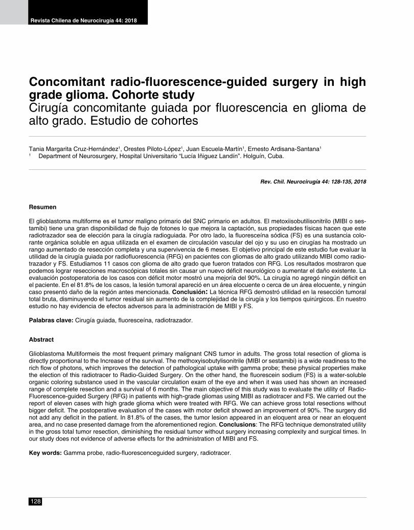
Revista Chilena de Neurocirugía 44: 2018 128 Concomitant radio-fluorescence-guided surgery in high grade glioma. Cohorte study Cirugía concomitante guiada por fluorescencia en glioma de alto grado. Estudio de cohortes Tania Margarita Cruz-Hernández 1 , Orestes Piloto-López 1 , Juan Escuela-Martín 1 , Ernesto Ardisana-Santana 1 1 Department of Neurosurgery, Hospital Universitario “Lucía Iñiguez Landín”. Holguín, Cuba. Rev. Chil. Neurocirugía 44: 128-135, 2018 Resumen El glioblastoma multiforme es el tumor maligno primario del SNC primario en adultos. El metoxiisobutilisonitrilo (MIBI o ses- tamibi) tiene una gran disponibilidad de flujo de fotones lo que mejora la captación, sus propiedades físicas hacen que este radiotrazador sea de elección para la cirugía radioguiada. Por otro lado, la fluoresceína sódica (FS) es una sustancia colo- rante orgánica soluble en agua utilizada en el examen de circulación vascular del ojo y su uso en cirugías ha mostrado un rango aumentado de resección completa y una supervivencia de 6 meses. El objetivo principal de este estudio fue evaluar la utilidad de la cirugía guiada por radiofluorescencia (RFG) en pacientes con gliomas de alto grado utilizando MIBI como radio- trazador y FS. Estudiamos 11 casos con glioma de alto grado que fueron tratados con RFG. Los resultados mostraron que podemos lograr resecciones macroscópicas totales sin causar un nuevo déficit neurológico o aumentar el daño existente. La evaluación postoperatoria de los casos con déficit motor mostró una mejoría del 90%. La cirugía no agregó ningún déficit en el paciente. En el 81.8% de los casos, la lesión tumoral apareció en un área elocuente o cerca de un área elocuente, y ningún caso presentó daño de la región antes mencionada. Conclusión: La técnica RFG demostró utilidad en la resección tumoral total bruta, disminuyendo el tumor residual sin aumento de la complejidad de la cirugía y los tiempos quirúrgicos. En nuestro estudio no hay evidencia de efectos adversos para la administración de MIBI y FS. Palabras clave: Cirugía guiada, fluoresceína, radiotrazador. Abstract Glioblastoma Multiformeis the most frequent primary malignant CNS tumor in adults. The gross total resection of glioma is directly proportional to the Increase of the survival. The methoxyisobutylisonitrile (MIBI or sestamibi) is a wide readiness to the rich flow of photons, which improves the detection of pathological uptake with gamma probe; these physical properties make the election of this radiotracer to Radio-Guided Surgery. On the other hand, the fluorescein sodium (FS) is a water-soluble organic coloring substance used in the vascular circulation exam of the eye and when it was used has shown an increased range of complete resection and a survival of 6 months. The main objective of this study was to evaluate the utility of Radio- Fluorescence-guided Surgery (RFG) in patients with high-grade gliomas using MIBI as radiotracer and FS. We carried out the report of eleven cases with high grade glioma which were treated with RFG. We can achieve gross total resections without bigger deficit. The postoperative evaluation of the cases with motor deficit showed an improvement of 90%. The surgery did not add any deficit in the patient. In 81.8% of the cases, the tumor lesion appeared in an eloquent area or near an eloquent area, and no case presented damage from the aforementioned region. Conclusions: The RFG technique demonstrated utility in the gross total tumor resection, diminishing the residual tumor without surgery increasing complexity and surgical times. In our study does not evidence of adverse effects for the administration of MIBI and FS. Key words: Gamma probe, radio-fluorescenceguided surgery, radiotracer.
Transcript of Concomitant radio-fluorescence-guided surgery in high grade … · eita Ciena de Neurocirugía 44:...
Revista Chilena de Neurocirugía 44: 2018
128
Concomitant radio-fluorescence-guided surgery in high grade glioma. Cohorte studyCirugía concomitante guiada por fluorescencia en glioma de alto grado. Estudio de cohortes
Tania Margarita Cruz-Hernández1, Orestes Piloto-López1, Juan Escuela-Martín1, Ernesto Ardisana-Santana1
1 Department of Neurosurgery, Hospital Universitario “Lucía Iñiguez Landín”. Holguín, Cuba.
Rev. Chil. Neurocirugía 44: 128-135, 2018
Resumen
El glioblastoma multiforme es el tumor maligno primario del SNC primario en adultos. El metoxiisobutilisonitrilo (MIBI o ses-tamibi) tiene una gran disponibilidad de flujo de fotones lo que mejora la captación, sus propiedades físicas hacen que este radiotrazador sea de elección para la cirugía radioguiada. Por otro lado, la fluoresceína sódica (FS) es una sustancia colo-rante orgánica soluble en agua utilizada en el examen de circulación vascular del ojo y su uso en cirugías ha mostrado un rango aumentado de resección completa y una supervivencia de 6 meses. El objetivo principal de este estudio fue evaluar la utilidad de la cirugía guiada por radiofluorescencia (RFG) en pacientes con gliomas de alto grado utilizando MIBI como radio-trazador y FS. Estudiamos 11 casos con glioma de alto grado que fueron tratados con RFG. Los resultados mostraron que podemos lograr resecciones macroscópicas totales sin causar un nuevo déficit neurológico o aumentar el daño existente. La evaluación postoperatoria de los casos con déficit motor mostró una mejoría del 90%. La cirugía no agregó ningún déficit en el paciente. En el 81.8% de los casos, la lesión tumoral apareció en un área elocuente o cerca de un área elocuente, y ningún caso presentó daño de la región antes mencionada. Conclusión: La técnica RFG demostró utilidad en la resección tumoral total bruta, disminuyendo el tumor residual sin aumento de la complejidad de la cirugía y los tiempos quirúrgicos. En nuestro estudio no hay evidencia de efectos adversos para la administración de MIBI y FS.
Palabras clave: Cirugía guiada, fluoresceína, radiotrazador.
Abstract
Glioblastoma Multiformeis the most frequent primary malignant CNS tumor in adults. The gross total resection of glioma is directly proportional to the Increase of the survival. The methoxyisobutylisonitrile (MIBI or sestamibi) is a wide readiness to the rich flow of photons, which improves the detection of pathological uptake with gamma probe; these physical properties make the election of this radiotracer to Radio-Guided Surgery. On the other hand, the fluorescein sodium (FS) is a water-soluble organic coloring substance used in the vascular circulation exam of the eye and when it was used has shown an increased range of complete resection and a survival of 6 months. The main objective of this study was to evaluate the utility of Radio-Fluorescence-guided Surgery (RFG) in patients with high-grade gliomas using MIBI as radiotracer and FS. We carried out the report of eleven cases with high grade glioma which were treated with RFG. We can achieve gross total resections without bigger deficit. The postoperative evaluation of the cases with motor deficit showed an improvement of 90%. The surgery did not add any deficit in the patient. In 81.8% of the cases, the tumor lesion appeared in an eloquent area or near an eloquent area, and no case presented damage from the aforementioned region. Conclusions: The RFG technique demonstrated utility in the gross total tumor resection, diminishing the residual tumor without surgery increasing complexity and surgical times. In our study does not evidence of adverse effects for the administration of MIBI and FS.
Key words: Gamma probe, radio-fluorescenceguided surgery, radiotracer.
129
Revista Chilena de Neurocirugía 44: 2018Trabajo Original
Introduction
In 1896 Becquerel discovers natural radioisotopes and De Hevesy invents the principle of “tracer” through his work with lead radiactivo. Radio-guided surgery (RGS) develops more less 60 years ago, today is used by surgeons to assess the degree of tumor resection and minimize the amount of healthy tis-sue to remover1-4.The MIBI (MIBI- 99mTc, methoxyiso-butylisonitrile, MIBI or sestamibi) has a wide availability rich photon flux, which improves the detection of abnormal uptake by gamma probe, these physi-cal properties make this radiotracer the choice for radioguided surgery, compared to other as thallium-2015. It was first described in 1980, to detect myocardial perfusion in coronarydis-ease(5, 6). The radiotracer uptake by the neoplastic cell depends on various factors such as regional flow blood, plasma potential and mitochondrial membrane, angiogenesis, and tissue metabolism, about 90% of tracer activ-ity is concentrated in the mitochondria. However physiological MIBI uptake by the choroid plexus is a disadvantage in the evaluation of deep lesions located in the paraventricular regions6.The lesion/bottom ratio is high with this tracer in tumors and suitable for techni-cal purposes. In addition, the scar tis-sue has no active uptake, so it is useful to distinguish tumor tissue during sur-gery7-14.Brain tumors have a high degree of ab-sorption of 99mTc-MIBI increased com-pared with that of the low-grade tumors, the Tc99m-MIBI absorption is related to the percentage of cells in S phase and level of tumor aneuploidy cerebral9.The impact of RFG in the updated treat-ing cancer patients is offering an essen-tial weapon in real time for surgeons in terms of determining the extent, loca-tion of the lesion, and the surgical mar-gins. The technique is based on using a radiotracer preferentially taken up by the tumor to mark the cancerous tissue, from normal tissue, this radiopharma-ceutical should be administered togeth-er before surgery15.
With the passage of years to go look-ing for technical aids, pre and intraop-erative images, making it possible to perform a complete as possible total tumor resection or infiltrative tumor le-sions those applying neuronavigation, intraoperative MRI, intraoperative ul-
trasound, cortical stimulation and finally the use of dye 5-amino levulinicAcid (5-ALA) and Fluorescein Sodium(FS) the latter has shown an increased range of complete resection and 6 months so-brevida16.In 1948 Moore and Peyton described the use of FS for locating brain tumors, which was subsequently abandoned its use due to own adverse reactions FS substance17.The FS is a water-soluble substance organic dye used in the ex-amination of blood vessels eye16.GBM is the most common malignant primary tumor of adults that applying a multimodal therapy (surgery, chemo-therapy, and radiotherapy) can achieve a median survival of 14 to 16 months, two years a 26-33% and less than 5% to five years18.There have been multiple studies in which direct relationship between the degree of tumor resection and pro-longed survival is shown, which cur-rently remains a point of contention between the neuro-oncologist18-20. Cur-rently, it is widely accepted, which can-not be identified functional brain areas, especially language center, only based on anatomical landmarks, plus a maxi-mum resection with minimal risks, it re-quires some functional single location pre and intraoperative. Radical resec-tion of gliomas carries the risk of injur-ing the eloquent functional areas due to the infiltrative nature of the lesion. The main role of surgery is to remove the tumor and its macroscopic limits as completely as possible. Although it has been possible to demonstrate the pres-ence of tumor cells imaging centime-ters beyond the alleged margin hence the importance to functional studies (spectroscopy MRI, PET-CT, SPECT-CT) in planning and surgical guide.There have been multiple attempts to intraoperative distinguish tumors from normal brain tissue: Using tissue pho-tosensitizers (chloro-aluminum phtha-locyanineTetrasulphonate) injection of dyes that cross the Blood-Brain Bar-rier (BBB) fluorescence-guided surgery (5-aminolevulinic acid) serial biopsies by freeze to discover the range, Dop-pler and intraoperative MRI guidance, most of these techniques lack the com-bination of ease of use and cost-efec-tividad11.Radioguided neurosurgery, is a tech-nique derived from nuclear medicine, introduced in 1985 by Martin, used for intraoperative identification of brain
tumors, due to emission by the same radiopharmaceutical, this can be done with a gamma probe or portable gam-ma camera5.This technique has already been used successfully in primary breast tumors, prostate, testicular, gastrointestinal, thyroid, parathyroid, melanoma and brain as well as in identifying sentinel nodes and metastases13.Studies published in 2012 and 2013 which combined the use of radiotrac-ers and fluorescent substances for identification in the sentinel lymph node biopsy in patients with breast cancer, squamous cell carcinoma of oral cavity and in cases of head and neck mela-noma21,22.It has designed a surgical trial compar-ing the results of Radio-Fluro Guided surgery with conventional surgery, aim-ing to demonstrate that the degree of resection of the tumor is greater with the RFG and with this progression free survival (PFS) and overall survival (OS). In this article we present the re-sults of Phase II.
Material and Method
A cohort study is performed, controlled and prospective of 11 patients with diagnoses of high grade gliomas, se-lected according to the inclusion crite-ria, who underwent Radio-fluorescence guided surgery in the period from Oc-tober 2014 to January 2015 to demon-strate that the practice of this approach is useful in our environment.RFG candidates who met the defined inclusion criteria were considered.
Inclusion criteria• Astrocytic tumors of high malig-
nancy, AA anaplastic astrocytomas (grade III) or glioblastoma multiforme GBM (Grade IV) without previous surgery.
• Patients aged ≥ 18 years to 70 years.
• Life expectancy ≥ 12 weeks.• Karnosfsky Index ≥ 70.• Laboratory parameters within normal
limits defined as:a) Hematopoietic: Hemoglobin ≥ 9
g/L, total leukocyte count ≥ 4 x 10⁹ cells/L, platelets ≥ 100 x 109/L.
b) Hepatic: liver function within normal limits and without liver disorders demonstrated by TGP, AST, GGT and alkaline phosphatase.
Revista Chilena de Neurocirugía 44: 2018
130
c) Renal function: Serum creatinine 132 mmol/L.
• Patients express written into the studio with his signature document voluntary informed consent.
• Tumor located in accessible areas to surgical resection.
Exclusion criteria• Patients who are pregnant or breast-
feeding.• Patients at the time of inclusion
present a chronic disease associ-ated phase of descompensation (eg. Heart disease, diabetes, hyperten-sion).
• Patients who have a history of bron-chial asthma.
• Fevers.• Severe septic processes.• Acute allergic or gravity States.• History of active malignant tumors
elsewhere.• Rejection by the patient.• Special locations such as 1. Lesiones
bilateral tumor.2. Invasion of the Corpus Callosum.3. Basal Ganglia.4. Brain stem.
As neuroimaging study, simple and en-hance image by magnetic resonance imaging (MRI) and single photon emis-sion tomography (SPECT) brain, with both techniques confirmed the pres-ence of uptake coincident with the le-sion described in the contrasted MRI was used, these procedures preop-erative were performed 72 hours after surgery (0.23-T Phillip MRI), can per-form the calculation of tumor volume. The residual tumor would be defined as uptake area, provided it is greater than 0.175 cm3, according to RANO criteria14,23. Tumor volume was calcu-lated by the computerized planimetric method and formula for the volume of an ellipsoid V = 4/3 π (a) (b) (c), was performed using the dimensions of the MRI contrasting obtained preoperative and postoperative, the latter were ob-tained within the first 48-72 hours after the operation, defining the residual vol-ume which presented enhancement by administering paramagnetic contrast.This study allowed us to calculate the preoperative tumor volume as15:• 35 cm3 Large.• ≤ 35 cm3 Small.
For postoperative volumetric assess we use the following nominación24.
Table 1 gives information about the de-gree of resection, the volume of tissue resected and the feature of the surgery.Dye uptake (FS): To describe the up-take of dye used the nomination sub-mitted by Bo Chenet al17, (Table 2).For a definition of eloquent area, de-fined as described by Sawaya16 elo-quent area (sensorimotor cortex, lan-guage center or visual, basal ganglia, hypothalamus, brainstem and corpus callosum) near eloquence (regions im-mediately adjacent to eloquent areas) and not eloquent (frontal lesions, tem-poropolar, right parietal-occipital, cer-ebellar hemisphere).Fulfilling the standards of Good Medi-cal Practice, before performing the procedure, the informedof consent was signed by patient and parent´s.The cut in the patient follow-up was conducted in the first six months after surgery, with neurological and imaging evaluation, fulfilling the protocol accord-ing to the histological type in each case.Phase III of the research are in prog-ress.Phase III: controlled, randomized, sin-gle-blind, where patients will be offered the Radio-fluorenceseguided surgery or conventional surgery, as methods of treatment for tumor pathology.Phase IV: Follow-up study with cutting at 6 and 12 months after surgery, with neurologic examination and imaging protocol as the disease.
Protocol RFGBrain SPECT with 20 mCi of Tc99m- MIBI, confirming the presence of coin-cident uptake (only) with the lesion de-scribed in contrasted MRI or CT, show-ing a high ratio injury / bottom (> 2).In each patient subsequent to brain SPECT, the respective surgical proce-dure was scheduled. Two hours before surgery was given 14 mCi of Tc99m- MIBI intravenously and the surgical de-tection probe explored.
ProceedThe main sites of concentration of MIBI are; heart and liver, after anesthesia, the use of leaded vest about the patient was implemented to reduce radiation to medical personnel.Intravenous injection of 14 mCi with 99m Tc-MIBI performed two hours before surgery. During anesthesia in-duction using fluorescein test with 200 mg of FS intradermally injection, it is expected 15 minutes, not allergic reac-tion, can proceed to the next step. Once craniotomy completed it proceeds to the administration of fluorescent sub-stance, then using the gamma probe to guide the intracerebral approach, directed primarily to normal brain tis-sue (bottom), is taken as a benchmark, then the gamma probe is directed to-wards the tumor (lesion), the difference is recorded. Due to the use of this dye will be tinged with mild, moderate or
Table 1.Data on surgeryDegree of resection Volume FeatureTotal ≤ 0.175 cm3 Absence of residual mass or uptake ring
in postoperative MRI
Subtotal > 0.175 cm3 Uptake residual tumor and measurable on postoperative MRI
Table 2.Description of the dye uptakeNomination Feature Intense yellow When the tumor intense greenish yellow
color evenly throughout the lesion is enhanced
Faint yellow When the tumor uptake is clear and yellow portions that do not capture
No uptake When there is no uptake
131
Revista Chilena de Neurocirugía 44: 2018
intense yellow color depending on the degree of disruption of the BBB. Once the resection of the lesion macroscopic fluorescence guided, the gamma probe to the tumor area is redirected, if activ-ity tumor is detecting (lesion) higher than the bottom (2: 1) and still existed intensity yellowing, we proceeds to total resection.Below check the decline in regional counting, to be equal to that of normal brain parenchyma in the gamma probe.
Results
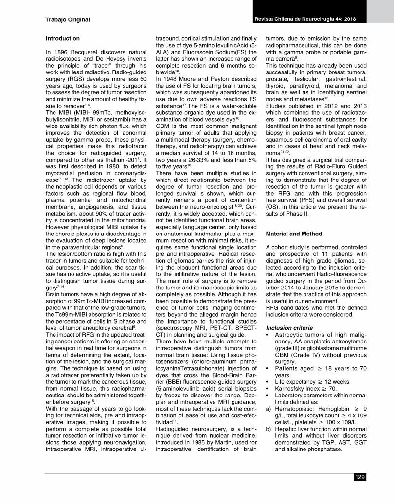
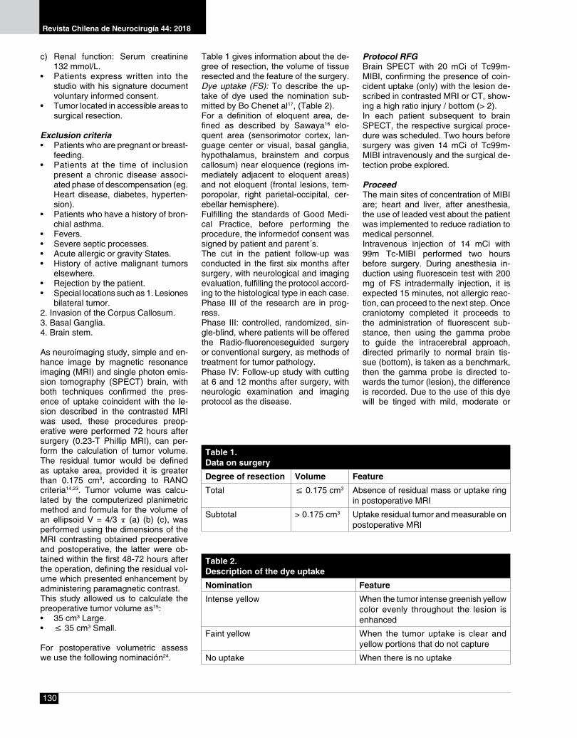
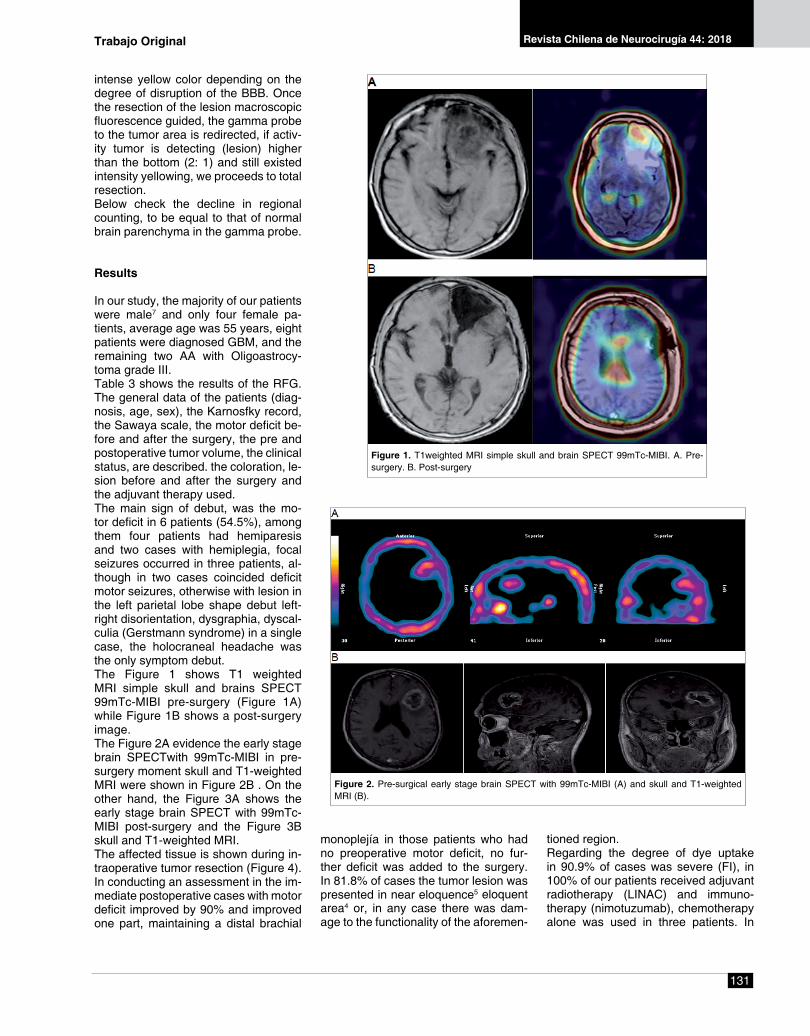
In our study, the majority of our patients were male7 and only four female pa-tients, average age was 55 years, eight patients were diagnosed GBM, and the remaining two AA with Oligoastrocy-toma grade III.Table 3 shows the results of the RFG. The general data of the patients (diag-nosis, age, sex), the Karnosfky record, the Sawaya scale, the motor deficit be-fore and after the surgery, the pre and postoperative tumor volume, the clinical status, are described. the coloration, le-sion before and after the surgery and the adjuvant therapy used. The main sign of debut, was the mo-tor deficit in 6 patients (54.5%), among them four patients had hemiparesis and two cases with hemiplegia, focal seizures occurred in three patients, al-though in two cases coincided deficit motor seizures, otherwise with lesion in the left parietal lobe shape debut left-right disorientation, dysgraphia, dyscal-culia (Gerstmann syndrome) in a single case, the holocraneal headache was the only symptom debut.The Figure 1 shows T1 weighted MRI simple skull and brains SPECT 99mTc-MIBI pre-surgery (Figure 1A) while Figure 1B shows a post-surgery image.The Figure 2A evidence the early stage brain SPECTwith 99mTc-MIBI in pre-surgery moment skull and T1-weighted MRI were shown in Figure 2B . On the other hand, the Figure 3A shows the early stage brain SPECT with 99mTc-MIBI post-surgery and the Figure 3B skull and T1-weighted MRI.The affected tissue is shown during in-traoperative tumor resection (Figure 4).In conducting an assessment in the im-mediate postoperative cases with motor deficit improved by 90% and improved one part, maintaining a distal brachial
monoplejía in those patients who had no preoperative motor deficit, no fur-ther deficit was added to the surgery. In 81.8% of cases the tumor lesion was presented in near eloquence5 eloquent area4 or, in any case there was dam-age to the functionality of the aforemen-
tioned region.Regarding the degree of dye uptake in 90.9% of cases was severe (FI), in 100% of our patients received adjuvant radiotherapy (LINAC) and immuno-therapy (nimotuzumab), chemotherapy alone was used in three patients. In
Figure 1. T1weighted MRI simple skull and brain SPECT 99mTc-MIBI. A. Pre-surgery. B. Post-surgery
Figure 2. Pre-surgical early stage brain SPECT with 99mTc-MIBI (A) and skull and T1-weighted MRI (B).
Trabajo Original
Revista Chilena de Neurocirugía 44: 2018
132
Table 3.General Result of RFG
No Diag-nosis
Age/ Sex
Kar-nosfky Score
Sawa-ya1
Scale
Pre-op-Motor Deficit
Pre-op tumoral volumen
Post-op Motor Deficit
Clinical Status2
Colora-ción3
Lesion/pottom-Pre-op
Lesion/pottom Post-op
Post-optumoral volumen
Adjuvant-Terapy4
1 GM 48/m 100 II Yes 123 cm3 No DD FI > 2/1 < 2/1 63,5 cm3 R, PCV, N
2 OA (gra-de) III
55/f 100 III No 65 cm3 No PFS FT > 2/1 < 2/1 11,4 cm3 R,N
3 GM 70/m 100 III Yes 33 cm3 No PFS FI > 2/1 < 2/1 3,4 cm3 R, PCV, N
4 AA 65/m 100 I No 71 cm3 No PFS FI > 2/1 < 2/1 1,7 cm3 R,N
5 GM 25/f 100 II Yes 87 cm3 No PFS FI > 2/1 < 2/1 31,2 cm3 R, PCV, N
6 GM 52/m 100 I Yes 48 cm3 No PFS FI > 2/1 < 2/1 0,5 cm3 R,N
7 GM 64/m 100 II No 42 cm3 No PFS FI > 2/1 < 2/1 1 cm3 R,N
8 GM 54/m 100 II Yes 47 cm3 Yes FI > 2/1 < 2/1 1,79 cm3 R,T,N
9 AA 68/f 100 III Yes 96 cm3 No FI > 2/1 < 2/1 0,17 cm3 R,T,N
10 GM 48/m 100 III Yes 59 cm3 No 2,87 cm3 R,N
11 GM 67/f 100 II No 43 cm3 No 10,24 cm3
R,N,T
Sawaya Scale (16):I: Area not eloquent, II: Area close to eloquence, III: Eloquent.State of the last follow-up:DD: Died by the disease, DAC: Died for another cause, DP: Disease in progression, PFS: Progression free survival.Degree of coloration:IF: Intense fluorescent, FT: Fluorescence tenua, NF: no fluorescence.Adjuvant therapy:R: Radiation therapy, PCV: procarbazine cisplatin vincristine, T: temozolamide, N: nimotuzumab.OA: Oligoastrocytoma.GM: glioblastoma multiforma.AA: anaplastic astrocytoma.
Figure 3. Post-surgical early stage brain SPECT with 99mTc-MIBI (A) and skull and T1-weighted MRI (B).
133
Revista Chilena de Neurocirugía 44: 2018
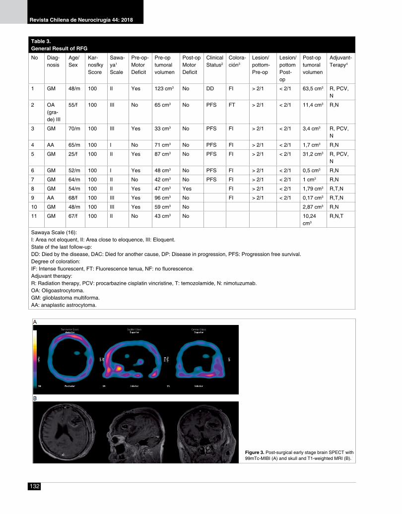
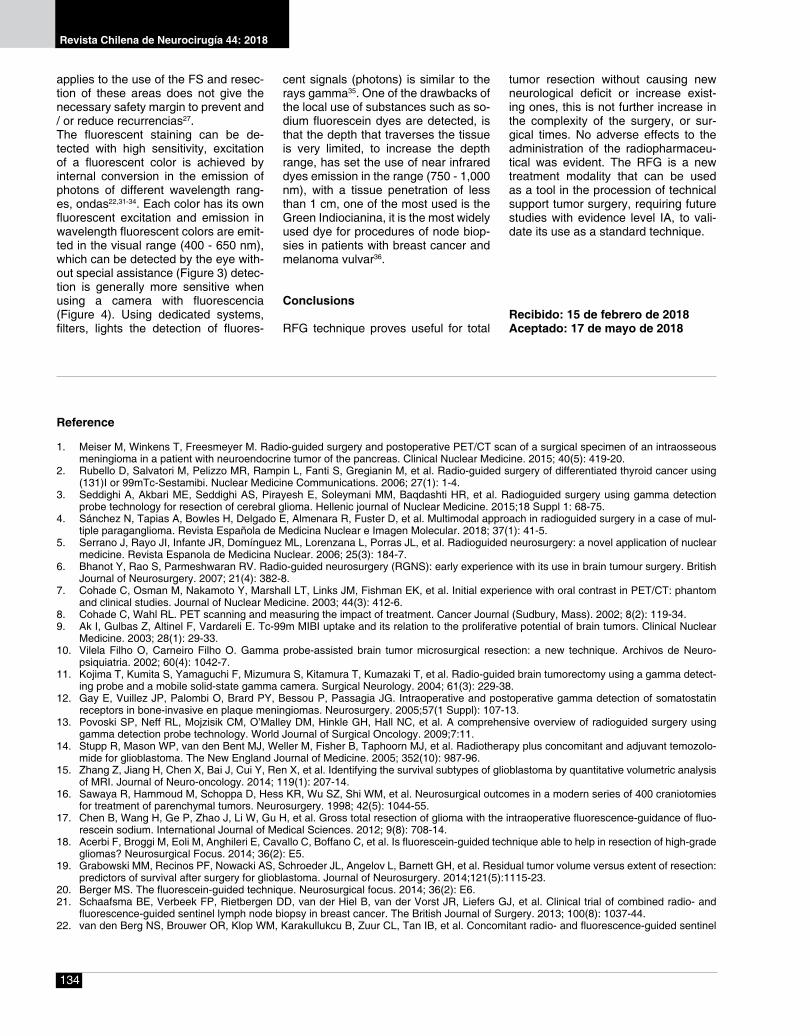
assessing preoperative tumor volume with postoperative tumor volume, they fell, with the lowest rates of postopera-tive residual volume of recent cases, which is related to the learning curve and have equipment reliability and the location not eloquent area. The back-ground / preoperative injury ratio was in all cases and postoperative > 2 was al-ways < 2, demonstrating that gets done the most complete resection of the le-sion and possible to confirm intraopera-tive real time.An example of an intraoperative image that shows how the injured tissue is observed with ultraviolet light is shown (Figure 5).
Discussion
The CRG using 99mTc-MIBI is not a common practice in neurosur-gery, in our study, the concomitant use of FS, made the procedure had a greater degree of tumor resection. The first description of CRG using Tc99m methoxyisobutylisonitrile Filho Vilela was made in 200210, for resec-tion of brain metastases in right parietal
lobe, assisted with gamma probe, two years after Kojima et al. report the use of the radiotracer in 13 patients with pri-mary or recurrentes11 astrocytomas16, in 2007, Bhanot et al, reported the use of Tc99m methoxyisobutylisonitrile, in a dose of 10 mCi (370 MBq) for assisted resection probe radius 13 patients with gliomas supratentoriales6,13.There are reports of other radiotracers como 111In- (DTPA) -D-Phe 1 pentet-reotide and 201 Tl in meningioma CRG the first plate and the second in one case report of resection of astrocytoma of the right temporoparietal region12,25.In the vast majority of cases reported by different groups complete resec-tion with the help of the gamma probe was performed with no adverse events or postsurgical complication, in the few cases of residual tumor after sur-gery confirmed by SPECT, the authors explain, the surgeon chose to leave remaining tumor although they indi-cated the probe due to the location in eloquent areas and little technical ex-perience, which made them hesitate to continue the surgery10,13.The radiation exposure of operating staff 99m Tc-MIBI has been previously
investigated11. The average whole body dose equivalent case was 25.8 and 27.9 14,9 μSv respectively for the surgeon, nurse and anesthesiology12. The United States Nuclear Regulatory Commission (USNRC) has set the annual occupa-tional exposure limit for adults and total effective dose equivalent 50,000 μSv and The International Commission on Radiological Protection (ICRP) has set an occupational exposure limit annual total dose for adults 20,000 μSv effec-tive by year13.The clinical trial from Schaafsma et al., evidenced that green indiocianina uses associated with Tc99m-nanocolloid in 32 patients with breast cancer, for detecting sentinel nodes, applying by local injection peri-areolar, conclud-ing the accuracy for detecting pre and intraoperative lymph affected, just as the shown by Brouwer studies et al. and van den Berg et al. with 11 and 14 patients respectively22,26, coinciding these studies in which the injection is local21,22,26.Using fluorescein sodium significantly increases the degree of tumor resec-tion, Díez-Valle et al, found areas of vague color matching infiltrated by tumor cells, areas which are not dis-played on the proven resonance27, obviously resection of these areas are crucial as a way to prevent recurrence and malignant progression of these tumoraciones15,17,18,23,24. Some studies suggest that the use of high doses of sodium fluorescein is a useful agent in-traoperative even without using equip-ment for visualización28. Shinoda et al., report on their study, that the degree of tumor resection total increase signifi-cantly with the use of FS at a dose of 20 mg / kg to 32 patients obtaining total resection in 27 of them to 84.4%, a sig-nificant difference when we compared with the level of total resection of the group control29.Koc et al, reported in their work a higher rate of complete resection with the use of guide FS in 47 patients in the control group, only 39 of them complete resec-tion (83%) was achieved, compared to 18 patients (54.5%) in the control group30.The study Chen Bo et al., in 2012 see light areas of contrast uptake around the tumor, which corresponded to areas adjacent edema, similar to that observed with the use of 5-ALA-Valle17. Díez et al., reports that these areas cor-respond to areas potentially infiltrated by tumor cells, this same mechanism
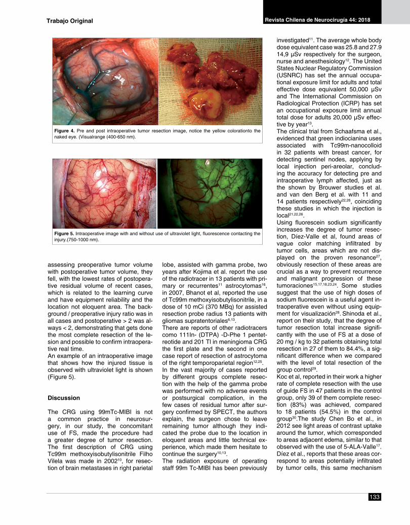
Figure 4. Pre and post intraoperative tumor resection image, notice the yellow colorationto the naked eye. (Visualrange (400-650 nm).
Figure 5. Intraoperative image with and without use of ultraviolet light, fluorescence contacting the injury.(750-1000 nm).
Trabajo Original
Revista Chilena de Neurocirugía 44: 2018
134
Reference
1. Meiser M, Winkens T, Freesmeyer M. Radio-guided surgery and postoperative PET/CT scan of a surgical specimen of an intraosseous meningioma in a patient with neuroendocrine tumor of the pancreas. Clinical Nuclear Medicine. 2015; 40(5): 419-20.
2. Rubello D, Salvatori M, Pelizzo MR, Rampin L, Fanti S, Gregianin M, et al. Radio-guided surgery of differentiated thyroid cancer using (131)I or 99mTc-Sestamibi. Nuclear Medicine Communications. 2006; 27(1): 1-4.
3. Seddighi A, Akbari ME, Seddighi AS, Pirayesh E, Soleymani MM, Baqdashti HR, et al. Radioguided surgery using gamma detection probe technology for resection of cerebral glioma. Hellenic journal of Nuclear Medicine. 2015;18 Suppl 1: 68-75.
4. Sánchez N, Tapias A, Bowles H, Delgado E, Almenara R, Fuster D, et al. Multimodal approach in radioguided surgery in a case of mul-tiple paraganglioma. Revista Española de Medicina Nuclear e Imagen Molecular. 2018; 37(1): 41-5.
5. Serrano J, Rayo JI, Infante JR, Domínguez ML, Lorenzana L, Porras JL, et al. Radioguided neurosurgery: a novel application of nuclear medicine. Revista Espanola de Medicina Nuclear. 2006; 25(3): 184-7.
6. Bhanot Y, Rao S, Parmeshwaran RV. Radio-guided neurosurgery (RGNS): early experience with its use in brain tumour surgery. British Journal of Neurosurgery. 2007; 21(4): 382-8.
7. Cohade C, Osman M, Nakamoto Y, Marshall LT, Links JM, Fishman EK, et al. Initial experience with oral contrast in PET/CT: phantom and clinical studies. Journal of Nuclear Medicine. 2003; 44(3): 412-6.
8. Cohade C, Wahl RL. PET scanning and measuring the impact of treatment. Cancer Journal (Sudbury, Mass). 2002; 8(2): 119-34.9. Ak I, Gulbas Z, Altinel F, Vardareli E. Tc-99m MIBI uptake and its relation to the proliferative potential of brain tumors. Clinical Nuclear
Medicine. 2003; 28(1): 29-33.10. Vilela Filho O, Carneiro Filho O. Gamma probe-assisted brain tumor microsurgical resection: a new technique. Archivos de Neuro-
psiquiatria. 2002; 60(4): 1042-7.11. Kojima T, Kumita S, Yamaguchi F, Mizumura S, Kitamura T, Kumazaki T, et al. Radio-guided brain tumorectomy using a gamma detect-
ing probe and a mobile solid-state gamma camera. Surgical Neurology. 2004; 61(3): 229-38.12. Gay E, Vuillez JP, Palombi O, Brard PY, Bessou P, Passagia JG. Intraoperative and postoperative gamma detection of somatostatin
receptors in bone-invasive en plaque meningiomas. Neurosurgery. 2005;57(1 Suppl): 107-13.13. Povoski SP, Neff RL, Mojzisik CM, O’Malley DM, Hinkle GH, Hall NC, et al. A comprehensive overview of radioguided surgery using
gamma detection probe technology. World Journal of Surgical Oncology. 2009;7:11.14. Stupp R, Mason WP, van den Bent MJ, Weller M, Fisher B, Taphoorn MJ, et al. Radiotherapy plus concomitant and adjuvant temozolo-
mide for glioblastoma. The New England Journal of Medicine. 2005; 352(10): 987-96.15. Zhang Z, Jiang H, Chen X, Bai J, Cui Y, Ren X, et al. Identifying the survival subtypes of glioblastoma by quantitative volumetric analysis
of MRI. Journal of Neuro-oncology. 2014; 119(1): 207-14.16. Sawaya R, Hammoud M, Schoppa D, Hess KR, Wu SZ, Shi WM, et al. Neurosurgical outcomes in a modern series of 400 craniotomies
for treatment of parenchymal tumors. Neurosurgery. 1998; 42(5): 1044-55.17. Chen B, Wang H, Ge P, Zhao J, Li W, Gu H, et al. Gross total resection of glioma with the intraoperative fluorescence-guidance of fluo-
rescein sodium. International Journal of Medical Sciences. 2012; 9(8): 708-14.18. Acerbi F, Broggi M, Eoli M, Anghileri E, Cavallo C, Boffano C, et al. Is fluorescein-guided technique able to help in resection of high-grade
gliomas? Neurosurgical Focus. 2014; 36(2): E5.19. Grabowski MM, Recinos PF, Nowacki AS, Schroeder JL, Angelov L, Barnett GH, et al. Residual tumor volume versus extent of resection:
predictors of survival after surgery for glioblastoma. Journal of Neurosurgery. 2014;121(5):1115-23.20. Berger MS. The fluorescein-guided technique. Neurosurgical focus. 2014; 36(2): E6.21. Schaafsma BE, Verbeek FP, Rietbergen DD, van der Hiel B, van der Vorst JR, Liefers GJ, et al. Clinical trial of combined radio- and
fluorescence-guided sentinel lymph node biopsy in breast cancer. The British Journal of Surgery. 2013; 100(8): 1037-44.22. van den Berg NS, Brouwer OR, Klop WM, Karakullukcu B, Zuur CL, Tan IB, et al. Concomitant radio- and fluorescence-guided sentinel
applies to the use of the FS and resec-tion of these areas does not give the necessary safety margin to prevent and / or reduce recurrencias27.The fluorescent staining can be de-tected with high sensitivity, excitation of a fluorescent color is achieved by internal conversion in the emission of photons of different wavelength rang-es, ondas22,31-34. Each color has its own fluorescent excitation and emission in wavelength fluorescent colors are emit-ted in the visual range (400 - 650 nm), which can be detected by the eye with-out special assistance (Figure 3) detec-tion is generally more sensitive when using a camera with fluorescencia (Figure 4). Using dedicated systems, filters, lights the detection of fluores-
cent signals (photons) is similar to the rays gamma35. One of the drawbacks of the local use of substances such as so-dium fluorescein dyes are detected, is that the depth that traverses the tissue is very limited, to increase the depth range, has set the use of near infrared dyes emission in the range (750 - 1,000 nm), with a tissue penetration of less than 1 cm, one of the most used is the Green Indiocianina, it is the most widely used dye for procedures of node biop-sies in patients with breast cancer and melanoma vulvar36.
Conclusions
RFG technique proves useful for total
tumor resection without causing new neurological deficit or increase exist-ing ones, this is not further increase in the complexity of the surgery, or sur-gical times. No adverse effects to the administration of the radiopharmaceu-tical was evident. The RFG is a new treatment modality that can be used as a tool in the procession of technical support tumor surgery, requiring future studies with evidence level IA, to vali-date its use as a standard technique.
Recibido: 15 de febrero de 2018Aceptado: 17 de mayo de 2018
135
Revista Chilena de Neurocirugía 44: 2018
lymph node biopsy in squamous cell carcinoma of the oral cavity using ICG-(99m)Tc-nanocolloid. European Journal of Nuclear Medicine and Molecular Imaging. 2012; 39(7): 1128-36.
23. Wen PY, Macdonald DR, Reardon DA, Cloughesy TF, Sorensen AG, Galanis E, et al. Updated response assessment criteria for high-grade gliomas: response assessment in neuro-oncology working group. Journal of Clinical Oncology. 2010; 28(11): 1963-72.
24. Stummer W, Pichlmeier U, Meinel T, Wiestler OD, Zanella F, Reulen HJ. Fluorescence-guided surgery with 5-aminolevulinic acid for resection of malignant glioma: a randomised controlled multicentre phase III trial. The Lancet Oncology. 2006; 7(5): 392-401.
25. Serrano J, Rayo JI, Infante JR, Dominguez L, Garcia-Bernardo L, Duran C, et al. Radioguided surgery in brain tumors with thallium-201. Clinical Nuclear Medicine. 2008; 33(12): 838-40.
26. Brouwer OR, Klop WM, Buckle T, Vermeeren L, van den Brekel MW, Balm AJ, et al. Feasibility of sentinel node biopsy in head and neck melanoma using a hybrid radioactive and fluorescent tracer. Annals of Surgical Oncology. 2012; 19(6): 1988-94.
27. Diez Valle R, Tejada Solis S, Idoate Gastearena MA, Garcia de Eulate R, Dominguez Echavarri P, Aristu Mendiroz J. Surgery guided by 5-aminolevulinic fluorescence in glioblastoma: volumetric analysis of extent of resection in single-center experience. Journal of Neuro-oncology. 2011; 102(1): 105-13.
28. Feigl GC, Ritz R, Moraes M, Klein J, Ramina K, Gharabaghi A, et al. Resection of malignant brain tumors in eloquent cortical areas: a new multimodal approach combining 5-aminolevulinic acid and intraoperative monitoring. Journal of Neurosurgery. 2010; 113(2): 352-7.
29. Shinoda J, Yano H, Yoshimura S, Okumura A, Kaku Y, Iwama T, et al. Fluorescence-guided resection of glioblastoma multiforme by us-ing high-dose fluorescein sodium. Technical note. Journal of Neurosurgery. 2003; 99(3): 597-603.
30. Koc K, Anik I, Cabuk B, Ceylan S. Fluorescein sodium-guided surgery in glioblastoma multiforme: a prospective evaluation. British journal of Neurosurgery. 2008; 22(1): 99-103.
31. van den Berg NS, Buckle T, KleinJan GH, van der Poel HG, van Leeuwen FWB. Multispectral Fluorescence Imaging During Robot-assisted Laparoscopic Sentinel Node Biopsy: A First Step Towards a Fluorescence-based Anatomic Roadmap. European Urology. 2017; 72(1): 110-7.
32. van den Berg NS, Miwa M, KleinJan GH, Sato T, Maeda Y, van Akkooi AC, et al. (Near-Infrared) Fluorescence-Guided Surgery Under Ambient Light Conditions: A Next Step to Embedment of the Technology in Clinical Routine. Annals of Surgical Oncology. 2016; 23(8): 2586-95.
33. KleinJan GH, Bunschoten A, van den Berg NS, Olmos RA, Klop WM, Horenblas S, et al. Fluorescence guided surgery and tracer-dose, fact or fiction? European Journal of Nuclear Medicine and Molecular Imaging. 2016; 43(10): 1857-67.
34. Yuan L, Lin W, Zheng K, He L, Huang W. Far-red to near infrared analyte-responsive fluorescent probes based on organic fluorophore platforms for fluorescence imaging. Chemical Society Reviews. 2013; 42(2): 622-61.
35. Van Den Berg NS, Buckle T, Kleinjan GI, Klop WM, Horenblas S, Van Der Poel HG, et al. Hybrid tracers for sentinel node biopsy. The quarterly Journal of Nuclear Medicine and Molecular Imaging. 2014; 58(2): 193-206.
36. Schaafsma BE, Mieog JS, Hutteman M, van der Vorst JR, Kuppen PJ, Lowik CW, et al. The clinical use of indocyanine green as a near-infrared fluorescent contrast agent for image-guided oncologic surgery. Journal of Surgical Oncology. 2011; 104(3): 323-32.
Correspondence autor:Orestes Piloto-LópezAve. 25 #15805 e/ 158 y 160, Playa.Teléfono: (537)[email protected]
Trabajo Original