Computerized preoperative planning for correction of...
12
ORIGINAL ARTICLE Computerized preoperative planning for correction of sagittal deformity of the spine Nicolas Aurouer Ibrahim Obeid Olivier Gille Vincent Pointillart Jean-Marc Vital Received: 18 August 2008 / Accepted: 27 May 2009 Ó Springer-Verlag 2009 Abstract Purpose Various methods of preoperative planning have been described for the correction of spinal sagittal defor- mities. They are reliable on condition that the thoraco- lumbar spine is totally fused and enable only the simulation of pedicle subtraction osteotomy (PSO). In this study, a new theoretical planning that can be used regardless of the etiology of the deformity and the type of osteotomy is described and assessed. Methods The spino-pelvic sagittal balance can be expressed by two parameters: pelvic tilt (PT) and center of both acoustic meati (CAM) overhang. These two parame- ters vary according to the type, number, level, and angu- lation of osteotomies. The general principle of the planning is to define the surgical program in order to obtain PT and CAM overhang as close as possible to the normal values. The theoretical planning is based on a trigonometric con- struction which depends on numerous factors and is chal- lenging to use in daily practice without the aid of a software tool. Modifications are proposed if the spine cannot be modeled as a solid beam due to unfused disks allowing relative motion. The SpineView software, which enables analysis and quick visualization of different cor- rection possibilities, is presented. The planning method is assessed in a prospective cohort of 11 patients by comparing planned values of spino-pelvic parameters to postoperative values. Results In all, 8 preoperative plans out of 11 were con- cordant with the postoperative results. Conclusions The preoperative planning enables the sur- geon to estimate the clinical effects of the different surgical techniques in order to choose the best procedure for a given patient. Keywords Spine Postural balance Osteotomy Software Planning techniques Introduction A kyphotic lumbar or thoracolumbar deformity of the spine leads to automatic compensatory phenomena in the adja- cent spinal segments, at the pelvis, and lower limbs. Intervertebral constraints and muscular efforts are then increased, thus reducing quality of life and functional ability [8, 12]. Correction of spinal deformities must enable the patient to maintain a balanced upright posture with minimum energy expenditure. Two main techniques can be used in order to correct a fixed sagittal imbalance: the pedicle subtraction osteotomy (PSO) and the multilevel Smith- Petersen osteotomies (SPO) [2]. PSO is essentially per- formed if the spine is totally inflexible as in the case of ankylosing spondylitis. Various methods of preoperative planning have been described in this type of case, where there is no relative motion between vertebrae [15, 16, 19, 20, 25]. These methods are reliable because the correction is obtained with only one osteotomy and the remainder of the spine behaves as a rigid body. However, patients with a partially flexible spine may require a surgical correction N. Aurouer (&) V. Pointillart J.-M. Vital Laboratoire d’Anatomie Me ´dico-Chirurgicale Applique ´e, Universite ´ Victor Se ´galen Bordeaux 2, 146 rue Le ´o Saignat, 33000 Bordeaux, France e-mail: [email protected]; [email protected] N. Aurouer I. Obeid O. Gille V. Pointillart J.-M. Vital Unite ´ de Pathologie Rachidienne, CHU de Bordeaux, Place Ame ´lie Raba-Le ´on, 33076 Bordeaux Cedex, France 123 Surg Radiol Anat DOI 10.1007/s00276-009-0524-9
Transcript of Computerized preoperative planning for correction of...

ORIGINAL ARTICLE
Computerized preoperative planning for correctionof sagittal deformity of the spine
Nicolas Aurouer Æ Ibrahim Obeid Æ Olivier Gille ÆVincent Pointillart Æ Jean-Marc Vital
Received: 18 August 2008 / Accepted: 27 May 2009
� Springer-Verlag 2009
Abstract
Purpose Various methods of preoperative planning have
been described for the correction of spinal sagittal defor-
mities. They are reliable on condition that the thoraco-
lumbar spine is totally fused and enable only the simulation
of pedicle subtraction osteotomy (PSO). In this study, a
new theoretical planning that can be used regardless of
the etiology of the deformity and the type of osteotomy is
described and assessed.
Methods The spino-pelvic sagittal balance can be
expressed by two parameters: pelvic tilt (PT) and center of
both acoustic meati (CAM) overhang. These two parame-
ters vary according to the type, number, level, and angu-
lation of osteotomies. The general principle of the planning
is to define the surgical program in order to obtain PT and
CAM overhang as close as possible to the normal values.
The theoretical planning is based on a trigonometric con-
struction which depends on numerous factors and is chal-
lenging to use in daily practice without the aid of a
software tool. Modifications are proposed if the spine
cannot be modeled as a solid beam due to unfused disks
allowing relative motion. The SpineView software, which
enables analysis and quick visualization of different cor-
rection possibilities, is presented. The planning method is
assessed in a prospective cohort of 11 patients by
comparing planned values of spino-pelvic parameters to
postoperative values.
Results In all, 8 preoperative plans out of 11 were con-
cordant with the postoperative results.
Conclusions The preoperative planning enables the sur-
geon to estimate the clinical effects of the different surgical
techniques in order to choose the best procedure for a given
patient.
Keywords Spine � Postural balance � Osteotomy �Software � Planning techniques
Introduction
A kyphotic lumbar or thoracolumbar deformity of the spine
leads to automatic compensatory phenomena in the adja-
cent spinal segments, at the pelvis, and lower limbs.
Intervertebral constraints and muscular efforts are then
increased, thus reducing quality of life and functional
ability [8, 12].
Correction of spinal deformities must enable the patient
to maintain a balanced upright posture with minimum
energy expenditure. Two main techniques can be used in
order to correct a fixed sagittal imbalance: the pedicle
subtraction osteotomy (PSO) and the multilevel Smith-
Petersen osteotomies (SPO) [2]. PSO is essentially per-
formed if the spine is totally inflexible as in the case of
ankylosing spondylitis. Various methods of preoperative
planning have been described in this type of case, where
there is no relative motion between vertebrae [15, 16, 19,
20, 25]. These methods are reliable because the correction
is obtained with only one osteotomy and the remainder of
the spine behaves as a rigid body. However, patients with a
partially flexible spine may require a surgical correction
N. Aurouer (&) � V. Pointillart � J.-M. Vital
Laboratoire d’Anatomie Medico-Chirurgicale Appliquee,
Universite Victor Segalen Bordeaux 2, 146 rue Leo Saignat,
33000 Bordeaux, France
e-mail: [email protected];
N. Aurouer � I. Obeid � O. Gille � V. Pointillart � J.-M. Vital
Unite de Pathologie Rachidienne, CHU de Bordeaux,
Place Amelie Raba-Leon, 33076 Bordeaux Cedex, France
123
Surg Radiol Anat
DOI 10.1007/s00276-009-0524-9
with PSO or SPO. The postoperative result is then more
difficult to assess. Mean improvement of lordosis is about
10� per SPO level, but there are important variations
depending on the preoperative degree of intervertebral
kyphosis. Further complicating the procedure, the sponta-
neous movements of the flexible adjacent segments are
difficult to predict. In such cases, a reliable preoperative
technique has not been described yet.
In this study, a preoperative theoretical planning for
treating different varieties of sagittal deformities, regard-
less of surgical correction technique, is described. The
specific computer software that enables this planning in
daily practice is presented and the effectiveness of the
method is assessed in a prospective cohort study.
Materials and methods
Biomechanical analysis
Spino-pelvic parameters
In the frontal plane, ideal balance is obtained by alignment
of the spine, so that the vertical line passing through the
center of C7 crosses the center of S1. In the sagittal plane,
the problem is more complex because of the considerable
morphological variability between individuals.
The works of Delmas [4] and the Duval-Beaupere group
[5, 13] are fundamental references to understand the ideal
spinal sagittal balance for a given patient. They have
revealed the close relationship between the sagittal spinal
curvatures, the spatial position of the pelvis, and its mor-
phology. Some other authors have confirmed that spinal
sagittal parameters are highly correlated with pelvic
parameters [1, 7, 10, 14, 22, 23].
In our analysis, the spino-pelvic sagittal balance is
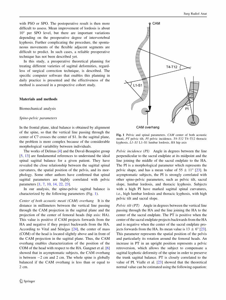
characterized by the following parameters (Fig. 1).
Center of both acoustic meati (CAM) overhang It is the
distance in millimeters between the vertical line passing
through the CAM projection in the sagittal plane and the
projection of the center of femoral heads (hip axis: HA).
This value is positive if CAM projects forwards from the
HA and negative if they project backwards from the HA.
According to Vital and Senegas [24], the center of mass
(COM) of the head is located slightly above and in front of
the CAM projection in the sagittal plane. Thus, the CAM
overhang enables characterization of the position of the
COM of the head with respect to the HA. Gangnet et al. [6]
showed that in asymptomatic subjects, the CAM overhang
is between -2 cm and 2 cm. The whole spine is globally
balanced if the CAM overhang is less than or equal to
2 cm.
Pelvic incidence (PI) Angle in degrees between the line
perpendicular to the sacral endplate at its midpoint and the
line joining the middle of the sacral endplate to the HA.
The PI is a morphological parameter which represents the
pelvic shape, and has a mean value of 55 ± 11� [23]. In
asymptomatic subjects, the PI is strongly correlated with
other spino-pelvic parameters, such as pelvic tilt, sacral
slope, lumbar lordosis, and thoracic kyphosis. Subjects
with a high PI have marked sagittal spinal curvatures,
i.e., high lumbar lordosis and thoracic kyphosis, with high
pelvic tilt and sacral slope.
Pelvic tilt (PT) Angle in degrees between the vertical line
passing through the HA and the line joining the HA to the
center of the sacral endplate. The PT is positive when the
center of the sacral endplate projects backwards from the HA
and is negative when the center of the sacral endplate pro-
jects forwards from the HA. Its mean value is 13 ± 6� [23].
This parameter represents the spatial position of the pelvis
and particularly its rotation around the femoral heads. An
increase in PT in an upright position represents a pelvic
retroversion, which allows the subject to compensate a
sagittal kyphotic deformity of the spine in order to preserve
the trunk sagittal balance. PT is closely correlated to the
value of PI. Vialle et al. [23] showed that the theoretical
normal value can be estimated using the following equation:
Fig. 1 Pelvic and spinal parameters. CAM center of both acoustic
meati, PT pelvic tilt, PI pelvic incidence, T4–T12 T4–T12 thoracic
kyphosis, L1–S1 L1–S1 lumbar lordosis, HA hip axis
Surg Radiol Anat
123

tPT = 0:37PI� 7 tPT = theoretical normal pelvic tiltð Þ
Lumbar L1–S1 lordosis Angle in degrees measured using
the Cobb technique between the L1 superior endplate and
the sacral endplate. This angle is negative when the
segment is in lordosis and positive in case of kyphosis. The
mean value is -62 ± 10�. L1–S1 lordosis is closely
correlated to the value of PI. Its theoretical normal value
can be estimated using the following equation (Gille O,
PhD Thesis, Ecole Nationale Superieure des Arts et
Metiers, Paris, 2006):
tL1S1 = 0:54PI + 32:56
tL1S1 = theoretical normal L1-S1 lumbar lordosisð Þ
Thoracic T4–T12 kyphosis Angle in degrees measured
using Cobb’s technique between T4 superior endplate and
the T12 inferior endplate. This angle is positive when the
segment is in kyphosis and negative in the case of lordosis.
The mean value is 39 ± 10� [23].
Natural history of the sagittal imbalance
A subject in an upright position can maintain balance
provided that the COM of the body projects onto the
polygon of support. Spinal kyphotic deformity induces a
forward translation of the COM of the trunk that can be
countered by several compensating mechanisms. If the
whole spine is fixed, as in ankylosing spondylitis, the
compensating mechanisms are located at the level of
the pelvis and lower limbs. The main compensating
mechanism is the pelvic retroversion, which is demon-
strated by increased PT. However, the spontaneous pelvic
retroversion can be insufficient to maintain balance if the
deformity is too great. The last means to shift the COM of
the trunk backward is to flex the knees and the ankles. In
this situation, the hips appear flexed, but they are actually
hyperextended (Fig. 2).
If a spinal segment adjacent to the deformity is flexible,
it can be involved in the compensating mechanisms. The
most typical example is the lumbar degenerative kyphosis
(Fig. 2b), where the thoracic spine is usually hypokyphotic
(at least at the beginning of the disease). The lumbar spine
is stiffened by arthritis and the thoracic spine is flexible, so
that the surgical correction of the lumbar deformity is
accompanied by a spontaneous restoration of the thoracic
kyphosis [11], which is necessary to consider at the time of
the preoperative planning.
Four types of sagittal spinal profile
Sagittal spinal profile can be described according to four
types of progressively greater deformity.
Fig. 2 Compensating
mechanisms of thoracolumbar
kyphotic deformities. a Ideal
sagittal balance, b loss of
lumbar lordosis compensated by
thoracic hypokyphosis and
pelvic retroversion, allowing the
subject to maintain a normal
CAM overhang. The pelvic
retroversion induces the rotation
of the hip range of motion
(ROM). In this case, the upright
subject has no ‘‘hip extension
reserve,’’ but his/her lower
limbs are still extended.
c Severe thoracolumbar
kyphosis with sagittal trunk
imbalance. The major pelvic
retroversion is associated with
hip hyperextension and knee
and ankle flexion. If ‘‘hip
extension reserve’’ is exhausted,
the increase of PT is equal to the
deviation of the femoral axis
from the vertical (a). As a result,
the measurement of the PT
allows us to take into account
the lower limb position. HipROM hip range of motion in
flexion–extension, PT pelvic
tilt, PI pelvic incidence
Surg Radiol Anat
123

The ideal sagittal balance It is the normal profile. All the
spino-pelvic parameters are normal, which means that
angulations of spine curvatures and PT are in accordance
with the value of the PI.
The compensated sagittal balance It is a local or regional
disturbance of the sagittal spinal profile, but trunk balance
is maintained (CAM overhang less than 2 cm) due to
compensating mechanisms (thoracic hypokyphosis, pelvic
retroversion and flexion of the knees).
The sagittal trunk imbalance It is a global disturbance of
the sagittal spinal profile, which means an increase in the
CAM overhang (greater than 2 cm).
The global sagittal imbalance It is a mechanical imbal-
ance, where the subject is unable to stand without the aid of
a crutch.
Surgical correction techniques
To correct a fixed sagittal imbalance there are two main
alternative techniques: pedicle subtraction osteotomy
(PSO) and Smith-Petersen osteotomy (SPO) (Fig. 3).
The principle of the PSO is based on resecting a pos-
terior wedge through the pedicle and part of the vertebral
body. This is accompanied by a shortening of the three
columns. Closure of the osteotomy enables a mean cor-
rection of 35� in the lumbar spine and 25� in the thoracic
spine [2]. However, there are variations related to the
morphology of each vertebra. In particular, vertebral shape
and height variations require a personalized analysis based
on lateral X-rays to predict the postoperative result.
In most cases, a PSO can only be performed at a single
vertebral level because of the risk of blood loss. In very
exceptional cases, the PSO can be envisaged at two levels.
SPO, also called Chevron osteotomies, are posterior
osteotomies that result in increased height of the anterior
column. They consist in resecting the upper and lower
articular processes at one or multiple levels. The space thus
created is closed by pressing the adjacent bones together.
The disk opening resulting from this type of osteotomy
exposes to the risk of pseudarthrosis, which is why we
systematically carry out intersomatic arthrodesis by trans-
foraminal lumbar interbody fusion (TLIF).
Mean improvement of lordosis is 10� per level, but there
are variations depending on the preoperative degree of
intervertebral kyphosis. In a previous unpublished study,
we established quantitative relationships between preop-
erative intervertebral lordosis and the potential for cor-
rection (Table 1).
In practice, it is difficult to carry out more than four
levels of SPO ? TLIF during the same procedure due to
the risk of blood loss.
Theoretical technique of planning
Normal parameters
The first step of the planning is the definition of the normal
theoretical parameters for a given subject:
• tCAM overhang has to be between -2 cm and 2 cm
[6].
• Pelvic tilt depends on PI according to the following
equation [23]: tPT = 0.37PI - 7.
• L1–S1 lumbar lordosis depends on PI according to the
following equation (Gille O, PhD Thesis, ENSAM,
Paris, 2006): tL1S1 = 0.54PI ? 32.56.
These theoretical normal parameters are distinguished
from the simulated parameters, i.e., sCAM overhang, sPT
and sL1S1 which represent the values that result from the
planning. In other words, the normal theoretical parameters
are the targets of the planning, but they cannot always be
Fig. 3 Surgical correction techniques: a pedicle subtraction osteot-
omy, b multilevel Smith Petersen osteotomies with transforaminal
lumbar interbody fusion
Table 1 Values for increasing lumbar lordosis according to preop-
erative intervertebral angle used for planning SPO ? TLIF
Preop intervertebral
angle
Potential for correction
with SPO ? TLIF
\-10� -5�[-10�-0[ -10�C0 -15�
Surg Radiol Anat
123

reached, so that the simulated parameters represent the best
compromise between the ideal solution and the technical
possibilities.
Hip hyperextension, knee flexion, and ankle flexion are
not directly considered in the planning, since the normal-
ization of the PT induces an automatic correction of these
clinical manifestations (Fig. 2).
Choice of surgical technique
The second step is the choice of the surgical technique
depending on the deformity etiology and the state of the
anterior spinal column.
If disk spaces seem to be mobile after posterior release,
we carry out multilevel SPO ? TLIF, since they enable a
more harmonious correction of the deformity. However,
in patients who have already been operated on and
present a very large posterior bone callus, we prefer to
do a PSO so that the spinal canal only has to be reached
once.
If the spine is in circumferential fusion, as in the case of
ankylosing spondylitis or in some cases of postoperative
flat back, we perform a PSO.
Regardless of the correction technique chosen, if a
fusion of L5–S1 disk is necessary, we carry out a cir-
cumferential arthrodesis with SPO ? TLIF at this level to
limit the risk of lumbosacral pseudarthrosis.
General principles of the planning
The preoperative planning is based on an elementary trig-
onometric construction (Fig. 4). The spine is modeled as a
rigid beam and different osteotomies are simulated in order
to normalize the spino-pelvic parameters (PT and CAM
overhang). First, the compensated balance is reproduced by
normalizing the CAM overhang up to the target of -2 cm.
Second, if it is surgically feasible, either the PSO angle or
the number of SPO ? TLIF is increased until the PT
angulation is normalized.
Adjustments of the method
In case of ankylosis spondylitis or flat-back after extensive
arthrodesis, the spine can be considered a solid beam and
the trigonometric construction is a model which is theo-
retically close to reality. If the thoracic spine is flexible,
like in lumbar degenerative kyphosis, this model requires
some modifications. The spontaneous increase of thoracic
kyphosis, which is induced by the correction of lumbar
lordosis, has to be considered [11]. Figure 5 shows that if
the thoracic spine is flexible, PT is underestimated by the
trigonometric construction, thus exposing the risk of un-
dercorrection. In other words, simulated PT (sPT) is an
underestimation of postop PT. For this reason, in this case,
the correction targets of the planning are not only the
Fig. 4 General principles of the
planning are based on a
trigonometric construction.
a Sagittal trunk imbalance. The
PI is equal to 70� and the PT is
equal to 62�. According to the
PI angle, the PT should be equal
to 19� (tPT = 0.37PI - 7).
b Normalization of the CAM
overhang due to a 10�osteotomy at a given level.
c At the same level, a correction
angle equal to 50� would be
required to normalize the PT.
CAM center of both acoustic
meati, PT pelvic tilt, PI pelvic
incidence, HA hip axis
Surg Radiol Anat
123
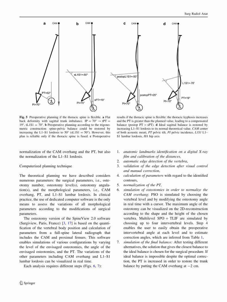
normalization of the CAM overhang and the PT, but also
the normalization of the L1–S1 lordosis.
Computerized planning technique
The theoretical planning we have described considers
numerous parameters: the surgical parameters, i.e., oste-
otomy number, osteotomy level(s), osteotomy angula-
tion(s), and the morphological parameters, i.e., CAM
overhang, PT, and L1–S1 lumbar lordosis. In clinical
practice, the use of dedicated computer software is the only
means to assess the variations of all morphological
parameters according to the modifications of surgical
parameters.
The osteotomy version of the SpineView 2.0 software
(Surgiview, Paris, France) [3, 17] is based on the quanti-
fication of the vertebral body position and calculation of
parameters from a full-spine lateral radiograph that
includes the CAM and proximal femurs. This software
enables simulations of various configurations by varying
the level of the envisaged osteotomies, the angle of the
envisaged osteotomies, and the PT. The variations of the
other parameters including CAM overhang and L1–S1
lumbar lordosis can be visualized in real time.
Each analysis requires different steps (Figs. 6, 7):
1. anatomic landmarks identification on a digital X-ray
film and calibration of the distances,
2. automatic edge detection of the vertebra,
3. validation of the edge detection after visual control
and manual correction,
4. calculation of parameters with regard to the identified
contours,
5. normalization of the PT,
6. simulation of osteotomies in order to normalize the
CAM overhang: PSO is simulated by choosing the
vertebral level and by modifying the osteotomy angle
in real time with a cursor. The maximum angle of the
osteotomy can be visualized on the 2D-reconstruction
according to the shape and the height of the chosen
vertebra. Multilevel SPO ? TLIF are simulated by
choosing up to four intervertebral levels. Step 4
enables the user to easily obtain the preoperative
intervertebral angle at each level and to estimate
correction angles, which are inferred from Table 1,
7. simulation of the final balance: After testing different
alternatives, the solution that gives the closest balance to
the ideal balance is chosen for the surgical procedure. If
ideal balance is impossible despite the optimal correc-
tion, the PT is increased in order to restore the trunk
balance by putting the CAM overhang at -2 cm.
Fig. 5 Preoperative planning if the thoracic spine is flexible. a Flat
back deformity with sagittal trunk imbalance. IP = 70� ? tPT =
19�, tL1S1 = 70�. b Preoperative planning according to the trigono-
metric construction: spino-pelvic balance could be restored by
increasing the L1–S1 lordosis to 50� (sL1S1 = 50�). However, this
plan is reliable only if the thoracic spine is fused. c Postoperative
results if the thoracic spine is flexible: the thoracic kyphosis increases
and the PT is greater than the planned value, leading to a compensated
balance (postop PT [ sPT). d Ideal sagittal balance is restored by
increasing L1–S1 lordosis to its normal theoretical value. CAM center
of both acoustic meati, PT pelvic tilt, PI pelvic incidence, L1S1 L1–
S1 lumbar lordosis, HA hip axis
Surg Radiol Anat
123
If the thoracic spine is flexible, the value of the sPT is an
underestimation of the postop PT. As a result, if the CAM
overhang is at -2 cm and the PT is normalized, the cor-
rection is still insufficient. The lumbar lordosis has to be
increased as close as possible to its theoretical value.
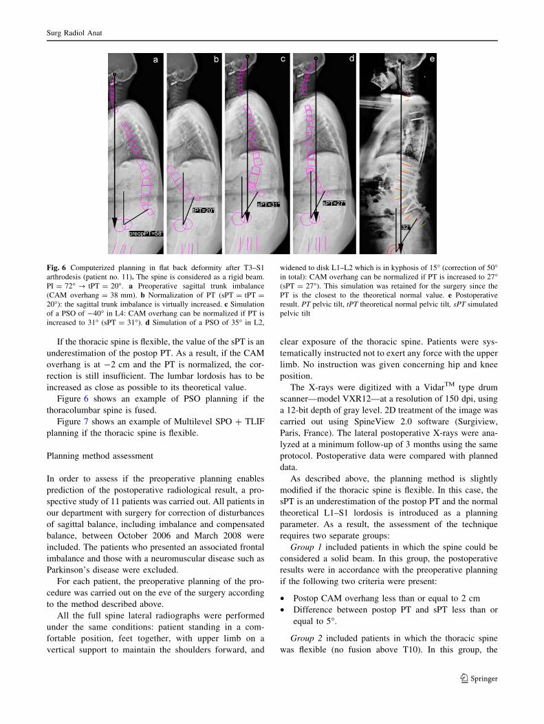
Figure 6 shows an example of PSO planning if the
thoracolumbar spine is fused.
Figure 7 shows an example of Multilevel SPO ? TLIF
planning if the thoracic spine is flexible.
Planning method assessment
In order to assess if the preoperative planning enables
prediction of the postoperative radiological result, a pro-
spective study of 11 patients was carried out. All patients in
our department with surgery for correction of disturbances
of sagittal balance, including imbalance and compensated
balance, between October 2006 and March 2008 were
included. The patients who presented an associated frontal
imbalance and those with a neuromuscular disease such as
Parkinson’s disease were excluded.
For each patient, the preoperative planning of the pro-
cedure was carried out on the eve of the surgery according
to the method described above.
All the full spine lateral radiographs were performed
under the same conditions: patient standing in a com-
fortable position, feet together, with upper limb on a
vertical support to maintain the shoulders forward, and
clear exposure of the thoracic spine. Patients were sys-
tematically instructed not to exert any force with the upper
limb. No instruction was given concerning hip and knee
position.
The X-rays were digitized with a VidarTM type drum
scanner—model VXR12—at a resolution of 150 dpi, using
a 12-bit depth of gray level. 2D treatment of the image was
carried out using SpineView 2.0 software (Surgiview,
Paris, France). The lateral postoperative X-rays were ana-
lyzed at a minimum follow-up of 3 months using the same
protocol. Postoperative data were compared with planned
data.
As described above, the planning method is slightly
modified if the thoracic spine is flexible. In this case, the
sPT is an underestimation of the postop PT and the normal
theoretical L1–S1 lordosis is introduced as a planning
parameter. As a result, the assessment of the technique
requires two separate groups:
Group 1 included patients in which the spine could be
considered a solid beam. In this group, the postoperative
results were in accordance with the preoperative planning
if the following two criteria were present:
• Postop CAM overhang less than or equal to 2 cm
• Difference between postop PT and sPT less than or
equal to 5�.
Group 2 included patients in which the thoracic spine
was flexible (no fusion above T10). In this group, the
Fig. 6 Computerized planning in flat back deformity after T3–S1
arthrodesis (patient no. 11). The spine is considered as a rigid beam.
PI = 72� ? tPT = 20�. a Preoperative sagittal trunk imbalance
(CAM overhang = 38 mm). b Normalization of PT (sPT = tPT =
20�): the sagittal trunk imbalance is virtually increased. c Simulation
of a PSO of -40� in L4: CAM overhang can be normalized if PT is
increased to 31� (sPT = 31�). d Simulation of a PSO of 35� in L2,
widened to disk L1–L2 which is in kyphosis of 15� (correction of 50�in total): CAM overhang can be normalized if PT is increased to 27�(sPT = 27�). This simulation was retained for the surgery since the
PT is the closest to the theoretical normal value. e Postoperative
result. PT pelvic tilt, tPT theoretical normal pelvic tilt, sPT simulated
pelvic tilt
Surg Radiol Anat
123

postoperative results were in accordance with the preop-
erative planning if the following three criteria were present:
• Postop CAM overhang less than or equal to 2 cm.
• Postop PT greater than or equal to sPT.
• Difference between postoperative lumbar lordosis
(postop L1–S1) and simulated lumbar L1–S1 lordosis
(sL1–S1) less than or equal to 10�.
Results
Eleven patients were included in our study: 9 women and 2
men with a mean age of 53.8 years (36–73). In all cases,
the surgical procedure was performed in accordance with
the planned number and level of the osteotomies.
Five of the 11 patients belonged to Group 1: 1 patient
presented an ankylosing spondylitis and 4 patients presented
a prior history of extensive thoraco-lumbar arthrodesis. The
results of the plan for this group are summarized in Table 2.
In all, 5 SPO ? TLIF were carried out in two patients and 4
PSO in three patients (T10, L2, L3, and L4). The mean
decrease of CAM overhang was 38 ± 20 mm and the mean
decrease of PT was 18.6 ± 7.2�. On average, the difference
between postop CAM overhang and sCAM overhang was
38 ± 54 mm, and the difference between postop PT and
sPT was 3.2 ± 1.6�. Individually, postoperative results were
in accordance with the preoperative planning in four cases
and were conflicting with the planning in one case. In this
case (patient no. 6), the CAM overhang was still much
greater than 2 cm even though two PSOs were carried out
(one in L3 and the other in T10). We noticed that the
angulation of each osteotomy was less than the value pre-
dicted by the planning. In Group 1 (with the exception of
patient no. 6, for whom a T10 PSO was carried out), tho-
racic T4T12 kyphosis had slight variation, if any at all. The
maximum variation reached 12� for patient no. 1 who had a
prior history of T6–L4 arthrodesis.
The other six patients belonged to Group 2. Three
patients had lumbar degenerative kyphosis without prior
Fig. 7 Computerized planning in lumbar degenerative kyphosis
(patient no. 5). The thoracic spine is flexible. PI = 71� ? tPT = 19�,
tL1–S1 = -70�. a Normalization of PT (sPT = tPT = 19�): the
sagittal trunk imbalance is virtually increased. b Simulation of
multilevel SPO ? TLIF. L5–S1 is sacralized and the preoperative
intervertebral lordosis are -3� in L4–L5, -4� in L3–L4, ?2� in L2–
L3 and -3� in L1–L2. According to Table 1, the correction angles
can be estimated as follows: -10� in L4–L5, -10� in L3–L4, -15� in
L2–L3 and -10� in L1–L2. With these values, sL1–S1 is equal to
-58�, which is still less than tL1S1 but we usually do not perform
more than 4 levels of SPO ? TLIF. c sPT is reduced to 15� in order to
normalize the CAM overhang. sPT is then an underestimation of the
postop PT because of the potential increase in thoracic kyphosis. (d)
Postoperative result. tPT theoretical normal pelvic tilt, sPT simulated
pelvic tilt, tL1–S1 theoretical normal L1–S1 lordosis, sL1–S1simulated L1–S1 lordosis
Surg Radiol Anat
123
history of spinal surgery and three patients presented a
prior history of localized lumbar arthrodesis. The results of
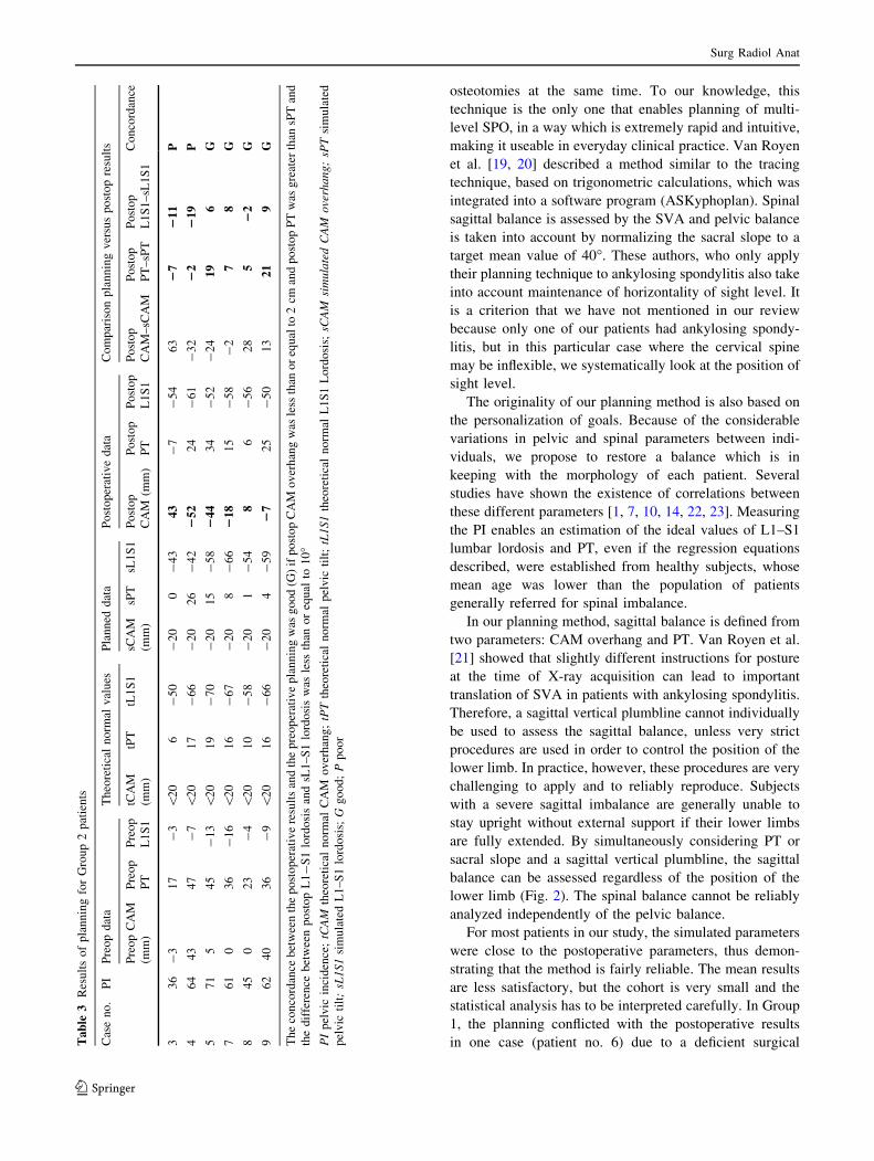
the plan for this group are summarized in Table 3. In all, 20
SPO ? TLIF and 1 PSO (L3) were carried out. The mean
decrease in CAM overhang was 26 ± 49 mm. The mean
decrease in PT was 17.8 ± 5.8� and the mean increase in
L1–S1 lumbar lordosis was 46.5 ± 6.5�. On average, the
difference between postop CAM overhang and sCAM
overhang was 27 ± 21 mm; the difference between postop
PT and sPT was 10.2 ± 7.9�; and the difference between
postop L1S1 lordosis and sL1S1 lordosis was 9.2 ± 5.7�.
Individually, two of the postoperative results were con-
flicting with the preoperative planning (patient nos. 3 and
4). For these two cases, the postoperative L1S1 lordoses
were greater than the simulated lordoses by 11� in one case
and 19� in the other. For patient no. 3, the CAM overhang
was still greater than 2 cm and the PT was paradoxically
negative (-7�). For patient no. 4, the postop PT was
greater than the sPT. In group 2, the T4–T12 kyphosis
increased in all cases, with a mean value of 23� (10–35�).
In all, 8 of the 11 preoperative plans were concordant
with the postoperative results according to the criteria
defined above. Considering the 11 patients, the mean cor-
rection of CAM overhang was 31 ± 38 mm and the mean
decrease of PT was 18 ± 6�. On average, the difference
between postop CAM overhang and sCAM overhang was
32 ± 38 mm, and the difference between postop PT and
sPT was 7 ± 7�.
Discussion
Anterior imbalance of the trunk corresponds to an
advanced spinal deformity. Surgical treatment implies
significant correction, the repercussions of which are dif-
ficult to determine without detailed planning. A number of
planning techniques has been described, but apart from the
restricted case of ankylosing spondylitis their precision is
poor. The trigonometric formula proposed by Ondra et al.
[15] is based solely on normalizing the C7 plumb line and
does not enable planning for global spino-pelvic sagittal
balance. Furthermore, some patients presented a non-fused
thoracic spine, which could not be exactly considered a
solid beam. We therefore chose to distinguish two groups
of patients according to flexibility of the thoracic spine and
the results of our review reinforce this: the patients in
group 2 presented a spontaneous increase in kyphosis of
23� on average.
Cervical lordosis can also vary spontaneously with
correction of the posture deformity, but the effect on the
CAM overhang is much less significant. In most cases, we
therefore do not take this parameter into account in our
planning method. However, it should be considered in
patients who present a major anterior imbalance and whose
cervical spine is flexible. In this case, the C7 plumb line or
the sagittal vertical axis (SVA) can be taken as reference.
The use of computer software enables us to work easily
with a large number of different parameters, each of which
can be of particular interest. We usually analyze the global
sagittal balance of the spine on the CAM overhang,
because this parameter takes into account the whole spine
and the pelvis position. It is disturbed when all spino-pelvic
compensating mechanisms are overstepped. Moreover, in
clinical practice CAM are easily identifiable on X-rays,
whereas C7 is hidden by the shoulders in 23–32% of the
cases depending on arm position [9].
The SpineView program was inspired by the tracing
technique described by Mangione (Vital JM, Mangione P,
Pedram M, Senegas J, Oral communication, Meeting of
the GRECO, Paris, 1999). This method only allowed one
or two simulations per patient since a new cut out had to
be made for each PSO level. Using computer software,
simulation at several different levels is very quick, and
it is possible to combine several corporeal or posterior
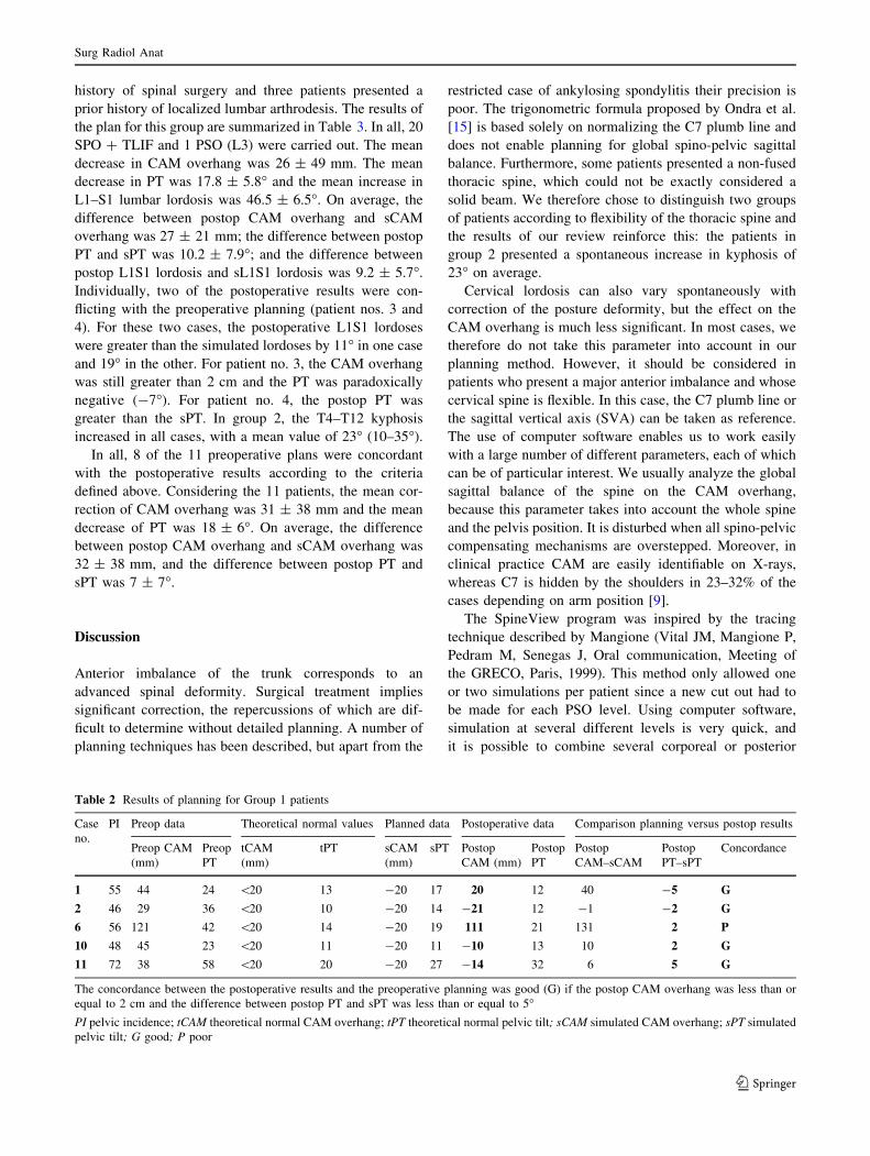
Table 2 Results of planning for Group 1 patients
Case
no.
PI Preop data Theoretical normal values Planned data Postoperative data Comparison planning versus postop results
Preop CAM
(mm)
Preop
PT
tCAM
(mm)
tPT sCAM
(mm)
sPT Postop
CAM (mm)
Postop
PT
Postop
CAM–sCAM
Postop
PT–sPT
Concordance
1 55 44 24 \20 13 -20 17 20 12 40 -5 G
2 46 29 36 \20 10 -20 14 -21 12 -1 -2 G
6 56 121 42 \20 14 -20 19 111 21 131 2 P
10 48 45 23 \20 11 -20 11 -10 13 10 2 G
11 72 38 58 \20 20 -20 27 -14 32 6 5 G
The concordance between the postoperative results and the preoperative planning was good (G) if the postop CAM overhang was less than or
equal to 2 cm and the difference between postop PT and sPT was less than or equal to 5�PI pelvic incidence; tCAM theoretical normal CAM overhang; tPT theoretical normal pelvic tilt; sCAM simulated CAM overhang; sPT simulated
pelvic tilt; G good; P poor
Surg Radiol Anat
123
osteotomies at the same time. To our knowledge, this
technique is the only one that enables planning of multi-
level SPO, in a way which is extremely rapid and intuitive,
making it useable in everyday clinical practice. Van Royen
et al. [19, 20] described a method similar to the tracing
technique, based on trigonometric calculations, which was
integrated into a software program (ASKyphoplan). Spinal
sagittal balance is assessed by the SVA and pelvic balance
is taken into account by normalizing the sacral slope to a
target mean value of 40�. These authors, who only apply
their planning technique to ankylosing spondylitis also take
into account maintenance of horizontality of sight level. It
is a criterion that we have not mentioned in our review
because only one of our patients had ankylosing spondy-
litis, but in this particular case where the cervical spine
may be inflexible, we systematically look at the position of
sight level.
The originality of our planning method is also based on
the personalization of goals. Because of the considerable
variations in pelvic and spinal parameters between indi-
viduals, we propose to restore a balance which is in
keeping with the morphology of each patient. Several
studies have shown the existence of correlations between
these different parameters [1, 7, 10, 14, 22, 23]. Measuring
the PI enables an estimation of the ideal values of L1–S1
lumbar lordosis and PT, even if the regression equations
described, were established from healthy subjects, whose
mean age was lower than the population of patients
generally referred for spinal imbalance.
In our planning method, sagittal balance is defined from
two parameters: CAM overhang and PT. Van Royen et al.
[21] showed that slightly different instructions for posture
at the time of X-ray acquisition can lead to important
translation of SVA in patients with ankylosing spondylitis.
Therefore, a sagittal vertical plumbline cannot individually
be used to assess the sagittal balance, unless very strict
procedures are used in order to control the position of the
lower limb. In practice, however, these procedures are very
challenging to apply and to reliably reproduce. Subjects
with a severe sagittal imbalance are generally unable to
stay upright without external support if their lower limbs
are fully extended. By simultaneously considering PT or
sacral slope and a sagittal vertical plumbline, the sagittal
balance can be assessed regardless of the position of the
lower limb (Fig. 2). The spinal balance cannot be reliably
analyzed independently of the pelvic balance.
For most patients in our study, the simulated parameters
were close to the postoperative parameters, thus demon-
strating that the method is fairly reliable. The mean results
are less satisfactory, but the cohort is very small and the
statistical analysis has to be interpreted carefully. In Group
1, the planning conflicted with the postoperative results
in one case (patient no. 6) due to a deficient surgicalTa
ble
3R
esu
lts
of
pla
nn
ing
for
Gro
up
2p
atie
nts
Cas
en
o.
PI
Pre
op
dat
aT
heo
reti
cal
no
rmal
val
ues
Pla
nn
edd
ata
Po
sto
per
ativ
ed
ata
Co
mp
aris
on
pla
nn
ing
ver
sus
po
sto
pre
sult
s
Pre
op
CA
M
(mm
)
Pre
op
PT
Pre
op
L1
S1
tCA
M
(mm
)
tPT
tL1
S1
sCA
M
(mm
)
sPT
sL1
S1
Po
sto
p
CA
M(m
m)
Po
sto
p
PT
Po
sto
p
L1
S1
Po
sto
p
CA
M–
sCA
M
Po
sto
p
PT
–sP
T
Po
sto
p
L1
S1
–sL
1S
1
Co
nco
rdan
ce
33
6-
31
7-
3\
20
6-
50
-2
00
-4
34
3-
7-
54
63
27
21
1P
46
44
34
7-
7\
20
17
-6
6-
20
26
-4
22
52
24
-6
1-
32
22
21
9P
57
15
45
-1
3\
20
19
-7
0-
20
15
-5
82
44
34
-5
2-
24
19
6G
76
10
36
-1
6\
20
16
-6
7-
20
8-
66
21
81
5-
58
-2
78
G
84
50
23
-4
\2
01
0-
58
-2
01
-5
48
6-
56
28
52
2G
96
24
03
6-
9\
20
16
-6
6-
20
4-
59
27
25
-5
01
32
19
G
Th
eco
nco
rdan
ceb
etw
een
the
po
sto
per
ativ
ere
sult
san
dth
ep
reo
per
ativ
ep
lan
nin
gw
asg
oo
d(G
)if
po
sto
pC
AM
ov
erh
ang
was
less
than
or
equ
alto
2cm
and
post
op
PT
was
gre
ater
than
sPT
and
the
dif
fere
nce
bet
wee
np
ost
op
L1
-S
1lo
rdo
sis
and
sL1
–S
1lo
rdo
sis
was
less
than
or
equ
alto
10
�P
Ip
elv
icin
cid
ence
;tC
AM
theo
reti
cal
no
rmal
CA
Mo
ver
han
g;
tPT
theo
reti
cal
no
rmal
pel
vic
tilt
;tL
1S
1th
eore
tica
ln
orm
alL
1S
1L
ord
osi
s;sC
AM
sim
ula
ted
CA
Mo
verh
an
g;
sPT
sim
ula
ted
pel
vic
tilt
;sL
1S
1si
mu
late
dL
1–
S1
lord
osi
s;G
go
od
;P
po
or
Surg Radiol Anat
123
technique. The global sagittal balance was not restored
because the angles of the 2 PSOs carried out were less
than the simulated values. The postoperative CAM over-
hang value was still very large (111 mm), which influ-
enced much the average values. Despite these results,
planning a PSO on a globally inflexible spine is the situ-
ation where the method is the most reliable because there
are few sources of error. In this scenario, the thoraco-
lumbar spine behaves like a rigid beam on either side of
the PSO. In the future, the development of navigation
techniques for spinal surgery could be a means of per-
forming the procedure exactly as planned [18]. In Group
2, there were more sources of planning errors because the
spontaneous variation of thoracic kyphosis is difficult to
predict. In the two cases where the planning was con-
flicting with postoperative results, the postoperative L1–S1
lordosis was much greater than the simulated lordosis, but
was close to the theoretical normal value. Patient no. 4 had
a postoperative sagittal balance close to the ideal value.
For patient no. 3, there remained postoperative trunk
sagittal imbalance with a CAM overhang of 43 mm,
whereas PT was negative (-7�). Paradoxically, the pelvis
was anteverted despite trunk anterior imbalance, which
conflicts with our theoretical reasoning. This may be
linked to poor posture at the time of the postoperative
X-ray potentially caused by the patient leaning on his/her
arms despite recommendations. Since this study, we have
changed our acquisition protocol using the ‘‘clavicle
position,’’ which does not require external support and
results in more accurate radiographic measures [9]. This
can also be explained by deficiencies of the gluteal mus-
cles, which are no longer able to retrovert the pelvis, and
therefore reduce the CAM overhang. This case emphasizes
that our planning does not consider muscular elements,
which is nevertheless essential in the sagittal balance. The
preoperative muscular assessment should also be taken
into account in the preoperative planning, but methods for
this remain to be defined.
Conclusion
In most cases, the theoretical planning presented in this
study enables the surgeon to estimate the results of the
surgical correction of sagittal deformities of the spine. As
long as the limitations of the technique are well under-
stood, all situations can be considered regardless of the
etiology of the deformity or the correction technique. The
best surgical procedure for a given patient can be deter-
mined, considering the global spino-pelvic sagittal balance
and the individual morphological variations. In clinical
practice, the SpineView software offers valuable assistance
to this planning, since numerous parameters can be taken
into account in a very short time.
References
1. Boulay C, Tardieu C, Hecquet J et al (2006) Sagittal alignment of
spine and pelvis regulated by pelvic incidence: standard values
and prediction of lordosis. Eur Spine J 15:415–422
2. Bridwell KH (2006) Decision making regarding Smith-Petersen
vs pedicle subtraction osteotomy vs vertebral column resection
for spinal deformity. Spine 31:S171–S178
3. Champain S, Benchikh K, Nogier A et al (2006) Validation of
new clinical quantitative analysis software applicable in spine
orthopaedic studies. Eur Spine J 15:982–991
4. Delmas A (1951) Types rachidiens de statique corporelle. Rev
Morpho-physiol Hum 4:27–32
5. Duval Beaupere G, Legaye J (2004) Composante sagittale de la
statique rachidienne. Rev Rhum 71:105–119
6. Gangnet N, Pomero V, Dumas R et al (2003) Variability of the
spine and pelvis location with respect to the gravity line: a three-
dimensional radiographic study using a force platform. Surg
Radiol Anat 25:424–433
7. Gelb DE, Lenke LG, Bridwell KH et al (1995) An analysis of
sagittal spinal alignment in 100 asymptomatic middle and older
aged volunteers. Spine 20:1351–1358
8. Glassman SD, Bridwell K, Dimar JR et al (2005) The impact
of positive sagittal balance in adult spinal deformity. Spine
30:2024–2029
9. Horton WC, Brown CW, Bridwell KH et al (2005) Is there an
optimal patient stance for obtaining a lateral 3600 radiograph? A
critical comparison of three techniques. Spine 30:427–433
10. Jackson RP, Hales C (2000) Congruent spinopelvic alignment
on standing lateral radiographs of adult volunteers. Spine 25:
2808–2815
11. Jang JS, Lee SH, Min JH et al (2009) Influence of lumbar lordosis
restoration on thoracic curve and sagittal position in lumbar
degenerative kyphosis patients. Spine 34:280–284
12. Lazennec JY, Ramare S, Arafati N et al (2000) Sagittal alignment
in lumbosacral fusion: relations between radiological parameters
and pain. Eur Spine J 9:47–55
13. Legaye J, Duval Beaupere G, Hecquet J et al (1998) Pelvic
incidence: a fundamental pelvic parameter for three-dimensional
regulation of spinal sagittal curves. Eur Spine J 7:99–103
14. Mangione P, Senegas J (1997) L’equilibre rachidien dans le plan
sagittal. Rev Chir Orthop Reparatrice Appar Mot 83:22–32
15. Ondra SL, Marzouk S, Koski T et al (2006) Mathematical cal-
culation of pedicle subtraction osteotomy size to allow precision
correction of fixed sagittal deformity. Spine. doi:10.1097/01.brs.
0000247950.02886.e5
16. Pigge RR, Scheerder FJ, Smit TH et al (2008) Effectiveness of
preoperative planning in the restoration of balance and view in
ankylosing spondylitis. Neurosurg Focus. doi:10.3171/foc/2008/
24/1/E7
17. Rajnics P, Pomero V, Templier A et al (2001) Computer-assisted
assessment of spinal sagittal plane radiographs. J Spinal Disord
14:135–142
18. Ruf M, Wagner R, Merk H et al (2006) Preoperative planning and
computer assisted surgery in ankylosing spondylitis (Abstract).
Z Orthop Ihre Grenzgeb 144:52–57
19. Van Royen BJ, De Gast A, Smit TH (2000) Deformity planning
for sagittal plane corrective osteotomies of the spine in anky-
losing spondylitis. Eur Spine J 9:492–498
Surg Radiol Anat
123

20. Van Royen BJ, Sheerder FJ, Jansen E et al (2007) ASKyphoplan:
a program for deformity planning in ankylosing spondylitis. Eur
Spine J 16:1445–1449
21. Van Royen BJ, Toussaint HM, Kingma I et al (1998) Accuracy of the
sagittal vertical axis in a standing lateral radiograph as a measure-
ment of balance in spinal deformities. Eur Spine J 7:408–412
22. Vaz G, Roussouly P, Berthonnaud E et al (2002) Sagittal morphol-
ogy and equilibrium of pelvis and spine. Eur Spine J 11:80–87
23. Vialle R, Levassor N, Rillardon L et al (2005) Radiographic
analysis of the sagittal alignment and balance of the spine in
asymptomatic subjects. J Bone Joint Surg Am 87:260–267
24. Vital JM, Senegas J (2006) Anatomical bases of the study of the
constraints to which the cervical spine is subject in the sagittal
plane. A study of the center of gravity of the head. Surg Radiol
Anat 8:169–173
25. Yang BP, Ondra SL (2006) A method for calculating the exact
angle required during pedicle subtraction osteotomy for fixed
sagittal deformity: comparison with the trigonometric method.
Neurosurgery. doi:10.1227/01.neu.0000232628.46247.15
Surg Radiol Anat
123