Comprehensive Definition of the SigH Regulon of Mycobacterium ...
16
RESEARCH ARTICLE Comprehensive Definition of the SigH Regulon of Mycobacterium tuberculosis Reveals Transcriptional Control of Diverse Stress Responses Jared D. Sharp 1☯¤a , Atul K. Singh 1☯ , Sang Tae Park 2¤b , Anna Lyubetskaya 3¤c , Matthew W. Peterson 5 , Antonio L. C. Gomes 3¤d , Lakshmi-Prasad Potluri 1 , Sahadevan Raman 2¤e , James E. Galagan 2,3,4,5 *, Robert N. Husson 1 * 1 Division of Infectious Diseases, Boston Children’s Hospital and Department of Pediatrics, Harvard Medical School, Boston, Massachusetts 02115, United States of America, 2 National Emerging Infectious Diseases Laboratories, Boston University, Boston, Massachusetts 02118, United States of America, 3 Bioinformatics Program, Boston University, Boston, Massachusetts 02215, United States of America, 4 Department of Biomedical Engineering, Boston University, Boston, Massachusetts 02215, United States of America, 5 Department of Microbiology, Boston University, Boston, Massachusetts 02215, United States of America ☯ These authors contributed equally to this work. ¤a Current address: Affinivax, Cambridge, Massachusetts 02139, United States of America ¤b Current address: Macrogen Clinical Laboratory, Rockville, Maryland 20850, United States of America ¤c Current address: Bioinformatics Program, Beth Israel Deaconess Hospital, Boston, Massachusetts 02115, United States of America ¤d Current address: Department of Systems Biology, Columbia University, New York, New York 10032, United States of America ¤e Current address: Division of Rheumatology, Brigham & Women’s Hospital, Boston, Massachusetts 02115, United States of America * [email protected] (RH); [email protected] (JG) Abstract Expression of SigH, one of 12 Mycobacterium tuberculosis alternative sigma factors, is induced by heat, oxidative and nitric oxide stresses. SigH activation has been shown to increase expression of several genes, including genes involved in maintaining redox equi- librium and in protein degradation. However, few of these are known to be directly regulated by SigH. The goal of this project is to comprehensively define the Mycobacterium tubercu- losis genes and operons that are directly controlled by SigH in order to gain insight into the role of SigH in regulating M. tuberculosis physiology. We used ChIP-Seq to identify in vivo SigH binding sites throughout the M. tuberculosis genome, followed by quantification of SigH-dependent expression of genes linked to these sites and identification of SigH-regu- lated promoters. We identified 69 SigH binding sites, which are located both in intergenic regions and within annotated coding sequences in the annotated M. tuberculosis genome. 41 binding sites were linked to genes that showed greater expression following heat stress in a SigH-dependent manner. We identified several genes not previously known to be regu- lated by SigH, including genes involved in DNA repair, cysteine biosynthesis, translation, and genes of unknown function. Experimental and computational analysis of SigH-regu- lated promoter sequences within these binding sites identified strong consensus -35 and PLOS ONE | DOI:10.1371/journal.pone.0152145 March 22, 2016 1 / 16 OPEN ACCESS Citation: Sharp JD, Singh AK, Park ST, Lyubetskaya A, Peterson MW, Gomes ALC, et al. (2016) Comprehensive Definition of the SigH Regulon of Mycobacterium tuberculosis Reveals Transcriptional Control of Diverse Stress Responses. PLoS ONE 11 (3): e0152145. doi:10.1371/journal.pone.0152145 Editor: Dipankar Chatterji, Indian Institute of Science, INDIA Received: December 1, 2015 Accepted: March 9, 2016 Published: March 22, 2016 Copyright: © 2016 Sharp et al. This is an open access article distributed under the terms of the Creative Commons Attribution License, which permits unrestricted use, distribution, and reproduction in any medium, provided the original author and source are credited. Data Availability Statement: Relevant data are within the paper and its Supporting Information files. ChIP-Seq datafiles are available at figshare at the following url: https://figshare.com/s/ 39ef281d71a9e0c6553e. Funding: This work was supported by the National Institute of Allergy and Infectious Diseases R01AI37901 to RNH; Boston Children's Hospital internal funding (no grant number) to RNH; and National Institute of General Medical Sciences R01GM114812 to JEG. JDS was supported in part by National Institutes of Health training grant
Transcript of Comprehensive Definition of the SigH Regulon of Mycobacterium ...
RESEARCH ARTICLE
Comprehensive Definition of the SigHRegulon ofMycobacterium tuberculosisReveals Transcriptional Control of DiverseStress ResponsesJared D. Sharp1☯¤a, Atul K. Singh1☯, Sang Tae Park2¤b, Anna Lyubetskaya3¤c, MatthewW. Peterson5, Antonio L. C. Gomes3¤d, Lakshmi-Prasad Potluri1, Sahadevan Raman2¤e,James E. Galagan2,3,4,5*, Robert N. Husson1*
1 Division of Infectious Diseases, Boston Children’s Hospital and Department of Pediatrics, Harvard MedicalSchool, Boston, Massachusetts 02115, United States of America, 2 National Emerging Infectious DiseasesLaboratories, Boston University, Boston, Massachusetts 02118, United States of America, 3 BioinformaticsProgram, Boston University, Boston, Massachusetts 02215, United States of America, 4 Department ofBiomedical Engineering, Boston University, Boston, Massachusetts 02215, United States of America,5 Department of Microbiology, Boston University, Boston, Massachusetts 02215, United States of America
☯ These authors contributed equally to this work.¤a Current address: Affinivax, Cambridge, Massachusetts 02139, United States of America¤b Current address: Macrogen Clinical Laboratory, Rockville, Maryland 20850, United States of America¤c Current address: Bioinformatics Program, Beth Israel Deaconess Hospital, Boston, Massachusetts02115, United States of America¤d Current address: Department of Systems Biology, Columbia University, New York, New York 10032,United States of America¤e Current address: Division of Rheumatology, Brigham &Women’s Hospital, Boston, Massachusetts02115, United States of America* [email protected] (RH); [email protected] (JG)
AbstractExpression of SigH, one of 12Mycobacterium tuberculosis alternative sigma factors, is
induced by heat, oxidative and nitric oxide stresses. SigH activation has been shown to
increase expression of several genes, including genes involved in maintaining redox equi-
librium and in protein degradation. However, few of these are known to be directly regulated
by SigH. The goal of this project is to comprehensively define theMycobacterium tubercu-losis genes and operons that are directly controlled by SigH in order to gain insight into the
role of SigH in regulatingM. tuberculosis physiology. We used ChIP-Seq to identify in vivoSigH binding sites throughout theM. tuberculosis genome, followed by quantification of
SigH-dependent expression of genes linked to these sites and identification of SigH-regu-
lated promoters. We identified 69 SigH binding sites, which are located both in intergenic
regions and within annotated coding sequences in the annotatedM. tuberculosis genome.
41 binding sites were linked to genes that showed greater expression following heat stress
in a SigH-dependent manner. We identified several genes not previously known to be regu-
lated by SigH, including genes involved in DNA repair, cysteine biosynthesis, translation,
and genes of unknown function. Experimental and computational analysis of SigH-regu-
lated promoter sequences within these binding sites identified strong consensus -35 and
PLOSONE | DOI:10.1371/journal.pone.0152145 March 22, 2016 1 / 16
OPEN ACCESS
Citation: Sharp JD, Singh AK, Park ST, LyubetskayaA, Peterson MW, Gomes ALC, et al. (2016)Comprehensive Definition of the SigH Regulon ofMycobacterium tuberculosis Reveals TranscriptionalControl of Diverse Stress Responses. PLoS ONE 11(3): e0152145. doi:10.1371/journal.pone.0152145
Editor: Dipankar Chatterji, Indian Institute of Science,INDIA
Received: December 1, 2015
Accepted: March 9, 2016
Published: March 22, 2016
Copyright: © 2016 Sharp et al. This is an openaccess article distributed under the terms of theCreative Commons Attribution License, which permitsunrestricted use, distribution, and reproduction in anymedium, provided the original author and source arecredited.
Data Availability Statement: Relevant data arewithin the paper and its Supporting Information files.ChIP-Seq datafiles are available at figshare at thefollowing url: https://figshare.com/s/39ef281d71a9e0c6553e.
Funding: This work was supported by the NationalInstitute of Allergy and Infectious DiseasesR01AI37901 to RNH; Boston Children's Hospitalinternal funding (no grant number) to RNH; andNational Institute of General Medical SciencesR01GM114812 to JEG. JDS was supported in part byNational Institutes of Health training grant
-10 promoter sequences, but with tolerance for non-consensus bases at specific positions.
This comprehensive identification and validation of SigH-regulated genes demonstrates an
extended SigH regulon that controls an unexpectedly broad range of stress response
functions.
IntroductionMycobacterium tuberculosis is a slow-growing but deadly pathogen that is able to cause achronic infection through its ability to adapt to the multiple environments that it encounters inthe human host during infection. During the course ofM. tuberculosis infection, ranging fromprimary infection with active bacterial replication, to latent infection with restricted replicationwithin granulomas, to cavitary tuberculosis (TB) where many bacteria are extracellular,M.tuberculosis is subject to an extensive and varying array of host-generated stresses. Amongthese are hypoxia, and several forms of oxidative and nitrosative stresses that can damage abroad range of bacterial macromolecules that are essential for viability [1, 2]. ForM. tuberculo-sis to persist in the face of these stresses requires extensive defense and repair mechanisms,many of which result from changes in gene expression in response to environmental cues.
TheM. tuberculosis genome encodes 13 sigma factors, the subunit of RNA polymerase thatbinds specific promoter sequences to initiate transcription [3]. In addition to SigA, the primarysigma factor that controls the expression of a large proportion of genes under most conditions,theM. tuberculosis genome encodes 12 alternative sigma factors that respond to specific signalsto regulate the transcription of genes and operons that are physically separated but functionallylinked [4, 5]. Ten of these sigma factors belong to the Type 4 or extracytoplasmic function(ECF) family, [6, 7]. The activity of the ECF sigma factors is often regulated by reversible bind-ing of the sigma factor by a cognate anti-sigma factor, which functions as a sensor of signalsfrom the environment [8, 9].
SigH is an ECF sigma factor ofM. tuberculosis that is activated by heat stress, oxidativestress and nitric oxide stress. SigH activity is regulated post-translationally by its anti-sigmafactor, RshA, which senses these stresses and releases SigH to bind to core RNA polymeraseand activate transcription of its regulon [10–13]. This sigma factor is required for full virulenceofM. tuberculosis in mouse and non-human primate (NHP) models of infection [14, 15]. Inprimate macrophages SigH is required for long-term infection and has been shown to affectthe expression of host genes in response toM. tuberculosis infection, with effects on host cellapoptosis [16]. Based on potent immunity in a non-human primate model, a sigH deletionstrain of M. tuberculosis has been suggested to be a candidate for development of vaccines toprotect against M. tuberculosis disease [17]. Analysis of a sigHmutantM. avium subsp. paratu-berculosis infection of bovine macrophages and a murine model of paratuberculosis suggestedthat sigH could play an important role in persistence and virulence of this mycobacterial spe-cies in infected animals [18].
Despite its importance for virulence and host responses to infection, only a limited numberof genes have been experimentally shown to be directly regulated by SigH [11, 19]. These genesinclude SigH itself and additional transcriptional regulators, heat shock genes required forrepair and degradation of damaged proteins, and components of the thioredoxin/thioredoxinreductase system required to maintain redox homeostasis. In addition to these genes that areknown to be directly regulated by SigH, increased stress-induced expression of several addi-tional genes in wild typeM. tuberculosis compared to sigH deletion strains has been observed
M. tuberculosis SigH Regulon and Control of Stress Responses
PLOS ONE | DOI:10.1371/journal.pone.0152145 March 22, 2016 2 / 16
T32HD055148. This project was funded in part withfederal funds from the National Institute of Allergy andInfectious Diseases, National Institutes of Health,Department of Health and Human Services, underContract no. HHSN272200800059C to JEG. Thefunders had no role in study design, data collectionand analysis, decision to publish, or preparation ofthe manuscript.
Competing Interests: The authors have declaredthat no competing interests exist.
[12, 14]. These data suggest that the direct SigH regulon may be substantially larger than is cur-rently known. Because SigH regulates expression of other transcription regulators, however,including SigE and SigB, two sigma factors that also regulate stress responses, differential geneexpression may result from indirect effects via these SigH-controlled regulators or by SigH-mediated changes in cell physiology that affect the activity other transcription factors.
We therefore undertook to comprehensively define the direct SigH regulon ofM. tuberculo-sis, using chromatin immunoprecipitation with massively parallel sequencing (ChIP-Seq) toidentify DNA regions bound by this sigma factor in vivo in theM. tuberculosis cell. We thencompared the stress-induced expression inM. tuberculosis wild type and ΔsigH strains of can-didate SigH-controlled genes identified by ChIP-Seq. For genes where we observed differentialgene expression between these strains, we performed 5’-RACE using RNA from wild type andΔsigH strains, to identify SigH-dependent transcription start points (TSPs). From these datawe derived a highly refined SigH-regulated promoter motif consensus.
Defining genes directly regulated by SigH as those i) that have a sequence bound by SigH, ii)that show differential gene expression in wild type versus ΔsigH strain and iii) that have a pro-moter sequence consistent with the SigH consensus, our data identify a minimum of 25 genesthat comprise the direct SigH regulon. The experimentally defined or predicted functions ofthese genes indicate that in addition to regulation of redox homeostasis and protein turnover,SigH plays a much broader role in recovery from stresses than was previously known, includingregulation of genes required for repair of DNA damage, recovery of ribosome function andtranslation, sulfur transport, and synthesis and salvage of sulfur-containing amino acids.
Materials and Methods
Chromatin Immunoprecipitation and Sequencing (ChIP-Seq)ChIP-Seq was performed as previously described with minor modifications [20]. The sigH-FLAG fusion was expressed using a tetracycline repressor (TetR)-regulated promoter [21].Prior to performing the ChIP-Seq experiments, the expression of SigH-FLAG was analyzed todetermine the optimal concentration of the anhydrotetracycline (aTc) inducer and the optimaltime point following induction. Based on these experiments,M. tuberculosis was grown at 37°Cin Middlebrook 7H9 broth (Becton Dickinson) supplemented with ADC [albumin (50 g l-1),dextrose (20 g l-1), NaCl (8.1 g l-1)], hydrolyzed casein (1g l-1), 0.2% glycerol, and 0.05%Tween 80. At OD600 = 0.5, aTc was added to the culture at a final concentration of 200 ng/mland cells were grown for 24h, followed by cross-linking with formaldehyde, quenching, pellet-ing, cell lysis, DNA shearing, immunoprecipitation with anti-FLAG antibody and further pro-cessing as described [20].
Genomic DNA enriched for SigH binding was then sequenced on the Illumina platformusing a GAIIx instrument (Boston University sequencing core facility). Significantly enrichedpeaks in the sequence data from the SigH-FLAG-expressing strain relative to the vector controlstrain were identified as previously described [22]. Briefly, coverage along the genome was cal-culated using Bowtie2 [23] and SamTools [24]. The total coverage at each position of thegenome was calculated and significantly enriched regions (P<0.01) were called using log-nor-mal distributions as described previously [20]. Continuous regions of enriched coverage thatwere 150 base pairs or more in length were selected for further analysis. In addition, a shift ofat least 60 nucleotides was required between the peak in forward and reverse read coverage, asassessed by a cross-correlation. Region coverage was normalized using mean coverage of anexperiment, correcting for differences in the number of reads among experiments. Exact bind-ing sites were determined as described by [25], and motifs present in the identified sites weredetermined using MEME [26, 27].
M. tuberculosis SigH Regulon and Control of Stress Responses
PLOS ONE | DOI:10.1371/journal.pone.0152145 March 22, 2016 3 / 16
The sigH-FLAG fusion was expressed in either wild typeM. tuberculosisH37Rv or a ΔsigHstrain derived from H37Rv [11]. Data from an independent experiment in each strain express-ing sigH-FLAG were compared to results from the ΔsigH strain containing the vector only andsignificantly enriched sites were identified as described above. In addition to the SigH bindingsites identified using the statistical approaches described above, additional candidate SigHbinding sites were identified by visual inspection of tdf files displayed using the IntegrativeGenomics Viewer [28]. Putative SigH target sequences were associated with binding sites basedon relative location. Intergenic binding sites were provisionally assigned possible regulatoryfunction for the immediate up- and downstream genes oriented away from the binding sites.Binding sites within a gene were provisionally associated with the gene itself or an immediatelyadjacent gene if oriented away from the binding site.
RNA Isolation and quantitative reverse transcription polymerase chainreaction (qRT-PCR)To evaluate whether binding sites identified by ChIP-Seq were linked to genes that showedSigH-dependent transcription, we performed quantitative reverse transcription PCR(qRT-PCR). H37Rv and ΔsigH strains were grown to mid-log phase (OD600 = 0.4–0.5) atwhich point the cultures were subjected to heat stress at 52°C for 15 min. To assess transcrip-tion following oxidative stress, both strains were grown to mid-log phase and exposed to 50μMplumbagin for 20 minutes. Cells were then harvested by centrifugation, re-suspended in TRIReagent (Molecular Resource Center) and mechanically disrupted (Magna Lyser, Roche). RNAwas extracted according to the manufacturer’s protocol and purified using an RNA cleanup kit(Qiagen), followed by two rounds of DNAse treatment. The quantity and quality of RNA wasdetermined by measuring absorbance at 260 and 280 nM using a Nanodrop instrument(Thermo Fisher). Reverse transcription was performed using the qScript cDNA synthesis kit(Quanta Bioscience).
Genes were selected for transcript quantification if they were adjacent to and oriented awayfrom the SigH binding sites identified in the ChIP-Seq experiments. If a binding site was withinthe 5’ half of the annotated coding sequence of a gene the expression of the 3’ region of thatgene was quantified. For some intragenic binding sites, we also determined whether a SigH-regulated antisense transcript was present. A map of all evaluated genes is shown in supple-mental S1 Fig. qRT-PCR of cDNA was performed by using the PerfeCTa SYBR Green Super-mix (Quanta Biosciences) on an Applied Biosystems 7300 real time PCR system using thefollowing protocol: denaturation at 95°C for 3 min., followed by 40 cycles of denaturation at95°C for 15 s and annealing/elongation at 60°C for 1 min. Data were analyzed using the ΔΔCT
method using the housekeeping gene sigA as the control [29]. The initial reactions were per-formed in technical duplicates and all genes with an expression ratio (wild type:ΔsigH)>2were analyzed in two biological replicates. The primers used for qRT-PCR are shown in S1Table.
Identification of transcription start points and determination of aconsensus SigH-regulated promoter sequenceGenes that were determined to have>2-fold higher expression in wild type relative to the ΔsigHstrain following heat stress were selected for experimental determination of transcription startpoints (TSPs). RNA Ligase Mediated-Rapid Amplification of cDNA Ends (5’-RLM-RACE) wasperformed on RNA extracted from H37Rv and the ΔsigH strain following 52°C heat stress asdescribed above. First, Terminator 5’-phosphate dependent exonuclease (Epicentre) was used todigest the total RNA sample containing mRNAs, rRNAs and tRNAs to produce an mRNA-
M. tuberculosis SigH Regulon and Control of Stress Responses
PLOS ONE | DOI:10.1371/journal.pone.0152145 March 22, 2016 4 / 16
enriched preparation. RNA 5’-polyphosphatase was then used to convert 5´-triphosphorylatedRNA (mRNA) into 5´-monophosphorylated RNA, which was then used for 5’ RACE adapterligation. The resulting 5’ adapter-ligated mRNA was then used for cDNA synthesis (qScript,Quanta Bioscience) and this cDNA was then used as the template for amplification of specificgenes. Two rounds of PCR were performed, using outer and then inner 3’ primers specific foreach gene, with an adapter-specific 5’ primer in each reaction. Amplicons from the inner PCRwere run on an agarose gel. PCR products present in reactions using RNA from wild type, butabsent in reactions using RNA from the ΔsigH strain were gel-purified and cloned into pGEM-T(Promega). In cases where PCR products from H37Rv and ΔsigH only had amplicons of similarsize, the amplicon from the wild type reaction was selected. The cloned PCR products weresequenced to identify the TSP as the locus-specific base immediately 3’ to the adapter sequence.The adapter sequence and the primers used for 5’-RACE experiments are shown in S2 Table.
The 50 bases 5’ of each TSP identified experimentally by 5’-RLM RACE, together withSigH-regulated promoters that had been previously identified experimentally, were analyzedusing the MEME algorithm [27], to determine conserved sequences among these promoterregions.
Results
Genome-wide identification of SigH binding sitesChIP-Seq was performed to identify the DNA sequences throughout theM. tuberculosisgenome that are bound by SigH. These experiments were performed using SigH fused to theFLAG epitope tag, expressed from an inducible TetR-regulated promoter [21]. A replicatingplasmid expressing sigH-FLAG was introduced into both H37Rv (wild type)M. tuberculosisand a ΔsigH strain derived from H37Rv [11]. SigH-FLAG was found to be optimally expressedat 24 hours of induction using 200 ng/ml of anhydrotetracycline (aTc), as determined by West-ern blotting (Fig 1A). As measured by qRT-PCR, the level of sigH-FLAG expression in H37Rvfollowing aTc-induction is approximately 60% of the expression of the level of sigH from thechromosomal copy of this gene following stimulation with heat stress, suggesting that the levelof induced expression would not likely result in binding of chromosomal DNA at sites that arenot bound by native SigH in response to stress (Fig 1B). To perform the ChIP-Seq
Fig 1. Expression of sigH and sigH-FLAG inM. tuberculosis. A. sigH-FLAG expression was induced inM. tuberculosis H37Rv (wild type) containing a vector expressing sigH-FLAG under control of a TetR-regulated promoter. Samples were obtained at serial time points following addition of aTc to a finalconcentration of 200 ng/ml, protein was extracted andWestern blotting was performed with an anti-FLAGantibody (Sigma-Aldrich). B.M. tuberculosis H37Rv (wild type) or the ΔsigH strain were exposed to 52°C for15 minutes. The same strains containing a vector expressing sigH-FLAG under control of a TetR-regulatedpromoter were induced by addition aTc to a final concentration of of 200 ng/ml (aTc). RNA was extracted after24h and qRT-PCRwas performed and analyzed as described in the Materials and Methods. The higher levelof induced sigH expression in wild type compared to the ΔsigH strain likely results from increased expressionin wild type of the native copy of sigH from its SigH-regulated promoter following induction of the TetR-regulated copy of sigH.
doi:10.1371/journal.pone.0152145.g001
M. tuberculosis SigH Regulon and Control of Stress Responses
PLOS ONE | DOI:10.1371/journal.pone.0152145 March 22, 2016 5 / 16
experiments, wild typeM. tuberculosisH37Rv or a Δ sigH strain was grown to mid-log phase atwhich point expression of sigH-FLAG was induced by adding aTc and incubating at 37°C for24h. DNA sites that were bound by SigH in these strains, but not in a strain in which the vectoronly was present, were identified using an optimized ChIP-Seq protocol as described in thematerials and methods [20].
62 sites showed significant SigH binding and 7 additional candidate binding sites were iden-tified by visual inspection of the data (S1 Fig and S3 Table). The ChIP-Seq binding data, repre-sented graphically, are shown in Fig 2. The upper panel shows a representative binding peakfor the region 5’ of sigE, which has a known SigH binding site, with the characteristic shift inthe position of the sequences bound in the forward versus the reverse DNA strands. The distri-bution and read coverage for binding sites throughout the genome are shown in the lowerpanel. The ChIP-Seq experiments identified binding sites 5’ of all but one of the 7 genes previ-ously confirmed to be directly regulated by SigH in our previous work. The exception was clpB,which is co-regulated by a heat responsive repressor whose binding site overlaps the SigH-reg-ulated promoter of this gene [11, 30]. In addition, binding sites were identified that are linked
Fig 2. ChIP-Seq results forM. tuberculosis SigH. A. Sequencing read coverage for a region with a knownSigH binding site 5’ of sigE (Rv1221) in two independent experiments. The total coverage is shown in blue,and the forward and reverse coverages are shown in red and green, respectively. The binding displays theexpected shift in position between the forward and reverse reads.B.Genome-wide fold read coverage.
doi:10.1371/journal.pone.0152145.g002
M. tuberculosis SigH Regulon and Control of Stress Responses
PLOS ONE | DOI:10.1371/journal.pone.0152145 March 22, 2016 6 / 16
to several genes identified in earlier microarray experiments that showed decreased expressionin ΔsigH strains relative to wild type [11, 12, 14]. We also identified several binding sites linkedto genes not previously shown to be SigH-regulated, suggesting a substantially expanded directSigH regulon. Notable among these genes are udgB, which encodes a DNA repair enzyme,rpmE, which encodes a zinc-binding ribosomal protein,msrB, which encodes an oxidizedmethionine repair enzyme, molybdopterin sulfuryltransferases, a set of genes encoding a cyste-ine biosynthesis pathway, and several genes of unknown function.
The sequences corresponding to each region of DNA bound by SigH were compiled andsearched for the presence of shared sequence features. As shown in Fig 3, this analysis identi-fied two conserved sequences, GGAA and GTT, separated by 19 base pairs. These sequencescorrespond to the most highly conserved bases of the -35 and -10 elements of the consensuspromoter sequence recognized by SigH that was derived from 7 genes previously shown to bedirectly SigH-regulated [11].
Quantification of SigH-dependent transcription from genes adjacent toSigH binding sitesSigma factor binding of DNA is expected to occur predominantly at promoters as part of RNApolymerase holoenzyme, resulting in transcription initiation. Previous ChIP-Seq studies inM.tuberculosis and other bacteria, however, have consistently found that not all transcription fac-tor binding sites identified by ChIP are associated with regulation of transcription of adjacentgenes under the conditions tested [31–33]. To determine which of the SigH binding sites thatwe identified are linked to SigH-dependent regulation of transcription, we isolated RNA fromheat-stressed wild typeM. tuberculosis and from the ΔsigH strain, and performed qRT-PCR tomeasure the expression of genes adjacent to SigH binding sites. For intergenic sites, we ana-lyzed transcripts for annotated genes that were 3’ of the binding site and oriented such that thebinding site could function as a promoter. For sites that occurred within annotated open read-ing frames we quantified transcripts within those genes and in the adjacent gene 3’ of the bind-ing sites. For a subset of intragenic binding sites we also looked for the presence of anti-sense
Fig 3. Analysis of of SigH binding sites identified by ChIP-Seq.MEME (http://meme-suite.org/) was usedto predict binding motifs de novo within SigH ChIP-Seq regions. In the heatmap shown in the center panel,columns are motif positions that correspond to the LOGO at the top, and rows are sequences from the SigHChIP-Seq binding sites. The colors of the blocks in the heatmap correspond to the colors of the bases shownin the LOGO. Sequences containing the motif are sorted according to the coverage of the correspondingChIP-Seq region as shown in the bar-plot to the right of the heatmap. The corresponding motif score(negative log10 p-value) for each sequence is shown in the bar-plot to the left. The core GGAA of the -35element and the cGTT of the -10 element are prominent even in this analysis of all binding sites.
doi:10.1371/journal.pone.0152145.g003
M. tuberculosis SigH Regulon and Control of Stress Responses
PLOS ONE | DOI:10.1371/journal.pone.0152145 March 22, 2016 7 / 16
transcripts. The location of SigH binding sites identified by Chip-Seq and the genes analyzedby qRT-PCR are shown in S1 Fig.
Using this approach and averaging the expression from three biological replicates, wefound that 41 of 69 unique binding sites (59%) were linked to transcripts that had more than2-fold greater expression in wild type compared to the ΔsigH strain (Table 1). This is a higherproportion of transcription factor binding sites associated with regulated transcription com-pared to many other ChIP studies, e.g. 25 of 67 (37%)M. tuberculosis SigF binding sites werelinked to SigF-dependent transcription in a ChIP-chip analysis, and a ChIP-Seq study
Table 1. Genes linked to SigH binding sites that show SigH-dependent expression following heat stress.
Rv No.a Gene H37Rv/ΔsigHb Gene product
Rv0016c pbpA 43.4±33.0 Probable penicillin-binding protein PbpA
Rv0100 29.7±24.4 Conserved hypothetical protein
Rv0101 nrp 2.5±0.2 Probable peptide synthetase Nrp
Rv0140 71.2±42.3 Conserved protein
Rv0141c 33.4±24.1 Unknown protein
Rv0303 2.7±0.8 Probable dehydrogenase/reductase
Rv0350 dnaK 21.6±9.2 Probable chaperone protein DnaK
Rv0488 34.8±11.0 Probable conserved integral membrane protein
Rv0654 4.1±0.6 Probable dioxygenase
Rv0759c 9.9±6.6 Conserved hypothetical protein
Rv0991c 40.3±12.2 Conserved serine rich protein
Rv1038c esxj 4.1±2.5 ESAT-6 like protein EsxJ
Rv1039c PPE15 8.3±7.9 PPE family protein PPE15
Rv1221 sigE 169.0±52.2 Alternative RNA polymerase sigma factor SigE
Rv1259 udgB 420.3±184.3 Probable uracil DNA glycosylase
Rv1298 rpmE 9.6±0.3 50S ribosomal protein L31
Rv1334 Mec 116.5±26.4 Possible hydrolase
Rv1471 trxB1 76.7±32.6 Probable thioredoxin
Rv1528c papA4 5.3±1.9 Probable conserved polyketide synthase associated protein
Rv1801 PPE29 17.3±4.7 PPE family protein PPE29
Rv1875 101.0±12.9 Conserved protein
Rv2204c 36.3±7.3 Conserved protein
Rv2266 cyp124 2.9±0.3 Probable cytochrome P450 124 Cyp124
Rv2332 mez 2.1±0.5 Probable [NAD] dependent malate oxidoreductase
Rv2373c dnaj2 5.7±2.5 Probable chaperone protein
Rv2386c mbtI 8.1±4.2 Isochorismate synthase MbtI
Rv2387 2.3±0.3 Conserved protein
Rv2400c subI 74.7±16.1 Probable sulfate-binding lipoprotein
Rv2454c 2.5±1.6 Probable oxidoreductase
RV2466c 516.1±103.6 Conserved protein
Rv2585c 2.4±1.1 Unknown protein
Rv2674 msrB 4.9±1.6 Probable peptide methionine sulfoxide reductase
Rv2706c 6.7±3.4 Hypothetical protein
Rv2707 11.6±4.8 Probable conserved alanine and leucine rich protein
Rv2710 sigB 15.6±8.5 RNA polymerase sigma factor SigB
Rv2906c trmD 12.6±4.3 Probable tRNA (guanine-N1)-methyltransferase TrmD
Rv3054c 1630.1±1187.7 Conserved hypothetical protein
Rv3056 dinP 4.7±0.4 Possible DNA-damage-inducible protein
(Continued)
M. tuberculosis SigH Regulon and Control of Stress Responses
PLOS ONE | DOI:10.1371/journal.pone.0152145 March 22, 2016 8 / 16
of>100M. tuberculosis transcription factors found transcription factor overexpressionresulted in changes in expression of genes linked to<10% of transcription factor binding sites[31, 32]. That study identified 36 SigH binding sites, of which 10 (28%) were linked to genesthat were significantly upregulated following sigH overexpression. We identified 26 of thesebinding sites in our experiments. It is not clear why we identified substantially more bindingsites in our study, however, our optimization of SigH-FLAG expression together with ourinclusion of a small number of manually identified sites likely contributed to the greater num-ber of binding sites identified in this work. The several binding sites that are uniquely presentin our ChIP-Seq results that are linked to SigH-regulated expression in our data and in previ-ous studies supports the validity of our binding data (Table 1).
Though over half of SigH binding sites in this study were linked to SigH-regulated tran-scription, a large proportion were not, indicating that a substantial number of the SigH bindingsites that we identified do not function as SigH-regulated promoters under the heat stress con-dition we tested, where SigH is strongly expressed. Two potential reasons for this finding are i)off-target (non-promoter) SigH binding in the ChIP-Seq experiments, and ii) effects of addi-tional regulatory factors, e.g. repressors or activators that may inhibit or enhance transcriptionfrom specific genes, and whose activity may be affected by different stresses. To address thefirst issue, we determined that expression of SigH-FLAG in response to aTc induction in theChIP experiments was lower than expression of SigH from the native chromosomal gene fol-lowing heat stress (Fig 1B), indicating that false positive binding sites are not likely to resultfrom excessive induced expression of SigH-FLAG in these experiments. This result suggeststhat SigH binds to sequences in theM. tuberculosis chromosome in a manner that does notlead to transcription initiation in the conditions we tested.
To address the second issue, we selected 15 genes that did not show heat-induced increasedexpression, plus two positive controls, Rv0759c, which showed low-level SigH-dependent heat-induced expression, and Rv2466c, which showed high-level SigH-dependent heat-inducedexpression. The wild type and ΔsigH strains were grown to mid-log phase and subjected to heatstress or to oxidative stress with 50 μM plumbagin for 20 minutes. We verified increased sigHexpression (17.3-fold) in response to plumbagin. The control genes Rv0759c and Rv2466cshowed 2.4-fold and 118.9-fold induction, respectively, in response to plumbagin. Of the
Table 1. (Continued)
Rv No.a Gene H37Rv/ΔsigHb Gene product
Rv3117 cysA3 6.6±1.6 Probable thiosulfate sulfurtransferase CysA3
Rv3206c moeB1 61.7±7.9 Probable molybdenum cofactor biosynthesis protein MoeB1
Rv3223c sigH 72.3±37.6 Alternative RNA polymerase sigma factor
Rv3279c birA 6.4±2.4 Possible bifunctional protein BirA
Rv3280 accD5 2.1±0.5 Probable propionyl-CoA carboxylase beta chain 5 AccD5
Rv3347c PPE55 42.4±11.5 PPE family protein PPE55
Rv3462c infA 8.4±2.2 Probable translation initiation factor if-1 InfA
Rv3463 573.6±110.9 Probable F420 dependent oxidoreductase
Rv3913 trxB2 67.2±25.2 Probable thioredoxin reductase
aRv numbers in bold were previously shown to have SigH-regulated expression in wild type compared to ΔsigH strain (11), or in a sigH overexpression
strain compared to wild type (31), and to be directly regulated by SigH based on primer extension (11) or ChIP-Seq (31) data.bValues are the ratio of expression of heat stressed/unstressed cells in wild type H37Rv divided by expression of heat stressed/unstressed cells in the
ΔsigH strain.
doi:10.1371/journal.pone.0152145.t001
M. tuberculosis SigH Regulon and Control of Stress Responses
PLOS ONE | DOI:10.1371/journal.pone.0152145 March 22, 2016 9 / 16
15 genes that had not shown heat-induced increased expression, 7 showed low-level inducedexpression (2.3–3.6-fold), but none showed high-level (>5-fold) increased expression (S4Table). The SigH binding sites linked to 2 of these genes, Rv0435c and Rv3596c (clpC1) containsequences that match the SigH -10 and -35 consensus promoter sequences (see below and S4Table)). MEME analysis did not identify an alternative consensus in the peaks linked to theother 5 genes that showed low-level plumbagin-induced expression. These data suggest thatSigH targets the same promoters in response to oxidative and heat stresses, and that for thegenes and conditions we tested, there are not stress-specific co-regulators that have majoreffects on gene expression.
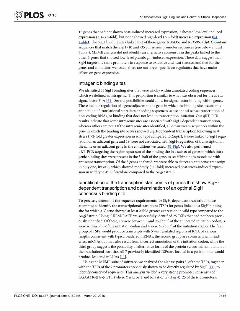
Intragenic binding sitesWe identified 33 SigH binding sites that were wholly within annotated coding sequences,which we defined as intragenic. This proportion is similar to what was observed for the E. colisigma factor FliA [34]. Several possibilities could allow for sigma factor binding within genes.These include regulation of a gene adjacent to the gene in which the binding site occurs, mis-annotation of translational start sites or coding sequences, sense or anti-sense transcription ofnon-coding RNAs, or binding that does not lead to transcription initiation. Our qRT-PCRresults indicate that some intragenic sites are associated with SigH-dependent transcription,whereas others are not. Of the intragenic sites identified, 10 downstream sequences within thegene in which the binding site occurs showed SigH-dependent transcription following heatstress (>2-fold greater expression in wild type compared to ΔsigH), 4 were linked to SigH regu-lation of an adjacent gene and 19 were not associated with SigH-regulation of transcription inthe same or an adjacent gene in the conditions we tested (S1 Fig). We also performedqRT-PCR targeting the region upstream of the binding site in a subset of genes in which intra-genic binding sites were present in the 3’ half of the gene, to see if binding is associated withantisense transcription. Of the 8 genes analyzed, we were able to detect an anti-sense transcriptin only one, Rv3056, which showed modestly (3.0-fold) increased heat stress-induced expres-sion in wild typeM. tuberculosis compared to the ΔsigH strain.
Identification of the transcription start points of genes that show SigH-dependent transcription and determination of an optimal SigHconsensus binding siteTo precisely determine the sequence requirements for SigH-dependent transcription, weattempted to identify the transcriptional start point (TSP) for genes linked to a SigH bindingsite for which a 3’ gene showed at least 2-fold greater expression in wild type compared to theΔsigH strain. Using 5’ RLM-RACE we successfully identified 25 TSPs that had not been previ-ously identified. Of these, 18 were between 5 and 250 bp 5’ of the annotated initiation codon, 3were within 5 bp of the initiation codon and 4 were>5 bp 3’ of the initiation codon. The firstgroup of TSPs would produce transcripts with 5’-untranslated regions of RNA of variouslengths consistent with typical leadered mRNAs, the second group are consistent with lead-erless mRNAs but may also result from incorrect annotation of the initiation codon, while thethird group suggests the possibility of alternative forms of the protein versus mis-annotation ofthe translational start site. All 7 previously identified TSPs are located in a position that wouldproduce leadered mRNAs [11].
Using the MEME suite of software, we analyzed the 40 base pairs 5’ of these TSPs, togetherwith the TSPs of the 7 promoters previously shown to be directly regulated by SigH [11], toidentify conserved sequences. This analysis yielded a very strong promoter consensus ofGGAAYR-(N17)-GTT (where Y is C or T and R is A or G) (Fig 4). 25 of these promoters,
M. tuberculosis SigH Regulon and Control of Stress Responses
PLOS ONE | DOI:10.1371/journal.pone.0152145 March 22, 2016 10 / 16
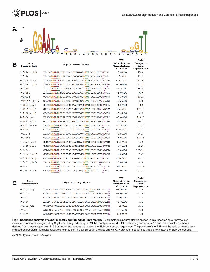
Fig 4. Sequence analysis of experimentally confirmed SigH promoters. 25 promoters experimentally identified in this research plus 7 previouslyidentified promoters recognized by SigH were analyzed using the MEME analysis suite.A. LOGO showing consensus -10 and -35 promoter elementsderived from these sequences. B. 25 promoter sequences that match the SigH consensus sequences. The position of the TSP and the ratio of heat stress-induced expression in wild type relative to expression in a ΔsigH strain are also shown.C. 7 promoter sequences that do not match the SigH consensus.
doi:10.1371/journal.pone.0152145.g004
M. tuberculosis SigH Regulon and Control of Stress Responses
PLOS ONE | DOI:10.1371/journal.pone.0152145 March 22, 2016 11 / 16
including 18 newly identified in this work, have sequences that match both the -10 and -35consensus sequence elements with appropriate spacing between these sequences. The remain-ing 7 promoters newly identified in this work do not match the consensus (Fig 4). In somecases, partial sequences corresponding to the -10 or -35 consensus are present at the appropri-ate position relative to the transcription start point. In addition to the sequence requirementsfor SigH binding indicated by these data, a key feature of the consensus is the restricted spacingbetween the -10 trimer and -35 hexamer. Of the 25 experimentally identified promoters, in 19there are 17 bases between the -35 hexamer and the -10 trimer. In two promoters there arebases and in 4 promoters there are 18 bases separating these promoter elements.
Comparison of SigH-dependent transcription following heat stress of the experimentallyvalidated promoters that contain the both the -10 and -35 consensus promoter sequences andthose that do not, shows a striking difference between these groups. The genes 3’ of the 25 pro-moters that match the consensus show a mean 173-fold SigH-dependent induction followingheat stress, with only 1 showing less than 8-fold induction (Fig 4). Among the 8 promotersthat do not match the consensus sequences, the median induction is 7.1 fold with only 3 show-ing greater than 3-fold induction. Similarly, the median binding enrichment in the ChIP-Seqexperiments was lower at sites containing promoters that lacked both the consensus promotersequences (11.6-fold) compared to sites that do match both of these sequences (18.6-fold).
DiscussionIn this work we comprehensively identified SigH binding sites throughout theM. tuberculosisgenome. We then determined whether these binding sites were associated with SigH-regulatedstress-induced transcription, and for those that showed increased expression in wild type com-pared a ΔsigH strain, we attempted to identify the transcriptional start point and evaluate can-didate promoter sequences 5’ of these genes. Based on the presence of a SigH binding site,SigH-dependent stress-induced transcription and promoter sequences that match the consen-sus sequences recognized by SigH, we identified for the first time at least 18 genes that aredirectly regulated by SigH, increasing the direct SigH regulon by over 3-fold. Though some ofthese genes were previously shown to be differentially expressed in wild typeM. tuberculosiscompared to a ΔsigH strain, we also identified several genes that are regulated by SigH thatwere not previously suspected of being part of the SigH regulon. Though our genome-wideapproach with follow-up validation has allowed us to markedly expand the number of genesdirectly regulated by SigH, these genes may not comprise the entire SigH regulon. For example,clpB, which has been shown to be negatively regulated by the HspR heat shock repressor thatbinds to a site that overlaps with the SigH-regulated promoter [11, 30] was not identified inour ChIP-Seq experiments. Other genes that are co-regulated by transcriptional repressors thatocclude SigH binding sites may not have been identified under the conditions we used. Forgenes linked to SigH binding sites that show SigH-dependent stress-induced expression, it islikely that our 5’-RACE experiments did not detect some SigH-regulated promoters.
The newly identified SigH-regulated genes markedly expand the functional roles of SigH-regulated gene expression in response to stress. Prior results had demonstrated that SigH isimportant forM. tuberculosis to re-establish redox homeostasis following oxidative stress,through regulation of several components of the thioredoxin/thioredoxin reductase system, aswell as other oxidoreductases, and for repair or removal of damaged proteins by the SigH-dependent induction of several heat shock proteins/chaperones [11, 12]. Our findings in thiswork indicate a broader role for direct regulation by SigH in recovery from oxidative and otherstresses. Direct SigH regulation of the DNA repair gene udgB for example, a gene that was notpreviously suspected to be SigH-regulated, indicates a role for SigH in recovery of genome
M. tuberculosis SigH Regulon and Control of Stress Responses
PLOS ONE | DOI:10.1371/journal.pone.0152145 March 22, 2016 12 / 16
integrity following genotoxic stress. SigH regulation of rpmE, which encodes a zinc-bindingribosomal protein gene indicates a role for SigH in recovery of translation by replenishment ofdamaged ribosomal proteins. The orthologue of this gene was shown to be regulated in Strepto-myces coelicolor by the SigH orthologue SigR [35], Also striking is the direct role of SigH in reg-ulating sulfur metabolism in the mycobacterial cell, including control of genes predicted to beinvolved in sulfur transport and incorporation of sulfur into molybdopterin. A critical roleSigH appears to be the synthesis and salvage of cysteine-containing proteins, which are particu-larly susceptible to oxidative stress. Our data show direct SigH regulation of genes encoding analternative cysteine biosynthetic pathway encoded by the operon Rv1334-Rv1336 (mec, cysOand cysM) and Rv3206c (moeZ ormoeB1) [36]. Further demonstrating a role for SigH inreplenishing sulfur-containing amino acids, we found that SigH also directly regulatesmsrB(Rv2674), which encodes an enzyme that repairs oxidized methionine residues.
Though 41 of 69 of SigH-binding sites were linked to SigH-dependent gene expression, theexpression of genes adjacent to many binding sites did not show evidence of SigH-regulationin response to heat stress. Testing the expression of several of these genes in response to plum-bagin-induced oxidative stress showed low level SigH-dependent expression for 7, howevermost did not show increased expression in response to either stress. The lack of gene regulationlinked to a subset of binding sites of sigma factors or transcription factors has been a consistentobservation in many ChIP-Seq studies [31–34]. In some cases these may be regulatory siteswhere transcription of the gene of interest is co-regulated by other transcription factors so thatchanges in gene expression were not observed under the conditions tested. For other bindingsites that we identified in the ChIP-Seq experiments where transcription regulation is not evi-dent, it is likely that binding does not indicate a site at which SigH regulates gene expression.Transcription initiation is a multi-step process that requires binding of RNA polymerase viasigma-promoter interactions, DNA melting and initiation of elongation, which is frequentlyaborted without the RNA polymerase being able to escape the promoter to transcribe full-length mRNAs [37, 38]. Thus, some sites to which SigH binds weakly may not achieve the ini-tial steps required for transcription initiation. This inference is supported by the markedlygreater frequency and magnitude of SigH-dependent transcription following heat stress frombinding sites that match the SigH promoter consensus sequences compared to those that donot. Conversely sites to which SigH binds strongly that are not linked to regulated transcriptionmay have sequence or structural characteristics that do not allow efficient completion of thesteps required for transcription initiation.
In previous work, based on a small number of genes directly regulated by SigH, we identifieda SigH binding consensus and subsequently performed extensive mutagenesis of the -10 and-35 sequences [11, 19]. This research identified optimal promoter sequences for SigH and forSigE, and identified position 6 of the -35 hexamer as important for distinguishing SigH fromSigE dependent promoters, though some promoters are recognized by both. The much largernumber of SigH-regulated promoters experimentally identified in this study, derived fromSigH-controlled genes identified using an unbiased genome-wide methodology, has allowed usto define the optimal and tolerated SigH promoter sequences more precisely. Notably, whileour results show that the optimal consensus is GGAAYR-(N17-18)-GTT, it is clear that varia-tion that was not previously known can be tolerated at the 2nd, 5th and 6th positions of the -35hexamer and at each position in the -10 trimer. In addition, while our previous results showedthat SigE prefers a pyrimidine at the 6th position of the -35 hexamer [19], results from thisstudy show that SigH strongly prefers a purine at this position.
In this work we have identified a greatly expanded direct SigH regulon that markedly broad-ens the role of SigH in stress response and recovery. In particular newly identified SigH-regu-lated genes indicate a direct role for SigH in DNA repair, sulfur metabolism, synthesis and
M. tuberculosis SigH Regulon and Control of Stress Responses
PLOS ONE | DOI:10.1371/journal.pone.0152145 March 22, 2016 13 / 16
salvage of sulfur-containing amino acids and recovery of translation. In addition to these spe-cific new insights into SigH function, this research demonstrates the value of genome-wideapproaches to understanding bacterial gene regulation, while highlighting the importance oftargeted follow-up experiments to more fully understand the implications of the genome leveldata.
Supporting InformationS1 Fig. Map of genomic loci containing SigH binding sites identified by ChIP-Seq. For eachSigH binding site, the region containing a binding site is shown. Black bars under each locusindicate the location of the binding site. The expression ratio (fold induction following heatstress in wild type divided by fold induction following heat stress in the ΔsigH strain) is shownfor genes adjacent to each binding site. Where an expression ratio>2 was observed, the arrowis black; where an expression ratio is<2 the arrow is grey. Arrows corresponding to genes thatwere not tested are white.(PDF)
S1 Table. Primers used for qRT-PCR.(DOCX)
S2 Table. Primers used for 5’-RACE.(PDF)
S3 Table. ChIP-Seq binding data.(XLSX)
S4 Table. Gene expression following oxidative stress.(DOCX)
AcknowledgmentsThe authors appreciate the helpful discussions of this work by members of the Husson andGalagan laboratories.
Author ContributionsConceived and designed the experiments: JDS AKS STP JEG RNH. Performed the experi-ments: JDS AKS STP SR. Analyzed the data: JDS AKS STP AL MWP ALCG LP SR JEG RNH.Wrote the paper: JDS AKS JEG RNH.
References1. Via LE, Lin PL, Ray SM, Carrillo J, Allen SS, Eum SY, et al. Tuberculous granulomas are hypoxic in
guinea pigs, rabbits, and nonhuman primates. Infect Immun. 2008; 76(6):2333–40. PMID: 18347040.doi: 10.1128/IAI.01515-07
2. Nathan C, Shiloh MU. Reactive oxygen and nitrogen intermediates in the relationship between mam-malian hosts and microbial pathogens. Proc Natl Acad Sci USA. 2000; 97(16):8841–8. PMID:10922044.
3. Camus JC, Pryor MJ, Medigue C, Cole ST. Re-annotation of the genome sequence ofMycobacteriumtuberculosis H37Rv. Microbiology. 2002; 148(Pt 10):2967–73. PMID: 12368430.
4. Rodrigue S, Provvedi R, Jacques P- E, Gaudreau L, Manganelli R. The sigma factors ofMycobacte-rium tuberculosis. FEMSMicrobiology Reviews. 2006; 30(6):926–41. doi: 10.1111/j.1574-6976.2006.00040.x PMID: 17064287.
5. Manganelli R. Sigma factors: Key Molecules inMycobacterium tuberculosis Physiology and Virulence.Microbiol Spectr. 2014; 2(1). doi: 10.1128/microbiolspec.MGM2-0007-2013
M. tuberculosis SigH Regulon and Control of Stress Responses
PLOS ONE | DOI:10.1371/journal.pone.0152145 March 22, 2016 14 / 16
6. Lonetto MA, Brown KL, Rudd KE, Buttner MJ. Analysis of the Streptomyces coelicolor sigE genereveals the existence of a subfamily of eubacterial RNA polymerase sigma factors involved in the regu-lation of extracytoplasmic functions. Proc Natl Acad Sci USA. 1994; 91(16):7573–7. PMID: 8052622;PubMed Central PMCID: PMC44444.
7. Paget MSB, Helmann JD. The sigma70 family of sigma factors. Genome Biol. 2003; 4(1):203. PMID:12540296; PubMed Central PMCID: PMC151288.
8. Helmann JD. Anti-sigma factors. Curr Opin Microbiol. 1999; 2(2):135–41. doi: 10.1016/S1369-5274(99)80024-1 PMID: 10322161.
9. Paget MS, Bae JB, Hahn MY, Li W, Kleanthous C, Roe JH, et al. Mutational analysis of RsrA, a zinc-binding anti-sigma factor with a thiol-disulphide redox switch. Mol Microbiol. 2001; 39(4):1036–47.PMID: 11251822
10. Song T, Dove SL, Lee KH, Husson RN. RshA, an anti-sigma factor that regulates the activity of themycobacterial stress response sigma factor SigH. Mol Microbiol. 2003; 50(3):949–59. PMID:14617153.
11. Raman S, Song T, Puyang X, Bardarov S, Jacobs Jr. W, Husson R. The alternative sigma factor SigHregulates major components of the oxidative and heat stress responses inMycobacterium tuberculo-sis. J Bacteriol. 2001; 183(20):6119–25. PMID: 11567012
12. Manganelli R, Voskuil MI, Schoolnik GK, Dubnau E, Gomez M, Smith I. Role of the extracytoplasmic-function sigma Factor sigmaH inMycobacterium tuberculosis global gene expression. Mol Microbiol.2002; 45(2):365–74. PMID: 12123450
13. Park ST, Kang CM, Husson RN. Regulation of the SigH stress response regulon by an essential proteinkinase inMycobacterium tuberculosis. Proc Natl Acad Sci U S A. 2008; 105(35):13105–10. PMID:18728196. doi: 10.1073/pnas.0801143105
14. Kaushal D, Schroeder BG, Tyagi S, Yoshimatsu T, Scott C, Ko C, et al. Reduced immunopathologyand mortality despite tissue persistence in aMycobacterium tuberculosismutant lacking alternativesigma factor, SigH. Proc Natl Acad Sci USA. 2002; 99(12):8330–5. doi: 10.1073/pnas.102055799PMID: 12060776; PubMed Central PMCID: PMC123067.
15. Mehra S, Golden NA, Stuckey K, Didier PJ, Doyle LA, Russell-Lodrigue KE, et al. TheMycobacteriumtuberculosis Stress Response Factor SigH Is Required for Bacterial Burden asWell as Immunopathol-ogy in Primate Lungs. J Infect Dis. 2012. doi: 10.1093/infdis/jis102 PMID: 22402035.
16. Dutta NK, Mehra S, Martinez AN, Alvarez X, Renner NA, Morici LA, et al. The stress-response factorSigH modulates the interaction betweenMycobacterium tuberculosis and host phagocytes. PLoSONE. 2012; 7(1):e28958. doi: 10.1371/journal.pone.0028958 PMID: 22235255; PubMed CentralPMCID: PMC3250399.
17. Kaushal D, Foreman TW, Gautam US, Alvarez X, Adekambi T, Rangel-Moreno J, et al. Mucosal vacci-nation with attenuated Mycobacterium tuberculosis induces strong central memory responses and pro-tects against tuberculosis. Nature Communications. 2015; 6:8533. doi: 10.1038/ncomms9533 PMID:26460802; PubMed Central PMCID: PMCPMC4608260.
18. Ghosh P, Wu C-w, Talaat AM. Key role for the alternative sigma factor, SigH, in the intracellular life ofMycobacterium avium subsp. paratuberculosis during macrophage stress. Infect Immun. 2013; 81(6):2242–57. doi: 10.1128/IAI.01273-12 PMID: 23569115; PubMed Central PMCID: PMC3676012.
19. Song T, Song SE, Raman S, Anaya M, Husson RN. Critical role of a single position in the -35 elementfor promoter recognition byMycobacterium tuberculosis SigE and SigH. J Bacteriol. 2008; 190(6):2227–30. PMID: 18192397. doi: 10.1128/JB.01642-07
20. Galagan JE, Minch K, Peterson M, Lyubetskaya A, Azizi E, Sweet L, et al. TheMycobacterium tuber-culosis regulatory network and hypoxia. Nature. 2013; 499(7457):178–83. doi: 10.1038/nature12337PMID: 23823726; PubMed Central PMCID: PMC4087036.
21. Klotzsche M, Ehrt S, Schnappinger D. Improved tetracycline repressors for gene silencing in mycobac-teria. Nucleic Acids Research. 2009; 37(6):1778–88. doi: 10.1093/nar/gkp015 PMID: 19174563
22. Galagan J, Lyubetskaya A, Gomes A. ChIP-Seq and the Complexity of Bacterial Transcriptional Regu-lation. Current topics in microbiology and immunology. 2013; 363:43–68. Epub 2012/09/18. doi: 10.1007/82_2012_257 PMID: 22983621.
23. Langmead B, Salzberg SL. Fast gapped-read alignment with Bowtie 2. Nat Meth. 2012; 9(4):357–9.doi: 10.1038/nmeth.1923 PMID: 22388286; PubMed Central PMCID: PMC3322381.
24. Li H, Handsaker B, Wysoker A, Fennell T, Ruan J, Homer N, et al. The Sequence Alignment/Map for-mat and SAMtools. Bioinformatics. 2009; 25(16):2078–9. doi: 10.1093/bioinformatics/btp352 PMID:19505943
M. tuberculosis SigH Regulon and Control of Stress Responses
PLOS ONE | DOI:10.1371/journal.pone.0152145 March 22, 2016 15 / 16
25. Gomes ALC, Abeel T, Peterson M, Azizi E, Lyubetskaya A, Carvalho L, et al. Decoding ChIP-seq with adouble-binding signal refines binding peaks to single-nucleotides and predicts cooperative interaction.Genome Research. 2014. doi: 10.1101/gr.161711.113 PMID: 25024162.
26. Bailey TL, Boden M, Buske FA, Frith M, Grant CE, Clementi L, et al. MEME SUITE: tools for motif dis-covery and searching. Nucleic Acids Research. 2009; 37(Web Server issue):W202–8. doi: 10.1093/nar/gkp335 PMID: 19458158; PubMed Central PMCID: PMC2703892.
27. Bailey TL, Elkan C. Fitting a mixture model by expectation maximization to discover motifs in biopoly-mers. Proc Int Conf Intell Syst Mol Biol. 1994; 2:28–36. PMID: 7584402.
28. Robinson JT, Thorvaldsdóttir H, Winckler W, Guttman M, Lander ES, Getz G, et al. Integrative geno-mics viewer. Nat Biotechnol. 2011; 29(1):24–6. doi: 10.1038/nbt.1754 PMID: 21221095; PubMed Cen-tral PMCID: PMC3346182.
29. Arany Z. High-Throughput Quantitative Real-Time PCR in Current Protocols in Human Genetics.Hoboken, NJ, USA: JohnWiley & Sons, Inc.; 2001.
30. Stewart GR, Wernisch L, Stabler R, Mangan JA, Hinds J, Laing KG, et al. Dissection of the heat-shockresponse inMycobacterium tuberculosis using mutants and microarrays. Microbiology (Reading,Engl). 2002; 148(Pt 10):3129–38. PMID: 12368446.
31. Hartkoorn RC, Sala C, Uplekar S, Busso P, Rougemont J, Cole ST. Genome-wide definition of the SigFregulon inMycobacterium tuberculosis. J Bacteriol. 2012; 194(8):2001–9. doi: 10.1128/JB.06692-11PMID: 22307756; PubMed Central PMCID: PMC3318452.
32. Minch KJ, Rustad TR, Peterson EJR, Winkler J, Reiss DJ, Ma S, et al. The DNA-binding network ofMycobacterium tuberculosis. Nature Communications. 2015; 6:5829. doi: 10.1038/ncomms6829PMID: 25581030.
33. Kahramanoglou C, Cortes T, Matange N, Hunt DM, Visweswariah SS, Young DB, et al. Genomic map-ping of cAMP receptor protein (CRPMt) inMycobacterium tuberculosis: relation to transcriptional startsites and the role of CRPMt as a transcription factor. Nucleic Acids Research. 2014; 42(13):8320–9.doi: 10.1093/nar/gku548 PMID: 24957601; PubMed Central PMCID: PMC4117774.
34. Fitzgerald DM, Bonocora RP, Wade JT. Comprehensive mapping of the Escherichia coli flagellar regu-latory network. PLoS Genet. 2014; 10(10):e1004649. doi: 10.1371/journal.pgen.1004649 PMID:25275371; PubMed Central PMCID: PMC4183435.
35. Paget MS, Molle V, Cohen G, Aharonowitz Y, Buttner MJ. Defining the disulphide stress response inStreptomyces coelicolor A3(2): identification of the sigmaR regulon. Mol Microbiol. 2001; 42(4):1007–20. PMID: 11737643
36. Burns KE, Baumgart S, Dorrestein PC, Zhai H, McLafferty FW, Begley TP. Reconstitution of a new cys-teine biosynthetic pathway inMycobacterium tuberculosis. J Am Chem Soc. 2005; 127(33):11602–3.doi: 10.1021/ja053476x PMID: 16104727; PubMed Central PMCID: PMC2536522.
37. Hsu LM. Promoter clearance and escape in prokaryotes. Biochim Biophys Acta. 2002; 1577(2):191–207. PMID: 12213652.
38. Revyakin A, Liu C, Ebright RH, Strick TR. Abortive initiation and productive initiation by RNA polymer-ase involve DNA scrunching. Science. 2006; 314(5802):1139–43. doi: 10.1126/science.1131398PMID: 17110577; PubMed Central PMCID: PMC2754787.
M. tuberculosis SigH Regulon and Control of Stress Responses
PLOS ONE | DOI:10.1371/journal.pone.0152145 March 22, 2016 16 / 16