Complications of Strabismus Surgery - Het Ot en Zien .... DeFaber_Strab OK Complications.pdf · 1...
13
1 Complications of Strabismus Surgery Tjeerd de Faber, Martha Tjon Rutger van Ruyven Alexis Damanakis Complications of Strabismus Surgery Tjeerd de Faber, Martha Tjon Rutger van Ruyven Alexis Damanakis Wat vind je erger? • Ondercorrectie • Overcorrectie
Transcript of Complications of Strabismus Surgery - Het Ot en Zien .... DeFaber_Strab OK Complications.pdf · 1...
1
Complicationsof Strabismus Surgery
Tjeerd de Faber, Martha Tjon Rutger van Ruyven Alexis Damanakis
Complicationsof Strabismus Surgery
Tjeerd de Faber, Martha Tjon Rutger van Ruyven Alexis Damanakis
Wat vind je erger?
• Ondercorrectie • Overcorrectie
2
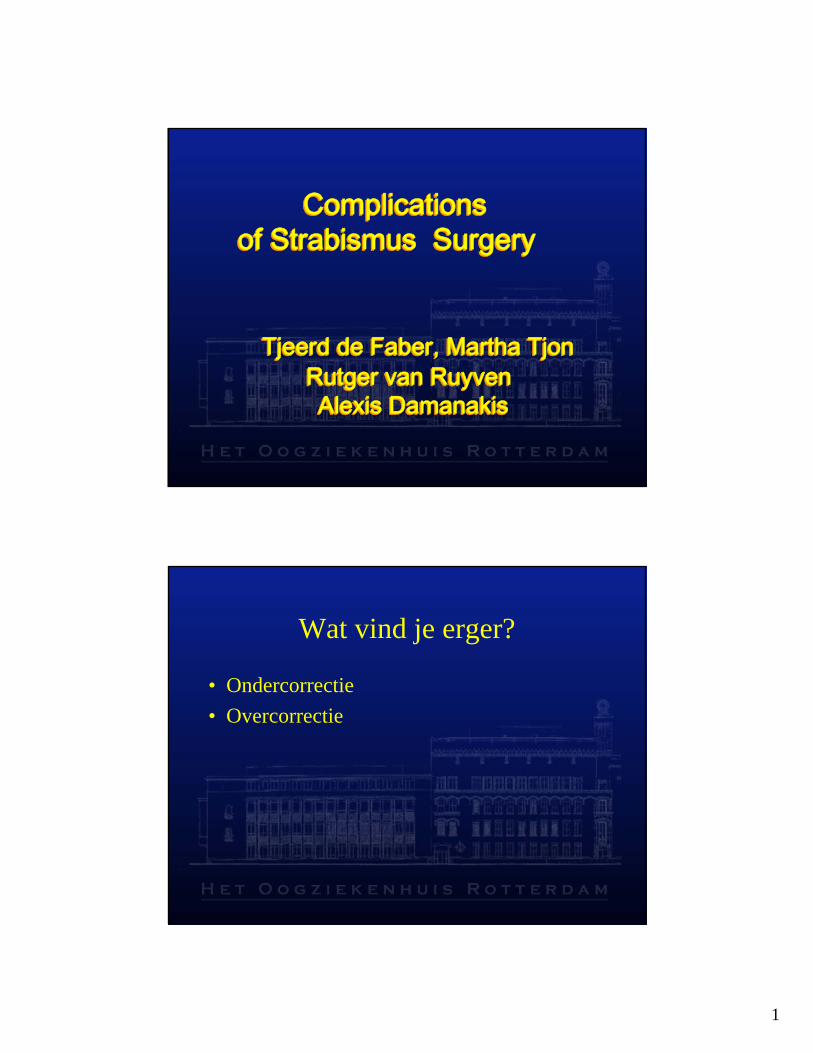
DELLENCorneal dellen are small areas of thinning associated with adjacent conjunctival swelling
DELLENCorneal dellen are small areas of thinning associated with adjacent conjunctival swelling
Prevention• Smooth closure of
the conjunctiva• Fornix incision
Prevention• Smooth closure of
the conjunctiva• Fornix incision
Treatment• Occlusion of the eye for one to two
days• Lubricating drops• Excision of the offending
conjunctiva
Treatment• Occlusion of the eye for one to two
days• Lubricating drops• Excision of the offending
conjunctiva
Visible line of previousmuscle insertion
Visible line of previousmuscle insertion
• When a muscle is recessed the former site of theoriginal muscle insertion becomes visiblethrough the conjunctiva
• There is no treatment for this minor complicationand reassurance and explanation are all that is required
• When a muscle is recessed the former site of theoriginal muscle insertion becomes visiblethrough the conjunctiva
• There is no treatment for this minor complicationand reassurance and explanation are all that is required
3
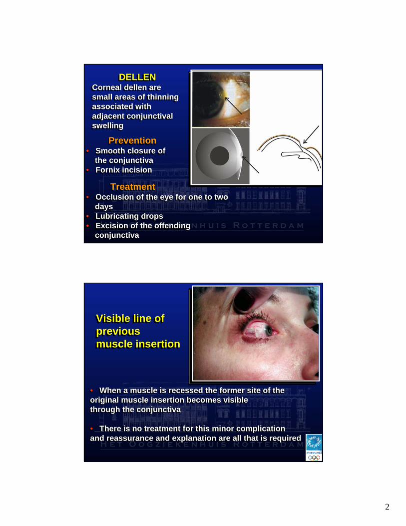
• Tenon’s capsule may prolapse through conjunctival woundsas a result of edema during the postoperative period
• Tenon’s capsule may prolapse through conjunctival woundsas a result of edema during the postoperative period
PreventionPrevention• Conjunctival closure should be meticulous with accurateapposition of the tissues
• Conjunctival closure should be meticulous with accurateapposition of the tissues
TreatmentTreatment• In most cases the prolapsed Tenon’s capsule will retractor disappear spontaneously without surgical intervention
• In most cases the prolapsed Tenon’s capsule will retractor disappear spontaneously without surgical intervention
Prolapse of Tenon’s capsuleProlapse of Tenon’s capsule
Increased vascularity of the conjunctivaIncreased vascularity of the conjunctiva
• Increased vascularity of the conjunctiva is common after strabismus operation
• The eye may not be constantly red but becomes erythematous with exposure to irritants
• Increased vascularity of the conjunctiva is common after strabismus operation
• The eye may not be constantly red but becomes erythematous with exposure to irritants

4
Chronic suture granuloma
• Localized, hyperemicmass over the muscleinsertion
• It represents a nonallergic foreign body reaction to the suture material
Chronic suture granuloma
• Localized, hyperemicmass over the muscleinsertion
• It represents a nonallergic foreign body reaction to the suture material
Prevention• Use of synthetic absorbable suturesPrevention• Use of synthetic absorbable sutures
Treatment• Topical corticosteroid drops or excision of the
granuloma
Treatment• Topical corticosteroid drops or excision of the
granuloma
Scleral perforation
Occurs intraoperativelyduring reattachment of a rectus muscle
Scleral perforation
Occurs intraoperativelyduring reattachment of a rectus muscle
Prevention• Prevent an excessively deep scleral entry by keeping the
needle parallel to the sclera• The scleral needle track should allow visualization of the
needle through the entire length
Prevention• Prevent an excessively deep scleral entry by keeping the
needle parallel to the sclera• The scleral needle track should allow visualization of the
needle through the entire length
Treatment• Dilate the pupil and study the retina• No treatment should be carried out unless retinal
detachment is present or impending
Treatment• Dilate the pupil and study the retina• No treatment should be carried out unless retinal
detachment is present or impending
5
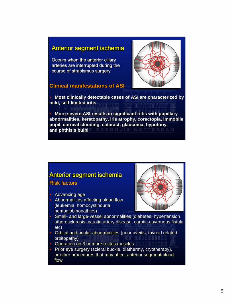
Anterior segment ischemia
Occurs when the anterior ciliary arteries are interrupted during the course of strabismus surgery
Anterior segment ischemia
Occurs when the anterior ciliary arteries are interrupted during the course of strabismus surgery
Clinical manifestations of ASI
• Most clinically detectable cases of ASI are characterized bymild, self-limited iritis
• More severe ASI results in significant iritis with pupillary abnormalities, keratopathy, iris atrophy, corectopia, immobilepupil, corneal clouding, cataract, glaucoma, hypotony, and phthisis bulbi
Anterior segment ischemiaAnterior segment ischemiaRisk factors
• Advancing age• Abnormalities affecting blood flow
(leukemia, homocystinouria, hemoglobinopathies)
• Small- and large-vessel abnormalities (diabetes, hypertensionatherosclerosis, carotid artery disease, carotic-cavernous fistula,etc)
• Orbital and ocular abnormalities (prior uveitis, thyroid related orbitopathy)
• Operation on 3 or more rectus muscles• Prior eye surgery (scleral buckle, diathermy, cryotherapy),
or other procedures that may affect anterior segment bloodflow
Risk factors
• Advancing age• Abnormalities affecting blood flow
(leukemia, homocystinouria, hemoglobinopathies)
• Small- and large-vessel abnormalities (diabetes, hypertensionatherosclerosis, carotid artery disease, carotic-cavernous fistula,etc)
• Orbital and ocular abnormalities (prior uveitis, thyroid related orbitopathy)
• Operation on 3 or more rectus muscles• Prior eye surgery (scleral buckle, diathermy, cryotherapy),
or other procedures that may affect anterior segment bloodflow

6
Anterior segment ischemiaAnterior segment ischemiaPrevention
• Limiting the number of rectus musclesoperated on
• Fornix incision• Use of botulinum toxin• Anterior ciliary vessel sparing
Prevention
• Limiting the number of rectus musclesoperated on
• Fornix incision• Use of botulinum toxin• Anterior ciliary vessel sparing
Treatment
• Cycloplegic agents• Topical and systemic corticosteroids• Hyperbaric oxygen• Control of intraocular pressure
Treatment
• Cycloplegic agents• Topical and systemic corticosteroids• Hyperbaric oxygen• Control of intraocular pressure
Slipped muscleSlipped muscle
A slipped muscle occurs when the muscle capsule rather than the muscle itself is sutured to the scleraA slipped muscle occurs when the muscle capsule rather than the muscle itself is sutured to the sclera
Prevention• Place sutures securely into muscle or tendon tissue
Prevention• Place sutures securely into muscle or tendon tissue
Treatment• Surgical exploration can identify the muscle• Reattachment of the muscle to the proper
scleral location
Treatment• Surgical exploration can identify the muscle• Reattachment of the muscle to the proper
scleral location
7
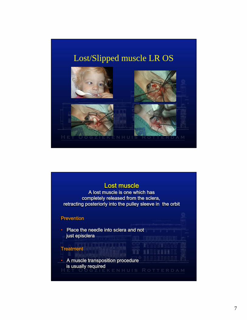
Lost/Slipped muscle LR OS
Lost muscleA lost muscle is one which has
completely released from the sclera, retracting posteriorly into the pulley sleeve in the orbit
Lost muscleA lost muscle is one which has
completely released from the sclera, retracting posteriorly into the pulley sleeve in the orbit
Prevention
• Place the needle into sclera and notjust episclera
Prevention
• Place the needle into sclera and notjust episclera
Treatment
• A muscle transposition procedureis usually required
Treatment
• A muscle transposition procedureis usually required

8
Postoperative appearance in a case of a lost right medial rectus muscle, after rectus muscle
transposition operation
(Hummelsheim operation augmented with resection of the transposed medial halves of the vertical recti)
Postoperative appearance in a case of a lost right medial rectus muscle, after rectus muscle
transposition operation
(Hummelsheim operation augmented with resection of the transposed medial halves of the vertical recti)
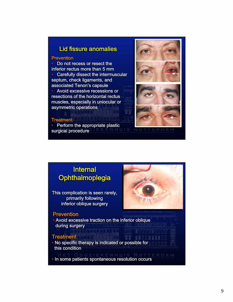
Lid fissure anomalies
• Lid displacement after vertical rectus surgery occurs in the samedirection as the shift in the insertionof the vertical rectus muscle
Lid fissure anomalies
• Lid displacement after vertical rectus surgery occurs in the samedirection as the shift in the insertionof the vertical rectus muscle
• MR or LR recessions may inducewidening of the palpebral fissure• MR or LR recessions may inducewidening of the palpebral fissure
• MR or LR resections may producenarrowing of the palpebral fissure• MR or LR resections may producenarrowing of the palpebral fissure
9
Lid fissure anomaliesLid fissure anomaliesPrevention• Do not recess or resect the inferior rectus more than 5 mm• Carefully dissect the intermuscularseptum, check ligaments, and associated Tenon’s capsule• Avoid excessive recessions orresections of the horizontal rectusmuscles, especially in uniocular or asymmetric operations
Prevention• Do not recess or resect the inferior rectus more than 5 mm• Carefully dissect the intermuscularseptum, check ligaments, and associated Tenon’s capsule• Avoid excessive recessions orresections of the horizontal rectusmuscles, especially in uniocular or asymmetric operations
Treatment• Perform the appropriate plasticsurgical procedure
Treatment• Perform the appropriate plasticsurgical procedure
Internal Ophthalmoplegia
This complication is seen rarely, primarily following
inferior oblique surgery
Internal Ophthalmoplegia
This complication is seen rarely, primarily following
inferior oblique surgery
Prevention• Avoid excessive traction on the inferior oblique
during surgery
Prevention• Avoid excessive traction on the inferior oblique
during surgery
Treatment• No specific therapy is indicated or possible for
this condition
• In some patients spontaneous resolution occurs
Treatment• No specific therapy is indicated or possible for
this condition
• In some patients spontaneous resolution occurs
10
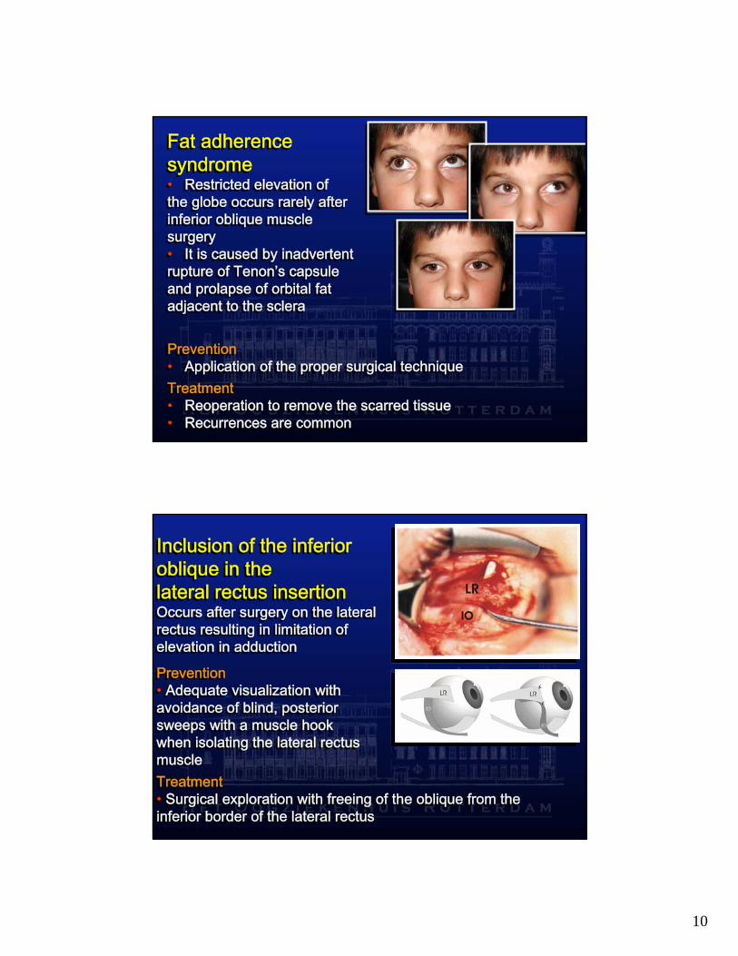
Fat adherence syndrome• Restricted elevation ofthe globe occurs rarely after inferior oblique muscle surgery• It is caused by inadvertent rupture of Tenon’s capsule and prolapse of orbital fat adjacent to the sclera
Fat adherence syndrome• Restricted elevation ofthe globe occurs rarely after inferior oblique muscle surgery• It is caused by inadvertent rupture of Tenon’s capsule and prolapse of orbital fat adjacent to the sclera
Prevention• Application of the proper surgical techniquePrevention• Application of the proper surgical techniqueTreatment• Reoperation to remove the scarred tissue• Recurrences are common
Treatment• Reoperation to remove the scarred tissue• Recurrences are common
Inclusion of the inferior oblique in the lateral rectus insertionOccurs after surgery on the lateral rectus resulting in limitation of elevation in adduction
Inclusion of the inferior oblique in the lateral rectus insertionOccurs after surgery on the lateral rectus resulting in limitation of elevation in adduction
Prevention• Adequate visualization withavoidance of blind, posterior sweeps with a muscle hookwhen isolating the lateral rectusmuscle
Prevention• Adequate visualization withavoidance of blind, posterior sweeps with a muscle hookwhen isolating the lateral rectusmuscleTreatment• Surgical exploration with freeing of the oblique from the inferior border of the lateral rectus
Treatment• Surgical exploration with freeing of the oblique from the inferior border of the lateral rectus
11
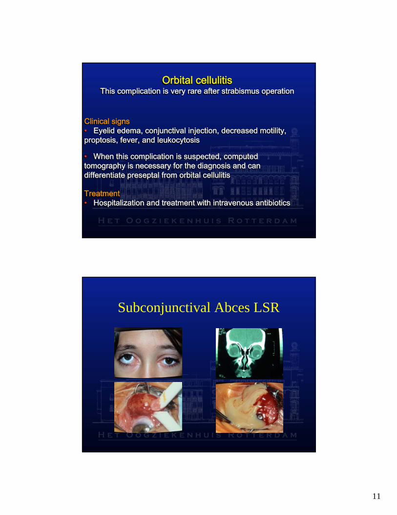
Orbital cellulitisThis complication is very rare after strabismus operation
Orbital cellulitisThis complication is very rare after strabismus operation
Clinical signs• Eyelid edema, conjunctival injection, decreased motility,proptosis, fever, and leukocytosis
Clinical signs• Eyelid edema, conjunctival injection, decreased motility,proptosis, fever, and leukocytosis
• When this complication is suspected, computedtomography is necessary for the diagnosis and can differentiate preseptal from orbital cellulitis
• When this complication is suspected, computedtomography is necessary for the diagnosis and can differentiate preseptal from orbital cellulitis
Treatment• Hospitalization and treatment with intravenous antibioticsTreatment• Hospitalization and treatment with intravenous antibiotics
Subconjunctival Abces LSR
12
EndophthalmitisEndophthalmitis
• This potentially blinding complication generally is associated with inadvertent scleral perforation
• While the incidence of scleral perforation is in the level of 10% the incidence of endophthalmitis after strabismus operation is very low (1 in 30.000)
• This potentially blinding complication generally is associated with inadvertent scleral perforation
• While the incidence of scleral perforation is in the level of 10% the incidence of endophthalmitis after strabismus operation is very low (1 in 30.000)
Postoperative diplopia
• Diplopia is quite common after surgery for comitant strabismus and usually lasts a few minutes, days, or weeks• Persistent postoperative diplopia is rare and more common in adults especially when they are overcorrected
Postoperative diplopia
• Diplopia is quite common after surgery for comitant strabismus and usually lasts a few minutes, days, or weeks• Persistent postoperative diplopia is rare and more common in adults especially when they are overcorrectedPrevention• Preoperative correction of the deviation with prisms
can give a hint for the possibility of postoperative diplopia• In adult patients avoid overcorrection• Use of adjustable sutures
Prevention• Preoperative correction of the deviation with prisms
can give a hint for the possibility of postoperative diplopia• In adult patients avoid overcorrection• Use of adjustable sutures
Treatment• Prisms• Occlusion of one eye• Reoperation
Treatment• Prisms• Occlusion of one eye• Reoperation

13
ESA 2007Mykonos20-23 Mei
www.esa2007.org


![Ocular Complications of Strabismus Surgeryfile.scirp.org/pdf/SS_2014092909455108.pdf · or retrobulbar injections [8]-[10]. Peribulbar anesthesia has been reported with fewer risks](https://static.fdocuments.us/doc/165x107/5aea83cd7f8b9ad73f8d424c/ocular-complications-of-strabismus-retrobulbar-injections-8-10-peribulbar-anesthesia.jpg)