Complications of Rhinosinusitis - Welcome to UTMB … · Complications of Rhinosinusitis All images...
55
Complications of Rhinosinusitis All images obtained via Google search unless otherwise specified. All images used without permission. Viet Pham, M.D. Faculty Advisor: Patricia Maeso, M.D. The University of Texas Medical Branch (UTMB Health) Department of Otolaryngology Grand Rounds Presentation April 22, 2010 Synopsis of Critical Sequelae
Transcript of Complications of Rhinosinusitis - Welcome to UTMB … · Complications of Rhinosinusitis All images...
Complications of
Rhinosinusitis
All images obtained via Google search unless otherwise
specified. All images used without permission.
Viet Pham, M.D.
Faculty Advisor: Patricia Maeso, M.D. The University of Texas Medical Branch (UTMB Health)
Department of Otolaryngology
Grand Rounds Presentation
April 22, 2010
Synopsis of Critical Sequelae
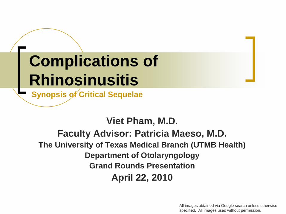
Outline
Sta
ndring S
, ed. G
ray'
s A
nato
my,
40th
Ed
.
Spain
: C
hurc
hill
Liv
ingsto
ne,
2008.
Anatomy
Rhinosinusitis
Acute
Chronic
Complications
Orbital
Intracranial
Bony
Conclusion
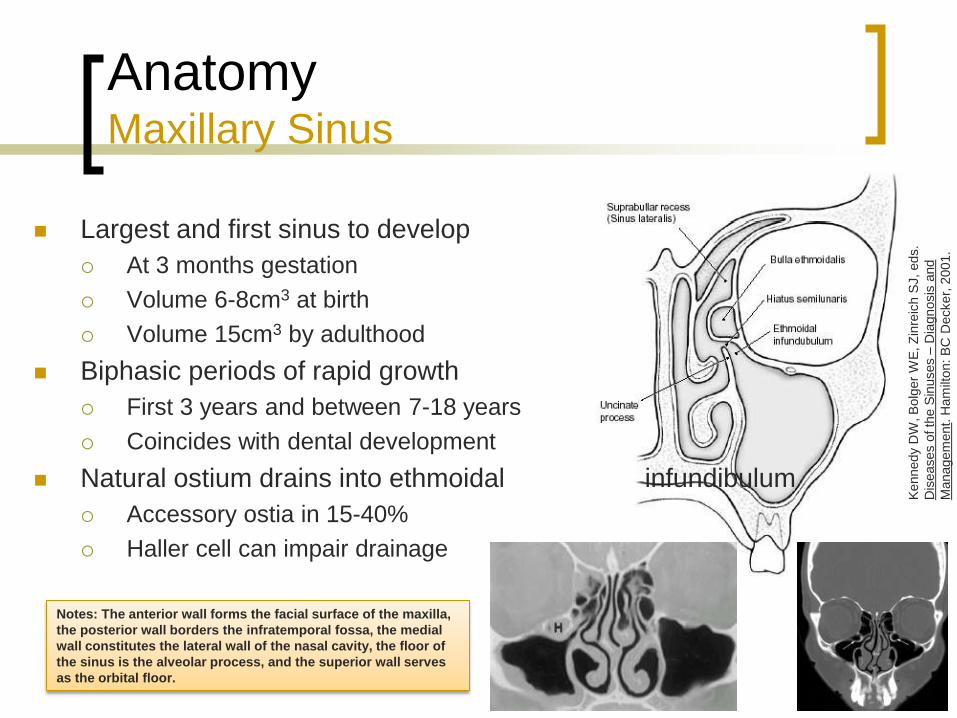
Anatomy Maxillary Sinus
Largest and first sinus to develop
At 3 months gestation
Volume 6-8cm3 at birth
Volume 15cm3 by adulthood
Biphasic periods of rapid growth
First 3 years and between 7-18 years
Coincides with dental development
Natural ostium drains into ethmoidal infundibulum
Accessory ostia in 15-40%
Haller cell can impair drainage
Kennedy
DW
, B
olg
er
WE
, Z
inre
ich S
J, eds.
Dis
eases o
f th
e S
inuses –
Dia
gnosis
and
Managem
ent.
Ham
ilton: B
C D
ecker,
2001.
Notes: The anterior wall forms the facial surface of the maxilla,
the posterior wall borders the infratemporal fossa, the medial
wall constitutes the lateral wall of the nasal cavity, the floor of
the sinus is the alveolar process, and the superior wall serves
as the orbital floor.

Anatomy Maxillary Sinus
Bailey, et al. 2006. pp 10.
Innervation via V2 distribution
Infraorbital nerve
Dehiscent intraorbital canal
in 14%
Vasculature
Maxillary artery and vein
Facial artery
First and second
molar roots dehiscent
in 2%
NOTES: Haller cell is an ethmoidal cell that
pneumatizes between maxillary sinus and
orbital floor.
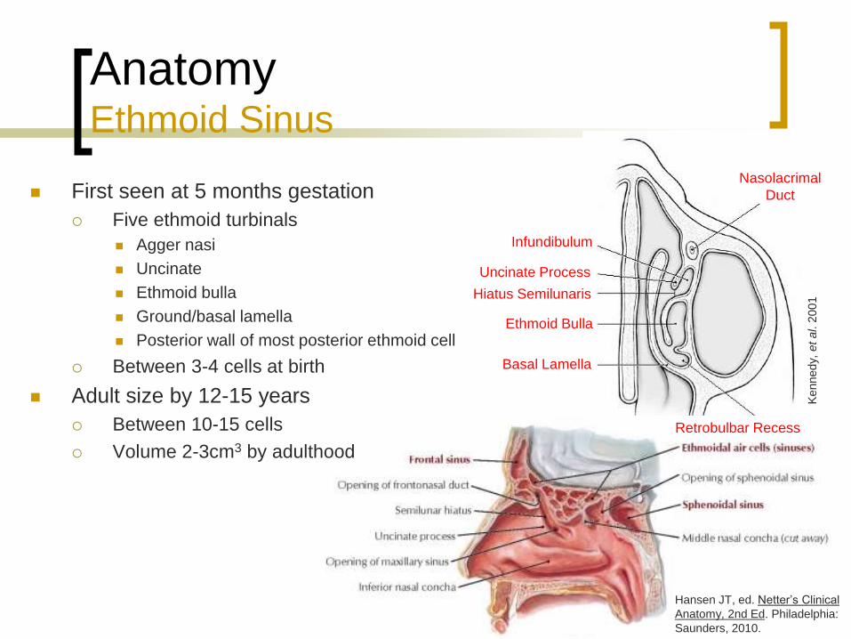
Anatomy Ethmoid Sinus
First seen at 5 months gestation
Five ethmoid turbinals
Agger nasi
Uncinate
Ethmoid bulla
Ground/basal lamella
Posterior wall of most posterior ethmoid cell
Between 3-4 cells at birth
Adult size by 12-15 years
Between 10-15 cells
Volume 2-3cm3 by adulthood
Hansen JT, ed. Netter’s Clinical
Anatomy, 2nd Ed. Philadelphia:
Saunders, 2010.
Kennedy,
et al. 2
001
Nasolacrimal
Duct
Infundibulum
Uncinate Process
Hiatus Semilunaris
Ethmoid Bulla
Basal Lamella
Retrobulbar Recess

NOTES: The lateral portions form the medial walls of the orbits, the sphenoid
establishes the posterior face, the superior surface is formed by the skull base of
the anterior cranial fossa, and many of the key structures of the lateral nasal wall,
derived from basal lamellas, extend posteroinferiorly from the skull base. The
lateral wall of the ethmoid sinus, or lamina papyracea, forms the paper-thin medial
wall of the orbit. The midline vertical plate of the ethmoid bone is composed of a
superior portion in the anterior cranial fossa called the crista galli and an inferior
portion in the nasal cavity called the perpendicular plate of the ethmoid bone that
contributes to the nasal septum. The anterior cranial fossa is separated from the
ethmoid air cells superiorly by the horizontal plate of the ethmoid bone, which is
composed of the thin medial cribriform plate and the thicker, more lateral ethmoid
roof. The ethmoid roof articulates with the cribriform plate at the lateral lamella of
the cribriform plate, which is the thinnest bone in the entire skull base.
The ethmoid sinuses are separated by a series of recesses demarcated by five
bony partitions or lamellae. These lamellae are named from the most anterior to
posterior: first (uncinate process), second (bulla ethmoidalis), third (ground or
basal lamella), fourth (superior turbinate), and fifth (supreme turbinate).
Anatomy
Ethmoid Sinus

Anatomy Ethmoid Sinus
Drainage
Anterior cells via ethmoid infundibulum
Posterior cells via sphenoethmoid recess
Innervation via V1 distribution
Branches from nasociliary nerve
Anterior and posterior ethmoids
Vasculature
Ophthalmic artery
Maxillary and ethmoid veins
Nasociliary Nerve
Anterior Ethmoidal Artery
Posterior Ethmoidal Artery
Ophthalmic
Nerve
Ophthalmic
artery
Posterior cells drain into superior meatus
Ophthalmic artery provides anterior and posterior
ethmoidal arteries
Cavernous sinus gives off maxillary and
ethmoidal veins
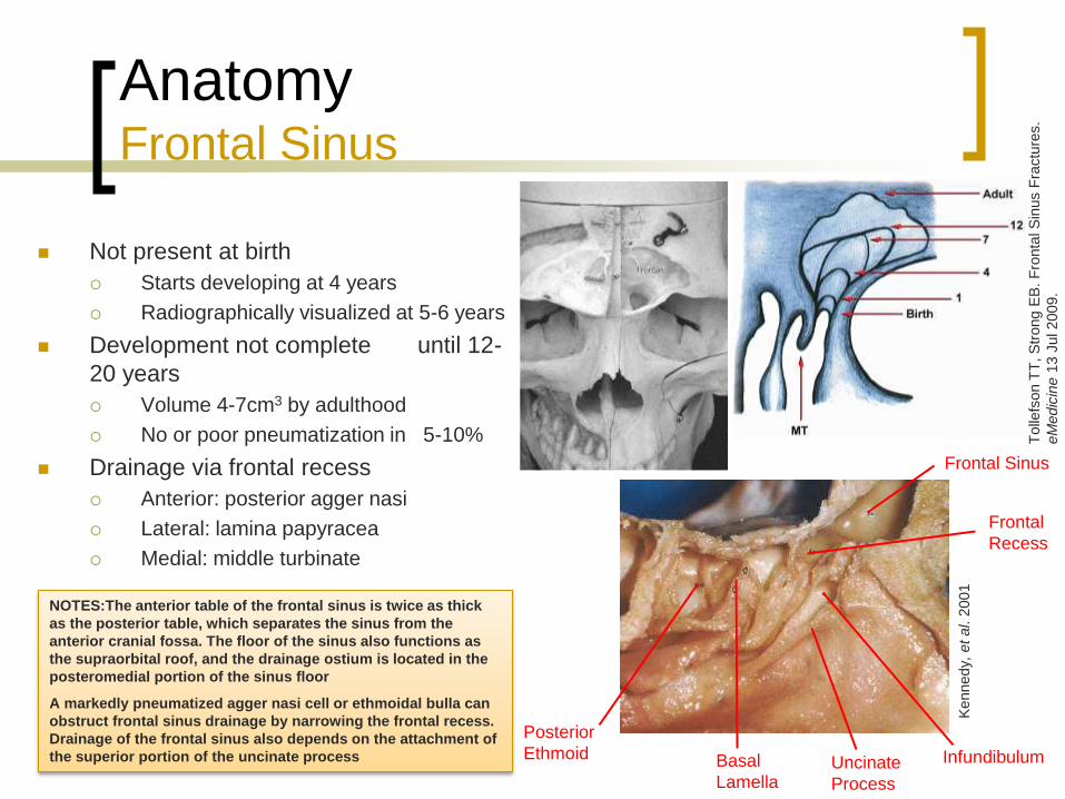
Anatomy Frontal Sinus
Not present at birth
Starts developing at 4 years
Radiographically visualized at 5-6 years
Development not complete until 12-
20 years
Volume 4-7cm3 by adulthood
No or poor pneumatization in 5-10%
Drainage via frontal recess
Anterior: posterior agger nasi
Lateral: lamina papyracea
Medial: middle turbinate
Tolle
fson T
T, S
trong E
B. F
ronta
l S
inus F
ractu
res.
eM
edic
ine 1
3 J
ul 2009.
Kennedy,
et al. 2
001
Frontal Sinus
Frontal
Recess
Basal
Lamella
Infundibulum
Posterior
Ethmoid Uncinate
Process
NOTES:The anterior table of the frontal sinus is twice as thick
as the posterior table, which separates the sinus from the
anterior cranial fossa. The floor of the sinus also functions as
the supraorbital roof, and the drainage ostium is located in the
posteromedial portion of the sinus floor
A markedly pneumatized agger nasi cell or ethmoidal bulla can
obstruct frontal sinus drainage by narrowing the frontal recess.
Drainage of the frontal sinus also depends on the attachment of
the superior portion of the uncinate process
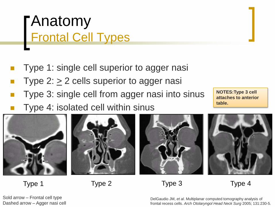
Anatomy Frontal Cell Types
Type 1: single cell superior to agger nasi
Type 2: > 2 cells superior to agger nasi
Type 3: single cell from agger nasi into sinus
Type 4: isolated cell within sinus
Type 1 Type 2 Type 3 Type 4
Sold arrow – Frontal cell type
Dashed arrow – Agger nasi cell DelGaudio JM, et al. Multiplanar computed tomography analysis of
frontal recess cells. Arch Otolaryngol Head Neck Surg 2005; 131:230-5.
NOTES:Type 3 cell
attaches to anterior
table.

Anatomy Frontal Sinus
Vasculature
Supraorbital artery and vein
Supratrochlear artery
Ophthalmic vein
Foramina of Breschet
Innervation via V1 distribution
Supraorbital
Supratrochlear
Supratrochlear
Nerve
Supraorbital
Nerve
Supratrochlear
Artery
Supraorbital
Artery
NOTES:Foramina of Breschet: small
venules that drain the sinus mucosa
into the dural veins

Anatomy Sphenoid Sinus
Evagination of nasal mucosa into sphenoid bone
First seen at 4 months gestation
Pneumatization begins in middle childhood
Minimal volume at birth
Volume 0.5-8cm3 by adult
Reaches adult size by 12-18 years
Sellar type (86%)
Presellar (11%)
Conchal (3%)
NOTES: Approximately 25% of bony capsules separating the
internal carotid artery from the sphenoid sinus are partially
dehiscent. An optic nerve prominence is present in 40% of
individuals with dehiscence in 6%.
In most cases, the posteroinferior end of the superior
turbinate was located in the same horizontal plane as the
floor of the sphenoid sinus. The ostium was located medial to
the superior turbinate in 83% of cases and lateral to it in 17%.

Anatomy Sphenoid Sinus
Innervation via sphenopalatine nerve
V2 distribution
Parasympathetics
Vasculature via maxillary artery and vein
Sphenopalatine artery
Pterygoid plexus

Acute Rhinosinusitis (ARS)
Inflammation of the nasal mucosa and lining of the
paranasal sinuses
Obstruction of sinus ostia
Impaired ciliary transport
Viral etiology in majority of cases
Superimposed bacterial infection in 0.5-2%
Symptoms for at least 7-10 days or worsening
after 5-7 days
Symptoms present for < 4 weeks
“Recurrent ARS” with > 4 episodes, lasting > 7-10
days
NOTES: Most viral upper respiratory tract infections are
caused by rhinovirus, but coronavirus, influenza A and B,
parainfluenza, respiratory syncytial virus, adenovirus, and
enterovirus are also causative agents.
Acute Rhinosinusitis (ARS)
Major symptoms
Facial pain/pressure
Facial congestion/fullness
Nasal obstruction
Nasal discharge/purulence
Minor symptoms
Headache
Fever (non-ARS)
Halitosis
Fatigue
Diagnosis with two major or one major and two minor
factors
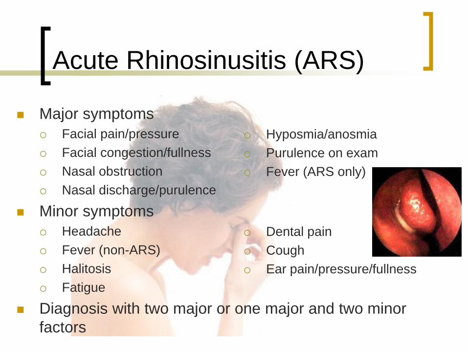
Hyposmia/anosmia
Purulence on exam
Fever (ARS only)
Dental pain
Cough
Ear pain/pressure/fullness
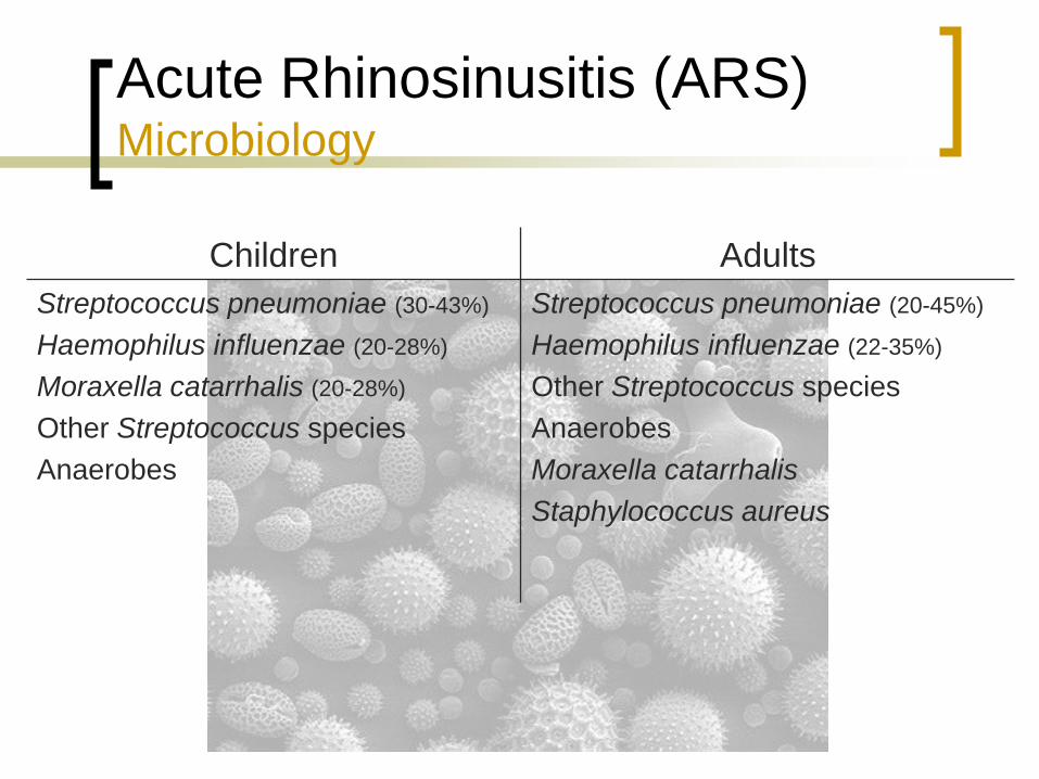
Acute Rhinosinusitis (ARS) Microbiology
Children Adults
Streptococcus pneumoniae (30-43%)
Haemophilus influenzae (20-28%)
Moraxella catarrhalis (20-28%)
Other Streptococcus species
Anaerobes
Streptococcus pneumoniae (20-45%)
Haemophilus influenzae (22-35%)
Other Streptococcus species
Anaerobes
Moraxella catarrhalis
Staphylococcus aureus

Chronic Rhinosinusitis (CRS)
Symptoms present for > 12 consecutive weeks
“Subacute” for symptoms between 4-12 weeks
Chronic inflammation
Bacterial, fungal, and viral
Allergic and immunologic
Anatomic
Genetic predisposition
No clear consensus on pathophysiology
NOTES: One of the major problems with identifying the pathogenesis of CRS is that neither symptoms, findings, nor
radiographs, taken independently, are sufficient basis for the diagnosis. One study showed that current symptom-based
criteria had only a 47% correlation with a positive CT scan result.
Stankiewicz JA, Chow JM: A diagnostic dilemma for chronic rhinosinusitis: definition accuracy and validity. Am J
Rhinol 2002; 16:199-202.

Chronic Rhinosinusitis (CRS) Microbiology
Children Adults
Anaerobes
Other Streptococcus species
Staphylococcus aureus
Streptococcus pneumoniae
Haemophilus influenzae
Pseudomonas aeruginosa
Anaerobes
Other Streptococcus species
Haemophilus influenzae
Staphylococcus aureus
Streptococcus pneumoniae
Moraxella catarrhalis

Complications of Sinusitis
Incidence has decreased with antibiotic use
Three main categories
Orbital (60-75%)
Intracranial (15-20%)
Bony (5-10%)
Radiography
Computed tomography (CT) best for orbit
Magnetic resonance imaging (MRI) best for intracranium
Siedek et al, 2010

Complications of Sinusitis Orbital
Most commonly involved complication site
Proximity to ethmoid sinuses
Periorbita/orbital septum is the only soft-tissue barrier
Valveless superior and inferior ophthalmic veins
Continuum of inflammatory/infectious changes
Direct extension through lamina papyracea
Impaired venous drainage from thrombophlebitis
Progression within 2 days
Children more susceptible
< 7 years – isolated orbital (subperiosteal abscess)
> 7 years – orbital and intracranial complications
NOTES:
-- close proximity of the orbit to the paranasal sinuses, particularly the ethmoids, make it the most commonly
involved structure in sinusitis complications; rarely from frontal or maxillary sinuses
-- pediatric population difference likely related to age-related sinus development
* pain and deterioration is not necessarily always present
* increase in WBC only found in 50%
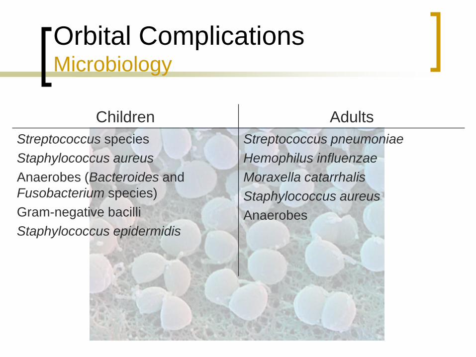
Orbital Complications Microbiology
Children Adults
Streptococcus species
Staphylococcus aureus
Anaerobes (Bacteroides and
Fusobacterium species)
Gram-negative bacilli
Staphylococcus epidermidis
Streptococcus pneumoniae
Hemophilus influenzae
Moraxella catarrhalis
Staphylococcus aureus
Anaerobes

Orbital Complications Chandler Criteria
Five classifications
Preseptal cellulitis
Orbital cellulitis
Subperiosteal abscess
Orbital abscess
Cavernous sinus thrombosis
Not exclusive, can occur concurrently
Bailey, et al. 2006.
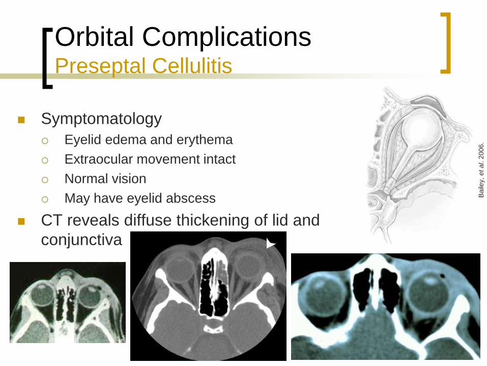
Orbital Complications Preseptal Cellulitis
Symptomatology
Eyelid edema and erythema
Extraocular movement intact
Normal vision
May have eyelid abscess
CT reveals diffuse thickening of lid and
conjunctiva
Baile
y, e
t al. 2
006.
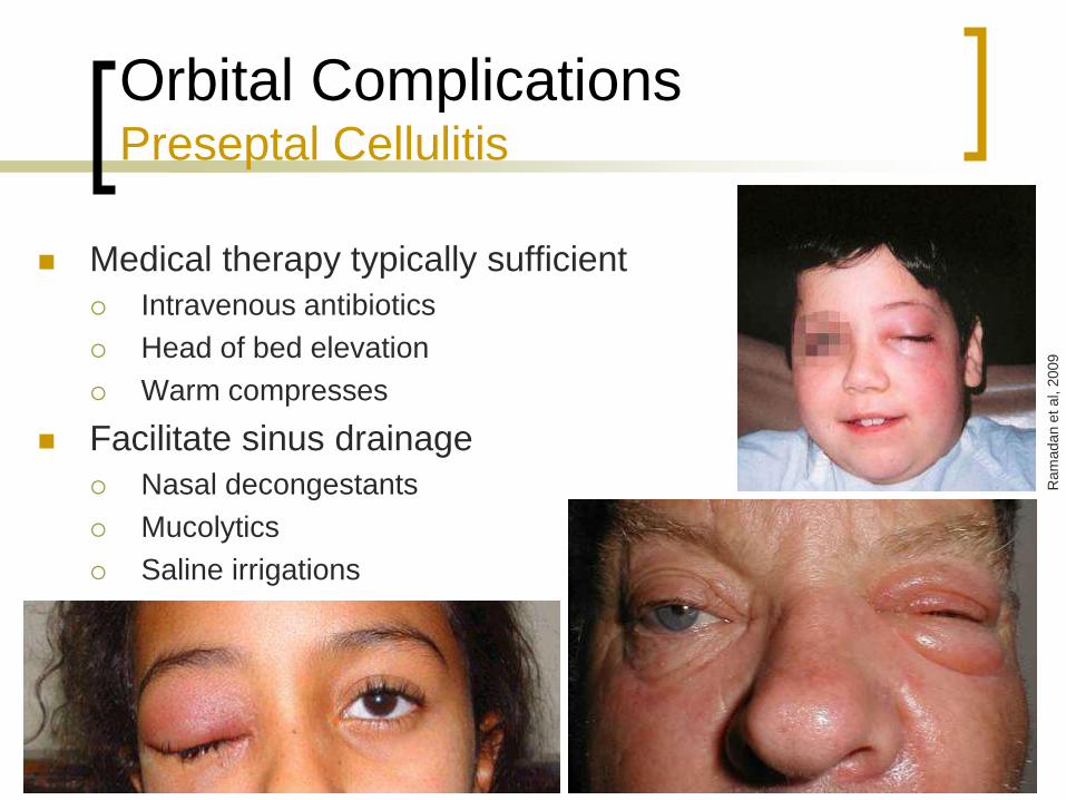
Orbital Complications Preseptal Cellulitis
Medical therapy typically sufficient
Intravenous antibiotics
Head of bed elevation
Warm compresses
Facilitate sinus drainage
Nasal decongestants
Mucolytics
Saline irrigations
Ram
adan e
t al, 2
009
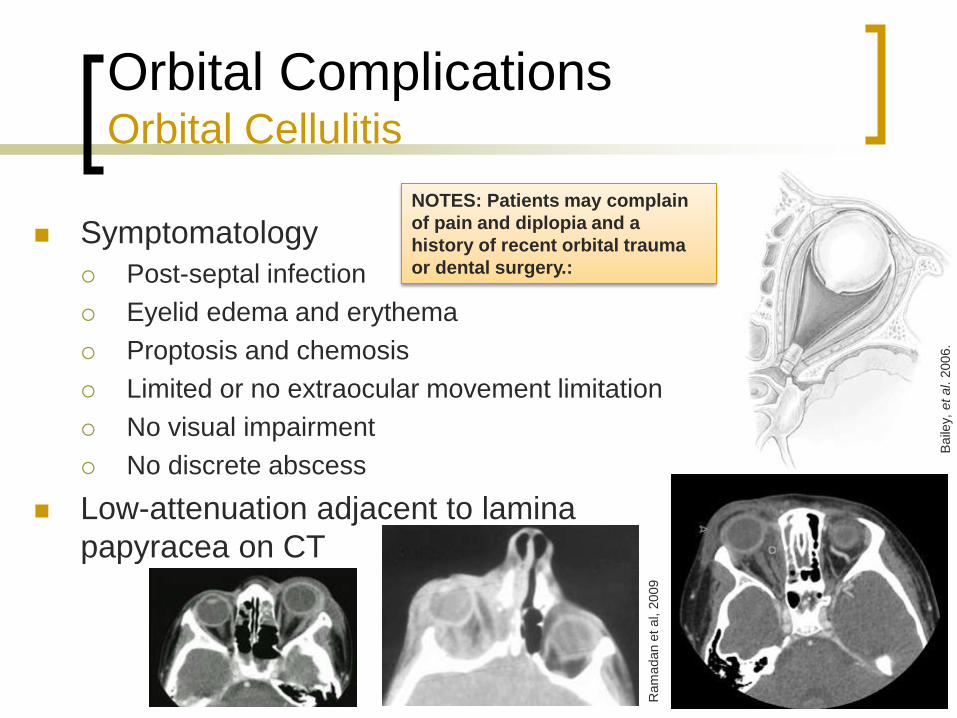
Orbital Complications Orbital Cellulitis
Symptomatology
Post-septal infection
Eyelid edema and erythema
Proptosis and chemosis
Limited or no extraocular movement limitation
No visual impairment
No discrete abscess
Low-attenuation adjacent to lamina
papyracea on CT
Baile
y, e
t al. 2
006.
Ram
adan e
t al, 2
009
NOTES: Patients may complain
of pain and diplopia and a
history of recent orbital trauma
or dental surgery.:
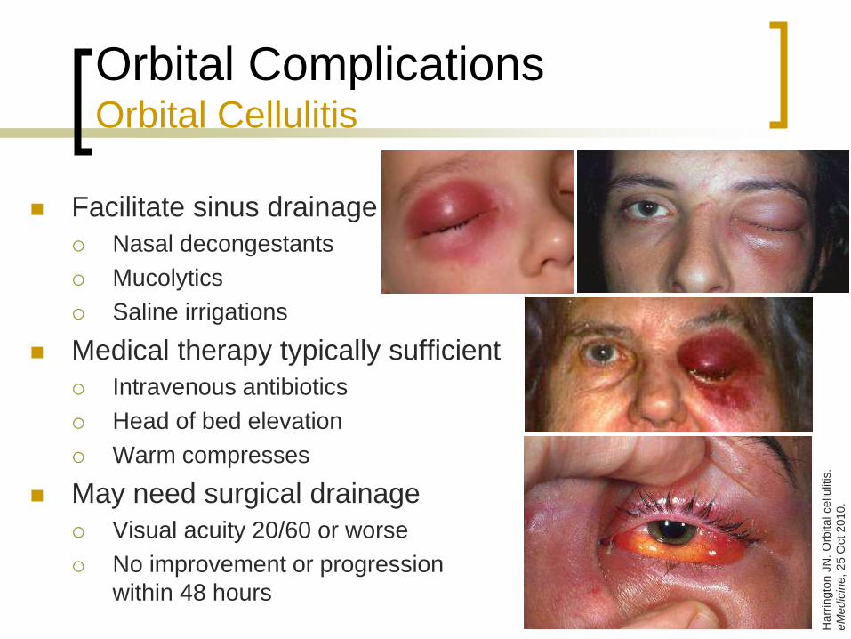
Orbital Complications Orbital Cellulitis
Facilitate sinus drainage
Nasal decongestants
Mucolytics
Saline irrigations
Medical therapy typically sufficient
Intravenous antibiotics
Head of bed elevation
Warm compresses
May need surgical drainage
Visual acuity 20/60 or worse
No improvement or progression
within 48 hours
Harr
ingto
n J
N. O
rbital cellu
litis
.
eM
edic
ine
, 25 O
ct 2010.
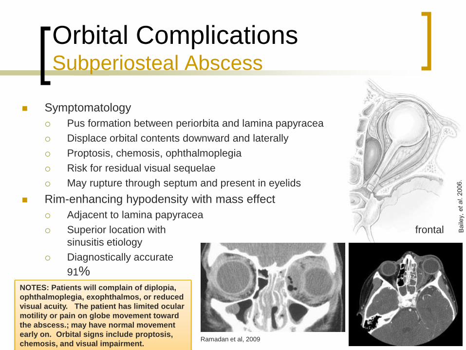
Orbital Complications Subperiosteal Abscess
Symptomatology
Pus formation between periorbita and lamina papyracea
Displace orbital contents downward and laterally
Proptosis, chemosis, ophthalmoplegia
Risk for residual visual sequelae
May rupture through septum and present in eyelids
Rim-enhancing hypodensity with mass effect
Adjacent to lamina papyracea
Superior location with frontal
sinusitis etiology
Diagnostically accurate 86-
91%
Ramadan et al, 2009
NOTES: Patients will complain of diplopia,
ophthalmoplegia, exophthalmos, or reduced
visual acuity. The patient has limited ocular
motility or pain on globe movement toward
the abscess.; may have normal movement
early on. Orbital signs include proptosis,
chemosis, and visual impairment.

Orbital Complications Subperiosteal Abscess
Surgical drainage
Worsening visual acuity or extraocular
movement
Lack of improvement after 48 hours
May be treated medically in 50-67%
Meta-analysis cure rate 26-93% (Coenraad 2009)
Combined treatment 95-100%
Orbital Complications Subperiosteal Abscess
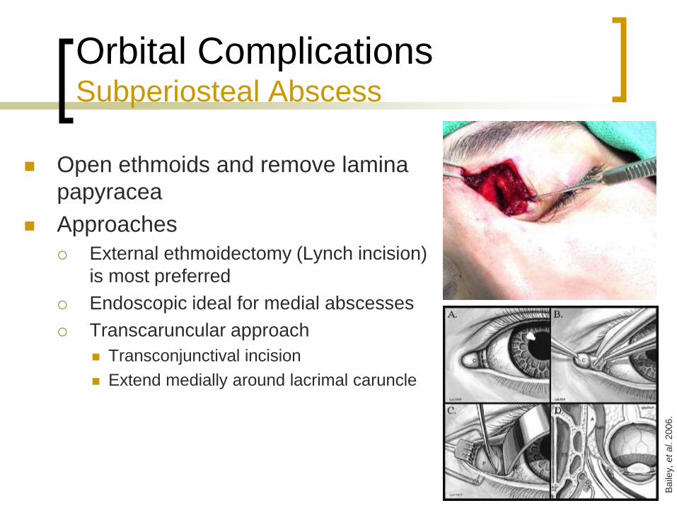
Open ethmoids and remove lamina
papyracea
Approaches
External ethmoidectomy (Lynch incision)
is most preferred
Endoscopic ideal for medial abscesses
Transcaruncular approach
Transconjunctival incision
Extend medially around lacrimal caruncle
Baile
y, e
t al. 2
006.
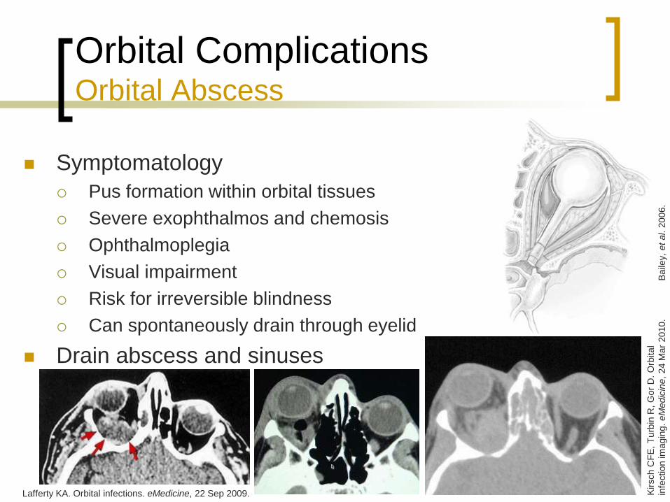
Orbital Complications Orbital Abscess
Symptomatology
Pus formation within orbital tissues
Severe exophthalmos and chemosis
Ophthalmoplegia
Visual impairment
Risk for irreversible blindness
Can spontaneously drain through eyelid
Drain abscess and sinuses
Baile
y, e
t al. 2
006.
Kirsch C
FE
, T
urb
in R
, G
or
D. O
rbital
infe
ction im
agin
g.
eM
edic
ine,
24 M
ar
2010.
Lafferty KA. Orbital infections. eMedicine, 22 Sep 2009.

Orbital Complications Orbital Abscess
Incise periorbita and drain intraconal abscess
Similar approaches as with subperiosteal
abscess
Lynch incision
Endoscopic
NOTES:Transcaruncular approach allegedly does not utilize a
facial incision.

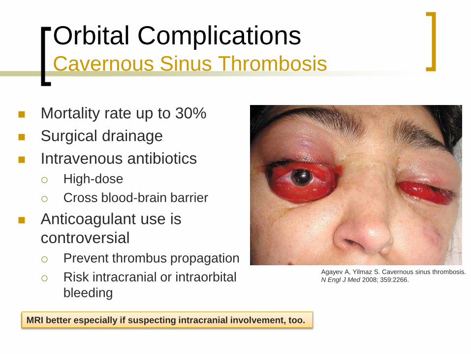
Orbital Complications Cavernous Sinus Thrombosis
Symptomatology
Orbital pain
Proptosis and chemosis
Ophthalmoplegia
Symptoms in contralateral eye
Associated with sepsis and meningismus
Radiology
Poor venous enhancement on CT
Better visualized on MRI
Contralateral involvement is distinguishing
feature of cavernous sinus thrombosis
MRI findings of heterogeneity and increased size
suggest the diagnosis
Orbital Complications Cavernous Sinus Thrombosis
Mortality rate up to 30%
Surgical drainage
Intravenous antibiotics
High-dose
Cross blood-brain barrier
Anticoagulant use is
controversial
Prevent thrombus propagation
Risk intracranial or intraorbital
bleeding
Agayev A, Yilmaz S. Cavernous sinus thrombosis.
N Engl J Med 2008; 359:2266.
MRI better especially if suspecting intracranial involvement, too.

Cavernous Sinus Thrombosis Anticoagulation
Beneficial
Southwick et al (1986)
Reduction in mortality
Not recommended for other dural sinus thrombosis
Levine et al (1988)
No change in mortality
Mortality reduction with added early
Bhatia et al (2002)
PTT ratio 1.5-2.5
INR 2-3
Anticoagulate for 3 months
Harmful
Bhatia et al (2002)
Fatal hemorrhagic cerebral
infarction
Subarachnoid hemorrhage
reversed with protamine
NOTES: 1980s were retrospective reviews
Bhatia was a literature review

Complications of Sinusitis Intracranial
Occurs more commonly in CRS
Mucosal scarring, polypoid changes
Hidden infectious foci with poor antibiotic penetration
Male teenagers affected more than children
Direct extension
Sinus wall erosion
Traumatic fracture lines
Neurovascular foramina (optic and olfactory nerves)
Hematogenous spread
Diploic skull veins
Ethmoid bone NOTES: Teenagers affected more because of developed frontal and sphenoid sinuses, and because they
are more prone to URI’s than adults.
Thrombophlebitis originating in the mucosal veins progressively involves the emissary veins of the
skull, the dural venous sinuses, the subdural veins, and, finally, the cerebral veins. By this mode, the
subdural space may be selectively infected without contamination of the intermediary structure; a
subdural empyema can exist without evidence of extradural infection or osteomyelitis.
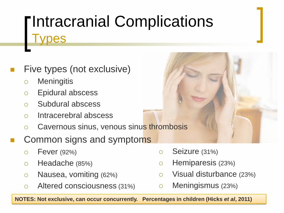
Intracranial Complications Types
Seizure (31%)
Hemiparesis (23%)
Visual disturbance (23%)
Meningismus (23%)
Five types (not exclusive)
Meningitis
Epidural abscess
Subdural abscess
Intracerebral abscess
Cavernous sinus, venous sinus thrombosis
Common signs and symptoms
Fever (92%)
Headache (85%)
Nausea, vomiting (62%)
Altered consciousness (31%)
NOTES: Not exclusive, can occur concurrently. Percentages in children (Hicks et al, 2011)

Intracranial Complications Meningitis
Most common intracranial complication of sinusitis
Symptomatology
Headache
Meningismus
Fever, septic
Cranial nerve palsies
Sinusitis is unusual cause of meningitis
Sphenoiditis
Ethmoiditis
Usually amenable with medical treatment
Drain sinuses if no improvement after 48 hours
Hearing loss and seizure sequelae
NOTES: Also incidence of neurologic sequelae such as hearing loss and seizure disorder.
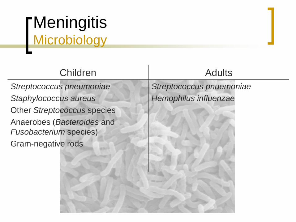
Meningitis Microbiology
Children Adults
Streptococcus pneumoniae
Staphylococcus aureus
Other Streptococcus species
Anaerobes (Bacteroides and
Fusobacterium species)
Gram-negative rods
Streptococcus pnuemoniae
Hemophilus influenzae
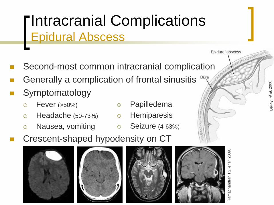
Intracranial Complications Epidural Abscess
Ram
achandra
n T
S, et al, 2
009.
Papilledema
Hemiparesis
Seizure (4-63%)
Second-most common intracranial complication
Generally a complication of frontal sinusitis
Symptomatology
Fever (>50%)
Headache (50-73%)
Nausea, vomiting
Crescent-shaped hypodensity on CT

Intracranial Complications Epidural Abscess
Lumbar puncture contraindicated
Prophylactic seizure therapy not necessary
Antibiotics Good intracerebral penetration
Typically for 4-8 weeks
Drain sinuses and abscess
Frontal sinus trephination
Frontal sinus cranialization
Stereotactic-guided drainage
NOTES: Will likely need antibiotics for 4-8
weeks; usually vancomycin and 3rd or 4th
generation cephalosporin
Prophylactic seizure therapy not necessary
unless there’s an associated subdural
abscess.
Intracranial Complications Subdural Abscess
Generally from frontal or ethmoid sinusitis
Symptomatology
Headaches
Fever
Nausea, vomiting
Hemiparesis
Lethargy, coma
Third-most common intracranial
complication, rapid deterioration
Mortality in 25-35%
Residual neurologic sequelae in 35-55%
Accompanies 10% of epidural abscesses
Intracranial Complications Subdural Abscess
Lumbar puncture potentially fatal
Aggressive medical therapy
Antibiotics
Anticonvulsants
Hyperventilation, mannitol
Steroids
Drain sinuses and abscess
Medical therapy possible if < 1.5cm
Craniotomy or stereotactic burr hole
Endoscopic or external sinus drainage
NOTES:Need antibiotics with good intracerebral penetration, typically
3-6 weeks
Craniotomy is favored over burr hole placement due to better exposure

Intracranial Complications Intracerebral Abscess
Uncommon, frontal and frontoparietal lobes
Generally from frontal sinusitis Sphenoid
Ethmoids
Symptomatology Headache (70%)
Mental status change (65%)
Focal neurological deficit (65%)
Fever (50%)
Mortality 20-30%
Neurologic sequelae 60%
Nausea, vomiting (40%)
Seizure (25-35%)
Meningismus (25%)
Papilledema (25%)
NOTES: May have mood swings
and behavioral changes with
frontal lobe involvement
Worsening headache with
meningismus suggests possible
rupture of the abscess.

Intracranial Complications Intracerebral Abscess
Lumbar puncture potentially fatal
Aggressive medical therapy
Antibiotics
Anticonvulsants
Hyperventilation, mannitol
Steroids
Drain sinuses and abscess
Medical therapy possible if abscess < 2.5cm
Excision or aspiration
Diagnostic aspiration if < 2.5cm or cerebritis
Stereotactic-guided aspiration
Endoscopic or external sinus drainage
NOTES: Antibiotic regimen is typically 6-8 weeks; typically ceftriaxone, vancomycin or nafcillin, and metronidazole
Corticosteroid use is controversial. Steroids can retard the encapsulation process, increase necrosis, reduce antibiotic penetration into the
abscess, increase the risk of ventricular rupture, and alter the appearance on CT scans. Steroid therapy can also produce a rebound effect
when discontinued. If used to reduce cerebral edema, therapy should be of short duration. The appropriate dosage, the proper timing, and
any effect of steroid therapy on the course of the disease are unknown. The procedures used are aspiration through a bur hole and complete
excision after craniotomy. Aspiration is the most common procedure and is often performed using a stereotactic procedure with the guidance
of CT scanning or MRI.
Intracranial Abscesses Microbiology
Children Adults
Anaerobes (anaerobic Streptococcus, Bacteroides, Fusobacterium species)
Staphylococcus aureus
Other Streptococcus species (Streptococcus milleri)
Gram-negative bacilli (Hemophilus influenzae)
Staphylococcus epidermidis
Eikenella corrodens
Polymicrobial
NOTES: Incidence of anaerobes in suppurative
intracranial complications range from 60-100%

Intracranial Complications Venous Sinus Thrombosis
Sagittal sinus most common
Retrograde thrombophlebitis from
frontal sinusitis
Extremely ill
Subdural abscess
Epidural abscess
Intracerebral abscess
Decreased cavernous carotid
artery flow void on MRI
Elevated mortality rate
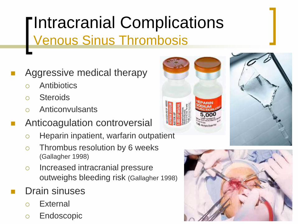
Intracranial Complications Venous Sinus Thrombosis
Aggressive medical therapy
Antibiotics
Steroids
Anticonvulsants
Anticoagulation controversial
Heparin inpatient, warfarin outpatient
Thrombus resolution by 6 weeks (Gallagher 1998)
Increased intracranial pressure
outweighs bleeding risk (Gallagher 1998)
Drain sinuses
External
Endoscopic
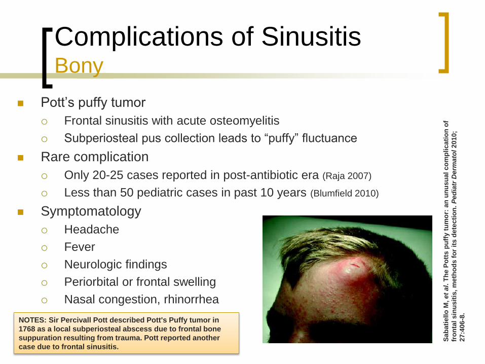
Complications of Sinusitis Bony
Pott’s puffy tumor
Frontal sinusitis with acute osteomyelitis
Subperiosteal pus collection leads to “puffy” fluctuance
Rare complication
Only 20-25 cases reported in post-antibiotic era (Raja 2007)
Less than 50 pediatric cases in past 10 years (Blumfield 2010)
Symptomatology
Headache
Fever
Neurologic findings
Periorbital or frontal swelling
Nasal congestion, rhinorrhea
Sab
ati
ello
M, et
al.
Th
e P
ott
s p
uff
y t
um
or:
an
un
usu
al co
mp
licati
on
of
fro
nta
l sin
usit
is, m
eth
od
s f
or
its d
ete
cti
on
. P
ed
iatr
Derm
ato
l 2010;
27:4
06
-8.
NOTES: Sir Percivall Pott described Pott's Puffy tumor in
1768 as a local subperiosteal abscess due to frontal bone
suppuration resulting from trauma. Pott reported another
case due to frontal sinusitis.

Complications of Sinusitis Bony
Associated with other abscesses in 60%
Pericranial
Periorbital
Epidural
Subdural
Intracranial
Cortical vein thrombosis
Frontocutaneous fistula
Upadhya
y S
. R
ecurr
ent P
ott's
puff
y t
um
or,
a r
are
clin
ical entity
. N
euro
l In
dia
2010; 58:8
15
-7.
Baile
y, e
t al. 2
006.
Blu
mfield
, et al. 2
010.
NOTES: Sir Percivall Pott described Pott's Puffy
tumor in 1768 as a local subperiosteal abscess due
to frontal bone suppuration resulting from trauma.
Pott reported another case due to frontal sinusitis.
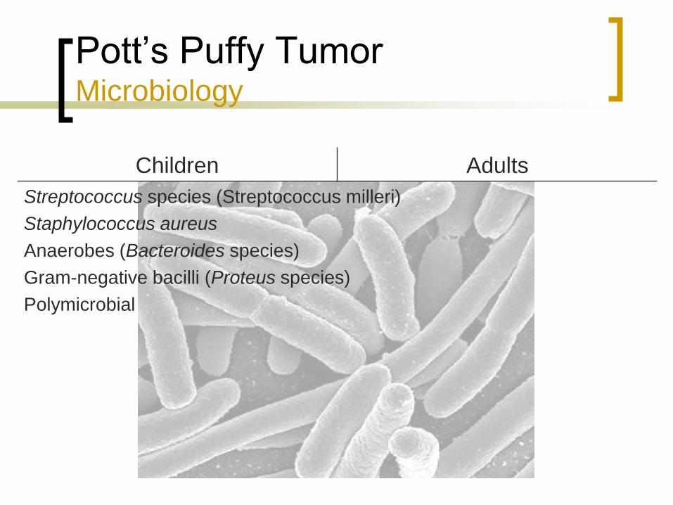
Pott’s Puffy Tumor Microbiology
Children Adults
Streptococcus species (Streptococcus milleri)
Staphylococcus aureus
Anaerobes (Bacteroides species)
Gram-negative bacilli (Proteus species)
Polymicrobial
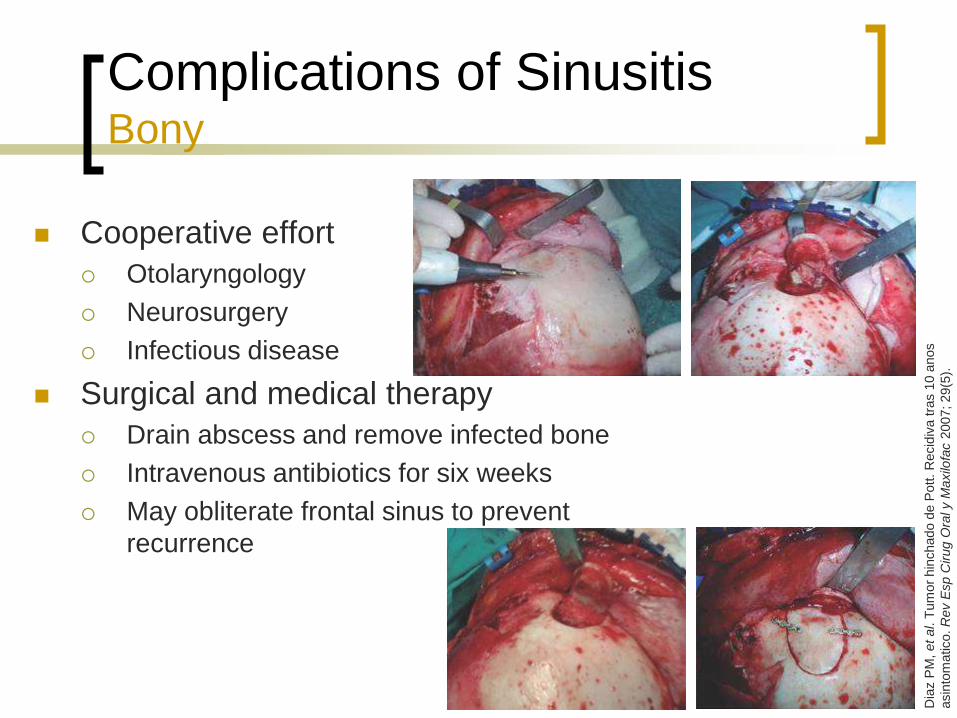
Complications of Sinusitis Bony
Cooperative effort
Otolaryngology
Neurosurgery
Infectious disease
Surgical and medical therapy
Drain abscess and remove infected bone
Intravenous antibiotics for six weeks
May obliterate frontal sinus to prevent
recurrence
Dia
z P
M, et al. T
um
or
hin
chado d
e P
ott. R
ecid
iva t
ras 1
0 a
nos
asin
tom
atico. R
ev E
sp C
irug O
ral y M
axilo
fac 2
007; 29(5
).

Conclusions
Complications are less common
with antibiotics
Orbital
Intracranial
Bony
Can result in drastic sequelae
Drain abscess and open involved
sinuses
Surgical involvement
Ophthalmology
Neurosurgery
(htt
p:/
/ww
w.s
mb
c-c
om
ics.c
om
)
References
Bailey BJ, Johnson, JT, Newlands SD, eds. Head and Neck Surgery – Otolaryngology, 4th Ed. Philadelphia: Lippincott, 2006:307-11, 406, 493-503.
Benninger MS, Ferguson BJ, Hadley JA, et al: Adult chronic rhinosinusitis: definitions, diagnosis, epidemiology, and pathophysiology. Otolaryngol Head Neck Surg 2003; 129:S1-S32.
Benson BE, Riauba L. Sinusitis, Acute. eMedicine 10 Feb 2009. Accessed 21 Mar 2011 <http://emedicine.medscape.com/article/232670-overview>.
Bhatia K, Jones NS. Septic cavernous sinus thrombosis secondary to sinusitis: area anticoagulants indicated? A review of the literature. J Laryngol Otol 2002; 116:667-76.
Blumfield E, Misra M. Pott's puffy tumor, intracranial, and orbital complications as the initial presentation of sinusitis in healthy adolescents, a case series. Emerg Radiol 2011 Mar 5 [Epub ahead of print].
Brook I. Brain abscess. eMedicine 26 Jun 2008. Accessed 10 Apr 2011 <http://emedicine.medscape.com/article/212946-overview>.
Brook I, Bajracharya H. Sinusitis, Chronic. eMedicine 17 Jun 2009. Accessed 21 Mar 2011 <http://emedicine.medscape.com/article/232791-overview>.
Brook I, Friedman EM. Intracranial complications of sinusitis in children: a sequela of periapical abscess. Ann Otol Rhinol Laryngol 1982; 91:41-3.
Caversaccio M, Heimgartner S, Aebi C. Orbital complications of acute pediatric rhinosinusitis: medical treatment versus surgery and analysis of the computer tomogram. Laryngorhinootologic 2005; 84:817-21.
Coenraad S, Buwalda J. Surgical or medical management of subperiosteal orbital abscess in children: a critical appraisal of the literature. Rhinology 2009; 47:18-23.
Chandler JR, Langenbrunner DJ, Stevens ER. The pathogenesis of orbital complications in acute sinusitis. Laryngoscope 1970; 80: 1414-28.
Dawodu ST, Lorenzo NY. Subdural empyema. eMedicine 11 Mar 2009. Accessed 10 Apr 2011 <http://emedicine.medscape.com/article/1168415-overview>.
Eweiss A, Mukonoweshuro W, Khalil HS. Cavernous sinus thrombosis secondary to contralateral sphenoid sinusitis: a diagnostic challenge. J Laryngol Otol 2010; 124:928-30.
Flint PW, et al, eds. Cummings Otolaryngology: Head and Neck Surgery, 5th Ed. Philadelphia: Mosby Elsevier, 2010. ch 47.
Gallagher RM, Gross CW, Phillips CD. Suppurative intracranial complications of sinusitis. Laryngoscope 1998; 108:1635-42.
References
Garcia GH, Harris GJ. Criteria for nonsurgical management of subperiosteal abscess of the orbit: analysis of outcomes 1988-1998. Ophthalmology 2000; 107:1454-8.
Giannoni CM, Sulek M, Friedman EM. Intracranial complications of sinusitis: A pediatric series. Am J Rhinol 1998; 12:173-8.
Goldberg AN, Oroszlan G, Anderson TD. Complications of frontal sinusitis and their management. Otolaryngol Clin North Am 2001; 34:211-25.
Greenberg MF, Pollard ZF. Medical treatment of pediatric subperiosteal orbital abscess secondary to sinusitis. J AAPOS 1998; 2:351-5.
Greenlee JE. Subdural empyema. In: Mandell GL, ed. Principles and Practice of Infectious Diseases, Vol 1. 4th Ed. New York: Churchill, 1994:900-3.
Gwaltney JM Jr. Acute community-acquired sinusitis. Clin Infect Dis 1996; 23:1209-23; quiz 1224-5.
Gwaltney JM, Scheld WM, Sande MA, et al. The microbial etiology and antimicrobial therapy of adults with acute community-acquired sinusitis: A fifteen-year experience at the University of Virginia and review of other selected studies. J Allergy Clin Immunol 1992; 90:457-62.
Herrmann BW, Forsen JW Jr. Simultaneous intracranial and orbital complications of acute rhinosinusitis in children. Int J Pediatr Otorhinolaryngol 2004; 68:619-25.
Hicks CW, Weber JG, Reid JR, Moodley M. Identifying and managing intracranial complications of sinusitis in children. Pediatr Infect Dis 2011; 30:222-6.
Janfaza P, Montgomery WW, Salman SD. Nasal cavities and paranasal sinuses. In: Janfaza P, Nadol JB, Galla R, et al, eds. Surgical Anatomy of the Head and Neck. Philadelphia: Lippincott Williams & Wilkins, 2001:259-318.
Karaman E, Hacizade Y, Isildak H, Kaytaz A. Pott's puffy tumor. J Craniofac Surg 2008; 19:1694-7.
Kayhan FT, Sayin I, Yazici ZM, Erdur O. Management of orbital subperiosteal abscess. J Craniofac Surg 2010; 21:1114-7.
Kuhn FA. Chronic frontal sinusitis: the endoscopic frontal recess approach. Operat Tech Otolaryngol Head Neck Surg 1996; 7:222-9.
Lanza DC, Kennedy DW. Adult rhinosinusitis defined. Otolaryngol Head Neck Surg 1997; 117:S1-S7.
Lee KJ, ed. Essential Otolaryngology - Head and Neck Surgery, 9th Ed. New York: McGraw-Hill, 2008. pp 365-6.
Levine SR, Twyman RE, Gilman S. The role of anticoagulation in cavernous sinus thrombosis. Neurology 1988; 38:517-22.
Marshall AH, Jones, NS. Osteomyelitis of the frontal bone secondary to frontal sinusitis. J Laryngol Otol 2000; 114:944-6.

References
Miaskiewicz B, Lukomski M, Starska K, Jozefowicz-Korezynska M. Orbital complication in acute and chronic sinusitis. H Pol Merkur Lekarski 2005; 19:388-9.
Oxford LE, McClay J. Complications of acute sinusitis in children. Otolaryngol Head Neck Surg 2005; 133:32-7.
Pasha R. Otolaryngology – Head and Neck Surgery, 2nd Ed. San Diego: Plural Publishing, 2006. pp 2-6.
Rahbar R, Petersen RA, DiCanzio J, et al. Management of orbital subperiosteal abscess in children. Arch Otolaryngol Head Neck Surg 2001; 127:281-6.
Raja V, Low C, Sastry A, Moriarty B. Pott’s puffy tumor following an insect bite. J Postgrad Med 2007; 53:114-6.
Ramachandran TS, Ramachandran A. Intracranial epidural abscess. eMedicine 9 Sep 2009. Accessed 10 Apr 2011 <http://emedicine.medscape.com/article/1165292-overview>.
Ramadan HH, Tewfik TL, Talavera F, et al. Pediatric sinusitis, medical treatment. eMedicine, 22 Apr 2009. Accessed 2 Apr 2011 <http://emedicine.medscape.com/article/873149-overview>.
Remmler D, Boles R. Intracranial complications of frontal sinusitis. Laryngoscope 1980; 90:1814-24.
Rosenfeld EA, Rowley AH. Infectious intracranial complications of sinusitis, other than meningitis, in children: 12-year review. Clin Infect Dis 1994; 18:750-4.
Schramm VL, Myers EN, Kennerdell JS. Orbital complications of acute sinusitis: Evaluation, management, and outcome. Otolaryngology 1978;86:221-30.
Souliere CR Jr, Antoine GA, Martin MP, et al. Selective non-surgical management of subperiosteal abscess of the orbit: computerized tomography and clinical course as indication for surgical drainage. Int J Pediatr Otolarynol 1990; 19:109-19.
Southwick FS, Richardson EP Jr, Swartz MN. Septic thrombosis of the dural venous sinuses. Medicine (Baltimore) 1986; 65:82-106.
Stankiewicz JA, Chow JM. A diagnostic dilemma for chronic rhinosinusitis: definition accuracy and validity. Am J Rhinol 2002; 16:199-202.
Vazquez E, Creixell S, Carreno JC, et al. Complicated acute pediatric bacterial sinusitis: imaging updated approach. Curr Probl Diagn Radiol 2004 May–Jun; 33:127-45.
Wald E. Microbiology of acute and chronic sinusitis in children. J Allergy Clin Immunol 1992; 90:452-60.
Wald E. Sinusitis in children. N Engl J Med 1992; 326:319-23.
References
Wallace MR, Rana A, Yadavalli GK. Epidural abscess. eMedicine 20 Apr 2009. Accessed 10 Apr 2011 <http://emedicine.medscape.com/article/232570-overview>.
Yogev R, Bar-Meir M. Management of brain abscesses in children. Pediatr Infect Dis J 2004; 23:157-9.
Younis RT, Lazar RH, Anand VK, Intracranial complications of sinusitis: A 15-year review of 39 cases. Ear Nose Throat J 2002; 81:636-44.
Younis RT, Lazar RH, Bustillo A, et al. Orbital infection as a complication of sinusitis:aAre diagnostic and treatment trends changing? Ear Nose Throat J 2002; 81:7715.