Complex Ultrasound Diagnosis of Thyroid Diseases
16
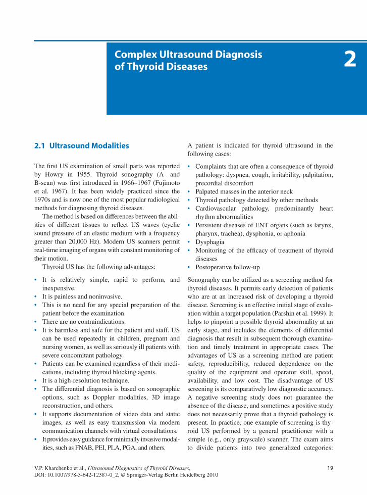
19 V.P. Kharchenko et al., Ultrasound Diagnostics of Thyroid Diseases, DOI: 10.1007/978-3-642-12387-0_2, © Springer-Verlag Berlin Heidelberg 2010 2 2.1 Ultrasound Modalities The first US examination of small parts was reported by Howry in 1955. Thyroid sonography (A- and B-scan) was first introduced in 1966–1967 (Fujimoto et al. 1967). It has been widely practiced since the 1970s and is now one of the most popular radiological methods for diagnosing thyroid diseases. The method is based on differences between the abil- ities of different tissues to reflect US waves (cyclic sound pressure of an elastic medium with a frequency greater than 20,000 Hz). Modern US scanners permit real-time imaging of organs with constant monitoring of their motion. Thyroid US has the following advantages: It is relatively simple, rapid to perform, and • inexpensive. It is painless and noninvasive. • This is no need for any special preparation of the • patient before the examination. There are no contraindications. • It is harmless and safe for the patient and staff. US • can be used repeatedly in children, pregnant and nursing women, as well as seriously ill patients with severe concomitant pathology. Patients can be examined regardless of their medi- • cations, including thyroid blocking agents. It is a high-resolution technique. • The differential diagnosis is based on sonographic • options, such as Doppler modalities, 3D image reconstruction, and others. It supports documentation of video data and static • images, as well as easy transmission via modern communication channels with virtual consultations. It provides easy guidance for minimally invasive modal- • ities, such as FNAB, PEI, PLA, PGA, and others. A patient is indicated for thyroid ultrasound in the following cases: Complaints that are often a consequence of thyroid • pathology: dyspnea, cough, irritability, palpitation, precordial discomfort Palpated masses in the anterior neck • Thyroid pathology detected by other methods • Cardiovascular pathology, predominantly heart • rhythm abnormalities Persistent diseases of ENT organs (such as larynx, • pharynx, trachea), dysphonia, or aphonia Dysphagia • Monitoring of the efficacy of treatment of thyroid • diseases Postoperative follow-up • Sonography can be utilized as a screening method for thyroid diseases. It permits early detection of patients who are at an increased risk of developing a thyroid disease. Screening is an effective initial stage of evalu- ation within a target population (Parshin et al. 1999). It helps to pinpoint a possible thyroid abnormality at an early stage, and includes the elements of differential diagnosis that result in subsequent thorough examina- tion and timely treatment in appropriate cases. The advantages of US as a screening method are patient safety, reproducibility, reduced dependence on the quality of the equipment and operator skill, speed, availability, and low cost. The disadvantage of US screening is its comparatively low diagnostic accuracy. A negative screening study does not guarantee the absence of the disease, and sometimes a positive study does not necessarily prove that a thyroid pathology is present. In practice, one example of screening is thy- roid US performed by a general practitioner with a simple (e.g., only grayscale) scanner. The exam aims to divide patients into two generalized categories: Complex Ultrasound Diagnosis of Thyroid Diseases
Transcript of Complex Ultrasound Diagnosis of Thyroid Diseases
19V.P. Kharchenko et al., Ultrasound Diagnostics of Thyroid Diseases, DOI: 10.1007/978-3-642-12387-0_2, © Springer-Verlag Berlin Heidelberg 2010
2
2.1 Ultrasound Modalities
The first US examination of small parts was reported by Howry in 1955. Thyroid sonography (A- and B-scan) was first introduced in 1966–1967 (Fujimoto et al. 1967). It has been widely practiced since the 1970s and is now one of the most popular radiological methods for diagnosing thyroid diseases.
The method is based on differences between the abil-ities of different tissues to reflect US waves (cyclic sound pressure of an elastic medium with a frequency greater than 20,000 Hz). Modern US scanners permit real-time imaging of organs with constant monitoring of their motion.
Thyroid US has the following advantages:
It is relatively simple, rapid to perform, and •inexpensive.It is painless and noninvasive.•This is no need for any special preparation of the •patient before the examination.There are no contraindications.•It is harmless and safe for the patient and staff. US •can be used repeatedly in children, pregnant and nursing women, as well as seriously ill patients with severe concomitant pathology.Patients can be examined regardless of their medi-•cations, including thyroid blocking agents.It is a high-resolution technique.•The differential diagnosis is based on sonographic •options, such as Doppler modalities, 3D image reconstruction, and others.It supports documentation of video data and static •images, as well as easy transmission via modern communication channels with virtual consultations.It provides easy guidance for minimally invasive modal-•ities, such as FNAB, PEI, PLA, PGA, and others.
A patient is indicated for thyroid ultrasound in the following cases:
Complaints that are often a consequence of thyroid •pathology: dyspnea, cough, irritability, palpitation, precordial discomfortPalpated masses in the anterior neck•Thyroid pathology detected by other methods•Cardiovascular pathology, predominantly heart •rhythm abnormalitiesPersistent diseases of ENT organs (such as larynx, •pharynx, trachea), dysphonia, or aphoniaDysphagia•Monitoring of the efficacy of treatment of thyroid •diseasesPostoperative follow-up•
Sonography can be utilized as a screening method for thyroid diseases. It permits early detection of patients who are at an increased risk of developing a thyroid disease. Screening is an effective initial stage of evalu-ation within a target population (Parshin et al. 1999). It helps to pinpoint a possible thyroid abnormality at an early stage, and includes the elements of differential diagnosis that result in subsequent thorough examina-tion and timely treatment in appropriate cases. The advantages of US as a screening method are patient safety, reproducibility, reduced dependence on the quality of the equipment and operator skill, speed, availability, and low cost. The disadvantage of US screening is its comparatively low diagnostic accuracy. A negative screening study does not guarantee the absence of the disease, and sometimes a positive study does not necessarily prove that a thyroid pathology is present. In practice, one example of screening is thy-roid US performed by a general practitioner with a simple (e.g., only grayscale) scanner. The exam aims to divide patients into two generalized categories:
Complex Ultrasound Diagnosis of Thyroid Diseases
20 2 Complex Ultrasound Diagnosis of Thyroid Diseases
those whose thyroids are grossly normal, and those with suspicious abnormalities in their thyroids.
Patients with thyroid abnormalities are subject to further complex qualified US. Complex US assumes the detection and certain differential diagnosis of dif-fuse changes and focal lesions, which is necessary to determine subsequent tactics.
US options utilized for the diagnosis of thyroid dis-eases include the following:
1. Grayscale 2. Tissue harmonics 3. Adaptive coloring 4. CDI 5. PDI 6. Grayscale 3D 7. Vascular 3D 8. 4D 9. Panoramic scan 10. Spectral pulsed wave Doppler 11. Others (multislice view, volume CT view, contrast
US, US elastography, etc.)
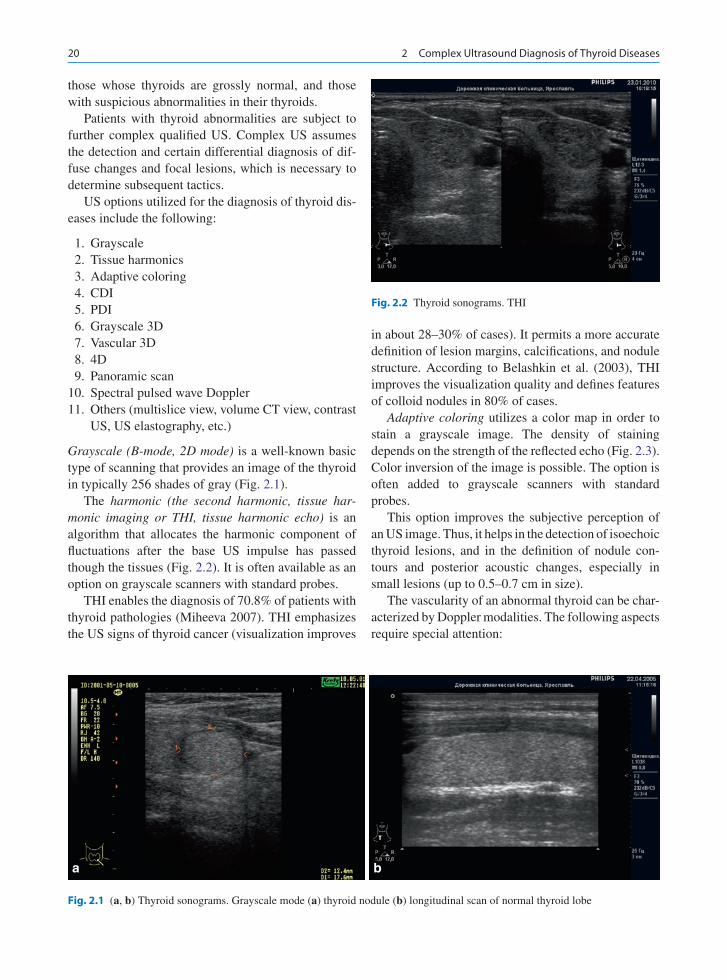
Grayscale (B-mode, 2D mode) is a well-known basic type of scanning that provides an image of the thyroid in typically 256 shades of gray (Fig. 2.1).
The harmonic (the second harmonic, tissue har-monic imaging or THI, tissue harmonic echo) is an algorithm that allocates the harmonic component of fluctuations after the base US impulse has passed though the tissues (Fig. 2.2). It is often available as an option on grayscale scanners with standard probes.
THI enables the diagnosis of 70.8% of patients with thyroid pathologies (Miheeva 2007). THI emphasizes the US signs of thyroid cancer (visualization improves
in about 28–30% of cases). It permits a more accurate definition of lesion margins, calcifications, and nodule structure. According to Belashkin et al. (2003), THI improves the visualization quality and defines features of colloid nodules in 80% of cases.
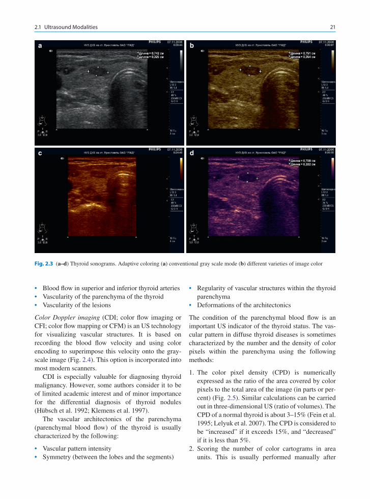
Adaptive coloring utilizes a color map in order to stain a grayscale image. The density of staining depends on the strength of the reflected echo (Fig. 2.3). Color inversion of the image is possible. The option is often added to grayscale scanners with standard probes.
This option improves the subjective perception of an US image. Thus, it helps in the detection of isoechoic thyroid lesions, and in the definition of nodule con-tours and posterior acoustic changes, especially in small lesions (up to 0.5–0.7 cm in size).
The vascularity of an abnormal thyroid can be char-acterized by Doppler modalities. The following aspects require special attention:
a b
Fig. 2.1 (a, b) Thyroid sonograms. Grayscale mode (a) thyroid nodule (b) longitudinal scan of normal thyroid lobe
Fig. 2.2 Thyroid sonograms. THI
212.1 Ultrasound Modalities
Blood flow in superior and inferior thyroid arteries•Vascularity of the parenchyma of the thyroid•Vascularity of the lesions•
Color Doppler imaging (CDI; color flow imaging or CFI; color flow mapping or CFM) is an US technology for visualizing vascular structures. It is based on recording the blood flow velocity and using color encoding to superimpose this velocity onto the gray-scale image (Fig. 2.4). This option is incorporated into most modern scanners.
CDI is especially valuable for diagnosing thyroid malignancy. However, some authors consider it to be of limited academic interest and of minor importance for the differential diagnosis of thyroid nodules (Hübsch et al. 1992; Klemens et al. 1997).
The vascular architectonics of the parenchyma (parenchymal blood flow) of the thyroid is usually characterized by the following:
Vascular pattern intensity•Symmetry (between the lobes and the segments)•
Regularity of vascular structures within the thyroid •parenchymaDeformations of the architectonics•
The condition of the parenchymal blood flow is an important US indicator of the thyroid status. The vas-cular pattern in diffuse thyroid diseases is sometimes characterized by the number and the density of color pixels within the parenchyma using the following methods:
1. The color pixel density (CPD) is numerically expressed as the ratio of the area covered by color pixels to the total area of the image (in parts or per-cent) (Fig. 2.5). Similar calculations can be carried out in three-dimensional US (ratio of volumes). The CPD of a normal thyroid is about 3–15% (Fein et al. 1995; Lelyuk et al. 2007). The CPD is considered to be “increased” if it exceeds 15%, and “decreased” if it is less than 5%.
2. Scoring the number of color cartograms in area units. This is usually performed manually after
a b
c d
Fig. 2.3 (a–d) Thyroid sonograms. Adaptive coloring (a) conventional gray scale mode (b) different varieties of image color
22 2 Complex Ultrasound Diagnosis of Thyroid Diseases
a
b
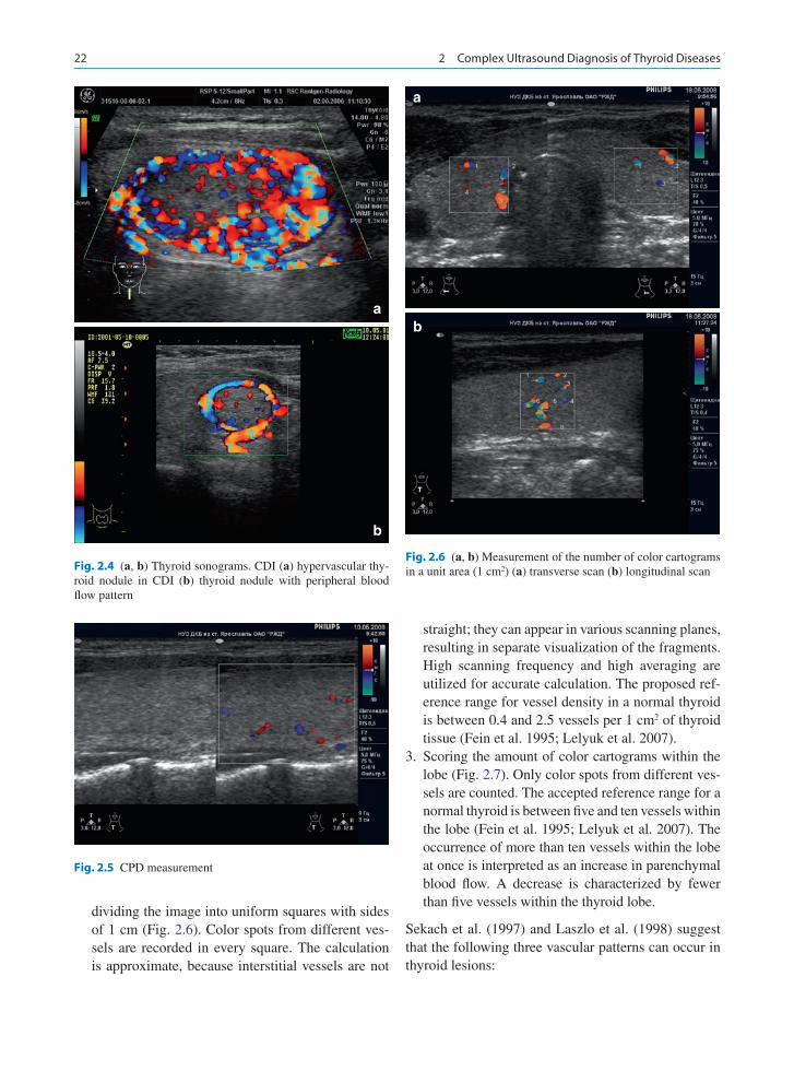
Fig. 2.4 (a, b) Thyroid sonograms. CDI (a) hypervascular thy-roid nodule in CDI (b) thyroid nodule with peripheral blood flow pattern
dividing the image into uniform squares with sides of 1 cm (Fig. 2.6). Color spots from different ves-sels are recorded in every square. The calculation is approximate, because interstitial vessels are not
straight; they can appear in various scanning planes, resulting in separate visualization of the fragments. High scanning frequency and high averaging are utilized for accurate calculation. The proposed ref-erence range for vessel density in a normal thyroid is between 0.4 and 2.5 vessels per 1 cm2 of thyroid tissue (Fein et al. 1995; Lelyuk et al. 2007).
3. Scoring the amount of color cartograms within the lobe (Fig. 2.7). Only color spots from different ves-sels are counted. The accepted reference range for a normal thyroid is between five and ten vessels within the lobe (Fein et al. 1995; Lelyuk et al. 2007). The occurrence of more than ten vessels within the lobe at once is interpreted as an increase in parenchymal blood flow. A decrease is characterized by fewer than five vessels within the thyroid lobe.
Sekach et al. (1997) and Laszlo et al. (1998) suggest that the following three vascular patterns can occur in thyroid lesions:
Fig. 2.5 CPD measurement
Fig. 2.6 (a, b) Measurement of the number of color cartograms in a unit area (1 cm2) (a) transverse scan (b) longitudinal scan
a
b
232.1 Ultrasound Modalities
1. Absence of blood flow both within the nodule and around it
2. Blood flow around the nodule3. Blood flow both within the nodule and around it
Some authors (Messina et al. 1996; Morozov 1997; Abdulhalimova et al. 1999) additionally describe an intranodular type of vascular pattern, where individual or multiple color signals are registered within the lesion.
Zubarev et al. (2000) suggest that the following three vascular patterns of thyroid nodules should be used in daily practice:
1. Perinodular: the blood flow is mainly in the periph-ery of the nodule
2. Mixed: vascularization occurs in the periphery of and within the nodule
3. Avascular: there is no sonographically discernible blood flow.
The thyroid nodules can also be divided into the fol-lowing groups according to the blood flow intensity:
1. Hypervascular nodules show a peripheral rim and multiple arterial and venous vessels within (the sign of a “color crown”)
2. Nodules with a medium degree of vascularization have 5–6 color spots within the nodule
3. Hypovascular nodules show 2–3 color spots4. Avascular nodules have no inner color spots and no
peripheral rim
CDI has some disadvantages, such as table distortions of the Doppler spectrum (aliasing artifact), baseline noise, and dependence on the angle of the US beam.
Power Doppler imaging (PDI) permits images of smaller vessels with sharper contours to be obtained. This increases the diagnostic value of US (Fig. 2.8). PDI demonstrates a decreased dependence on the angle between the US beam and the blood flow, shows no aliasing artifact, and has a lower noise level. Therefore, PDI is three- to fivefold more sensitive than CDI (Lagalla et al. 1994, Adler et al. 1995; Spiezia et al. 1996). According to Zubarev (1997), PDI increases diagnostic sensitivity to thyroid pathology from 36 to 79% and specificity from 58 to 62% as compared with CDI.
PDI has some disadvantages, such as its high depen-dence on the motions of surrounding structures (lead-ing to “motion artifacts”) and the staining of perivascular areas.
Fast computer processing of US images permits the three-dimensional (3D) reconstruction of the thyroid structure, lesions, the vascular tree, and surrounding tis-sues (Fig. 2.9). This option may be incorporated into ordinary US scanners as additional software. Data acquisition is achieved by a freehand scan with a usual 2D probe. Such cases demand subsequent computa-tional processing of the data obtained. Some scanners can be equipped with special probes for mechanical 3D scanning. 3D imaging confers many advantages, such as the possibility of viewing planes that are usually inaccessible, and improved accuracy of volume estima-tion. It is useful for archiving US data in an objective form suitable for delayed analysis and digital transfer.
In comparison with 2D PDI, 3D reconstruction of vascular structures (3D power Doppler imaging or 3DPD) enables more specific diagnoses of neoplasms based on the objective visual data for the structure
a b
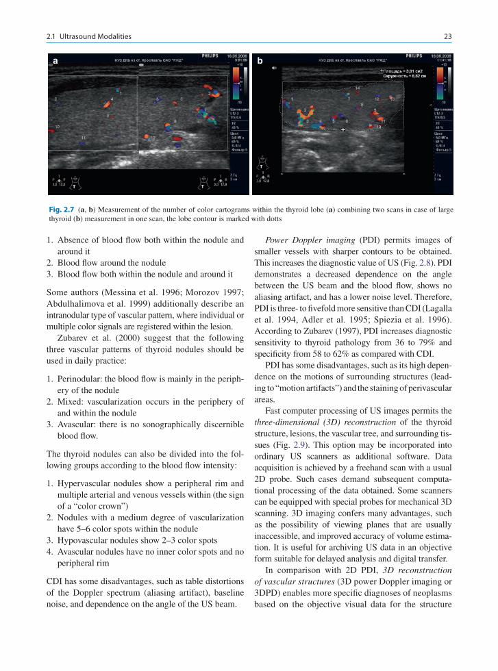
Fig. 2.7 (a, b) Measurement of the number of color cartograms within the thyroid lobe (a) combining two scans in case of large thyroid (b) measurement in one scan, the lobe contour is marked with dotts
24 2 Complex Ultrasound Diagnosis of Thyroid Diseases
a
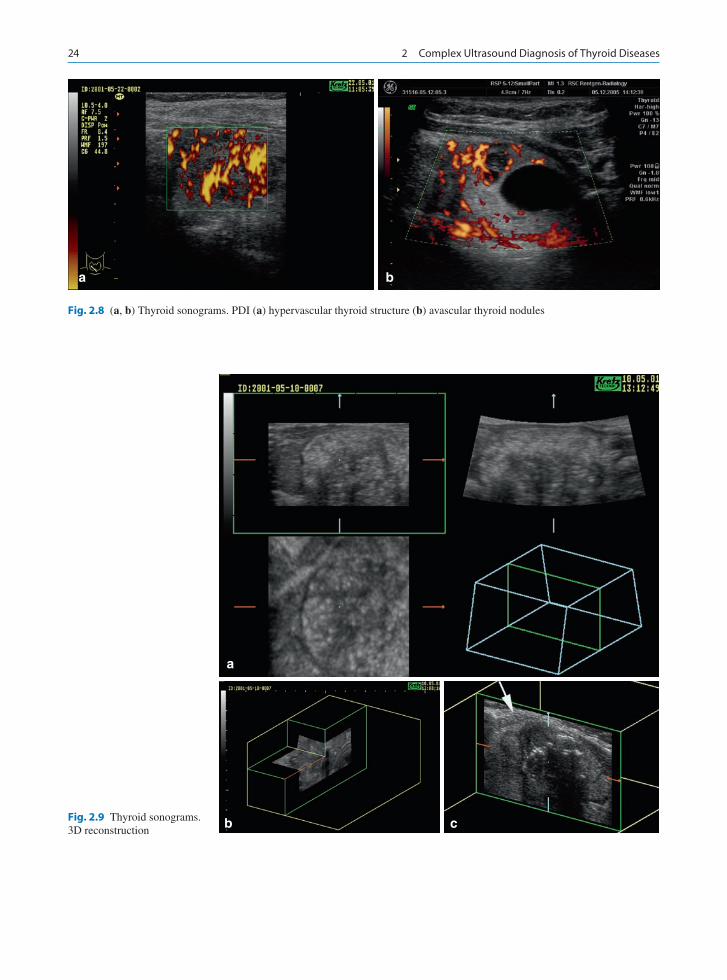
cbFig. 2.9 Thyroid sonograms. 3D reconstruction
a b
Fig. 2.8 (a, b) Thyroid sonograms. PDI (a) hypervascular thyroid structure (b) avascular thyroid nodules
252.1 Ultrasound Modalities
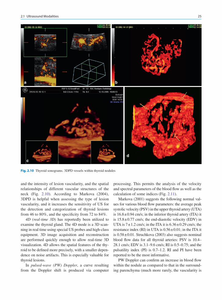
and the intensity of lesion vascularity, and the spatial relationships of different vascular structures of the neck (Fig. 2.10). According to Markova (2004), 3DPD is helpful when assessing the type of lesion vascularity, and it increases the sensitivity of US for the detection and categorization of thyroid lesions from 46 to 80%, and the specificity from 72 to 84%.
4D (real-time 3D) has reportedly been utilized to examine the thyroid gland. The 4D mode is a 3D scan-ning in real time using special US probes and high-class equipment. 3D image acquisition and reconstruction are performed quickly enough to allow real-time 3D visualization. 4D allows the spatial features of the thy-roid to be defined more precisely, with a smaller depen-dence on noise artifacts. This is especially valuable for thyroid lesions.
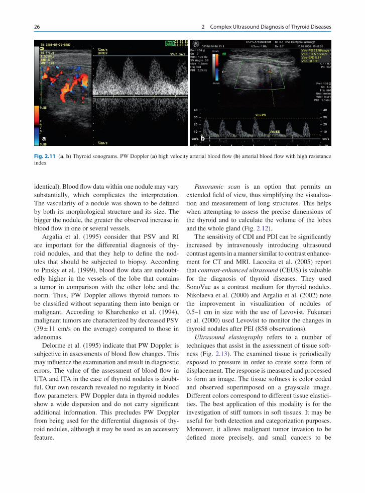
In pulsed-wave (PW) Doppler, a curve resulting from the Doppler shift is produced via computer
processing. This permits the analysis of the velocity and spectral parameters of the blood flow as well as the calculation of some indices (Fig. 2.11).
Markova (2001) suggests the following normal val-ues for various blood flow parameters: the average peak systolic velocity (PSV) in the upper thyroid artery (UTA) is 16.8 ± 0.94 cm/s; in the inferior thyroid artery (ITA) it is 15.8 ± 0.77 cm/s; the end-diastolic velocity (EDV) in UTA is 7 ± 1.2 cm/s; in the ITA it is 6.36 ± 0.29 cm/s; the resistance index (RI) in UTA is 0.56 ± 0.01; in the ITA it is 0.58 ± 0.01. Struchkova (2003) also suggests nominal blood flow data for all thyroid arteries: PSV is 10.4–28.1 cm/s; EDV is 3.1–9.6 cm/s; RI is 0.5–0.75; and the pulsatility index (PI) is 0.7–1.2. RI and PI have been reported to be the most informative.
PW Doppler can confirm an increase in blood flow within the nodule as compared to that in the surround-ing parenchyma (much more rarely, the vascularity is
a b
cd
Fig. 2.10 Thyroid sonograms. 3DPD vessels within thyroid nodules
26 2 Complex Ultrasound Diagnosis of Thyroid Diseases
identical). Blood flow data within one nodule may vary substantially, which complicates the interpretation. The vascularity of a nodule was shown to be defined by both its morphological structure and its size. The bigger the nodule, the greater the observed increase in blood flow in one or several vessels.
Argalia et al. (1995) consider that PSV and RI are important for the differential diagnosis of thy-roid nodules, and that they help to define the nod-ules that should be subjected to biopsy. According to Pinsky et al. (1999), blood flow data are undoubt-edly higher in the vessels of the lobe that contains a tumor in comparison with the other lobe and the norm. Thus, PW Doppler allows thyroid tumors to be classified without separating them into benign or malignant. According to Kharchenko et al. (1994), malignant tumors are characterized by decreased PSV (39 ± 11 cm/s on the average) compared to those in adenomas.
Delorme et al. (1995) indicate that PW Doppler is subjective in assessments of blood flow changes. This may influence the examination and result in diagnostic errors. The value of the assessment of blood flow in UTA and ITA in the case of thyroid nodules is doubt-ful. Our own research revealed no regularity in blood flow parameters. PW Doppler data in thyroid nodules show a wide dispersion and do not carry significant additional information. This precludes PW Doppler from being used for the differential diagnosis of thy-roid nodules, although it may be used as an accessory feature.
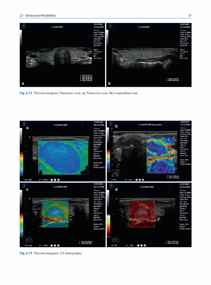
Panoramic scan is an option that permits an extended field of view, thus simplifying the visualiza-tion and measurement of long structures. This helps when attempting to assess the precise dimensions of the thyroid and to calculate the volume of the lobes and the whole gland (Fig. 2.12).
The sensitivity of CDI and PDI can be significantly increased by intravenously introducing ultrasound contrast agents in a manner similar to contrast enhance-ment for CT and MRI. Lacocita et al. (2005) report that contrast-enhanced ultrasound (CEUS) is valuable for the diagnosis of thyroid diseases. They used SonoVue as a contrast medium for thyroid nodules. Nikolaeva et al. (2000) and Argalia et al. (2002) note the improvement in visualization of nodules of 0.5–1 cm in size with the use of Levovist. Fukunari et al. (2000) used Levovist to monitor the changes in thyroid nodules after PEI (858 observations).
Ultrasound elastography refers to a number of techniques that assist in the assessment of tissue soft-ness (Fig. 2.13). The examined tissue is periodically exposed to pressure in order to create some form of displacement. The response is measured and processed to form an image. The tissue softness is color coded and observed superimposed on a grayscale image. Different colors correspond to different tissue elastici-ties. The best application of this modality is for the investigation of stiff tumors in soft tissues. It may be useful for both detection and categorization purposes. Moreover, it allows malignant tumor invasion to be defined more precisely, and small cancers to be
a b
Fig. 2.11 (a, b) Thyroid sonograms. PW Doppler (a) high velocity arterial blood flow (b) arterial blood flow with high resistance index
272.1 Ultrasound Modalities
a b
Fig. 2.12 Thyroid sonogram. Panoramic scan. (a) Transverse scan. (b) Longitudinal scan
a
c d
b
Fig. 2.13 Thyroid sonogram. US elastography
28 2 Complex Ultrasound Diagnosis of Thyroid Diseases
diagnosed (Ophir 1999; Doyley 2000; Lindop et al. 2006). Tanaka et al. (2006) reported a high efficacy of ultrasound elastography for the differential diagnosis of abnormal lymph nodes of the neck.
In multislice viewing, a 3D US image is converted into a series of consecutive sections corresponding to intervals of 0.5–5 mm in any plane, similar to CT rep-resentations. This aids in the analysis of thyroid images, and makes it objective.
Enhancements to traditional procedures and advances in new technologies are leading to continual improvements in the accuracy and value of diagnostic ultrasound.
2.2 Technology Used in Ultrasound Examinations of the Thyroid Gland
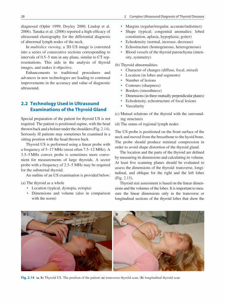
Special preparation of the patient for thyroid US is not required. The patient is positioned supine, with the head thrown back and a bolster under the shoulders (Fig. 2.14). Seriously ill patients may sometimes be examined in a sitting position with the head thrown back.
Thyroid US is performed using a linear probe with a frequency of 5–17 MHz (most often 7.5–12 MHz). A 3.5–5 MHz convex probe is sometimes more conve-nient for measurements of large thyroids. A sector probe with a frequency of 2.5–5 MHz may be required for the substernal thyroid.
An outline of an US examination is provided below:
(a) The thyroid as a wholeLocation (typical, dystopia, ectopia)•Dimensions and volume (also in comparison •with the norm)
Margins (regular/irregular, accurate/indistinct)•Shape (typical; congenital anomalies: lobed •constitution, aplasia, hypoplasia; goiter)Echodensity (normal, increase, decrease)•Echostructure (homogeneous, heterogeneous)•Blood vessels of the thyroid parenchyma (inten-•sity, symmetry)
(b) Thyroid abnormalitiesCharacter of changes (diffuse, focal, mixed)•Location (in lobes and segments)•Number of lesions•Contours (sharpness)•Borders (smoothness)•Dimensions (in three mutually perpendicular planes)•Echodensity, echostructure of focal lesions•Vascularity•
(c) Mutual relations of the thyroid with the surround-ing structures
(d) The status of regional lymph nodes
The US probe is positioned on the front surface of the neck and moved from the breastbone to the hyoid bone. The probe should produce minimal compression in order to avoid shape distortion of the thyroid gland.
The location and the parts of the thyroid are defined by measuring its dimensions and calculating its volume. At least five scanning planes should be evaluated to assess the dimensions of the thyroid: transverse, longi-tudinal, and oblique for the right and the left lobes (Fig. 2.15).
Thyroid size assessment is based on the linear dimen-sions and the volumes of the lobes. It is important to mea-sure the linear dimensions only in the transverse or longitudinal sections of the thyroid lobes that show the
a b
Fig. 2.14 (a, b) Thyroid US. The position of the patient (a) transverse thyroid scan, (b) longitudinal thyroid scan
292.2 Technology Used in Ultrasound Examinations of the Thyroid Gland
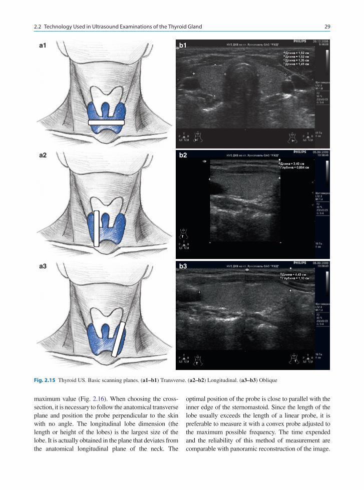
maximum value (Fig. 2.16). When choosing the cross-section, it is necessary to follow the anatomical transverse plane and position the probe perpendicular to the skin with no angle. The longitudinal lobe dimension (the length or height of the lobes) is the largest size of the lobe. It is actually obtained in the plane that deviates from the anatomical longitudinal plane of the neck. The
optimal position of the probe is close to parallel with the inner edge of the sternomastoid. Since the length of the lobe usually exceeds the length of a linear probe, it is preferable to measure it with a convex probe adjusted to the maximum possible frequency. The time expended and the reliability of this method of measurement are comparable with panoramic reconstruction of the image.
a1 b1
a2 b2
a3 b3
Fig. 2.15 Thyroid US. Basic scanning planes. (a1–b1) Transverse. (a2–b2) Longitudinal. (a3–b3) Oblique
30 2 Complex Ultrasound Diagnosis of Thyroid Diseases
a1 b1
a2 b2
a3 b3
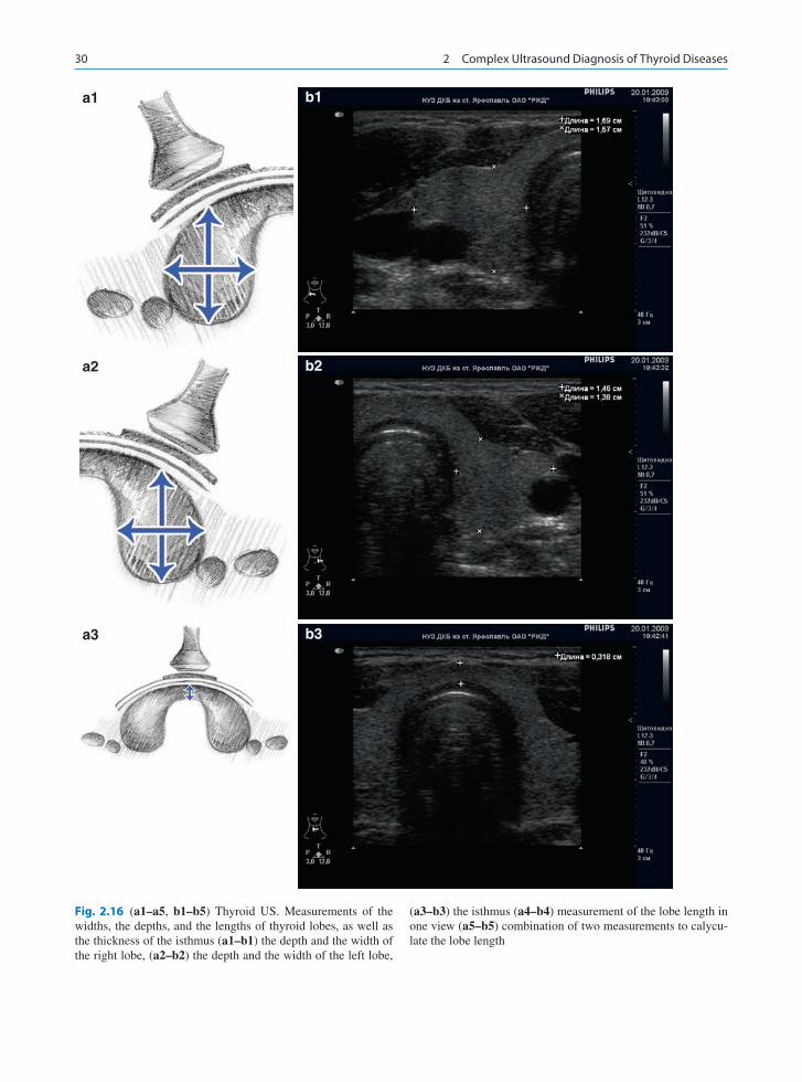
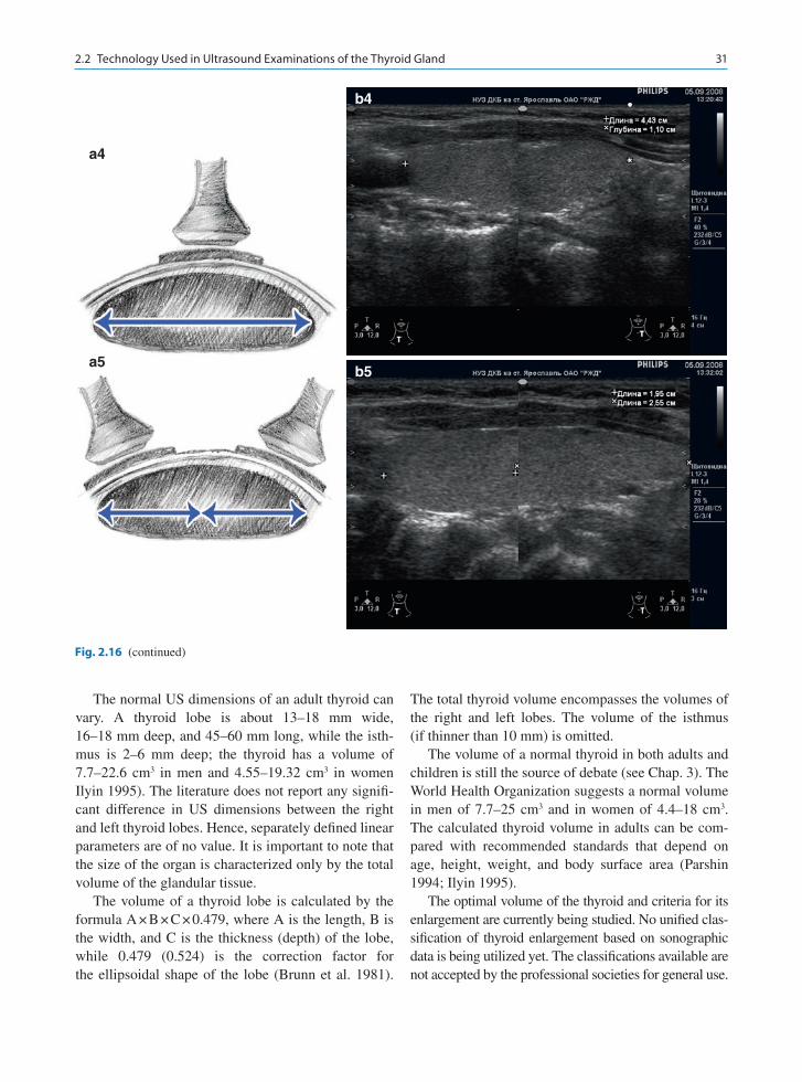
Fig. 2.16 (a1–a5, b1–b5) Thyroid US. Measurements of the widths, the depths, and the lengths of thyroid lobes, as well as the thickness of the isthmus (a1–b1) the depth and the width of the right lobe, (a2–b2) the depth and the width of the left lobe,
(a3–b3) the isthmus (a4–b4) measurement of the lobe length in one view (a5–b5) combination of two measurements to calycu-late the lobe length
312.2 Technology Used in Ultrasound Examinations of the Thyroid Gland
The normal US dimensions of an adult thyroid can vary. A thyroid lobe is about 13–18 mm wide, 16–18 mm deep, and 45–60 mm long, while the isth-mus is 2–6 mm deep; the thyroid has a volume of 7.7–22.6 cm3 in men and 4.55–19.32 cm3 in women Ilyin 1995). The literature does not report any signifi-cant difference in US dimensions between the right and left thyroid lobes. Hence, separately defined linear parameters are of no value. It is important to note that the size of the organ is characterized only by the total volume of the glandular tissue.
The volume of a thyroid lobe is calculated by the formula A × B × C × 0.479, where A is the length, B is the width, and C is the thickness (depth) of the lobe, while 0.479 (0.524) is the correction factor for the ellipsoidal shape of the lobe (Brunn et al. 1981).
The total thyroid volume encompasses the volumes of the right and left lobes. The volume of the isthmus (if thinner than 10 mm) is omitted.
The volume of a normal thyroid in both adults and children is still the source of debate (see Chap. 3). The World Health Organization suggests a normal volume in men of 7.7–25 cm3 and in women of 4.4–18 cm3. The calculated thyroid volume in adults can be com-pared with recommended standards that depend on age, height, weight, and body surface area (Parshin 1994; Ilyin 1995).
The optimal volume of the thyroid and criteria for its enlargement are currently being studied. No unified clas-sification of thyroid enlargement based on sonographic data is being utilized yet. The classifications available are not accepted by the professional societies for general use.
a4
b4
a5b5
Fig. 2.16 (continued)
32 2 Complex Ultrasound Diagnosis of Thyroid Diseases
They anchor the US data to the degree of enlargement of the thyroid gland based on imperfect palpation and visual assessment (for example the 1994 WHO scale).
At the same time, only one aspect is important in most cases: whether the patient’s thyroid volume differs from the norm. Many authors suggest that presenting the degree of deviation as a percentage may be of ben-efit for the dynamic assessment of changes in thyroid volume during treatment.
2.3 Basic Mistakes in Thyroid Ultrasound
Factors that result in inaccurate US assessment of the status of the thyroid gland can be divided into the following groups:
1. ObjectiveAnatomical, physiological, and constitutional •features of the patient leading to a decrease in visualizationLimitations of the equipment (the quality of the •scanner, the characteristics of the probes, etc.)
2. Subjective
Insufficient US specialist experience•Faulty thyroid US technique•
High intra- and interobserver variations in thyroid sonography are largely due to the quality of the equip-ment and the skill level of the operator. According to Bataeva et al. (2006), an expert fails to reproduce the results of the 2D thyroid volume calculation in 8.7% of cases; assessments performed by different experts differ by 12.8%. Measurements taken in 3D methods of surface reconstruction and segmentation show dif-ferences of 4 and 4.8%, respectively. One disadvan-tage of using US in some cases is a low detection efficacy for thyroid dystopia and ectopia. Retrosternal location of the thyroid below the tracheal bifurcation significantly limits the possibility of ultrasonography (see Chap. 10).
High-resolution US equipment allows several thy-roid pathologies to be detected, which used to be con-sidered the norm. For example, medium- and large-sized cellular patterns (multiple fine hypo- and anechoic lesions 2–4 mm in size) that were interpreted as the norm years ago are now considered abnormal. This
kind of sonogram is widely seen for people living in iodine-deficient regions, and precedes a diffuse endemic goiter. It corresponds to colloid cystic change with dila-tion of the follicles due to extra colloid accumulation. The hyperechoic points within such follicles are a consequence of the dense consistency of the colloid. Another type of small change is inflammatory foci dur-ing the initial stages of autoimmune thyroid disease (AITD). These relate to a tendency for a decrease in echodensity and a slight heterogeneity of thyroid tissue resulting from small foci of lymphoplasmacytic infil-tration and edema.
Alternatively, there are hyperdiagnostic cases when normal thyroid structures are interpreted as nodules. This especially concerns structures behind the left lobe or along the posterior margin of the inferior compart-ment of the right lobe. This type of error can be caused by the proximity of the esophagus, which may be reported as a nodule if imaged only with a transverse scan. The normal vascular pattern of the inferior thy-roid artery (ITA) can result in hyperdiagnostics of thy-roid nodules. In some cases, the ITA trunk does not split into fine branches when entering the inferior pole of the thyroid lobe. It may be traced within the lobe, where it borders a roundish region of healthy tissue that imitates a nodule.
Lymph nodes adjacent to the isthmus frequently complicate the diagnosis. The enlarged lymph nodes present in AITD are often located close to the upper or inferior part of the isthmus and can be interpreted as thyroid nodules. Differential diagnosis may benefit from the use of the highest probe frequency, as it per-mits a more detailed image. It is necessary to pay attention to specific features of lymph nodes, such as form, echostructure with differentiation of the corti-cal and central parts, and type of blood flow. The opposite type of error, where nodules of the thyroid isthmus are interpreted as neck lymph nodes, are quite rare.
Differential diagnosis of thyroid nodules and inflam-matory foci in AITD is rather difficult (see Chap. 5). In this case, it is necessary to consider the sharpness and regularity of the contours of the lesion, its form, and the blood flow pattern. Nevertheless, the correct diagnosis sometimes requires long observations or the use of other diagnostic modalities.
Rare neck pathologies may be misdiagnosed due to insufficient experience of the US operator. In many cases, adenomas and hyperplasia of the parathyroid
332.3 Basic Mistakes in Thyroid Ultrasound
glands are interpreted as thyroid nodules. This pathol-ogy is relatively rare, so general practitioners or US spe-cialists who do not practice at a specialized endocrinology center are not aware of it and have little experience in its diagnosis (see Chap. 11). The same can be said about the diverticulum of the esophagus (see Chap. 12). In most cases, sonographers have a mental image of “typi-cal” thyroid nodules based on their own experience, so any lesion differing from this typical image should be interpreted with extra caution.
Special opportunities for differential diagnosis are provided by auxiliary methods, such as turning the patient’s head, compression of the neck tissues with fingers, swallowing, and others.
Note that modern equipment permits accurate US visualization of solid thyroid nodules larger than 2–3 mm in size. The presence of smaller lesions is preferably reported without the term “nodule.” Drawing
a conclusion about the lesion is expedient only when it can be clearly visualized in at least two mutually perpendicular scans.
It is very important to adhere to the correct exami-nation technique. Thyroid structure including small parts can be estimated only with linear probes at fre-quencies of 7.5 MHz and higher. Convex abdominal probes may be used to measure the lengths of thyroid lobes or for large thyroids. The use of a convex probe alone for thyroid examination results in multiple severe errors and discredits the field of sonography.
Rational timescales for thyroid US are as follows:Normal thyroid: once every two years for preven-•tive purposesBenign diffuse or nodular thyroid pathology: 1–2 •times a year to monitor the dynamics of the diseaseInoperable thyroid malignancy: once every two •months to define the stage.
http://www.springer.com/978-3-642-12386-3